eliciting action potentials from epidermal stimulation of … of laser stimulated action potentials...
TRANSCRIPT

AFRL-RH-BR-TR-2009-0022
Eliciting Action Potentials from
Epidermal Stimulation of Skin
Receptors Using Ultrashort Laser Pulses
Douglas N. Goddard
Michelle Imholte
Northrop Grumman Information Technology
Nichole Jindra
Robert J. Thomas
Human Effectiveness Directorate
Directed Energy Bioeffects Division
Optical Radiation Branch
February 2009
Interim Report for October 2006 thru December 2006
© 2005 ACME Corp. This work is copyrighted. The United States has for itself and others
acting on its behalf an unlimited, paid-up, nonexclusive, irrevocable worldwide license. Any
other form of use is subject to copyright restrictions.
Air Force Research Laboratory
711 Human Performance Wing
Human Effectiveness Directorate
Directed Energy Bioeffects Division
Optical Radiation Branch
Brooks City-Base, TX 78235
Approved for public release;
distribution unlimited. Public Affairs
Case File Number 09-161,
21 April 2009; Brooks City-Base, TX

NOTICE AND SIGNATURE PAGE
Using Government drawings, specifications, or other data included in this document for
any purpose other than Government procurement does not in any way obligate the U.S.
Government. The fact that the Government formulated or supplied the drawings,
specifications, or other data does not license the holder or any other person or corporation;
or convey any rights or permission to manufacture, use, or sell any patented invention that
may relate to them.
This report was cleared for public release by the 311th
Public Affairs Office at Brooks City
Base, TX and is available to the general public, including foreign nationals. Copies may be
obtained from the Defense Technical Information Center (DTIC) (http://www.dtic.mil).
The experiments reported were conducted according to the “Guide for the Care and Use
of Laboratory Animals,” Institute of Laboratory Animal Resources, National Research
Council.
AFRL-RH-BR-TR-2009-0022 HAS BEEN REVIEWED AND IS APPROVED FOR
PUBLICATION IN ACCORDANCE WITH ASSIGNED DISTRIBUTION STATEMENT.
__________//SIGNED//_______________ ALAN J. RICE, Capt, USAF
Work Unit Manager 711 HPW/RHDO __________//SIGNED//_______________ GARRETT D. POLHAMUS, Ph.D.
Chief, Directed Energy Bioeffects Division
Human Effectiveness Directorate
711 Human Performance Wing
Air Force Research Laboratory
This report is published in the interest of scientific and technical information exchange, and its
publication does not constitute the Government’s approval or disapproval of its ideas or findings.
i
DISTRIBUTION A, APPROVED FOR PUBLIC RELEASE; DISTRIBUTION UNLIMITED
REPORT DOCUMENTATION PAGE Form Approved
OMB No. 0704-0188 Public reporting burden for this collection of information is estimated to average 1 hour per response, including the time for reviewing instructions, searching existing data sources, gathering and maintaining the data needed, and completing and reviewing this collection of information. Send comments regarding this burden estimate or any other aspect of this collection of information, including suggestions for reducing this burden to Department of Defense, Washington Headquarters Services, Directorate for Information Operations and Reports (0704-0188), 1215 Jefferson Davis Highway, Suite 1204, Arlington, VA 22202-4302. Respondents should be aware that notwithstanding any other provision of law, no person shall be subject to any penalty for failing to comply with a collection of information if it does not display a currently valid OMB control number. PLEASE DO NOT RETURN YOUR FORM TO THE ABOVE ADDRESS.
1. REPORT DATE (DD-MM-YYYY)
February 2009 2. REPORT TYPE
Interim Technical Report 3. DATES COVERED (From - To)
October 2006- December 2006
4. TITLE AND SUBTITLE
Eliciting Action Potentials from Epidermal Stimulation of Skin
Receptors Using Ultrashort Laser Pulses
5a. CONTRACT NUMBER
FA865008-D-6930 5b. GRANT NUMBER
5c. PROGRAM ELEMENT NUMBER
0602203F 6. AUTHOR(S)
Nichole M. Jindra, Douglas N. Goddard, Michelle L. Imholte, Robert J.
Thomas
5d. PROJECT NUMBER
7757 5e. TASK NUMBER
B2
5f. WORK UNIT NUMBER
39 7. PERFORMING ORGANIZATION NAME(S) AND ADDRESS(ES)
Air Force Research Laboratory Northrop Grumman -IT
711 Human Performance Wing
Human Effectiveness Directorate
Directed Energy Bioeffects
Optical Radiation Branch
Brooks City-Base, TX 78235-5214
8. PERFORMING ORGANIZATION REPORT NUMBER 711 Human Performance Wing 4241 Woodcock Dr., Ste B-100
Human Effectiveness Directorate San Antonio, TX 78228
Directed Energy Bioeffects
Optical Radiation Branch
Brooks City-Base, TX 78235-5214
711 HPW/ RHDO
9. SPONSORING / MONITORING AGENCY NAME(S) AND ADDRESS(ES) 10. SPONSOR/MONITOR’S ACRONYM(S)
Air Force Research Laboratory
711 Human Performance Wing
Human Effectiveness Directorate
Directed Energy Bioeffects
Optical Radiation Branch
Brooks City-Base, TX 78235-5214
711 HPW/RHDO
11. SPONSOR/MONITOR’S REPORT
NUMBER(S)
AFRL-RH-BR-TR-2009-0022 12. DISTRIBUTION / AVAILABILITY STATEMENT
13. SUPPLEMENTARY NOTES Public Affairs Case File No. 09-161, 21 Apr 09
14. ABSTRACT
Measurements of laser stimulated action potentials in the sciatic nerve of leopard frogs (Rana pipiens) were made using two
ultrashort pulsed infrared lasers. The dorsal sides of the frog’s hind limbs were exposed to 1540 nm and 1064 nm wavelengths at
three separate spot sizes: 2 mm, 3 mm, and 4 mm. Energy density thresholds were determined for eliciting an action potential at
each experimental condition. Results from these exposures showed similar evoked potential thresholds for both wavelengths.
Skin ablation was observed at temperature increases as low as 0.7 ⁰C, so we believe the primary skin damage mechanism to be stress confinement. Determining the method of receptor activation was outside the scope of this study. While the exact
mechanism still remains unknown, it is possible to elicit action potentials from transdermal exposures of ultrashort lasers.
15. SUBJECT TERMS
16. SECURITY CLASSIFICATION OF:
17. LIMITATION OF ABSTRACT
18. NUMBER OF PAGES
19a. NAME OF RESPONSIBLE PERSON
Capt. Alan J. Rice a. REPORT
U
b. ABSTRACT
U
c. THIS PAGE
U
SAR
16
19b. TELEPHONE NUMBER (include
area code)
N/A Standard Form 298 (Rev. 8-98)
Prescribed by ANSI Std. 239.18

ii
DISTRIBUTION A, APPROVED FOR PUBLIC RELEASE; DISTRIBUTION UNLIMITED
This page intentionally left blank
iii
DISTRIBUTION A, APPROVED FOR PUBLIC RELEASE; DISTRIBUTION UNLIMITED
Table of Contents
Table of Contents ......................................................................................................................... iii
Figures .......................................................................................................................................... iii
Tables ........................................................................................................................................... iii
Abstract ......................................................................................................................................... 1
Background ................................................................................................................................... 1
Objectives ...................................................................................................................................... 2
Methods ......................................................................................................................................... 2
Leopard Frogs ............................................................................................................................ 2
Nerve Preparation ...................................................................................................................... 3
Laser Set-Up .............................................................................................................................. 3
Probit Analysis .......................................................................................................................... 4
Results ........................................................................................................................................... 5
Discussion ..................................................................................................................................... 6
Conclusions ................................................................................................................................... 9
Acknowledgement ....................................................................................................................... 10
Bibliography ................................................................................................................................ 10
Figures
Figure 1: Laser energy penetration and nerve receptor locations in skin layers. .......................... 2 Figure 2: Generic schematic of experimental set-up. ..................................................................... 4
Figure 3: Action potential from a 2-mm exposure on the frog’s calf, using a 1540 nm laser. ...... 5 Figure 4: Radiant exposure (J/cm
2) values for skin damage and AP thresholds at 1064 nm. ....... 6
Figure 5: Skin damage and APl thresholds for radiant energy exposures at 1540 nm. .................. 6 Figure 6: Ablation of tissue on frog skin. ....................................................................................... 7 Figure 7: AP50 probability vs. temperature rise data for 4-mm 1540 nm exposures. ..................... 9
Tables
Table 1: Action potential thresholds (J/cm2). AP50 is the energy level at which 50% of exposures
will elicit an action potential. UFL = upper fiducial limit. LFL = lower fiducial limit .............. 5 Table 2: Spot size versus the average temperature ........................................................................ 8

iv
DISTRIBUTION A, APPROVED FOR PUBLIC RELEASE; DISTRIBUTION UNLIMITED
This page intentionally left blank

1
DISTRIBUTION A, APPROVED FOR PUBLIC RELEASE; DISTRIBUTION UNLIMITED
Abstract
Measurements of laser stimulated action potentials in the sciatic nerve of leopard frogs (Rana
pipiens) were made using two ultrashort pulsed infrared lasers. The dorsal sides of the frog’s
hind limbs were exposed to 1540 nm and 1064 nm wavelengths at three separate spot sizes: 2
mm, 3 mm, and 4 mm. Energy density thresholds were determined for eliciting an action
potential at each experimental condition. Results from these exposures showed similar evoked
potential thresholds for both wavelengths. Skin ablation was observed at temperature increases
as low as 0.7 ⁰C, so we believe the primary skin damage mechanism to be stress confinement.
Determining the method of receptor activation was outside the scope of this study. While the
exact mechanism still remains unknown, it is possible to elicit action potentials from
transdermal exposures of ultrashort lasers.
Background
Electrical stimulation is commonly used to stimulate action potentials in neurons for both
medical and research applications. Electrical signals are applied to a nerve, initiating the
voltage change that will start a chain reaction along the axon. Once began, the signal is passed
along the axonal tract. Unfortunately, electrical stimulation systems possess characteristics that
create problems in this kind of work. Besides low spatial specificity (electrical stimulation will
activate several nerve tracts simultaneously), difficulties include tissue damage from electrode
installation and unnatural action potential responses (Izzo, et al., 2006). Recent studies have
found that a laser source can be used to induce an action potential in the nervous system as well
as, if not better than, electrical methods. Kao et al. have shown that there are few differences
between optical and electrical stimulation on the activation of the nerves (Kao, Wells, & Jansen,
2005). Laser excitation of neural activity provides a contact-free, spatially selective, artifact-
free method of stimulation without incurring tissue damage (Kao, Wells, & Jansen, 2005). The
small spot sizes used by laser systems allow for pin point accuracy when stimulating nerve
tracts and the low irradiance levels help to minimize introduction of extra energy into the action
potential response. The amplitude of an action potential is directly proportional to the strength
of its triggering event so the lower energies used by laser stimulations are very important in
achieving greater specificity. Most of the studies were performed directly on the nerves and
were within laser-tissue interaction parameters that led Wells to suspect thermal confinement
mechanisms for action potential elicitation.
Clinically, indirect stimulation of nerves has been conducted by radiating the skin with lasers
(typically a CO2 or visible wavelength laser) and activating skin nerve fibers. This technique is
typically used for determining pain thresholds, pain desensitization studies, and physiological
studies of nociceptive pathways. (Perchet et al., 2008; Kneebone, 2007; Moore, 2004; Arendt-
Nielsen, 1988) To date, we have been unable to find any data on laser-evoked potentials using
ultrashort lasers.
This work is a pilot study to determine if action potentials can be induced using near-infrared
ultrashort pulses directed through the skin of an animal. Much work has been performed by this
laboratory into the damage thresholds of ultrashort lasers, and this has led to a desire to clarify
whether or not these lasers are causing any unseen secondary reactions at low energy levels.
Unlike longer pulsed lasers which operate in thermal confinement parameters, ultrashort pulses

2
DISTRIBUTION A, APPROVED FOR PUBLIC RELEASE; DISTRIBUTION UNLIMITED
operate within stress confinement parameters. Thus, any reaction could be thermal, mechanical,
or a combination of these or other mechanisms. If they can indeed elicit action potentials,
further work will be required to determine the exact method.
Figure 1: Illustration of laser energy penetration and nerve receptor locations in skin layers.
Objectives
The goal of this work was to determine the feasibility of an electro-potential response of neural
receptors due to ultrashort pulsed laser exposures.† This was tested during an experiment
performed on Leopard Frogs. An electrode was placed in the sciatic nerve of the frogs to record
action potentials elicited from laser exposures on the surface skin of the calf. Three spot sizes
and two different infrared wavelengths were used in the study. The data was then analyzed to
determine threshold values for action potential stimulations.
Methods
Leopard Frogs
In vivo sciatic nerve experiments were performed using Leopard Frogs (Rana pipiens) from the
Carolina Biological Supply Company in North Carolina. The frogs ranged in torso length from
3 to 4 inches and were euthanized via a double pithing technique. To maintain a constant body
temperature during the experiment, the cold-blooded frogs were placed on a saline bag that had
† * The animals involved in this study were procured, maintained, and used in accordance with the Federal Animal Welfare Act,
"Guide for the Care and Use of Laboratory Animals," prepared by the Institute of Laboratory Animal Resources National
Research Council, and DoD Regulation 40-33 Secnavinst 3900.38C AFMAN 40-401(1) DARPAINST 18 USUHSINST 3203
“The Care and Use of Laboratory animals in DOD Programs.” Brooks City-Base, TX has been fully accredited by the
Association for Assessment and Accreditation of Laboratory Animal Care, International (AAALAC) since 1967.
(Protocol HEDO-06-12).

3
DISTRIBUTION A, APPROVED FOR PUBLIC RELEASE; DISTRIBUTION UNLIMITED
been warmed to approximately 20-22 ˚C. This was to ensure that the nerve receptors remained
within their effective ranges and that the frogs were not negatively affected by cold ambient
temperatures in the lab.
Since the subject was an amphibian, the water content of the skin was very high. In order to
minimize variability in the data obtained during this experiment, saline was periodically applied
to the skin to maintain hydration. Excess solution was blotted off using gauze.
Nerve Preparation
Nerve preparation started by making a centrally located incision from the knee to the upper
thigh, removing the skin from the dorsal side and exposing the trunk of the nerve at the knee.
Muscular fascia was incised and removed to expose the rest of the nerve. A piece of latex was
placed between the nerve and the underlying muscle in order to minimize any collateral
electrical signals. After the nerve was prepared, insulated stainless steel needle electrodes
(Chalgreen Enterprises, Inc. 111-637-24TP, Disposable, Monopolar EMG Needle Electrodes,
37mm x 26 gauge) were inserted into the nerve located approximately 15 mm above the knee.
Baseline data was collected to verify the system’s isolation from other electrical sources and to
ensure correct electrode placement. To initialize each experiment, the sciatic nerve was directly
stimulated by the laser to verify that the electrode was reading correctly and that the nerve had
not been damaged by the insertion. Compound nerve action potential (CNAP) responses were
recorded with BioPac Systems Inc. MP100 interfaced to a computer running Acknowledge
software v3.73. The CNAP is the algebraic sum of many individual “all-or-none” action
potentials arising more or less simultaneously in a large number of individual axons
(Physiology Dept., McGill University, 2005). All action potentials are measured using a
differential medical amplifier and extracellular recording electrodes, which measure the
summed electrical response of all excited axons in the nerve. The recordings for this project
were manually triggered prior to the exposure and recorded 5 seconds of data. All signals were
amplified 1000 times and electrically filtered with a 50 to 5000 Hz bandpass filter.
Once the initial direct testing of the sciatic nerve had been conducted, the laser was focused on
the animal’s calf. The surface of the skin was randomly irradiated using varying energy levels,
with one second in between each shot. This lag time was necessary to prevent overheating of
the Er:Glass laser and was maintained for both lasers to eliminate unnecessary variation
between the exposure procedures.
Laser Set-Up
Optical stimulation was performed using two infrared laser sources. A Q-switched Nd:YAG
laser emitting 1064 nm was first used to verify experimental methods. The Nd:YAG laser was
pulsed at a repetition rate of 10 Hz with a pulse duration of 15 ns. The energy of the laser was
controlled using a half wave plate and a polarizing beam splitter, and the generated pulses were
sampled using a 90/10 non-polarizing beamsplitter by an Ophir Laser Star energy meter using
#1Z0230 power head. The laser was then directed into a faraday cage where it would be focused
into the desired spot size using a 500 mm bi-convex lens. (Figure 2)

4
DISTRIBUTION A, APPROVED FOR PUBLIC RELEASE; DISTRIBUTION UNLIMITED
Figure 2: Generic schematic of experimental set-up.
Each infrared laser used possessed a Gaussian spatial beam profile. Beam diameters and profiles
were measured using linagraph laser burn paper. Using only one pulse per exposure, three beam
diameters were used during the experiment: 2 mm, 3 mm and 4 mm.
Once the methods were verified with the Nd:YAG laser, an Er:Glass (Erbium-glass) laser
emitting 1540-nm was employed to further evaluate optical stimulation and for statistical
comparison with another wavelength. The Er:Glass laser was mechanically q-switched to a
pulse duration of 55 ns. Due to the system’s high energy, only 1 shot per minute is allowed. The
energy of the laser is varied by adjusting the flash lamp energy. Again the beam was directed
into the faraday cage and focused to the same spot sizes as with the 1064-nm Nd:YAG laser: 2
mm, 3 mm and 4 mm.
Data showed considerable variability between the animals in respect to the pigmentation
placement. While melanin has only a small role in energy absorption at infrared wavelengths,
early skin exposures showed noticeable differences between skin damage threshold energy
levels for dark and light skin patches. Thus, for greater consistency, only lightly pigmented skin
data was used in the analysis. Each AP50 is represented in units of fluence (J/cm2).
Probit Analysis
Probit analysis was developed in order to analyze discrete data collected by experiments
involving threshold response rate in biological systems. This is computed using the EZ-Probit
program designed by Dr. Clarence Cain and Capt. Lonnie Manning at Brooks City- Base in San
Antonio, Texas (Cain & Manning, 1996). This method has been employed as a statistical tool to
determine the probability of dose-response curves for action potential (AP) responses in the
sciatic nerve. In this case the thresholds probabilities are reported as AP50 -- the radiant
exposure dose which has a 50% probability of creating a response. The values presented here
are for 100% probability, without consideration of additional experimental uncertainties. Also,
the slope of the probit line is calculated between the ED84 and ED50 values. A high value for
slope would represent high value for data certainty, with minimal sample-to-sample variability
affecting results.

5
DISTRIBUTION A, APPROVED FOR PUBLIC RELEASE; DISTRIBUTION UNLIMITED
Results
Positive responses, such as that seen in Figure 3, were recorded for every parameter tested in
this project. These viable action potentials obtained from the laser exposures are presented in
Table 1. Measurements were taken from multiple subjects and calculated by combining all data
for each spot size and wavelength.
Table 1: Action potential thresholds (J/cm
2). AP50 is the energy level at which 50% of exposures will elicit an
action potential. UFL = upper fiducial limit. LFL = lower fiducial limit
The action potentials elicited demonstrated very similar activation trends among the 1064- and
1540-nm lasers. At each wavelength, the 2-mm spot sizes required approximately 1 J/cm2 of
radiant energy to achieve an action potential versus the larger spot sizes which required less
than 0.5 J/cm2. (Figure 4 and Figure 5) This data also shows that the skin became damaged at
levels below AP thresholds for both wavelengths when using a 4-mm spot size. The most
notable difference between the two lasers was that skin damage occurred below the action
potential threshold when using the 3 mm beam at 1064 nm, but not at 1540 nm. (Figure 4 and
Figure 5)
ACTION POTENTIAL THRESHOLDS
Spot Size 1064 nm 1540 nm
AP50 UFL LFL SLOPE AP50 UFL LFL SLOPE
2 mm 0.900 1.262 1.470 5.483 1.331 1.838 1.981 74.783
3 mm 0.497 0.557 0.443 25.910 0.449 0.712 0.195 3.423
4 mm 0.430 0.520 0.297 3.678 0.323 0.350 0.296 37.950
Figure 3: Action potential elicited from a 2-mm exposure on the frog’s calf, using the 1540
nm laser at 1.71 J/cm2.

6
DISTRIBUTION A, APPROVED FOR PUBLIC RELEASE; DISTRIBUTION UNLIMITED
Figure 4: Radiant exposure (J/cm
2) values for skin damage and action potential thresholds at 1064
nm.
Figure 5: Skin damage and action potential thresholds for radiant energy exposures at 1540 nm.
Discussion
A major impediment to this project was the skin’s tendency to ablate, even at very low energy
levels (0.169 J/cm2). As seen in Figure 6a, the ablation was inconsistent and varied depending
on pigmentation and location of the exposure site. Dark pigmented tissue required less energy
and had larger ablation diameters. Thermal data from these areas show temperature rises as low
as 0.689 oC so ablation due to thermal effects are most likely not the reason for this. Many of
these low powered exposures did not exhibit the same charred responses around the crater
perimeter as the ablations seen with high radiant exposures. (Figure 6b) The most probable
cause, although confirmation of this will require additional experimentation, would be
photomechanical damage due to stress confinement.
7
DISTRIBUTION A, APPROVED FOR PUBLIC RELEASE; DISTRIBUTION UNLIMITED
p
Equation 1: The criterion for stress confinement is given by where τp is the laser pulse
length, δ the penetration depth of laser light in tissue, and σ is the speed of sound in tissue.
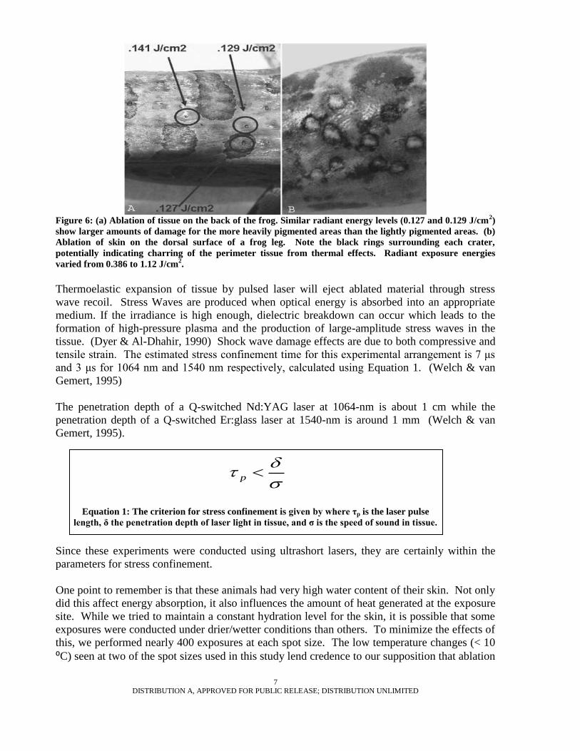
Figure 6: (a) Ablation of tissue on the back of the frog. Similar radiant energy levels (0.127 and 0.129 J/cm
2)
show larger amounts of damage for the more heavily pigmented areas than the lightly pigmented areas. (b)
Ablation of skin on the dorsal surface of a frog leg. Note the black rings surrounding each crater,
potentially indicating charring of the perimeter tissue from thermal effects. Radiant exposure energies
varied from 0.386 to 1.12 J/cm2.
Thermoelastic expansion of tissue by pulsed laser will eject ablated material through stress
wave recoil. Stress Waves are produced when optical energy is absorbed into an appropriate
medium. If the irradiance is high enough, dielectric breakdown can occur which leads to the
formation of high-pressure plasma and the production of large-amplitude stress waves in the
tissue. (Dyer & Al-Dhahir, 1990) Shock wave damage effects are due to both compressive and
tensile strain. The estimated stress confinement time for this experimental arrangement is 7 μs
and 3 μs for 1064 nm and 1540 nm respectively, calculated using Equation 1. (Welch & van
Gemert, 1995)
The penetration depth of a Q-switched Nd:YAG laser at 1064-nm is about 1 cm while the
penetration depth of a Q-switched Er:glass laser at 1540-nm is around 1 mm (Welch & van
Gemert, 1995).
Since these experiments were conducted using ultrashort lasers, they are certainly within the
parameters for stress confinement.
One point to remember is that these animals had very high water content of their skin. Not only
did this affect energy absorption, it also influences the amount of heat generated at the exposure
site. While we tried to maintain a constant hydration level for the skin, it is possible that some
exposures were conducted under drier/wetter conditions than others. To minimize the effects of
this, we performed nearly 400 exposures at each spot size. The low temperature changes (< 10
⁰C) seen at two of the spot sizes used in this study lend credence to our supposition that ablation
8
DISTRIBUTION A, APPROVED FOR PUBLIC RELEASE; DISTRIBUTION UNLIMITED
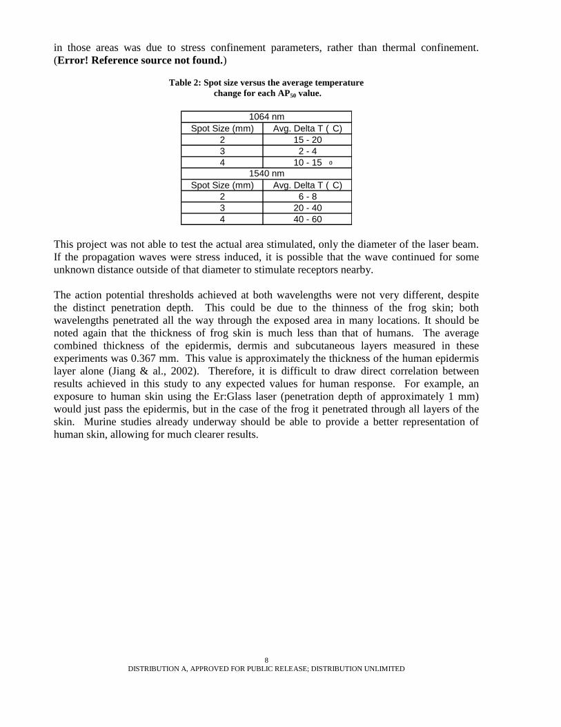
in those areas was due to stress confinement parameters, rather than thermal confinement.
(Error! Reference source not found.)
Table 2: Spot size versus the average temperature
change for each AP50 value.
Spot Size (mm) Avg. Delta T (
⁰
C)
2 15 - 20
3 2 - 4
4 10 - 15
Spot Size (mm) Avg. Delta T (
⁰
C)
2 6 - 8
3 20 - 40
4 40 - 60
1540 nm
1064 nm
This project was not able to test the actual area stimulated, only the diameter of the laser beam.
If the propagation waves were stress induced, it is possible that the wave continued for some
unknown distance outside of that diameter to stimulate receptors nearby.
The action potential thresholds achieved at both wavelengths were not very different, despite
the distinct penetration depth. This could be due to the thinness of the frog skin; both
wavelengths penetrated all the way through the exposed area in many locations. It should be
noted again that the thickness of frog skin is much less than that of humans. The average
combined thickness of the epidermis, dermis and subcutaneous layers measured in these
experiments was 0.367 mm. This value is approximately the thickness of the human epidermis
layer alone (Jiang & al., 2002). Therefore, it is difficult to draw direct correlation between
results achieved in this study to any expected values for human response. For example, an
exposure to human skin using the Er:Glass laser (penetration depth of approximately 1 mm)
would just pass the epidermis, but in the case of the frog it penetrated through all layers of the
skin. Murine studies already underway should be able to provide a better representation of
human skin, allowing for much clearer results.

9
DISTRIBUTION A, APPROVED FOR PUBLIC RELEASE; DISTRIBUTION UNLIMITED
Figure 7: AP50
probability vs.
temperature rise
data for 1540 nm
exposures with a 4
mm spot size.
While the parameters of this experiment did not provide any conclusive information, the data
from the 4-mm 1540 nm exposures did provide some insight as to future possibilities for this
kind of work. This larger spot size provides for a more linear temperature rise, allowing for
more predictable energy requirements to estimate surface damage before exposures. (Figure 7)
Conclusions
The results presented in this document are only the beginning of a new line of research in the
systematic characterization of neural stimulation with ultrashort-pulsed lasers. For this research
we have studied the effects at two wavelengths and three individual beam diameters. The most
significant finding provided by this study was that smaller beam diameters were needed to avoid
tissue damage while still causing stimulation. As the results show, larger beam diameters have
much lower thresholds in terms of radiant exposure for both neural stimulation and skin
damage. As the laser beam diameters increased, the damage threshold decreased. The action
potential threshold for the larger spots is lower, since the laser is stimulating a greater number
of neurons. Therefore, based upon our findings, the ideal spot size would be 3 mm since it
required lower laser energy to stimulate action potentials, and did so at energy levels below
those that cause skin damage. It was shown that tissue ablation occurred well before the
average surface temperature of the skin reached 100⁰ C, which may be explained by laser
induced breakdown or stress confinement mechanisms. Indeed, skin damage frequently
occurred before action potentials were stimulated at beam diameters of 4 mm for each
wavelength. This phenomenon will certainly require additional studies to determine the exact
mechanism of damage; whether it be thermal, mechanical, or a combination of the two.
It became obvious that the differences between frog skin pigmentation and morphology from
that of humans makes them ill suited as human skin damage threshold models. A mammalian
study (currently underway) should provide the necessary data to determine the best wavelength
for creating action potentials without causing skin damage. It could also provide more precise
data on skin damage, since water content of the skin will not be such an issue.

10
DISTRIBUTION A, APPROVED FOR PUBLIC RELEASE; DISTRIBUTION UNLIMITED
Finally, this study was conducted with two wavelengths common in the medical and photonics
industries. Additional wavelengths should be studied to determine if different penetration
depths or powers could yield more optimal results.
Acknowledgement
We would like to thank the Air Force Research Lab, Northrop Grumman Information
Technologies (Contract # F41624-02-D-7003) and Pittsburg State University for their support.
Bibliography
1. Arendt-Nielson, L., and Bjerring, P., Sensory and Pain Threshold Characteristics to
Laser Stimuli, Journal of Neurology, Neurosurgery, and Psychiatry 51, 35 (1988).
2. Cain, C., & Manning, L. (1996). Analyzing Yes/No Data on a Log Scale. Brooks City-
Base, TX: Tech Report AL/OE-TR-1996-0102.
3. Hendry, S., & Hsiao, S. (2003). Fundamental neuroscience, 2nd Edition. Boston:
Academic Press.
4. Izzo, A., Pathria, J., Suh, E., Whitlonb, D., Jansen, E., & Richter, C. (2006). Selectivity
of Optical Stimulation in the Auditory System. Proceedings of SPIE Vol. 6078, 6078P-
1.
5. Jiang, S., & al., e. (2002). Effects of Thermal Properties and Geometric Dimensions on
Skin Burn injuries. Burns, 713-717.
6. Kao, C., Wells, J., & Jansen, E. (2005). Application of Infrared Light for in vivo Neural
Stimulation. Journal of Biomedical Optics , 1-12.
7. Kneebone, Wm J., Basic Principles of Low-Level Laser Therapy and Clinical
Applications for Pain Relief, Dynamic Chiropractic 25 (18), 1 (2007).
8. Lefaucher, J.P., Debray, S., & Jarry, G. (2001). Nd:YAG Laser Evoked Potentials.
Muscle and Nerve, 496-500
9. Moore, K., Lasers and Pain Treatment, (The Czech Society for the Use of Lasers in
Medicine, 2004).
10. Perchet, C., Godinho, F., Mazza, S., et al., Evoked Potentials to Nociceptive Stimuli
Delivered by CO2 or ND:YAP Lasers, Clinical Neurophysiology 119, 2615 (2008).
11. Physiology Dept., McGill University. (2005). Retrieved March 2007, from The
Compound Action Potential of the Frog's Sciatic nerve:
http://www.medicine.mcgill.ca/physio/vlab/PDF/cap2005-06.pdf
12. Welch, A. J., & van Gemert, M. J. (1995). Optical-Thermal Response of Laser-
Irradiated Tissue. New York: Plenum Press.
13. W. Precht & Linas, R. (1976). Frog Neurobiology: A Handbook, New York, Springer-
Verlag