elimination of different heavy metals in contaminated soil ... · elimination of different heavy...
TRANSCRIPT

J. Mater. Environ. Sci. 7 (7) (2016) 2603-2616 Khalil et al.
ISSN : 2028-2508
CODEN: JMESC
2603
Elimination of different heavy metals in contaminated soil using indigenous
microorganisms and nanoparticle in the El-Rahawy village, Egypt
N.M. Khalil, H.S. El-Sheshtawy*, D. Aman Egyptian Petroleum Research Institute, Nasr City, 11727 Cairo, Egypt
Received 04 Mar 2016, Revised 28 Apr 2016, Accepted 03 May 2016
* E-mail: Email: [email protected] (H.S. El-Sheshtawy); Phone: +202 22745902; Fax: + 202 227727433
Abstract El-Rahawy drain, Egypt is exposure to various sources of contamination, including heavy metals which are considered
serious growing problem for agricultural and human activities. The soil sample was collected from El-Rahawy village and
the physicochemical properties of soil sample were determined. The aim of this study was to select autochthonous
microorganisms that are capable to elimination of heavy metals in the polluted soil sample in the presences of biosurfactants
and graphene oxide nanoparticle. These bacterial isolates were identified by 16sRNA analysis as Pseudomonas putida
KM434858 strain A1-26 (B1) and Bacillus subtilis DS=15029 (B2). The characterization of graphene oxide nanoparticle was
determined by XRD and transmission electron microscopy (TEM) analysis. The surface properties and the removal of heavy
metals were tested in all microcosms. The best results were obtained for the bacterial strain (B2) which showed decreased in
the surface tension from 72 to 32mNm-1
. While, the emulsification power percentage increased up to 98 % in microcosm
containing the bacteria-producing biosurfactant and graphene oxide nanoparticle. The concentration of Fe decreased from
56764into 1370 mg/l, Cu 109-0 mg/l, Co 98.8- 0 mg/l, Ni 76.2-0 mg/l, Cr 68-0 mg/l, Zn 92.6-0 mg/l. The results indicate
that, these bacterial strains may be useful and environmentally friendly approach for removing pollutants, heavy metals from
contaminated soil in the presence of biosurfactants and nanoparticle.
Key words: microorganisms, biosorbtion, bioremoval, biosurfactants, nanoparticles
1. Introduction Heavy metals are natural compounds of the environment, being present in rocks, soil, plants and animals. They
occur in various forms: as minerals in rock, sand and ground; bound in organic or inorganic molecules are
attached to particles in the air [1]. Metals are present in the solid phase and in solution, as free ions, or adsorbed
to soil colloidal particles [2]. In universal, it is very difficult to distinguish between the natural metal
enhancement and resulting from anthropogenic sources.
Heavy metals pollution has become one of the most serious ecological problems today. With the fast development
of industries such as metal plating facilities, mining operations, fertilizer industries, tanneries, batteries, sheet
manufacture and pesticides, etc. Heavy metals wastewaters are directly or indirectly throw into the environment
progressively; especially in developing countries. Release of heavy metals without proper treatment pass a
significant threat to public health because of its persistence and accumulation in the food chain. Unlike organic
contaminants, heavy metals are not biodegradable and tend to accumulate in living organisms and abundant
heavy metal ions are recognized to be poisonous or carcinogenic [3, 4].
Heavy metals have been extensively deliberated and their effects on individual health regularly reviewed by
international bodies such as the World Health Organization (WHO). Each heavy metal has unique toxicity or
function. For examples zinc and copper can enhance microbial development at low concentrations, but represses
growth at high concentrations [5]. Heavy metals such as copper, iron, chromium and nickel are fundamental

J. Mater. Environ. Sci. 7 (7) (2016) 2603-2616 Khalil et al.
ISSN : 2028-2508
CODEN: JMESC
2604
minerals since they play an important role in biological systems, where cadmium and lead are non-major metals,
they are toxic, even in trace amounts [6]. The evacuation of the toxic metal particles from waste water is being
received by numerous techniques, for instance, adsorption, biosorption, ion exchange etc. Among them,
adsorption is generally utilized because of its easy, effective and least effort. During the years, different
adsorbents including inorganic materials, polymers, wood squanders, and carbon-based materials have been
contemplated to take out overwhelming metal particles. Carbon-based materials, for example, carbon nanotube,
activated carbon, graphene, graphene oxide (GO) has attracted specific thoughtfulness regarding be utilized to
adsorb substantial metal particles [7].
Biosorption of heavy metals from aqueous solutions is a comparatively new procedure that has been proven a
very promising process in the removal of heavy metal contaminants. The prime feature of biosorption is its high
efficiency in reducing the heavy metal ions and the use of inexpensive biosorbents. Biosorption methods are
especially appropriate to treat dilute heavy metal wastewater. Typical biosorbents can be derived from three
origins as follows up Apiratikul and Pavasant [8]: (1) non-living biomass such as bark, lignin, shrimp, krill, crab
shell, etc.; (2) Algal biomass; (3) microbial biomass, e.g. Bacteria, fungi and yeast.
Bioremediation technology has provided an alternative to conventional methods for remediation the metal-
polluted soils [9]. The microbiological processes are significant in determining metal mobility and have actual
possibility application in bioremediation of metal contamination [10]. According to Ge et al. [5] the various
approaches have been possessed to repair of metal contaminated soil by bioremediation process.
In Egypt, the Nile river is the lifeline provisioning water to tens of millions people. It flows into the
Mediterranean Sea by its two major branches, Damietta and Rosetta, which are streamingthrough the Nile delta
wetland [11, 12]. El- Rahawy drain is located in the southern part of the Nile Delta, Egypt, El-Rahawy drain is
about 12.41 km with amoderate length of 4.5 Km, which outlet on Rosetta branch. This drain passes through El-
Rahawy village and many villages distributed along it receive agricultural and domestic wastes without treatment
or partial treatment.In addition to sewage of El-Giza governorate that discharged directly into a Rosetta branch of
the River Nile [12]. The drain is encompassed by high density of population area and wide agricultural lands [5,
6]. Which are considered a serious growing problem for agricultural and human activities (Figure 1).
The present work is to report the description of the physicochemical characteristics of soil sample collected from
El-Rahawy village, Giza, Egypt. Also, this study is an investigation of the ability of isolated bacterial strains
towards biosorption or bioremediation of different heavy metals in soil samples by producing biosurfactants. The
elimination of heavy metals from soil sample is done using graphene oxide (GO) nanoparticle or a combination
between the in-situ biosurfactant and nanoparticle.
Figure 1: Location of El-Rahawy village, Egypt
2. Experimental 2.1. Sample Collection

J. Mater. Environ. Sci. 7 (7) (2016) 2603-2616 Khalil et al.
ISSN : 2028-2508
CODEN: JMESC
2605
One soil sample was collected from agricultural land which irrigated by El-Rahawy drain from El- Rahawy
village, Giza, Egypt. The sample was collected by clean stainless steel shovel from 0-15 cm depth.
2.2 Chemical and physical characterization of soil
The soil sample was air-dried at room temperature. The soil pH was determined by a pH meter with a soil/water
ratio of 1:2.5 in an aqueous suspension. pH was determined according to ASTM D-1293 using digital pH meter
model metler Toledo-Seven Go. The electrical conductivity (EC) was established using 1:2.5 soil – distilled water
ratio, Conductivity and resistivity was determined on site using a digital conductivity meter WTW 330I according
to ASTM D-1125. In addition to the Anions and cations were determined according to ASTM D-4327 and D-
6919, respectively using ion chromatography. The instrument used was Dionex IC model ICS 1100 equipped
together with high capability columns (AS9 and CS12) for anion and cations, respectively. The heavy metals in
soil were analyzed according to the method of the Environmental Protection Agency (EPA) method 3050A
(1992). Soil sample was digestedby acid, with a mixture of HCCl3 (tri-chloromethane acid), HNO3 (nitric acid)
and HClO4 (perchloric acid) H2O2 (hydrogen peroxide), using Teflon flak. Heavy metals were determined using
Flame Atomic Absorption Spectrophotometer model Zenit 700p approving to ASTM D4691.
2.3 Preparation and characterization of graphene oxide
Graphene oxide (GO) was prepared from the commercial graphite utilizing a modified Hummers’ procedure
(GO-H). Typically, Concentrated H2SO4 (360 ml) was added to a blend of graphite (7.5 g) and NaNO3 (7.5 g),
and the mixture was cooled down to 0 °C in an ice bath. KMnO4 (45 g) was added quietly in little doses to keep
the reaction temperature below 20 °C. The solution was heated to 35 °C and stirred for 3 h. Then 3 % H2O2 (1.5
L) was slowly added. This had a pronounced exothermal effect at 98 °C. The reaction mixture was whiskered for
30 min and, ultimately, the mixture was centrifuged (3700 RPM for 30 min), after which the supernatant was
poured away. The residual solid material was then washed with 600 ml of water and centrifuged again, this
process being reduplicated until the pH was neutral [13].
X-ray powder diffraction Analysis (XRD) was taken out using Shimadzu XD-1 diffractometer in 2θ range
between 20 and 80°. The morphology of the samples was distinguished by high-resolution transmission electron
microscopy (HR-TEM) spectroscopy, the TEM pictures were gained by a JEOL 2100F.
2.4 Culture media
Four different culture media were used in this study. The nutrient agar medium was used for isolation of bacterial
isolates from soil sample. This medium contains the following peptone 5; Sodium chloride 5; Beef extract 3. The
final pH was adjusted at 7.0-7.3. Dox medium was used for isolation of fungi isolates, this medium containing
sucrose 30; NaNO3; KH2PO4 1; MgSO4.7H2O 0.5; KCl 0.5; FeSO4.7H2O 0.01, the pH at 7.3±0.2. UGSY medium
was used for isolation of yeast strains;this medium contains the following urea 1; Glucose 100; Sunflower 100;
yeast extract 10, the pH was adjusted at 6.0 [14]. Mineral salt medium (MSM) was used for screening the
production of biosurfactants by the microbial isolates and the elimination or removal of heavy metal in a soil
sample. This medium was containing (g/l) glucose 20; CaCl2 0.2; KH2PO4 2; Na2HPO4 2; MgSO4.7H2O 0.01;
FeSO4.7H2O 0.001; NaNO3 2.5; NaCl, 0.8; KCl, 0.8, the pH at 7.3±0.2,yeast extract, 3%, and 1 ml of a trace
element solution. Trace element solution contained ZnSO4.7H2O, 525 mg/l; MnSO4.4H2O, 200 mg/l;
CuSO4.5H2O, 705 mg/l; NaMnO4.2H2O, 15 mg/l, CoCl2.6H2O, 200 mg/l; H3BO3, 15 mg/l; NiSO4.6H2O, 27
mg/l[15]. All the above four media were indented as Grams per liter of distilled water, and subsequentlyautoclave
sterilized at 121°C for 20 min. The biosorption or elimination of heavy metals processes used to modify MSM
medium (removing any source of heavy metals in the constituent medium).
2.5 Isolation of microorganisms from soil sample
One gram of soil sample was taken for the serial dilution process. 100μl of the different dilution of the extract
was inoculated in the nutrient agar medium for isolation of bacterial isolates, the constituent of medium was
mentioned above. The pH was adjusted at 7.0. The cultures were then incubated at 30 °C for 48 h. Also, the

J. Mater. Environ. Sci. 7 (7) (2016) 2603-2616 Khalil et al.
ISSN : 2028-2508
CODEN: JMESC
2606
different dilution of the extract was inoculated in Dox and UGSY media for isolation of fungi and yeast isolates.
The pH was adjusted at 6.0. The cultures were then incubated at 25 °C for 72 h. All the above media was supplied
with agar (20 g/L, Merck). Thereafter, plate count in the range of 30 and 300 colonies was recorded [16].
2.6 Selection of the most predominate microbial isolates
Two bacterial isolates and one yeast isolate were chosen for further studies due to the maximum predominated
growth on the nutrient and UGCY agar media.
2.7 Screening of the predominate microbial isolates for biosurfactants production
The bacterial isolates and yeast isolates isolated from soil sample were screened for the production of
biosurfactants. One loopful of microbial growth obtained on nutrient agar plates after 24 h incubation for bacteria
and 72 h for yeast was inoculated on nutrient broth medium and incubated in a rotary shaker 150 RPM at 30 °C
for 18 h. For the biosurfactants production 2ml of inoculum was taken and incubated in 100 ml of MSM broth in
500 ml Erlenmeyer flasks at 30 °C for 120h [15].
2.8 Analysis of producing biosurfactant
Surface properties including surface tension and Emulsification index (E24) were determined as indicated for the
production of biosurfactant.
2.8.1 Surface tension
The surface tension was measured with Krüss K6 tensiometer. The bacterial supernatant solution (free cell
culture) (50 ml) was tested at 25 °C to evaluate the surface tension of biosurfactant [17]. The surface tension was
expressed as mN/m.
2.8.2 Emulsification activity (E24)
E24 of the produced biosurfactant in the supernatant were measured by adding kerosene (6ml) (Dearomatized
kerosene was supplied by the Alexandria Petroleum Refining Company, Alexandria, Egypt) to the aquatic phase
(supernatant culture or cell free culture) and vortexing for 2 min. After 24 h, the emulsion index (E24) was
calculated conferring to the nextequation [18].
(E24) = 100 (height of the emulsion layer/ the total height)
2.8.3 Genomic DNA isolation and PCR analysis of the most promising bacterial isolates
The bacterial isolates were selected for further study. An analysis of 16S rRNA was performed to taxonomically
characterize the bacterial isolates (B1 and B2) (Sigma Scientific Services Co., Egypt). The bacterial cells were
harvested up to 2 × 10⁹
by centrifugation for 10 min at 5000 xg. Supernatant was removed and pellets were
collected and suspended in 180 µl of digestion solution. The sample was incubated at 56°C use a shaking water
bath, rocking platform until the cells are completely lysed ~30 min. 20 µl of RNase A Solution was added, mix
by vortexing and incubated the mixture for 10 min at room temperature. 200 µl of Lysis solution was added to the
sample, 400 µl of 50 % ethanol was added. The prepared lysate was transferred to a GeneJETTM
Genomic DNA
purification column. 80 µl of Elution buffer was added to the center of the GeneJETTM
Genomic DNA purified
column membrane to wash genomic DNA. The sample was incubated for 2 min at room temperature and
centrifuge for 1 min at 8000 xg. The purification column was discarded and used the purified DNA in PCR. A
polymerase chain reaction (PCR) was made by using Maxima Hot Start PCR Master Mix (Thermo K1051). PCR
was performed using (5'-AGA GTT TGA TCC TGG CTC AG-3') (5'-GGT TAC CTT GTT ACG ACTT-3') as
forward and reverse primer, respectively for (B1) bacterial isolate while the PCR was also performed using (5'-
GCTTGCTCCCTGATGTTAGC-3') (5'-CGGTACCTTGACGGACCTAA-3') as forward and reverse primer for
(B2) bacterial isolate. PCR was cleaned up to the PCR product using the GeneJET™ PCR Purification Kit. A 45
µl of Binding Buffer was added to the finished PCR mixture. The mix was then thoroughly transferred from step
1 to the GeneJET™ purification column. The mixture was then centrifuged for 30–60 s at> 12,000 xg, and then
J. Mater. Environ. Sci. 7 (7) (2016) 2603-2616 Khalil et al.
ISSN : 2028-2508
CODEN: JMESC
2607
the flow was discarded. A 100 µl wash buffer was added to the GeneJET™ purification column, centrifuged for
30–60 s, discarded the flow-through and site the purification column back into the combination tube. The mixture
was centrifuged at empty purification column for asupplemental 1 min to fully remove any residual wash buffer.
The purification column was transferred to 1.5 ml micro centrifuge tube. A 25 µl of elution buffer were then
added to the center of the column membrane which subsequently centrifuged for 1 min, dismiss the column and
store the purified DNA at 20 °C. Following purification of the PCR products, the DNA sequence of the
favorableclone was subjected to a similarsearch, BLAST on the NCBI website (http://www.ncbi.nlm.nih.gov),
and preserved into GenBank. Many relevant 16S rRNA gene sequences with validly published names were
chosen as references from the Gen-Bank.
2.8.4 Biosorption or bioremoval of heavy metals using bacterial isolates in the participation of biosurfactants
and nanoparticle
The isolated bacteria were adapted to metal sorption or removal abilities on soil sample in the existence of
Graphene oxide (GO) nanoparticle and the produced biosurfactants as the modified method of Uzel and Ozdemir
[19]. After 12 h of incubation period at exponential growth phase, 2 ml of inoculum containing two bacterial
isolates separatelywas added into 100 ml MSM of 500 ml Erlenmeyer conical flasks. 1g of soil sample including
different kinds of heavy metals was added into the media in the presence or the absence of (GO) nanoparticle.
The control medium (MSM with soil and without the culture) was also maintained at (pH 7). The test and control
medium were incubated on a rotary incubator on 150 RPM at 30°C at different time intervals (24, 48, 72, 96,
120h). After the incubation, the bacterial count, surface tension and emulsification power were determined. The
soil was filtered using filter papers and filtrated soils were dried and digested with acids (HCl and HNO3 in the
ratio of 3:1) and analyzed the different heavy metals in the treated and control soil by using atomic absorption
spectrophotometer [20].
3. Results and discussions 3.1. Physicochemical properties of soil sample
pH is an important property of soil that determines the acidity or alkalinity, which affects the chemical reactions
between water and soil minerals.pH of the soil sample was found within the allowable limits (Table1).
There is a powerful relationship between soil pH and nutrient availability. Alkaline soils with pH ranging 7-8 are
commonly deficient in Zn2+, Fe
3+, P
5+ and also the uptake of various plant nutrients is pH dependent. Most of the
primary nutrients like nitrogen, phosphorous, potassium and secondary nutrients like calcium, magnesium and
sulfur are preferableconsumed by the plants when the pH rang are 5.5-7.9. The uptake of most of the
micronutrients also takes place at low pH 20. At pH 5.5, fungi and algae generally dominate the soils while at
higher pH levels the bacteria are predominating [21].
The electrical conductivity (EC) which may create a salinity problem over a long period of time was found 0.075
× 10-2
MS /cm, whereas the permissible limit is 4 ms/cm (Table1).
In supplement, the amount of salinity (104.6 mg/l) fell below WHO\FAO acceptable limit of 2000 mg/l. Also
Total Dissolved Solids (TDS) is known to be negatively charged and therefore attract more heavy metal ions,
reducing their condensation in water [21].
Table 1:The physicochemical properties of the soil sample
3.2. Some anions and cations of the soil sample extract
Constituents Experimental results According to WHO \
FAO
Total Dissolved Solids
(TDS) 540.1 mg/l 500 mg/l
Conductivity 0.075 × 10⁻²
mS/cm 4 mS/cm
Salinity 104.6 mg/l 200 mg/l
pH 8.17 @ 25 °C 6.5-9

J. Mater. Environ. Sci. 7 (7) (2016) 2603-2616 Khalil et al.
ISSN : 2028-2508
CODEN: JMESC
2608
Concentration of sulfate (anion) was 90.000 mg/L in our sample which is considered lower than permissible value
(500.000) mg/L according toTanzania Bureau of Standards (TBS) [22] as shown in Table (2). But the increase of
nitrate (anion) in the sample as illustrated in Table (2) indicated that waste water which use for irrigation is rich in
nitrate [23].
The concentration of potassium (cation) in the present soil sample was found to be 36.57 ppm (Table 2). The
optimum levelswanted for the crop agriculture are recognized to be 180-300 ppm while, below 60 ppm deficiency
symptoms may occur [23].
So the soil of the present study is found to be deficient in potassium. Potassium is the main nutrient source after
nitrogen and phosphorous, which is considered essential for the plant growth. Potassium is an enzyme activator
that excesses photosynthesis and decreasesharvest loading [24, 25]. The availability and leaching of potassium
have become a limiting factor for crop production in many types of soils.
The soil sample was also found deficient in calcium (cation) content (Table 2). Whereas, the deficiency
symptoms were generally occur below 500 ppm [26]. Physiologically calcium is important in plant nutrition. It
regulates the growth, IAA as is shown hormone, a calmodulin barrier across the cell membrane to regulate inter
cellular cation and anion balance [27].
Magnesium (cation) plays an active role in plant growth and metabolism. It arranges the ATP enzymes, carbon
dioxide stabilization, cellular pH control, chlorophyl content, chloroplast pigmentation and many other functions
of crop development [27].The amount of exchangeable Mg is less than the exchangeable soil complexes.
Magnesium content was found to be 11.92 ppm value (Table 2) and is less than the permissible limit. The
magnesium deficiency occurs below 60 ppm [26].
Table 2: Determination of some anions and cations from extract of soil sample
Cations Concentration
(mg/I)
Concentration
according to
WHO (mg/I)
Anions Concentration
(mg/I)
Concentration
according to
(TBS) (mg/I)
Sodium 67.99 200.00 Fluoride 8.70 1-1.5
Potassium 36.57 180-300 Nitrate 48.00 20,00
Magnesium 11.92 150 Sulfate 90.000 500.00
Calcium 48.80 75
(WHO): World Health of Organization; (TBS): TanzaniaBureau of Standards.
3.3. Heavy metals of soil sample
Copper does not deteriorate in the environment and can be cumulative in plants and animals when it is found in
soil. In copper-rich soils only a restricted number of plants have anopportunity of survival. Due to the effects
ofplants, copper is a serious threat to the manufacture of farmlands. Copper can seriously impact, the proceedings
of certain farmlands, rely upon the acidity of the soil and the existence of organic matter. The results indicated
that value of copper in the soil sample was found to be 68.4 mg/L (ppm). The value of Cu in the sample was
found greater than the permissible limits (1.000 ppm) according to WHO\FAO [28] soil standard (Table 3). So
the soil of the present study was found adequate in copper.
The soil concentration of nickel was found 76.2 ppm. The analysis of Ni showed that the concentration of Ni
higher than the permissible limit and there is a need of remediation. Nickel has been considered to be an essential
trace element for human and animal health. In living systems, it is related with DNA and RNA molecules and
also a regulatory element for the various enzyme systems
The concentration of cobalt in the soil was found 58.8ppm, the permissible limit in plants recommended by WHO
is not detected.
The concentration of zinc was found to be 92.6ppm, whereas the maximum permissible limit 3.000ppm (Table 3).
This is suggestive of input of effluents into the soil from El-Rahawy drain to irrigate El-Rahway land. The major
scientific and medicinal advantage in iron is as a fundamental metal, but toxicological considerations are
important in terms of accidental acute exposition and chronic iron over load. With rare exclusions, virtually all

J. Mater. Environ. Sci. 7 (7) (2016) 2603-2616 Khalil et al.
ISSN : 2028-2508
CODEN: JMESC
2609
living organisms are dependent on iron for subsistence. Regardless of the ubiquitous allocation and abundance of
iron in the biosphere, iron-dependent life must contend with the antithetical hazards of iron insufficiency and iron
overload, each with its serious or fatal consequences. Iron is a normal ingredient of soils, but its concentration can
be influenced by some industries. It has been reported that civil soils presented different heavy metal features
[29]. In the present study high concentration of iron was recorded at 56764 ppm, while the concentration of this
element at 3000 ppm according WHO\FAO [28] soil standard.
Table 3: Concentration of heavy metals in soil sample and standards according to WHO\FAO
3.4. Characterization of graphene oxide nanoparticle
High transparency of graphene oxide (GO) to the electron beam has been scanned by transmission electron
microscopy (TEM) image (Figure 2). It is obvious that GO is partially transparent with some wrinkles and has
lateral distance of several micrometers with the small holes caused by overexposure to sonication.
Figure 2: TEM images for graphene oxide (GO)
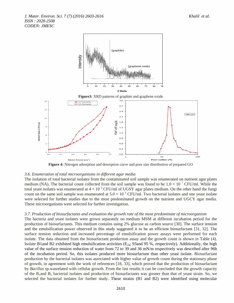
XRD patterns of graphite and graphene oxide are showed in Figure (3). As can be seen, a sharp 2 θ reflection at
26.050 in the natural graphene originated from interlayer spacing and there is a clear peak at 2θ = 8.6°
corresponding to the inter-layer empty of 0.83 nm in the XRD pattern of graphene oxide due to the introduction
of oxygen-containing series and the insertion of moisture. The XRD and TEM figures confirmed that the GO
nanosheets successfully prepared.
Figure (4) exhibited the nitrogen adsorption–desorption isotherms and pore size distribution of the GO. Nitrogen
sorption–desorption isotherms display IV-type curves, which is representative for mesoporous materials. The
BET surface area of the GO is calculated to be 57.4 cm2/g.
3.5. Isolation of different microorganisms in a soil sample
Eighteen bacterial strains were isolated on (NA) medium that was established at 30 °C for 4 weeks. While three
fungi isolates were isolated from Dox medium. Two yeasts isolate were isolated from soil sample on UGSY agar
medium.
Metals in soil
sample
Experimental results
(ppm)
According to WHO\FAO(1991)
Standards in sediment (ppm)
Cu 68.4 1.000
Zn 92.6 3.000
Ni 76..2 0.02
Fe 56764 3000. 0
Co 58.8 NA
J. Mater. Environ. Sci. 7 (7) (2016) 2603-2616 Khalil et al.
ISSN : 2028-2508
CODEN: JMESC
2610
Figure3: XRD patterns of graphite and graphene oxide
Figure 4: Nitrogen adsorption and desorption curve and pore size distribution of prepared GO
3.6. Enumeration of total microorganisms in different agar media
The isolation of total bacterial isolates from the contaminated soil sample was enumerated on nutrient agar plates
medium (NA). The bacterial count collected from the soil sample was found to be 1.0 × 10 7 CFU/ml. While the
total yeast isolates was enumerated at 4 × 10 3 CFU/ml of UGSY agar plates medium. On the other hand the fungi
count on the same soil sample was enumerated at 5.0 × 10 3 CFU/ml. Two bacterial isolates and one yeast isolate
were selected for further studies due to the most predominated growth on the nutrient and UGCY agar media.
These microorganisms were selected for further investigation.
3.7. Production of biosurfactants and evaluation the growth rate of the most predominate of microorganism
The bacteria and yeast isolates were grown separately on medium MSM at different incubation period for the
production of biosurfactants. This medium contains using 2% glucose as carbon source [30]. The surface tension
and the emulsification power observed in this study suggested it to be an efficient biosurfactant [31, 32]. The
surface tension reduction and increased percentage of emulsification power assays were performed for each
isolate. The data obtained from the biosurfactant production assay and the growth count is shown in Table (4).
Isolate B1and B2 exhibited high emulsification activities (E24; 93and 95 %, respectively). Additionally, the high
value of the surface tension reduction of water from 72 to 39 and 36 mN/m respectively was described after 96h
of the incubation period. So, this isolates produced more biosurfactant than other yeast isolate. Biosurfactant
production by the bacterial isolates was associated with higher value of growth count during the stationary phase
of growth, in agreement with the work of references [18, 33], which proved that the production of biosurfactant
by Bacillus sp.wasrelated with cellular growth. From the last results it can be concluded that the growth capacity
of the B1and B2 bacterial isolates and production of biosurfactants was greater than that of yeast strain. So, we
selected the bacterial isolates for further study. These strains (B1 and B2) were identified using molecular
0 10 20 30 40 50 60 70 80 90
2 theta
(graphite)
(graphene oxide)Inte
nsity
0
3
6
9
12
15
18
0,0 0,2 0,4 0,6 0,8 1,0
Relative Pressure (p /po)
Ads
orbe
d V
olum
e at
STP
(cc/
g)
GO Adsorption
GO Desorption
0,0
0,3
0,6
0,9
1,2
1,5
1,8
2,1
2,4
2,7
3,0
1 10 100
Pore radius (Å)
Vp /drp x
103 (c
c/g.Å
)GO

J. Mater. Environ. Sci. 7 (7) (2016) 2603-2616 Khalil et al.
ISSN : 2028-2508
CODEN: JMESC
2611
identification completed by enlarging and sequencing the 16S rRNA gene sequences. The results of the
identification method demonstrated that the two isolated bacteria belong to Pseudomonas putida KM434858
strain A1-26 (B1) 94.21 %, Bacillus subtilis DS=15029 (B2) 99.94%. Literature search revealed that various
speciesof Pseudomonas and Bacillus are most common strains produced by biosurfactants [34, 35].
Table 4: Surface properties of biosurfactants and growth count of bacterial and yeast isolates at different time intervals
Microorganism
Incubation
period
(h)
Surface properties
Log count Surface
tension
(mN/m)
Emulsification
power (%)
Bacterial
isolate (B1)
0 57 30 6.0
24 49 72 6.2
48 45 77 7.0
72 41 85 7.0
96 39 93 7.1
120 43 80 7.0
Bacterial
isolate (B2)
0 56 31 6.5
24 50 70 7.1
48 50 77 7.3
72 40 93 7.4
96 36 95 7.4
120 39 85 7.7
Yeast isolate
0 54 25 5.9
24 43 10 6.2
48 40 12 6.1
72 40 15 6.2
96 39 10 6.5
120 47 10 6.7
3.8. Elimination of heavy metals by bacterial strains and nanoparticle
Biosorption or bioremoval of heavy metals from soil sample collected from El-Rahawy village is a relatively new
process that has been confirmed a very promising process in the removal of heavy metal contaminants. The major
advantage of biosorptionis its high effectiveness in reducing the heavy metal ions and the use of cheap
biosorbents [8].
3.8.1. Determination of growth rate and some surface properties of media after biosorption process
Biosurfactants, a group of surface active molecules synthesized by different microorganisms, qualify to decrease
both surface and intersurface tension and increased the emulsification power [36].The anionic nature of
biosurfactants can also capture the metal ions through electrostatic interactions or complexation [37].
In this research, bacterial (B1 and B2) strains were grown separately in MSM medium. This medium contains 2%
glucose with/without nanoparticle and 1 g soil sample containing different concentration of heavy metals. Data
are shown in (Table 5) the nanoparticle (np) having surface properties which indicated by the reduction of surface
tension and increased emulsification power percentage, in the microcosm containing heavy metals and (GO) np.
De Windt et al. [38]; Shan et al. [39] were illustrated that nanoparticle catalysts are more popular than common
catalysts due to their individual properties and high obtainable active specific surface. Moreover, Addition of
nanoparticle (np) to the previous media enhanced the bacterial growth rate and the biosurfactant efficiency.This
achievement confirmed that the nanoparticles were susceptible of helping the microbial activities which agrees
with several other studies Banat et al. [40] and El-Sheshtawy et al. [15].The higher growth rate, best produced

J. Mater. Environ. Sci. 7 (7) (2016) 2603-2616 Khalil et al.
ISSN : 2028-2508
CODEN: JMESC
2612
biosurfactants by (lower surface tension and the higher emulsification power) were obtained in microcosms
containing the heavy metals, (GO) np and two different bacterial isolates separately.
Table 5: Surface properties and growth count of bacterial strains after biosorption process
Sample
Surface properties
Log
count Surface
tension
(mN/m)
Emulsification
power (%)
Control 55 9 0
Heavy metal+ graphene oxide
(GO) np 38 77 0
Heavy metal+ (B1) 40 92 7.2
Heavy metal+ (B2) 36 94 7.8
Heavy metal+ (GO) np+ (B1) 35 95 8.1
Heavy metal+ graphene oxide
(GO) np+ B2 32 98 8.9
Control: MEM medium containing only heavy metals.
3.8.2. Atomic absorbance spectrophotometer (AAS)
The heavy metals in the treated by microorganisms and control extract soil samples were analyzed using atomic
absorption spectrophotometer [20]. Data in Table (6) were shown that the heavy metals was adsorpted on the
surface of GO (np) in microcosm containing the heavy metals and GO (np). By comparison of (TEM) images of
GO before and after adsorption (Fig. 5). The authors observed that, the stable and most transparent locations are
likelyto be a monolayer graphene oxide, whereas the darker area indicated metal ions adsorption in graphene
oxide as in Figure (5).
Table 6: Elimination of heavy metals by two different bacterial strains
with/without nanoparticle using atomic adsorption Spectrophotometer
Sample Heavy metals concentration (mg/l)
Fe Cu Co Ni Cr Zn
Control 2979 109 98.8 76.2 68 92.6
Heavy metal+ graphene
oxide (GO) np 1400 N.D. N.D. N.D. N.D. N.D.
Heavy metal+ (B1) 2200 50 20 15 27 18
Heavy metal+ (B2) 2090 25 5 10 15 8
Heavy metal+ (GO)
np+ (B1) 1440 10 1 8 10 4
Heavy metal+
grapheme oxide (GO)
np+ B2
1370 N.D. N.D. N.D. N.D. N.D.

J. Mater. Environ. Sci. 7 (7) (2016) 2603-2616 Khalil et al.
ISSN : 2028-2508
CODEN: JMESC
2613
Figure 5: TEM images for GO a. before adsorption of heavy metal b. after adsorption of heavy metal
Zeta potential was examined inpreparing GO and the outcome demonstrates that GO fluid scattering is pH-
sensitive (Figure 6). The surface charge of GO is exceptionally negative charged and zeta potential turns out to be
more negative in pH 7 (at which the experimental occurred). This marvel, which is illustrated in Figure (7), may
be because of the ionization of the different groups (carboxylic and/or hydroxyl bunch) on the surface of GO
nanosheets [41]. It likewise recommends that stable GO colloid has been framed as a result of electrostatic
aversion [42].
As a subordinate of graphene, one atom thick GO sheets has not just amazingly high surface region up to 2620
m2/g (theoretical value) which can be from cheap natural graphite in large scale additionally oxygen-containing
practical gatherings, for example, – OH, – COOH, and C=O at its edges and surfaces [43]. These oxygen
containing gatherings on GO sheet makes it less demanding to be scattered in water and act as binding sites for
metal ions complexation [44]. Plus, electron donor–acceptor connection between the delocalized electron of
graphene layers (electron donors) and heavy metals (electron acceptors) is additionally thought to be in charge of
the adsorption [45]. The previous properties of GO surface explain its signifying capacity to sorb heavy metals.
According to the survey, GO has higher extreme sorption abilities for Pb (II), Cd (II), Zn (II), Cu (II), Co (II), and
U (VI) than any other currently reported material. The extraordinary capacity of GO to sorb metal cations from
solution suggests potential uses in water treatment and contaminant remediation [46].
Figure 6: Zeta potential of graphene oxide (GO)
Figure 7: The panel on the right shows a cartoon representation (C, gray; O, red; H, blue) of the sequence of
ionization. The ionized groups are highlighted.

J. Mater. Environ. Sci. 7 (7) (2016) 2603-2616 Khalil et al.
ISSN : 2028-2508
CODEN: JMESC
2614
The bacterial strains (B1 and B2) absorbed the heavy metals on the surface cell by the nature of biosurfactants
produced by these strains. Data in Table (5) also represented that these bacterial isolates can be produced
biosurfactants in culture media. From the above results, these bacterial strains in this study belonging to
Pseudomonas putida KM434858 strain A1-26 (B1), Bacillus subtilis DS=15029 (B2) by identify of 16sRNA.
From the literature search suggested that lipopeptide (surfactin) biosurfactant produced by Bacillus sp. and
rhamnolipids biosurfactant produced by Pseudomonas sp. [31, 34]. Amino acid analysis of surfactin biosurfactant
exhibited presence of glutamic acid, isoleucine, leucine, tyrosine, glutamine, proline, alanine, Valine, threonine
and expertise acid. Presence of expertise acid and glutamic acid in the biosurfactant is of special interest as they
are negatively charged (anionic nature) amino acids and can assist in removing cationic metal ions contamination
soil[47, 48]. Herman et al.[49]; Ochoa-Loza, [50]were illustrated that the anionic nature of rhamnolipids, are
accomplished to eliminate metals from ecosystem and ions such as cadmium, copper, lanthanum, lead and zinc
due to their complexation ability. Gautam and Tyagi, [51] was indicated that the rhamnolipids biosurfactant
proved to be capable of removing Cd, Pb and Zn from the soil. The mechanism by which the rhamnolipids
reduced metal toxicity may involve a combination of rhamnolipids complexation of Cd and rhamnolipids
interaction with the cell surface to alter Cd uptake. The higher absorbance or removal of heavy metals on the
surface bacterial strain was observed in microcosm containing the heavy metals, GO (np) and B1 strain. On the
other hand the completely heavy metals absorbance was obtained in microcosm containing the heavy metals, GO
(np) and B2 strain. The presence of biosurfactant with nanoparticle help the bacterial strains to remove or
eliminate heavy metals more than the microcosms containing the bacterial strains and heavy metals separately
after 96 h,which seems to be newly and valuable elimination of heavy metals of contaminatedsoil. In the previous
work, El-Sheshtawy et al. [15] was studied the existence of biosurfactant with different types of nanoparticles
assists the bacterial isolates to consume the iso-paraffins more than n-paraffins. The total degradation of some
various membered rings of polyaromatics and the proportionbioremediation of other polyaromatics increased in
microcosms containing two variouskinds of nanoparticles with biosurfactant withinseven days. However, until
now, very restricted studies have been reported on nanoparticles effect on the bioremediation of heavy metals
contaminated soil in the presence of biosurfactants. Thus, there is multitude of aspects wanted to be studied. Each
kind of nanoparticles has their especial characters. Meanwhile, the chosen of the optimal group of nanoparticles
and microorganisms is considered a great help of the reaction rates. Suitable nanoparticle concentration should be
examined referring to that the excessive concentration can be poisonous to microorganism, thus, it may
minimizethe reaction rate. The reaction conditions in the existence of nanoparticles and microorganisms must be
researched to detect out the optimal conditions for reaction [52].
Conclusions Bioremoval of heavy metals from soil samples collected from El-Rahawy village is a relatively new process that
has been assured a very promising process in the elimination of heavy metal contaminants. In this work, two
bacterial strains were isolated from the El - Rahawy village. The production of biosurfactants with high growth
rate was obtained by these bacterial strains. The removal of heavy metals from soil contamination was studied by
grapheme oxide GO (np), two bacterial strains separately and combination between GO (np) and the two bacterial
strains. The different heavy metals could be adsorpted on the surface of graphene oxide nanoparticle in
microcosm containing the heavy metals and GO (np). The extraordinary ability of GO to sorb metal cations from
solution indicates potential uses in water treatment and contaminant remediation. The bacterial strains (B1 and
B2) were absorbed the heavy metals on the surface cell by the production of biosurfactants in the culture medium.
The enhancement of the removal metals from culture medium was studied by a combination between bacteria-
producing biosurfactants and GO nanoparticle. The application of anionic biosurfactant and specific
concentration of GO nanoparticle in extraction of metal from soil sample can be greener and environment
friendly approach.
References 1. Lasheen M.R., Ammar N.S., Desalin. Water Treat. 52 (2014) 3271.

J. Mater. Environ. Sci. 7 (7) (2016) 2603-2616 Khalil et al.
ISSN : 2028-2508
CODEN: JMESC
2615
2. Silveira M.L.A., Alleoni L.R.F., Guimarães G.L.R., Scien. Agri. 60 (2003) 793.
3. Medici L., Bellanova J., Belviso C., Cavalcante F., Lettino A., Pasquale P., Fiore R.S.,Appl. Clay Sci.
53(2010) 414.
4. Yang Z., Wang Y., Shen Z., Niu J., Tang, Z., J. Hazard. Mater. 166 (2009) 1186.
5. Ge H.W., Lian M.F., Wen F.Z., Yun Y.F., Jian F.Y., Ming, T., J. Hazard Mater. 162 (2009) 50.
6. Ibrahim S.A., Authman M.M.N., Gaber H.S., El-Kasheif M.A., Inter. J. environ. Sci. engin. 4 (2013) 57.
7. Dong Z., Zhang F., Wang D., Liu X., Jin J.,J. solid state chem. 224 (2015) 88.
8. Apiratikul R., Pavasant P., Bioresour. Technol. 99 (2008) 2766.
9. Aweng E.R.M., Suhaimi O., J. Appl. Sci. Environ. Sani. 6 (2011) 463.
10. Opaluwa O.Da., Aremu M.Oa., Ogbo L.Ob., Abiola K.Ab., Odiba I.Ec., Abubakar M. Ma, Nweze N.Od.,
Adv. Appl. Sci. Res. 3 (2012) 780.
11. Badr M.H., Elewa A., Shehata M.B., Mohamed L.F., Abdelaziz G.S., Bull. Environ. Res. 9(2) (2006) 35.
12. El Bouraie M.M., Motawea E.A., Gehad G.M., Yehia M.M., Suoseura-Finnish Peatland Society, Research
Notes. 62 (2011) 31.
13. Botasa C., Álvareza P., Blancoa P., Grandaa M., Blanco C., Santamaría R., Romasantab L., Verdejob
R., López-Manchadob M., Menéndeza R., Carbon. 65 (2013) 156.
14. Daverey A., Pakshirajan K., Coll. Surf. B: Biointer. 79 (2010) 246.
15. El-Sheshtawy H.S., Khalil N.M., Ahmed W., Abdallah R.I., Marine Poll. Bull. 87 (2014a) 191.
16. Benson H.J., A Laboratory Manual in General Microbiology, Brown Company Publishers, Dubuque Lowa,
067137643, (1995).
17. Nitschke M., Pastore G., Bioresour. Technol. 97 (2006) 336.
18. Cooper D.G., Goldenberg B.G., Appl. Environ. Microbiol. 53 (1987) 224.
19. Uzel A., Ozdemir G., Bioresour. Technol. 100 (2009) 542.
20. Mathiyazhagan N., Natarajan D., J Biorem. Biodeg. http://dx.doi.org/10.4172/2155-6199.1000115, (2011).
21. Imran M., Khan A.-UL-H., Hassan A.-UL., Kanwal F., Liviu, M., Amir M., Iqbal A.M., Asian J Chem. 22
(2010) 4823.
22. Tanzania Bureau of Standards, For Receiving Water, Effluents and soils (TBS 789) (2003).
23. Ewa E.E., Iwara A.I., Adeyemi J.A., Eja E.I., Ajake A.O., Otu C.A., J. Environ. Studies. 1 (2011), 8-16.
24. Malik D.M., Chaudhry R.A., Hassan G., Proc. of the workshop on the role of potassium in improving
fertilizer used efficiency, planning and development division, GOP, Islamabad, (1989)71.
25. Rashid M., Bajwa M.I., Shacong A.A., Proc. Natl. cong. Soil Science. 13 (1990) 20-22.
26. Doahue R.L., Miller R.W., Shickluna J.C., An introduction to soils and plant growth, prentice hall Inc.
Englewood Cliffs, New Jersey, (1977) 626.
27. Dawaki M.U., Dikko A.U., Noma S.S., Aliyu U.A., Food and Energy Security. 2 (2013) 20-33.
28. WHO/FAO. Joint FAO/WHO food standard program codex alimentarius commission13th session. Report of
the thirty eight session of the codex committee on food hygiene. Houston, United States of America,
ALINORM 07/30/13, (2007).
29. Yang Z., Wang Y., Shen Z., Niu J., Tang Z., J. Hazard. Mater.166 (2009) 1186.
30. Mulligan C.N., Gibbs B.F., Factors influencing the economics of biosurfactants. Marcel Dekker Inc, New
York, (1993) 329.
31. Kim P.I., Ryu J., Kim Y.H., Chi Y.T., J. Micro. Biotechnol. 20 (2010) 138.
32. de Faria A.F., Teodoro-Martinez D.S., Barbosa G.N.O., Vaz B.G., Silva I.S., Garcia J.S., Process Biochem.
46 (2011) 1951.
33. Vater J., Prog. Colloid Polym. Sci. 72 (1986)12.
34. El-Sheshtawy H.S., Doheim M.M., Egy. J. Petr. 23 (2014b) 1.
35. El-Sheshtawy H.S., Omayma E.A., El-Tabei A.S., Doheim M.M., Int. J. Pharm. Sci. Rev. Res. 33 (2015) 222.
36. Mukherjee S., Das P., Sen R., Trends Biotechnol. 24 (2006) 509.
37. Mulligan C.N., Yong R.N., Gibbs B.F., Engin. Geology 60 (2001) 371.
38. De Windt W., Peter A., Willy V. Environ. Microbiol. 7 (2005) 314.

J. Mater. Environ. Sci. 7 (7) (2016) 2603-2616 Khalil et al.
ISSN : 2028-2508
CODEN: JMESC
2616
39. Shan G.B., Xing J.M., Zhang H.Y., Liu H.Z., Appl. Environ. Microbiol. 71 (2005) 4497.
40. Banat I.M., Makkar R.S., Cameotra S.S., Appl. Microbiol. Biotechnol. 53 (2000) 495.
41. Wang H., Yuana X., Wuc Y., Huanga H., Zenga G., Liua Y., Wanga X., Lina N., Qid Y., Appl. Surf. Sci. 279
(2013) 432.
42. Li D., Müller M.B., Gilje S., Kaner R.B., Wallace G.G., Nature Nanotech. 3(2008) 101.
43. Dong Z., Zhang F., Wang D., Liu X., Jin J., J. Solid State Chem. 224(2015) 88.
44. Lei Y., Chen Fei., Luo Y., Zhang L., Chem. Phys. Letters 593(2014) 122..
45. Yang S., Li L., Pei Z., Li C., Lv J., Xie J., Wen B., Zhang S., Coll. Surf. A: Phys. Eng. Aspects 457(2014)
100.
46. Duster T.A., Szymanowski J.E.S., Na C., Showalter A.R., Bunker B.A., Fein J.B., Coll. Surf. A: Phys. Eng.
Aspects 466 (2015) 28.
47. Mulligan C.N., Yong C.N., Gibbs B.F., Environ. Progr. 18 (1999) 31.
48. Lai C.C., Huang Y.C., Wei Y.H. Chang J.S., J. Hazard Mater. 167(2009)609.
49. Herman D.C., Artiola J.F., Miller R.M., Environ. Sci. Technol. 29(1995) 2280.
50. Ochoa-Loza F., Ph.D. dissertation, University of Arizona, Tucson, (1998).
51. Gautam, K.K., Tyagi V.K., J. Oleo Sci. 55(2006) 155.
52. Zhang X., Song Y.S., Tyagi R., Surampall R. Chemosphere 82(2011) 489.
(2016) ; http://www.jmaterenvironsci.com/