elongation factor 2 and fragile x mental retardation...
TRANSCRIPT

Neuron
Article
Elongation Factor 2 and Fragile X MentalRetardation Protein Control the DynamicTranslation of Arc/Arg3.1 Essential for mGluR-LTDSungjin Park,1,7 Joo Min Park,1,7 Sangmok Kim,1 Jin-Ah Kim,1 Jason D. Shepherd,4 Constance L. Smith-Hicks,2
Shoaib Chowdhury,1 Walter Kaufmann,2 Dietmar Kuhl,5 Alexey G. Ryazanov,6 Richard L. Huganir,1,3 David J. Linden,1
and Paul F. Worley1,2,*1Department of Neuroscience2Department of Neurology3Howard Hughes Medical Institute
Johns Hopkins University School of Medicine, Baltimore, MD 20205, USA4The Picower Institute for Learning and Memory, Massachusetts Institute of Technology, Cambridge, MA 02139, USA5Molecular Neurobiology, Department of Biology-Chemistry-Pharmacy, Freie Universitat Berlin, 14195 Berlin, Germany6Department of Pharmacology, University of Medicine and Dentistry of New Jersey-Robert Wood Johnson Medical School,
Piscataway, NY 08854, USA7These authors contributed equally to this work*Correspondence: [email protected]
DOI 10.1016/j.neuron.2008.05.023
SUMMARY
Group I metabotropic glutamate receptors (mGluR)induce long-term depression (LTD) that requiresprotein synthesis. Here, we demonstrate thatArc/Arg3.1 is translationally induced within 5 min ofmGluR activation, and this response is essential formGluR-dependent LTD. The increase in Arc/Arg3.1translation requires eEF2K, a Ca2+/calmodulin-de-pendent kinase that binds mGluR and dissociatesupon mGluR activation, whereupon it phosphory-lates eEF2. Phospho-eEF2 acts to slow the elonga-tion step of translation and inhibits general proteinsynthesis but simultaneously increases Arc/Arg3.1translation. Genetic deletion of eEF2K results in a se-lective deficit of rapid mGluR-dependent Arc/Arg3.1translation and mGluR-LTD. This rapid translationalmechanism is disrupted in the fragile X diseasemouse (Fmr1 KO) in which mGluR-LTD does not re-quire de novo protein synthesis but does requireArc/Arg3.1. We propose a model in which eEF2K-eEF2 and FMRP coordinately control the dynamictranslation of Arc/Arg3.1 mRNA in dendrites that iscritical for synapse-specific LTD.
INTRODUCTION
Long-lasting forms of synaptic plasticity require de novo protein
synthesis (Kelleher et al., 2004; Wang and Tiedge, 2004).
N-methyl-D-aspartate (NMDA) receptor-dependent long-term
potentiation (LTP) of the Schaffer collateral-CA1 synapse lasting
longer than �60 min is blocked by agents that halt translation.
Late-phase NMDA-dependent LTD in hippocampal slice cul-
tures is also protein synthesis dependent (Kauderer and Kandel,
70 Neuron 59, 70–83, July 10, 2008 ª2008 Elsevier Inc.
2000). De novo protein synthesis is also required for forms of
long-term depression (LTD) that are induced by group I metabo-
tropic glutamate receptor (mGluR) activation or by paired-pulse
low-frequency stimulation (PP-LFS) (Huber et al., 2000). In con-
trast to NMDA receptor-dependent LTP and LTD, where the re-
quirement for protein synthesis is delayed, mGluR-LTD requires
de novo protein synthesis within 5–10 min. (Huber et al., 2000).
The products of de novo protein synthesis are hypothesized to
be ‘‘captured’’ at active synapses (Frey and Morris, 1997), but
their identity remains unknown. Arc/Arg3.1 is an immediate-early
gene (IEG) that is induced by NMDA receptor activation in vivo
(Link et al., 1995; Lyford et al., 1995) and mediates a postsynaptic
endocytic pathway by interacting with endophilin 2/3 and
dynamin that selectively traffics AMPA receptors (AMPAR)
(Chowdhury et al., 2006). The activity-dependent expression of
Arc/Arg3.1 mRNA and protein underlie a homeostatic mecha-
nism that maintains a precise level of AMPAR-dependent excit-
ability in conditions of persistently increased or decreased
synaptic input (Shepherd et al., 2006). Studies presented here
demonstrate that Arc/Arg3.1 is also required for mGluR and
PP-LFS LTD. In contrast to changes in Arc/Arg3.1 expression
that occur over hours to days in homeostatic plasticity, mGluR
activation results in increases in Arc/Arg3.1 protein within
3–5 min. Using biochemical and genetic approaches, we dem-
onstrate that mGluR evokes rapid Arc/Arg3.1 translation via
a signaling pathway that involves eukaryotic elongation factor
2 kinase (eEF2K) and eukaryotic elongation factor 2 (eEF2).
eEF2 is required for the elongation step of translation (Ryazanov
et al., 1988), while phospho-eEF2 acts as a potent inhibitor of the
elongation step in a manner that is similar to chemical protein
synthesis inhibitors, such as cycloheximide (Begueret et al.,
1977; Obrig et al., 1971). Our data support a model in which
rapid translational upregulation of Arc/Arg3.1 is required for
mGluR-dependent LTD, and this translational induction is gener-
ated as a consequence of local and transient inhibition of the
translation of other mRNAs via phospho-eEF2.
Neuron
eEF2K-Dependent Arc Translation Mediates mGluR-LTD
Our studies also provide insights into the molecular basis of al-
tered synaptic plasticity in fragile X mental retardation syndrome.
Fragile X syndrome, which is the most common inherited cause
of mental retardation and autism (O’Donnell and Warren, 2002),
is caused by an expansion of CGG in the 50 untranslated region of
the fragile X mental retardation protein (FMRP) gene (Fmr1) that
reduces its expression. FMRP binds to G-quartet-containing
RNAs through the RGG box (Darnell et al., 2001; Schaeffer
et al., 2001). FMRP functions as a translational repressor of spe-
cific synaptic mRNAs by a proposed mechanism that may
involve BC1 RNA, a nontranslatable message abundant in den-
drites (Zalfa et al., 2003; but see also Iacoangeli et al., 2008).
FMRP is also reported to function as part of a RISC nuclease
complex that represses translation by RNA interference (Caudy
et al., 2002; Ishizuka et al., 2002; Jin et al., 2004). In Fmr1 knock-
out (KO) mice, Arc/Arg3.1, a-CaMKII, and MAP1B proteins are
reported to be elevated both in total brain and synaptosomal
fractions (Zalfa et al., 2003). Notably, an increased association
of Arc/Arg3.1 mRNA with polyribosomes suggests that a greater
fraction of Arc/Arg3.1 mRNA is being actively translated in Fmr1
KO. The notion that misregulated expression of these proteins
might underlie cognitive deficits in fragile X syndrome is sup-
ported by physiological studies that implicate FMRP in altered
synaptic plasticity. Fmr1 KO mice show robust mGluR-LTD
(Huber et al., 2002) that is insensitive to protein synthesis inhib-
itors (Hou et al., 2006; Nosyreva and Huber, 2006), suggesting
that, when translation is derepressed due to the absence of
FMRP, ‘‘LTD proteins’’ that are normally produced in a stimu-
lus-dependent manner are now continuously synthesized and
are sufficient to sustain mGluR-LTD without the requirement
for de novo protein synthesis. Insight into the molecular basis
of mGluR and FMRP-dependent translation is provided by the
observations that the efficacy of FMRP to inhibit translation is
regulated by phosphorylation (Ceman et al., 2003) and that
mGluR activation results in dephosphorylation of FMRP by
PP2A within 1 min that is linked to rapid translational upregula-
tion of a target protein, SAPAP3 (Narayanan et al., 2007). A sim-
ilar rapid mGluR- and FMRP-dependent increase in a-CaMKII,
PSD-95 (Muddashetty et al., 2007), and MAP1B (Davidkova
and Carroll, 2007) has been reported, and MAP1B is suggested
to play a role in glutamate receptor trafficking in cultured
neurons. In the present study, we find that rapid, de novo
Arc/Arg3.1 expression is disrupted in Fmr1 KO neurons, and
Arc/Arg3.1/Fmr1 double KO mice show reduced mGluR-LTD.
These observations suggest that eEF2K/eEF2 and FMRP-
dependent translational pathways are cofunctional in controlling
a rapid and transient switch of the translational machinery
to mRNAs that are essential for mGluR-dependent synaptic
depression.
RESULTS
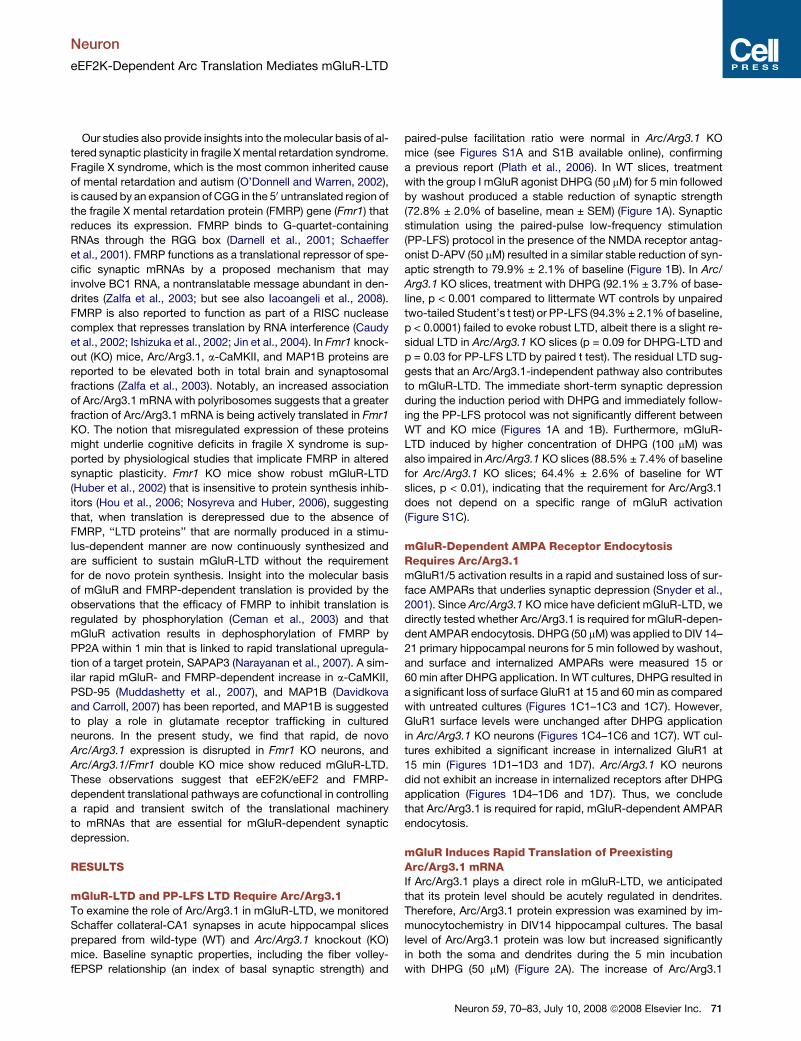
mGluR-LTD and PP-LFS LTD Require Arc/Arg3.1To examine the role of Arc/Arg3.1 in mGluR-LTD, we monitored
Schaffer collateral-CA1 synapses in acute hippocampal slices
prepared from wild-type (WT) and Arc/Arg3.1 knockout (KO)
mice. Baseline synaptic properties, including the fiber volley-
fEPSP relationship (an index of basal synaptic strength) and
paired-pulse facilitation ratio were normal in Arc/Arg3.1 KO
mice (see Figures S1A and S1B available online), confirming
a previous report (Plath et al., 2006). In WT slices, treatment
with the group I mGluR agonist DHPG (50 mM) for 5 min followed
by washout produced a stable reduction of synaptic strength
(72.8% ± 2.0% of baseline, mean ± SEM) (Figure 1A). Synaptic
stimulation using the paired-pulse low-frequency stimulation
(PP-LFS) protocol in the presence of the NMDA receptor antag-
onist D-APV (50 mM) resulted in a similar stable reduction of syn-
aptic strength to 79.9% ± 2.1% of baseline (Figure 1B). In Arc/
Arg3.1 KO slices, treatment with DHPG (92.1% ± 3.7% of base-
line, p < 0.001 compared to littermate WT controls by unpaired
two-tailed Student’s t test) or PP-LFS (94.3% ± 2.1% of baseline,
p < 0.0001) failed to evoke robust LTD, albeit there is a slight re-
sidual LTD in Arc/Arg3.1 KO slices (p = 0.09 for DHPG-LTD and
p = 0.03 for PP-LFS LTD by paired t test). The residual LTD sug-
gests that an Arc/Arg3.1-independent pathway also contributes
to mGluR-LTD. The immediate short-term synaptic depression
during the induction period with DHPG and immediately follow-
ing the PP-LFS protocol was not significantly different between
WT and KO mice (Figures 1A and 1B). Furthermore, mGluR-
LTD induced by higher concentration of DHPG (100 mM) was
also impaired in Arc/Arg3.1 KO slices (88.5% ± 7.4% of baseline
for Arc/Arg3.1 KO slices; 64.4% ± 2.6% of baseline for WT
slices, p < 0.01), indicating that the requirement for Arc/Arg3.1
does not depend on a specific range of mGluR activation
(Figure S1C).
mGluR-Dependent AMPA Receptor EndocytosisRequires Arc/Arg3.1mGluR1/5 activation results in a rapid and sustained loss of sur-
face AMPARs that underlies synaptic depression (Snyder et al.,
2001). Since Arc/Arg3.1 KO mice have deficient mGluR-LTD, we
directly tested whether Arc/Arg3.1 is required for mGluR-depen-
dent AMPAR endocytosis. DHPG (50 mM) was applied to DIV 14–
21 primary hippocampal neurons for 5 min followed by washout,
and surface and internalized AMPARs were measured 15 or
60 min after DHPG application. In WT cultures, DHPG resulted in
a significant loss of surface GluR1 at 15 and 60 min as compared
with untreated cultures (Figures 1C1–1C3 and 1C7). However,
GluR1 surface levels were unchanged after DHPG application
in Arc/Arg3.1 KO neurons (Figures 1C4–1C6 and 1C7). WT cul-
tures exhibited a significant increase in internalized GluR1 at
15 min (Figures 1D1–1D3 and 1D7). Arc/Arg3.1 KO neurons
did not exhibit an increase in internalized receptors after DHPG
application (Figures 1D4–1D6 and 1D7). Thus, we conclude
that Arc/Arg3.1 is required for rapid, mGluR-dependent AMPAR
endocytosis.
mGluR Induces Rapid Translation of PreexistingArc/Arg3.1 mRNAIf Arc/Arg3.1 plays a direct role in mGluR-LTD, we anticipated
that its protein level should be acutely regulated in dendrites.
Therefore, Arc/Arg3.1 protein expression was examined by im-
munocytochemistry in DIV14 hippocampal cultures. The basal
level of Arc/Arg3.1 protein was low but increased significantly
in both the soma and dendrites during the 5 min incubation
with DHPG (50 mM) (Figure 2A). The increase of Arc/Arg3.1
Neuron 59, 70–83, July 10, 2008 ª2008 Elsevier Inc. 71
Neuron
eEF2K-Dependent Arc Translation Mediates mGluR-LTD
Figure 1. Arc/Arg3.1 Is Required for Hippo-
campal mGluR-LTD
Field excitatory postsynaptic potentials (fEPSPs)
were recorded in the hippocampal Schaffer collat-
eral-CA1 synapses derived from Arc/Arg3.1 KO
mice and compared to WT littermate controls.
(A) Average time course of the change in fEPSP
slope induced by the group I mGluR agonist (R,S)-
DHPG (50 mM, for 5 min). LTD of WT mice was
72.8% ± 2.0% of baseline at t = 70 min (n = 10). In
Arc/Arg3.1 KO, fEPSPs were 92.1% ± 3.7% of the
baseline at t = 70 min (n = 9). p < 0.001 when com-
pared to littermate WT. Error bars indicate the stan-
dard error of the mean. Measurements correspond
to the time points indicated on the time course
graph in this and all subsequent figures.
(B) Time course of the change in fEPSP slope pro-
duced by paired-pulse low-frequency stimulation
(PP-LFS: at 1 Hz, 50 ms interstimulus interval,
for 15 min) in the presence of the NMDA receptor
antagonist D-APV (50 mM). LTD of WT mice was
79.9% ± 2.1% of baseline at t = 80 min (n = 12).
In Arc/Arg3.1 KO mice, fEPSPs were 94.3% ±
2.1% of the baseline at t = 80 min (n = 13).
p < 0.0001. Scale bars, 0.5 mV/10 ms.
(C) Five minutes of DHPG application resulted in
a loss of surface GluR1 at 15 min (n = 20, ***p <
0.005) and 60 min (n = 19, *p < 0.05) after DHPG
application, compared to untreated controls in
WT hippocampal cultures. Arc/Arg3.1 KO neurons
did not exhibit any changes in surface GluR1
levels after DHPG treatment. Representative pic-
tures of cultures are shown using an LUT scale
where white is high intensity and dark red is low
intensity. CTL, control.
(D) Five minutes of DHPG application resulted in
an increase of internalized GluR1 at 15 min (n =
20, *p < 0.05) compared with untreated cultures.
Arc/Arg3.1 KO neurons did not exhibit changes
in internalized GluR1 levels after DHPG treatment.
Error bars indicate SEM in this and all subsequent
figures.
protein was blocked by the protein synthesis inhibitor emetine,
indicating a role for de novo translation. The induced Arc/
Arg3.1 immunoreactivity in both proximal and distal dendrites
was detected within 5 min of mGluR activation, and there was
no evidence of a concentration gradient that might occur with
rapid transport of Arc/Arg3.1 from the soma. The rapidity and
distribution of the response suggests that Arc/Arg3.1 is synthe-
sized locally in dendrites and is consistent with the observation
that mGluR-LTD is expressed in isolated dendrites (Huber
et al., 2000). Similar levels of Arc/Arg3.1 induction during 5 min
incubation of DHPG were observed by western blot analysis us-
ing forebrain cultures (Figure S2A). Treatment with BDNF (10 ng/
ml) also increased Arc/Arg3.1 protein expression, but, in contrast
to DHPG, this was evident only after 40 min (Figure S2A).
The rapid increase of Arc/Arg3.1 protein could be mediated by
an enhanced rate of translation or by a stable level of translation
together with reduced degradation. As reported previously (Rao
72 Neuron 59, 70–83, July 10, 2008 ª2008 Elsevier Inc.
et al., 2006), the proteosome inhibitor MG132 increased
Arc/Arg3.1 protein but did not block the ability of DHPG to further
increase Arc/Arg3.1 (Figure S2B). Induction of Arc/Arg3.1 by
DHPG at 5 min was blocked by 5 min pretreatment of emetine
or cycloheximide (Figures 2A7 and 2B). These data support the
notion that Arc/Arg3.1 induction following DHPG treatment in-
volves an increase in the rate of de novo protein translation.
To examine the possible role of de novo transcription of
Arc/Arg3.1 mRNA, we monitored the effect of the transcription
blocker actinomycin D. Actinomycin D (10 mM, 5 min pretreat-
ment and 5 min with or without DHPG) did not alter the DHPG-in-
duced increase of Arc/Arg3.1 protein (Figures 2C and 2E). DHPG
did evoke a modest increase of Arc/Arg3.1 mRNA, but this was
detected only after 20 min (Figure 2F). The time course of
the delayed Arc/Arg3.1 protein expression by DHPG or BDNF
correlated with the mRNA induction, and actinomycin D
blocked this response (data not shown). These observations

Neuron
eEF2K-Dependent Arc Translation Mediates mGluR-LTD
Figure 2. Arc/Arg3.1 Protein Is Rapidly Syn-
thesized by Group I mGluR Activation
(A) Stimulation of hippocampal neurons with
DHPG (50 mM) for 5 min increased Arc/Arg3.1 im-
munoreactivity in both cell body (1.34 ± 0.063 of
untreated soma, n = 13) and dendrites (1.58 ±
0.095 of untreated dendrites, n = 38). The rapid in-
crease of Arc/Arg3.1 was blocked by the protein
synthesis inhibitor emetine (10 ng/ml, 10 min).
(B) High-dose cycloheximide (CHX, 50 mM, total
10 min: 5 min pretreatment and 5 min with or with-
out DHPG) blocked the induction of Arc/Arg3.1
by DHPG (5 min).
(C) Transcription inhibitor actinomycin D (ActD:
10 mM, 5 min pretreatment and 5 min with or
without DHPG) did not block the induction of
Arc/Arg3.1 by DHPG (5 min).
(D)Low-dose CHX increased the level of Arc/Arg3.1
protein. Neurons were treated with vehicle or vari-
ous doses of CHX for 10 min. Total protein synthe-
sis was measured by counting the incorporation of35S methionine and cysteine in TCA precipitant.
(E) Statistical analysis of western blots. Five
minute treatment of DHPG significantly increased
the level of Arc/Arg3.1. Inhibition of new protein
synthesis by high dose of cycloheximide not only
blocked the induction of Arc/Arg3.1 protein but
also slightly decreased the level of Arc/Arg3.1
upon stimulation with DHPG. Inhibition of tran-
scription by actinomycin D did not affect the level
of Arc/Arg3.1. Low-dose CHX (50–100 nM, 5 min
pretreatment and 5 min with or without DHPG)
increased the level of Arc/Arg3.1, which was not
further induced by DHPG. *p < 0.05, **p < 0.01.
(F) The level of Arc/Arg3.1 mRNA was measured
using real-time RT-PCR. Stimulation of neurons
with BDNF (10 ng/ml) and forskolin (50 mM) in-
duced the level of Arc/Arg3.1 mRNA 40 min and
20 min after stimulation, respectively. DHPG
slightly increased the level of Arc/Arg3.1 mRNA
at 20 and 40 min after stimulation.
*p < 0.05, **p < 0.01, ***p < 0.005.
suggest that the rapid increase in de novo translation requires
Arc/Arg3.1 mRNA that is present in neurons prior to DHPG
stimulation, while the delayed Arc/Arg3.1 expression is coupled
to de novo transcription. We note that Arc/Arg3.1 mRNA is de-
tected in dendrites of unstimulated cultured neurons (Giorgi
et al., 2007), and we detect Arc/Arg3.1 mRNA in stratum radia-
tum of the hippocampal CA1 region from home-caged mice
(Figure S3).
Low Dose Cycloheximide Can IncreaseArc/Arg3.1 Protein ExpressionIn examining the dose dependence of cycloheximide’s actions,
we noted that the level of Arc/Arg3.1 protein rapidly increased
when neurons are treated with low doses (Figure 2D). For exam-
ple, 100 nM cycloheximide increased Arc/Arg3.1 protein within
10 min. Even at these low doses, cycloheximide effectively re-
duced general protein synthesis. 100 nM cycloheximide reduced
the total incorporation of 35S-labeled methionine and cysteine
into TCA precipitant to �60%. Previous studies have noted the
paradoxical action of low-dose cycloheximide to increase the
synthesis of specific proteins and rationalized this action by sug-
gesting that global reduction of elongation can increase the
availability of factors that are required for translation initiation
of specific transcripts that are poorly initiated under control con-
ditions (Fernandez et al., 2005; Gupta and Ono, 1997; Perlman
and Feldman, 1982; Scheetz et al., 2000; Walden and Thach,
1986). This notion contributed to our analysis of the eEF2 path-
way (below), since activated eEF2 inhibits elongation and can
paradoxically increase translation of certain mRNAs (Chotiner
et al., 2003; Scheetz et al., 2000).
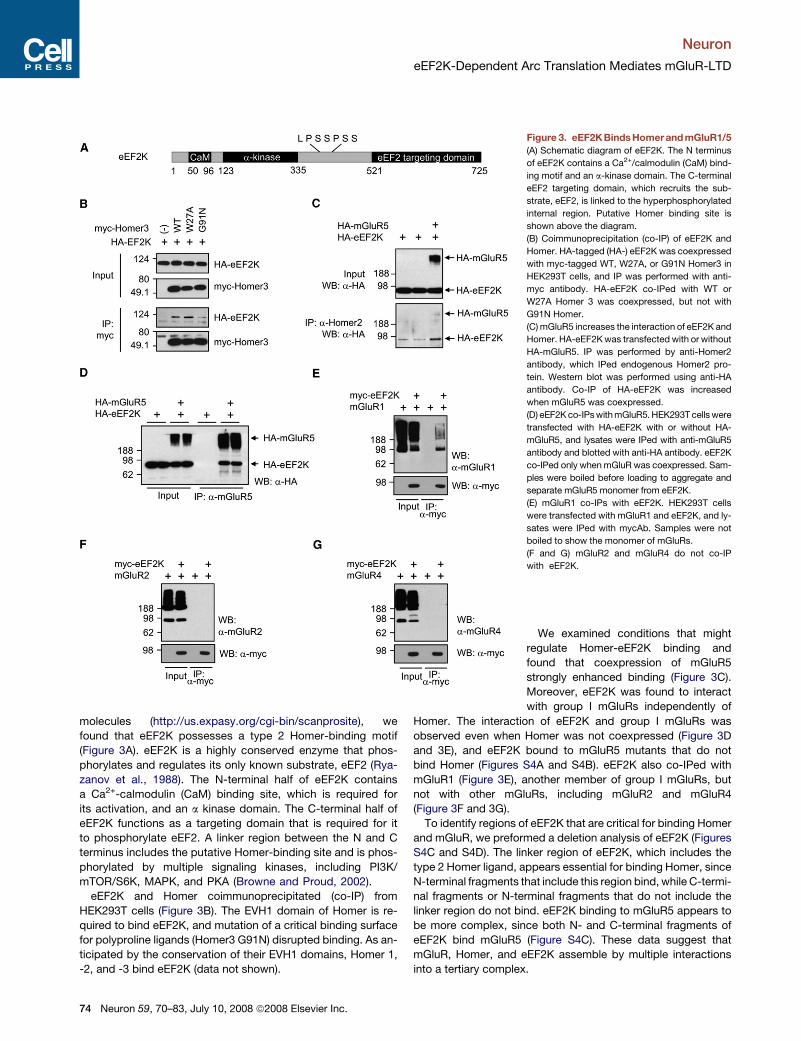
eEF2K Physically Associates with Homerand Group I mGluRsHomer proteins bind group I mGluRs and play a role in their
signaling by also binding signaling partners, including IP3R
(Tu et al., 1998). Homer proteins bind two known sequence
motifs: PPxxF (type 1) and LPSSPSS (type 2) (Yuan et al.,
2003). When we searched for candidate Homer-binding
Neuron 59, 70–83, July 10, 2008 ª2008 Elsevier Inc. 73
Neuron
eEF2K-Dependent Arc Translation Mediates mGluR-LTD
molecules (http://us.expasy.org/cgi-bin/scanprosite), we
found that eEF2K possesses a type 2 Homer-binding motif
(Figure 3A). eEF2K is a highly conserved enzyme that phos-
phorylates and regulates its only known substrate, eEF2 (Rya-
zanov et al., 1988). The N-terminal half of eEF2K contains
a Ca2+-calmodulin (CaM) binding site, which is required for
its activation, and an a kinase domain. The C-terminal half of
eEF2K functions as a targeting domain that is required for it
to phosphorylate eEF2. A linker region between the N and C
terminus includes the putative Homer-binding site and is phos-
phorylated by multiple signaling kinases, including PI3K/
mTOR/S6K, MAPK, and PKA (Browne and Proud, 2002).
eEF2K and Homer coimmunoprecipitated (co-IP) from
HEK293T cells (Figure 3B). The EVH1 domain of Homer is re-
quired to bind eEF2K, and mutation of a critical binding surface
for polyproline ligands (Homer3 G91N) disrupted binding. As an-
ticipated by the conservation of their EVH1 domains, Homer 1,
-2, and -3 bind eEF2K (data not shown).
Figure 3. eEF2K BindsHomer and mGluR1/5
(A) Schematic diagram of eEF2K. The N terminus
of eEF2K contains a Ca2+/calmodulin (CaM) bind-
ing motif and an a-kinase domain. The C-terminal
eEF2 targeting domain, which recruits the sub-
strate, eEF2, is linked to the hyperphosphorylated
internal region. Putative Homer binding site is
shown above the diagram.
(B) Coimmunoprecipitation (co-IP) of eEF2K and
Homer. HA-tagged (HA-) eEF2K was coexpressed
with myc-tagged WT, W27A, or G91N Homer3 in
HEK293T cells, and IP was performed with anti-
myc antibody. HA-eEF2K co-IPed with WT or
W27A Homer 3 was coexpressed, but not with
G91N Homer.
(C) mGluR5 increases the interaction of eEF2K and
Homer. HA-eEF2K was transfected with or without
HA-mGluR5. IP was performed by anti-Homer2
antibody, which IPed endogenous Homer2 pro-
tein. Western blot was performed using anti-HA
antibody. Co-IP of HA-eEF2K was increased
when mGluR5 was coexpressed.
(D) eEF2K co-IPs with mGluR5. HEK293T cells were
transfected with HA-eEF2K with or without HA-
mGluR5, and lysates were IPed with anti-mGluR5
antibody and blotted with anti-HA antibody. eEF2K
co-IPed only when mGluR was coexpressed. Sam-
ples were boiled before loading to aggregate and
separate mGluR5 monomer from eEF2K.
(E) mGluR1 co-IPs with eEF2K. HEK293T cells
were transfected with mGluR1 and eEF2K, and ly-
sates were IPed with mycAb. Samples were not
boiled to show the monomer of mGluRs.
(F and G) mGluR2 and mGluR4 do not co-IP
with eEF2K.
We examined conditions that might
regulate Homer-eEF2K binding and
found that coexpression of mGluR5
strongly enhanced binding (Figure 3C).
Moreover, eEF2K was found to interact
with group I mGluRs independently of
Homer. The interaction of eEF2K and group I mGluRs was
observed even when Homer was not coexpressed (Figure 3D
and 3E), and eEF2K bound to mGluR5 mutants that do not
bind Homer (Figures S4A and S4B). eEF2K also co-IPed with
mGluR1 (Figure 3E), another member of group I mGluRs, but
not with other mGluRs, including mGluR2 and mGluR4
(Figure 3F and 3G).
To identify regions of eEF2K that are critical for binding Homer
and mGluR, we preformed a deletion analysis of eEF2K (Figures
S4C and S4D). The linker region of eEF2K, which includes the
type 2 Homer ligand, appears essential for binding Homer, since
N-terminal fragments that include this region bind, while C-termi-
nal fragments or N-terminal fragments that do not include the
linker region do not bind. eEF2K binding to mGluR5 appears to
be more complex, since both N- and C-terminal fragments of
eEF2K bind mGluR5 (Figure S4C). These data suggest that
mGluR, Homer, and eEF2K assemble by multiple interactions
into a tertiary complex.
74 Neuron 59, 70–83, July 10, 2008 ª2008 Elsevier Inc.

Neuron
eEF2K-Dependent Arc Translation Mediates mGluR-LTD
The Interaction of eEF2K with mGluR Is Dynamicand Is Modulated by Ca2+ and mGluR ActivityThe kinase activity of eEF2K is known to be regulated by Ca2+
via its Ca2+-CaM-binding domain (Nairn and Palfrey, 1987;
Ryazanov, 1987). To test whether Ca2+ modulates the mGluR5-
eEF2K binding, co-IP experiments were performed using lysates
from cotransfected HEK293T cells in the presence of defined
concentrations of free Ca2+ (Figure 4A). Co-IP was robust at
[Ca2+] less than 1 mM but markedly decreased at concentrations
>10 mM. mGluR5 binding to a C-terminal fragment of eEF2K that
lacks the CaM-binding domain but retains binding to mGluR5
was not inhibited by [Ca2+] (Figure S4E). These results indicate
that [Ca2+] can modulate the interaction of group I mGluRs
with eEF2K and suggest a role for CaM binding to eEF2K.
Figure 4. Dynamic Interaction of eEF2K and mGluR5
(A) Calcium dissociates eEF2K from mGluR5. HEK293T cells were transfected
with HA-eEF2K with or without myc-mGluR5, and cells were harvested with ly-
sis buffer without calcium or containing various concentrations of free calcium.
Calmodulin (CaM) (25 mg/ml) was also added to the lysis buffer as indicated.
Binding was decreased at [Ca2+] higher than 10 mM.
(B) Phospho-eEF2 was not detected in the hippocampus of eEF2K KO, while
the level of total eEF2, GluR1, Glur2/3, mGluR5, a-CaMKII, Arc/Arg3.1, and ac-
tin was not altered in eEF2K KO mice compared to WT littermate controls.
(C) Synaptoneurosomes, prepared from the forebrain of eEF2K KO and WT
mice, were stimulated with vehicle or DHPG for 20 min. Synaptoneurosomes
were then lysed and immunoprecipitated with anti-eEF2K antibody. mGluR5
co-IPed with eEF2K only in WT samples. Stimulation of synaptoneurosomes
with DHPG decreased the co-IP of mGluR5.
We used eEF2K KO mice in our analysis of mGluR-eEF2K
binding. eEF2K KO mice were viable and fertile (Ryazanov,
2002) and showed the anticipated absence of phosphorylated
eEF2 (Thr56) (Figure 4B). The levels of several synaptic proteins
were not altered in the hippocampus of KO mice (Figure 4B).
Synaptoneurosomes from forebrains of WT and eEF2K KO
mice were prepared and stimulated with DHPG for 20 min.
Co-IP experiments using anti-eEF2K antibody confirmed that
native mGluR5 associated with eEF2K (Figure 4C). The co-IP
of mGluR5 was reduced when synaptoneurosomes were stimu-
lated with DHPG. Interaction of endogenous mGluR5 and
eEF2K was also reduced upon DHPG stimulation of cultured
neurons (Figure S4F). We conclude that mGluR and eEF2K
associate in vivo and that their interaction is reduced by
mGluR activation.
Group I mGluRs Dynamically Regulatethe Phosphorylation of eEF2Activated eEF2K selectively phosphorylates eEF2 (Ryazanov
et al., 1988). To assess whether mGluR activates this pathway
in conditions that evoke LTD, we monitored the level of phos-
pho-eEF2 in hippocampal slices of either WT or eEF2K KO
mice using the same stimulus parameters that induce mGluR-
LTD. Activation of mGluR increased the phosphorylation of
eEF2 in the stratum pyramidal (s.p.), and stratum radiatum (s.r.)
of the hippocampal CA1 region within 5 min (Figure 5A). By
30 min after washout of DHPG, the level of phospho-eEF2 was
reduced to prestimulation level. No phosphorylation of eEF2
was detected in eEF2K KO slices. The transient induction of
phospho-eEF2 by DHPG was confirmed by western blot analysis
in hippocampal slices (Figure S5A).
To further examine dendritic localization of eEF2K activity,
DIV14 neurons were stimulated with DHPG for 5 min and
stained with phospho-eEF2 and PSD95, a marker for excitatory
synapses (Figure 5B). Phospho-eEF2 was present in dendritic
shafts and the cell body. Phospho-eEF2 also showed a distinct
punctal distribution in spines that colocalized with PSD95.
Staining was absent in eEF2K KO cultures (data not shown).
This result is consistent with a previous report that translational
regulators, including eEF2K, are enriched in synaptic fractions
(Asaki et al., 2003).
Phosphorylation of eEF2 is known to inhibit translational
elongation. Therefore, we examined the prediction that global
protein translation might be transiently reduced co-incident with
the transient increase of phospho-eEF2. Stimulation of neurons
with DHPG for 5 min transiently decreased the incorporation of35S amino acids into TCA precipitants, and this effect was re-
versed 20 min after washout of DHPG (Figure S5B). A previous
study reported that DHPG rapidly increased protein synthesis
in synaptoneurosomes (Weiler et al., 2004). We observed that
DHPG did not induce p-eEF2 in synaptoneurosomes (data not
shown), and it is possible that the eEF2-dependent translational
mechanism is not maintained in broken cell preparations.
Rapid De Novo Arc/Arg3.1 Translation Is SelectivelyAbsent in eEF2K KO NeuronsArc/Arg3.1 expression was examined in DIV14 forebrain neuro-
nal cultures prepared from WT and eEF2K KO mice. The
Neuron 59, 70–83, July 10, 2008 ª2008 Elsevier Inc. 75

Neuron
eEF2K-Dependent Arc Translation Mediates mGluR-LTD
steady-state expression of Arc/Arg3.1 protein was identical
in WT and eEF2K KO neurons; however, the increase in
Arc/Arg3.1 protein 5 min after DHPG in WT neurons was absent
in eEF2K KO neurons in both biochemical (Figures 5C and 5D)
and immunocytochemical assays (Figure S6). By contrast,
Arc/Arg3.1 protein was induced to the same extent in WT and
eEF2K KO neurons 60 min after DHPG stimulation. Arc/Arg3.1
mRNA was identical in WT and eEF2K KO neurons prior to appli-
cation of DHPG and increased identically at 40 min after stimu-
lation in both WT and eEF2K KO neurons (Figure 5E). Accord-
ingly, the lack of rapid induction of Arc/Arg3.1 protein in the
eEF2K KO neurons is not due to reduced Arc/Arg3.1 mRNA
expression. We also note that mGluR signaling that is required
for induction of Arc/Arg3.1 mRNA and the delayed increase of
Arc/Arg3.1 protein are intact in eEF2K KO neurons. Moreover,
Figure 5. Rapid Induction of Arc/Arg3.1 by
Group I mGluRs Is Dependent on eEF2K
(A) Hippocampal slices were prepared from WT
and eEF2K KO mice and were stimulated with
DHPG for 5 min. phospho-eEF2 (p-eEF2, red) in
area CA1 was increased by DHPG within 5 min
and declined by 30 min following washout. Spec-
ificity of phospho-eEF2 was confirmed by staining
of eEF2K KO slices. s.p., stratum pyramidal; s.r.,
stratum radiatum.
(B) Cultured hippocampal neurons were treated
with DHPG for 5 min and stained with phospho-
eEF2 (red) and PSD95 (green) antibodies on DIV14.
phospho-eEF2 showed punctal distribution in den-
dritic spines and dendritic shafts. phospho-eEF2 in
spines colocalized with PSD95 (arrows). (B2), (B3),
and (B4) are enlarged images of the rectangular
region of (B1).
(C and D) mGluR-dependent rapid synthesis of
Arc/Arg3.1 is absent in eEF2K KO neurons. Neu-
rons from the forebrains of WT or eEF2K KO mice
were cultured for DIV14 and treated with DHPG
(50 mM, 5 min). Phosphorylation of eEF2 was unde-
tectable in eEF2K KO neurons. No difference in the
level of mGluR5 was observed between WT and
eEF2K KO neurons. An arrowhead indicates a non-
specific band. p values were obtained by paired t
test comparing basal and drug-treated levels.
p values for comparison of WT and eEF2K KO
mice were obtained by Student’s t test. *p < 0.05,
**p < 0.01, n = 8. Error bars are SEM.
(E) Arc/Arg3.1 mRNA expression is not altered in
eEF2K KO neurons. The level of Arc/Arg3.1
mRNA was measured in WT and eEF2K KO neu-
rons following the stimulation with DHPG.
(F) Low-dose cycloheximide (CHX) increases Arc/
Arg3.1 protein expression. Cultured eEF2K KO
neurons were treated with indicated doses of
CHX for 10 min. *p < 0.05, n = 8.
Arc/Arg3.1 protein expression is identical
in the hippocampus of WT and eEF2K KO
mice (Figure 4B), indicating that eEF2K
is not required for basal expression of
Arc/Arg3.1 protein in vivo.
If the failure of DHPG to induce rapid synthesis of Arc/
Arg3.1 protein in the eEF2K KO neurons is due to a selective
interruption of the action of phospho-eEF2, we predicted that
a low dose of cycloheximide, which does not require eEF2K
or phopho-eEF2 to inhibit the elongation step, should induce
the synthesis of Arc/Arg3.1 protein in eEF2K KO neurons.
Treatment of DIV14 eEF2K KO neurons with low-dose cyclo-
heximide (50 nM and 100 nM) increased the level of
Arc/Arg3.1 protein in eEF2K KO neurons (Figure 5F), similar
to WT neurons (Figure 2D). High-dose cycloheximide (>1 uM)
did not induce Arc/Arg3.1 in either WT and eEF2K KO neurons.
The ability of low-dose cycloheximide to rescue rapid
Arc/Arg3.1 induction indicates that mechanisms that mediate
rapid Arc/Arg3.1 translation subsequent to inhibition of
elongation are intact in eEF2K KO neurons.
76 Neuron 59, 70–83, July 10, 2008 ª2008 Elsevier Inc.
Neuron
eEF2K-Dependent Arc Translation Mediates mGluR-LTD
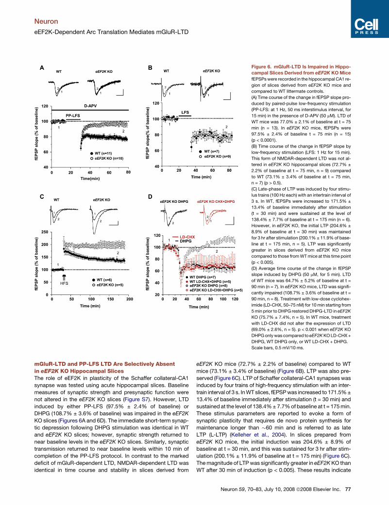
Figure 6. mGluR-LTD Is Impaired in Hippo-
campal Slices Derived from eEF2K KO Mice
fEPSPs were recorded in the hippocampal CA1 re-
gion of slices derived from eEF2K KO mice and
compared to WT littermate controls.
(A) Time course of the change in fEPSP slope pro-
duced by paired-pulse low-frequency stimulation
(PP-LFS: at 1 Hz, 50 ms interstimulus interval, for
15 min) in the presence of D-APV (50 mM). LTD of
WT mice was 77.0% ± 2.1% of baseline at t = 75
min (n = 13). In eEF2K KO mice, fEPSPs were
97.5% ± 2.4% of baseline t = 75 min (n = 15)
(p < 0.0001).
(B) Time course of the change in fEPSP slope by
low-frequency stimulation (LFS: 1 Hz for 15 min).
This form of NMDAR-dependent LTD was not al-
tered in eEF2K KO hippocampal slices (72.7% ±
2.2% of baseline at t = 75 min, n = 9) compared
to WT (73.1% ± 3.4% of baseline at t = 75 min,
n = 7) (p > 0.5).
(C) Late-phase of LTP was induced by four stimu-
lus trains (100 Hz each) with an intertrain interval of
3 s. In WT, fEPSPs were increased to 171.5% ±
13.4% of baseline immediately after stimulation
(t = 30 min) and were sustained at the level of
138.4% ± 7.7% of baseline at t = 175 min (n = 6).
However, in eEF2K KO, the initial LTP (204.6% ±
8.9% of baseline at t = 30 min) was maintained
for 3 hr after stimulation (200.1% ± 11.9% of base-
line at t = 175 min, n = 5). LTP was significantly
greater in slices derived from eEF2K KO mice
compared to those from WT mice at this time point
(p < 0.005).
(D) Average time course of the change in fEPSP
slope induced by DHPG (50 mM, for 5 min). LTD
of WT mice was 64.7% ± 5.2% of baseline at t =
90 min (n = 7). In eEF2K KO mice, LTD was signifi-
cantly impaired (108.7% ± 3.6% of baseline at t =
90 min, n = 8). Treatment with low-dose cyclohex-
imide (LD-CHX, 50–75 nM) for 10 min starting from
5 min prior to DHPG restored DHPG-LTD in eEF2K
KO (75.7% ± 7.4%, n = 5). In WT mice, treatment
with LD-CHX did not alter the expression of LTD
(69.0% ± 2.6%, n = 5). p < 0.001 when eEF2K KO
DHPG only was compared to eEF2K KO LD-CHX +
DHPG, WT DHPG only, or WT LD-CHX + DHPG.
Scale bars, 0.5 mV/10 ms.
mGluR-LTD and PP-LFS LTD Are Selectively Absentin eEF2K KO Hippocampal SlicesThe role of eEF2K in plasticity of the Schaffer collateral-CA1
synapse was tested using acute hippocampal slices. Baseline
measures of synaptic strength and presynaptic function were
not altered in the eEF2K KO slices (Figure S7). However, LTD
induced by either PP-LFS (97.5% ± 2.4% of baseline) or
DHPG (108.7% ± 3.6% of baseline) was impaired in the eEF2K
KO slices (Figures 6A and 6D). The immediate short-term synap-
tic depression following DHPG stimulation was identical in WT
and eEF2K KO slices; however, synaptic strength returned to
near baseline levels in the eEF2K KO slices. Similarly, synaptic
transmission returned to near baseline levels within 10 min of
completion of the PP-LFS protocol. In contrast to the marked
deficit of mGluR-dependent LTD, NMDAR-dependent LTD was
identical in time course and stability in slices derived from
eEF2K KO mice (72.7% ± 2.2% of baseline) compared to WT
mice (73.1% ± 3.4% of baseline) (Figure 6B). LTP was also pre-
served (Figure 6C). LTP of Schaffer collateral-CA1 synapses was
induced by four trains of high-frequency stimulation with an inter-
train interval of 3 s. In WT slices, fEPSP was increased to 171.5% ±
13.4% of baseline immediately after stimulation (t = 30 min) and
sustained at the level of 138.4% ± 7.7% of baseline at t = 175 min.
These stimulus parameters are reported to evoke a form of
synaptic plasticity that requires de novo protein synthesis for
maintenance longer than �60 min and is referred to as late
LTP (L-LTP) (Kelleher et al., 2004). In slices prepared from
eEF2K KO mice, the initial induction was 204.6% ± 8.9% of
baseline at t = 30 min, and this was sustained for 3 hr after stim-
ulation (200.1% ± 11.9% of baseline at t = 175 min) (Figure 6C).
The magnitude of LTP was significantly greater in eEF2K KO than
WT after 30 min of induction (p < 0.005). These results indicate
Neuron 59, 70–83, July 10, 2008 ª2008 Elsevier Inc. 77

Neuron
eEF2K-Dependent Arc Translation Mediates mGluR-LTD
that eEF2K KO disrupts mGluR-LTD but does not alter NMDAR-
dependent LTD or early LTP. The apparent enhancement of late-
phase LTP deserves further study.
Our proposed mechanism for the mGluR-LTD deficit in
the eEF2K KO slices is linked to failure to rapidly translate
Arc/Arg3.1. Since low-dose cycloheximide induced Arc/Arg3.1
synthesis and did not depend on phospho-eEF2, we examined
the possibility that cycloheximide could rescue mGluR-LTD in
slices from eEF2K KO mice. We found that a 10 min exposure
to 50–75 nM cycloheximide (low-dose CHX: LD-CHX) beginning
5 min prior to addition of DHPG rescued mGluR-dependent LTD
in the eEF2K KO slice (75.7% ± 7.4% of baseline, p < 0.001,
compared to DHPG only in eEF2K KO slices) (Figure 6D). The
same treatment of WT slices did not substantially alter the time
course of mGluR-LTD (69.0% ± 2.6% of baseline, p > 0.5, com-
pared to DHPG only in WT slices). Low-dose cycloheximide had
no effect on baseline synaptic transmission in the absence of
mGluR stimulation (101.2% ± 2.0% for WT slices; 100.4% ±
4.6% for eEF2K KO slices). These observations confirm that
mGluR signaling required for mGluR-LTD is selectively impaired
in eEF2K KO in a manner that can be rescued by transient appli-
cation of low-dose cycloheximide.
mGluR-LTD, but Not Homeostatic Plasticity,Is Disrupted in eEF2K KO Neurons in CultureTo further assess the selectivity of the eEF2K KO effect on neu-
ronal function, we examined two forms of neuronal plasticity that
can be assayed in primary neuronal cultures. Treatment of cul-
tures with DHPG for 5 min to evoke mGluR-LTD reduced the ratio
of surface to total GluR2/3 by �30% in WT neurons but did not
significantly reduce this measure in eEF2K KO neurons (Figures
S8A and S8B). This result parallels the deficit of mGluR-LTD seen
in acute slices. Cultures were also assayed for homeostatic
adaptations of surface AMPA receptors since this response
is markedly altered in Arc/Arg3.1 KO neurons (Shepherd et al.,
2006). Treatment of eEF2K KO cortical cultures for 2 days with
either tetrodotoxin (TTX) or bicuculline evoked homeostatic in-
creases and decreases of surface GluR1 that were identical to
WT neurons (Figure S8C). Thus, eEF2K KO results in a selective
disruption of mGluR-dependent LTD.
Fmr1 KO Disrupts Rapid, but Not Delayed,Induction of Arc/Arg3.1 ProteinWe examined the role of the eEF2K/eEF2/Arc mechanism in the
aberrant plasticity described in Fmr1 KO mice. FMRP has been
reported to bind Arc/Arg3.1 mRNA (Iacoangeli et al., 2008; Zalfa
et al., 2003) and is hypothesized to inhibit translation prior to
mGluR stimulation (Bear et al., 2004; Narayanan et al., 2007).
To assess whether FMRP might be critical for either rapid or de-
layed induction of Arc/Arg3.1 protein following mGluR stimula-
tion, we prepared primary neuronal cultures from Fmr1 KO
mice and stimulated with DHPG. Arc/Arg3.1 expression in unsti-
mulated cultures was not consistently different between WT and
Fmr1 KO neurons. Moreover, Arc/Arg3.1 protein increased
60 min after DHPG stimulation in Fmr1 KO neurons identically
as in WT neurons (Figure 7A). However, the rapid increase of
Arc/Arg3.1 protein following DHPG stimulation was absent in
Fmr1 KO neurons (Figure 7A). DHPG activated mGluR/eEF2K
78 Neuron 59, 70–83, July 10, 2008 ª2008 Elsevier Inc.
signaling in the Fmr1 KO neurons since phospho-eEF2 was
identically induced as in WT neurons (Figure 7A). Assays of
Arc/Arg3.1 protein stability and induction following proteasome
inhibition with MG132 did not reveal differences between WT
and Fmr1 KO neurons (Figures S9B and S9C). Biochemical ex-
periments to monitor Arc/Arg3.1 expression using acute hippo-
campal slices revealed that basal Arc/Arg3.1 expression was
highly variable even when normalized to total protein or actin,
indicating a limitation of this preparation (Taubenfeld et al.,
2002). When examined histochemically, basal Arc/Arg3.1 varied
through the thickness of the slice (data not shown). We con-
clude that Fmr1 KO neurons selectively lack the ability to rapidly
upregulate Arc/Arg3.1 expression. The reported increase of
Arc/Arg3.1 mRNA in polysome fractions from Fmr1 KO mice
(Zalfa et al., 2003) suggests that failure to detect a DHPG-
evoked rapid increase of Arc/Arg3.1 protein is linked to elevated
constitutive expression.
Arc/Arg3.1 Is Required for mGluR-LTDand PP-LFS LTD in Fmr1 KO MiceIn anticipation of physiological studies to assess the role of
Arc/Arg3.1 in synaptic plasticity of Fmr1 KO mice, we examined
Arc/Arg3.1 protein expression in hippocampus. Arc/Arg3.1 pro-
tein has previously been reported to be modestly upregulated in
both total brain and synaptosomal fractions of Fmr1 KO mice
(Zalfa et al., 2003). However, in our assays, Arc/Arg3.1 protein
was not consistently different in hippocampus (either in vivo or
in acute slices) or cortex when care was taken to sacrifice
mice without behavioral activation. We generated mice in which
both Fmr1 (in FVB background) and Arc/Arg3.1 (in B6 back-
ground) were deleted. Double Arc/Arg3.1/Fmr1 KO (DKO) mice
are viable, fertile, and not different from WT mice in size or
postnatal survival. Indices of basal synaptic transmission were
normal in Fmr1 KO and Arc/Arg3.1/Fmr1 DKO (Figures S9D
and S9E). As reported previously (Nosyreva and Huber, 2006),
DHPG evoked a sustained reduction of synaptic strength
(68.2% ± 2.6% of baseline for Fmr1 single KO slices; 73.0% ±
6.6% of baseline for FVB WT slices; Figure 7C). Jackson labora-
tory provides Fmr1 KO mice in the FVB background, and the
magnitude of LTD was not significantly different from FVB WT
mice. As reported previously in studies of Fmr1 KO in the
B6 background, mGluR-LTD was not inhibited by high-dose
cycloheximide (60 mM) (Figure 7B). In Arc/Arg3.1/Fmr1 DKO (in
B6/FVB), DHPG evoked an initial reduction of synaptic strength
that was not different from WT, Arc/Arg3.1 KO, or Fmr1 KO.
However, expression of DHPG-evoked LTD was significantly
impaired in Arc/Arg3.1/Fmr1 DKO (85.9% ± 4.1%, p < 0.01
compared to Fmr1 single KO or FVB WT). PP-LFS LTD was
also impaired in Arc/Arg3.1/Fmr1 DKO (88.3% ± 2.1% of base-
line for Arc/Arg3.1/Fmr1 DKO slices; 75.5% ± 3.7% of baseline
for Fmr1 single KO; 80.5% ± 2.6% of baseline for FVB WT slices,
p < 0.05 when Arc/Arg3.1/Fmr1 DKO was compared to Fmr1 sin-
gle KO or FVB WT; Figure 7D). These results indicate that
Arc/Arg3.1 is required for mGluR-LTD in both WT and Fmr1
KO neurons. Deletion of Arc/Arg3.1 does not entirely prevent
DHPG or PP-LFS LTD, suggesting that additional mechanisms
contribute to the aberrant LTD in Fmr1 KO mice.

Neuron
eEF2K-Dependent Arc Translation Mediates mGluR-LTD
Figure 7. LTD Is Impaired in Hippocampal
Slices Derived from Arc/Arg3.1/Fmr1 Dou-
ble KO Mice
(A) DIV14 Fmr1 KO neurons were treated with
DHPG as indicated in Figure 5C. Rapid but not de-
layed synthesis of Arc/Arg3.1 was absent in Fmr1
KO. The regulation of phospho-eEF2 was intact in
Fmr1 KO neurons.
(B) High-dose cycloheximide (60 mM: HD-CHX) did
not block DHPG-LTD of Fmr1 KO slices. In the
presence of a high dose of cycloheximide,
DHPG-LTD of Fmr1 KO was 72.3% ± 4.8% of
baseline at t = 105 min (n = 5), while DHPG-LTD
in WT (FVB) slices was blocked (fEPSP was
95.5% ± 2.9% of baseline at t = 105 min (n = 4);
p < 0.01 when Fmr1 KO was compared to FVB
WT).
(C) Average time course of fEPSP slope of
Arc/Arg3.1/Fmr1 double KO (DKO) mice. mGluR-
LTD was induced by DHPG (50 mM, for 5 min).
DHPG-LTD of Arc/Arg3.1/Fmr1 DKO was 85.9% ±
4.1% of baseline at t = 75 min (n = 8). In Fmr1
KO, DHPG-LTD was 68.2% ± 2.6% of baseline
at t = 75 min (n = 6). In WT, DHPG-LTD was
73.0% ± 6.6% of baseline at t = 75 min (n = 5).
p < 0.01 when Arc/Arg3.1/Fmr1 DKO was com-
pared to either WT or Fmr1 KO. fEPSPs of
post-DHPG in Fmr1 KO were not significantly
different from those in FVB WT.
(D) Time course of the change in fEPSP slope by
PP-LFS. PP-LFS LTD of Arc/Arg3.1/Fmr1 DKO
was 88.3% ± 2.1% of baseline at t = 65 min (n =
6). In Fmr1 KO, PP-LFS LTD was 75.5% ± 3.7%
of baseline at t = 65 min (n = 8). In FVB WT, PP-
LFS LTD was 80.5% ± 2.6% of baseline at t = 65
min (n = 8). p < 0.05 when Arc/Arg3.1/Fmr1 DKO
was compared to either WT or Fmr1 KO. fEPSPs
of post-DHPG in Fmr1 KO were not significantly
different from those in FVB WT (p = 0.4).
Scale bars, 0.5 mV/10 ms.
DISCUSSION
Arc/Arg3.1 Plays a Central Rolein mGluR-Dependent LTDThe present study identifies Arc/Arg3.1 as a molecule that is re-
quired for certain forms of LTD that are known to be dependent
on rapid, de novo protein synthesis, including mGluR-LTD and
PP-LFS-LTD. Upon activation of group I mGluRs, Arc/Arg3.1
protein is rapidly upregulated in a process that requires de
novo translation. Arc/Arg3.1 protein functions as a regulatory
factor for a postsynaptic endocytic signaling pathway that in-
cludes endophilins 2 and -3 and dynamin, which together medi-
ate selective endocytosis of AMPA type glutamate receptors
(Chowdhury et al., 2006). Accordingly, the local and rapid in-
crease of Arc/Arg3.1 protein can be linked to the selective down-
regulation of AMPAR at specific synapses during LTD (Figure 8).
While group I mGluR activation results in a modest rise of
Arc/Arg3.1 mRNA, the translation that underlies mGluR-LTD
utilizes Arc/Arg3.1 mRNA that is present in neurons prior to stim-
ulation. Arc/Arg3.1 is regulated as an immediate-early gene that
is strongly induced during learning-related behaviors (Guzowski
et al., 1999) and expressed in discrete populations of neurons
that are part of behaviorally activated networks even when
rodents are resting in home cage (Marrone et al., 2008).
Arc/Arg3.1 mRNA is present in dendritic regions of CA1 neurons
of mice sacrificed immediately from their home cage (Figure S3),
indicating that basal Arc/Arg3.1 mRNA can be either activity
independent or related to prior neuronal activity. The rapid trans-
lation of dendritic Arc/Arg3.1 mRNA rationalizes the known
dependence of mGluR-LTD on rapid de novo protein synthesis
and independence of de novo RNA synthesis (Huber et al.,
2000). It also suggests that prior neuronal activity that drives
the expression of Arc/Arg3.1 mRNA could modify subsequent
mGluR-dependent plasticity.
In further support for the notion that Arc/Arg3.1 is an essential
LTD molecule, we find that mGluR-LTD in Fmr1 KO mice is also
Neuron 59, 70–83, July 10, 2008 ª2008 Elsevier Inc. 79
Neuron
eEF2K-Dependent Arc Translation Mediates mGluR-LTD
dependent on Arc/Arg3.1. This is notable because mGluR-LTD
in Fmr1 KO mice is distinctly different from mGluR-LTD in WT
mice in several ways, including insensitivity to proteosome inhib-
itors, protein synthesis inhibitors, and inhibitors of ERK signaling
(Hou et al., 2006). Disruption of Arc/Arg3.1 function is the first
experimental manipulation shown to reduce mGluR-LTD in
Fmr1 KO slices, but it is likely that additional molecular mecha-
nisms contribute to the response since mGluR-LTD is not entirely
absent in the Arc/Arg3.1/Fmr1 DKO. Since FMRP functions
to repress translation of specific mRNAs, the dependence
mGluR-LTD in the Fmr1 KO mouse suggests that Arc/Arg3.1
protein that is present prior to mGluR stimulation can mediate
this response and thereby confers resistance to protein synthe-
sis inhibitors.
Group I mGluRs and Rapid De Novo Translation;Convergence of eEF2K/eEF2 and FMRP PathwaysmGluR activation induces the rapid translation of Arc/Arg3.1 and
requires eEF2K. eEF2K binds Homer and group I mGluRs and
dissociates from this complex in a Ca2+-dependent manner.
Phospho-eEF2 is present at excitatory synapses and dendrites
and is rapidly upregulated in cultured neurons and hippocampal
slices by stimulation of group I mGluRs with DHPG. The ability of
phospho-eEF2 to inhibit translation is thought to be general for
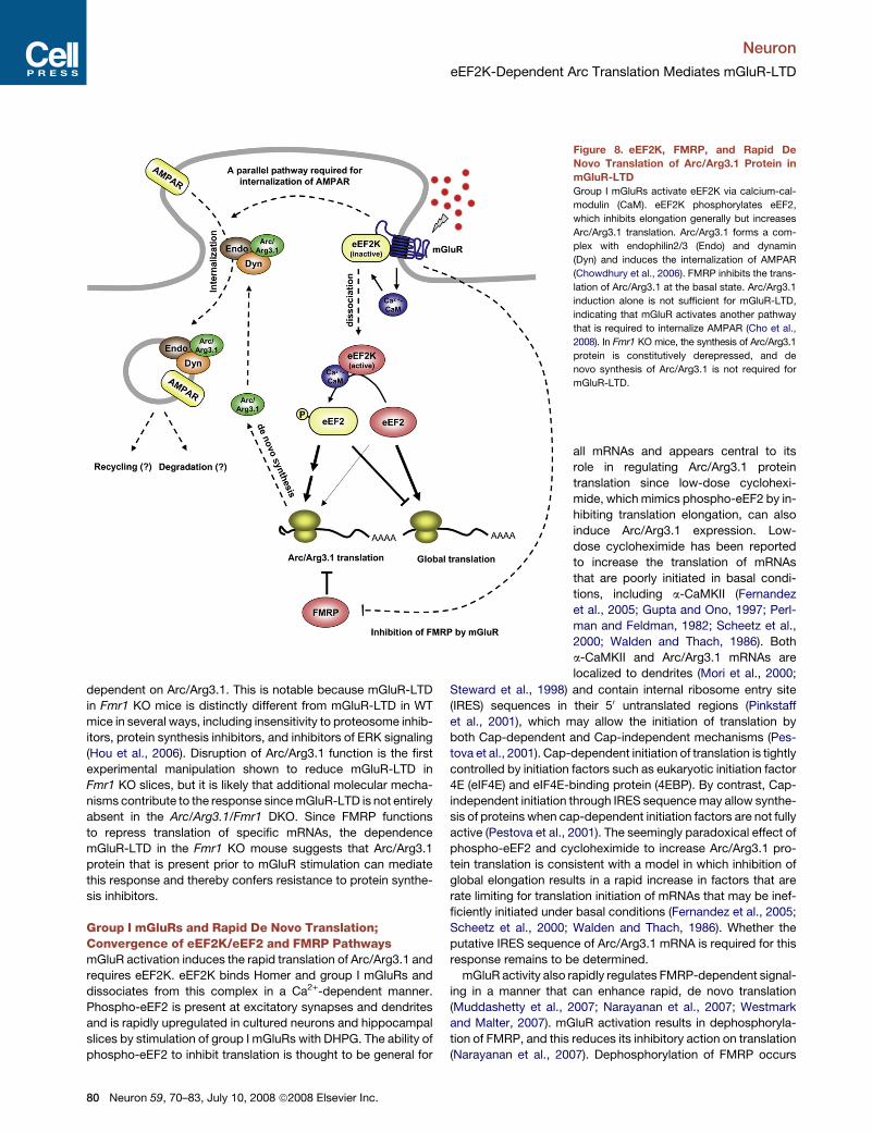
Figure 8. eEF2K, FMRP, and Rapid De
Novo Translation of Arc/Arg3.1 Protein in
mGluR-LTD
Group I mGluRs activate eEF2K via calcium-cal-
modulin (CaM). eEF2K phosphorylates eEF2,
which inhibits elongation generally but increases
Arc/Arg3.1 translation. Arc/Arg3.1 forms a com-
plex with endophilin2/3 (Endo) and dynamin
(Dyn) and induces the internalization of AMPAR
(Chowdhury et al., 2006). FMRP inhibits the trans-
lation of Arc/Arg3.1 at the basal state. Arc/Arg3.1
induction alone is not sufficient for mGluR-LTD,
indicating that mGluR activates another pathway
that is required to internalize AMPAR (Cho et al.,
2008). In Fmr1 KO mice, the synthesis of Arc/Arg3.1
protein is constitutively derepressed, and de
novo synthesis of Arc/Arg3.1 is not required for
mGluR-LTD.
all mRNAs and appears central to its
role in regulating Arc/Arg3.1 protein
translation since low-dose cyclohexi-
mide, which mimics phospho-eEF2 by in-
hibiting translation elongation, can also
induce Arc/Arg3.1 expression. Low-
dose cycloheximide has been reported
to increase the translation of mRNAs
that are poorly initiated in basal condi-
tions, including a-CaMKII (Fernandez
et al., 2005; Gupta and Ono, 1997; Perl-
man and Feldman, 1982; Scheetz et al.,
2000; Walden and Thach, 1986). Both
a-CaMKII and Arc/Arg3.1 mRNAs are
localized to dendrites (Mori et al., 2000;
Steward et al., 1998) and contain internal ribosome entry site
(IRES) sequences in their 50 untranslated regions (Pinkstaff
et al., 2001), which may allow the initiation of translation by
both Cap-dependent and Cap-independent mechanisms (Pes-
tova et al., 2001). Cap-dependent initiation of translation is tightly
controlled by initiation factors such as eukaryotic initiation factor
4E (eIF4E) and eIF4E-binding protein (4EBP). By contrast, Cap-
independent initiation through IRES sequence may allow synthe-
sis of proteins when cap-dependent initiation factors are not fully
active (Pestova et al., 2001). The seemingly paradoxical effect of
phospho-eEF2 and cycloheximide to increase Arc/Arg3.1 pro-
tein translation is consistent with a model in which inhibition of
global elongation results in a rapid increase in factors that are
rate limiting for translation initiation of mRNAs that may be inef-
ficiently initiated under basal conditions (Fernandez et al., 2005;
Scheetz et al., 2000; Walden and Thach, 1986). Whether the
putative IRES sequence of Arc/Arg3.1 mRNA is required for this
response remains to be determined.
mGluR activity also rapidly regulates FMRP-dependent signal-
ing in a manner that can enhance rapid, de novo translation
(Muddashetty et al., 2007; Narayanan et al., 2007; Westmark
and Malter, 2007). mGluR activation results in dephosphoryla-
tion of FMRP, and this reduces its inhibitory action on translation
(Narayanan et al., 2007). Dephosphorylation of FMRP occurs
80 Neuron 59, 70–83, July 10, 2008 ª2008 Elsevier Inc.
Neuron
eEF2K-Dependent Arc Translation Mediates mGluR-LTD
within 1 min of mGluR activation and parallels the time course of
mGluR-dependent phosphorylation of eEF2. Polysome profiles
from WT and Fmr1 KO brain indicate that FMRP is associated
with mRNAs that are not in active translation. In the absence of
FMRP, specific mRNAs including Arc/Arg3.1 shift to fractions
that include actively translating polyribosomes (Zalfa et al.,
2003). Group I mGluRs are reported to rapidly reduce the histo-
chemical colocalization of FMRP and Fmr1 mRNA at synapses in
association with increased translation of de novo FMRP (Antar
et al., 2004). While this is an indirect assay of translational regu-
lation, it supports a model in which mGluR activity reduces
FMRP binding to specific mRNAs and thereby provides a dy-
namic increase in mRNAs in close proximity to synapses for
translation. FMRP is not required for mGluR-dependent induc-
tion of phospho eEF2 (Figure 7A), suggesting that mGluR-
FMRP and mGluR-eEF2K are parallel pathways. Accordingly,
the rapid, de novo Arc/Arg3.1 translational response required
for normal mGluR-LTD appears to require both a transient rever-
sal of FMRP’s repressive effect on translation and the general
inhibition of the elongation step by phospho-eEF2 (Figure 8).
De Novo Translated Arc/Arg3.1 Is SpecificallyRequired for Normal mGluR-LTDArc/Arg3.1 protein that mediates mGluR-LTD appears to be
functionally distinct from the total pool of Arc/Arg3.1 protein
expressed in neurons. For example, the basal expression of
Arc/Arg3.1 protein in eEF2K KO neurons is identical to that in WT
neurons, yet mGluR-LTD and PP-LFS LTD, which are dependent
on Arc/Arg3.1, are absent in the eEF2K KO. Arc/Arg3.1 protein in
eEF2K KO neurons is functional, since homeostatic scaling,
which is also dependent on Arc/Arg3.1 (Shepherd et al., 2006),
is intact. This apparent paradox is resolved with the observa-
tions that eEF2K KO neurons selectively lack rapid, de novo
Arc/Arg3.1 induction, and this deficit is rescued by brief applica-
tion of low-dose cycloheximide that also rescues mGluR-LTD.
Thus, the eEF2-dependent pool of Arc/Arg3.1 protein appears
uniquely capable of mediating LTD. In another example demon-
strating the distinct properties of Arc/Arg3.1 related to its mode
of translation, Arc/Arg3.1 protein is expressed at near normal
levels in Fmr1 KO mice, and Arc/Arg3.1 protein is induced after
40 min of DHPG stimulation; however, the rapid increase of
Arc/Arg3.1 at 5 min is absent. In this regard, expression of
Arc/Arg3.1 protein in the Fmr1 KO appears identical to the
eEF2K KO, but with the technical caveat that we cannot detect
changes in basal translation of Arc/Arg3.1 that are predicted
from other studies to be increased in Fmr1 KO neurons (Zalfa
et al., 2003). Our data suggest that an elevation of constitutive
Arc/Arg3.1 translation in Fmr1 KO underlies mGluR LTD that is
aberrantly independent of de novo translation but dependent
on Arc/Arg3.1. This model rationalizes the distinctly different
mGluR-LTD phenotypes in eEK2K and Fmr1 KOs even though
they show a similar absence of acute mGluR Arc/Arg3.1 induc-
tion. The relative amount of this eEF2K/FMRP-dependent pool
of Arc/Arg3.1 protein is typically small compared to the
Arc/Arg3.1 level of the delayed response to DHPG or BDNF in
neuronal cultures, and we could not detect differences in basal
and induced Arc/Arg3.1 in WT and eEF2K KO neurons using
conventional histochemical techniques. We infer that proteins
translated by the eEF2K/FMRP-dependent mechanism are
uniquely available for mGluR-LTD and anticipate that new imag-
ining methods with high spatial and temporal resolution may
shed light on this prediction.
Our studies also indicate that mGluR-LTD requires a second
signal in addition to rapid, de novo Arc/Arg3.1 protein. Experi-
ments that use cycloheximide to rescue mGluR-LTD in the
eEF2K KO demonstrate that cycloheximide alone is sufficient
to induce Arc/Arg3.1 protein but does not induce LTD unless ac-
companied by mGluR activation. Similarly, low-dose cyclohexi-
mide induces Arc/Arg3.1 in WT neurons but does not induce
LTD without concurrent mGluR activation. These observations
rationalize why misregulated expression of Arc/Arg3.1 protein
in the Fmr1 KO, and presumably other proteins, does not oc-
clude mGluR-LTD. The dual dependence of Arc/Arg3.1 protein
and mGluR activity for aberrant LTD in Fmr1 KO mice provides
a supporting rationale for treatment regimens of Fragile X syn-
drome that include group I mGluR antagonists (Bear et al.,
2004; McBride et al., 2005). In other studies, we have reported
that mGluR-LTD requires the rapid cleavage of the neuronal
pentraxin receptor protein (NPR) in a process that involves the
extracellular metalloprotease tumor necrosis factor-a converting
enzyme (TACE/ADAM17) (Cho et al., 2008). The pentraxin do-
main, released of its transmembrane tether, appears to capture
AMPA receptors at the site of endocytosis, and this mechanism
is required for mGluR-LTD in both the hippocampus and cere-
bellum. It is possible that this NPR pathway functions in con-
junction with the Arc/Arg3.1 endosomal pathway and offers a
potential new target for agents to modify mGluR-LTD.
EXPERIMENTAL PROCEDURES
Constructs, cell culture, real-time RT-PCR, metabolic labeling, and fluores-
cence in situ hybridization assay are included in Supplemental Experimental
Procedures.
Antibodies
Anti-phospho-eEF2 (Thr56: rabbit polyclonal) and total-eEF2 (rabbit poly-
clonal) from Cell Signaling; eEF2K (rabbit polyclonal) and mGluR1 (mouse
monoclonal) from BD Biosciences; mGluR5, mGluR2, and PSD-95 from Up-
state; mGluR4 from Zymed; horse radish peroxidase (HRP) conjugated HA an-
tibody, HRP-conjugated myc antibody, myc (mouse monoclonal), and actin
(mouse monoclonal) from Santa Cruz; Arc/Arg3.1 (Lyford et al., 1995);
N-GluR1 antibody (Shepherd et al., 2006).
AMPA Receptor Trafficking Experiments and Immunostaining
Labeling of surface or internalized pool of AMPA receptor was performed as
described with minor modifications (Shepherd et al., 2006). Briefly, surface
GluR1-containing AMPA receptors were labeled by adding 2.5 mg of GluR1-
N JH1816 pAb to the neuronal growth media and subsequently incubated at
37�C for 15 or 60 min after 5 min DHPG application. To visualize surface and
internalized GluR1, Alexa 555 secondary was added in excess live at 10�C.
Neurons were fixed, permeabilized, and subsequently exposed to Alexa 488
secondary to stain internalized receptors (background in the nonpermeabi-
lized control was negligible). Immunocytochemistry of cultured neurons was
performed as described (Shepherd et al., 2006). Immunohistochemistry of
phospho-eEF2 in WT and eEF2K mice was performed as described (Ram-
irez-Amaya et al., 2005) with slight modifications. See Supplemental Experi-
mental Procedures for details.
Neuron 59, 70–83, July 10, 2008 ª2008 Elsevier Inc. 81
Neuron
eEF2K-Dependent Arc Translation Mediates mGluR-LTD
Electrophysiology
Field recording of excitatory postsynaptic potential (fEPSP) of hippocampal
CA1 neurons of postnatal day (P)21–30 male mice as described with minor
modifications (Huber et al., 2000). mGluR-LTD was induced by an mGluR1/5
agonist, (R,S)-3,5-DHPG, for 5 min (Tocris, 50 mM, unless otherwise indicated),
or by paired-pulse low-frequency stimulation (PP-LFS: 50 ms interstimulus
interval, 1 Hz, for 15 min) in the presence of D-APV (Tocris, 50 mM). NMDAR-
dependent LTD was induced by using 900 single pulses delivered at 1 Hz
(Huber et al., 2000).
LTP was measured in Schaffer collateral-CA1 synapses in hippocampal sli-
ces derived from 8- to 10-week-old male mice. Late-phase LTP (L-LTP) was
induced by four trains of high-frequency stimulation (HFS) (100 Hz, 1 s) with
3 s of intertrain interval. See Supplemental Experimental Procedures for
details.
Western Blotting, Immunoprecipitation Assay,
and Surface Biotinylation Assay
Western blotting, IP assay, and surface biotinylation assay were performed as
previously described (Chowdhury et al., 2006; Cho et al., 2008). See Supple-
mental Experimental Procedures for details.
SUPPLEMENTAL DATA
The Supplemental Data include Supplemental Experimental Procedures and
figures and can be found with this article online at http://www.neuron.org/
cgi/content/full/59/1/70/DC1/.
ACKNOWLEDGMENTS
This work was supported by NIMH grants MH053608 (P.F.W.), MH068830
(R.L.H.), MH51106 (D.J.L.), NIDA grant DA00266 (P.F.W.), NIA grant
AG019890 (A.G.R.), GM057300 (A.G.R.), and FRAXA (W.K.). We thank Marlin
Dehoff and Glory Harris for animal husbandry and David Lieberman for helpful
discussions.
Received: August 21, 2007
Revised: December 22, 2007
Accepted: May 17, 2008
Published: July 9, 2008
REFERENCES
Antar, L.N., Afroz, R., Dictenberg, J.B., Carroll, R.C., and Bassell, G.J. (2004).
Metabotropic glutamate receptor activation regulates fragile x mental retarda-
tion protein and FMR1 mRNA localization differentially in dendrites and at
synapses. J. Neurosci. 24, 2648–2655.
Asaki, C., Usuda, N., Nakazawa, A., Kametani, K., and Suzuki, T. (2003). Local-
ization of translational components at the ultramicroscopic level at postsynap-
tic sites of the rat brain. Brain Res. 972, 168–176.
Bear, M.F., Huber, K.M., and Warren, S.T. (2004). The mGluR theory of fragile X
mental retardation. Trends Neurosci. 27, 370–377.
Begueret, J., Perrot, M., and Crouzet, M. (1977). Ribosomal proteins in the fun-
gus Podospora anserina: evidence for an electrophoretically altered 60S
protein in a cycloheximide resistant mutant. Mol. Gen. Genet. 156, 141–144.
Browne, G.J., and Proud, C.G. (2002). Regulation of peptide-chain elongation
in mammalian cells. Eur. J. Biochem. 269, 5360–5368.
Caudy, A.A., Myers, M., Hannon, G.J., and Hammond, S.M. (2002). Fragile
X-related protein and VIG associate with the RNA interference machinery.
Genes Dev. 16, 2491–2496.
Ceman, S., O’Donnell, W.T., Reed, M., Patton, S., Pohl, J., and Warren, S.T.
(2003). Phosphorylation influences the translation state of FMRP-associated
polyribosomes. Hum. Mol. Genet. 12, 3295–3305.
Cho, R.W., Park, J.M., Wolff, S.B.E., Xu, D., Hopf, C., Kim, J.-a., Reddy, R.C.,
Petralia, R.S., Perin, M.S., Linden, D.J., and Worley, P.F. (2008). mGluR1/5-
82 Neuron 59, 70–83, July 10, 2008 ª2008 Elsevier Inc.
dependent long-term depression requires the regulated ectodomain cleavage
of neuronal pentraxin NPR by TACE. Neuron 57, 858–871.
Chotiner, J.K., Khorasani, H., Nairn, A.C., O’Dell, T.J., and Watson, J.B. (2003).
Adenylyl cyclase-dependent form of chemical long-term potentiation triggers
translational regulation at the elongation step. Neuroscience 116, 743–752.
Chowdhury, S., Shepherd, J.D., Okuno, H., Lyford, G., Petralia, R.S., Plath,
N., Kuhl, D., Huganir, R.L., and Worley, P.F. (2006). Arc/Arg3.1 interacts
with the endocytic machinery to regulate AMPA receptor trafficking. Neuron
52, 445–459.
Darnell, J.C., Jensen, K.B., Jin, P., Brown, V., Warren, S.T., and Darnell, R.B.
(2001). Fragile X mental retardation protein targets G quartet mRNAs important
for neuronal function. Cell 107, 489–499.
Davidkova, G., and Carroll, R.C. (2007). Characterization of the role of micro-
tubule-associated protein 1B in metabotropic glutamate receptor-mediated
endocytosis of AMPA receptors in hippocampus. J. Neurosci. 27, 13273–
13278.
Fernandez, J., Yaman, I., Huang, C., Liu, H., Lopez, A.B., Komar, A.A., Cap-
rara, M.G., Merrick, W.C., Snider, M.D., Kaufman, R.J., et al. (2005). Ribosome
stalling regulates IRES-mediated translation in eukaryotes, a parallel to pro-
karyotic attenuation. Mol. Cell 17, 405–416.
Frey, U., and Morris, R.G. (1997). Synaptic tagging and long-term potentiation.
Nature 385, 533–536.
Giorgi, C., Yeo, G.W., Stone, M.E., Katz, D.B., Burge, C., Turrigiano, G., and
Moore, M.J. (2007). The EJC factor eIF4AIII modulates synaptic strength and
neuronal protein expression. Cell 130, 179–191.
Gupta, K.C., and Ono, E. (1997). Stimulation of Sendai virus C’ protein synthe-
sis by cycloheximide. Biochem. J. 321, 811–818.
Guzowski, J.F., McNaughton, B.L., Barnes, C.A., and Worley, P.F. (1999).
Environment-specific expression of the immediate-early gene Arc in hippo-
campal neuronal ensembles. Nat. Neurosci. 2, 1120–1124.
Hou, L., Antion, M.D., Hu, D., Spencer, C.M., Paylor, R., and Klann, E. (2006).
Dynamic translational and proteasomal regulation of fragile X mental retarda-
tion protein controls mGluR-dependent long-term depression. Neuron 51,
441–454.
Huber, K.M., Kayser, M.S., and Bear, M.F. (2000). Role for rapid dendritic pro-
tein synthesis in hippocampal mGluR-dependent long-term depression.
Science 288, 1254–1257.
Huber, K.M., Gallagher, S.M., Warren, S.T., and Bear, M.F. (2002). Altered syn-
aptic plasticity in a mouse model of fragile X mental retardation. Proc. Natl.
Acad. Sci. USA 99, 7746–7750.
Iacoangeli, A., Rozhdestvensky, T.S., Dolzhanskaya, N., Tournier, B., Schutt,
J., Brosius, J., Denman, R.B., Khandjian, E.W., Kindler, S., and Tiedge, H.
(2008). On BC1 RNA and the fragile X mental retardation protein. Proc. Natl.
Acad. Sci. USA 105, 734–739.
Ishizuka, A., Siomi, M.C., and Siomi, H. (2002). A Drosophila fragile X protein
interacts with components of RNAi and ribosomal proteins. Genes Dev. 16,
2497–2508.
Jin, P., Zarnescu, D.C., Ceman, S., Nakamoto, M., Mowrey, J., Jongens, T.A.,
Nelson, D.L., Moses, K., and Warren, S.T. (2004). Biochemical and genetic in-
teraction between the fragile X mental retardation protein and the microRNA
pathway. Nat. Neurosci. 7, 113–117.
Kauderer, B.S., and Kandel, E.R. (2000). Capture of a protein synthesis-depen-
dent component of long-term depression. Proc. Natl. Acad. Sci. USA 97,
13342–13347.
Kelleher, R.J., 3rd, Govindarajan, A., and Tonegawa, S. (2004). Translational
regulatory mechanisms in persistent forms of synaptic plasticity. Neuron 44,
59–73.
Link, W., Konietzko, U., Kauselmann, G., Krug, M., Schwanke, B., Frey, U., and
Kuhl, D. (1995). Somatodendritic expression of an immediate early gene is reg-
ulated by synaptic activity. Proc. Natl. Acad. Sci. USA 92, 5734–5738.
Lyford, G.L., Yamagata, K., Kaufmann, W.E., Barnes, C.A., Sanders, L.K., Co-
peland, N.G., Gilbert, D.J., Jenkins, N.A., Lanahan, A.A., and Worley, P.F.
(1995). Arc, a growth factor and activity-regulated gene, encodes a novel
Neuron
eEF2K-Dependent Arc Translation Mediates mGluR-LTD
cytoskeleton-associated protein that is enriched in neuronal dendrites. Neuron
14, 433–445.
Marrone, D.F., Schaner, M.J., McNaughton, B.L., Worley, P.F., and Barnes,
C.A. (2008). Immediate-early gene expression at rest recapitulates recent ex-
perience. J. Neurosci. 28, 1030–1033.
McBride, S.M., Choi, C.H., Wang, Y., Liebelt, D., Braunstein, E., Ferreiro, D.,
Sehgal, A., Siwicki, K.K., Dockendorff, T.C., Nguyen, H.T., et al. (2005).
Pharmacological rescue of synaptic plasticity, courtship behavior, and
mushroom body defects in a Drosophila model of fragile X syndrome. Neu-
ron 45, 753–764.
Mori, Y., Imaizumi, K., Katayama, T., Yoneda, T., and Tohyama, M. (2000). Two
cis-acting elements in the 30 untranslated region of alpha-CaMKII regulate its
dendritic targeting. Nat. Neurosci. 3, 1079–1084.
Muddashetty, R.S., Kelic, S., Gross, C., Xu, M., and Bassell, G.J. (2007). Dys-
regulated metabotropic glutamate receptor-dependent translation of AMPA
receptor and postsynaptic density-95 mRNAs at synapses in a mouse model
of fragile X syndrome. J. Neurosci. 27, 5338–5348.
Nairn, A.C., and Palfrey, H.C. (1987). Identification of the major Mr 100,000
substrate for calmodulin-dependent protein kinase III in mammalian cells as
elongation factor-2. J. Biol. Chem. 262, 17299–17303.
Narayanan, U., Nalavadi, V., Nakamoto, M., Pallas, D.C., Ceman, S., Bassell,
G.J., and Warren, S.T. (2007). FMRP phosphorylation reveals an immediate-
early signaling pathway triggered by group I mGluR and mediated by PP2A.
J. Neurosci. 27, 14349–14357.
Nosyreva, E.D., and Huber, K.M. (2006). Metabotropic receptor-dependent
long-term depression persists in the absence of protein synthesis in the mouse
model of fragile X syndrome. J. Neurophysiol. 95, 3291–3295.
Obrig, T.G., Culp, W.J., McKeehan, W.L., and Hardesty, B. (1971). The mech-
anism by which cycloheximide and related glutarimide antibiotics inhibit
peptide synthesis on reticulocyte ribosomes. J. Biol. Chem. 246, 174–181.
O’Donnell, W.T., and Warren, S.T. (2002). A decade of molecular studies of
fragile X syndrome. Annu. Rev. Neurosci. 25, 315–338.
Perlman, J., and Feldman, J.F. (1982). Cycloheximide and heat shock in-
duce new polypeptide synthesis in Neurospora crassa. Mol. Cell. Biol. 2,
1167–1173.
Pestova, T.V., Kolupaeva, V.G., Lomakin, I.B., Pilipenko, E.V., Shatsky, I.N.,
Agol, V.I., and Hellen, C.U. (2001). Molecular mechanisms of translation initia-
tion in eukaryotes. Proc. Natl. Acad. Sci. USA 98, 7029–7036.
Pinkstaff, J.K., Chappell, S.A., Mauro, V.P., Edelman, G.M., and Krushel, L.A.
(2001). Internal initiation of translation of five dendritically localized neuronal
mRNAs. Proc. Natl. Acad. Sci. USA 98, 2770–2775.
Plath, N., Ohana, O., Dammermann, B., Errington, M.L., Schmitz, D., Gross, C.,
Mao, X., Engelsberg, A., Mahlke, C., Welzl, H., et al. (2006). Arc/Arg3.1 is
essential for the consolidation of synaptic plasticity and memories. Neuron
52, 437–444.
Ramirez-Amaya, V., Vazdarjanova, A., Mikhael, D., Rosi, S., Worley, P.F., and
Barnes, C.A. (2005). Spatial exploration-induced Arc mRNA and protein ex-
pression: evidence for selective, network-specific reactivation. J. Neurosci.
25, 1761–1768.
Rao, V.R., Pintchovski, S.A., Chin, J., Peebles, C.L., Mitra, S., and Finkbeiner,
S. (2006). AMPA receptors regulate transcription of the plasticity-related
immediate-early gene Arc. Nat. Neurosci. 9, 887–895.
Ryazanov, A.G. (1987). Ca2+/calmodulin-dependent phosphorylation of elon-
gation factor 2. FEBS Lett. 214, 331–334.
Ryazanov, A.G. (2002). Elongation factor-2 kinase and its newly discovered
relatives. FEBS Lett. 514, 26–29.
Ryazanov, A.G., Shestakova, E.A., and Natapov, P.G. (1988). Phosphorylation
of elongation factor 2 by EF-2 kinase affects rate of translation. Nature 334,
170–173.
Schaeffer, C., Bardoni, B., Mandel, J.L., Ehresmann, B., Ehresmann, C., and
Moine, H. (2001). The fragile X mental retardation protein binds specifically
to its mRNA via a purine quartet motif. EMBO J. 20, 4803–4813.
Scheetz, A.J., Nairn, A.C., and Constantine-Paton, M. (2000). NMDA receptor-
mediated control of protein synthesis at developing synapses. Nat. Neurosci.
3, 211–216.
Shepherd, J.D., Rumbaugh, G., Wu, J., Chowdhury, S., Plath, N., Kuhl, D., Hu-
ganir, R.L., and Worley, P.F. (2006). Arc/Arg3.1 mediates homeostatic synaptic
scaling of AMPA receptors. Neuron 52, 475–484.
Snyder, E.M., Philpot, B.D., Huber, K.M., Dong, X., Fallon, J.R., and Bear, M.F.
(2001). Internalization of ionotropic glutamate receptors in response to mGluR
activation. Nat. Neurosci. 4, 1079–1085.
Steward, O., Wallace, C.S., Lyford, G.L., and Worley, P.F. (1998). Synaptic ac-
tivation causes the mRNA for the IEG Arc to localize selectively near activated
postsynaptic sites on dendrites. Neuron 21, 741–751.
Taubenfeld, S.M., Stevens, K.A., Pollonini, G., Ruggiero, J., and Alberini, C.M.
(2002). Profound molecular changes following hippocampal slice preparation:
loss of AMPA receptor subunits and uncoupled mRNA/protein expression. J.
Neurochem. 81, 1348–1360.
Tu, J.C., Xiao, B., Yuan, J.P., Lanahan, A.A., Leoffert, K., Li, M., Linden, D.J.,
and Worley, P.F. (1998). Homer binds a novel proline-rich motif and links group
1 metabotropic glutamate receptors with IP3 receptors. Neuron 21, 717–726.
Walden, W.E., and Thach, R.E. (1986). Translational control of gene expression
in a normal fibroblast. Characterization of a subclass of mRNAs with unusual
kinetic properties. Biochemistry 25, 2033–2041.
Wang, H., and Tiedge, H. (2004). Translational control at the synapse. Neuro-
scientist 10, 456–466.
Weiler, I.J., Spangler, C.C., Klintsova, A.Y., Grossman, A.W., Kim, S.H., Ber-
taina-Anglade, V., Khaliq, H., de Vries, F.E., Lambers, F.A., Hatia, F., et al.
(2004). Fragile X mental retardation protein is necessary for neurotransmit-
ter-activated protein translation at synapses. Proc. Natl. Acad. Sci. USA
101, 17504–17509.
Westmark, C.J., and Malter, J.S. (2007). FMRP mediates mGluR5-dependent
translation of amyloid precursor protein. PLoS Biol. 5, e52.
Yuan, J.P., Kiselyov, K., Shin, D.M., Chen, J., Shcheynikov, N., Kang, S.H.,
Dehoff, M.H., Schwarz, M.K., Seeburg, P.H., Muallem, S., and Worley, P.F.
(2003). Homer binds TRPC family channels and is required for gating of
TRPC1 by IP3 receptors. Cell 114, 777–789.
Zalfa, F., Giorgi, M., Primerano, B., Moro, A., Di Penta, A., Reis, S., Oostra, B.,
and Bagni, C. (2003). The fragile X syndrome protein FMRP associates with
BC1 RNA and regulates the translation of specific mRNAs at synapses. Cell
112, 317–327.
Neuron 59, 70–83, July 10, 2008 ª2008 Elsevier Inc. 83