embryology laboratory - profsue.comprofsue.com/page3/files/embryology.pdf · embryology laboratory...
TRANSCRIPT

Embryology Laboratory
Purpose: To provide a hands-on opportunity for the biology student to review spermatogenesis, oogenesis, fertilization and comparative embryology of early embryos. The student will examine the relationship between phylogeny and embryology, as well as the effects of the environment on embryo development. Objectives: 1. To allow the student the opportunity to review gamete production. 2. To allow the student the opportunity to learn some of the mechanisms of
sexual fertilization. 3. The student should be able to differentiate between 2-cell, 4-cell, morula,
blastula and gastrula of the early starfish embryo. 4. The student should begin to understand the underlying principles of
fertilization, cell division, differentiation and determination. 5. The student should understand the significance of the 3 germ layers and
resultant tissues that they contribute to. 6. The student should realize the significance of the extra-embryonic
membranes and their functions in both bird and mammal. 7. The student should be able to identify major structures of the 72-hour
chick embryo. 8. The student will understand the relationship between phylogeny and
ontogeny. 9. The student will identify mutagenic and teratogenic influences in the
environment of developing embryos.
Gametogenesis and Fertilization The theory of preformation suggested that the embryo contains all of its descendants, a series of successively smaller embryos within embryos. Epigenesis is an Aristotelian concept suggesting that the form of an embryo emerges gradually from a relatively formless egg. This is the theory that we recognize today. The process of embryology is an incredibly fascinating series of reactions. Embryonic development includes cell division, determination, differentiation and morphogenesis. In gametogenesis it is common for the dedicated spermatogonium to produce 4 haploid sperm. In the female the two divisions of meiosis are unequal which results in one large ovum with a disproportionate amount of
1
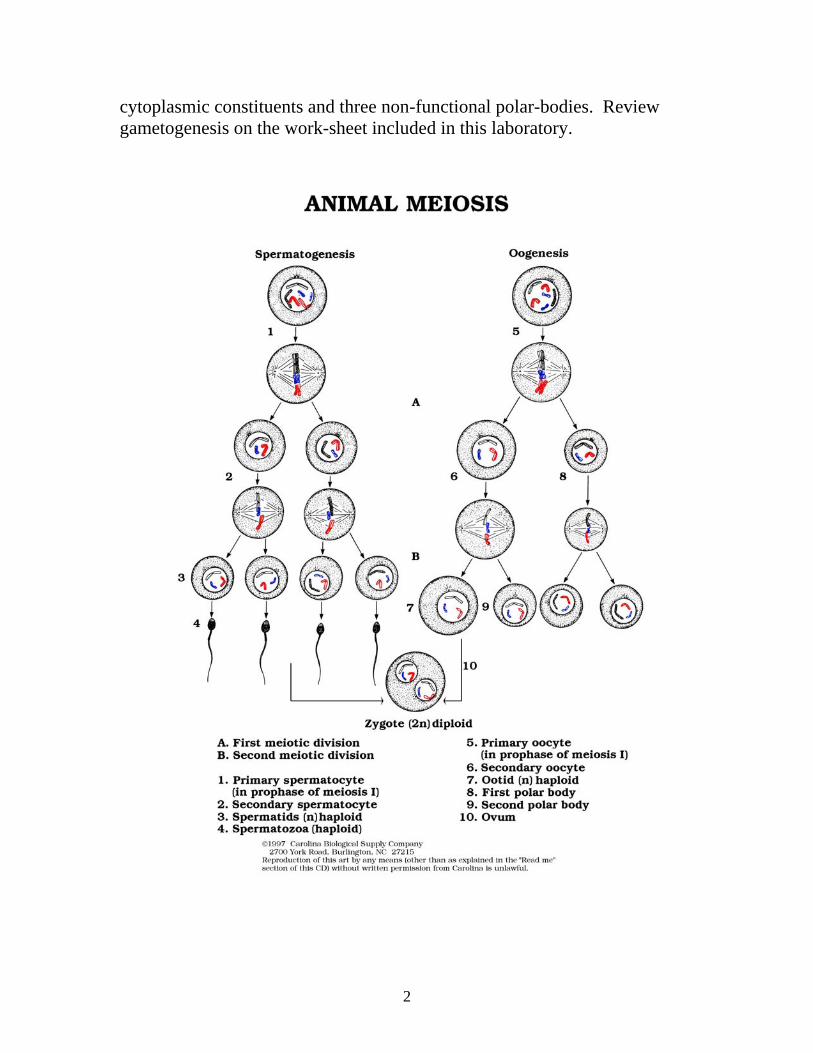
cytoplasmic constituents and three non-functional polar-bodies. Review gametogenesis on the work-sheet included in this laboratory.
2

Fertilization, syngamy, occurs with the union of a sperm and an egg, both highly specialized cell types, produced in the gonads of parents (sea urchins represent common model). The major events are: (1) The acrosomal reaction: upon contacting the jelly coat of the egg, the acrosomal vesicle of the sperm releases hydrolytic proteins that dissolve a pathway for the sperm through the jelly. An acrosomal process (actin) pushes through the jelly pathway until contact with the vitteline layer of the egg where the bindin protein attachs to the vitelline layer. The plasma membrane of the two gametes fuses and the sperm enters the cytoplasm of the egg. (2) The fast-block to polyspermy prevents more than one sperm from entering the egg. The union of the gamete membranes opens ion channels that allow sodium ions to flow into the egg cell and change the membrane potential thereby changing the voltage across the membrane. This membrane depolarization bars other sperm from entering the egg. (3) The cortical reaction: the sperm-egg fusion causes calcium ions to be released into the cytoplasm of the egg (probably from the ER) which causes vesicles in the cortex to fuse with the PM (plasma membrane.) The cortical granules release enzymes into the perivitelline space, and thereby allow it to become more fluid through the uptake of water. This may elevate the vitelline layer where enzymes polymerize molecules to form a hardened fertilization membrane. Slow-block to polyspermy: about a minute after fertilization, the membrane depolarization has returned to normal, reducing the effects of the fast-block, but now the slow-block to polyspermy is in place. (4) Activation of the egg: the higher concentration of calcium ions stimulates the extrusion of H+ from the egg, changing the pH from 6.8 to 7.3. The rate of protein synthesis increases dramatically. Twenty minutes later the sperm nucleus is swollen and merges with the egg nucleus to yield the 2N zygote. First cell division occurs in about 90 minutes.
Fertilization in the Sea Urchin
1. Upon contact, acrosome releases hydrolytic enzymes, breaking down jelly coat.
3

2. Acrosomal process pushes through, merging membranes. 3. Depolarization of the membrane of ovum yields fast block to
polyspermy. 4. Sperm nucleus enters cytoplasm. 5. Ca++ ions released from ER into cytoplasm. 6. H+ ions are extruded -----alkaline condition. 7. Cortical granules release contents into perivitelline space --- swelling,
raising and hardening of the vitelline layer ---slow block to polyspermy. 8. Alkaline conditions ---- activation of egg ---- increased respiration and
protein synthesis (translation of existing mRNA).
Fertilization in Mammals
1. Sperm migrate through follicle cells and bind to receptors in zona pellucida.
2. Acrosomal reaction releases hydrolytic enzymes into zona pellucida. 3. Sperm membrane binds to receptors on egg membrane --- fast block. 4. Cortical granules release contents causing slow block to polyspermy. 5. Slow block --- changes in zona pellucida. 6. Whole sperm enters egg, basal body of flagellum --- two centrosomes
(with centriole) for mitotic spindle. 7. After the first division, the chromosomes from male and female unite.
Embryology While viewing and contemplating embryology and the incredible processes of fertilization, differentiation, division, cellular movement and change in morphology we stand in awe. While many of the processes are different in terms of location and phylogenic association, they share some incredible similarities. From the very first division of the fertilized egg, the cells are often very different. While in some organisms the cells exhibit totipotency (the ability to express the entire genome), such as in the Homo sapiens embryo, some organisms do not. The structure and organization of the egg itself differs from organism to organism. Eggs that are cast adrift into an aquatic environment may have very large amounts of yolk, the nutrient filled material that the embryos will rely on to complete their development. Others, often simpler in nature such as the sea urchin ova, may have smaller amounts. We will examine
4

development of simpler organisms and move to the complex. Model organisms will be chosen in this progression. Cleavage consists of rapid cell division that produces a ball of cells from the starfish zygote. Most animal zygotes nourish themselves on yolk, where many nutrients are stored. The embryo does not grow, but the cell is simply partitioned into many smaller cells, called blastomeres (cells cycle between the S and M phases of mitosis). Increased surface to volume ratios enhance uptake of gases and other substances. An axis is formed by creation of two poles: animal pole (where polar body is budded) and vegetal pole (the opposite end). In frogs, the animal hemisphere is characterized by gray hue due to deposit of melanin whereby the vegetal pole is characterized by yellow yolk deposit. Note the structure and development of the sea urchin embryo. We see that the zygote moves from the one-cell stage through two vertical divisions, a horizontal division and then proceeds to the morula stage (a solid ball of cells). The cells continue dividing and a fluid-filled cavity develops (blastocoel). The structure is now referred to as a blastula with ciliated cells forming the hollow ball. A designated area on the surface of the starfish blastula invaginates (buckles into the cavity) and we observe early gastrulation of the embryo. These cells are pulled into the blastocoel by migrating mesenchyme cells and the primitive digestive system of developing embryo is then begun. The development of the archenteron designates late gastrulation. During this time cells are differentiating, changing personality, through a series of morphological and biochemical events. The three germ layers of the gastrula are ectoderm, mesoderm and endoderm. The resultant tissues from these primary germ layers will be discussed later. The larva is formed which then ultimately gives rise to the adult starfish.
5

6

7

8

While we will not pursue the development of the fish we note that it shares much in common with the frog which is the phylogenic bridge between aquatic and land environs. The frog egg itself is somewhat different as it contains a moderate amount of yolk. We notice that the blastocoel is off-center. Cells change shape near a specialized location the blastopore. The cells then allow migration of others into the blastocoel to form the germ layers, epiderm, mesoderm and endoderm. The archenteron, as in the starfish, gives rise to the digestive system. It is pulled through the developing embryo and becomes a primitive gut. A longitudinal indentation, the neural plate, begins to form on the surface of the developing egg. Thus begins the formation of the central nervous system (CNS): spine, brain and peripheral nerves. The process is called neurulation and is exhibited in vertebrates. The neural groove forms the posterior-anterior axis. The sides of the groove move up and enclose the longitudenal coelum (cavity) which will ultimately give rise to the CNS. The process of neurulation is incredibly complex. If errors are made at the stage, the organisms will probably be selected out of the population (die). In man, NTD (Neural Tube Disorders) are responsible for spina bifida, encephalopathy and anencephaly and other developmental disorders of the CNS. (An important note: NTD’s in man seem to be reduced when the mother’s diet is supplemented with folic acid.) As amphibians made the great trek to terra firma (dry land) the special adaptations that some of the population developed enabled them to be successful. Those without special adaptations died, often without progeny. However, some of the organisms were successful. Remember that to be successful, an organism must live and reproduce. An incredible structure evolved, ensuring the survival in this new environment - the egg, with a shell and extra-embryonic membranes. The structure of the amniotic egg is rather interesting. It is exhibited in reptilian and avian development and even a few mammals. The egg develops 4 membranes after fertilization. We will examine the avian egg. The egg is usually developed in the female of the species, fertilized within and shed into the environment into a protected place, such as a nest in the ground or in trees.
9

10

11

12

13

The egg has a large amount of yolk and four extraembryonic membranes: the yolk sac, the amnion, the allantois and the chorion. The yolk sac protects and segregates the yolk. It is responsible for making available the enclosed nutrients to the developing zygote. The amniotic membrane surrounds the developing embryo and protects it both from mechanical shock and desiccation. The allantois acts as a uric acid disposal site for wastes produced as development proceeds. The chorion in conjunction with the allantois, acts as a gas exchange system through which O2 can be provided for the developing zygote and CO2 can be released to the environment. The embryology of the bird proceeds as indicated in the diagrams. The division of cells is limited to a group of cells, the blastodisc, that sit on the surface of the large sac of yolk. Cells from an upper layer, the epiblast, migrate through a structure called the primitive streak, a longitudinal depression marking the posterior-anterior axis. The migrating cells then contribute to the hypoblast as well as enter the blastocoel which is flattened. The germ layers develop into respective tissues in a similar fashion as the frog (the aquatic ancestors) and organogenesis proceeds. While mammals existed during the time of the very successful, but extinct, dinosaurs, they did not flourish and undergo speciation until the extinction of the great land animals. Many phylogenic changes were successful and ultimately most mammals developed the ability to retain the developing zygote in a specialized structure, the uterus. Mammalian development is somewhat different from reptilian in some important ways. The typical mammalian ovum exhibits little or no yolk. This nutrient supply that many other embryos rely on is replaced by a system that is integrated with the mothers in a complex and interesting way. Note the structure of the fertilized and developing zygote. The trophoblast, a layer of cells of the zygote combines with cells of the mother’s uterus to form a complex structure, the placenta. The placenta then is the conduit for nutrient and gas exchange between mother and embryo. We see that the epilblast and the hypoblast are evident and a primitive streak occurs in a similar fashion to the bird. Organogenesis proceeds in a fashion similar to our aquatic ancestor, the frog.
14

15

16

17
18

We are attempting to generalize today to grasp the overall view of embryology. We examine the simple and proceed to the complex. Later we will discuss more aspects of this incredibly complex process of development. It is very important that we have an appreciation for the similarities and differences in development of our vertebrate relatives.
Questions for Embryology Laboratory
1. Draw and describe the structure of spermatozoa.
19

2. Differentiate between ovum, morula, blastula and gastrula. 3. Describe the amount of yolk in the following eggs: sea urchin, frog and chick. 4. Name and state the function of the four extraembryonic membranes in both avian and mammalian development.
20

5. Draw and label the morula stage of development of a sea urchin or starfish embryo: 6. Draw and label the blastula stage of development of a sea urchin or starfish embryo:
21

7. Draw and label the early and late gastrula stages of development of a sea urchin or starfish embryo: 8. Draw and label the 36-48 – hour chick embryo sagittal section:
22

9. Identify four major component processes in development. 10. Describe environmental conditions (both internal and external) that may be teratogenic to the developing embryo. 11. Cite a reputable source and attach a printout of a study linking environmental pollutants and increasing mutagenic effects.
23