embryology of some congenital cardiac anomalies · embryology. the basic difficulty in any such...
TRANSCRIPT
Thorax (1965), 20, 158.
Embryology of some congenital cardiac anomaliesE. W. T. MORRIS
From St. Thomas's Hospital Medical School, London
The purpose of this paper is to propose explana-tions for the nature of some commonly occurringcongenital cardiac anomalies in terms of humanembryology. The basic difficulty in any suchattempt is that, in the course of the developmentof the human heart, there are two stages. Duringthe first, the embryological process results in theformation of a four-chambered heart separatedby an interatrial and interventricular septum intoa right and left part; during the second, the organso formed grows in size, and changes occur in theform and proportions of its component parts toachieve those of the definitive heart. As Grant(1962) points out, little is known about the secondstage. In congenital heart disease, since the hearthas to function continuously so long as it is alive,errors of form occurring in the first period arelikely to influence changes occurring in the second.These latter may be such as to render difficult theelucidation of those errors of formation in thefirst stage which is the object of this paper.The views put forward are based on the dissec-
tion of 25 infant hearts which showed suchabnormalities (see Table). This study was under-taken because views still current on the aetiologyof some forms of congenital heart disease arethought to be erroneous. The earlier ones (Keith,1902, 1905) were propounded, in the light oftheories then current, that ontogeny repeatedphylogeny, and the abnormalities were conse-quently seen as arrests of development at a stagewhen the evolving heart resembled some phylo-genetic precursor of man, such as a reptile. Theseviews have been elaborated (Spitzer, 1923) andare included in recent clinical books (Brown,1950; Taussig, 1960) and seem widely held despiterecent papers (Kramer, 1942; Foxon, 1955) whichcast doubts on them.There is a central difficulty inherent in any
attempt to explain abnormalities on the assump-tion that they represent a deviation to the formof a phylogenetic ancestor: no one knows, or isever likely to know, what the hearts of theamphibian and reptilian ancestors of man werelike. The assumption that they would haveresembled the hearts of modern amphibians or
reptiles seems open to doubt since the livingorders of these two classes are, for the most part,highly specialized and therefore less likely toresemble their early common ancestor in thestructure and form of their organs.The steps in ontogeny only repeat phylogeny
in a very general way. For example, in its earlystages the mammalian heart resembles the fishheart in that each chamber is undivided, and theblood is impelled by it into a single ventral aortaand thence into paired aortic arch arteries. Ata later stage, the common chambers becomedivided into a right and a left, a step that occursin phylogeny when pulmonary respiration isestablished.At the most these stages may resemble early
steps in the embryology of the presumed ancestorand not the definitive heart of such a precursor.There seems little justification now for describingcongenital abnormalities of the heart in terms ofan atavism to the form of heart found in amodern reptile. Moreover, these explanationswere mainly given at a time when the accountof the development of the mammalian heartshowed many gaps. Now that these have beenlargely filled in to give an almost complete pictureit is worth while to try to explain examples ofsome congenital abnormalities in terms of failureeither of initiation or completion of some stepor steps known to occur in the normal heart.When this has been done some abnormalities
will remain which are inexplicable in terms ofsimple arrest: in these, some stage has notproceeded normally. In attempting to unravelwhat has happened, it is legitimate to try toconfine the explanation to a presumed diversionof a step known to occur normally. Equally, inaccordance with Ocean's dictum 'essentia non suntmultiplicanda praeter necessitatem', it is undesir-able to introduce into the explanation a stage notknown to occur in normal human embryology.An assumption of this sort is made by somewriters (Lev and Saphir, 1945) when theypostulate that, in the development of an abnormalheart, the bulbar septum is derived from ridgeswhich normally occur in the developing heart of
158
on May 2, 2020 by guest. P
rotected by copyright.http://thorax.bm
j.com/
Thorax: first published as 10.1136/thx.20.2.158 on 1 M
arch 1965. Dow
nloaded from

Embryology of some congenital cardiac anomalies 159
" + +vI ~ ~ +"fl __ + + +
N'i + + + +
-l + +
eo. + +
IXlo +
Y# I
o1 ++ + + +
We + +
m1 ++ + +
cn :I + +as2e1 +
" ++ ++ + +
z -z-z
0< -z
- I + +
o +
,,IrI + + + ±
t1
+ + o
+ +02
+ + + +
++ + + +"!+ + °r
I-I + IC*** * * *.... . . . . . . .
0)C
00
Co~~~~~~~~~0o .. ..o E UC 0OC Y
~~~::~~~:: 0 -:0)0)
.2 .~. .
0) ) 4 t* ' U0 ) LUU0.. 0. 0 0..2. Y >t c! ; ..
U.0~~~~~~~~~~~~~~~~~~~~~~~~~~~~. a ,o
.0 C,0 .0~~UU0)~~~~~ ~~0
0S' : IDo
oAO o co
0 000 C
U~~~~~~r -.
I E
I ;;
;ha
.X1;9LO.54
k.
I
on May 2, 2020 by guest. P
rotected by copyright.http://thorax.bm
j.com/
Thorax: first published as 10.1136/thx.20.2.158 on 1 M
arch 1965. Dow
nloaded from
E. W. T. Morris
modern reptiles in order to explain overriding ofthe aorta and transposition of the great arteries.At no stage in the development of the humanheart are there two ventral aortae arisingindependently from the ventricles as there are intypical modern reptiles. This is, of course, not tobe confused with the fact that double aortic archesmay occur as the result of the persistence of bothright and left fourth arch arteries, for, when theseare found, they are connected to the left ventricleby a single ascending aorta.
After some study of and thought about thespecimens, various features and patterns offeatures displayed by them began to emerge. Evenin the most complicated abnormalities it wasfound possible to suggest embryological explana-tions without invoking any steps now known notto occur in normal development. These observa-tions and the embryological explanation for themare given below. The terms 'proximal' and 'distal'relate to the direction of blood flow through theheart.
ABNORMALITIES OF THE VENTRICLE AND BULBUSCORDIS
Reference to embryological texts (Hamilton,Boyd, and Mossman, 1962; Kramer, 1942;Odgers, 1938) shows that defects in the septationof this part of the heart can result in varyingdegrees of failure either of development or ofunion of the component parts of the septum.There are five structures or groups of structuresinvolved in the formation of this septum; fromproximal to distal, (a) the muscular inter-ventricular septum, (b) the atrioventricularcushions forming both (i) the atrioventricular, and
-
f-
1.t.I.
(ii) the interventricular parts of the parsmembranacea septi, (c) the proximal, and (d) thedistal bulbar swellings, and (e) the aortico-pulmonary septum.Of these, the last to develop is the inter-
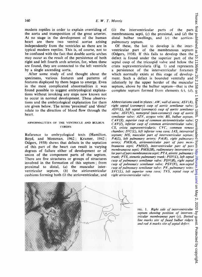
ventricular part of the membranous septum(Odgers, 1938). If this fails to develop then thedefect is found under the superior part of theseptal cusp of the tricuspid valve and below thecrista supraventricularis (Fig. l) and representsa persistence of the interventricular foramenwhich normally exists at this stage of develop-ment. Such a defect is bounded ventrally andinferiorly by the upper border of the muscularseptum, above by the bulbar septum-that is thecomplete septum formed from elements (c), (d),
Abbreviations used in plates: A W, wall ofaorta; AS V(R),right septal (coronary) cusp of aortic semilunar valve;ASV(L), left septal (coronary) cusp of aortic semilunarvalve; ASV(N), nonseptal (non-coronary) cusp of aorticsemilunar valve; AZV, azygos vein; BS, bulbar septum;CAV(S), superior cusp of common atrioventricular valve;CAV(I), inferior cusp of common atrioventricular valve;CS, crista supraventricularis; CVC, common venouschamber; IVC(L), left inferior vena cava; IAS, interatrialseptum; MS, muscular part of interventricular septum;PA(L), left pulmonary artery; PA(R), right pulmonaryartery; PMS(A), atrioventricular part of pars mcn7-branacea septi; PMS(I), interventricular part of parsmembranacea septi; PMS(IR), rudimentary interventricu-larpart ofpars membranacea septi; PTA, atretic pulmonarytrunk; PTS, stenotic pulmonary trunk; PSV(L), left septalcusp of pulmonary semilunar valve; PS V(R), right septalcusp of pulmonary semilunar valve; PSV(N), non-septalcusp ofpulmonary semilunar valve; PV, pulmonary veins;SVC(L), left superior vena cava, TVS, septal cusp ofright atrioventricular valve.
FIG. 1. Right side of interventricularseptum showing position of interven-tricular membranous part (x). Dottedline marks site of fused bulbar ridgesand rod A marks site of septal defect.
160
on May 2, 2020 by guest. P
rotected by copyright.http://thorax.bm
j.com/
Thorax: first published as 10.1136/thx.20.2.158 on 1 M
arch 1965. Dow
nloaded from
Embryology of some congenital cardiac anomalies
and (e) already described; it is limited dorsallyby the atrioventricular part of the membranousseptum. This is continuous with the right wall ofthe aorta in the region below and between theright coronary and non-coronary cusps of theaortic valve, and has the superior part of theseptal cusp of the tricuspid valve continuous withit ventrally (Fig. 2).
()
FIG. 2. Coronal section through right wall of aorta,membranous septum, septal cusp of tricuspid valve, andtnuscular septum.
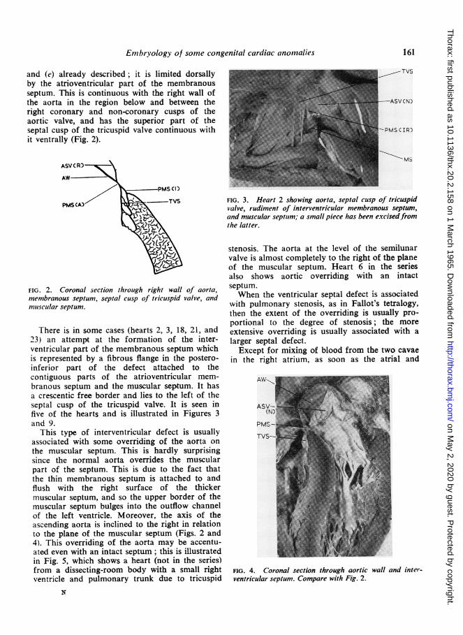
There is in some cases (hearts 2, 3, 18, 21, and23) an attempt at the formation of the inter-ventricular part of the membranous septum whichis represented by a fibrous flange in the postero-inferior part of the defect attached to thecontiguous parts of the atrioventricular mem-branous septum and the muscular septum. It hasa crescentic free border and lies to the left of theseptal cusp of the tricuspid valve. It is seen infive of the hearts and is illustrated in Figures 3and 9.
This type of interventricular defect is usuallyassociated with some overriding of the aorta onthe muscular septum. This is hardly surprisingsince the normal aorta overrides the muscularpart of the septum. This is due to the fact thatthe thin membranous septum is attached to andflush with the right surface of the thickermuscular septum, and so the upper border of themuscular septum bulges into the outflow channelof the left ventricle. Moreover, the axis of theascending aorta is inclined to the right in relationto the plane of the muscular septum (Figs. 2 and4). This overriding of the aorta may be accentu-ated even with an intact septum; this is illustratedin Fig. 5, which shows a heart (not in the series)from a dissecting-room body with a small rightventricle and pulmonary trunk due to tricuspid
N
TVS
Ms
FIG. 3. Heart 2 showing aorta, septal cusp of tricuspidvalve, rudiment of interventricular membranous septum,and muscular septutm; a small piece has been excisedfiromthe latter.
stenosis. The aorta at the level of the semilunarvalve is almost completely to the right of the planeof the muscular septum. Heart 6 in the seriesalso shows aortic overriding with an intactseptum.When the ventricular septal defect is associated
with pulmonary stenosis, as in Fallot's tetralogy,then the extent of the overriding is usually pro-portional to the degree of stenosis; the moreextensive overriding is usually associated with alarger septal defect.
Except for mixing of blood from the two cavaein the right atrium, as soon as the atrial and
AWo
PM'-
TVS-
FIG. 4. Coronal section through aortic wall and inter-ventricular septum. Compare with Fig. 2.
161
on May 2, 2020 by guest. P
rotected by copyright.http://thorax.bm
j.com/
Thorax: first published as 10.1136/thx.20.2.158 on 1 M
arch 1965. Dow
nloaded from

E. W. T. Morris
ever small, has always been in the same position,relative to the cusps of the aortic valve, as in thenormal heart, that is adjacent to the commissurebetween the two coronary cusps (Fig. 6b). Thissuggests that in those cases the bulbus cordis hasbeen normally divided by the distal bulbar swell-ings, which have also formed the semilunar valves,but that in the subsequent growth of the foetalheart the infundibulum and pulmonary trunkhave failed to grow normally.
PSV (PSV (PSVASV (
ASV (ASV (
a
FIG. 5. View into open right ventricle and pulmonarytrunk to show aorta overriding the muscular septum.
ventricular septa are complete the circulation inthe embryo is divided into two separate circuits,one to the fore and the other to the hind end ofthe embryo, and it is the placental oxygenatedblood that goes to the former. On returning fromthe fore end, this blood then nourishes the hindend on its way back to the placenta. Thispreferential treatment of the head andforelimb region is of course reflected in therelatively greater rate of growth of this partcompared with the hind end during foetal life.If there is a ventricular septal defect with aorticoverriding, then the blood from the right ventriclecan now reach the end of the aortic arch in twoways: either via the pulmonary trunk and ductusarteriosus or via the overriding aorta. Thoma's(1893) first law suggests that there is a haemo-dynamic principle at work in biology accordingto which the calibre of a vessel is controlled bythe volume of blood passing through it. More-over, as Hughes (1943) points out, 'the dynamicrelationship between vessels and circulationcharacteristic of the adult circulatory system isearly acquired in the mbryo'; once initiated,there is, in Fallot's tetralogy and allied abnorm-alities, a factor that reduces the flow through thepulmonary trunk, and, once initiated, this seems
to be progressive and results in an enlargementof the aorta at the expense of the pulmonarychannel.
In hearts (Nos. 6, 17, and 22) showingpulmonary stenosis, the pulmonary trunk, how-
di
c
dii
FIG. 6. Sections through the aorta and pulmonary trunkat the level of semilunar valves (except d ii): (a) normal,A-B represents plane of bulbar septum; (b) stenosis ofpulmonary trunk; (c) atresia; (d i) undivided bulbus cordis;and (d ii) just above the valve showing pulmonary stenosis.
An even more advanced stage of this process
was found in several of the hearts (Nos. 2, 3, 7,12, 24, and 25); these appear at first sight to beexamples of undivided bulbus, with a single largevessel lying astride the septal defect. Furtherinvestigation, however, suggests that these are
extreme cases of pulmonary stenosis with com-
plete occlusion of the pulmonary trunk, becauseeach one shows a small blind recess in thesuperior part of the right ventricle anterior to theplane of the crista supraventricularis, that is inthe position of the normal infundibulum (Fig. 7).Attached to the external wall of this recess bya fibrous cord (Fig. 8) is a patent channel lyingon the surface of the aortic wall and, at the levelof the aortic valve (which possesses three normalcusps), it is in the position indicated in Figure 6c.
Moreover, in three of the hearts (Nos. 7, 12,and 24), which show an aorta arising mainly from
........ --.AMS --
b
162
on May 2, 2020 by guest. P
rotected by copyright.http://thorax.bm
j.com/
Thorax: first published as 10.1136/thx.20.2.158 on 1 M
arch 1965. Dow
nloaded from

Embryology of some congenital cardiac anomalies
MS-
FIG. 7. Heart 2. View of the right ventricle showinginfundibular recess (R).
the right ventricle but with some overlap of themuscular septum, inspection of the left ventricleshows a small diverticulum between the septal.cusp of the mitral valve and the flange represent-ing the interventricular part of the membranousseptum, that is in the position of the outflowchannel of the left ventricle. Arising from theexternal surface of the heart, corresponding tothe diverticulum and dorsal to the aorta, is afibrous cord which is continuous with a patentvessel giving rise to the right and left pulmonaryarteries. These appear to be cases of transpositionof the aorta and pulmonary trunk with oblitera-tion of the proximal part of the latter. If thisinterpretation is correct it is interesting to notethat, even when the vessels are transposed, it isthe now dorsally situated pulmonary trunk thathas become occluded, suggesting that some factoris operating to produce pulmonary stenosis orocclusion whatever the position of the vessel.The hearts in this series, whatever the orienta-
tion of the great arteries relative to the sagittalplane of the body may be, all show the sameorientation of the cusps of their semilunar valvesrelative to the other vessel, that is the arrangementof the cusps of either valve in relation to the cuspsof the valve in the other channel is the same asin the normal heart. Even when the pulmonarytrunk is represented by a fibrous cord, it liesadjacent to the commissure between the twocoronary cusps of the aortic valve (Fig. 6c). This
suggests that the division of the bulbus cordis andthe formation of the semilunar valves have beennormally initiated by the four distal bulbarswellings whatever has been the subsequent courseof development. It seems unnecessary to postulatethe occurrence in the development of these heartsof a step which is a return to the form of aphylogenetic reptilian precursor with two aortaearising from the heart. In the hearts that showtransposition of the aorta and pulmonary trunk,the end product could result from an alterationin the position of the proximal and distal bulbarswellings so as to cause the pulmonary trunk toarise from the left ventricle and the aorta fromthe right.
It is of interest that several writers (Bremer,1928; de Vries and Saunders, 1962; Grant, 1962)have suggested that the position of the variousridges forming the bulbar septum may bedetermined by a pre-existing flow of bloodthrough the bulbus. They postulate two streams,one from the right and the other from the leftpart of the ventricle, flowing simultaneouslythrough the bulbus cordis in such a way that,between the two streams, there are regions ofreduced flow into which the ridges tend to grow.In the normally developing heart the position ofthe streams entering the bulbus is such as tocause them to spiral around each other in theircourse through the channel and so result in theformation of a spiral septum which rotatesthrough approximately 180 degrees in a right-handed spiral from proximal to distal. Differences
PACLI
Pv-
FIG. 8. Heart 7 showing proximal part of pulmonarytrunk.
1 63
on May 2, 2020 by guest. P
rotected by copyright.http://thorax.bm
j.com/
Thorax: first published as 10.1136/thx.20.2.158 on 1 M
arch 1965. Dow
nloaded from
E. W. T. Morris
-.......
4;f <
._ /f. --qAX,..x; Ds;'i >s .. h L,
*: V\E _ y
:: \;\t. I:
*'X, S..
IF,-)__
C7.
FIG. 9. Heart 20 showing compound defectin the bulbar and membranous septa throw-ing both outflow channels and the roots of
,M. the great vessels into communication. Thearrow leads into the aorta; (x) narks theposterior cusp of the common semilunarvalves. (See Fig. 6, d i.)
in the relative positions of the bulbus cordis andanlagen of the right and left ventricles to eachother could result in the two streams takingdifferent courses in the bulbus and so causing theridges and the septum derived from them to format different sites in the bulbus cordis. This, in turn,could account for the varying degrees of trans-position of the great arteries.Although the type of ventricular septal defect
that results from failure of the interventricularpart of the membranous septum to develop is thecommonest, others occur. One is situated justproximal to the semilunar valves in that part ofthe septum, formed from the bulbar ridges, whichseparates the distal part of the outflow channelof each ventricle (Morris, 1957). It occurs sub-jacent to the commissure between the two septalcusps of both aortic and pulmonary valves andis explicable as a failure of the fusion thatnormally occurs between the contiguous bordersof the septa formed from the bulbar ridges: thisdefect was described by Keith (1909).A rarer defect in the same relative position, but
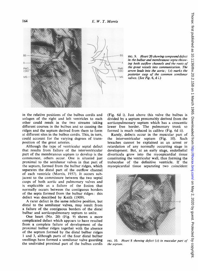
distal to the semilunar valves, may result froma failure of the contiguous borders of the distalbulbar and aorticopulmonary septum to unite.One heart (No. 20) (Fig. 9) shows a more
complicated defect which appears to have resultedfrom a complete failure of development of theproximal bulbar ridges together with the absenceof the septum formed by the distal bulbar ridges1 and 3, although parts of the four distal bulbarswellings have formed a semilunar valve guardingthe undivided proximal part of the bulbus cordis
(Fig. 6d i). Just above this valve the bulbus isdivided by a septum presumably derived from theaorticopulmonary septum which has a crescenticlower free border. The pulmonary trunk soformed is much reduced in calibre (Fig. 6d ii).
Rarely, defects occur in the muscular part ofthe interventricular septum (Fig. 10). Suchbreaches cannot be explained as an arrest orretardation of any normally occurring stage indevelopment. But, at an early stage, endothelialdiverticula grow into the myoepicardial tissueconstituting the ventricular wall, thus forming thetrabeculae of the definitive ventricle. If themyoepicardial tissue separating two coincident
FIG. 10. Heart 8 showing defect (x) in muscular part ofthe septum.
164
'r
on May 2, 2020 by guest. P
rotected by copyright.http://thorax.bm
j.com/
Thorax: first published as 10.1136/thx.20.2.158 on 1 M
arch 1965. Dow
nloaded from
Embryology of some congenital cardiac anomalies
diverticula growing from opposite sides of themuscular septum were to break down, such adefect would result. Keith (1909) supports such apossibility when, describing this defect, he says'apparently interstices in the original muscularspongework of the septum which have remainedunobliterated'. A breakdown of tissue is notunknown in the course of development; thefenestration of the dorsal mesocardium to formthe definitive transverse sinus of the pericardiumis an example.
ABNORMALITIES OF THE ATRIOVENTRICULAR CANAL
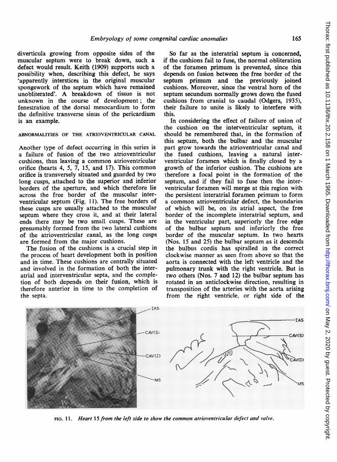
Another type of defect occurring in this series isa failure of fusion of the two atrioventricularcushions, thus leaving a common atrioventricularorifice (hearts 4, 5, 7, 15, and 17). This commonorifice is transversely situated and guarded by twolong cusps, attached to the superior and inferiorborders of the aperture, and which therefore lieacross the free border of the muscular inter-ventricular septum (Fig. 11). The free borders ofthese cusps are usually attached to the muscularseptum where they cross it, and at their lateralends there may be two small cusps. These arepresumably formed from the two lateral cushionsof the atrioventricular canal, as the long cuspsare formed from the major cushions.The fusion of the cushions is a crucial step in
the process of heart development both in positionand in time. These cushions are centrally situatedand involved in the formation of both the inter-atrial and interventricular septa, and the comple-tion of both depends on their fusion, which istherefore anterior in time to the completion ofthe septa.
IAS
C-CAV(SC
-CAVCID
So far as the interatrial septum is concerned,if the cushions fail to fuse, the normal obliterationof the foramen primum is prevented, since thisdepends on fusion between the free border of theseptum primum and the previously joinedcushions. Moreover, since the ventral horn of theseptum secundum normally grows down the fusedcushions from cranial to caudal (Odgers, 1935),their failure to unite is likely to interfere withthis.
In considering the effect of failure of union ofthe cushion on the interventricular septum, itshould be remembered that, in the formation ofthis septum, both the bulbar and the muscularpart grow towards the atrioventricular canal andthe fused cushions, leaving a natural inter-ventricular foramen which is finally closed by agrowth of the inferior cushion. The cushions aretherefore a focal point in the formation of theseptum, and if they fail to fuse then the inter-ventricular foramen will merge at this region withthe persistent interatrial foramen primum to forma common atrioventricular defect, the boundariesof which will be, on its atrial aspect, the freeborder of the incomplete interatrial septum, andin the ventricular part, superiorly the free edgeof the bulbar septum and inferiorly the freeborder of the muscular septum. In two hearts(Nos. 15 and 25) the bulbar septum as it descendsthe bulbus cordis has spiralled in the correctclockwise manner as seen from above so that theaorta is connected with the left ventricle and thepulmonary trunk with the right ventricle. But intwo others (Nos. 7 and 12) the bulbar septum hasrotated in an anticlockwise direction, resulting intransposition of the arteries with the aorta arisingfrom the right ventricle, or right side of the
FIG. 11. Heart 15 from the left side to show the common atrioventricular defect and valve.
165
on May 2, 2020 by guest. P
rotected by copyright.http://thorax.bm
j.com/
Thorax: first published as 10.1136/thx.20.2.158 on 1 M
arch 1965. Dow
nloaded from
E. W. T. Morris
4# -
i--K--I __
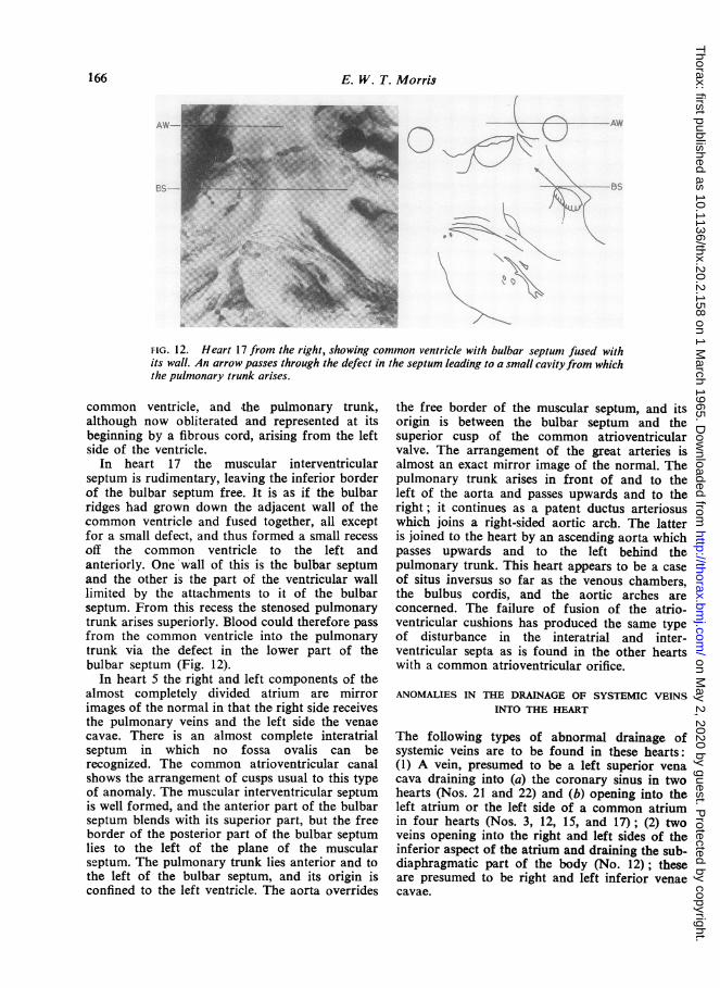
FIG. 12. Heart 17 from the right, showing common ventricle with bulbar septunm fused withits wall. An arrow passes through the defect in the septum leading to a small cavityfrom whichthe pulmonary trunk arises.
common ventricle, and the pulmonary trunk,although now obliterated and represented at itsbeginning by a fibrous cord, arising from the leftside of the ventricle.
In heart 17 the muscular interventricularseptum is rudimentary, leaving the inferior borderof the bulbar septum free. It is as if the bulbarridges had grown down the adjacent wall of thecommon ventricle and fused together, all exceptfor a small defect, and thus formed a small recessoff the common ventricle to the left andanteriorly. One wall of this is the bulbar septumand the other is the part of the ventricular walllimited by the attachments to it of the bulbarseptum. From this recess the stenosed pulmonarytrunk arises superiorly. Blood could therefore passfrom the common ventricle into the pulmonarytrunk via the defect in the lower part of thebulbar septum (Fig. 12).
In heart 5 the right and left components of thealmost completely divided atrium are mirrorimages of the normal in that the right side receivesthe pulmonary veins and the left side the venaecavae. There is an almost complete interatrialseptum in which no fossa ovalis can berecognized. The common atrioventricular canalshows the arrangement of cusps usual to this typeof anomaly. The muscular interventricular septumis well formed, and the anterior part of the bulbarseptum blends with its superior part, but the freeborder of the posterior part of the bulbar septumlies to the left of the plane of the muscularseptum. The pulmonary trunk lies anterior and tothe left of the bulbar septum, and its origin isconfined to the left ventricle. The aorta overrides
the free border of the muscular septum, and itsorigin is between the bulbar septum and thesuperior cusp of the common atrioventricularvalve. The arrangement of the great arteries isalmost an exact mirror image of the normal. Thepulmonary trunk arises in front of and to theleft of the aorta and passes upwards and to theright; it continues as a patent ductus arteriosuswhich joins a right-sided aortic arch. The latteris joined to the heart by an ascending aorta whichpasses upwards and to the left behind thepulmonary trunk. This heart appears to be a caseof situs inversus so far as the venous chambers,the bulbus cordis, and the aortic arches areconcerned. The failure of fusion of the atrio-ventricular cushions has produced the same typeof disturbance in the interatrial and inter-ventricular septa as is found in the other heartswith a common atrioventricular orifice.
ANOMALIES IN THE DRAINAGE OF SYSTEMIC VEINSINTO THE HEART
The following types of abnormal drainage ofsystemic veins are to be found in these hearts:(1) A vein, presumed to be a left superior venacava draining into (a) the coronary sinus in twohearts (Nos. 21 and 22) and (b) opening into theleft atrium or the left side of a common atriumin four hearts (Nos. 3, 12, 15, and 17); (2) twoveins opening into the right and left sides of theinferior aspect of the atrium and draining the sub-diaphragmatic part of the body (No. 12); theseare presumed to be right and left inferior venaecavae.
166
on May 2, 2020 by guest. P
rotected by copyright.http://thorax.bm
j.com/
Thorax: first published as 10.1136/thx.20.2.158 on 1 M
arch 1965. Dow
nloaded from

Embryology of some congenital cardiac anomalies
(la) This condition is consistent with a persist-ence of the left common and anterior cardinalveins as a continuous channel. The normal fateof these veins, first described by Marshall (1850),is as follows: When the cross connexion betweenthe two anterior cardinal veins, which forms theleft brachiocephalic vein, is developed, the wholeof the blood from the left forelimb and the leftside of the head and neck drains via this into theright common cardinal vein and so into the rightatrium. The remainder of the left anterior cardinalvein below the new channel forms the terminalpart of the definitive left superior intercostal vein.The distal part of the left common cardinal veinis obliterated and forms a fibrous cord. This isconnected above to the left superior intercostalvein and is continuous below with the patentproximal part of the common cardinal vein, thedefinitive oblique vein of Marshall, which drainsinto the left extremity of the coronary sinus,which is formed from the left horn of the sinusvenosus.
(lb) These hearts show a vein, believed to bethe left common cardinal vein, opening posteriorlyinto the superior aspect of the left side of theatrium (Fig. 13). This suggests that the left hornof the sinus venosus has been absorbed into theposterior wall of the left side of the atrium,leaving only the right horn to be incorporatedin the right side. Such a state of affairs couldoccur if the constriction separating the common
SVCL) |.
AZV-|LPV-f ..a.,w '1 E
l N[. v 1
_ | _l|
FIG. 13. Heart 12 viewedfrom the left, showing the leftsuperior and inferior venae cavae.
atrium from the sinus venosus, usually describedas occurring at an early stage, during Streeter'sHorizon x, had been shallow and symmetrical,for it is the asymmetry of this inflexion that leadsto both a reduction in the size of the sinu-atrialopening and its movement to the right side ofthe dorsal wall of the atrium. The venous partof the heart would then consist of an anteriorcommon chamber, the atrium, separated from aposterior common chamber, the sinus venosus,by a shallow uniform inflexion of the wall. More-over it is the deep indentation of the left wallthat forms both the dorsal wall of the atrium tothe left of the sinu-atrial opening and the anteriorwall of the left horn of the sinus venosus. If thisfails, the sinus venosus will be a symmetricalchamber with no horns, and the veins will openinto its lateral ends. If this were the case someremnant of a left venous valve would be foundin the left side of the chamber in the form of aleft crista terminalis running anterior to theentrance of the left vena cava and continuingdown the lateral wall of the venous chamber onthe left. The chamber should be smooth-walledposterior to the cristae and with musculae pectinatiarising from it anteriorly. In three of the heartsthese features are present, and in one, althoughthe venous chamber is smooth-walled posteriorlyand trabeculated anteriorly and in the auricles,no crista is visible. In addition, if this theory istenable there cannot be a normal coronary sinus,and examination shows that in none of the fourhearts is it found.
(2) In this heart there are two veins openinginferiorly into the right and left sides of thecommon venous chamber, which are taken to beinferior venae cavae (Fig. 13). If this is correctand they have been normally derived from a rightand left hepato-cardiac channel, then these shouldalso open into the posterior part of the venouschamber, and the right one should be guardedby a normal valve continuous with the lower endof the right crista terminalis and the left oneshould be bordered anteriorly by a similar valve,a mirror image of that on the right. In this heartthere is such a structure. In the remaining threehearts there is a single inferior vena cava.
In these four hearts it is suggested that thevenous chamber is, as in the normal heart, a com-pound one, but, instead of the whole of the sinusvenosus-both right and left horns-beingabsorbed into the right atrium, the symmetricalsinus venosus is incorporated into the posteriorwall. The left common cardinal vein will thereforeopen into the left side of this common chamberand the right one into the other side.
167
on May 2, 2020 by guest. P
rotected by copyright.http://thorax.bm
j.com/
Thorax: first published as 10.1136/thx.20.2.158 on 1 M
arch 1965. Dow
nloaded from

E. W. T. Morris
At the time of the initiation of the septumprimum the dorsal wall of the venous chamberis therefore formed by the dorsal wall of the sinusvenosus, and the dorsal mesocardium will beattached to this wall, and it is on the internalwall at this point that the septum primumnormally forms. Although there is an attempt atinteratrial septum formation in each of the fourhearts, it is far from complete, even in two ofthe hearts in which the atrioventricular canal isdivided.
ANOMALIES IN THE DRAINAGE OF PULMONARY VEINS
INTO THE HEART
In hearts 3, 4, 7, 12, 15, 16, and 17, the pulmonaryveins do not drain normally, and in four of these(Nos. 7, 12, 16, and 17) the pulmonary veins haveno direct connexion with the heart (Fig. 14). Thearrangement is similar in each of the hearts inthe larger group; there is a transverse channellying behind the venous heart chamber, and intothe lateral ends of this the right and leftpulmonary veins drain, but this transversechannel has no connexion with the heart. Thepulmonary veins are derived from the pulmonaryplexus, and this is then tapped by an outgrowthfrom the dorsal wall of the left atrium (Auer,1948; Neill, 1956). If this outgrowth failed, thepulmonary veins would be derived solely from thepulmonary plexus and would have no openinginto the heart. In all the hearts with this anomalyother defects of the venous end of the heart are
0.T.0-r'
pT, 4-_ _A - b
PArr A * r
PV-
FIG. 14. Heart 12 turned upward to show the ventralsurface of the lung roots. The pulmonary veins are con-
nected by a common channel. The pulmonary trunk showsatresia proximally.
present. The pulmonary plexus has numerousconnexions with both the cardinal and the vitello-umbilical system of veins and, in the absence ofthe normal drainage via the common pulmonaryvein, any of these connexions may enlarge.Numerous papers have been written on thevariety of forms of this anomalous pulmonarydrainage. Considering the small volume of bloodthat passes through the foetal lung, the size ofthese connexions is not important until birth andpost-natal survival increase the pulmonary flow,when they must be adequate to carry this. In thethree newborn hearts, the connexions are small,whereas in two others, which were from infantswho survived birth by 30 hours and 12 weeksrespectively, the communications between thepulmonary and other veins were well developed.
In two hearts the pulmonary veins drain intothe venous chamber and presumably thereforesome form of common pulmonary vein must havedeveloped, but in each case the abnormality ofdouble superior venae cavae is present, and sothis must have grown out from the posterior wallof the sinus venosus.
SUMMARY
It has been pointed out that certain structuresoccurring in the normal heart can be recognizedin most of the specimens. For example, all thehearts show a muscular structure, taken to bethe lower border of the bulbar septum, which atone stage in development forms part of theboundary of the interventricular foramennormally existing at that stage and which thenseparates the outflow channels of the pulmonarytrunk and aorta. Whatever the orientation ofthese channels relative to each other, this freeborder can be recognized, even when one of theorifices has become obliterated, and is representedby a recess in the ventricle. In these cases theproximal part of the vessel is replaced by a fibrouscord. Furthermore, if semilunar valves arepresent, the cusps bear a constant relationship tothe septum. This indicates that the initial forma-tion of the bulbar septum is a very constantfeature and that, while there are obviously factorswhich influence the orientation and the degreeand direction of twist of the developing bulbarseptum, its actual formation is relativelyuninfluenced by the occurrence of abnormalitiesin that region of the heart.The junction of the anterior part of the bulbar
septum with the muscular interventricular septumrarely fails to take place when the latter is present.In those hearts, as in Fallot's tetralogy, in which
168
on May 2, 2020 by guest. P
rotected by copyright.http://thorax.bm
j.com/
Thorax: first published as 10.1136/thx.20.2.158 on 1 M
arch 1965. Dow
nloaded from
Embryology of some congenital cardiac anomalies
the last stage of the interventricular septum-theinterventricular membranous septum-has beenarrested, an attempt at its formation can beobserved. This takes the form of a fibrous ridgein the posterior border of the defect and bearsthe same relationship to the right aortic wall, theatrioventricular part of the membranous septum,and the septal cusp of the tricuspid valve as inthe normal heart. Moreover this relationshippersists if, owing to malrotation of the bulbarseptum, it is the right wall of the pulmonary trunkto which the atrioventricular membranous septumis attached.An explanation is offered for the occurrence of
defects in the muscular interventricular septum.When fusion of the atrioventricular cushions
fails to take place, other defects also occur andthese are consequent upon the earlier failure ofcushion fusion.An explanation is offered for the occurrence of
a superior and inferior vena cava on the left sideof the venous chamber, and it is suggested thatthe failure of the pulmonary veins to draindirectly into the venous end of the heart has asimple embryological explanation.The majority of cardiac anomalies seem to be
compatible with an embryological explanation,and any resemblance to the hearts seen in othervertebrate classes is coincidental.
I wish to express my gratitude to Professors D. V.Davies and C. H. Barnett for help and advice and toMr. A. Pache for the photographs.
REFERENCESAuer, J. (1948). The development of the pulmonary veins in man.
Anat. Rec., 101, 581.Bremer, J. L. (1928). Part 1. An interpretation of the development of
the heart. Part 2. The left aorta ofreptiles. Amer. J. Anat., 42, 307.Brown, J. W. (1950). Congenital Heart Disease, 2nd ed. Staples,
London.de Vries, P. A., and Saunders, J. B. de C. M. (1962). Development of
the ventricles and spiral outflow tract in the human heart.Contr. Embryol. Carneg. Instn, 37, 87 [No. 256].
Foxon, G. E. H. (1955). Problems of the double circulation invertebrates. Biol. Rev., 30, 196.
Grant, R. P. (1962). The embryology of ventricular flow pathways inman. Circulation, 25, 756.
Hamilton, W. J., Boyd, J. D., and Mossman, H. W. (1962). HumanEmbryology, 3rd ed. Heffer, Cambridge.
Hughes, A. F. W. (1943). The histogenesis of the arteries of the chickembryo. J. Anat. (Lond.), 77, 266.
Keith, A. (1902). The anatomy of the valvular mechanism round thevenous orifices of the right and left auricles, with some observa-tions on the morphology of the heart. Ibid., 37, proc. ii.(1905). Exhibition of thirty malformed human hearts. Ibid., 39,proc. xiv.- (1909). Hunterian lectures on malformations of the heart.
Lancet, ii, 359, 433, 519.Kramer, T. C. (1942). The partitioning of the truncus and conus and
the formation of the membranous portion of the interventricularseptum in the human heart. Amer. J. Anat., 71, 343.
Lev, M., and Saphir, 0. (1945). A theory of transposition of thearterial trunks based on the phylogenetic and ontogeneticdevelopment of the heart. Arch. Path., 39, 172.
Marshall, J. (1850). On the development of the great anterior veinsin man and mammalia. Phil. Trans., 140, 133.
Morris, E. W. T. (1957). The interventricular septum. Thorax, 12, 304.Neill, C. A. (1956). The development of the pulmonary veins with
reference to the embryology of anomalous pulmonary venousreturn. Pediatrics, 18, 880.
Odgers, P. N. B. (1935). The formation of the venous valves, theforamen secundum and the septum secundum in the humanheart. J. Anat. (Lond.), 69, 412.- (1938). The development of the pars membranacea septi in the
human heart. Ibid., 72, 247.Spitzer, A. (1923). 'Yber den Bauplan des normalen und missbildeten
Herzens. Virchows Arch. path. Anat., 243, 81.Taussig, H. B. (1960). Congenital Malformations of the Heart, 2nd ed.
Harvard University Press, Cambridge, Mass.Thoma, R. (1893). Untersuchungen dber die Histogenese und Histo-
mechanik des Gefasssystems. Enke, Stuttgart.
169
on May 2, 2020 by guest. P
rotected by copyright.http://thorax.bm
j.com/
Thorax: first published as 10.1136/thx.20.2.158 on 1 M
arch 1965. Dow
nloaded from