embryonic and larval development in the caecilian ...ib.berkeley.edu/labs/mwake/papers/156.pdf ·...
TRANSCRIPT
Embryonic and Larval Development in the CaecilianIchthyophis kohtaoensis (Amphibia, Gymnophiona):A Staging TableNicole Dunker,1 Marvalee H. Wake,2* and Wendy M. Olson2
1 Department of Zoology, Technical University of Darmstadt, Darmstadt, Germany2 Department of Integrative Biology and Museum of Vertebrate Zoology, University of California,Berkeley, California
ABSTRACT Little is known about the developmentalbiology of caecilians—tropical, elongate, limbless, mostlyfossorial amphibians that are members of the Order Gym-nophiona. Ichthyophis kohtaoensis (Family Ichthyophi-idae; southeast Asia) is an oviparous species in whichmaternal care of the clutch is provided. The clutch is laidin a burrow on land, and the embryos develop in their eggmembranes, curved around a large yolk mass. Larvae areaquatic and exhibit characteristic features that are notpresent in the terrestrial adults. Because accurate de-scriptions of ontogenies and the establishment of stan-dardized stages of embryonic and larval development areuseful for both experimental and comparative embryology,a staging table for I. kohtaoensis was developed based onexternal morphological features. Development from theend of neurulation to metamorphosis was divided into 20stages. Principal diagnostic features include development
of the lateral line organs, formation of three pairs of ex-ternal gills, development of the eyes, changes in yolkstructure, changes in the structure of the cloacal apertureand growth of the tail, including the formation and regres-sion of the tail fin. This study provides a comparison withdescriptions of embryonic stages of I. glutinosus and Hy-pogeophis rostratus and with a recent staging table for theaquatic, viviparous caecilian Typhlonectes compressi-cauda, the only other caecilians for which reasonably com-plete ontogenetic information exists in the literature.Comparisons with established staging tables for selectedfrogs and salamanders are also presented. J. Morphol.243:3–34, 2000. © 2000 Wiley-Liss, Inc.
KEY WORDS: Ichthyophis kohtaoensis; amphibians; cae-cilians; ontogeny; development; staging
Caecilians, members of the Order Gymnophiona,are tropical, limbless, elongate, tailless or nearly so,amphibians that occupy subterranean, leaf-litter, orsemi-aquatic to aquatic habitats. They are amongthe least studied of vertebrates. Rather little isknown of the development of caecilians, especiallycompared to the relative wealth of knowledge aboutselected frogs and salamanders.
The development of Ichthyophis glutinosus (Fam-ily Ichthyophiidae) was described and beautifullyillustrated by Sarasin and Sarasin (1887–1890).Cleavage in amphibian eggs is typically holoblasticand unequal. In I. glutinosus, the only caecilian forwhich cleavage has been described (Sarasin andSarasin, 1887–1890), cleavage is nearly meroblastic,dividing the egg into numerous separate blas-tomeres and a residual multinucleate mass of cyto-plasm. At the tail-bud stage, caecilians are similarto salamanders and have a relatively long head andpharyngeal region, including the gill plates, thatprojects above the yolk mass, but they have only ashort tail bud. In contrast, in anurans the gill plateregion lies dorsal to the yolk mass with only theanterior most part of the head projecting beyond theyolk, but a relatively long tail bud extends beyond
the yolk. A number of specific features of develop-ment are evaluated below.
There are very few descriptions of caecilian devel-opment from early stages to metamorphosis or birth(Sarasin and Sarasin, 1887–1890; Brauer, 1897,1899; Sammouri et al., 1990); some authors havedescribed only one or a few specimens (not staged)available for study. Sarasin and Sarasin (1887–1890) and Brauer (1899) presented detailed descrip-tions of the changes in the course of development,referring variously to the formation of gills, eyes, thetentacle, the lateral line organs, changes in yolkstructure, and general external morphology. Brauer(1899) provided a rather complete series of drawingsof early embryos of Hypogeophis rostratus, with
Contract grant sponsor: National Science Foundation; Contractgrant number: IBN95-27681.
Current address for Nicole Dunker, Anatomy, Medical Faculty 3.1,University of Saarland, 66421 Homburg/Saar, Germany.
*Correspondence to: Marvalee H. Wake, M.D., Department of Integra-tive Biology, University of California, Berkeley, CA 94720-3140, USA.E-mail: [email protected]
JOURNAL OF MORPHOLOGY 243:3–34 (2000)
© 2000 WILEY-LISS, INC.
some later stages of that species and a few for Gran-disonia alternans; Marcus described the develop-ment of specific elements of Hypogeophis rostratus,but mostly internal features so only his work rele-vant to external morphology is cited herein; Sarasinand Sarasin (1887–1890) described various develop-mental stages of Ichthyophis glutinosus, includingthose of very early development. However, thesestudies did not present their results in the form of astaging table.
The continuously changing appearance of em-bryos and larvae during ontogenesis necessitates amethod of quantifying the progress of development.Tables of normal stages of development have beenestablished for a number of amphibian species,nearly all anuran and urodelan (see Duellman andTrueb, 1986). Each normal stage is designated by anumber and represents a specific (but highly subjec-tive) interval in ontogeny, defined by the presence orabsence of ontogenetic character states, such asbody size or morphology (Bartsch et al., 1997). Bysubdividing development into discrete stages, it ispossible to group and compare individuals at ap-proximately the same point in ontogeny. Stagingtables are useful tools and have long been used inthe study of amphibian development. Only onesuch table exists for caecilians; it is for a highlyderived taxon. Sammouri et al. (1990) presented adetailed description of the embryonic developmentof Typhlonectes compressicauda, a viviparousaquatic caecilian of the family Typhlonectidae thathas many developmental features unique to thefamily. They included a formal staging table andcompared development in T. compressicauda withthat of the oviparous frog Alytes obstetricans.
We describe the development of Ichthyophiskohtaoensis, an oviparous species with free-livinglarvae from Thailand, and compared it to that re-ported for Ichthyophis glutinosus, Hypogeophis ros-tratus, and Typhlonectes compressicauda. This com-parison affords an examination of development incaecilians representing both basal and derived taxa,and provides a basis for evaluation of evolutionarytrends in development in caecilians. The two speciesof Ichthyophis are members of the Family Ich-thyophiidae. The families Rhinatrematidae (themost basal caecilian family) and Ichthyophiidae con-stitute the sister-group to all other caecilians. H.rostratus is oviparous and direct-developing (i.e., de-velopment through metamorphosis occurs beforehatching in terrestrially laid clutches), thus obviat-ing the free-living aquatic larval stage, and is amember of the derived family Caeciliidae. The mo-notypic Hypogeophis occurs in the Seychelles Is-lands. T. compressicauda, with its obligate vivipar-ity, and with other features of its biology that areconstrued as correlates of its aquatic habitus, repre-sents a highly derived clade of caecilians; it occurs inSouth America. The present study provides develop-mental information, including a staging table, for a
second oviparous ichthyophiid caecilian, I. kohtaoen-sis (Fig. 1A), thus facilitating comparison within agenus and family of caecilians.
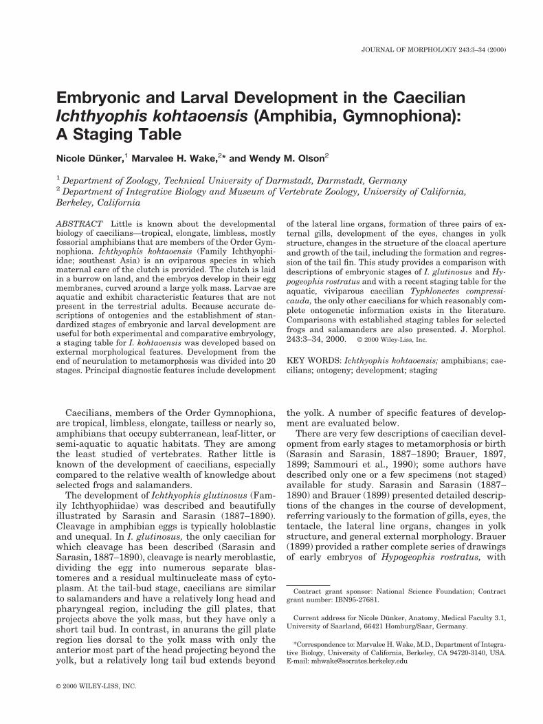
Ichthyophis kohtaoensis typifies the biology ofoviparous caecilians, so far as is known (see Himst-edt, 1996). Copulation in I. kohtaoensis has neverbeen observed, but internal fertilization is pre-sumed, as for all caecilians, via the male’s insertinghis phallodaeum into the vent of the female to effectsperm transport (see Wake, 1972, for summary). Inthe course of the rainy season from May to October,during which 90% of the annual rainfall in northeastThailand occurs and floods the rice fields, the fe-males lay their fertilized, developing eggs in moistcavities in the ground. I. kohtaoensis practices ma-ternal care of the laid clutch, as has been reportedfor several egg-laying species (e.g., Sarasin andSarasin, 1887–1890; Sanderson, 1936). The eggs of aclutch are bound together with strands that extendfrom the jelly coats, forming a cluster; the parentalfemale curls around the clutch and turns the eggsat regular intervals. Similar to embryos of
Fig. 1. Ichthyophis kohtaoensis. An adult specimen with atotal body length of approximately 300 mm is shown in A. Larvae,approximately 100–150 mm in length, are shown in B. Photos byW. Himstedt.
4 N. DUNKER ET AL.
F1
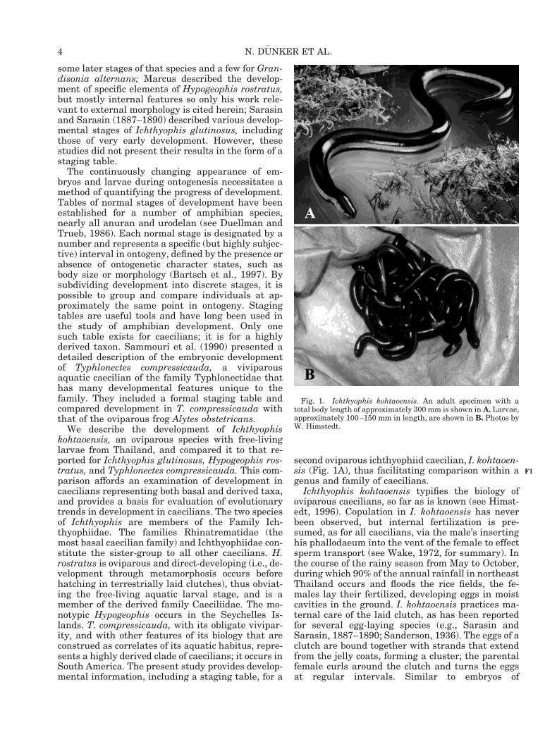
salamanders, embryos of I. kohtaoensis and appar-ently of most caecilians, except for typhlonectids,develop three pairs of external gills (Figs. 2–6).Later in the wet season, the Ichthyophis embryoshatch and, as larvae, move out of the burrows toponds and small streams. Also similar to thesalamander condition, but unlike anurans, larvae ofI. kohtaoensis (Fig. 1B) closely resemble their re-spective adults morphologically, physiologically, andtrophically. In many caecilians that have aquaticlarvae, such as I. kohtaoensis, the gills are lost soonafter hatching, leaving only a gill chamber that in-cludes gill rudiments on either side. Hatchling Ich-thyophis larvae also possess a well-developed caudaltail fin, and electroreceptive (ampullary organs) andmechanoreceptive (neuromasts) lateral-line sensoryorgans on the head and trunk. The lateral yellowstripes (see Fig. 1A) and the unique sensory tentaclecharacteristic of adults do not develop until meta-morphosis.
To our knowledge, there is not yet available atable of normal stages of development for oviparouscaecilians. Consequently we evaluate our data onthe development of Ichthyophis kohtaoensis andpresent a staging table based on external morpho-logical characters (Table 1). Because the presentstudy is intended to provide a staging table for avery poorly known species, a detailed description ofthe embryonic and larval development is given andas many characters are described as possible. Thosefeatures facilitate a comparison to other caecilians,and to frogs and salamanders, allowing a muchbroader comparison of development among amphib-ians.
MATERIALS AND METHODSCollection and Maintenance of theSpecimens
Because oviparous caecilians that provide paren-tal care have proven difficult to breed in captivity to
Fig. 3. Photomicrographs of embryonic stages of Ichthyophiskohtaoensis. Lettering sequence of embryos continues from Fig-ure 2: G, stage 32; H, stage 33; I, stage 34; J and K, stage 36; L,stage 37. Note further reduction of the yolk, with complete enclo-sure in the abdominal folds occurring at stage 37 (L). Only twoexternal gills are present in stage 36 (J, K); gills are stripped offafter hatching (stage 37; L). Scale bar 5 1 mm.
Fig. 2. Photomicrographs of embryonic stages of Ichthyophiskohtaoensis: A, stage 21; B, stage 23; C, stage 26; D, stage 28; E,stage 30; F, stage 31. Note the progressive development of thegills and gill filaments, and the reduction of the yolk mass. Scalebar 5 1 mm.
5DEVELOPMENT OF ICHTHYOPHIS KOHTAOENSIS
F2-F6
T1
date, Prof. Werner Himstedt (Department of Zool-ogy, Technical University of Darmstadt, Germany)collected the specimens of Ichthyophis kohtaoensisin Thailand, where the species is widespread andoccupies diverse geographical and climatic regions ofthe country. I. kohtaoensis occurs in the warm plainsnear sea level in the vicinity of Bangkok and insouth Thailand, in regions with moderate tempera-tures between 25–30°C, and at elevations of 2,000 min the mountains of north Thailand, where the tem-peratures in January drop to the freezing point. Inthe summers of 1994 and 1995, during July or Au-gust, females with egg clutches were collected in theprovince of Ubon (district Khemarat) in northeastThailand. Embryos and larvae were raised from thefertilized eggs in the laboratory. Clutches averageapproximately 30–40 eggs. Twelve out of 30 eggclutches collected were selected for our study. Stages
of development at the time of collection ranged fromunpigmented embryos to stages close to hatching. Inthe field, the females and their egg clutches wereusually found nestled in small cavities in the groundor under moist moss; in the laboratory, females werenot separated from their eggs, but were maintainedwith their clutches in tanks with moist moss.Clutches were raised at room temperature (approx-imately 20°C) and ambient light cycle.
Preparation of Developmental Series,Observations, and Data Collection
We took eggs from clutches at various stages ofdevelopment regularly during each week beforehatching. Descriptions are based on preserved ma-terial. Embryos were dissected free from the sur-rounding egg membranes using forceps, fixed inBouin’s fixative (picric acid, formaldehyde, and gla-cial acetic acid; see Presnell and Schreibman, 1997),then measured and described. Larvae were kept inaquaria at about 27°C and fed pieces of meat, then
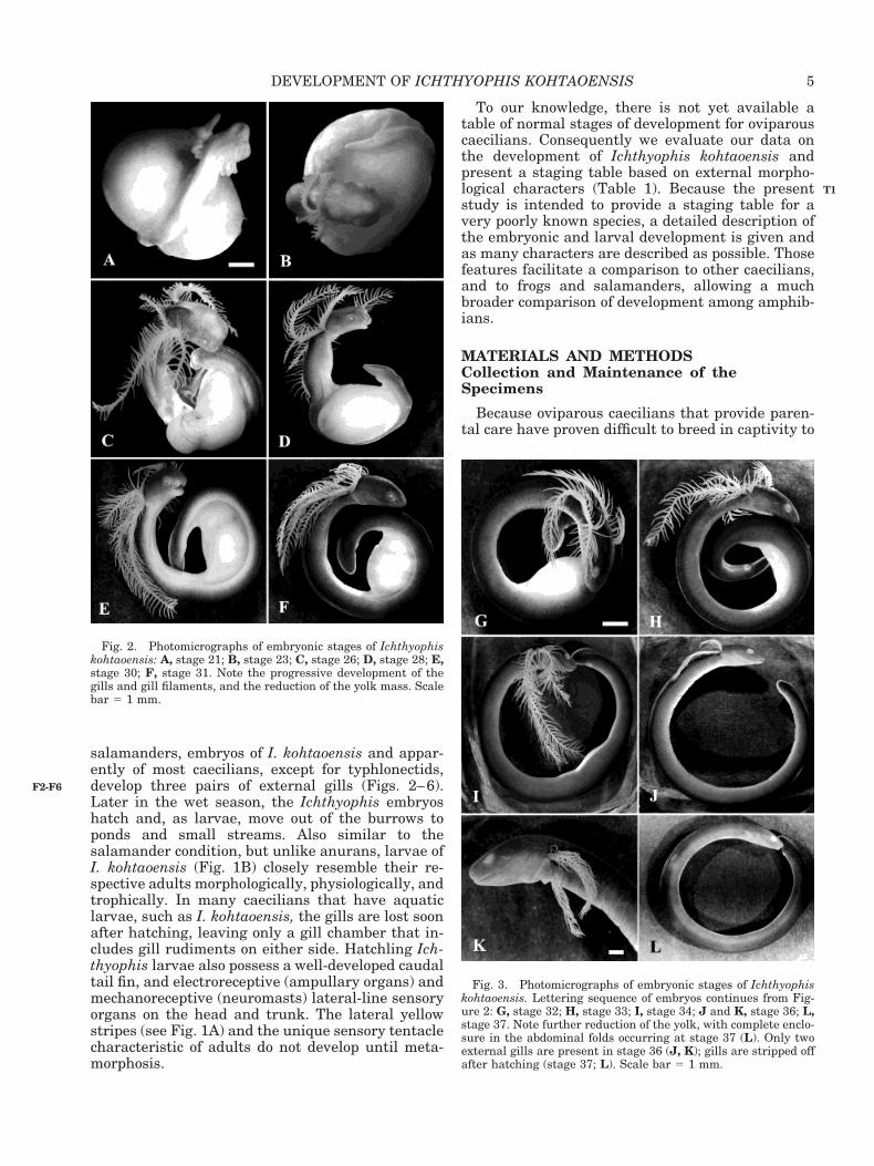
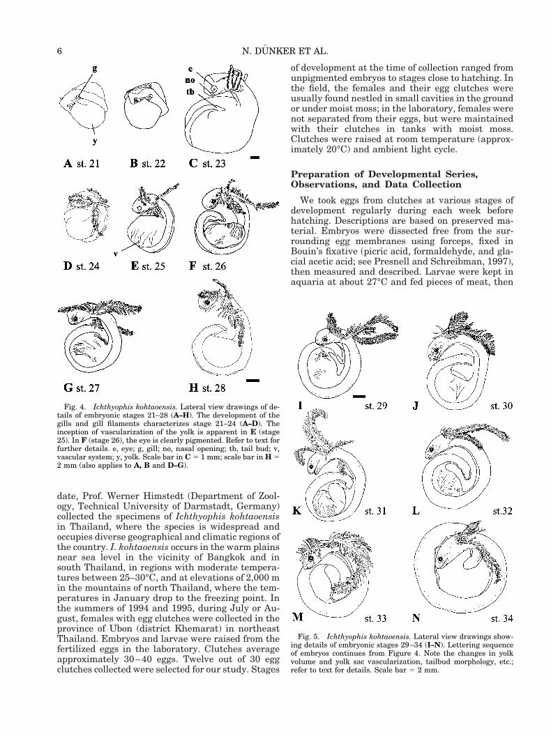
Fig. 5. Ichthyophis kohtaoensis. Lateral view drawings show-ing details of embryonic stages 29–34 (I–N). Lettering sequenceof embryos continues from Figure 4. Note the changes in yolkvolume and yolk sac vascularization, tailbud morphology, etc.;refer to text for details. Scale bar 5 2 mm.
Fig. 4. Ichthyophis kohtaoensis. Lateral view drawings of de-tails of embryonic stages 21–28 (A–H). The development of thegills and gill filaments characterizes stage 21–24 (A–D). Theinception of vascularization of the yolk is apparent in E (stage25). In F (stage 26), the eye is clearly pigmented. Refer to text forfurther details. e, eye; g, gill; no, nasal opening; tb, tail bud; v,vascular system; y, yolk. Scale bar in C 5 1 mm; scale bar in H 52 mm (also applies to A, B and D–G).
6 N. DUNKER ET AL.
selected specimens were fixed from the day of hatch-ing until metamorphosis on a monthly basis andwere similarly measured and described. Therefore, anearly complete chronological survey of develop-ment from early embryonic stages through larvalmetamorphosis was available. Additionally, juve-niles were preserved 1 month after metamorphosis.They were not described in the staging table, buttheir morphology is considered in the Results sec-tion. Early developmental stages corresponding toblastulation, gastrulation, and the beginning of neu-rulation are not represented in our series. In somecases, only one specimen per stage was available dueto the rarity of the material.
Photographs were taken using a Wild stereomi-croscope with a Photoautomat. Drawings of embry-onic and larval stages were made using a Wild ste-reomicroscope and a camera lucida. Measurementsof total body length of embryos were taken fromcamera lucida drawings. Larvae were measured di-rectly. Total length of embryos is the longest dimen-sion of the specimens in dorsal view, and was mea-sured to the most posterior edge of the curled tailstem. The lengths in millimeters given in Table 2are averages in round numbers, as there is sufficientvariation to invalidate the use of measurements totenths of millimeters. Additionally, the maximumlength and width of the yolk mass and the maximumgill length were measured, and the number of gillfilaments was counted.
For scanning electron microscopy (SEM), the spec-imens were dehydrated in a graded series of ethanoland dried in a Samdri-PVT-3B (Tousimis ResearchCorp., Rockville, MD) critical point dryer. Specimenswere mounted on stubs using double-sided tape andsilver paste, and then sputter coated with gold pal-ladium in a Polaron E5400 (Energy Beam Science,
Agawam, MA) unit. Specimens were viewed with aISI-DS130 scanning electron microscope and photo-graphed with a Polaroid camera. The SEM wasequipped with the SEMICAPS imaging system soimages could be stored digitally and sent to the labcomputer for further processing.
Because no egg clutches were observed being ovi-posited, the exact developmental time and age ofspecimens is not known. Total developmental timewas estimated by combining data from severalclutches with overlapping developmental stages.Counting backwards from the day of hatching, theestimated time of development ranges between 85and 90 days following oviposition. Metamorphosisoccurs approximately 9 to 12 months after hatching.
Many authors use the staging table of Gosner(1960) to identify developmental stages of frogs, andthat of Harrison (1969) for salamanders. Because wecould not specify stages of Ichthyophis glutinosusbased on Sarasin and Sarasin’s (1887–1890) descrip-tion, or that by Brauer (1899) of Hypogeophis rostra-tus, we compared our material of I. kohtaoensis tothat presented in the staging table for the vivipa-rous aquatic caecilian Typhlonectes compressicauda(Sammouri et al., 1990). Principal diagnostic fea-tures were those from Sammouri et al. and Nieuwk-oop and Faber (1967), and include development ofthe lateral line organs, formation of external gills,including the size of the gills and the number of gillfilaments, development of the eyes, changes in yolkstructure, and growth of the tail, including the for-mation and regression of the tail fin (Table 1). Forthe description of the development of the lateral lineorgans, we refer to the terminology of Hetheringtonand Wake (1979). Because few data are available forcaecilians, Table 2 states total length, weight of thespecimens, length and width of the yolk, and size ofthe gill and the number of gill filaments, though insome cases only one sample per stage was available.
RESULTS
We recognize 20 discrete developmental stages inour Ichthyophis kohtaoensis specimens, from embryosat the end of neurulation through mid-metamorphosis,based on readily discernible changes in major aspectsof external morphology (e.g., lateral line organs,mouth opening, eyes, nasal openings, gills, tail, tail fin,cloacal region, yolk). The major features of each stageare summarized in Table 1 and described in detailherein. The table begins with stage 21 for two reasons:absence of material representing early stages of devel-opment through neurulation, and comparability of ourearliest embryo with stage 21 of Typhlonectes compres-sicauda (Sammouri et al., 1990) (see Discussion).
Body Pigmentation
Earliest embryos of Ichthyophis kohtaoensis arealmost white; at stage 21 they have only a few dif-
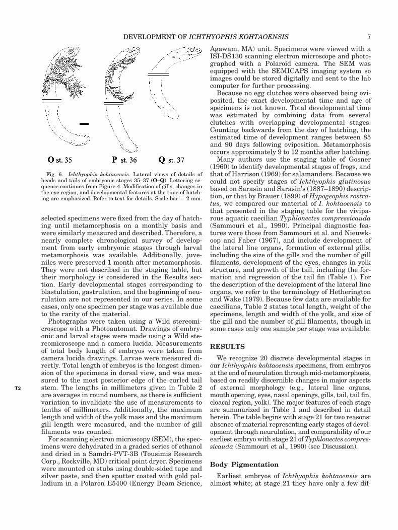
Fig. 6. Ichthyophis kohtaoensis. Lateral views of details ofheads and tails of embryonic stages 35–37 (O–Q). Lettering se-quence continues from Figure 4. Modification of gills, changes inthe eye region, and developmental features at the time of hatch-ing are emphasized. Refer to text for details. Scale bar 5 2 mm.
7DEVELOPMENT OF ICHTHYOPHIS KOHTAOENSIS
T2
TABLE 1. Normal table of development of Ichthyophis kohtaoensis
Stage 21 (n 5 1)● embryo curls around a large yolk mass; head and tail tip nearly touch● pigmentation covers anterior third of the body; melanophores scattered diffusely● neural folds in contact in tail and forebrain region, approaching but not touching in hindbrain region● mandibular arch divided into paired club-shaped upper maxillary buds and paired mandibular elements; hyoid arch and
three branchial arches developed; paired mandibular elements just touch, forming a deep heart-shaped angle at the site ofcontact
● optic vesicles stand out distinctly; no eye pigmentation; lens discernible as central, dense, round disc● otic vesicles, small white dots with a dense central disc, are discernible on either side of the neural folds in the region of the
rhombencephalic groove● olfactory pits evidenced by folds● three short external gills; no gill filaments● tail bud short, without tail fin, elevated from yolk● cloacal opening is triangular-shaped, bordered by two lateral cloacal swellings● yolk mass homogeneous, without constrictions; vascular system not discernible
Stage 22 (n 5 2)● paired mandibular elements continuous ventrally; club-shaped maxillary buds expanded ventro-laterally to border the
stomodeum● nasal pits large, lateral, round to oval● gill filaments present on base (or base and top) of first gill and base of second gill
Stage 23 (n 5 2)● pigmentation covers anterior two-thirds of body● club-shaped protrusions of the maxillary buds flank the lateral sides of the stomodeum; close contact between maxillary buds
and lateral nasal walls forms a nasobranchial rim● protrusion of optic vesicles and lenses; grooves encircle eyes and separate them from surrounding tissue● first and second gill more elongate and bearing more filaments● cloacal opening slit-shaped, bordered by two lateral cloacal swellings
Stage 24 (n 5 2)● pigmentation covers anterior 75% of the body● first appearance of lateral line organs (neuromasts)● mandibular elements fully continuous; maxillary buds connected; formation of laterally closed mouth opening, clearly
distinguishable in lateral view● third gill bears gill filaments● tail bud pointed; fin formation begins● yolk mass with smooth constrictions
Stage 25 (n 5 1)● pigmentation more dense, extends over all the body● nasal, supra- and infraorbital neuromast rows developed; anlage of oral and postorbital neuromast rows; first appearance of
neuromasts on trunk, more developed anteriorly; 24 elevated neuromasts distributed regularly on anterior half of the body,then 2–4 flat neuromasts with broader distance between them, then a gap lacking neuromasts, then 3 elevated neuromastson tail tip; trunk neuromasts situated more laterally than dorsally
● otic vesicles no longer distinguishable● gill chamber begins to form, as epithelial outpocketing at the base of the second and third gills● vascular system now discernible on yolk surface
Stage 26 (n 5 2)● anterior part of the body clearly elevated from yolk● initiation of two additional lateral line rows above the eyes; development of oral and postorbital neuromast rows; neuromasts
on trunk still more elevated anteriorly; 40–42 elevated neuromasts regularly arranged along 75% of trunk; gap betweenneuromasts on anterior part of the trunk and tail tip is reduced
● eye pigmentation clearly discernible● triangular-shaped rostro-ventral nasal openings surrounded by nasal swellings● tail fin higher, further developed● vascular system of the yolk further developed
Stage 27 (n 5 1)● lateral line organs in head region distinguishable as rows of white dots; anlage of V-shaped mandibular neuromast row on
chin; first appearance of ampullary organs; three rows of lateral line organs above the eyes; neuromasts on trunk situatedslightly more dorsally than laterally
● lower jaw elongates and extends posteriorly; mouth opening appears narrower● nasal openings triangular to tear-shaped, moved to a more frontal position● tail bud thickened, round; tail fin broadened, more delineated
Stage 28 (n 5 2)● appearance of gular neuromast rows on throat and supraspiracular neuromast rows above the gills; trunk neuromasts evenly
distributed along anteroposterior axis; trunk neuromasts situated more dorsally than laterally on anterior part of trunk, morelaterally in posterior region and especially on tail tip
● roundish lower jaw not fully developed, forms lip-like structure● anlage of tentacle apparent near eye● yolk is slightly apple-shaped
Stage 29 (n 5 1)● head elongated● inception of ampullary organ row below the eyes; neuromasts become elevated in head region
8 N. DUNKER ET AL.
● mouth development further advanced; narrow lower jaw extends posteriorly; mouth deeply notched laterally● yolk mass elongated with heart-shaped curvatures (shape similar to Sarasins’ fig. 1b)
Stage 30 (n 5 6)● straightening of head curvature● neural folds connected in head region; suture still present● two rows of lateral line organs (one row of neuromasts and one row of ampullary organs) below the eyes and three rows (one
neuromast and two ampullary) projecting behind the eyes● tentacle anlage apparent as an opacity or small indentation anterior to the eye● cloacal region further developed; cloacal slit elongated, encircled by rim of swollen tissue, bordered laterally by two oval
elongated buds● first bending of yolk mass, resulting in a heart- to U-shaped curvature (similar to Sarasins’ fig. 2); late stage 30, yolk mass
with S-shaped curvature to complete S-shaped twist (similar to Sarasins’ fig. 3)Stage 31 (n 5 1)
● head clearly flattened and elongated● initiation of skin glands on chin● inception of second row of ampullary organs below the eyes● occlusion of the jaws; mouth resembles the deeply notched, flat subterminal structure of larvae● clearly reduced double S-shaped yolk tube with curvatures transverse to the longitudinal axis of the embryo (similar to
Sarasins’ fig. 4)Stage 32 (n 5 3)
● skin glands on chin are distinct● three complete rows of lateral line organs (one neuromast and two ampullary rows) below the eyes; dark gray pigmentation
allows distinction between neuromasts (large solid white dots) and ampullary organs (small fine white dots)● tail tip becomes arrow-shaped and slightly curved ventrally; tail fin broadened; ventral incision between cloacal region and
end of tail fin● cloacal aperture further differentiated; slit-shaped cloacal opening encircled by elongated elevated rim and bordered by two
pronounced button-shaped swellings, which are now also visible in lateral view● beginning of yolk enclosure in abdominal folds and formation of spindle-shaped yolk mass; yolk mass spiralized; spirals run
parallel to longitudinal axis of embryo; two symmetrical yolk halves separated by gutStage 33 (n 5 4)
● skin covering the eyes thickened; tear-shaped skin configuration overlies eyes and tentacle anlagen● elongated and retracted cloacal opening bordered by crescent- to bean-shaped cloacal swellings● large parts of reduced yolk enclosed in abdominal folds; externally, yolk discernible as large elevation; yolk mass spindle- to
corkscrew-shapedStage 34 (n 5 3)
● neural folds in head region connected without suture● trunk neuromasts are larger, more accentuated, beginning to sink into the skin● tear-shaped skin pad thicker and cloudier; eyes barely discernible● reduced yolk mass nearly completely enclosed in abdominal fold; external yolk visible as one or several elevation(s); yolk
mass resembles zigzag-shaped tube (similar to Sarasins’ fig. 5a)Stage 35 (n 5 3)
● neuromasts have central circular indentations; ampullary organs begin to sink into the skin● tear-shaped frontal nasal openings enlarged, becoming deeper● yolk mass nearly to completely enclosed in abdominal folds; no external yolk visible or only a slim yellow stripe; beginning of
compartmentation of yolk tube; yolk mass appears to be segmented into several portions, which are aligned as a stack ofcoins
Stage 36 (n 5 3)● neuromasts and ampullary organs barely discernible, sunken into skin; some neuromasts represented by small holes,
especially in nasal row and anterior part of infraorbital row● only two external gills remain; third gill degenerated and internalized in the gill chamber; gill chamber nearly fully covered
by epithelial fold● elongated, deeply retracted cloacal slit bordered by flattened, elongated wall of thickened tissue lacking pigment; wall is
flattened at posterior end and more elevated anteriorlyStage 37 (n 5 3)
● hatching begins● V-shaped mandibular neuromast row represented by small holes● two remaining gills are stripped off immediately after hatching; no external gills; opening to gill chamber elongates● tail tip more rounded than arrow-shaped; tail fin less broad, no longer delineated
Stage 38 (n 5 4)● inception of lateral yellow stripes● neuromasts, resembling thick white dots with small central grooves, are considerably larger and are circumscribed by a dark
circle or groove; no ampullary organs on chin● further enlargement of the tear-shaped frontal nasal openings● narrow tail fin curved dorsally
Stage 39 (n 5 3)● broadened, intensively yellow lateral stripes, clearly discernible also on head● ventral side becomes pigmented, including chin; skin glands barely discernible● supra- and infraorbital rows have fewer neuromasts, and the supraorbital row is no longer curved● tail fin no longer discernible, only a narrow dorsal fimbris in region of former fin; tail tip no longer compressed laterally but
begins to resemble rounded tail of adults
9DEVELOPMENT OF ICHTHYOPHIS KOHTAOENSIS
TABLE 1. (Continued)
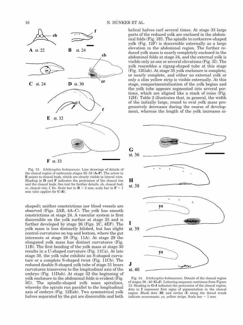
fusely scattered melanophores on the dorsum (Fig.2A). Pigmentation covers the anterior third of thebody at this stage. Pigmentation extends posteri-orly, covering the anterior two-thirds of the body atstage 23. At stage 24 pigmented cells cover the an-terior 75% of the body and at stage 25, the pigmen-tation is denser and extends over the entire body.The dark gray pigmentation of older embryonicstages (see stage 32; Fig. 3G) allows the distinctionbetween elements of the lateral line system becausedenser neuromast rows are indicated by largerwhite dots, and fainter peripheral ampullary rowsare represented by smaller white dots. At stage 39the ventral side of the body becomes pigmented,including the cloacal region. The characteristicpaired lateral yellow stripes of adult I. kohtaoensis(Fig. 1A) begin to develop on either side of the trunkat stage 38. The stripes broaden, become intensivelyyellow, and are clearly discernible on the head aswell as the body at stage 39 (Figs. 10HI, 14IJ).
Neural Folds
At stage 21, the neural folds are in contact in thetail and forebrain regions, and approach but do notyet touch in the hindbrain region (Figs. 2A, 7A). Theneural folds are in contact in the head region atstage 30, but a suture and a small triangular-shapedregion at the anterior end of the suture, both lackingpigment, are still present in the hindbrain region. Atstage 34 the neural folds in the head region arefinally fully fused, without an apparent suture zone.
Lateral Line Organs
Lateral line organs are not discernible at stages21 to 23 (Figs. 4A–C, 7A–C, 9AB). At stage 24 thefirst lateral line organs appear, represented by linesof pigmentless dots near the eyes (Figs. 4D, 7D). Inthe tail bud region, three pronounced elevated dotsare observed, but the structures are absent on eitherside of the trunk. Based on their position and size,the organs are neuromasts, and are larger and morelinearly arranged than ampullary organs, whichfirst appear at stage 27 (see below). The neuromast
rows of stage 25 (Fig. 9C), running from the nasalopenings to and encircling the eyes, are well devel-oped and represent the nasal, supra- and infraor-bital neuromast rows described by Hetheringtonand Wake (1979; their terminology will be followedin this description). Two additional rows of neuro-masts occur, one projecting from the mouth opening(oral row) and one projecting from the eye (postor-bital row), forming a “V.” The first neuromasts alsoappear on the trunk (Fig. 4E). They are more fullydeveloped anteriorly and are situated more laterallythan dorsally. At this stage, 24 elevated neuromastsare regularly distributed on the anterior half of thebody, followed by two to four flat neuromasts thatare more widely separated. Then a gap, lacking neu-romasts, is observed, followed by the three elevatedneuromasts on the tail tip.
At stage 26, two additional lateral line rows abovethe eyes appear, and the oral and postorbital neuro-mast rows are further developed (Fig. 7E). The neu-romasts on the trunk are still more elevated anteri-orly. Forty to 42 elevated neuromasts are regularlyarranged along 75% of the trunk, and the gap be-tween the neuromasts on the anterior part of thetrunk and tail tip is reduced, with increased num-bers of neuromasts proceeding posteriorly. The neu-romast organs of the head region are distinguishableas rows of larger light dots; the first ampullary or-gans, resembling small fine dots, appear at stage 27(Fig. 9D). Subsequently, a V-shaped row of neuro-masts is established on the chin (mandibular neuro-mast row; Fig. 7F–H) and three rows of lateral lineorgans (one row of neuromasts and two rows of am-pullary organs) are present above the eyes. How-ever, the organs occur only above the right eye of ourspecimen; asymmetric development is not unusual(see Hetherington and Wake, 1979). Rows of neuro-masts on the throat (gular neuromasts) and groupsof neuromasts above the gills (supraspiracular rows)appear at stage 28 (Figs. 7F, 9E). The neuromasts onthe trunk are evenly distributed and situated moredorsally than laterally on the anterior part of thetrunk. However, they are found more laterally onthe trunk in posterior regions and especially on thetail tip. The gaps between the anterior neuromasts
● cloacal region pigmented; slit-shaped cloacal opening bordered by notched folds arranged transversely to cloaca, resembling ascar
Stage 40 (n 5 1)● metamorphic stage● clearly reduced lateral line organs; no ampullary organs discernible on dorsal head, so only one row of lateral line organs
visible above the eyes; neuromasts no longer accentuated, indicating beginning of degeneration of lateral line system;neuromasts resorbed posteriorly in the dorsal supra- and infraorbital rows; some neuromasts missing in the oral, postorbital,and supraspiracular rows; entire nasal neuromast row is resorbed
● funnel-shaped tentacle sheath clearly discernible; appearance of tentacle orifice with distinct tentacle fold● gill chamber opening begins to close● round tail resembles adult tail
Characteristic features of embryonic and larval stages from the end of neurulation (stage 21) through the onset of metamorphosis(stage 40) are summarized.
10 N. DUNKER ET AL.
F7
TABLE 1. (Continued)
and the posterior group, and between that group andthose on the tail tip, are closed by a continuous lineof neuromasts at regular intervals. The neuromastrows in the head region become elevated and resem-ble rows of dome-shaped protrusions rather thanlines of unpigmented dots at stage 29 (Fig. 7G).Additionally, a row of ampullary organs forms belowthe eyes. At stage 30 several new rows develop: onerow of neuromasts and one row of ampullary organsnow lie below the eyes, and a row of neuromasts andtwo rows of ampullary organs extend behind theeyes (Figs. 7H, 9F). A second row of ampullary or-gans occurs below the eyes at stage 31 (Fig. 8I). Atstage 32 the rows of lateral line organs include ad-ditional structures below the eyes.
As the body pigmentation progressively becomesdarker, the lateral line organs are more prominentand stand out strikingly as large solid white dots(neuromasts) or small fine white dots (ampullaryorgans). By stage 32, the dark gray pigmentationpermits distinction between neuromast and ampul-lary rows. At stage 34 the neuromasts of the trunkare sunken into the skin and are not as elevated asin previous stages, though they are slightly largerand more accentuated at this stage of development(Fig. 5N). The ampullary organs begin to sink intothe skin at stage 35. At this stage the neuromastshave central, circle-like indentations. At stage 36both kinds of lateral line organs are well sunken intothe skin and are barely discernible. Some neuro-masts are represented by small holes, especially inthe nasal region and the anterior part of the infraor-bital row. The V-shaped mandibular neuromast rowresembles a row of small holes at stage 37. At stage38 the neuromasts are considerably larger, but wellsunken, and resemble thick white dots with small
central grooves; the dots are circumscribed by darkcircles. Ampullary organs on the chin are no longerdiscernible at this stage (Fig. 8L). The supra- andinfraorbital lateral line rows exhibit fewer neuro-masts at stage 39; the supraorbital rows are nolonger curved and thus are no longer in contact withthe nasal row (Fig. 8M). At metamorphic stage 40the lateral line organs are clearly reduced. No am-pullary organs are discernible on the dorsum of thehead and only one row of lateral line organs is ob-servable above the eyes (Fig. 8N). Additionally, theneuromasts are no longer accentuated, indicatingdegeneration of the lateral line system. Neuromastsare resorbed posteriorly in the supra- and infraor-bital rows; in the oral, postorbital, and supraspi-racular rows some neuromasts are missing, and theentire nasal row is absent. Finally, 1 month aftermetamorphosis apparently commences, the postor-bital, supraspiracular, gular, and the body neuro-mast rows are fully resorbed, the mandibular, su-praorbital, and infraorbital rows are reduced, andthe oral row consists only of scattered neuromasts,no longer forming a row projecting from the mouthopening.
We examined the development of the lateral linesystem at the ultrastructural level at stages 27, 30,and 35. In scanning electron micrographs, ampul-lary organs resemble small, deep pits (see Fig. 15A).At stages 27 and 30, the early neuromasts are rep-resented by dome-shaped protrusions (Fig. 15B–D)that are covered by epidermis because the organshave not yet broken through the skin. At stage 35,however, the organs resemble shallow grooves, asthey have sunken into the skin (Fig. 15E,F). Thekinocilia and stereocilia in the neuromasts and thecentral position of several bundles of microvilli in
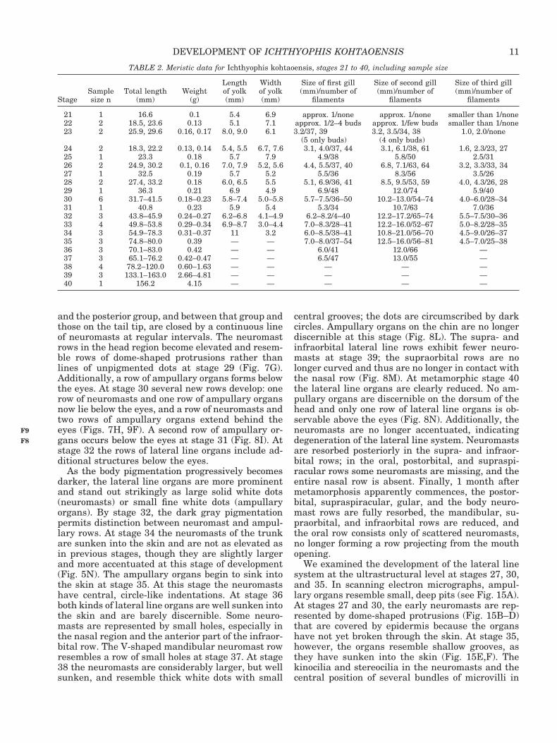
TABLE 2. Meristic data for Ichthyophis kohtaoensis, stages 21 to 40, including sample size
StageSamplesize n
Total length(mm)
Weight(g)
Lengthof yolk(mm)
Widthof yolk(mm)
Size of first gill(mm)/number of
filaments
Size of second gill(mm)/number of
filaments
Size of third gill(mm)/number of
filaments
21 1 16.6 0.1 5.4 6.9 approx. 1/none approx. 1/none smaller than 1/none22 2 18.5, 23.6 0.13 5.1 7.1 approx. 1/2–4 buds approx. 1/few buds smaller than 1/none23 2 25.9, 29.6 0.16, 0.17 8.0, 9.0 6.1 3.2/37, 39
(5 only buds)3.2, 3.5/34, 38
(4 only buds)1.0, 2.0/none
24 2 18.3, 22.2 0.13, 0.14 5.4, 5.5 6.7, 7.6 3.1, 4.0/37, 44 3.1, 6.1/38, 61 1.6, 2.3/23, 2725 1 23.3 0.18 5.7 7.9 4.9/38 5.8/50 2.5/3126 2 24.9, 30.2 0.1, 0.16 7.0, 7.9 5.2, 5.6 4.4, 5.5/37, 40 6.8, 7.1/63, 64 3.2, 3.3/33, 3427 1 32.5 0.19 5.7 5.2 5.5/36 8.3/56 3.5/2628 2 27.4, 33.2 0.18 6.0, 6.5 5.5 5.1, 6.9/36, 41 8.5, 9.5/53, 59 4.0, 4.3/26, 2829 1 36.3 0.21 6.9 4.9 6.9/48 12.0/74 5.9/4030 6 31.7–41.5 0.18–0.23 5.8–7.4 5.0–5.8 5.7–7.5/36–50 10.2–13.0/54–74 4.0–6.0/28–3431 1 40.8 0.23 5.9 5.4 5.3/34 10.7/63 7.0/3632 3 43.8–45.9 0.24–0.27 6.2–6.8 4.1–4.9 6.2–8.2/4–40 12.2–17.2/65–74 5.5–7.5/30–3633 4 49.8–53.8 0.29–0.34 6.9–8.7 3.0–4.4 7.0–8.3/28–41 12.2–16.0/52–67 5.0–8.2/28–3534 3 54.9–78.3 0.31–0.37 11 3.2 6.0–8.5/38–41 10.8–21.0/56–70 4.5–9.0/26–3735 3 74.8–80.0 0.39 — — 7.0–8.0/37–54 12.5–16.0/56–81 4.5–7.0/25–3836 3 70.1–83.0 0.42 — — 6.0/41 12.0/66 —37 3 65.1–76.2 0.42–0.47 — — 6.5/47 13.0/55 —38 4 78.2–120.0 0.60–1.63 — — — — —39 3 133.1–163.0 2.66–4.81 — — — — —40 1 156.2 4.15 — — — — —
11DEVELOPMENT OF ICHTHYOPHIS KOHTAOENSIS
F9
F8
the ampullary receptors can be seen (Fig. 15A,F)because the amorphous substance secreted by spe-cialized cells of the lateral line organs has dried andretracted during dehydration. At all stages exam-ined, the epidermis includes the ciliated cells (Fig.15A) typical of amphibian skin development.
Stomodeum
At stage 21, the mandibular arch is already di-vided into paired upper maxillary buds and paired
mandibular elements. Additionally, the hyoid archand three branchial arches are well developed (Fig.7A). However, the components of the lower jaw arenot connected; the mandibular elements only touch,forming a deep, heart-shaped angle at the site ofcontact (Fig. 7A). The paired mandibular elementsare continuous ventrally at stage 22, and the club-shaped maxillary buds have expanded ventro-laterally to border the stomodeum (Figs. 7B, 9A). Atstage 23 the maxillary buds flank the lateral sides ofthe stomodeum (Fig. 7C). The maxillary buds andthe lateral nasal walls are in close contact, forminga nasobranchial rim (Fig. 9B). The maxillary budsare connected at stage 24, and the mandibular ele-ments are fully continuous, so that the lateral junc-ture of the mouth opening is clearly distinguishablein the lateral view (Fig. 7D). As the lower jaw elon-
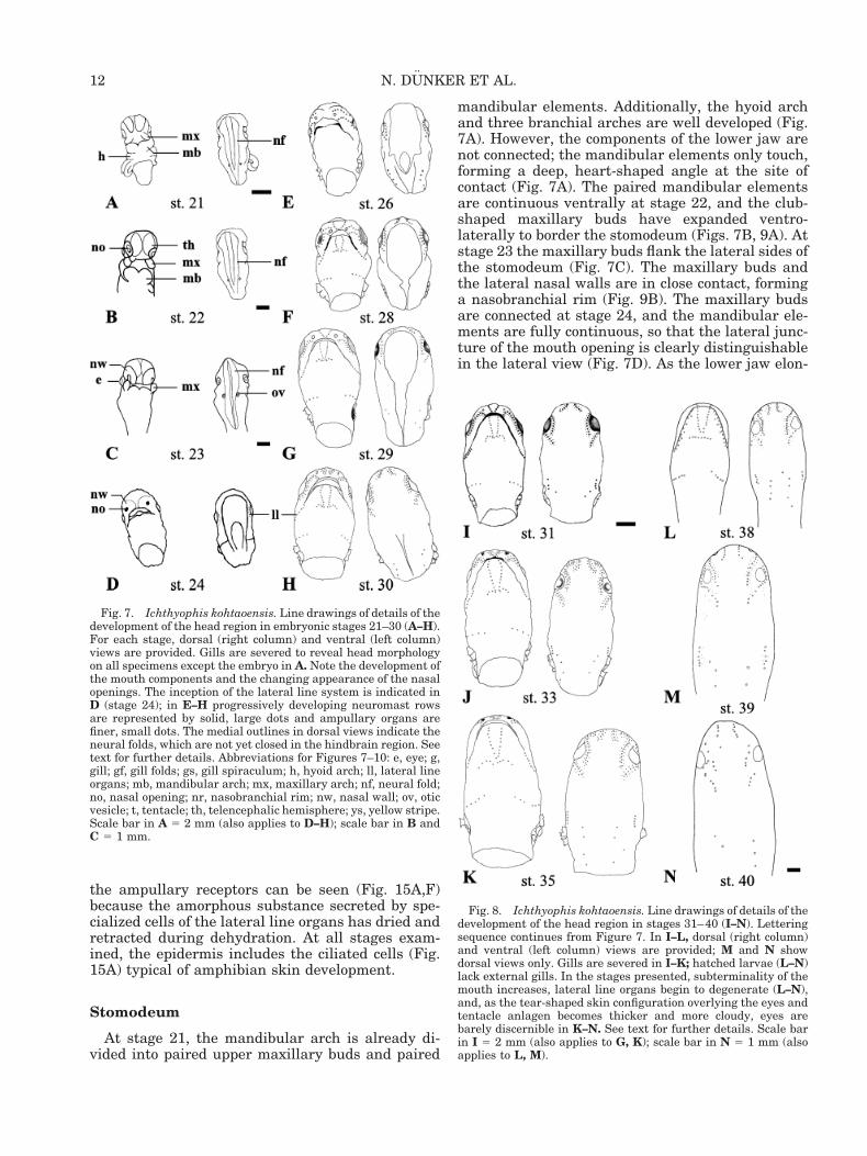
Fig. 7. Ichthyophis kohtaoensis. Line drawings of details of thedevelopment of the head region in embryonic stages 21–30 (A–H).For each stage, dorsal (right column) and ventral (left column)views are provided. Gills are severed to reveal head morphologyon all specimens except the embryo in A. Note the development ofthe mouth components and the changing appearance of the nasalopenings. The inception of the lateral line system is indicated inD (stage 24); in E–H progressively developing neuromast rowsare represented by solid, large dots and ampullary organs arefiner, small dots. The medial outlines in dorsal views indicate theneural folds, which are not yet closed in the hindbrain region. Seetext for further details. Abbreviations for Figures 7–10: e, eye; g,gill; gf, gill folds; gs, gill spiraculum; h, hyoid arch; ll, lateral lineorgans; mb, mandibular arch; mx, maxillary arch; nf, neural fold;no, nasal opening; nr, nasobranchial rim; nw, nasal wall; ov, oticvesicle; t, tentacle; th, telencephalic hemisphere; ys, yellow stripe.Scale bar in A 5 2 mm (also applies to D–H); scale bar in B andC 5 1 mm.
Fig. 8. Ichthyophis kohtaoensis. Line drawings of details of thedevelopment of the head region in stages 31–40 (I–N). Letteringsequence continues from Figure 7. In I–L, dorsal (right column)and ventral (left column) views are provided; M and N showdorsal views only. Gills are severed in I–K; hatched larvae (L–N)lack external gills. In the stages presented, subterminality of themouth increases, lateral line organs begin to degenerate (L–N),and, as the tear-shaped skin configuration overlying the eyes andtentacle anlagen becomes thicker and more cloudy, eyes arebarely discernible in K–N. See text for further details. Scale barin I 5 2 mm (also applies to G, K); scale bar in N 5 1 mm (alsoapplies to L, M).
12 N. DUNKER ET AL.
gates and extends caudally, the mouth opening ap-pears narrower at stage 27. At stage 28 the lowerjaw, which is roundish and not yet fully developed,forms a lip-like structure (Figs. 9E, 7F). Mouth de-velopment is further advanced at stage 29; the nar-row lower jaw extends further caudally, and themouth opening becomes deeply notched laterally(Fig. 7G). The upper and lower jaw occlude, closingthe mouth, at stage 31 (Fig. 8I). At this stage themouth presents the deeply notched flat subterminalstructure characteristic of larvae.
Eyes
The eyes of Ichthyophis kohtaoensis embryos areapparent in stage 21 (Fig. 2A). At that stage theoptic vesicles lack pigment but stand out distinctly,and the lens is discernible as a dense round central
disc. Protrusion of eyes and lenses is observable atstage 23, at which grooves encircle the optic vesiclesand separate them from the surrounding tissue(Figs. 2B, 9B). Eye pigmentation appears at stage 26(Figs. 2C, 4F). The eyes are covered by unpigmented,cloudy, translucent skin. At stage 33 the skin cover-ing the eyes is thickened, forming a tear-shapedwhite configuration that overlies the eyes and thetentacle anlagen (Fig. 3H). The tear-shaped skinpad becomes cloudier and the eyes are barely dis-cernible at stage 34 (Fig. 3I).
Tentacle
The tentacle anlage is first discernible externallyat stage 28. At stage 30, the tentacle anlage is anopacity in a small indentation or groove near the eyeat the frontal border of the cloudy translucent skinthat covers the eyes (Fig. 9F). At stage 33 the ten-tacle aperture is represented by a white dense tissuefold, which is continuous with the thickened tear-shaped configuration that covers the eyes (Fig. 9G).At metamorphic stage 40 a funnel-shaped tentaclesheath is clearly discernible, and the tentacle foldappears in the tentacle orifice (Fig. 10I).
Otic Vesicles
Otic vesicles are discernible from stage 21 to stage24 on either side of the neural folds in the region ofthe rhombencephalic groove as small white dotswith dense central discs (Figs. 7A–C, 9AB). At stage25 otic vesicles are no longer distinguishable exter-nally. They are sunken somewhat into the develop-ing cranium and covered with a denser epidermallayer.
Nasal Openings
The olfactory pits of early embryonic stages areevidenced by folds (see stage 21; Fig. 4A). At stage22 large, lateral, oval nasal pits are present. Atstage 23 the nasal walls open laterally to form anasobranchial rim, a connection between the nasalpits and the maxillary buds (Figs. 4C, 7C, 9B). Thetriangular rostro-ventral nasal openings are sur-rounded by enlarged nasal swellings at stage 26(Fig. 7E). At stage 27 the triangular to tear-shapednasal openings have moved to more frontal positionson the head (Fig. 9D). At stage 35, prior to hatching,the enlarged tear-shaped nasal openings becomedeeper, and further enlargement is observed atstage 38.
Gills and Gill Chamber
The embryos of Ichthyophis kohtaoensis bearthree pairs of external gills, which first appear asslightly curved knobs or short elongated swellingssituated laterally in the cephalic region on either
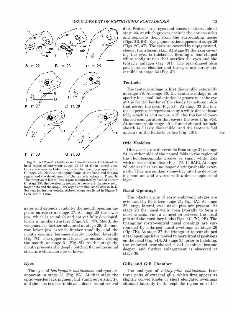
Fig. 9. Ichthyophis kohtaoensis. Line drawings of details of thehead region of embryonic stages 22–33 (A–G) in lateral view.Gills are severed in C–G; the gill chamber opening is apparent inC (stage 25). Note the changing shape of the head and the jawregion and the development of the tentacle anlage in F and G.The inception of lateral line organs is indicated by dashed lines inC (stage 25); the developing neuromast rows are the more solid,larger dots and the ampullary organs are fine, small dots in D–G.See text for further details. Abbreviations are listed in Figure 7.Scale bar 5 1 mm.
13DEVELOPMENT OF ICHTHYOPHIS KOHTAOENSIS
F10
side of the head by stage 21 (Figs. 2A, 4A). At thisstage the short gills lack filaments and gill slitsbetween gill bars are not perforated. The gill fila-ments form in sequence, proximal to distal, as out-pocketings of the gill tissue. They extend at rightangles to the length of the ramus and are spacednearly equidistantly, reaching approximately 3 mmin length. The first bud-shaped gill filaments appeareither on the base or the base and top of the first gilland the base of the second gill at stage 22 (Fig. 4B).At stage 23 the first and second gills are more elon-gated and bear more filaments (Figs. 2B, 4C, 9B).Filaments of the third gill appear at stage 24, whenthe gills are already elongated (Fig. 4D). From stage29 until hatching (stage 37), the second (middle) gillis considerably longer, sometimes double in size,compared to the first (anterior) and third (posterior)gills (see Table 2). Similarly, the filaments of thesecond gill are more numerous and appear longer
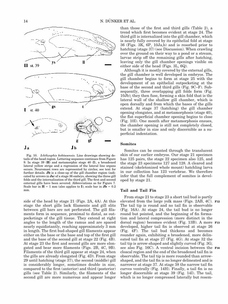
than those of the first and third gills (Table 2), atrend which first becomes evident at stage 24. Thethird gill is internalized into the gill chamber, whichis nearly fully covered by its epithelial fold at stage36 (Figs. 3K, 6P, 10Ja,b) and is resorbed prior tohatching (stage 37) (see Discussion). When crawlingover the ground on their way to a pond or a stream,larvae strip off the remaining gills after hatching,leaving only the gill chamber openings visible oneither side of the head (Figs. 3L, 6Q).
Although it is mostly covered by the external gills,the gill chamber is well developed in embryos. Thegill chamber begins to form at stage 25 with thedevelopment of an epithelial outpocketing at thebase of the second and third gills (Fig. 9C–F). Sub-sequently, three overlapping gill folds form (Fig.10Jb); they then fuse, forming a skin fold that is thelateral wall of the shallow gill chamber, which isopen dorsally and from which the bases of the gillsextend. At stage 37 (hatching) the gill chamberopening elongates, and at metamorphosis (stage 40),the flat superficial chamber opening begins to close(Fig. 10I). One month after metamorphosis ensues,the chamber opening is still not completely closed,but is smaller in size and only discernible as a su-perficial indentation.
Somites
Somites can be counted through the translucentskin of our earlier embryos. Our stage 21 specimenhas 125 pairs, the stage 22 specimen also 125, andthe stage 23 specimens 127 and 128. A cleared andstained (skeletonized whole mount) hatchling larvain our collection has 123 vertebrae. We thereforeinfer that the full complement of somites is devel-oped by stage 21.
Tail and Tail Fin
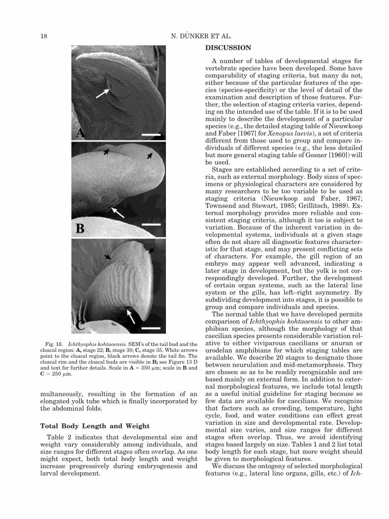
From stage 21 to stage 23 a short tail bud is partlyelevated from the large yolk mass (Figs. 2AB, 4C).The tail tip is round and no tail fin is observable(Fig. 16A). At stage 24, the tail bud is no longerround but pointed, and the beginning of fin forma-tion and lateral compression (more distinct in thedorsal region) becomes evident (Fig. 13B). A moredeveloped, higher tail fin is observed at stage 26(Fig. 4F). The tail bud thickens and becomesrounder again, exhibiting a broadened, more delin-eated tail fin at stage 27 (Fig. 4G). At stage 32 thetail tip is arrow-shaped and slightly curved (Fig. 3G;see also Fig. 16C). A ventral incision between thecloacal region and the end of the broadened tail fin isobservable. The tail tip is more rounded than arrow-shaped, and the tail fin is no longer delineated and isnarrower at stage 37. At stage 38 the narrow tail fincurves ventrally (Fig. 14H). Finally, a tail fin is nolonger discernible at stage 39 (Fig. 14I). The tail,which is no longer compressed laterally but resem-
Fig. 10. Ichthyophis kohtaoensis. Line drawings showing de-tails of the head region. Lettering sequence continues from Figure9. In stage 39 (H) and metamorphic stage 40 (I), a broadenedlateral yellow stripe and a regression of the lateral line organsoccurs. Neuromast rows are represented by circles; see text forfurther details. Jb is a close-up of the gill chamber region (indi-cated by arrows in Ja) of a stage 36 embryo, showing the three gillfolds and the internalization of the third gill. The first and secondexternal gills have been severed. Abbreviations as for Figure 7.Scale bar in H 5 1 mm (also applies to I); scale bar in Jb 5 0.2mm.
14 N. DUNKER ET AL.
F12
F13
F16
bles the round tail of adults, exhibits a narrow dor-sal fimbris in the region of the former fin. At meta-morphic stage 40 the tail has achieved thecircumference of the adult tail. Finally, 1 monthafter metamorphosis began, the tail tip is no longerpointed but obtuse, similar to the tail tip of adults.
Cloacal Region
Differentiation of the cloacal region is evident atstage 21. At stages 21 and 22, the triangular-shapedcloacal opening is bordered by two lateral round toelongated cloacal swellings (Figs. 13A, 16A). Thecloacal opening, still bordered by lateral cloacalswellings, becomes slit-shaped at stage 23 (see alsostage 24 in Fig. 13C). At stage 30 the elongatedcloacal slit is encircled by a rim of swollen tissue andbordered by two lateral elongated buds (Figs. 13D,16B). The cloacal aperture further differentiates atstage 32; the slit-shaped cloacal opening is sur-rounded by an elongated elevated rim and is bor-
dered by two pronounced button-shaped swellings.The latter are also clearly visible in the lateral viewof the tail tip (Fig. 13E). At stage 33 the elongatedand clearly retracted cloacal opening is bordered bycrescent- to bean-shaped cloacal swellings (Figs. 3H,13F; see also stage 35 in 16C). The elongated wall ofthickened tissue which borders the retracted cloacalslit at stage 36 lacks pigment and is elevated ante-riorly but flattened posteriorly (Fig. 14G). At stage39 the cloacal region is pigmented and the slit-shaped cloaca is bordered by notched folds, arrangedtransversely to the slit (Fig. 14I); the cloacal aper-ture has a scar-shaped configuration at this devel-opmental stage and through metamorphosis (Fig.14J). Finally, 1 month after metamorphosis, the re-tracted cloacal slit is bordered by faint wrinkles orfolds.
Yolk
From stage 21 to stage 23 the large yolk mass ishomogeneous, slightly oval to bilobed (heart-
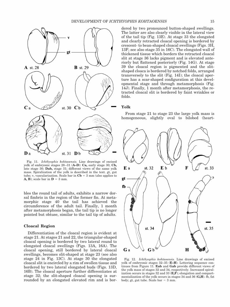
Fig. 12. Ichthyophis kohtaoensis. Line drawings of excisedyolk of embryonic stages 32–35 (E–H). Lettering sequence con-tinues from Figure 11. Eab and Gab provide different views ofthe yolk mass of stages 32 and 34, respectively. Increased spiral-ization occurs in stages 32 and 33 (E,F); elongation and compart-mentalization of the yolk occurs in stages 34 and 36 (G,H). fb, fatbody; gt, gut tube. Scale bar 5 3 mm.
Fig. 11. Ichthyophis kohtaoensis. Line drawings of excisedyolk of embryonic stages 28–31 (A–D): Ca, early stage 30; Cb,late stage 30; Dab, stage 31; different views of the same yolkmass. Spiralization of the yolk is described in the text. gt, guttube; v, vascularization. Scale bar in Cb 5 3 mm (also applies toA, B); scale bar in D 5 3 mm.
15DEVELOPMENT OF ICHTHYOPHIS KOHTAOENSIS
shaped); neither constrictions nor blood vessels areobserved (Figs. 2AB, 4A–C). The yolk has smoothconstrictions at stage 24. A vascular system is firstdiscernible on the yolk surface at stage 25 and isfurther developed by stage 26 (Figs. 2C, 4EF). Theyolk mass is less distinctly bilobed, but has slightcentral curvatures on top and bottom, where the gutintersects at stage 28 (Fig. 11A). At stage 29 theelongated yolk mass has distinct curvatures (Fig.11B). The first bending of the yolk mass at stage 30results in a U-shaped curvature (Fig. 11Ca). At latestage 30, the yolk tube exhibits an S-shaped curva-ture or a complete S-shaped twist (Fig. 11Cb). Thereduced double S-shaped yolk tube of stage 31 bearscurvatures transverse to the longitudinal axis of theembryo (Fig. 11Dab). At stage 32 the beginning ofyolk enclosure in the abdominal folds is evident (Fig.3G). The spindle-shaped yolk mass spiralizes,whereby the spirals run parallel to the longitudinalaxis of embryo (Fig. 12Eab). Two symmetrical yolkhalves separated by the gut are discernible and both
helical halves curl several times. At stage 33 largeparts of the reduced yolk are enclosed in the abdom-inal folds (Fig. 3H). The spindle to corkscrew-shapedyolk (Fig. 12F) is discernible externally as a largeelevation in the abdominal region. The further re-duced yolk mass is nearly completely enclosed in theabdominal folds at stage 34, and the external yolk isvisible only as one or several elevations (Fig. 3I). Theyolk resembles a zigzag-shaped tube at this stage(Fig. 12Gab). At stage 35 yolk enclosure is complete,or nearly complete, and either no external yolk oronly a slim yellow strip is visible externally. At thisstage, compartmentalization of the yolk begins andthe yolk tube appears segmented into several por-tions, which are aligned like a stack of coins (Fig.12H). Table 2 illustrates that, in general, the widthof the initially large, round to oval yolk mass pro-gressively decreases during the course of develop-ment, whereas the length of the yolk increases si-
Fig. 14. Ichthyophis kohtaoensis. Details of the cloacal regionof stages 36–40 (G–J). Lettering sequence continues from Figure13. Shading in G–I indicates the protrusion of the cloacal region;dots in I represent first signs of pigmentation in the cloacalregion. Black dots (H) and circles (I) along the dorsal trunkindicate neuromasts. ys, yellow stripe. Scale bar 5 1 mm.
Fig. 13. Ichthyophis kohtaoensis. Line drawings of details ofthe cloacal region of embryonic stages 22–33 (A–F). The arrow inE points to cloacal buds, which are clearly visible in lateral view.Shading in D and F indicates the protrusion of the cloacal rimand the cloacal buds. See text for further details. cb, cloacal bud;cr, cloacal rim; f, fin. Scale bar in B 5 3 mm; scale bar in F 5 1mm (also applies for C–E).
16 N. DUNKER ET AL.
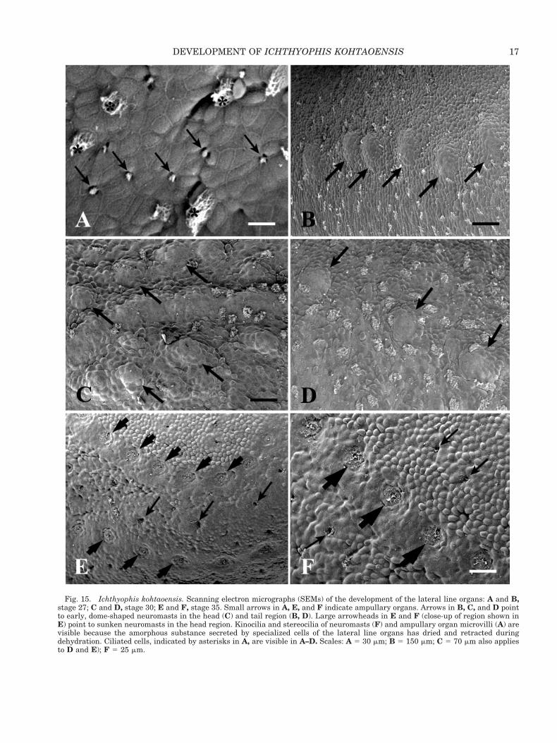
Fig. 15. Ichthyophis kohtaoensis. Scanning electron micrographs (SEMs) of the development of the lateral line organs: A and B,stage 27; C and D, stage 30; E and F, stage 35. Small arrows in A, E, and F indicate ampullary organs. Arrows in B, C, and D pointto early, dome-shaped neuromasts in the head (C) and tail region (B, D). Large arrowheads in E and F (close-up of region shown inE) point to sunken neuromasts in the head region. Kinocilia and stereocilia of neuromasts (F) and ampullary organ microvilli (A) arevisible because the amorphous substance secreted by specialized cells of the lateral line organs has dried and retracted duringdehydration. Ciliated cells, indicated by asterisks in A, are visible in A–D. Scales: A 5 30 mm; B 5 150 mm; C 5 70 mm also appliesto D and E); F 5 25 mm.
17DEVELOPMENT OF ICHTHYOPHIS KOHTAOENSIS
multaneously, resulting in the formation of anelongated yolk tube which is finally incorporated bythe abdominal folds.
Total Body Length and Weight
Table 2 indicates that developmental size andweight vary considerably among individuals, andsize ranges for different stages often overlap. As onemight expect, both total body length and weightincrease progressively during embryogenesis andlarval development.
DISCUSSION
A number of tables of developmental stages forvertebrate species have been developed. Some havecomparability of staging criteria, but many do not,either because of the particular features of the spe-cies (species-specificity) or the level of detail of theexamination and description of those features. Fur-ther, the selection of staging criteria varies, depend-ing on the intended use of the table. If it is to be usedmainly to describe the development of a particularspecies (e.g., the detailed staging table of Nieuwkoopand Faber [1967] for Xenopus laevis), a set of criteriadifferent from those used to group and compare in-dividuals of different species (e.g., the less detailedbut more general staging table of Gosner [1960]) willbe used.
Stages are established according to a set of crite-ria, such as external morphology. Body sizes of spec-imens or physiological characters are considered bymany researchers to be too variable to be used asstaging criteria (Nieuwkoop and Faber, 1967;Townsend and Stewart, 1985; Grillitsch, 1989). Ex-ternal morphology provides more reliable and con-sistent staging criteria, although it too is subject tovariation. Because of the inherent variation in de-velopmental systems, individuals at a given stageoften do not share all diagnostic features character-istic for that stage, and may present conflicting setsof characters. For example, the gill region of anembryo may appear well advanced, indicating alater stage in development, but the yolk is not cor-respondingly developed. Further, the developmentof certain organ systems, such as the lateral linesystem or the gills, has left–right asymmetry. Bysubdividing development into stages, it is possible togroup and compare individuals and species.
The normal table that we have developed permitscomparison of Ichthyophis kohtaoensis to other am-phibian species, although the morphology of thatcaecilian species presents considerable variation rel-ative to either viviparous caecilians or anuran orurodelan amphibians for which staging tables areavailable. We describe 20 stages to designate thosebetween neurulation and mid-metamorphosis. Theyare chosen so as to be readily recognizable and arebased mainly on external form. In addition to exter-nal morphological features, we include total lengthas a useful initial guideline for staging because sofew data are available for caecilians. We recognizethat factors such as crowding, temperature, lightcycle, food, and water conditions can effect greatvariation in size and developmental rate. Develop-mental size varies, and size ranges for differentstages often overlap. Thus, we avoid identifyingstages based largely on size. Tables 1 and 2 list totalbody length for each stage, but more weight shouldbe given to morphological features.
We discuss the ontogeny of selected morphologicalfeatures (e.g., lateral line organs, gills, etc.) of Ich-
Fig. 16. Ichthyophis kohtaoensis. SEM’s of the tail bud and thecloacal region: A, stage 22; B, stage 30; C, stage 35. White arrowspoint to the cloacal region, black arrows denote the tail fin. Thecloacal rim and the cloacal buds are visible in B; see Figure 13 Dand text for further details. Scale in A 5 350 mm; scale in B andC 5 250 mm.
18 N. DUNKER ET AL.
thyophis kohtaoensis and compare it to that of thecongeneric I. glutinosus, then to the development ofother caecilians, especially Typhlonectes compressi-cauda, a representative of a more derived familywith a viviparous, rather than egg-laying, reproduc-tive mode. We analyze our data in two ways: first, adirect comparison of each of the selected morpholog-ical features across species; second, a comparison ofoverall ontogeny of I. kohtaoensis with that of otherspecies of caecilians in order to summarize salientsimilarities and differences in ontogenies. Finally, inthose cases in which developmental morphology isparticularly illustrative, we compare the ontogeny ofI. kohtaoensis to that of selected oviparous anddirect-developing frogs and salamanders, with spe-cific reference to normal tables, in order to illustratesimilarities and differences among taxa of amphibi-ans. These several comparisons provide a baselinefor further study of the evolution of patterns of de-velopment.
Ontogeny of External MorphologicalFeatures
Lateral line system. The lateral line system is acollection of epidermal sense organs (mechanorecep-tive neuromasts and electroreceptive ampullary or-gans) and their nerves, distributed over the headand along the body in many aquatic amphibians.Early work on the lateral line system involved onlythe identification of mechanoreceptors, the neuro-masts. The ampullary organs or “Nebenohren” ofgymnophiones were described in 1887 by Sarasinand Sarasin, and Coggi (1905) compared them to theampullae of Lorenzini of sharks. However, ampul-lary organs were not examined again for nearly acentury. In fact, it was long assumed that amphibi-ans lacked ampullary organs and had only neuro-masts (Bennett, 1971). Hetherington and Wake(1979) reviewed the biology of lateral-line receptorsin caecilians and corroborated the work of the Sara-sins and Coggi, finding both neuromasts and amp-ullary organs in the lateral line system of gym-nophiones. It is now understood that, in general,salamanders and caecilians have both neuromastsand ampullary organs as the receptors in their lat-eral line systems; anurans have only neuromasts(Fritzsch, 1981; Fritzsch and Wahnschaffe, 1983;Fritzsch and Munz, 1986).
Free-living larvae of several caecilian genera (e.g.,Ichthyophis, Caudacaecilia, Epicrionops, Sylvacae-cilia; Taylor, 1960, 1970; Hetherington and Wake,1979; Fritzsch and Wake, 1986; Himstedt andFritzsch, 1990; Wake, pers. obs.) possess a lateral-line system composed of both neuromasts and amp-ullary organs. Ampullary organs appear to be re-stricted to the head (Sarasin and Sarasin, 1887–1890; Fritzsch and Wake, 1986), where they are lessnumerous than neuromasts. Neuromasts occur inseries along the body as well as on the head; how-
ever, they are more concentrated on the head. Ourobservations of ampullary organs associated withrows of neuromasts on the heads of I. kohtaoensisembryos at both light microscopic and ultrastruc-tural levels (see Results) parallel those of Fritzsch etal. (1985) and Fritzsch and Wake (1986) for larvae ofthat species. In addition, lateral line organs (unspec-ified, but likely neuromasts) are reported for embry-onic Hypogeophis (Brauer, 1899) and for Sylvacae-cilia (then Geotrypetes) grandisonae (Largen et al.,1972; however, both neuromasts and ampullary or-gans are present in the latter [Wake, pers. obs.]).Further, Fritzsch and Wake (1986) observed struc-tures that conform to descriptions of ampullary or-gans found in larval urodeles and caecilians in em-bryos of Typhlonectes compressicauda and T.natans, as well as on the heads of adult T. natansand H. rostratus. These organs lie within the epider-mis and have a characteristic prominent central pitthat resembles the flask-like ampullary organs ofvarious freshwater fishes. Fritzsch and Wake (1986)remarked upon the association of the presence ofampullary organs with aquatic life and proposedthat adults of Typhlonectes retain ampullary organsfor electroreception in the murky, slow-flowing wa-ters in which the animals live.
Sarasin and Sarasin’s (1887–1890) examination ofthe formation of lateral line organs revealed that inearly embryos of Ichthyophis glutinosus, repre-sented by their figure 38 on Plate IV (comparable toour stage 26), neuromasts (“Hugelorgane”) resembledome-shaped structures, slightly protruding beyondthe epidermis (also see Sarasins’ fig. 11, Plate VI),whereas later the tip of the organ sinks in to form adepression or shallow groove (see Sarasins’ fig. 12,Plate VI), very similar to the pattern of developmentin I. kohtaoensis. In larvae of I. kohtaoensis we founda distribution of lateral line organs rather similar tothe pattern observed for Ichthyophis sp. by Hether-ington and Wake (1979). However, they state thatampullary organs occasionally occur only along oneside of a neuromast row, as in the instance of organsoccurring only lateral to the supraorbital series. Incontrast, three rows of lateral line organs (one row ofneuromasts and two rows of ampullary organs), nottwo, are consistently discernible above and belowthe eyes of I. kohtaoensis larvae. Additionally, thedistribution of ampullary organs differs from that inHetherington and Wake’s material; in I. kohtaoensislarvae ampullary organs are no longer concentratedon the chin, as they are in embryos of that species,but disappear or have sunk into the skin. The small,round pigmentless structures discernible in the chinregion are skin glands. During the course of larvaldevelopment the lateral line organs (neuromastsand ampullary organs) degenerate and by metamor-phosis are no longer discernible (Sarasin and Sara-sin, 1887–1890, and our observations).
Arrangements of neuromasts and ampullary or-gans vary among salamanders, though they bear
19DEVELOPMENT OF ICHTHYOPHIS KOHTAOENSIS
some similarities to the caecilian condition. For ex-ample, in Triturus alpestris and Ambystoma mexi-canum, a linear arrangement of up to eight neuro-masts occurs (Fritzsch and Bolz, 1986; Fritzsch andWake, 1986). In these species the ampullary organsform groups of two to five organs, in contrast to thesituation observed in Ichthyophis, in which the or-gans are in a more linear arrangement. In the axo-lotl A. mexicanum, ampullary organs occur at thebases of the gills. However, similar to their distribu-tion in I. kohtaoensis, the ampullary organ clustersaccompany the rows of neuromasts, are concen-trated around the external nares and around theeyes, and are absent on the body and the tail. At theultrastructural level, salamander ampullary organsare small, irregularly shaped pits distinct from largeneuromasts (see figs. 5 and 6 in Fritzsch andWahnschaffe, 1983), comparable to our SEM results(Fig. 15). The linear arrangement of kinocilia andstereocilia in the neuromasts and the central posi-tion of several bundles of microvilli in the ampullaryreceptor are visible when the amorphous substance(consisting of neutral mucopolysaccharides) fillingthe canal has dried and retracted during dehydra-tion (see Fig. 15, and Fritzsch and Wahnschaffe’sfigures). Neuromast morphology in caecilians is gen-erally similar to that of fishes and salamanders;however, the ampullary organs of salamanders andteleost fishes, lack kinocilia, but have many mi-crovilli (Fritzsch and Wahnschaffe, 1983).
In general, those amphibians that adopt a pre-dominately terrestrial life history at metamorphosislose their lateral line systems, whereas entirelyaquatic amphibians retain their lateral line organsafter metamorphosis (Shelton, 1970; Russel, 1976).There are some exceptions to this generalization;newts that are terrestrial but return to water tobreed have large neuromasts that break through theskin and occupy a superficial position in a shallowdepression during the aquatic phase, whereas theysink below the epidermal surface, are covered by oneor two layers of epidermal cells, and appear to “de-differentiate” during terrestrial life (Fritzsch andWahnschaffe, 1983). However, the generalizationholds for caecilians; terrestrial animals lose theirlateral line sensory structures at metamorphosis, aswe observed in Ichthyophis kohtaoensis and Sarasinand Sarasin (1887–1890) and Breckenridge et al.(1987) saw in I. glutinosus, but the aquaticTyphlonectes has ampullary organs (but not neuro-masts) in both pre-birth and adult states (summa-rized in Fritzsch and Munz, 1986).
Tentacle. Caecilians possess a sensory organunique among vertebrates, the tentacle, which isapparently involved in tactile and chemoreceptivefunctions (see Billo and Wake, 1987). This smallprotrusile organ lying between the nasal openingand the eye is associated with the vomeronasal or-gan and its glands, and develops from several struc-tures usually associated with the eye, such as the
lower eyelid, the interpalpebral space, the conjunc-tiva, the Harderian gland, the retractor and levatormuscles, and their nerves (summarized in Billo andWake, 1987). The tentacle apparatus is similar in itsbauplan among all adult gymnophiones; however,sizes and shapes of the structures and their propor-tions differ among species and the tentacle apper-ture varies in its position relative to the eye and thenostril. The tentacle of Ichthyophis glutinosus wasdescribed by Sarasin and Sarasin (1887–1890). Inthat species, the specialized fold is recurved, elon-gate, and highly protrusile. Sarasin and Sarasin(1887–1890) also examined tentacle development inI. glutinosus and reported that in embryonic stagesnear hatching, the epithelium along the frontal edgeof the eyes is thickened, resembling a tissue knot(see Sarasins’ fig. 61, Plate XIX). In a 9.5 cm larvathe tissue proliferation enlarges and a central cavityappears, which has no external contact (Sarasins’figs. 62 and 63, Plate XIX). In the course of develop-ment the growth cone continues sinking in and asmall indentation develops from the outer surface(Sarasins’ fig. 73, Plate XIX). At that stage, shown inSarasins’ figure 58 (Plate XIX), only a small pit infront of the eyes is visible externally in larvae, sim-ilar to the situation in our stage 30 and 33. Accord-ing to the Sarasins, the formation of the tentacle foldbegins in embryos prior to hatching, illustrated infigure 59 (Plate XIX). In those stages a tentacleorifice and a small “papilla,” the tentacle fold, arediscernible in front of the eye, similar to the situa-tion in metamorphic stage 40 of I. kohtaoensis. Fi-nally, in late larval stages, which exhibit the exter-nal features of young terrestrial adults (see theSarasins’ fig. 60, Plate XIX), the tentacle aperturemoves in the direction of the upper lip, and thetentacle sheath and the involuted tentacle fold elon-gate. The tentacle of I. kohtaoensis is constructedvery similarly to that of I. glutinosus, based on ourobservations of external developmental and adultmorphology.
Billo and Wake (1987) examined tentacle develop-ment in Dermophis mexicanus using transverse,sagittal, and frontal head sections. They reportedthat a tentacle anlage appeared to be presentventro-rostral to the eye in the form of a minuteectodermal elevation in 14–15 mm TL D. mexicanusembryos, which appears comparable to stage 28 ofIchthyophis kohtaoensis, at which the tentacle an-lage first appears. In 22–23 mm Dermophis embryosthe tentacle anlage is surrounded by an ectodermalrim and resembles a depressed plug. This stage oftentacle development appears comparable to the sit-uation found in Ichthyophis embryos of stage 33, inwhich the tentacle aperture represents a white,dense tissue fold. Billo and Wake (1987) reportedthat in 34–35 mm fetuses the tentacle anlage hasdifferentiated by evagination into a tiny fold withina small sheath. In 44 mm fetuses the tentacle aper-ture still has the same ventrorostral position rela-
20 N. DUNKER ET AL.
tive to the eye, which is covered by skin. By compar-ison, at our metamorphic stage 40 a funnel-shapedtentacle sheath is clearly discernible and the tenta-cle appears in the tentacle orifice.
A direct comparison of tentacle development inthe species examined in these studies is difficultbecause they have different modes of reproductionand development (oviparous vs. viviparous); em-bryos, fetuses or larvae, and adults are differentsizes at approximately the same developmentalstages; and neither Billo and Wake (1987) nor Sara-sin and Sarasin (1887–1890) provided staging crite-ria other than total body length. Furthermore, wedid not have sectioned material of Ichthyophiskohtaoensis available, which would have permitteda more specific comparison. We do note that, like thecondition reported by Sarasin and Sarasin (1887–1890) but contradicted by Breckenridge et al. (1987)in I. glutinosus (they state that the tentacle appearsexternally at metamorphosis), in I. kohtaoensis atentacle anlage is visible externally in embryonicstages, although the development of the tentacleapparatus continues after hatching and metamor-phosis. We do not have more advanced larval stages,at which the tentacle moves rostrally in the direc-tion of the lip, available for examination.
Gills. Three pairs of external gills develop insalamanders and caecilians, whereas external gillsare poorly developed or transitory, if present at all,in anuran embryos. Among caecilian taxa that haveterrestrial adults, the gills of embryos typically aretriradiate, elongate and plumose, no matter whatthe reproductive mode (oviparity, direct develop-ment, or viviparity), but they are a single pair ofhighly dilated sacs in the viviparous aquatictyphlonecids (Peters, 1874, 1875; Parker, 1956; Tay-lor, 1968; Wake, 1969; Nussbaum and Wilkinson,1989; Wilkinson, 1989; Sammouri et al., 1990; Ex-brayat and Hraoui-Bloquet, 1991, 1992; Hraoui-Bloquet and Exbrayat, 1994; Wilkinson and Nuss-baum, 1997). Embryos of Ichthyophis glutinosus(Sarasin and Sarasin, 1887–1890), Hypogeophis ros-tratus (Brauer, 1897, 1899) and Geotrypetes seraphi-nii (Parker, 1956) have three pairs of gills through-out their embryonic development until the gills arelost at hatching or metamorphosis; in larvae of somespecies, three gill rudiments persist (e.g., Epicrion-ops petersi and E. bicolor; Wake, pers. obs.). How-ever, Seshachar (1942) and Ramaswami (1954)stated that Gegenophis carnosus embryos have onlytwo pairs of gills (also M. Wake, pers. obs.). In Gym-nopis multiplicata the external gills have threefringed rami arising from a short stump attached tothe head (Wake, 1969). In contrast, gill rami inembryos of most taxa reported in the literature aredirectly attached to the head.
Brauer (1899) described the development oftriramous, filamentous gills from three knobs oneach side of the head of embryos of Hypogeophisrostratus. He reported that the three rami develop
from these outpocketings of the gill tissue, as we andthe Sarasins observed in Ichthyophis. It was notpossible for us to examine early gill developmentbecause we lack early stages of development in ourseries, so we are not certain whether the three gillsin Ichthyophis embryos develop in parallel or with atime delay. However, in contrast to gill developmentin Hypogeophis, where the third gill does not appearuntil the time that the gill filaments have appearedon the first gill, in Ichthyophis embryos all threegills are well developed at that stage. Brauer (1899)stated that at “full” development in Hypogeophis themiddle ramus is slightly longer than the anteriorand posterior. Brauer reported a maximum length ofonly 7 mm for the second gill and a maximum lengthof 5 mm for the first gill in a 40 mm total lengthHypogeophis embryo; he stated that the filaments ofthe third gill remain small in size and number.According to Brauer and to Breckenridge and Jayas-inghe (1979), the differences in lengths of gill ramiare even more pronounced in I. glutinosus (which wealso observed in I. kohtaoensis; see Table 2). Marcus(1908) observed gill development in Hypogeophisand Grandisonia, but mostly added information toBrauer’s report and commented on comparative gillhomologies.
Wake (1967, 1969) described gill ontogeny andstructure in embryos of the viviparous Gymnopismultiplicata; total length was her sole “staging” cri-terion, so it is used here as a basis for comparison. A10.5 mm embryo has two 2.5–3.5 mm long gill ramiwith 20–25 filaments on each side of the head. A16.5 mm Ichthyophis embryo, at our earliest devel-opmental stage (21), has only 1 mm long gill budswithout any filaments, but has three rather thantwo external gills. A 37 mm Gymnopis embryo has a10 mm long posterior gill, a 6 mm long anterior gill,and a newly developed 2.5 mm long middle ramuswith 10–12 short filaments that extend at right an-gles the length of the ramus, similar to the situationin Ichthyophis. The 36.3 mm stage 29 of Ichthyophishas first (6.3 mm) and third (5.9 mm) gills of com-parable size, but the second or middle gill is muchlonger at 12 mm and its numbers of gill filaments ishigher, between 40 and 74. Similar to Wake’s (1967,1969) 52 mm and 54 mm embryos, our comparablestages 33 (53.8 mm) and 34 (54.9 mm) have a middlegill (12 mm in stage 33; 10.8 mm in stage 34) that isnearly twice as long as the anterior gill (6.8 mm instage 33, 6 mm in stage 34) and the posterior gill (6mm in stage 33, 4.5 mm in stage 34). All threeexternal gills are significantly longer and have con-siderably more filaments in Ichthyophis embryosthan do Gymnopis embryos at approximately theoverall size.
The unique large, sac-like gills of typhlonectidshave frequently been described (see above), withconjecture about their function and their restrictionto that aquatic lineage. Sammouri et al. (1990) pro-vide a detailed description of the development of the
21DEVELOPMENT OF ICHTHYOPHIS KOHTAOENSIS
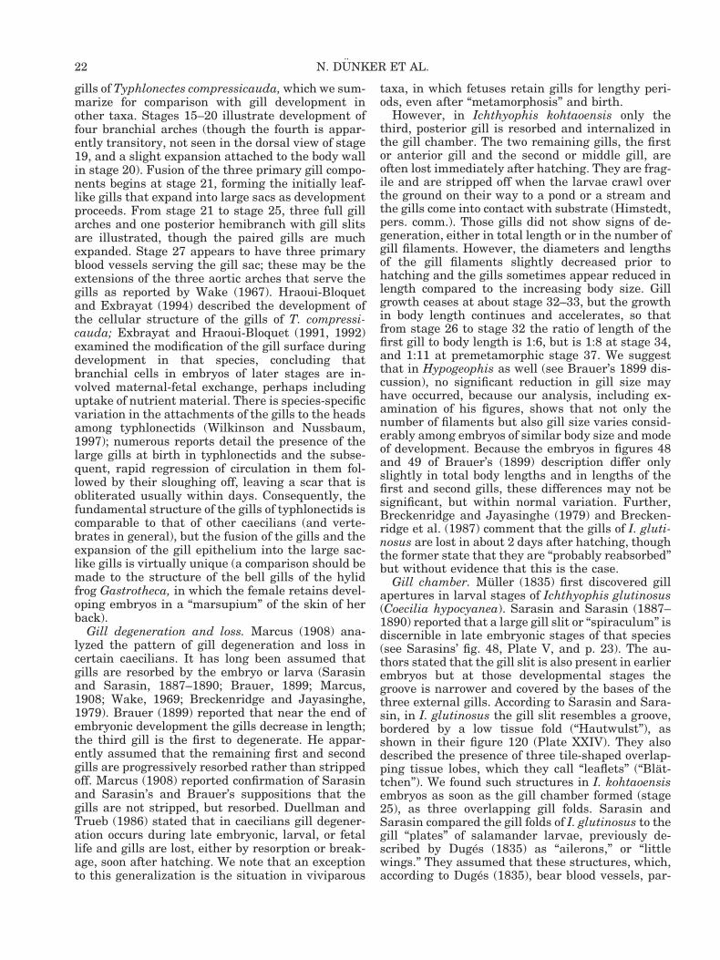
gills of Typhlonectes compressicauda, which we sum-marize for comparison with gill development inother taxa. Stages 15–20 illustrate development offour branchial arches (though the fourth is appar-ently transitory, not seen in the dorsal view of stage19, and a slight expansion attached to the body wallin stage 20). Fusion of the three primary gill compo-nents begins at stage 21, forming the initially leaf-like gills that expand into large sacs as developmentproceeds. From stage 21 to stage 25, three full gillarches and one posterior hemibranch with gill slitsare illustrated, though the paired gills are muchexpanded. Stage 27 appears to have three primaryblood vessels serving the gill sac; these may be theextensions of the three aortic arches that serve thegills as reported by Wake (1967). Hraoui-Bloquetand Exbrayat (1994) described the development ofthe cellular structure of the gills of T. compressi-cauda; Exbrayat and Hraoui-Bloquet (1991, 1992)examined the modification of the gill surface duringdevelopment in that species, concluding thatbranchial cells in embryos of later stages are in-volved maternal-fetal exchange, perhaps includinguptake of nutrient material. There is species-specificvariation in the attachments of the gills to the headsamong typhlonectids (Wilkinson and Nussbaum,1997); numerous reports detail the presence of thelarge gills at birth in typhlonectids and the subse-quent, rapid regression of circulation in them fol-lowed by their sloughing off, leaving a scar that isobliterated usually within days. Consequently, thefundamental structure of the gills of typhlonectids iscomparable to that of other caecilians (and verte-brates in general), but the fusion of the gills and theexpansion of the gill epithelium into the large sac-like gills is virtually unique (a comparison should bemade to the structure of the bell gills of the hylidfrog Gastrotheca, in which the female retains devel-oping embryos in a “marsupium” of the skin of herback).
Gill degeneration and loss. Marcus (1908) ana-lyzed the pattern of gill degeneration and loss incertain caecilians. It has long been assumed thatgills are resorbed by the embryo or larva (Sarasinand Sarasin, 1887–1890; Brauer, 1899; Marcus,1908; Wake, 1969; Breckenridge and Jayasinghe,1979). Brauer (1899) reported that near the end ofembryonic development the gills decrease in length;the third gill is the first to degenerate. He appar-ently assumed that the remaining first and secondgills are progressively resorbed rather than strippedoff. Marcus (1908) reported confirmation of Sarasinand Sarasin’s and Brauer’s suppositions that thegills are not stripped, but resorbed. Duellman andTrueb (1986) stated that in caecilians gill degener-ation occurs during late embryonic, larval, or fetallife and gills are lost, either by resorption or break-age, soon after hatching. We note that an exceptionto this generalization is the situation in viviparous
taxa, in which fetuses retain gills for lengthy peri-ods, even after “metamorphosis” and birth.
However, in Ichthyophis kohtaoensis only thethird, posterior gill is resorbed and internalized inthe gill chamber. The two remaining gills, the firstor anterior gill and the second or middle gill, areoften lost immediately after hatching. They are frag-ile and are stripped off when the larvae crawl overthe ground on their way to a pond or a stream andthe gills come into contact with substrate (Himstedt,pers. comm.). Those gills did not show signs of de-generation, either in total length or in the number ofgill filaments. However, the diameters and lengthsof the gill filaments slightly decreased prior tohatching and the gills sometimes appear reduced inlength compared to the increasing body size. Gillgrowth ceases at about stage 32–33, but the growthin body length continues and accelerates, so thatfrom stage 26 to stage 32 the ratio of length of thefirst gill to body length is 1:6, but is 1:8 at stage 34,and 1:11 at premetamorphic stage 37. We suggestthat in Hypogeophis as well (see Brauer’s 1899 dis-cussion), no significant reduction in gill size mayhave occurred, because our analysis, including ex-amination of his figures, shows that not only thenumber of filaments but also gill size varies consid-erably among embryos of similar body size and modeof development. Because the embryos in figures 48and 49 of Brauer’s (1899) description differ onlyslightly in total body lengths and in lengths of thefirst and second gills, these differences may not besignificant, but within normal variation. Further,Breckenridge and Jayasinghe (1979) and Brecken-ridge et al. (1987) comment that the gills of I. gluti-nosus are lost in about 2 days after hatching, thoughthe former state that they are “probably reabsorbed”but without evidence that this is the case.
Gill chamber. Muller (1835) first discovered gillapertures in larval stages of Ichthyophis glutinosus(Coecilia hypocyanea). Sarasin and Sarasin (1887–1890) reported that a large gill slit or “spiraculum” isdiscernible in late embryonic stages of that species(see Sarasins’ fig. 48, Plate V, and p. 23). The au-thors stated that the gill slit is also present in earlierembryos but at those developmental stages thegroove is narrower and covered by the bases of thethree external gills. According to Sarasin and Sara-sin, in I. glutinosus the gill slit resembles a groove,bordered by a low tissue fold (“Hautwulst”), asshown in their figure 120 (Plate XXIV). They alsodescribed the presence of three tile-shaped overlap-ping tissue lobes, which they call “leaflets” (“Blat-tchen”). We found such structures in I. kohtaoensisembryos as soon as the gill chamber formed (stage25), as three overlapping gill folds. Sarasin andSarasin compared the gill folds of I. glutinosus to thegill “plates” of salamander larvae, previously de-scribed by Duges (1835) as “ailerons,” or “littlewings.” They assumed that these structures, which,according to Duges (1835), bear blood vessels, par-
22 N. DUNKER ET AL.
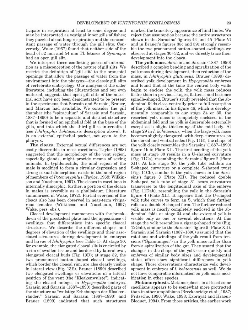
ticipate in respiration at least to some degree andmay be interpreted as vestigial inner gills of fishes;they puzzled about lung respiration and the concom-itant passage of water through the gill slits. Con-versely, Wake (1967) found that neither side of thehead of 52 mm and 54 mm TL fetuses of Gymnopishad an open gill slit.
We interpret these conflicting pieces of informa-tion as a misconception of the nature of gill slits. Werestrict the definition of “gill slit” to the branchialopenings that allow the passage of water from theenvironment into the pharynx—the classic gill slitsof vertebrate embryology. Our analysis of the olderliterature, including the illustrations and our ownmaterial, suggests that open gill slits of the ances-tral sort have not been demonstrated to be presentin the specimens that Sarasin and Sarasin, Brauer,and Marcus had available. We consider the gillchamber (the “spiraculum” of Sarasin and Sarasin,1887–1890) to be a separate and distinct structurethat is formed of an epithelial fold at the base of thegills, and into which the third gill is internalized(see Ichthyophis kohtaoensis description above). Itis an external epithelial pocket, not open to thepharynx.
The cloaca. External sexual differences are noteasily discernible in most caecilians. Taylor (1968)suggested that the morphology of the vent region,especially glands, might provide means of sexinganimals. In typhlonectids, the anal region of themale is modified to form a circular depression andstrong sexual dimorphism exists in the anal regionof members of Potomotyphlus (Taylor, 1968; Wilkin-son and Nussbaum, 1997). The cloaca in caecilians isinternally dimorphic; further, a portion of the cloacain males is eversible as a phallodeum (literaturesummarized in Wake, 1972). Partial eversion of thecloaca also has been observed in near-term vivipa-rous females (Wilkinson and Nussbaum, 1997;Wake, pers. obs.).
Cloacal development commences with the break-down of the proctodeal plate and the appearance ofswellings that differentiate into specific cloacalstructures. We describe the different shapes anddegrees of elevation of the swellings and their asso-ciated structures during development in embryosand larvae of Ichthyophis (see Table 1). At stage 30,for example, the elongated cloacal slit is encircled bya rim of swollen tissue and bordered by lateral oval,elongated cloacal buds (Fig. 13D); at stage 32, thetwo pronounced button-shaped cloacal swellings,which border the cloacal rim, are also clearly visiblein lateral view (Fig. 13E). Brauer (1899) describedtwo elongated swellings or elevations in a lateralposition of the vent (the “Kloakenwulste”), indicat-ing the cloacal anlage, in Hypogeophis embryos.Sarasin and Sarasin (1887–1890) described parts ofthe structure as “wulstige Erhebungen der Kloaken-rander.” Sarasin and Sarasin (1887–1890) andBrauer (1899) indicated that such structures
marked the transitory appearance of hind limbs. Wereject that assumption because the entire structuresshown in the Sarasins’ figures 44 and 45 (Plate V)and in Brauer’s figures 38c and 39c strongly resem-ble the two pronounced button-shaped swellings weobserved at stages 30–32, and we directly trace theirdevelopment into the cloaca.
The yolk mass. Sarasin and Sarasin (1887–1890)described a complex bending and spiralization of theyolk mass during development, then reduction of themass, in Ichthyophis glutinosus. Brauer (1899) de-scribed yolk development in Hypogeophis embryosand found that at the time the ventral body wallsbegin to enclose the yolk, the yolk mass reducesfaster than in previous stages, flattens, and becomesspindle-shaped. Brauer’s study revealed that the ab-dominal folds close ventrally prior to full resorptionof the yolk mass. In his figure 48, which is develop-mentally comparable to our stage 34 or 35, theresorbed yolk mass is completely enclosed in theabdominal fold and no yolk is discernible externallyexcept as a slight thickening of the mid-body. Atstage 29 in I. kohtaoensis, when the large yolk massbecomes slightly elongated, with deep curvatures onthe dorsal and ventral sides (Fig. 11B), the shape ofthe yolk closely resembles the Sarasins’ (1887–1890)figure 1b in Plate XII. The first bending of the yolkmass at stage 30 results in a U-shaped curvature(Fig. 11Ca), resembling the Sarasins’ figure 2 (PlateXII). At late stage 30, the yolk tube exhibits anS-shaped curvature or a complete S-shaped twist(Fig. 11Cb), similar to the yolk shown in the Sara-sins’s figure 3 (Plate XII). The reduced doubleS-shaped yolk tube of stage 31 bears curvaturestransverse to the longitudinal axis of the embryo(Fig. 11Dab), resembling the yolk in the Sarasin’sfigure 4 (Plate XII). It appears that the U-shapedyolk tube curves to form an S, which then furtherrolls to a double S-shaped form. The further reducedyolk mass is nearly completely enclosed in the ab-dominal folds at stage 34 and the external yolk isvisible only as one or several elevations. At thisstage the yolk resembles a zigzag-shaped tube (Fig.12Gab), similar to the Sarasins’ figure 5 (Plate XII).Sarasin and Sarasin (1887–1890) assumed that therotations and windings of the yolk result from ten-sions (“Spannungen”) in the yolk mass rather thanfrom a spiralization of the gut. They stated that thechanges in the shape of the yolk occur quickly andembryos of similar body sizes and developmentalstates often show significant differences in yolkshape. Those observations characterize yolk devel-opment in embryos of I. kohtaoensis as well. We donot have comparable information on yolk mass mod-ification in other species.
Metamorphosis. Metamorphosis in at least somecaecilians appears to be somewhat more protractedthan in other amphibians (Breckenridge et al., 1987;Fritzsche, 1990; Wake, 1993; Exbrayat and Hraoui-Bloquet, 1994). From those articles, the earlier work
23DEVELOPMENT OF ICHTHYOPHIS KOHTAOENSIS
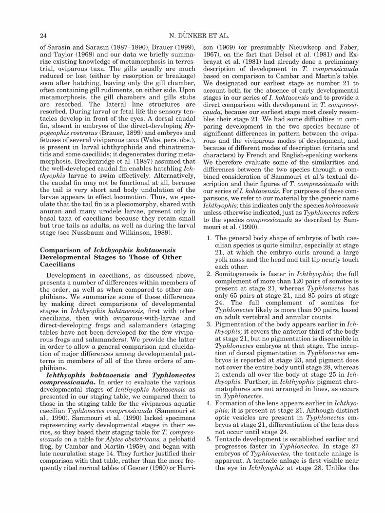
of Sarasin and Sarasin (1887–1890), Brauer (1899),and Taylor (1968) and our data we briefly summa-rize existing knowledge of metamorphosis in terres-trial, oviparous taxa. The gills usually are muchreduced or lost (either by resorption or breakage)soon after hatching, leaving only the gill chamber,often containing gill rudiments, on either side. Uponmetamorphosis, the gill chambers and gills stubsare resorbed. The lateral line structures areresorbed. During larval or fetal life the sensory ten-tacles develop in front of the eyes. A dorsal caudalfin, absent in embryos of the direct-developing Hy-pogeophis rostratus (Brauer, 1899) and embryos andfetuses of several viviparous taxa (Wake, pers. obs.),is present in larval ichthyophiids and rhinatrema-tids and some caeciliids; it degenerates during meta-morphosis. Breckenridge et al. (1987) assumed thatthe well-developed caudal fin enables hatchling Ich-thyophis larvae to swim effectively. Alternatively,the caudal fin may not be functional at all, becausethe tail is very short and body undulation of thelarvae appears to effect locomotion. Thus, we spec-ulate that the tail fin is a plesiomorphy, shared withanuran and many urodele larvae, present only inbasal taxa of caecilians because they retain smallbut true tails as adults, as well as during the larvalstage (see Nussbaum and Wilkinson, 1989).
Comparison of Ichthyophis kohtaoensisDevelopmental Stages to Those of OtherCaecilians
Development in caecilians, as discussed above,presents a number of differences within members ofthe order, as well as when compared to other am-phibians. We summarize some of these differencesby making direct comparisons of developmentalstages in Ichthyophis kohtaoensis, first with othercaecilians, then with oviparous-with-larvae anddirect-developing frogs and salamanders (stagingtables have not been developed for the few vivipa-rous frogs and salamanders). We provide the latterin order to allow a general comparison and elucida-tion of major differences among developmental pat-terns in members of all of the three orders of am-phibians.
Ichthyophis kohtaoensis and Typhlonectescompressicauda. In order to evaluate the variousdevelopmental stages of Ichthyophis kohtaoensis aspresented in our staging table, we compared them tothose in the staging table for the viviparous aquaticcaecilian Typhlonectes compressicauda (Sammouri etal., 1990). Sammouri et al. (1990) lacked specimensrepresenting early developmental stages in their se-ries, so they based their staging table for T. compres-sicauda on a table for Alytes obstetricans, a pelobatidfrog, by Cambar and Martin (1959), and began withlate neurulation stage 14. They further justified theircomparison with that table, rather than the more fre-quently cited normal tables of Gosner (1960) or Harri-
son (1969) (or presumably Nieuwkoop and Faber,1967), on the fact that Delsol et al. (1981) and Ex-brayat et al. (1981) had already done a preliminarydescription of development in T. compressicaudabased on comparison to Cambar and Martin’s table.We designated our earliest stage as number 21 toaccount both for the absence of early developmentalstages in our series of I. kohtaoensis and to provide adirect comparison with development in T. compressi-cauda, because our earliest stage most closely resem-bles their stage 21. We had some difficulties in com-paring development in the two species because ofsignificant differences in pattern between the ovipa-rous and the viviparous modes of development, andbecause of different modes of description (criteria andcharacters) by French and English-speaking workers.We therefore evaluate some of the similarities anddifferences between the two species through a com-bined consideration of Sammouri et al.’s textual de-scription and their figures of T. compressicauda withour series of I. kohtaoensis. For purposes of these com-parisons, we refer to our material by the generic nameIchthyophis; this indicates only the species kohtaoensisunless otherwise indicated, just as Typhlonectes refersto the species compressicauda as described by Sam-mouri et al. (1990).
1. The general body shape of embryos of both cae-cilian species is quite similar, especially at stage21, at which the embryo curls around a largeyolk mass and the head and tail tip nearly toucheach other.
2. Somitogenesis is faster in Ichthyophis; the fullcomplement of more than 120 pairs of somites ispresent at stage 21, whereas Typhlonectes hasonly 65 pairs at stage 21, and 85 pairs at stage24. The full complement of somites forTyphlonectes likely is more than 90 pairs, basedon adult vertebral and annular counts.
3. Pigmentation of the body appears earlier in Ich-thyophis; it covers the anterior third of the bodyat stage 21, but no pigmentation is discernible inTyphlonectes embryos at that stage. The incep-tion of dorsal pigmentation in Typhlonectes em-bryos is reported at stage 23, and pigment doesnot cover the entire body until stage 28, whereasit extends all over the body at stage 25 in Ich-thyophis. Further, in Ichthyophis pigment chro-matophores are not arranged in lines, as occursin Typhlonectes.
4. Formation of the lens appears earlier in Ichthyo-phis; it is present at stage 21. Although distinctoptic vesicles are present in Typhlonectes em-bryos at stage 21, differentiation of the lens doesnot occur until stage 24.
5. Tentacle development is established earlier andprogresses faster in Typhlonectes. In stage 27embryos of Typhlonectes, the tentacle anlage isapparent. A tentacle anlage is first visible nearthe eye in Ichthyophis at stage 28. Unlike the
24 N. DUNKER ET AL.
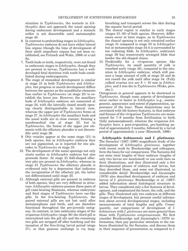
situation in Typhlonectes, the tentacle in Ich-thyophis does not approach the nasal openingduring embryonic development and a tentacleorifice is not discernible until metamorphicstage 40.
6. In contrast to prehatching stages in Ichthyophis,Typhlonectes embryos do not have either lateralline organs (though the time of development oftheir adult ampullary organs has not been es-tablished; see Fritzsch and Wake, 1986) or a tailfin.
7. Tooth buds or teeth, respectively, were not foundin embryonic stages in Ichthyophis, though theyare present in larvae; Typhlonectes has a well-developed fetal dentition with tooth buds estab-lished during embryogenesis.
8. The stage of stomodeal development is similarat stage 21 in both Typhlonectes and Ichthyo-phis, but progress in mouth development differsbetween the species as the mandibular elementsfuse earlier in Typhlonectes (at stage 23) to es-tablish the lower jaw. However, the maxillarybuds of Ichthyophis embryos are connected atstage 24, with the laterally closed mouth open-ing clearly distinguishable in lateral view,whereas the upper jaw of Typhlonectes forms atstage 27. In Ichthyophis the maxillary buds andthe nasal walls are in close contact, forming anasobranchial rim at stage 23, but inTyphlonectes, the fusion of the upper jaw ele-ments with the olfactory placodes is not discern-ible until stage 26.
9. Otic vesicles appear at the same stage (21) inboth species, but in Ichthyophis embryos theyare not pigmented, as is reported for otic pla-codes in Typhlonectes at stage 23.
10. The development of the nasal openings not onlystarts earlier in Ichthyophis embryos but alsoproceeds faster. At stage 21 fold-shaped olfac-tory pits are present in Ichthyophis, whereas instage 21 Typhlonectes the olfactory placode isevidenced only by a depression that indicatesthe invagination of the olfactory pit, the latternot differentiated until stage 25.
11. Although external gills are present in embryosof both species, they are very different in struc-ture. Ichthyophis embryos possess three pairs ofgill rami-bearing filaments, whereas embryonicand fetal stages of Typhlonectes have sac-likegills. In the live-bearing Typhlonectes, thepaired external gills are not lost until aftermetamorphosis and birth, and are thereforefunctional throughout the pre-juvenile ontog-eny. In contrast, in late embryonic stages of theoviparous Ichthyophis (stage 36) the third gill isinternalized into the gill slit and the remainingtwo gills are stripped off after hatching and theinception of the free-living larval period (stage37), so that gaseous exchange is via lung-
breathing and transport across the skin duringthe aquatic larval period.
12. The cloacal region is similar in early stages(stages 21–30) of both species. However, differ-ences occur at later stages, as in Typhlonectesthe cloacal opening is not only retracted earlier(stage 31 compared to stage 33 in Ichthyophis)but at metamorphic stage 34 it is surrounded byten radiating folds. In Ichthyophis, embryonicstage 39 has transversely arranged folds thatborder the slit-shaped cloaca.
13. Predictably for a viviparous species likeTyphlonectes, its small quantity of yolk isresorbed early (stage 28), compared to the situ-ation in Ichthyophis embryos, which still pos-sess a large amount of yolk at stage 28 and donot resorb the yolk until after stage 34. (Fullyyolked ovarian ova are 8 3 10 mm in Ichthyo-phis, and 2 mm dia in Typhlonectes [Wake, pers.obs.]).
Ontogenesis in general appears to be shortened inTyphlonectes, though some aspects of developmentoccur earlier or faster in Ichthyophis (e.g., somito-genesis, appearance and extent of pigmentation, ap-pearance of the lens). These disjunctions may becorrelated with life history features, in that embryosand fetuses of the viviparous Typhlonectes are main-tained for 7–9 months from fertilization to birth(fully metamorphosed), whereas the oviparous Ich-thyophis have a post-oviposition, pre-hatching pe-riod of approximately 3 months, followed by a larvalperiod of approximately a year (Himstedt, 1996).
Ichthyophis kohtaoensis and I. glutinosus.The Sarasins’ (1887–1890) magnificent study of thedevelopment of Ichthyophis glutinosus, togetherwith recent work by Breckenridge and colleagues,form the basis for our comparisons. The Sarasins didnot state total lengths of their embryos (lengths ofonly two larvae are mentioned) or use scale bars ontheir illustrations, and they illustrated only a fewdevelopmental phases of embryos and larvae, butthey described those, and some not illustrated, inconsiderable detail. Breckenridge and Jayasinghe(1979) also described development of embryos andlarvae of I. glutinosus; Breckenridge et al. (1987)added information about histological structure inlarvae. They considered only a few features of devel-opment, and emphasized the heart, the yolk, and thegills. They illustrated only two embryos at differentstages of development, but present useful informa-tion about several developmental stages, includingmeasurements of total lengths and gills. Conse-quently, our comparisons of development of I.kohtaoensis with I. glutinosus are more general thanthose with Typhlonectes compressicauda. We firstconsider Breckenridge and Jayasinghe’s (1979) in-formation, then pay particular attention to the em-bryos illustrated by the Sarasins, and discuss themin their sequence of presentation as compared to I.
25DEVELOPMENT OF ICHTHYOPHIS KOHTAOENSIS
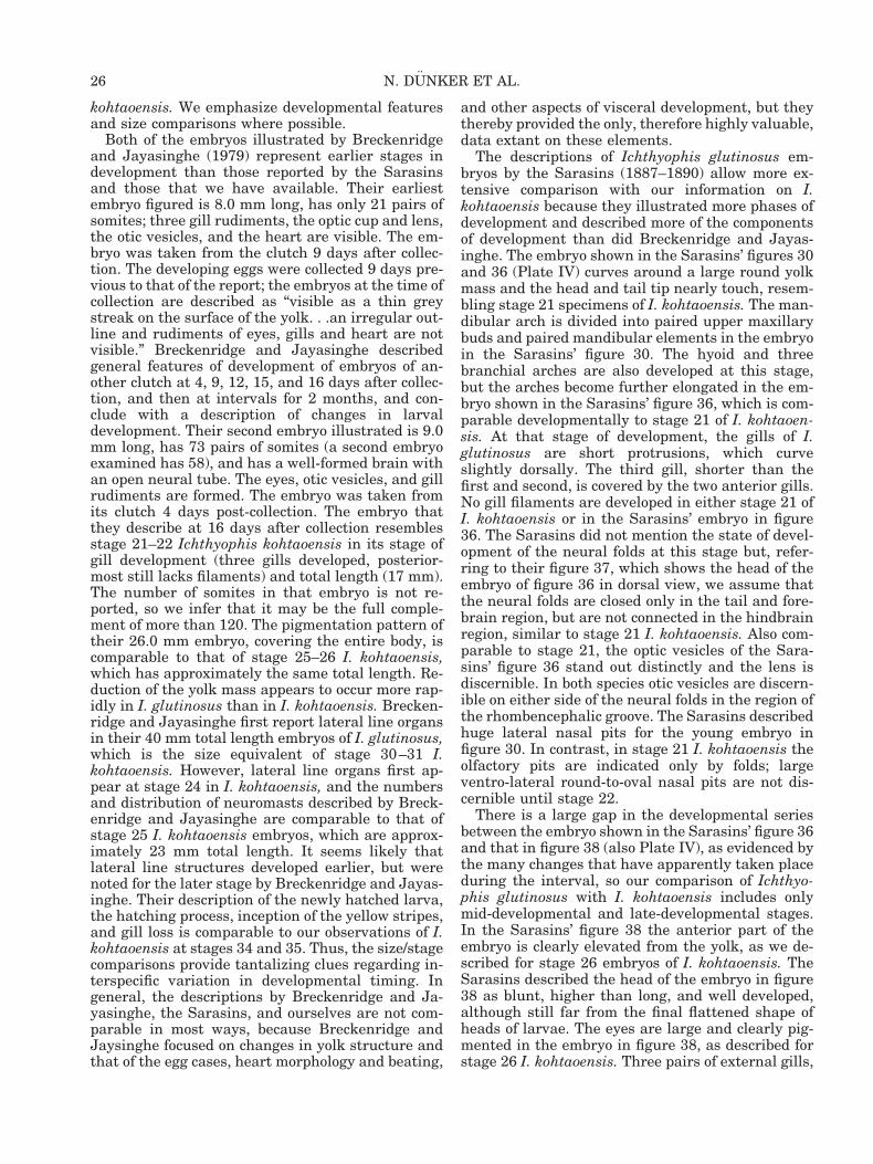
kohtaoensis. We emphasize developmental featuresand size comparisons where possible.
Both of the embryos illustrated by Breckenridgeand Jayasinghe (1979) represent earlier stages indevelopment than those reported by the Sarasinsand those that we have available. Their earliestembryo figured is 8.0 mm long, has only 21 pairs ofsomites; three gill rudiments, the optic cup and lens,the otic vesicles, and the heart are visible. The em-bryo was taken from the clutch 9 days after collec-tion. The developing eggs were collected 9 days pre-vious to that of the report; the embryos at the time ofcollection are described as “visible as a thin greystreak on the surface of the yolk. . .an irregular out-line and rudiments of eyes, gills and heart are notvisible.” Breckenridge and Jayasinghe describedgeneral features of development of embryos of an-other clutch at 4, 9, 12, 15, and 16 days after collec-tion, and then at intervals for 2 months, and con-clude with a description of changes in larvaldevelopment. Their second embryo illustrated is 9.0mm long, has 73 pairs of somites (a second embryoexamined has 58), and has a well-formed brain withan open neural tube. The eyes, otic vesicles, and gillrudiments are formed. The embryo was taken fromits clutch 4 days post-collection. The embryo thatthey describe at 16 days after collection resemblesstage 21–22 Ichthyophis kohtaoensis in its stage ofgill development (three gills developed, posterior-most still lacks filaments) and total length (17 mm).The number of somites in that embryo is not re-ported, so we infer that it may be the full comple-ment of more than 120. The pigmentation pattern oftheir 26.0 mm embryo, covering the entire body, iscomparable to that of stage 25–26 I. kohtaoensis,which has approximately the same total length. Re-duction of the yolk mass appears to occur more rap-idly in I. glutinosus than in I. kohtaoensis. Brecken-ridge and Jayasinghe first report lateral line organsin their 40 mm total length embryos of I. glutinosus,which is the size equivalent of stage 30–31 I.kohtaoensis. However, lateral line organs first ap-pear at stage 24 in I. kohtaoensis, and the numbersand distribution of neuromasts described by Breck-enridge and Jayasinghe are comparable to that ofstage 25 I. kohtaoensis embryos, which are approx-imately 23 mm total length. It seems likely thatlateral line structures developed earlier, but werenoted for the later stage by Breckenridge and Jayas-inghe. Their description of the newly hatched larva,the hatching process, inception of the yellow stripes,and gill loss is comparable to our observations of I.kohtaoensis at stages 34 and 35. Thus, the size/stagecomparisons provide tantalizing clues regarding in-terspecific variation in developmental timing. Ingeneral, the descriptions by Breckenridge and Ja-yasinghe, the Sarasins, and ourselves are not com-parable in most ways, because Breckenridge andJaysinghe focused on changes in yolk structure andthat of the egg cases, heart morphology and beating,
and other aspects of visceral development, but theythereby provided the only, therefore highly valuable,data extant on these elements.
The descriptions of Ichthyophis glutinosus em-bryos by the Sarasins (1887–1890) allow more ex-tensive comparison with our information on I.kohtaoensis because they illustrated more phases ofdevelopment and described more of the componentsof development than did Breckenridge and Jayas-inghe. The embryo shown in the Sarasins’ figures 30and 36 (Plate IV) curves around a large round yolkmass and the head and tail tip nearly touch, resem-bling stage 21 specimens of I. kohtaoensis. The man-dibular arch is divided into paired upper maxillarybuds and paired mandibular elements in the embryoin the Sarasins’ figure 30. The hyoid and threebranchial arches are also developed at this stage,but the arches become further elongated in the em-bryo shown in the Sarasins’ figure 36, which is com-parable developmentally to stage 21 of I. kohtaoen-sis. At that stage of development, the gills of I.glutinosus are short protrusions, which curveslightly dorsally. The third gill, shorter than thefirst and second, is covered by the two anterior gills.No gill filaments are developed in either stage 21 ofI. kohtaoensis or in the Sarasins’ embryo in figure36. The Sarasins did not mention the state of devel-opment of the neural folds at this stage but, refer-ring to their figure 37, which shows the head of theembryo of figure 36 in dorsal view, we assume thatthe neural folds are closed only in the tail and fore-brain region, but are not connected in the hindbrainregion, similar to stage 21 I. kohtaoensis. Also com-parable to stage 21, the optic vesicles of the Sara-sins’ figure 36 stand out distinctly and the lens isdiscernible. In both species otic vesicles are discern-ible on either side of the neural folds in the region ofthe rhombencephalic groove. The Sarasins describedhuge lateral nasal pits for the young embryo infigure 30. In contrast, in stage 21 I. kohtaoensis theolfactory pits are indicated only by folds; largeventro-lateral round-to-oval nasal pits are not dis-cernible until stage 22.
There is a large gap in the developmental seriesbetween the embryo shown in the Sarasins’ figure 36and that in figure 38 (also Plate IV), as evidenced bythe many changes that have apparently taken placeduring the interval, so our comparison of Ichthyo-phis glutinosus with I. kohtaoensis includes onlymid-developmental and late-developmental stages.In the Sarasins’ figure 38 the anterior part of theembryo is clearly elevated from the yolk, as we de-scribed for stage 26 embryos of I. kohtaoensis. TheSarasins described the head of the embryo in figure38 as blunt, higher than long, and well developed,although still far from the final flattened shape ofheads of larvae. The eyes are large and clearly pig-mented in the embryo in figure 38, as described forstage 26 I. kohtaoensis. Three pairs of external gills,
26 N. DUNKER ET AL.
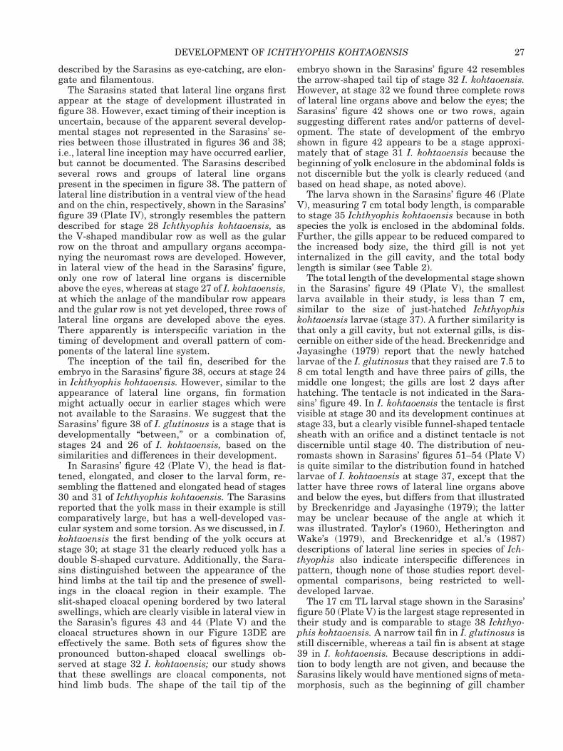
described by the Sarasins as eye-catching, are elon-gate and filamentous.
The Sarasins stated that lateral line organs firstappear at the stage of development illustrated infigure 38. However, exact timing of their inception isuncertain, because of the apparent several develop-mental stages not represented in the Sarasins’ se-ries between those illustrated in figures 36 and 38;i.e., lateral line inception may have occurred earlier,but cannot be documented. The Sarasins describedseveral rows and groups of lateral line organspresent in the specimen in figure 38. The pattern oflateral line distribution in a ventral view of the headand on the chin, respectively, shown in the Sarasins’figure 39 (Plate IV), strongly resembles the patterndescribed for stage 28 Ichthyophis kohtaoensis, asthe V-shaped mandibular row as well as the gularrow on the throat and ampullary organs accompa-nying the neuromast rows are developed. However,in lateral view of the head in the Sarasins’ figure,only one row of lateral line organs is discernibleabove the eyes, whereas at stage 27 of I. kohtaoensis,at which the anlage of the mandibular row appearsand the gular row is not yet developed, three rows oflateral line organs are developed above the eyes.There apparently is interspecific variation in thetiming of development and overall pattern of com-ponents of the lateral line system.
The inception of the tail fin, described for theembryo in the Sarasins’ figure 38, occurs at stage 24in Ichthyophis kohtaoensis. However, similar to theappearance of lateral line organs, fin formationmight actually occur in earlier stages which werenot available to the Sarasins. We suggest that theSarasins’ figure 38 of I. glutinosus is a stage that isdevelopmentally “between,” or a combination of,stages 24 and 26 of I. kohtaoensis, based on thesimilarities and differences in their development.
In Sarasins’ figure 42 (Plate V), the head is flat-tened, elongated, and closer to the larval form, re-sembling the flattened and elongated head of stages30 and 31 of Ichthyophis kohtaoensis. The Sarasinsreported that the yolk mass in their example is stillcomparatively large, but has a well-developed vas-cular system and some torsion. As we discussed, in I.kohtaoensis the first bending of the yolk occurs atstage 30; at stage 31 the clearly reduced yolk has adouble S-shaped curvature. Additionally, the Sara-sins distinguished between the appearance of thehind limbs at the tail tip and the presence of swell-ings in the cloacal region in their example. Theslit-shaped cloacal opening bordered by two lateralswellings, which are clearly visible in lateral view inthe Sarasin’s figures 43 and 44 (Plate V) and thecloacal structures shown in our Figure 13DE areeffectively the same. Both sets of figures show thepronounced button-shaped cloacal swellings ob-served at stage 32 I. kohtaoensis; our study showsthat these swellings are cloacal components, nothind limb buds. The shape of the tail tip of the
embryo shown in the Sarasins’ figure 42 resemblesthe arrow-shaped tail tip of stage 32 I. kohtaoensis.However, at stage 32 we found three complete rowsof lateral line organs above and below the eyes; theSarasins’ figure 42 shows one or two rows, againsuggesting different rates and/or patterns of devel-opment. The state of development of the embryoshown in figure 42 appears to be a stage approxi-mately that of stage 31 I. kohtaoensis because thebeginning of yolk enclosure in the abdominal folds isnot discernible but the yolk is clearly reduced (andbased on head shape, as noted above).
The larva shown in the Sarasins’ figure 46 (PlateV), measuring 7 cm total body length, is comparableto stage 35 Ichthyophis kohtaoensis because in bothspecies the yolk is enclosed in the abdominal folds.Further, the gills appear to be reduced compared tothe increased body size, the third gill is not yetinternalized in the gill cavity, and the total bodylength is similar (see Table 2).
The total length of the developmental stage shownin the Sarasins’ figure 49 (Plate V), the smallestlarva available in their study, is less than 7 cm,similar to the size of just-hatched Ichthyophiskohtaoensis larvae (stage 37). A further similarity isthat only a gill cavity, but not external gills, is dis-cernible on either side of the head. Breckenridge andJayasinghe (1979) report that the newly hatchedlarvae of the I. glutinosus that they raised are 7.5 to8 cm total length and have three pairs of gills, themiddle one longest; the gills are lost 2 days afterhatching. The tentacle is not indicated in the Sara-sins’ figure 49. In I. kohtaoensis the tentacle is firstvisible at stage 30 and its development continues atstage 33, but a clearly visible funnel-shaped tentaclesheath with an orifice and a distinct tentacle is notdiscernible until stage 40. The distribution of neu-romasts shown in Sarasins’ figures 51–54 (Plate V)is quite similar to the distribution found in hatchedlarvae of I. kohtaoensis at stage 37, except that thelatter have three rows of lateral line organs aboveand below the eyes, but differs from that illustratedby Breckenridge and Jayasinghe (1979); the lattermay be unclear because of the angle at which itwas illustrated. Taylor’s (1960), Hetherington andWake’s (1979), and Breckenridge et al.’s (1987)descriptions of lateral line series in species of Ich-thyophis also indicate interspecific differences inpattern, though none of those studies report devel-opmental comparisons, being restricted to well-developed larvae.
The 17 cm TL larval stage shown in the Sarasins’figure 50 (Plate V) is the largest stage represented intheir study and is comparable to stage 38 Ichthyo-phis kohtaoensis. A narrow tail fin in I. glutinosus isstill discernible, whereas a tail fin is absent at stage39 in I. kohtaoensis. Because descriptions in addi-tion to body length are not given, and because theSarasins likely would have mentioned signs of meta-morphosis, such as the beginning of gill chamber
27DEVELOPMENT OF ICHTHYOPHIS KOHTAOENSIS
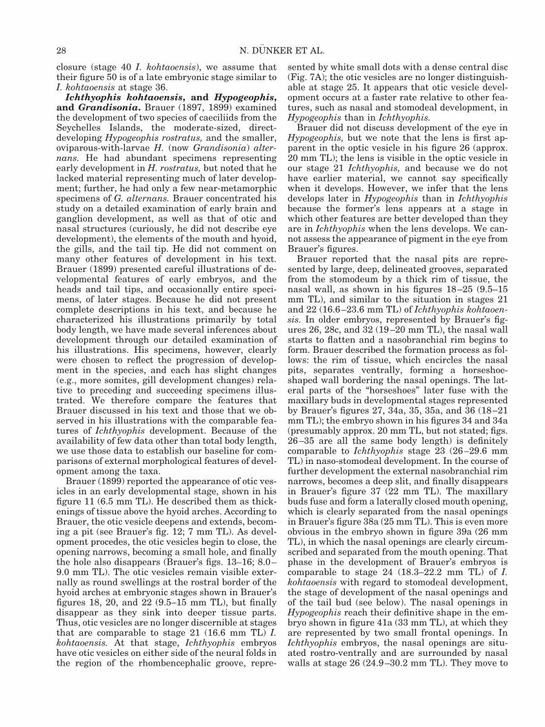
closure (stage 40 I. kohtaoensis), we assume thattheir figure 50 is of a late embryonic stage similar toI. kohtaoensis at stage 36.
Ichthyophis kohtaoensis, and Hypogeophis,and Grandisonia. Brauer (1897, 1899) examinedthe development of two species of caeciliids from theSeychelles Islands, the moderate-sized, direct-developing Hypogeophis rostratus, and the smaller,oviparous-with-larvae H. (now Grandisonia) alter-nans. He had abundant specimens representingearly development in H. rostratus, but noted that helacked material representing much of later develop-ment; further, he had only a few near-metamorphicspecimens of G. alternans. Brauer concentrated hisstudy on a detailed examination of early brain andganglion development, as well as that of otic andnasal structures (curiously, he did not describe eyedevelopment), the elements of the mouth and hyoid,the gills, and the tail tip. He did not comment onmany other features of development in his text.Brauer (1899) presented careful illustrations of de-velopmental features of early embryos, and theheads and tail tips, and occasionally entire speci-mens, of later stages. Because he did not presentcomplete descriptions in his text, and because hecharacterized his illustrations primarily by totalbody length, we have made several inferences aboutdevelopment through our detailed examination ofhis illustrations. His specimens, however, clearlywere chosen to reflect the progression of develop-ment in the species, and each has slight changes(e.g., more somites, gill development changes) rela-tive to preceding and succeeding specimens illus-trated. We therefore compare the features thatBrauer discussed in his text and those that we ob-served in his illustrations with the comparable fea-tures of Ichthyophis development. Because of theavailability of few data other than total body length,we use those data to establish our baseline for com-parisons of external morphological features of devel-opment among the taxa.
Brauer (1899) reported the appearance of otic ves-icles in an early developmental stage, shown in hisfigure 11 (6.5 mm TL). He described them as thick-enings of tissue above the hyoid arches. According toBrauer, the otic vesicle deepens and extends, becom-ing a pit (see Brauer’s fig. 12; 7 mm TL). As devel-opment procedes, the otic vesicles begin to close, theopening narrows, becoming a small hole, and finallythe hole also disappears (Brauer’s figs. 13–16; 8.0–9.0 mm TL). The otic vesicles remain visible exter-nally as round swellings at the rostral border of thehyoid arches at embryonic stages shown in Brauer’sfigures 18, 20, and 22 (9.5–15 mm TL), but finallydisappear as they sink into deeper tissue parts.Thus, otic vesicles are no longer discernible at stagesthat are comparable to stage 21 (16.6 mm TL) I.kohtaoensis. At that stage, Ichthyophis embryoshave otic vesicles on either side of the neural folds inthe region of the rhombencephalic groove, repre-
sented by white small dots with a dense central disc(Fig. 7A); the otic vesicles are no longer distinguish-able at stage 25. It appears that otic vesicle devel-opment occurs at a faster rate relative to other fea-tures, such as nasal and stomodeal development, inHypogeophis than in Ichthyophis.
Brauer did not discuss development of the eye inHypogeophis, but we note that the lens is first ap-parent in the optic vesicle in his figure 26 (approx.20 mm TL); the lens is visible in the optic vesicle inour stage 21 Ichthyophis, and because we do nothave earlier material, we cannot say specificallywhen it develops. However, we infer that the lensdevelops later in Hypogeophis than in Ichthyophisbecause the former’s lens appears at a stage inwhich other features are better developed than theyare in Ichthyophis when the lens develops. We can-not assess the appearance of pigment in the eye fromBrauer’s figures.
Brauer reported that the nasal pits are repre-sented by large, deep, delineated grooves, separatedfrom the stomodeum by a thick rim of tissue, thenasal wall, as shown in his figures 18–25 (9.5–15mm TL), and similar to the situation in stages 21and 22 (16.6–23.6 mm TL) of Ichthyophis kohtaoen-sis. In older embryos, represented by Brauer’s fig-ures 26, 28c, and 32 (19–20 mm TL), the nasal wallstarts to flatten and a nasobranchial rim begins toform. Brauer described the formation process as fol-lows: the rim of tissue, which encircles the nasalpits, separates ventrally, forming a horseshoe-shaped wall bordering the nasal openings. The lat-eral parts of the “horseshoes” later fuse with themaxillary buds in developmental stages representedby Brauer’s figures 27, 34a, 35, 35a, and 36 (18–21mm TL); the embryo shown in his figures 34 and 34a(presumably approx. 20 mm TL, but not stated; figs.26–35 are all the same body length) is definitelycomparable to Ichthyophis stage 23 (26–29.6 mmTL) in naso-stomodeal development. In the course offurther development the external nasobranchial rimnarrows, becomes a deep slit, and finally disappearsin Brauer’s figure 37 (22 mm TL). The maxillarybuds fuse and form a laterally closed mouth opening,which is clearly separated from the nasal openingsin Brauer’s figure 38a (25 mm TL). This is even moreobvious in the embryo shown in figure 39a (26 mmTL), in which the nasal openings are clearly circum-scribed and separated from the mouth opening. Thatphase in the development of Brauer’s embryos iscomparable to stage 24 (18.3–22.2 mm TL) of I.kohtaoensis with regard to stomodeal development,the stage of development of the nasal openings andof the tail bud (see below). The nasal openings inHypogeophis reach their definitive shape in the em-bryo shown in figure 41a (33 mm TL), at which theyare represented by two small frontal openings. InIchthyophis embryos, the nasal openings are situ-ated rostro-ventrally and are surrounded by nasalwalls at stage 26 (24.9–30.2 mm TL). They move to
28 N. DUNKER ET AL.
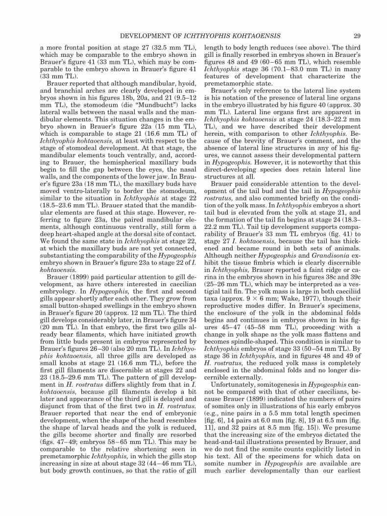
a more frontal position at stage 27 (32.5 mm TL),which may be comparable to the embryo shown inBrauer’s figure 41 (33 mm TL), which may be com-parable to the embryo shown in Brauer’s figure 41(33 mm TL).
Brauer reported that although mandibular, hyoid,and branchial arches are clearly developed in em-bryos shown in his figures 18b, 20a, and 21 (9.5–12mm TL), the stomodeum (die “Mundbucht”) lackslateral walls between the nasal walls and the man-dibular elements. This situation changes in the em-bryo shown in Brauer’s figure 22a (15 mm TL),which is comparable to stage 21 (16.6 mm TL) ofIchthyophis kohtaoensis, at least with respect to thestage of stomodeal development. At that stage, themandibular elements touch ventrally, and, accord-ing to Brauer, the hemispherical maxillary budsbegin to fill the gap between the eyes, the nasalwalls, and the components of the lower jaw. In Brau-er’s figure 23a (18 mm TL), the maxillary buds havemoved ventro-laterally to border the stomodeum,similar to the situation in Ichthyophis at stage 22(18.5–23.6 mm TL). Brauer stated that the mandib-ular elements are fused at this stage. However, re-ferring to figure 23a, the paired mandibular ele-ments, although continuous ventrally, still form adeep heart-shaped angle at the dorsal site of contact.We found the same state in Ichthyophis at stage 22,at which the maxillary buds are not yet connected,substantiating the comparability of the Hypogeophisembryo shown in Brauer’s figure 23a to stage 22 of I.kohtaoensis.
Brauer (1899) paid particular attention to gill de-velopment, as have others interested in caecilianembryology. In Hypogeophis, the first and secondgills appear shortly after each other. They grow fromsmall button-shaped swellings in the embryo shownin Brauer’s figure 20 (approx. 12 mm TL). The thirdgill develops considerably later, in Brauer’s figure 34(20 mm TL). In that embryo, the first two gills al-ready bear filaments, which have initiated growthfrom little buds present in embryos represented byBrauer’s figures 26–30 (also 20 mm TL). In Ichthyo-phis kohtaoensis, all three gills are developed assmall knobs at stage 21 (16.6 mm TL), before thefirst gill filaments are discernible at stages 22 and23 (18.5–29.6 mm TL). The pattern of gill develop-ment in H. rostratus differs slightly from that in I.kohtaoensis, because gill filaments develop a bitlater and appearance of the third gill is delayed anddisjunct from that of the first two in H. rostratus.Brauer reported that near the end of embryonicdevelopment, when the shape of the head resemblesthe shape of larval heads and the yolk is reduced,the gills become shorter and finally are resorbed(figs. 47–49; embryos 58–65 mm TL). This may becomparable to the relative shortening seen inpremetamorphic Ichthyophis, in which the gills stopincreasing in size at about stage 32 (44–46 mm TL),but body growth continues, so that the ratio of gill
length to body length reduces (see above). The thirdgill is finally resorbed in embryos shown in Brauer’sfigures 48 and 49 (60–65 mm TL), which resembleIchthyophis stage 36 (70.1–83.0 mm TL) in manyfeatures of development that characterize thepremetamorphic state.
Brauer’s only reference to the lateral line systemis his notation of the presence of lateral line organsin the embryo illustrated by his figure 40 (approx. 30mm TL). Lateral line organs first are apparent inIchthyophis kohtaoensis at stage 24 (18.3–22.2 mmTL), and we have described their developmentherein, with comparison to other Ichthyophis. Be-cause of the brevity of Brauer’s comment, and theabsence of lateral line structures in any of his fig-ures, we cannot assess their developmental patternin Hypogeophis. However, it is noteworthy that thisdirect-developing species does retain lateral linestructures at all.
Brauer paid considerable attention to the devel-opment of the tail bud and the tail in Hypogeophisrostratus, and also commented briefly on the condi-tion of the yolk mass. In Ichthyophis embryos a shorttail bud is elevated from the yolk at stage 21, andthe formation of the tail fin begins at stage 24 (18.3–22.2 mm TL). Tail tip development supports compa-rability of Brauer’s 33 mm TL embryos (fig. 41) tostage 27 I. kohtaoensis, because the tail has thick-ened and became round in both sets of animals.Although neither Hypogeophis and Grandisonia ex-hibit the tissue fimbris which is clearly discerniblein Ichthyophis, Brauer reported a faint ridge or ca-rina in the embryos shown in his figures 38c and 39c(25–26 mm TL), which may be interpreted as a ves-tigial tail fin. The yolk mass is large in both caeciliidtaxa (approx. 9 3 6 mm; Wake, 1977), though theirreproductive modes differ. In Brauer’s specimens,the enclosure of the yolk in the abdominal foldsbegins and continues in embryos shown in his fig-ures 45–47 (45–58 mm TL), proceeding with achange in yolk shape as the yolk mass flattens andbecomes spindle-shaped. This condition is similar toIchthyophis embryos of stage 33 (50–54 mm TL). Bystage 36 in Ichthyophis, and in figures 48 and 49 ofH. rostratus, the reduced yolk mass is completelyenclosed in the abdominal folds and no longer dis-cernible externally.
Unfortunately, somitogenesis in Hypogeophis can-not be compared with that of other caecilians, be-cause Brauer (1899) indicated the numbers of pairsof somites only in illustrations of his early embryos(e.g., nine pairs in a 5.5 mm total length specimen[fig. 6], 14 pairs at 6.0 mm [fig. 8], 19 at 6.5 mm [fig.11], and 32 pairs at 8.5 mm [fig. 15]). We presumethat the increasing size of the embryos dictated thehead-and-tail illustrations presented by Brauer, andwe do not find the somite counts explicitly listed inhis text. All of the specimens for which data onsomite number in Hypogeophis are available aremuch earlier developmentally than our earliest
29DEVELOPMENT OF ICHTHYOPHIS KOHTAOENSIS
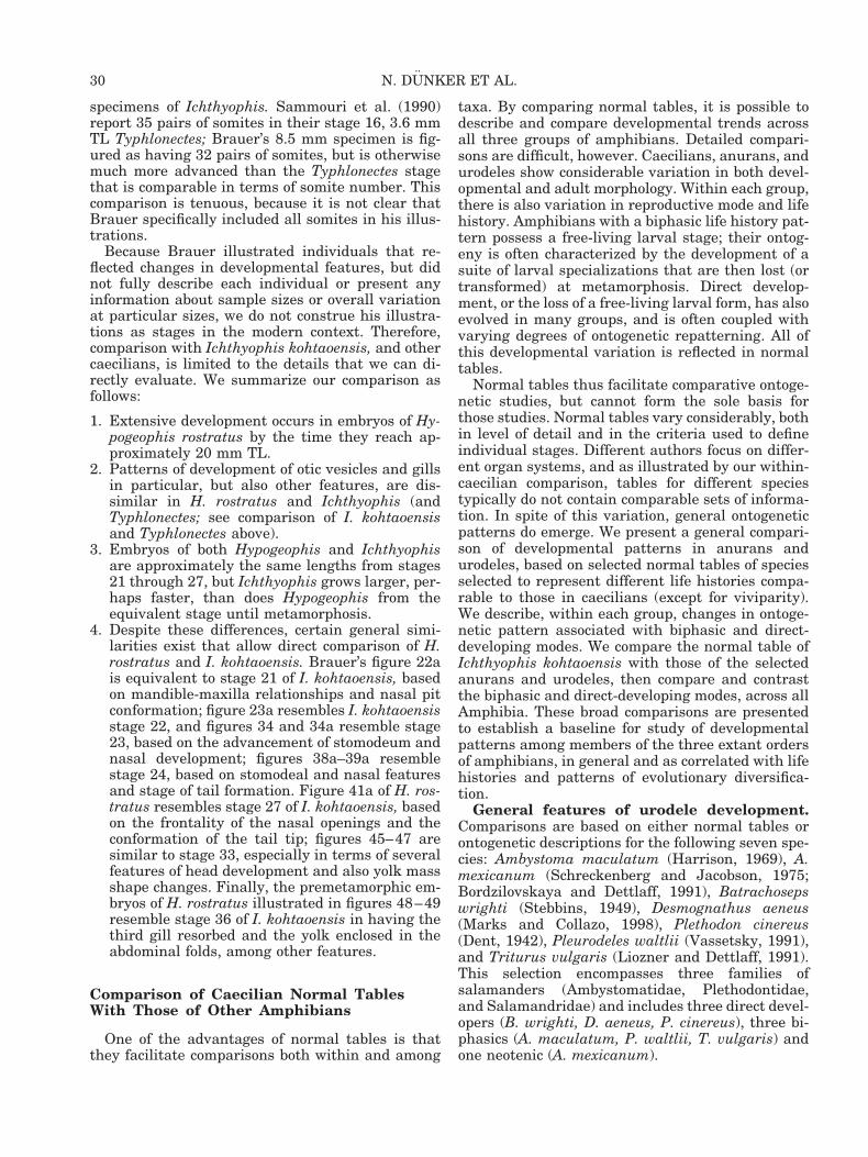
specimens of Ichthyophis. Sammouri et al. (1990)report 35 pairs of somites in their stage 16, 3.6 mmTL Typhlonectes; Brauer’s 8.5 mm specimen is fig-ured as having 32 pairs of somites, but is otherwisemuch more advanced than the Typhlonectes stagethat is comparable in terms of somite number. Thiscomparison is tenuous, because it is not clear thatBrauer specifically included all somites in his illus-trations.
Because Brauer illustrated individuals that re-flected changes in developmental features, but didnot fully describe each individual or present anyinformation about sample sizes or overall variationat particular sizes, we do not construe his illustra-tions as stages in the modern context. Therefore,comparison with Ichthyophis kohtaoensis, and othercaecilians, is limited to the details that we can di-rectly evaluate. We summarize our comparison asfollows:
1. Extensive development occurs in embryos of Hy-pogeophis rostratus by the time they reach ap-proximately 20 mm TL.
2. Patterns of development of otic vesicles and gillsin particular, but also other features, are dis-similar in H. rostratus and Ichthyophis (andTyphlonectes; see comparison of I. kohtaoensisand Typhlonectes above).
3. Embryos of both Hypogeophis and Ichthyophisare approximately the same lengths from stages21 through 27, but Ichthyophis grows larger, per-haps faster, than does Hypogeophis from theequivalent stage until metamorphosis.
4. Despite these differences, certain general simi-larities exist that allow direct comparison of H.rostratus and I. kohtaoensis. Brauer’s figure 22ais equivalent to stage 21 of I. kohtaoensis, basedon mandible-maxilla relationships and nasal pitconformation; figure 23a resembles I. kohtaoensisstage 22, and figures 34 and 34a resemble stage23, based on the advancement of stomodeum andnasal development; figures 38a–39a resemblestage 24, based on stomodeal and nasal featuresand stage of tail formation. Figure 41a of H. ros-tratus resembles stage 27 of I. kohtaoensis, basedon the frontality of the nasal openings and theconformation of the tail tip; figures 45–47 aresimilar to stage 33, especially in terms of severalfeatures of head development and also yolk massshape changes. Finally, the premetamorphic em-bryos of H. rostratus illustrated in figures 48–49resemble stage 36 of I. kohtaoensis in having thethird gill resorbed and the yolk enclosed in theabdominal folds, among other features.
Comparison of Caecilian Normal TablesWith Those of Other Amphibians
One of the advantages of normal tables is thatthey facilitate comparisons both within and among
taxa. By comparing normal tables, it is possible todescribe and compare developmental trends acrossall three groups of amphibians. Detailed compari-sons are difficult, however. Caecilians, anurans, andurodeles show considerable variation in both devel-opmental and adult morphology. Within each group,there is also variation in reproductive mode and lifehistory. Amphibians with a biphasic life history pat-tern possess a free-living larval stage; their ontog-eny is often characterized by the development of asuite of larval specializations that are then lost (ortransformed) at metamorphosis. Direct develop-ment, or the loss of a free-living larval form, has alsoevolved in many groups, and is often coupled withvarying degrees of ontogenetic repatterning. All ofthis developmental variation is reflected in normaltables.
Normal tables thus facilitate comparative ontoge-netic studies, but cannot form the sole basis forthose studies. Normal tables vary considerably, bothin level of detail and in the criteria used to defineindividual stages. Different authors focus on differ-ent organ systems, and as illustrated by our within-caecilian comparison, tables for different speciestypically do not contain comparable sets of informa-tion. In spite of this variation, general ontogeneticpatterns do emerge. We present a general compari-son of developmental patterns in anurans andurodeles, based on selected normal tables of speciesselected to represent different life histories compa-rable to those in caecilians (except for viviparity).We describe, within each group, changes in ontoge-netic pattern associated with biphasic and direct-developing modes. We compare the normal table ofIchthyophis kohtaoensis with those of the selectedanurans and urodeles, then compare and contrastthe biphasic and direct-developing modes, across allAmphibia. These broad comparisons are presentedto establish a baseline for study of developmentalpatterns among members of the three extant ordersof amphibians, in general and as correlated with lifehistories and patterns of evolutionary diversifica-tion.
General features of urodele development.Comparisons are based on either normal tables orontogenetic descriptions for the following seven spe-cies: Ambystoma maculatum (Harrison, 1969), A.mexicanum (Schreckenberg and Jacobson, 1975;Bordzilovskaya and Dettlaff, 1991), Batrachosepswrighti (Stebbins, 1949), Desmognathus aeneus(Marks and Collazo, 1998), Plethodon cinereus(Dent, 1942), Pleurodeles waltlii (Vassetsky, 1991),and Triturus vulgaris (Liozner and Dettlaff, 1991).This selection encompasses three families ofsalamanders (Ambystomatidae, Plethodontidae,and Salamandridae) and includes three direct devel-opers (B. wrighti, D. aeneus, P. cinereus), three bi-phasics (A. maculatum, P. waltlii, T. vulgaris) andone neotenic (A. mexicanum).
30 N. DUNKER ET AL.
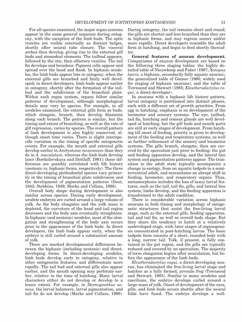
For all species examined, the major organ systemsappear in the same general sequence during ontog-eny, with the exception of the limb buds. The opticvesicles are visible externally as distinct bulges,shortly after neural tube closure. The visceralarches then develop, giving rise to the external gillbuds and stomodeal elements. The tailbud appears,followed by the otic, then olfactory vesicles. The tailfin develops and broadens. Pigment cells appear andspread over the head and flank. In biphasic urode-les, the limb buds appear late in ontogeny, when theexternal gills are branched and fairly well devel-oped; in direct-developers, limb buds appear earlierin ontogeny, shortly after the formation of the tail-bud and the subdivision of the branchial plate.Within each organ system, organs follow similarpatterns of development, although morphologicaldetails may vary by species. For example, in allurodeles examined, the external gills form as buds,which elongate, branch, then develop filamentsalong each branch. The pattern is similar, but thetiming and extent of branching, as well as the rate ofgill regression, varies by species. The overall patternof limb development is also highly conserved, al-though onset time varies. There is slight interspe-cific variation in the timing of specific ontogeneticevents. For example, the mouth and external gillsdevelop earlier in Ambystoma mexicanum than theydo in A. maculatum, whereas the limb buds developlater (Bordzilovskaya and Dettlaff, 1991); these dif-ferences are possibly correlated with life history(neotenic vs. biphasic forms, respectively). The threedirect-developing plethodontid species vary primar-ily in the timing of branchial plate subdivision andthe development of pigmentation patterns (Dent,1942; Stebbins, 1949; Marks and Collazo, 1998).
Overall body shape during development is alsosimilar across species. During early development,urodele embryos are curled around a large volume ofyolk. As the body elongates and the yolk mass isdepleted, the curvature of the head and tail regiondecreases and the body axis eventually straightens.In biphasic (and neotenic) urodeles, most of the elon-gation and straightening of the body axis occursprior to the appearance of the limb buds. In directdevelopers, the limb buds appear early, when theembryo is still curled around a substantial amountof yolk.
There are marked developmental differences be-tween the biphasic (including neotenic) and direct-developing forms. In direct-developing urodeles,limb buds develop early in ontogeny, relative toother ontogenetic features, and differentiate morerapidly. The tail bud and external gills also appearearlier, and the mouth opening may perforate ear-lier, relative to the time of hatching. Many larvalcharacters either do not develop or develop to alesser extent. For example, in Desmognathus ae-neus, the larval balancers, larval pigmentation, andtail fin do not develop (Marks and Collazo, 1998).
During ontogeny, the tail remains short and round;the gills are shorter and less branched than they arein biphasic forms, and may regress sooner and/ormore rapidly. Direct developers resemble the adultform at hatching, and begin to feed shortly thereaf-ter.
General features of anuran development.Comparisons of anuran development are based onthe following three staging tables: the highly de-tailed table of Nieuwkoop and Faber (1967; Xenopuslaevis, a biphasic, secondarily fully aquatic anuran),the generalized table of Gosner (1960; widely usedfor staging of biphasic anurans), and the table ofTownsend and Stewart (1985; Eleutherodactylus co-qui, a direct-developer).
In anurans with a biphasic life history pattern,larval ontogeny is partitioned into distinct phases,each with a different set of growth priorities. Fromegg to hatching, emphasis is on development of thelocomotor and sensory systems. The eye, tailbud,tail fin, hatching and cement glands are well devel-oped at hatching, but the gill buds and mouth partsare still at early stages of development. From hatch-ing till onset of feeding, priority is given to develop-ment of the feeding and respiratory systems, as wellas further refinement of the sensory and locomotorsystems. The gills branch, elongate, then are cov-ered by the operculum just prior to feeding; the gutand feeding apparatus develop, and the lateral linesystem and pigmentation patterns appear. The tran-sition to the adult state typically accompanies achange in ecology, from an aquatic tadpole to a semi-terrestrial adult, and necessitates an abrupt shift infeeding, locomotor, and respiratory organs. Thus,metamorphosis includes the loss of many larval fea-tures, such as the tail, tail fin, gills, and lateral linesystem; limbs develop, and the feeding apparatus istransformed to the adult condition.
There is considerable variation across biphasicanurans in both timing and morphology of ontoge-netic structures that assist the free-living larvalstage, such as the external gills, feeding apparatus,tail and tail fin, as well as overall body shape. Butthey share the tendency to hatch at a relativelyundeveloped stage, with later stages of organogene-sis concentrated in post-hatching larvae. The basictadpole form consists of a short, rounded body witha long, narrow tail. Yolk, if present, is fully con-tained in the gut region, and the gills are typicallyreduced and covered by an operculum. The majorityof torso elongation begins after neurulation, but be-fore the appearance of the limb buds.
Eleutherodactylus coqui, a direct-developing anu-ran, has eliminated the free-living larval stage andhatches as a fully formed, juvenile frog (Townsendand Stewart, 1985). Similar to many urodeles andcaecilians, the embryo develops curled around alarge mass of yolk. Onset of development of the eyes,gills, and limb buds occurs shortly after the neuralfolds have fused. The embryo develops a well-
31DEVELOPMENT OF ICHTHYOPHIS KOHTAOENSIS
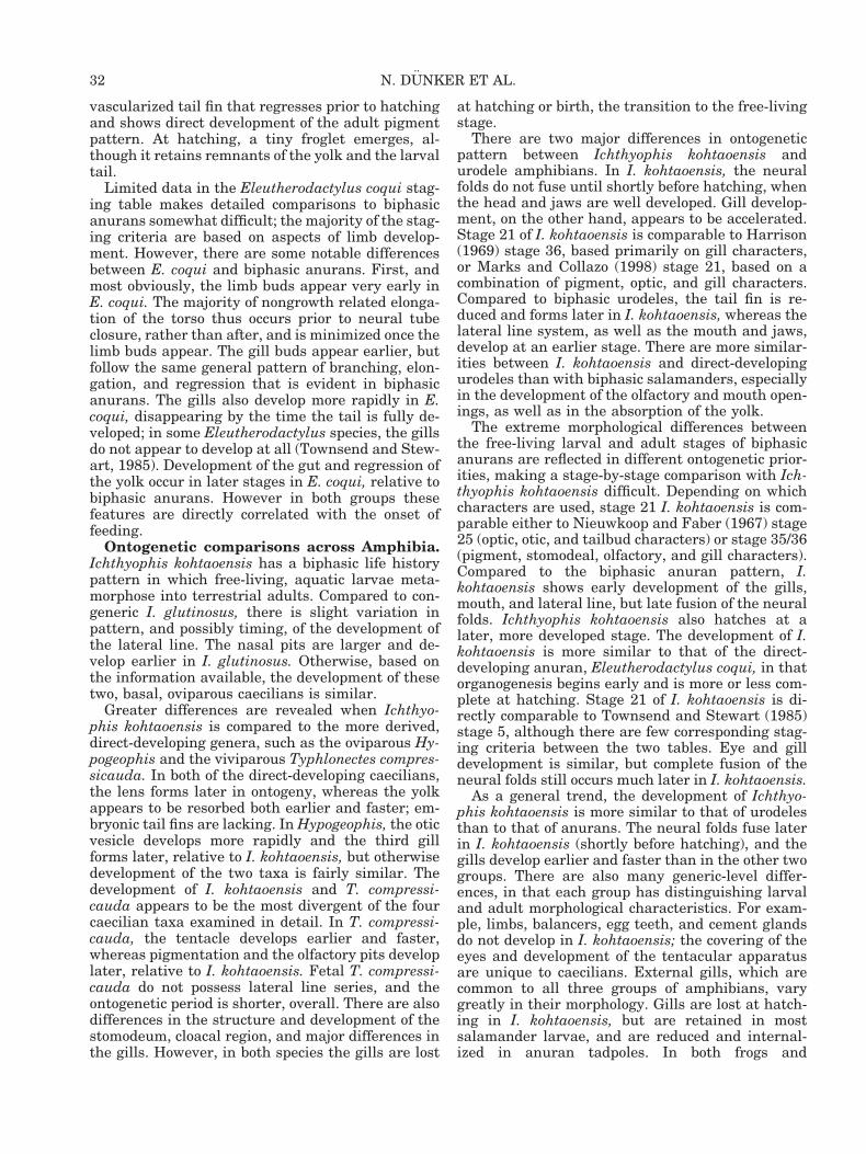
vascularized tail fin that regresses prior to hatchingand shows direct development of the adult pigmentpattern. At hatching, a tiny froglet emerges, al-though it retains remnants of the yolk and the larvaltail.
Limited data in the Eleutherodactylus coqui stag-ing table makes detailed comparisons to biphasicanurans somewhat difficult; the majority of the stag-ing criteria are based on aspects of limb develop-ment. However, there are some notable differencesbetween E. coqui and biphasic anurans. First, andmost obviously, the limb buds appear very early inE. coqui. The majority of nongrowth related elonga-tion of the torso thus occurs prior to neural tubeclosure, rather than after, and is minimized once thelimb buds appear. The gill buds appear earlier, butfollow the same general pattern of branching, elon-gation, and regression that is evident in biphasicanurans. The gills also develop more rapidly in E.coqui, disappearing by the time the tail is fully de-veloped; in some Eleutherodactylus species, the gillsdo not appear to develop at all (Townsend and Stew-art, 1985). Development of the gut and regression ofthe yolk occur in later stages in E. coqui, relative tobiphasic anurans. However in both groups thesefeatures are directly correlated with the onset offeeding.
Ontogenetic comparisons across Amphibia.Ichthyophis kohtaoensis has a biphasic life historypattern in which free-living, aquatic larvae meta-morphose into terrestrial adults. Compared to con-generic I. glutinosus, there is slight variation inpattern, and possibly timing, of the development ofthe lateral line. The nasal pits are larger and de-velop earlier in I. glutinosus. Otherwise, based onthe information available, the development of thesetwo, basal, oviparous caecilians is similar.
Greater differences are revealed when Ichthyo-phis kohtaoensis is compared to the more derived,direct-developing genera, such as the oviparous Hy-pogeophis and the viviparous Typhlonectes compres-sicauda. In both of the direct-developing caecilians,the lens forms later in ontogeny, whereas the yolkappears to be resorbed both earlier and faster; em-bryonic tail fins are lacking. In Hypogeophis, the oticvesicle develops more rapidly and the third gillforms later, relative to I. kohtaoensis, but otherwisedevelopment of the two taxa is fairly similar. Thedevelopment of I. kohtaoensis and T. compressi-cauda appears to be the most divergent of the fourcaecilian taxa examined in detail. In T. compressi-cauda, the tentacle develops earlier and faster,whereas pigmentation and the olfactory pits developlater, relative to I. kohtaoensis. Fetal T. compressi-cauda do not possess lateral line series, and theontogenetic period is shorter, overall. There are alsodifferences in the structure and development of thestomodeum, cloacal region, and major differences inthe gills. However, in both species the gills are lost
at hatching or birth, the transition to the free-livingstage.
There are two major differences in ontogeneticpattern between Ichthyophis kohtaoensis andurodele amphibians. In I. kohtaoensis, the neuralfolds do not fuse until shortly before hatching, whenthe head and jaws are well developed. Gill develop-ment, on the other hand, appears to be accelerated.Stage 21 of I. kohtaoensis is comparable to Harrison(1969) stage 36, based primarily on gill characters,or Marks and Collazo (1998) stage 21, based on acombination of pigment, optic, and gill characters.Compared to biphasic urodeles, the tail fin is re-duced and forms later in I. kohtaoensis, whereas thelateral line system, as well as the mouth and jaws,develop at an earlier stage. There are more similar-ities between I. kohtaoensis and direct-developingurodeles than with biphasic salamanders, especiallyin the development of the olfactory and mouth open-ings, as well as in the absorption of the yolk.
The extreme morphological differences betweenthe free-living larval and adult stages of biphasicanurans are reflected in different ontogenetic prior-ities, making a stage-by-stage comparison with Ich-thyophis kohtaoensis difficult. Depending on whichcharacters are used, stage 21 I. kohtaoensis is com-parable either to Nieuwkoop and Faber (1967) stage25 (optic, otic, and tailbud characters) or stage 35/36(pigment, stomodeal, olfactory, and gill characters).Compared to the biphasic anuran pattern, I.kohtaoensis shows early development of the gills,mouth, and lateral line, but late fusion of the neuralfolds. Ichthyophis kohtaoensis also hatches at alater, more developed stage. The development of I.kohtaoensis is more similar to that of the direct-developing anuran, Eleutherodactylus coqui, in thatorganogenesis begins early and is more or less com-plete at hatching. Stage 21 of I. kohtaoensis is di-rectly comparable to Townsend and Stewart (1985)stage 5, although there are few corresponding stag-ing criteria between the two tables. Eye and gilldevelopment is similar, but complete fusion of theneural folds still occurs much later in I. kohtaoensis.
As a general trend, the development of Ichthyo-phis kohtaoensis is more similar to that of urodelesthan to that of anurans. The neural folds fuse laterin I. kohtaoensis (shortly before hatching), and thegills develop earlier and faster than in the other twogroups. There are also many generic-level differ-ences, in that each group has distinguishing larvaland adult morphological characteristics. For exam-ple, limbs, balancers, egg teeth, and cement glandsdo not develop in I. kohtaoensis; the covering of theeyes and development of the tentacular apparatusare unique to caecilians. External gills, which arecommon to all three groups of amphibians, varygreatly in their morphology. Gills are lost at hatch-ing in I. kohtaoensis, but are retained in mostsalamander larvae, and are reduced and internal-ized in anuran tadpoles. In both frogs and
32 N. DUNKER ET AL.
salamanders, gills are lost at metamorphosis (butare retained in neotenic forms).
Interestingly, the ontogenetic pattern of Ichthyo-phis kohtaoensis, a caecilian with a biphasic lifehistory, more strongly resembles the patterns ofdirect-developing anurans and urodeles, comparedto the ancestral biphasic forms. However, this maybe an artifact of the normal tables themselves. Thetables of direct-developing anurans and urodeles aretypically highly generalized and encompass largeintervals of ontogeny. Such tables tend to emphasizelimb development as the main staging criterion, sothere are relatively fewer criteria to match withdata for limbless caecilians. As a result, it is ofteneasier to compare the tables to those of other speciesand to assign comparable stages. The strong resem-blance of the biphasic caecilian pattern to the direct-developing anuran and urodele patterns may alsoreflect a fundamental ontogenetic difference be-tween the three groups of amphibians. Biphasic cae-cilians, in general, show less divergence of larvaland adult forms during ontogeny, relative to bipha-sic anurans and urodeles. The metamorphic transi-tion is thus less abrupt and exerts less influence onthe timing and compartmentalization of ontogeneticevents.
We stress that the evolution of development is anintegrated and integrative field of study, rich inempirical and heuristic value. We present our com-parisons of patterns of development among diverseamphibians to help to establish a baseline for fur-ther study of species with different evolutionary anddevelopmental histories that result in markedly dif-ferent phenotypes. We anticipate that such studieswill facilitate a more intensive and better informedanalysis of pattern and process of the evolution ofdevelopment.
ACKNOWLEDGMENTS
We thank Werner Himstedt (Technical Universityof Darmstadt [TUD]) for generously providing thespecimens. We appreciate the assistance with illus-trations from K. Klitz (Museum of Vertebrate Zool-ogy, University of California, Berkeley [UCB]) andA. Klepper (TUD). N. Dunker especially thanks P.Sicurello, Staff Research Associate in the ElectronMicroscope Laboratory (UCB), for her excellenttechnical assistance and SEM training, and W.P.Wang from the BioAAPE Image Analysis Labora-tory at UCB for his help in preparing the digitalfigures. Financial support was provided by NSFgrant IBN95-27681 and Chancellor’s Professorshipfunds to M.H. Wake.
LITERATURE CITED
Bartsch P, Gemballa S, Piotrowski T. 1997. The embryonic andlarval development of Polypterus senegalensis Cuvier, 1829; its
staging with reference to external and skeletal features, behav-ior and locomotory habits. Acta Zool (Stockholm) 78:309–328.
Bennett MVL. 1971. Electroreception. In: Hoar WS, Randall DJ,editors. Fish physiology. New York: Academic Press. p 493–574.
Billo R, Wake MH. 1987. Tentacle development in Dermophismexicanus (Amphibia; Gymnophiona) with an hypothesis oftentacle origin. J Morphol 192:101–111.
Bordzilovskaya NP, Dettlaff TA. 1991. The axolotl Ambystomamexicanum. In: Detlaff TA, Vassetzky SG, editors. Animal spe-cies for developmental studies, vol. 2. Vertebrates. Translatedby Gause GG Jr, Vassetzky SG. New York, London: Consult-ants Bureau. p 203–230.
Brauer A. 1897. Beitrage zur Kenntnis der Entwicklungsge-schichte und Anatomie der Gymnophionen. Zool Jahrb Anat10:277–472.
Brauer A. 1899. Beitrage zur Kenntniss der Entwicklung undAnatomie der Gymnophionen. Zool Jahrb Anat 12:477–508.
Breckenridge WR, Jayasinghe S. 1979. Observations on the eggsand larvae of Ichthyophis glutinosus. Ceylon J Sci 13:187–202.
Breckenridge WR, Nathanael S, Pereira L. 1987. Some aspects ofthe biology and development of Ichthyophis glutinosus (Am-phibia: Gymnophiona). J Zool London 211:437–449.
Cambar R, Martin S. 1959. Table chronologique du developpe-ment embryonnaire et larvaire du Crapaud accoucheur (Alytesobstetricans Laur.). Actes Soc Linn Bordeaux 98:1–20.
Coggi A. 1905. Le ampolle di Lorenzini nei Gimnofioni. Monit ZoolItal 16:49–56.
Delsol M, Flatin J, Exbrayat JM, Bons J. 1981. Developpement deTyphlonectes compressicaudus, Amphibien apode vivipare. Hy-potheses sur sa nutrition embryonnaire et larvaire par un ec-totrophoblaste. C R Acad Sci Paris 293:281–285.
Duellman WE, Trueb L. 1986. Biology of amphibians. New York:McGraw Hill. 670 p.
Duges A. 1835. Recherches sur l’osteologie et la myologie desBatraciens a leurs differens ages. Mem Acad Sci Paris (Sc MathPhys) 6:21, 23, 159, 201, 209.
Exbrayat JM, Hraoui-Bloquet S. 1991. Morphologie de l’epithe-lium branchial des embryons de Typhlonectes compressicaudus(Amphibien Gymnophione) etudie en microscopie electronique abalayage. Bull Soc Herp France 57:45–62.
Exbrayat JM, Hraoui-Bloquet S. 1992. Evolution de la surfacebranchiale des embryons de Typhlonectes compressicaudus,Amphibien Gymnophione vivipare, au cours du developpement.Bull Soc Zool France 117:340.
Exbrayat JM, Hraoui-Bloquet S. 1994. Un exemple d’heterochronie:la metamorphose chez les gymnophiones. Bull Soc Zool France119:117–126.
Exbrayat JM, Delsol M, Flatin J. 1981. Premiere remarques surla gestation chez Typhlonectes compressicaudus (Dumeril etBibron, 1841), Amphibien Apode vivipare. C R Acad Sci Paris292:417–420.
Fritzsch B. 1981. The pattern of lateral line afferents in urodeles.A horseradish-peroxidase study. Cell Tissue Res 218:581–594.
Fritzsch B. 1990. The evolution of metamorphosis in amphibians.J Neurobiol 21:1011–1021.
Fritzsch B, Bolz D. 1986. On the development of electroreceptiveampullary organs of Triturus alpestris (Amphibia: Urodela).Amphib Rept 7:1–9.
Fritzsch B, Munz H. 1986. Electroreception in amphibians. In:Bullock TH, Heiligenberg W, editors. Electroreception. NewYork: John Wiley & Sons. p 483–496.
Fritzsch B, Wahnschaffe U. 1983. The electroreceptive ampullaryorgans of urodeles. Cell Tissue Res 229:483–503.
Fritzsch B, Wake MH. 1986. The distribution of ampullary organsin Gymnophiona. J Herpetol 20:90–93.
Fritzsch B, Wahnschaffe U, Crapon de Caprona MD, Himstedt W.1985. Anatomical evidence for electroreception in larval Ich-thyophis kohtaoensis. Naturwissenschaften 72:102–104.
Gosner KL. 1960. A simplified table for staging anuran embryosand larvae with notes on identification. Herpetologica 16:183–190.
33DEVELOPMENT OF ICHTHYOPHIS KOHTAOENSIS
Grillitsch B. 1989. On the relevance of certain external featuresin staging ranoid larvae (Amphibia: Anura). Fort Zool 35:269–275.
Harrison RG. 1969. Harrison stages and description of the nor-mal table of development of the spotted salamander, Amblys-toma punctatum (Linn.). In: Wilens S, editor. Organization anddevelopment of the embryo. New Haven: Yale University Press.p 44–66.
Hetherington TE, Wake MH. 1979. The lateral line system inlarval Ichthyophis (Amphibia: Gymnophiona). Zoomorphology93:209–225.
Himstedt W. 1996. Die Blindwuhlen. Magdeburg: Die NeueBrehm-Bucherei Bd. 630. 160 p.
Himstedt W, Fritzsch B. 1990. Behavioral evidence for electrore-ception in larvae of Ichthyophis kohtaoensis (Amphibia; Gym-nophiona). Zool Jahrb Physiol 94:486–492.
Hraoui-Bloquet S, Exbrayat JM. 1994. Developpement desbranchies chez les embryons de Typhlonectes compressicaudus,amphibien gymnophione vivipare. Ann Sci Nat Zool Paris 15:33–46.
Largen MJ, Morris PA, Yalden DW. 1972. Observations on thecaecilian Geotrypetes grandisonae Taylor (Amphibia; Gym-nophiona) from Ethiopia. Mon Zool Ital N S Suppl IV 8:185–205.
Liozner LD, Dettlaff TA. 1991. The newts Triturus vulgaris andTriturus cristatus. In: Detlaff TA, Vassetzky SG, editors. Ani-mal species for developmental studies, vol. 2. Vertebrates.Translated by Gause GG Jr, Vassetzky SG. New York, London:Consultants Bureau. p 145–165.
Marcus H. 1908. Beitrage zur Kenntnis der Gymnophionen. I.Uber das Schlundspaltengebiet. Arch mikr Anat 71:695–744.
Marks SB, Collazo A. 1998. Direct development in Desmognathusaeneus (Caudata: Plethodontidae): A staging table. Copeia1998:637–648.
Muller J. 1835. Uber die Kiemenlocher der jungen Coecilia hypo-cyanea. Arch Anat Physiol 1835:391–398.
Nieuwkoop PD, Faber F. 1967. Normal table of Xenopus laevis(Daudin). A systematical and chronological survey of the devel-opment from the fertilized egg till the end of metamorphosis.Amsterdam: North-Holland Publishing Company. 252 p.
Nussbaum RA, Wilkinson M. 1989. On the classification and ofcaecilians (Amphibia: Gymnophiona), a critical review. Herpe-tol Monogr 3:1–42.
Parker HW. 1956. Viviparous caecilians and amphibian phylog-eny. Nature 178:250–252.
Peters W. 1874. Uber die Entwicklung der Caecilien und be-sonders der Caecilia compressicauda. Monatsb Akad Wiss Ber-lin 1874:45–49.
Peters W. 1875. Uber die Entwicklung der Caecilien. MonatsbAkad Wiss Berlin 1875:483–486.
Presnell JK, Schreibman MP. 1997. Humason’s animal tissuetechniques. Baltimore: Johns Hopkins University Press. 572 p.
Ramaswami LS. 1954. The external gills of Gegenophis embryos.Anat Anz 101:120–123.
Rose CS, Reiss JO. 1993. Metamorphosis and the vertebrateskull: ontogenetic patterns and developmental mechanisms. In:
Hanken J, Hall BK, editors. The skull, vol. 1. Chicago: Univer-sity of Chicago Press. p 289–346.
Russel IJ. 1976. Amphibian lateral line receptors. In: Llinas R,Precht W, editors. Frog neurobiology. Berlin: Springer-Verlag.p 513–550.
Sammouri R, Renous S, Exbrayat JM, Lescure J. 1990. Devel-oppement embryonnaire de Typhlonectes compressicaudus(Amphibia, Gymnophiona). Ann Sci Nat Zool Paris 11:135–163.
Sanderson IT. 1936. Animal treasure. New York: Viking Press.325 p.
Sarasin P, Sarasin F. 1887–1890. Ergebnisse naturwissen-schaftlicher Forschungen auf Ceylon in den Jahren 1884–1886.Band II: Zur Entwicklungsgeschichte und Anatomie der ceylo-nesischen Blindwuhle Ichthyophis glutinosus L. Wiesbaden:C.W. Kreidel Verlag.
Schreckenberg GM, Jacobson AG. 1975. Normal stages of devel-opment of the axolotl, Ambystoma mexicanum. Dev Biol 42:391–400.
Seshachar BR. 1942. The eggs and embryos of Gegenophis carno-sus. Bedd Curr Sci 1942:439–441.
Shelton P. 1970. The lateral line system at metamorphosis inXenopus laevis (Daudin). J Embryol Morphol 24:511–524.
Stebbins RC. 1949. Observations on laying, development, andhatching of the eggs of Batrachoseps wrighti. Copeia 1949:161–168.
Taylor EH. 1960. On the caecilian species Ichthyophis mono-chrous and Ichthyophis glutinosus with descriptions of relatedspecies. Univ Kansas Sci Bull 40:37–120.
Taylor EH. 1968. The caecilians of the world. A taxonomic review.Lawrence: Univ Kansas Press. 848 p.
Taylor EH. 1970. The lateral-line sensory system in the caecilianfamily Ichthyophiidae (Amphibia: Gymnophiona). Univ KansasSci Bull 48:861–868.
Townsend DS, Stewart MM. 1985. Direct development in Eleu-therodactylus coqui (Anura: Leptodactylidae): a staging table.Copeia 1985:423–436.
Vassetzky SG. 1991. The Spanish newt Pleurodeles waltlii. In:Dettlaff TA, Vassetzky SG, editors. Animal species for develop-mental studies, vol. 2. Vertebrates. Translated by Gause GG Jr,Vassetzky SG. New York, London: Consultants Bureau. p 167–201.
Wake MH. 1967. Gill structure in the caecilian genus Gymnopis.Bull So Calif Acad Sci 66:109–116.
Wake MH. 1969. Gill ontogeny in embryos of Gymnopis. (Am-phibia: Gymnophiona). Copeia 1969:183–184.
Wake MH. 1972. Evolutionary morphology of the caecilian uro-genital system. IV. The cloaca. J Morphol 136:353–366.
Wake MH. 1993. Evolution of oviductal gestation in amphibians.J Exp Zool 266:394–413.
Wilkinson M. 1989. On the status of Nectocaecilia fasciata Taylor,with a discussion of the phylogeny of the Typhlonectidae (Am-phibia: Gymnophiona). Herpetologica 45:23–36.
Wilkinson M, Nussbaum RA. 1997. Comparative morphology andevolution of the lungless caecilian Atretochoana eiselti (Taylor)(Amphibia: Gymnophiona: Typhlonectidae). Biol J Linn Soc62:39–109.
34 N. DUNKER ET AL.