emergency pitfalls
TRANSCRIPT

Emergency Medicine:Avoiding the Pitfalls andImproving the Outcomes
Edited by
Amal Mattu, MDProgram Director, Emergency Medicine ResidencyCo-Program Director, Emergency Medicine/InternalMedicine Combined ResidencyAssociate Professor of Emergency MedicineUniversity of Maryland School of MedicineBaltimore, Maryland
and
Deepi Goyal, MDAssociate Program DirectorMayo Emergency Medicine ResidencyAssistant Professor of Emergency MedicineMayo clinic College of MedicineRochester, Minnesota

Emergency Medicine: Avoiding the Pitfalls andImproving the Outcomes

This text is dedicated to the residents and faculty inEmergency Medicine at the University of Maryland MedicalCenter for providing the inspiration for this work; to my col-league Deepi Goyal, a true friend, scholar, and role model; toMary Banks and Blackwell Publishing for supporting thiswork; to my children Nikhil, Eleena, and Kamran for provid-ing the greatest inspiration in my life; and to my wife Sejalfor her incredible support of all that I do, and without whomnone of this would be possible.
Amal Mattu, MD
This text is dedicated to all those whose support and inspira-tion brighten my every day: to the Emergency Medicine res-idents, faculty, and nurses at Mayo Medical Center whosecuriosity, patience, and passion benefits all those they touch;to Amal Mattu, a colleague, teacher, and mentor who I feeltruly fortunate to call a friend; and most importantly to my wife Bhargavi and my children Kiran and Seeta whoseunfailing support and understanding have made this all possible.
Deepi Goyal, MD
Emergency Medicine:Avoiding the Pitfalls andImproving the Outcomes
Edited by
Amal Mattu, MDProgram Director, Emergency Medicine ResidencyCo-Program Director, Emergency Medicine/InternalMedicine Combined ResidencyAssociate Professor of Emergency MedicineUniversity of Maryland School of MedicineBaltimore, Maryland
and
Deepi Goyal, MDAssociate Program DirectorMayo Emergency Medicine ResidencyAssistant Professor of Emergency MedicineMayo clinic College of MedicineRochester, Minnesota

© 2007 by Blackwell PublishingBMJ Books is an imprint of the BMJ Publishing Group Limited, used underlicence
Blackwell Publishing, Inc., 350 Main Street, Malden, Massachusetts 02148-5020, USABlackwell Publishing Ltd, 9600 Garsington Road, Oxford OX4 2DQ, UKBlackwell Publishing Asia Pty Ltd, 550 Swanston Street, Carlton, Victoria 3053,Australia
The right of the Author to be identified as the Author of this Work has beenasserted in accordance with the Copyright, Designs and Patents Act 1988.
All rights reserved. No part of this publication may be reproduced, stored in aretrieval system, or transmitted, in any form or by any means, electronic,mechanical, photocopying, recording or otherwise, except as permitted by theUK Copyright, Designs and Patents Act 1988, without the prior permission ofthe publisher.
First published 20071 2007
Library of Congress Cataloging-in-Publication Data
Emergency medicine: avoiding the pitfalls and improving the outcomes/editedby Amal Mattu and Deepi Goyal.p.; cm.Includes bibliographical references.ISBN-13: 978-1-4051-4166-6 (pbk.:alk. paper)1. Emergency medicine. I. Mattu, Amal. II. Goyal, Deepi.[DNLM: 1. Emergencies. 2. Emergency Medicine. WB 105 E541 2007]RC86.7.E4444 2007616.02’5–dc22
2006027486
ISBN: 978-1-4051-4166-6
A catalogue record for this title is available from the British Library
Set in 9/12 pt Meridien Roman by Charon Tec Ltd (A Macmillan Company),Chennai, Indiawww.charontec.comPrinted and bound in Singapore by Markono Print Media Pte Ltd
Commissioning Editor: Mary BanksEditorial Assistant: Victoria PittmanDevelopment Editor: Lauren BrindleyProduction Controller: Rachel Edwards
For further information on Blackwell Publishing, visit our website:http://www.blackwellpublishing.com
The publisher’s policy is to use permanent paper from mills that operate a sustainable forestry policy, and which has been manufactured from pulpprocessed using acid-free and elementary chlorine-free practices. Furthermore,the publisher ensures that the text paper and cover board used have met acceptable environmental accreditation standards.
Blackwell Publishing makes no representation, express or implied, that the drugdosages in this book are correct. Readers must therefore always check that anyproduct mentioned in this publication is used in accordance with the prescribinginformation prepared by the manufacturers. The author and the publishers donot accept responsibility or legal liability for any errors in the text or for the misuse or misapplication of material in this book.

Contributors vii
Preface ix
1. Evaluation and Management of Patients withChest Syndromes 1Richard A. Harrigan & Michael A. DeAngelis
2. Management of the Dyspneic Patient in the ED 17Jairo I. Santanilla & Peter M.C. DeBlieux
3. Evaluation and Management of the Patient with Abdominal Pain 25Joseph P. Martinez
4. Management of Patients with Acute Back Pain in the ED 33Michael E. Winters
5. Headache Management 39Stephen Schenkel
6. Evaluation and Management of the Patient with Neck Pain 46Joshua Broder & Anita L’Italien
7. Trauma Management in the ED 55David E. Manthey & Bret A. Nicks
8. Management of Infectious Diseases 63David J. Karras, Wayne A. Satz & Jeffrey Barrett
9. Wound Care in Emergency Medicine 72Siamak Moayedi & Mercedes Torres
10. Management of the Pregnant Patient in the ED 79Kristine Thompson
11. Pediatric Care in the ED 85Ghazala Sharieff
12. Management of Hematology/Oncology Patients in the ED 93Robert L. Rogers
13. Management of Intoxicated/Violent Patients 99Yesha Patel & Gus M. Garmel
Index 109
Contents
v

Jeffrey Barrett, MDAssistant ProfessorDepartment of Emergency MedicineTemple University School of MedicinePhiladelphia, PA
Joshua Broder, MD, FACEPAssociate Residency Program DirectorAssistant Clinical ProfessorDivision of Emergency MedicineDepartment of SurgeryDuke University Medical CenterDurham, NC
Michael A. DeAngelis, MDAssistant Professor of Emergency MedicineDepartment of Emergency MedicineTemple University School of MedicinePhiladelphia, PA
Peter M. C. DeBlieux, MDProfessor of Clinical MedicineLouisiana State University School of Medicine in New OrleansNew Orleans, LA
Gus M. Garmel, MD, FACEP, FAAEMCo-Program Director, Stanford/Kaiser EM ResidencyClinical Associate ProfessorEmergency Medicine (Surgery)Stanford University School of MedicineandSenior Emergency PhysicianThe Permanente Medical GroupKaiser Santa Clara, CA
Richard A. Harrigan, MDAssociate Professor of Emergency MedicineDepartment of Emergency MedicineTemple University Hospital and School of MedicinePhiladelphia, PA
David J. Karras, MD, FACEP, FAAEMProfessor of Emergency MedicineAssociate Chair for Academic AffairsDepartment of Emergency MedicineTemple University School of MedicinePhiladelphia, PA
Anita L’Italien, MDChief ResidentDepartment of Emergency MedicineUniversity of North Carolina at Chapel HillChapel Hill, NC
David E. Manthey, MDDirector, Undergraduate Medical EducationAssociate ProfessorWake Forest University School of MedicineWinston-Salem, NC
Joseph P. Martinez, MDAssistant Professor of Emergency MedicineAssistant Dean for Student AffairsUniversity of Maryland School of MedicineBaltimore, MD
Siamak Moayedi, MDAssistant ProfessorDepartment of Emergency MedicineUniversity of Maryland School of MedicineBaltimore, MD
Bret A. Nicks, MDAssistant Medical DirectorWake Forest University School of MedicineWinston-Salem, NC
Yesha Patel, MDAssistant Professor, Emergency MedicineTufts University School of MedicineTufts-New England Medical CenterBoston, MA
Contributors
vii
Robert L. Rogers, MD, FAAEM, FACEP, FACPAssistant Professor of Emergency Medicine Director of Undergraduate Medical EducationThe University of Maryland School of MedicineBaltimore, MD
Jairo I. Santanilla, MDChief ResidentLouisiana State University School of Medicine in New OrleansNew Orleans, LA
Wayne A. Satz, MDAssociate ProfessorDepartment of Emergency MedicineTemple UniversityPhiladelphia, PA
Stephen Schenkel, MD, MPPAssistant ProfessorUniversity of Maryland School of MedicineBaltimore, MD
Ghazala Sharieff, MDDirector of Pediatric Emergency MedicinePalomar-Pomerado Health System/California EmergencyPhysiciansandAssociate Clinical ProfessorChildren’s Hospital and Health CenterUniversity of CaliforniaSan Diego, CA
Mercedes Torres, MDChief ResidentDepartment of Emergency MedicineUniversity of Maryland School of MedicineBaltimore, MD
Kristine Thompson, MDClinical Instructor, Emergency MedicineDepartment for Emergency MedicineMayo ClinicJacksonville, FL
Michael E. Winters, MDAssistant Professor of Emergency Medicine and MedicineProgram Director, Combined Emergency Medicine/InternalMedicine Training ProgramUniversity of Maryland School of MedicineBaltimore, MD
viii | Contributors
Emergency Medicine is a high-risk specialty. The seasonedpractitioner is well aware that that even the most mundaneof patients may, at any moment, be on the brink of a cata-strophic outcome. The emergency physician must, therefore,be ever wary of these “disasters in waiting.” It seems thatmost pitfalls in emergency medicine, many of which result inmedicolegal consequences, occur not purely due to a lack ofknowledge but rather to simply “letting one’s guard down.”This text was created in order to focus the attention of emer-gency physicians on these common pitfalls. The text is notcomprehensive in scope, but rather it focuses the readers’attention on an assortment of chief complaints and patientgroups that are frequently encountered in EmergencyMedicine. The authors of each chapter were chosen for theirexpertise in the respective topics, and they have focused theirtext on potential pitfalls in everyday clinical practice that
represent high risk for patient morbidity, mortality, and litiga-tion. At the end of each chapter, they have provided impor-tant pearls for improving patient outcomes. Although the textis primarily intended for use by the seasoned practitioner,physicians-in-training should find many teaching points thatwill assist their education as well.Finally, we hope that the reader will not relegate this text tothe bookshelf alongside other voluminous, dusty referencebooks. Rather, we hope that the reader finds the text ofappropriate size and practicality to read cover-to-cover and touse frequently during everyday practice in the EmergencyDepartment and other acute-care settings.
Amal Mattu, MDDeepi Goyal, MD
Preface
ix
Introduction
Chest pain is a common emergency department (ED) com-plaint with a well-known differential diagnosis. Yet comparedto the abdomen, the chest contains relatively few structures(e.g., the heart, the lungs, the great vessels, the esophagus) toconsider as the source of the complaint when evaluating apatient with chest pain. In these few structures, however,there exists the potential for several life-threatening maladies,some of which unfortunately occur rather commonly. Inpatients with chest pain, initial attention is often devoted toestablishing the presence or absence of acute coronary syn-drome (ACS), but indeed there are several other syndromes ofcritical importance and clinical relevance to consider. In thischapter, we consider six pitfalls related to ACS, followed by avariety of pitfalls related to other diseases of the chest: aorticdissection (AD), pulmonary embolism (PE), pericarditis, pneu-mothorax, esophageal rupture, and finally, herpes zoster.
Pitfall | Over-reliance on the classic presenceof chest pain for the diagnosis of acutemyocardial infarction (MI)
Although chest pain has long been considered the hallmarkclinical feature of acute myocardial infarction (MI), it is impor-tant to recognize that the absence of chest pain in no wayexcludes the diagnosis. In a large observational study, Canto etal. examined the presenting complaints of nearly 435,000patients with confirmed MI enrolled in the National Registry ofMyocardial Infarction 2 (NRMI-2) database and found thatone-third of the patients presented to the hospital withoutchest pain [1]. Other studies have reported similar findings. Inone study, over 20% of 2096 patients diagnosed with acute MIpresented with symptoms other than chest pain [2]. In anothersmaller study, nearly half (47%) of 721 patients hospitalized foracute MI presented to the ED without chest pain [3]. Risk fac-tors associated with the absence of chest pain included age,female gender, non-white race, diabetes mellitus, and a priorhistory of congestive heart failure or stroke (see Table 1.1) [1].
In the elderly population, chest pain is reported less fre-quently according to the NRMI-2 database, patients experi-encing an acute MI without chest pain are, on average, 7years older (74 versus 67 years) [1]. Uretsky et al. reported amean age of 69.1 years in those patients without chest painas compared to 58.7 years in those with chest pain [4].Under the age of 85, chest pain is still present in the major-ity of patients but other non-pain symptoms (referred to as“anginal equivalents”) such as shortness of breath, syncope,weakness, and confusion are common. Over the age of 85,60–70% of patients with acute MI present without chestpain; shortness of breath is the most frequent anginal equiv-alent in this population [5].
Women are more likely than men to experience acute MIwithout chest pain [1–3, 6]. In one study, women over theage of 65 were the most prevalent group to experience acuteMI without chest pain [6]. In another study of 515 womensurveyed after experiencing an acute MI, only 57% reportedchest pain at the time of their MI. The most frequent anginalequivalents reported were shortness of breath (58%), weak-ness (55%), unusual fatigue (43%), cold sweats (39%), anddizziness (39%) [7].
Patients with diabetes mellitus are at increased risk foracute MI and are more likely to present without chest pain[1, 8]. Medically unrecognized acute MI has been noted inup to 40% of patients with diabetes as compared to 25% ofthe non-diabetic population [8]. Although the NRMI-2database noted that diabetics were more likely to experienceacute MI without chest pain (32.6% versus 25.4%), two-thirds of those who experienced acute MI without chestpain were still non-diabetics [1].
KEY FACT | Over the age of 85, 60–70% of patientswith acute MI present without chest pain.
Chapter 1 | Evaluation and Management of Patients withChest Syndromes
Richard A. Harrigan & Michael A. DeAngelis
1
Table 1.1 Risk factors for painless acute MI [1].
Risk Factors % Without Chest Pain
Prior heart failure 51Prior stroke 47Age � 75 years 45Diabetes mellitus 38Non-white 34Women 39
Patients experiencing an acute MI without chest pain aremore likely to suffer delays in their care. Analysis of theNRMI-2 database revealed that these patients were lesslikely to receive aspirin, heparin, or beta-adrenergic blockersin the initial 24 h and were much less likely to receive fibri-nolysis or primary angioplasty (25.3% versus 74.0%) [1].They were also more likely to die in the hospital comparedto patients who presented with chest pain (23.3% versus9.3%) [1]. Uretsky et al. reported a nearly 50% mortalityrate in patients hospitalized with acute MI who presentedwithout chest pain compared to an 18% mortality rate inthose presenting with chest pain [4]. The 30- and 365-daymortality rates have also been noted to be higher in thisgroup [2]. Clearly, populations other than diabetics are atrisk to present without chest pain while having an acute MI;women and the elderly are among those groups identified tobe at particular risk.
Pitfall | Exclusion of cardiac ischemia based on reproducible chest walltenderness
ED visits for chest pain comprise 5–8% of all ED cases [9].The etiologies of chest pain range from benign to life threat-ening. The goal of the emergency physicians (EP) is to iden-tify the life-threatening causes, including acute MI. Rulingout acute MI in the clinically stable patient presenting withchest pain and a non-diagnostic ECG represents a particularchallenge to the EP.
Certain chest pain characteristics have been shown todecrease the likelihood of acute MI. Lee et al. examinedmultiple chest pain characteristics to identify patients at lowrisk for acute MI. The combination of three variables – sharpor stabbing pain, no history of angina or acute MI, and painthat was pleuritic, positional, or reproducible – defined avery low-risk group [10]. Other studies have concluded thatpositional chest pain suggests a non-ACS etiology [11, 12].Chest pain localized to a small area of the chest is oftenthought to suggest a musculoskeletal etiology. In one study,however, 27 of 403 patients (7%) with acute MI localizedtheir pain to an area as small as a coin [13].
Chest wall tenderness, or reproducible chest pain, is aclinical feature that may persuade the EP to make a diagno-sis of musculoskeletal pain. On examining the patient, theEP should be careful in determining if the pain induced bychest palpation is the same pain as the presenting pain. If there is no defined injury or event that could have led to asoft tissue injury, the EP should be reluctant to render adiagnosis of musculoskeletal pain.
Several studies have shown that chest wall tenderness canbe misleading. In two separate studies, as many as 15% ofpatients diagnosed with acute MI had some degree of chestwall tenderness on examination [4, 14]. In another study,17/247 (7%) of patients with acute MI or unstable anginahad their pain partially or fully reproduced on chest wallpalpation [10]. More recently, Disla et al. noted that 6% ofpatients with chest wall tenderness on their initial examina-tion were ultimately diagnosed with acute MI [15].
Several other studies have demonstrated that chest walltenderness “suggests” a non-ACS etiology of chest pain. In oneprospective observational study, the presence of chest walltenderness reduced the probability of acute MI (LR, 0.2; 95%CI, 0.1–1.0) [16]. Panju et al. and Chun and McGee con-cluded after separate meta-analyses that chest wall tendernessdecreased the likelihood of acute MI (LR, 0.2–0.4; LR, 0.3respectively) [17, 18]. However, considering the pre-test prob-ability of acute MI noted in both meta-analyses (12.5–17.4%),the post-test probability of acute MI was still 4.3–6.3%.
Although certain chest pain characteristics decrease thelikelihood of acute MI, none is powerful enough to supportdischarging at-risk patients without additional testing. Inpatients with chest pain, chest wall tenderness may suggestthat acute MI is less likely but it does not effectively rule out the diagnosis. Given the potential implications of missingthe diagnosis of acute MI, using chest wall tenderness as anindependent rule out strategy is not recommended in patientsat risk for ACS.
Pitfall | Assumption that acute MI cannot be diagnosed with a 12-lead ECG in thepresence of pre-existing left bundle branchblock or ventricular paced rhythm
The 12-lead ECG is an invaluable tool in the diagnosis ofacute MI; in fact, it is the defining test of an ST-segment ele-vation MI (STEMI). There is a tendency to proffer diagnosticsurrender when confronted with a patient presenting withsigns and symptoms of ACS and an ECG that demonstrateseither left bundle branch block (LBBB) or ventricular pacedrhythm (VPR); the decision may be made to “wait for the car-diac enzymes” to establish a diagnosis. In fact, whereas thesetwo electrocardiographic entities may confound or obscurethe diagnosis of STEMI, there are published criteria that offerfairly specific (if not sensitive) evidence of STEMI in the faceof LBBB and VPR.
LBBBDelayed depolarization of ventricular myocardium in patientswith LBBB results in the following characteristic findings:
1. QRS complex width � 0.12 s;2. broad QS or rS pattern in the right precordial leads (leadsV1, V2, and sometimes V3);
KEY FACT | 7% of patients with acute MI or unstableangina had their pain partially or fully reproduced onchest wall palpation.
2 | Chapter 1
3. monophasic R-wave in the lateral leads (some, if not all, ofleads I, aVL, V5, and V6); the absence of a q-wave in lateralleads.
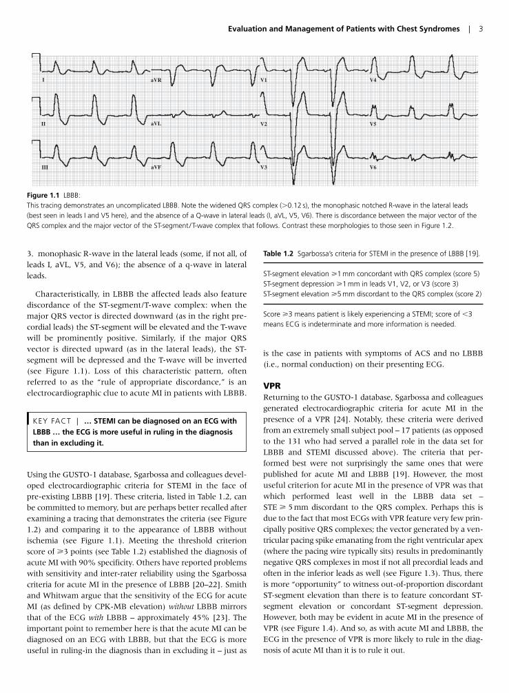
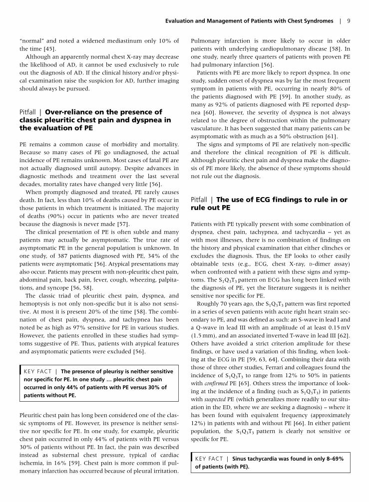
Characteristically, in LBBB the affected leads also featurediscordance of the ST-segment/T-wave complex: when themajor QRS vector is directed downward (as in the right pre-cordial leads) the ST-segment will be elevated and the T-wavewill be prominently positive. Similarly, if the major QRSvector is directed upward (as in the lateral leads), the ST-segment will be depressed and the T-wave will be inverted(see Figure 1.1). Loss of this characteristic pattern, oftenreferred to as the “rule of appropriate discordance,” is anelectrocardiographic clue to acute MI in patients with LBBB.
Using the GUSTO-1 database, Sgarbossa and colleagues devel-oped electrocardiographic criteria for STEMI in the face ofpre-existing LBBB [19]. These criteria, listed in Table 1.2, canbe committed to memory, but are perhaps better recalled afterexamining a tracing that demonstrates the criteria (see Figure1.2) and comparing it to the appearance of LBBB withoutischemia (see Figure 1.1). Meeting the threshold criterionscore of �3 points (see Table 1.2) established the diagnosis ofacute MI with 90% specificity. Others have reported problemswith sensitivity and inter-rater reliability using the Sgarbossacriteria for acute MI in the presence of LBBB [20–22]. Smithand Whitwam argue that the sensitivity of the ECG for acuteMI (as defined by CPK-MB elevation) without LBBB mirrorsthat of the ECG with LBBB – approximately 45% [23]. Theimportant point to remember here is that the acute MI can bediagnosed on an ECG with LBBB, but that the ECG is moreuseful in ruling-in the diagnosis than in excluding it – just as
is the case in patients with symptoms of ACS and no LBBB(i.e., normal conduction) on their presenting ECG.
VPRReturning to the GUSTO-1 database, Sgarbossa and colleaguesgenerated electrocardiographic criteria for acute MI in thepresence of a VPR [24]. Notably, these criteria were derivedfrom an extremely small subject pool – 17 patients (as opposedto the 131 who had served a parallel role in the data set forLBBB and STEMI discussed above). The criteria that per-formed best were not surprisingly the same ones that werepublished for acute MI and LBBB [19]. However, the mostuseful criterion for acute MI in the presence of VPR was thatwhich performed least well in the LBBB data set –STE � 5 mm discordant to the QRS complex. Perhaps this isdue to the fact that most ECGs with VPR feature very few prin-cipally positive QRS complexes; the vector generated by a ven-tricular pacing spike emanating from the right ventricular apex(where the pacing wire typically sits) results in predominantlynegative QRS complexes in most if not all precordial leads andoften in the inferior leads as well (see Figure 1.3). Thus, thereis more “opportunity” to witness out-of-proportion discordantST-segment elevation than there is to feature concordant ST-segment elevation or concordant ST-segment depression.However, both may be evident in acute MI in the presence ofVPR (see Figure 1.4). And so, as with acute MI and LBBB, theECG in the presence of VPR is more likely to rule in the diag-nosis of acute MI than it is to rule it out.
KEY FACT | … STEMI can be diagnosed on an ECG withLBBB … the ECG is more useful in ruling in the diagnosisthan in excluding it.
Evaluation and Management of Patients with Chest Syndromes | 3
I
II
III aVF
aVL
aVR V1
V2
V3
V4
V5
V6
Figure 1.1 LBBB:This tracing demonstrates an uncomplicated LBBB. Note the widened QRS complex (�0.12 s), the monophasic notched R-wave in the lateral leads(best seen in leads I and V5 here), and the absence of a Q-wave in lateral leads (I, aVL, V5, V6). There is discordance between the major vector of theQRS complex and the major vector of the ST-segment /T-wave complex that follows. Contrast these morphologies to those seen in Figure 1.2.
Table 1.2 Sgarbossa’s criteria for STEMI in the presence of LBBB [19].
ST-segment elevation �1 mm concordant with QRS complex (score 5)ST-segment depression �1 mm in leads V1, V2, or V3 (score 3)ST-segment elevation �5 mm discordant to the QRS complex (score 2)
Score �3 means patient is likely experiencing a STEMI; score of �3means ECG is indeterminate and more information is needed.
4 | Chapter 1
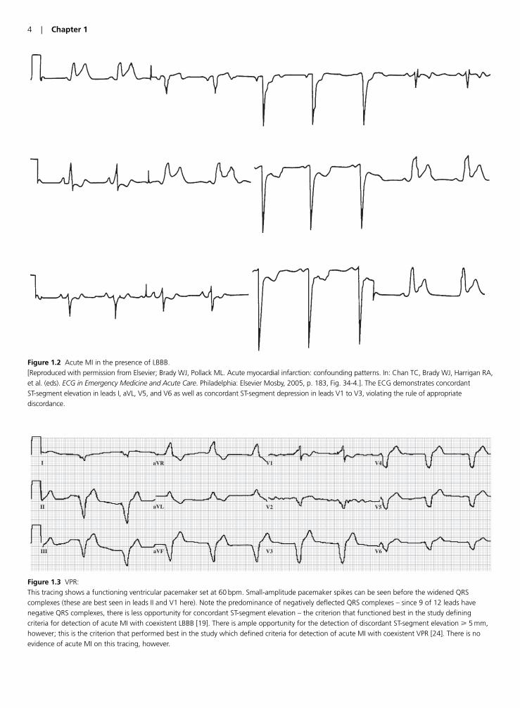
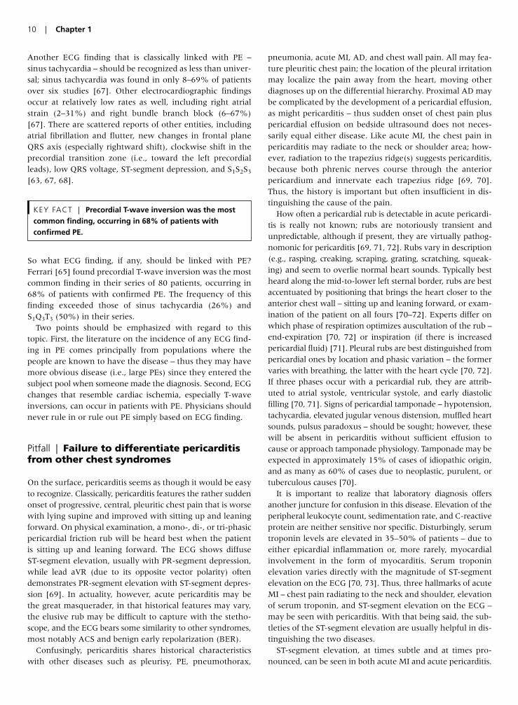
Figure 1.2 Acute MI in the presence of LBBB. [Reproduced with permission from Elsevier; Brady WJ, Pollack ML. Acute myocardial infarction: confounding patterns. In: Chan TC, Brady WJ, Harrigan RA, et al. (eds). ECG in Emergency Medicine and Acute Care. Philadelphia: Elsevier Mosby, 2005, p. 183, Fig. 34-4.]. The ECG demonstrates concordant ST-segment elevation in leads I, aVL, V5, and V6 as well as concordant ST-segment depression in leads V1 to V3, violating the rule of appropriatediscordance.
I
II
III aVF
aVL
aVR V1
V2
V3
V4
V5
V6
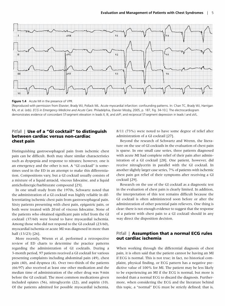
Figure 1.3 VPR:This tracing shows a functioning ventricular pacemaker set at 60 bpm. Small-amplitude pacemaker spikes can be seen before the widened QRScomplexes (these are best seen in leads II and V1 here). Note the predominance of negatively deflected QRS complexes – since 9 of 12 leads havenegative QRS complexes, there is less opportunity for concordant ST-segment elevation – the criterion that functioned best in the study definingcriteria for detection of acute MI with coexistent LBBB [19]. There is ample opportunity for the detection of discordant ST-segment elevation � 5 mm,however; this is the criterion that performed best in the study which defined criteria for detection of acute MI with coexistent VPR [24]. There is noevidence of acute MI on this tracing, however.
Pitfall | Use of a “GI cocktail” to distinguishbetween cardiac versus non-cardiac chest pain
Distinguishing gastroesophageal pain from ischemic chestpain can be difficult. Both may share similar characteristicssuch as dyspepsia and response to nitrates; however, one isan emergency and the other is not. A “GI cocktail” is some-times used in the ED in an attempt to make this differentia-tion. Compositions vary, but a GI cocktail usually consists ofa mixture of a liquid antacid, viscous lidocaine, and a liquidanticholinergic/barbiturate compound [25].
In one small study from the 1970s, Schwartz noted thatthe administration of a GI cocktail was highly reliable in dif-ferentiating ischemic chest pain from gastroesophageal pain.Sixty patients presenting with chest pain, epigastric pain, orboth were treated with 20 ml of viscous lidocaine. None ofthe patients who obtained significant pain relief from the GIcocktail (37/60) were found to have myocardial ischemia.Among those who did not respond to the GI cocktail (23/60),myocardial ischemia or acute MI was diagnosed in more thanhalf (13/23) [26].
More recently, Wrenn et al. performed a retrospectivereview of ED charts to determine the practice patternsregarding the administration of GI cocktails. During a 3-month period, 97 patients received a GI cocktail for variouspresenting complaints including abdominal pain (49), chestpain (40), and dyspnea (4). Over two-thirds of the patients(66/97) also received at least one other medication and themedian time of administration of the other drug was 9 minbefore the GI cocktail. The most common medications givenincluded opiates (56), nitroglycerin (22), and aspirin (10).Of the patients admitted for possible myocardial ischemia,
8/11 (73%) were noted to have some degree of relief afteradministration of a GI cocktail [27].
Beyond the research of Schwartz and Wrenn, the litera-ture on the use of GI cocktails in the evaluation of chest painis sparse. In one small case series, three patients diagnosedwith acute MI had complete relief of their pain after admin-istration of a GI cocktail [28]. One patient, however, didreceive nitroglycerin in parallel with the GI cocktail. Inanother slightly larger case series, 7% of patients with ischemicchest pain got relief of their symptoms after receiving a GIcocktail [29].
Research on the use of the GI cocktail as a diagnostic testin the evaluation of chest pain is clearly limited. In addition,the interpretation of this test remains difficult because theGI cocktail is often administered soon before or after theadministration of other potential pain relievers. One thing isclear: there is not enough evidence to suggest that the responseof a patient with chest pain to a GI cocktail should in anyway direct the disposition decision.
Pitfall | Assumption that a normal ECG rulesout cardiac ischemia
When working through the differential diagnosis of chestpain, it is often said that the patient cannot be having an MIif ECG is normal. This is not true; in fact, no historical com-plaint, physical finding, or ECG pattern has a negative pre-dictive value of 100% for MI. The patient may be less likelyto be experiencing an MI if the ECG is normal, but more isneeded than a normal ECG to discard the diagnosis. Further-more, when considering the ECG and the literature behindthis topic, a “normal” ECG must be strictly defined; that is,
Evaluation and Management of Patients with Chest Syndromes | 5
I
II
III
VI
aVF
aVL
aVR V1
V2
V3
V4
V5
V6
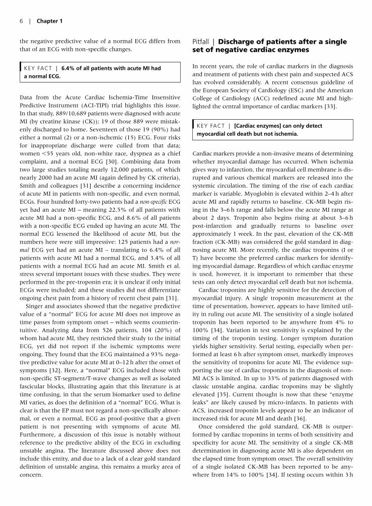
Figure 1.4 Acute MI in the presence of VPR. [Reproduced with permission from Elsevier; Brady WJ, Pollack ML. Acute myocardial infarction: confounding patterns. In: Chan TC, Brady WJ, HarriganRA, et al. (eds). ECG in Emergency Medicine and Acute Care. Philadelphia, Elsevier Mosby, 2005, p. 187, Fig. 34-10.]. The electrocardiogramdemonstrates evidence of concordant ST-segment elevation in leads II, III, and aVF; and reciprocal ST-segment depression in leads I and aVL.
the negative predictive value of a normal ECG differs fromthat of an ECG with non-specific changes.
Data from the Acute Cardiac Ischemia-Time InsensitivePredictive Instrument (ACI-TIPI) trial highlights this issue.In that study, 889/10,689 patients were diagnosed with acuteMI (by creatine kinase (CK)); 19 of those 889 were mistak-enly discharged to home. Seventeen of those 19 (90%) hadeither a normal (2) or a non-ischemic (15) ECG. Four risksfor inappropriate discharge were culled from that data;women �55 years old, non-white race, dyspnea as a chiefcomplaint, and a normal ECG [30]. Combining data fromtwo large studies totaling nearly 12,000 patients, of whichnearly 2000 had an acute MI (again defined by CK criteria),Smith and colleagues [31] describe a concerning incidenceof acute MI in patients with non-specific, and even normal,ECGs. Four hundred forty-two patients had a non-specific ECGyet had an acute MI – meaning 22.5% of all patients withacute MI had a non-specific ECG, and 8.6% of all patientswith a non-specific ECG ended up having an acute MI. Thenormal ECG lessened the likelihood of acute MI, but thenumbers here were still impressive: 125 patients had a nor-mal ECG yet had an acute MI – translating to 6.4% of allpatients with acute MI had a normal ECG, and 3.4% of allpatients with a normal ECG had an acute MI. Smith et al.stress several important issues with these studies. They wereperformed in the pre-troponin era; it is unclear if only initialECGs were included; and these studies did not differentiateongoing chest pain from a history of recent chest pain [31].
Singer and associates showed that the negative predictivevalue of a “normal” ECG for acute MI does not improve astime passes from symptom onset – which seems counterin-tuitive. Analyzing data from 526 patients, 104 (20%) ofwhom had acute MI, they restricted their study to the initialECG, yet did not report if the ischemic symptoms wereongoing. They found that the ECG maintained a 93% nega-tive predictive value for acute MI at 0–12 h after the onset ofsymptoms [32]. Here, a “normal” ECG included those withnon-specific ST-segment/T-wave changes as well as isolatedfascicular blocks, illustrating again that this literature is attime confusing, in that the serum biomarker used to defineMI varies, as does the definition of a “normal” ECG. What isclear is that the EP must not regard a non-specifically abnor-mal, or even a normal, ECG as proof-positive that a givenpatient is not presenting with symptoms of acute MI.Furthermore, a discussion of this issue is notably withoutreference to the predictive ability of the ECG in excludingunstable angina. The literature discussed above does notinclude this entity, and due to a lack of a clear gold standarddefinition of unstable angina, this remains a murky area ofconcern.
Pitfall | Discharge of patients after a singleset of negative cardiac enzymes
In recent years, the role of cardiac markers in the diagnosisand treatment of patients with chest pain and suspected ACShas evolved considerably. A recent consensus guideline ofthe European Society of Cardiology (ESC) and the AmericanCollege of Cardiology (ACC) redefined acute MI and high-lighted the central importance of cardiac markers [33].
Cardiac markers provide a non-invasive means of determiningwhether myocardial damage has occurred. When ischemiagives way to infarction, the myocardial cell membrane is dis-rupted and various chemical markers are released into thesystemic circulation. The timing of the rise of each cardiacmarker is variable. Myoglobin is elevated within 2–4 h afteracute MI and rapidly returns to baseline. CK-MB begin ris-ing in the 3–6 h range and falls below the acute MI range atabout 2 days. Troponin also begins rising at about 3–6 hpost-infarction and gradually returns to baseline overapproximately 1 week. In the past, elevation of the CK-MBfraction (CK-MB) was considered the gold standard in diag-nosing acute MI. More recently, the cardiac troponins (I orT) have become the preferred cardiac markers for identify-ing myocardial damage. Regardless of which cardiac enzymeis used, however, it is important to remember that thesetests can only detect myocardial cell death but not ischemia.
Cardiac troponins are highly sensitive for the detection ofmyocardial injury. A single troponin measurement at thetime of presentation, however, appears to have limited util-ity in ruling out acute MI. The sensitivity of a single isolatedtroponin has been reported to be anywhere from 4% to100% [34]. Variation in test sensitivity is explained by thetiming of the troponin testing. Longer symptom durationyields higher sensitivity. Serial testing, especially when per-formed at least 6 h after symptom onset, markedly improvesthe sensitivity of troponins for acute MI. The evidence sup-porting the use of cardiac troponins in the diagnosis of non-MI ACS is limited. In up to 33% of patients diagnosed withclassic unstable angina, cardiac troponins may be slightlyelevated [35]. Current thought is now that these “enzymeleaks” are likely caused by micro-infarcts. In patients withACS, increased troponin levels appear to be an indicator ofincreased risk for acute MI and death [36].
Once considered the gold standard, CK-MB is outper-formed by cardiac troponins in terms of both sensitivity andspecificity for acute MI. The sensitivity of a single CK-MBdetermination in diagnosing acute MI is also dependent onthe elapsed time from symptom onset. The overall sensitivityof a single isolated CK-MB has been reported to be any-where from 14% to 100% [34]. If testing occurs within 3 h
KEY FACT | [Cardiac enzymes] can only detectmyocardial cell death but not ischemia.
KEY FACT | 6.4% of all patients with acute MI had a normal ECG.
6 | Chapter 1
of symptom onset, the sensitivity of CK-MB is only 25–50%.After 3 h, the sensitivity is increased, ranging from 40% to100%. Because CK-MB rises relatively quickly, serial test-ing, even over a relatively short time period, has beenshown to increase the sensitivity considerably. In one study,a change in a 2-h CK-MB level had a sensitivity of 93.2% foracute MI [37].
Myoglobin is found in both skeletal and cardiac muscle,thereby limiting its specificity. Because myoglobin is rapidlyreleased after myocardial injury, it has been identified as apotential early indicator of acute MI. The sensitivity of a sin-gle myoglobin at the time of presentation, however, hasbeen noted to be as low as 21% [34]. Serial testing signifi-cantly improves the diagnostic utility of myoglobin. In onestudy, doubling of the level 1–2 h after the initial measure-ment was nearly 100% sensitive for the diagnosis of acuteMI [38].
More recent studies have looked into the use of serialmeasurements of multiple markers. McCord et al. noted thatwhen myoglobin and troponin were drawn at presentationand at 90 min, the sensitivity for acute MI was 96.9% andthe negative predictive value was 99.6% [39]. Ng et al.reported similar results utilizing a three-marker approachand a 90-min accelerated pathway, reporting nearly 100%sensitivity and 100% negative predicative value for acute MI[40]. It is critical to remember, however, that cardiacenzymes will not be reliably elevated in the setting of car-diac ischemia.
Ultimately, determining the disposition of patients with sus-pected ACS requires the EP to gather and interpret manypieces of information. The combined data from the history,physical, ECG, and cardiac markers should guide the EP inmanaging a patient with chest pain or suspected ACS. Singledetermination of cardiac markers at the time of presentationappears to be inadequate to exclude the diagnosis of acuteMI and provides no information about the possibility of car-diac ischemia.
Pitfall | Over-reliance on a “classic”presentation for diagnosis of AD
Acute dissection of the thoracic aorta is, unfortunately, bothchallenging to diagnose and potentially lethal if the diagno-sis is missed. Furthermore, misattributing the chest pain ofacute AD to ACS can lead to disastrous results as anticoagu-lant and fibrinolytic therapy are staples of the treatment of
the latter [41, 42]. Classically, the patient with AD has a his-tory of hypertension and experiences the sudden onset ofprofound ripping or tearing chest pain that radiates to theback (interscapular region – perhaps migrating to the lowback) [43]. It is important to note, however, that theabsence of this history in no way excludes the diagnosis;symptoms may be atypical – or may even be absent. Indeed,one report [43] looking at pooled data from 16 studies,found a history of any pain to be only 90% sensitive for thediagnosis of acute AD (CI 85–94%) (see Table 1.3), withmore precise and classic pain descriptions faring less well.Data reported from the International Registry of AcuteAortic Dissection (IRAD) [44] included 464 patients from 12referral centers; some type of pain was reported in 94% ofType A dissections and 98% of Type B dissections; it was chestpain in 79% and 63%, respectively. The pain was abrupt inonset in roughly 85% of all dissections, and it was character-ized as severe or the “worst ever” in 90% of both groups.Interestingly, it was classified as “sharp” (64%) more oftenthan “ripping or tearing” (51%) [44]. Since the classic descrip-tion has been well-documented to be less than universal,knowledge of atypical presentations of AD together with anawareness of risk factors enhances diagnostic capability.
KEY FACT | Single determinations of cardiac markers atthe time of presentation appear to be inadequate toexclude the diagnosis of acute MI and provide noinformation about the possibility of cardiac ischemia.
Evaluation and Management of Patients with Chest Syndromes | 7
Table 1.3 Sensitivity of clinical history of pain in acute thoracic AD [43].
ConfidencePain Description Sensitivity (%) Intervals (%)
Any pain 90 85–94Chest pain 67 56–77Anterior chest pain 57 48–66Posterior chest pain 32 24–40Back pain 32 19–47Abdominal pain 23 16–31Sudden-onset pain 84 80–89Severe pain 90 88–92Ripping/tearing pain 39 14–69
Risk factors for AD [43].
• Hypertension• Bicuspid aortic valve• Previous cardiac surgery, particularly aortic valve
replacement• Coarctation of the aorta• Marfan syndrome• Ehlers–Danlos syndrome• Turner syndrome• Giant cell arteritis• Third-trimester pregnancy• Cocaine abuse• Trauma
So how do patients with acute AD present, if not withchest pain, or indeed any pain? Syncope was reported in 13%of Type A AD in IRAD; 2% of those patients did not haveany pain or neurological findings (only 4% of Type B dissec-tions presented with syncope) [44]. Others have reportedsyncope (at times painless) in acute AD as well [42, 45–47].Another common diagnosis associated with acute AD is acutestroke, this being mediated by flap occlusion of a carotidartery in Type A dissection. IRAD data found 17/289 (6%)to present with acute stroke symptoms [44]; the morebroadly defined finding of a new focal neurologic deficit wasreported in 17% of pooled studies [43]. The neurologicdeficit may be peripheral rather than central, due to the siteof occlusion; motor and sensory findings in a lower extrem-ity have been reported with acute AD in the absence of pain[48]. AD may also present as an acutely painful ischemic legor as acute chest pain radiating to the back with simultaneousincontinence and bilateral lower extremity paralysis. Otheratypical presentations of acute AD include abdominal or flankpain, hoarseness (recurrent laryngeal nerve compression),swelling and bruising of the neck, cough (mainstem bronchuscompression), dysphagia (esophageal compression), Horner’ssyndrome (sympathetic chain compression), pulsatile stern-oclavicular joint, superior vena cava syndrome, and testicular/groin pain [43–46, 49–51].
Pitfall | Use of the chest X-ray to excludethe diagnosis of AD
AD is the most common fatal condition involving the aorta[45]. Left untreated, about 75% of patients with AD involvingthe ascending aorta will die within 2 weeks. If diagnosed earlyand treated successfully, the 5-year survival rate approaches75% [49]. Because early diagnosis is so important, the EPmust maintain a high level of suspicion for AD. In the settingof chronic hypertension, AD should be considered in anypatient with sudden and severe chest or back pain.
When AD is being considered, a chest X-ray should beobtained and examined for abnormalities of the aortic sil-houette. This is best accomplished with a standing pos-teroanterior (PA) view. Portable anteroposterior (AP) viewsmay falsely enlarge the cardiomediastinal silhouette and lat-eral chest X-rays rarely show evidence of AD [52]. Manyradiographic findings have been noted in AD but unfortu-nately the majority of these findings are subjective and notwell defined. Although the chest X-ray may suggest thediagnosis, it is rarely definitive.
Radiographic findings in AD may include widening of the mediastinum, abnormalities of the aortic knob and aortic contour, increased aortic diameter, left-sided pleural effusion,tracheal deviation, and esophageal deviation [49, 53]. Thedouble density sign is observed when the false lumen is lessradiopaque than the true lumen [49]. The calcium sign, con-sisting of the displacement of the aorta’s intimal calcification
from the aortic knob by 1 cm or more, is highly suggestive ofAD but is only present in a minority of cases [43, 49].
Widened mediastinum, defined as a measurement � 8 cmat the level of the aortic knob, is considered by many to bethe most sensitive radiographic finding. According to onestudy, widening of the mediastinum and widening of theaortic knob were the only two radiographic features of sig-nificance in predicting dissection [54]. A tortuous aorta,common in hypertensive patients, may widen the medi-astinum and be hard to distinguish from AD. Other causes ofmediastinal widening include adenopathy, lymphoma, andan enlarged thyroid.
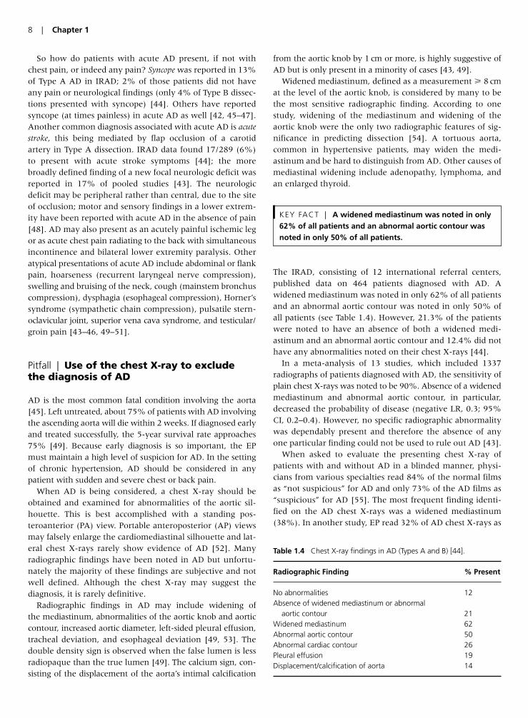
The IRAD, consisting of 12 international referral centers,published data on 464 patients diagnosed with AD. Awidened mediastinum was noted in only 62% of all patientsand an abnormal aortic contour was noted in only 50% ofall patients (see Table 1.4). However, 21.3% of the patientswere noted to have an absence of both a widened medi-astinum and an abnormal aortic contour and 12.4% did nothave any abnormalities noted on their chest X-rays [44].
In a meta-analysis of 13 studies, which included 1337radiographs of patients diagnosed with AD, the sensitivity ofplain chest X-rays was noted to be 90%. Absence of a widenedmediastinum and abnormal aortic contour, in particular,decreased the probability of disease (negative LR, 0.3; 95%CI, 0.2–0.4). However, no specific radiographic abnormalitywas dependably present and therefore the absence of anyone particular finding could not be used to rule out AD [43].
When asked to evaluate the presenting chest X-ray ofpatients with and without AD in a blinded manner, physi-cians from various specialties read 84% of the normal filmsas “not suspicious” for AD and only 73% of the AD films as“suspicious” for AD [55]. The most frequent finding identi-fied on the AD chest X-rays was a widened mediastinum(38%). In another study, EP read 32% of AD chest X-rays as
KEY FACT | A widened mediastinum was noted in only62% of all patients and an abnormal aortic contour wasnoted in only 50% of all patients.
8 | Chapter 1
Table 1.4 Chest X-ray findings in AD (Types A and B) [44].
Radiographic Finding % Present
No abnormalities 12Absence of widened mediastinum or abnormal
aortic contour 21Widened mediastinum 62Abnormal aortic contour 50Abnormal cardiac contour 26Pleural effusion 19Displacement/calcification of aorta 14
“normal” and noted a widened mediastinum only 10% ofthe time [45].
Although an apparently normal chest X-ray may decreasethe likelihood of AD, it cannot be used exclusively to ruleout the diagnosis of AD. If the clinical history and/or physi-cal examination raise the suspicion for AD, further imagingshould always be pursued.
Pitfall | Over-reliance on the presence ofclassic pleuritic chest pain and dyspnea inthe evaluation of PE
PE remains a common cause of morbidity and mortality.Because so many cases of PE go undiagnosed, the actualincidence of PE remains unknown. Most cases of fatal PE arenot actually diagnosed until autopsy. Despite advances indiagnostic methods and treatment over the last severaldecades, mortality rates have changed very little [56].
When promptly diagnosed and treated, PE rarely causesdeath. In fact, less than 10% of deaths caused by PE occur inthose patients in which treatment is initiated. The majorityof deaths (90%) occur in patients who are never treatedbecause the diagnosis is never made [57].
The clinical presentation of PE is often subtle and manypatients may actually be asymptomatic. The true rate ofasymptomatic PE in the general population is unknown. Inone study, of 387 patients diagnosed with PE, 34% of thepatients were asymptomatic [56]. Atypical presentations mayalso occur. Patients may present with non-pleuritic chest pain,abdominal pain, back pain, fever, cough, wheezing, palpita-tions, and syncope [56, 58].
The classic triad of pleuritic chest pain, dyspnea, andhemoptysis is not only non-specific but it is also not sensi-tive. At most it is present 20% of the time [58]. The combi-nation of chest pain, dyspnea, and tachypnea has beennoted be as high as 97% sensitive for PE in various studies.However, the patients enrolled in these studies had symp-toms suggestive of PE. Thus, patients with atypical featuresand asymptomatic patients were excluded [56].
Pleuritic chest pain has long been considered one of the clas-sic symptoms of PE. However, its presence is neither sensi-tive nor specific for PE. In one study, for example, pleuriticchest pain occurred in only 44% of patients with PE versus30% of patients without PE. In fact, the pain was describedinstead as substernal chest pressure, typical of cardiacischemia, in 16% [59]. Chest pain is more common if pul-monary infarction has occurred because of pleural irritation.
Pulmonary infarction is more likely to occur in olderpatients with underlying cardiopulmonary disease [58]. Inone study, nearly three quarters of patients with proven PEhad pulmonary infarction [56].
Patients with PE are more likely to report dyspnea. In onestudy, sudden onset of dyspnea was by far the most frequentsymptom in patients with PE, occurring in nearly 80% ofthe patients diagnosed with PE [59]. In another study, asmany as 92% of patients diagnosed with PE reported dysp-nea [60]. However, the severity of dyspnea is not alwaysrelated to the degree of obstruction within the pulmonaryvasculature. It has been suggested that many patients can beasymptomatic with as much as a 50% obstruction [61].
The signs and symptoms of PE are relatively non-specificand therefore the clinical recognition of PE is difficult.Although pleuritic chest pain and dyspnea make the diagno-sis of PE more likely, the absence of these symptoms shouldnot rule out the diagnosis.
Pitfall | The use of ECG findings to rule in orrule out PE
Patients with PE typically present with some combination ofdyspnea, chest pain, tachypnea, and tachycardia – yet aswith most illnesses, there is no combination of findings onthe history and physical examination that either clinches orexcludes the diagnosis. Thus, the EP looks to other easilyobtainable tests (e.g., ECG, chest X-ray, D-dimer assay)when confronted with a patient with these signs and symp-toms. The S1Q3T3 pattern on ECG has long been linked withthe diagnosis of PE, yet the literature suggests it is neithersensitive nor specific for PE.
Roughly 70 years ago, the S1Q3T3 pattern was first reportedin a series of seven patients with acute right heart strain sec-ondary to PE, and was defined as such: an S-wave in lead I anda Q-wave in lead III with an amplitude of at least 0.15 mV(1.5 mm), and an associated inverted T-wave in lead III [62].Others have avoided a strict criterion amplitude for thesefindings, or have used a variation of this finding, when look-ing at the ECG in PE [59, 63, 64]. Combining their data withthose of three other studies, Ferrari and colleagues found theincidence of S1Q3T3 to range from 12% to 50% in patientswith confirmed PE [65]. Others stress the importance of look-ing at the incidence of a finding (such as S1Q3T3) in patientswith suspected PE (which generalizes more readily to our situ-ation in the ED, where we are seeking a diagnosis) – where ithas been found with equivalent frequency (approximately12%) in patients with and without PE [66]. In either patientpopulation, the S1Q3T3 pattern is clearly not sensitive or specific for PE.
KEY FACT | Sinus tachycardia was found in only 8–69%of patients (with PE).
KEY FACT | The presence of pleurisy is neither sensitivenor specific for PE. In one study … pleuritic chest painoccurred in only 44% of patients with PE versus 30% ofpatients without PE.
Evaluation and Management of Patients with Chest Syndromes | 9
Another ECG finding that is classically linked with PE –sinus tachycardia – should be recognized as less than univer-sal; sinus tachycardia was found in only 8–69% of patientsover six studies [67]. Other electrocardiographic findingsoccur at relatively low rates as well, including right atrialstrain (2–31%) and right bundle branch block (6–67%)[67]. There are scattered reports of other entities, includingatrial fibrillation and flutter, new changes in frontal planeQRS axis (especially rightward shift), clockwise shift in theprecordial transition zone (i.e., toward the left precordialleads), low QRS voltage, ST-segment depression, and S1S2S3
[63, 67, 68].
So what ECG finding, if any, should be linked with PE?Ferrari [65] found precordial T-wave inversion was the mostcommon finding in their series of 80 patients, occurring in68% of patients with confirmed PE. The frequency of thisfinding exceeded those of sinus tachycardia (26%) andS1Q3T3 (50%) in their series.
Two points should be emphasized with regard to thistopic. First, the literature on the incidence of any ECG find-ing in PE comes principally from populations where thepeople are known to have the disease – thus they may havemore obvious disease (i.e., large PEs) since they entered thesubject pool when someone made the diagnosis. Second, ECGchanges that resemble cardiac ischemia, especially T-waveinversions, can occur in patients with PE. Physicians shouldnever rule in or rule out PE simply based on ECG finding.
Pitfall | Failure to differentiate pericarditisfrom other chest syndromes
On the surface, pericarditis seems as though it would be easyto recognize. Classically, pericarditis features the rather suddenonset of progressive, central, pleuritic chest pain that is worsewith lying supine and improved with sitting up and leaningforward. On physical examination, a mono-, di-, or tri-phasicpericardial friction rub will be heard best when the patient is sitting up and leaning forward. The ECG shows diffuse ST-segment elevation, usually with PR-segment depression,while lead aVR (due to its opposite vector polarity) oftendemonstrates PR-segment elevation with ST-segment depres-sion [69]. In actuality, however, acute pericarditis may bethe great masquerader, in that historical features may vary,the elusive rub may be difficult to capture with the stetho-scope, and the ECG bears some similarity to other syndromes,most notably ACS and benign early repolarization (BER).
Confusingly, pericarditis shares historical characteristicswith other diseases such as pleurisy, PE, pneumothorax,
pneumonia, acute MI, AD, and chest wall pain. All may fea-ture pleuritic chest pain; the location of the pleural irritationmay localize the pain away from the heart, moving otherdiagnoses up on the differential hierarchy. Proximal AD maybe complicated by the development of a pericardial effusion,as might pericarditis – thus sudden onset of chest pain pluspericardial effusion on bedside ultrasound does not neces-sarily equal either disease. Like acute MI, the chest pain inpericarditis may radiate to the neck or shoulder area; how-ever, radiation to the trapezius ridge(s) suggests pericarditis,because both phrenic nerves course through the anteriorpericardium and innervate each trapezius ridge [69, 70].Thus, the history is important but often insufficient in dis-tinguishing the cause of the pain.
How often a pericardial rub is detectable in acute pericardi-tis is really not known; rubs are notoriously transient andunpredictable, although if present, they are virtually pathog-nomonic for pericarditis [69, 71, 72]. Rubs vary in description(e.g., rasping, creaking, scraping, grating, scratching, squeak-ing) and seem to overlie normal heart sounds. Typically bestheard along the mid-to-lower left sternal border, rubs are bestaccentuated by positioning that brings the heart closer to theanterior chest wall – sitting up and leaning forward, or exam-ination of the patient on all fours [70–72]. Experts differ onwhich phase of respiration optimizes auscultation of the rub –end-expiration [70, 72] or inspiration (if there is increasedpericardial fluid) [71]. Pleural rubs are best distinguished frompericardial ones by location and phasic variation – the formervaries with breathing, the latter with the heart cycle [70, 72].If three phases occur with a pericardial rub, they are attrib-uted to atrial systole, ventricular systole, and early diastolicfilling [70, 71]. Signs of pericardial tamponade – hypotension,tachycardia, elevated jugular venous distension, muffled heartsounds, pulsus paradoxus – should be sought; however, thesewill be absent in pericarditis without sufficient effusion tocause or approach tamponade physiology. Tamponade may beexpected in approximately 15% of cases of idiopathic origin,and as many as 60% of cases due to neoplastic, purulent, ortuberculous causes [70].
It is important to realize that laboratory diagnosis offersanother juncture for confusion in this disease. Elevation of theperipheral leukocyte count, sedimentation rate, and C-reactiveprotein are neither sensitive nor specific. Disturbingly, serumtroponin levels are elevated in 35–50% of patients – due toeither epicardial inflammation or, more rarely, myocardialinvolvement in the form of myocarditis. Serum troponinelevation varies directly with the magnitude of ST-segmentelevation on the ECG [70, 73]. Thus, three hallmarks of acuteMI – chest pain radiating to the neck and shoulder, elevationof serum troponin, and ST-segment elevation on the ECG –may be seen with pericarditis. With that being said, the sub-tleties of the ST-segment elevation are usually helpful in dis-tinguishing the two diseases.
ST-segment elevation, at times subtle and at times pro-nounced, can be seen in both acute MI and acute pericarditis.
KEY FACT | Precordial T-wave inversion was the mostcommon finding, occurring in 68% of patients withconfirmed PE.
10 | Chapter 1
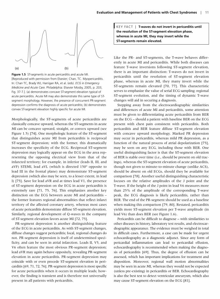
Morphologically, the ST-segments of acute pericarditis areclassically concave upward, whereas the ST-segments in acuteMI can be concave upward, straight, or convex upward (seeFigure 1.5) [74]. One morphologic feature of the ST-segmentthat distinguishes acute MI from pericarditis is reciprocal ST-segment depression; with the former, this dramaticallyincreases the specificity of the ECG. Reciprocal ST-segmentdepression may logically appear on the ECG in the area rep-resenting the opposing electrical view from that of theinfarcted territory; for example, in inferior (leads II, III, andaVF) STEMI, lead aVL (which is directed 150° opposite tolead III in the frontal plane) may demonstrate ST-segmentdepression (which also may be seen, to a lesser extent, in leadI) [75]. Save for lead aVR and at times lead V1, the presenceof ST-segment depression on the ECG in acute pericarditis isextremely rare [71, 75, 76]. This emphasizes another key distinction on the ECG between acute MI and pericarditis –the former features regional abnormalities that reflect infarctterritory of the affected coronary artery, whereas most casesof acute pericarditis demonstrate diffuse ST-segment elevation.Similarly, regional development of Q-waves in the companyof ST-segment elevation favors acute MI [72, 75].
PR-segment depression is another distinguishing featureof the ECG in acute pericarditis. As with ST-segment changes,diffuse changes suggest pericarditis; focal, regional changes donot. PR-segment depression is itself of undetermined speci-ficity, and can be seen in atrial infarction. Leads II, V5, andV6 often feature the most obvious PR-segment depression;lead aVR may again behave oppositely, revealing PR-segmentelevation in acute pericarditis. PR-segment depression maycoincide with or even precede ST-segment elevation in peri-carditis [69, 71, 72, 76]. PR-segment depression is most specificfor acute pericarditis when it occurs in multiple leads; how-ever, the finding is transient and is therefore not universallypresent in all patients with pericarditis.
Like the PR- and ST-segments, the T-wave behaves differ-ently in acute MI and pericarditis. While both diseases canfeature T-wave inversions following ST-segment elevation,there is an important distinction: T-waves do not invert inpericarditis until the resolution of ST-segment elevationphase, whereas in acute MI, they many invert while theST-segments remain elevated [70, 77]. This characteristicserves to emphasize the value of serial ECG sampling; regionalST-segment evolution, and the timing of dynamic T-wavechanges will aid in securing a diagnosis.
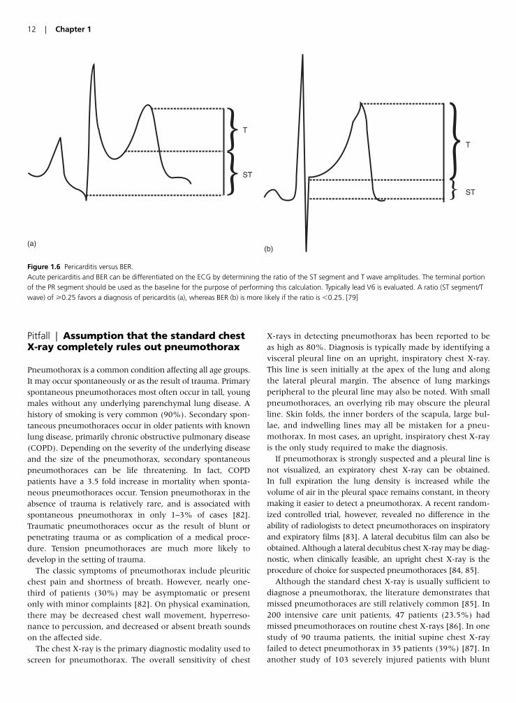
Stepping away from the electrocardiographic similaritiesand differences of acute MI and pericarditis, some attentionmust be given to differentiating acute pericarditis from BERon the ECG – should a patient with baseline BER on the ECGpresent with chest pain consistent with pericarditis. Bothpericarditis and BER feature diffuse ST-segment elevationwith concave upward morphology. Marked PR depressionmay occur in pericarditis, whereas mild PR depression, as afunction of the natural process of atrial depolarization [75],may be seen on any ECG, including those with BER. Oneuseful distinguishing factor is that the ST-segment elevationof BER is stable over time (i.e., should be present on old trac-ings), whereas the ST-segment elevation of acute pericarditis,though not given to minute-to-minute change (unlike ACS),should be absent on old ECGs, should they be available forcomparison [78]. Another useful distinguishing characteristicfocuses on the relative amplitudes of the J point and theT-wave. If the height of the J point in lead V6 measures morethan 25% of the amplitude of the corresponding T-wavepeak, the ECG diagnosis is likely pericarditis, rather thanBER. The end of the PR-segment should be used as a baselinewhen making this comparison [79, 80]. Restated, pericarditisyields more ST-segment elevation per T-wave amplitude (inlead V6) than does BER (see Figure 1.6).
Pericarditis can be difficult to diagnose – with similarities toother diseases in history, laboratory test results, and electrocar-diographic appearance. The evidence must be weighed in totalin difficult cases. Furthermore, a case can be made for urgentechocardiography as a diagnostic adjunct. Since any form ofpericardial inflammation can lead to pericardial effusion,echocardiography is recommended when making the diagno-sis of pericarditis [69]. Thus, the degree of effusion can beassessed, which has important implications for treatment anddisposition. Moreover, regional wall motion abnormalitiesmay be seen in acute MI, whereas they would not be expected(unless pre-existing) in pericarditis or BER. Echocardiographyis also the best test to detect ventricular aneurysm, which alsomay cause ST-segment elevation on the ECG [81].
KEY FACT | T-waves do not invert in pericarditis untilthe resolution of the ST-segment elevation phase,whereas in acute MI, they may invert while theST-segments remain elevated.
Evaluation and Management of Patients with Chest Syndromes | 11
(a) (b)
Figure 1.5 ST-segments in acute pericarditis and acute MI. [Reproduced with permission from Elsevier; Chan, TC. Myopericarditis.In: Chan TC, Brady WJ, Harrigan RA, et al. (eds). ECG in EmergencyMedicine and Acute Care. Philadelphia: Elsevier Mosby, 2005, p. 203, Fig. 37-7.]. (a) demonstrates concave ST-segment elevation typical ofacute pericarditis. Acute MI may also demonstrate this same type of ST-segment morphology. However, the presence of concurrent PR-segmentdepression confirms the diagnosis of acute pericarditis; (b) demonstratesconvex ST-segment elevation highly specific for acute MI.
Pitfall | Assumption that the standard chestX-ray completely rules out pneumothorax
Pneumothorax is a common condition affecting all age groups.It may occur spontaneously or as the result of trauma. Primaryspontaneous pneumothoraces most often occur in tall, youngmales without any underlying parenchymal lung disease. Ahistory of smoking is very common (90%). Secondary spon-taneous pneumothoraces occur in older patients with knownlung disease, primarily chronic obstructive pulmonary disease(COPD). Depending on the severity of the underlying diseaseand the size of the pneumothorax, secondary spontaneouspneumothoraces can be life threatening. In fact, COPDpatients have a 3.5 fold increase in mortality when sponta-neous pneumothoraces occur. Tension pneumothorax in theabsence of trauma is relatively rare, and is associated withspontaneous pneumothorax in only 1–3% of cases [82].Traumatic pneumothoraces occur as the result of blunt orpenetrating trauma or as complication of a medical proce-dure. Tension pneumothoraces are much more likely todevelop in the setting of trauma.
The classic symptoms of pneumothorax include pleuriticchest pain and shortness of breath. However, nearly one-third of patients (30%) may be asymptomatic or presentonly with minor complaints [82]. On physical examination,there may be decreased chest wall movement, hyperreso-nance to percussion, and decreased or absent breath soundson the affected side.
The chest X-ray is the primary diagnostic modality used toscreen for pneumothorax. The overall sensitivity of chest
X-rays in detecting pneumothorax has been reported to beas high as 80%. Diagnosis is typically made by identifying avisceral pleural line on an upright, inspiratory chest X-ray.This line is seen initially at the apex of the lung and alongthe lateral pleural margin. The absence of lung markingsperipheral to the pleural line may also be noted. With smallpneumothoraces, an overlying rib may obscure the pleuralline. Skin folds, the inner borders of the scapula, large bul-lae, and indwelling lines may all be mistaken for a pneu-mothorax. In most cases, an upright, inspiratory chest X-rayis the only study required to make the diagnosis.
If pneumothorax is strongly suspected and a pleural line isnot visualized, an expiratory chest X-ray can be obtained. In full expiration the lung density is increased while the volume of air in the pleural space remains constant, in theorymaking it easier to detect a pneumothorax. A recent random-ized controlled trial, however, revealed no difference in theability of radiologists to detect pneumothoraces on inspiratoryand expiratory films [83]. A lateral decubitus film can also beobtained. Although a lateral decubitus chest X-ray may be diag-nostic, when clinically feasible, an upright chest X-ray is theprocedure of choice for suspected pneumothoraces [84, 85].
Although the standard chest X-ray is usually sufficient todiagnose a pneumothorax, the literature demonstrates thatmissed pneumothoraces are still relatively common [85]. In200 intensive care unit patients, 47 patients (23.5%) hadmissed pneumothoraces on routine chest X-rays [86]. In onestudy of 90 trauma patients, the initial supine chest X-rayfailed to detect pneumothorax in 35 patients (39%) [87]. Inanother study of 103 severely injured patients with blunt
12 | Chapter 1
(a)
T
ST
(b)
T
ST
Figure 1.6 Pericarditis versus BER. Acute pericarditis and BER can be differentiated on the ECG by determining the ratio of the ST segment and T wave amplitudes. The terminal portionof the PR segment should be used as the baseline for the purpose of performing this calculation. Typically lead V6 is evaluated. A ratio (ST segment/Twave) of �0.25 favors a diagnosis of pericarditis (a), whereas BER (b) is more likely if the ratio is �0.25. [79]
trauma, 27 (26%) had pneumothoraces missed on their ini-tial chest X-ray only to be picked up on thoracic CT [88]. Inyet another study, one-third of all traumatic pneumotho-races were missed on the initial chest X-ray and diagnosed onabdominal CT [89]. If the initial chest X-ray is inconclusiveand there is a significant suspicion of pneumothorax, CTimaging should be pursued in any high-risk patient group(COPD, trauma, mechanically ventilated). As the diagnosticsensitivity of a test (chest CT) increases, the issue of clinicalrelevance emerges. Clearly some trivial pneumothoracesfound only on chest CT need no treatment.
Pitfall | Excluding the diagnosis ofBoerhaave’s syndrome due to an absenceof antecedent retching or vomiting
First described by the Dutch physician Herman Boerhaavein 1724, Boerhaave’s syndrome refers to rupture of theesophagus – and is associated with high morbidity and mor-tality. At times referred to as spontaneous rupture of theesophagus, Boerhaave’s syndrome is probably best thoughtof as rupture due to the development of a tear after a rise inthe intraluminal pressure of this structure. The classic triadfor this syndrome includes forceful emesis, chest pain, andsubcutaneous emphysema. Patients usually appear very ill,prefer to sit up and lean forward, and may have lateralizingpulmonary findings on examination (rales, wheezing,decreased breath sounds) in addition to the subcutaneousemphysema, if it is present. Chest X-ray abnormalitiesinclude atelectasis, infiltrates, and pleural effusion, usuallyon the left because 90% of cases are due to a tear in the leftposterolateral wall of the lower third of the esophagus,which communicates with the left pleural cavity in 80% ofcases. Pneumomediastinum and hydropneumothorax maybe apparent on the chest X-ray as well. Definitive diagnosisis usually made by computed tomographic scan of the tho-rax or by esophagram, although false negative studies mayoccur with either [90–93].
In one literature review [90] antecedent retching or vomit-ing was absent in 21% of cases of Boerhaave’s syndrome.Thus, it should be emphasized that the diagnosis should notbe excluded in the absence of this historical feature. Indeed,Boerhaave’s syndrome has been reported after a variety ofevents, some less dramatic than others. Belching [94], simply swallowing a sandwich [95], violent cough [96],defecation, childbirth, weight lifting, asthma attacks, seizures,
and blunt abdominal trauma [97, 98] have all been reportedas precipitant events for Boerhaave’s syndrome. It has beenreported to complicate the vomiting associated with acuteMI [99]. It should especially be considered in patients withchest pain after a recent esophageal endoscopic procedure.Notably, it is also seen in children [97, 98], and may presentwith a right-sided esophageal tear – leading to findings onphysical examination and chest X-ray on the right side ratherthan the classic occurrence on the left [91, 98]. Thus, as withmost diseases, atypical isolated features of the history andphysical examination, and even negative initial diagnostictests, should not dissuade the EP from pursuing the diagnosisof Boerhaave’s syndrome if the patient appears ill and thediagnosis remains possible yet illusive.
Pitfall | Failure to evaluate a patient withchest tenderness for herpes zoster
We have all seen patients in a less-than optimal setting (e.g.,in a chair; multiple layers of clothes on; no curtain for pri-vacy) where we take the chest pain history and find that thepain is reproducible with palpation on physical examina-tion. When entertaining the diagnosis of chest wall pain orcostochondritis, consider herpes zoster (shingles) as well.
Herpes zoster is generally a clinical diagnosis. It occurs inpatients due to reactivation of latent varicella zoster virus,dormant in the dorsal root ganglia. It is seen in both childrenand adults, although incidence varies directly with age [100,101]. Annualized incidence is 1.5–3.0 case per 1000 per-sons; in patients �75 years of age, this rate increases to 10cases per 1000 persons [100]. The increased incidence withage, as well as an association with states of impaired cell-mediated immunity (e.g., immunosuppressive therapy, can-cer, human immunodeficiency virus) is evident, but anoutbreak of herpes zoster is not specific for a state ofimpaired immunity [100, 102]. Indeed, herpes zoster devel-ops in approximately 20,000 apparently healthy childreneach year in the USA; chicken pox at an age of less than 1year is a risk factor [102].
Herpes zoster typically presents with abnormal skin sensa-tions (itching, tingling, and/or pain – which may be severe)in a dermatomal distribution that precede the appearance ofskin lesions – typically by 1–5 days [100], although visiblelesions may not develop for a week to 10 days [103, 104].Zoster sine herpete is an uncommon variant in which thelesions never appear [104]. The history together with visibleevidence of the lesions (classically an erythematous macu-lopapular rash which progresses to the vesicular stage, fol-lowed by pustulation, ulceration, and finally crusting beforedisappearance) in a dermatomal distribution is key to thediagnosis [100]. Pain on light touch (allodynia) or overlysensitive skin (hyperesthesia) in a dermatomal distributionis also consistent with the diagnosis; these findings may precede the outbreak of the skin lesions [104]. Generally
KEY FACT | Antecedent retching or vomiting wasabsent in 21% of cases of Boerhaave’s syndrome … thediagnosis should not be excluded in the absence of thishistorical feature.
Evaluation and Management of Patients with Chest Syndromes | 13
speaking, the rash is unilateral, does not cross the midline,and is confined to one dermatome in immunocompetentpersons. Overlap with adjacent dermatomes is relativelycommon (20%), and the appearance of a few lesions outsidethe affected dermatome is also not unusual [100]. Resolutionoccurs over 2–4 weeks, although it may be followed by thepersistence of pain – so-called post-herpetic neuralgia.
Thus, in patients with a presumptive diagnosis of chestwall pain, carefully inspect the skin for signs of herpeszoster. Furthermore, if no lesions are visible, but the history(pain, oftentimes severe, in a band-like, dermatomal distri-bution, and perhaps accompanied by itching or paresthesias)and physical examination (hyperesthesia or allodynia in thesame dermatomal distribution) are consistent with the pro-dromal stage of herpes zoster, instruct the patient to watchcarefully for the appearance of any lesions. Prompt treat-ment (generally within 3 days of appearance of the rash)with antiviral therapy is indicated [100].
Pearls for Improving Patient Outcomes
• Do not exclude the diagnosis of acute cardiac ischemia or MIbased on the absence of pain, especially when evaluating dia-betic patients, the elderly, and women.
• Never use reproducible chest wall tenderness to exclude thediagnosis of acute MI.
• When the ECG shows LBBB or VPR, examine it closely for signsof inappropriately large, discordant ST-segment elevation; con-cordant ST-segment elevation; or concordant ST-segmentdepression (in the right precordial leads) – these may indicate anacute MI.
• Never use the response to antacids as a diagnostic test for dis-tinguishing cardiac versus gastric pain.
• Neither a single normal ECG nor a single negative set of cardiacenzymes should be used to rule out acute cardiac ischemia.
• The chest X-ray can be used to suggest the diagnosis of AD, butit cannot definitively exclude the diagnosis.
• Consider AD and PE in the differential diagnosis of patients pre-senting with syncope.
• Pleuritic chest pain should prompt diagnostic consideration ofPE as well as acute pericarditis.
• Precordial T-wave inversions in patients with chest pain shouldprompt consideration of not only acute cardiac ischemia butalso of acute PE.
• Boerhaave’s syndrome should be considered in the differentialdiagnosis for all patients with chest pain, even in the absence ofa history of retching or vomiting.
• Always visualize the skin whenever a patient has reproduciblechest well tenderness, and look for evidence of herpes zoster.
References
1. Canto JG, Shlipak MG, Rogers WJ, et al. Prevalence, clinical
characteristics, and mortality among patients with myocardial
infarction presenting without chest pain. J Am Med Assoc 2000;
283:3223–9.
2. Dorsch MF, Lawrence RA, Sapsford RJ, et al. Poor prognosis of
patients presenting with symptomatic myocardial infarction
but without chest pain. Heart 2001; 86:494–8.
3. Gupta M, Tabas JA, Kohn MA. Presenting complaint among
patients with myocardial infarction who present to an urban,
public hospital emergency department. Ann Emerg Med 2002;
40:180–6.
4. Uretsky BF, Farquahr DS, Berezin AF, et al. Symptomatic
myocardial infarction without chest pain: prevalence and clini-
cal course. Am J Cardiol 1977; 40:498–503.
5. Bayer AJ, Chadha JS, Farag RR, et al. Changing presentation of
myocardial infarction with increasing old age. J Am Geriatr Soc
1986; 34:263–6.
6. Lusiani L, Perrone A, Pesavento R, et al. Prevalence, clinical
features, and acute course of atypical myocardial infarction.
Angiology 1994; 45:49–55.
7. McSweeney JC, Cody M, O’Sullivan P, et al. Women’s early
warning symptoms of acute myocardial infarction. Circulation
2003; 108:2619–23.
8. Jacoby RM, Nesto RW. Acute myocardial infarction in the dia-
betic patient: pathophysiology, clinical course, and prognosis.
J Am Coll Cardiol 1992; 20:736–44.
9. Boie ET. Initial evaluation of chest pain. Emerg Med Clin N Am
2005; 23:937–57.
10. Lee TH, Cook EF, Weisberg MC, et al. Acute chest pain in the
emergency room. Identification and examination of low risk
patients. Arch Intern Med 1985; 145:85–9.
11. Lee TH, Rouan GW, Weisberg MC, et al. Clinical characteristics
and natural history of patients with acute myocardial infarction
sent home from the emergency room. Am J Cardiol 1987;
60:219–24.
12. Solomon CG, Lee TH, Cook EF, et al. Comparison of clinical
presentation of acute myocardial infarction in patients older
than 65 years of age to younger patients: the Multicenter Chest
Pain Study experience. Am J Cardiol 1989; 63:772–6.
13. Swap CJ, Nagurney JT. Value and limitations of chest pain his-
tory in the evaluation of patients with suspected acute coronary
syndromes. J Am Med Assoc 2005; 294:2623–9.
14. Tierney WM, Roth BJ, Psaty B, et al. Predictors of myocardial
infarction in emergency room patients. Crit Care Med 1985;
13:526–31.
15. Disla E, Rhim HR, Reddy A, et al. Costochondritis: a prospective
analysis in an emergency department setting. Arch Intern Med
1994; 154:2466–9.
16. Goodacre S, Locker T, Morris F, et al. How useful are the clinical
features in the diagnosis of acute, undifferentiated chest pain?
Acad Emerg Med 2002; 9:203–8.
17. Panju AA, Hemmelgarn BR, Gordon G, et al. Is this patient hav-
ing a myocardial infarction? J Am Med Assoc 1998; 280:1256–63.
18. Chun AA, McGee SR. Bedside diagnosis of coronary artery dis-
ease: a systematic review. Am J Med 2004; 117:334–43.
19. Sgarbossa EB, Pinski SL, Barbagelata A, et al. Electrocardio-
graphic diagnosis of acute myocardial infarction in the presence
of left bundle branch block. New Engl J Med 1996; 334:481–7.
20. Shapiro NI, Fisher J, Zimmer GD, et al. Validation of electrocar-
diographic criteria for diagnosing acute myocardial infarction in
the presence of left bundle branch block [Abstract]. Acad Emerg
Med 1998; 5:508.
14 | Chapter 1
21. Shlipak MG, Lyons WL, Go AS, et al. Should the electrocardio-
gram be used to guide therapy for patients with left bundle
branch block and suspected acute myocardial infarction? J Am
Med Assoc 1999; 281:714–9.
22. Kontos MC, McQueen RH, Jesse RL, et al. Can MI be rapidly
identified in emergency department patients who have left
bundle branch block? Ann Emerg Med 2001; 37:431–8.
23. Smith SW, Whitwam W. Acute coronary syndromes. Emerg Med
Clin N Am 2006; 53–89.
24. Sgarbossa EB, Pinski SL, Gates KB, et al. Early electrocardio-
graphic diagnosis of acute myocardial infarction in the presence
of ventricular paced rhythm. Am J Cardiol 1996; 77:423–4.
25. Welling LR, Waston WA. The emergency department treatment
of dyspepsia with antacids and oral lidocaine. Ann Emerg Med
1990; 19:785–8.
26. Schwartz GR. Xylocaine viscous as aid in the differential diag-
nosis of chest pain. JACEP 1976; 5:981–3.
27. Wrenn K, Slovis CM, Gongaware J. Using the “GI cocktail”: a
descriptive study. Ann Emerg Med 1995; 26:687–90.
28. Servi RJ. Relief of myocardial ischemia pain with a gastroin-
testinal cocktail. Am J Emerg Med 1985; 3:208–9.
29. Bennett JR, Atkinson M. The differentiation between
oesophageal and cardiac pain. Lancet 1966; 2:1123–7.
30. Pope JH, Aufderheide TP, Ruthazer R, et al. Missed diagnosis of
acute cardiac ischemia in the emergency department. New Engl
J Med 2000; 342:1163–70.
31. Smith SW, Zvosek DL, Sharkey SW, Henry TD (eds). The ECG in
Acute MI: An Evidence-based Manual of Reperfusion Therapy.
Philadelphia: Lippincott Williams & Wilkins, 2002, pp. 19–27.
32. Singer AJ, Brogan GX, Valentine SM, et al. Effect of duration
from symptom onset on the negative predictive value of a nor-
mal ECG for exclusion of acute myocardial infarction. Ann
Emerg Med 1997; 29:575–9.
33. The Joint European Society of Cardiology/American College of
Cardiology Committee. Myocardial infarction redefined – a con-
sensus document of the Joint European Society of Cardiology/
American College of Cardiology Committee for the Redefinition
of Myocardial Infarction. J Am Coll Cardiol 2000; 36:959–69.
34. Balk EM, Iaonnidis JP, Salem D, et al. Accuracy of biomarkers
to diagnose acute cardiac ischemia in the emergency depart-
ment: a meta-analysis. Ann Emerg Med 2001; 37:478–94.
35. Malasky BR, Alpert JS. Diagnosis of myocardial injury by bio-
chemical markers: problems and promises. Cardiol Rev 2002;
10:307.
36. Antman EM, Tanasijevic MJ, Thompson B, et al. Cardiac specific
troponin I levels to predict the risk of mortality in patients with
acute coronary syndromes. New Engl J Med 1996; 335:1342–9.
37. Fesmire FM, Percy RF, Bardoner JB, et al. Serial creatine kinase
(CK) MB testing during the emergency department evaluation
of chest pain: utility of a 2-hour delta CK-MB of �1.6 ng/ml.
Am Heart J 1998; 136:237–44.
38. Pope JH, Selker HP. Diagnosis of acute cardiac ischemia. Emerg
Med Clinic N Am 2003; 21:27–59.
39. McCord J, Nowak RM, McCullough PA, et al. Ninety-minute
exclusion of acute myocardial infarction by use of quantitative
point-of-care testing of myoglobin and troponin I. Circulation
2001; 104:1483–7.
40. Ng SM, Krishnaswamy P, Morrisey R, et al. Ninety-minute
accelerated critical pathway for chest pain evaluation. Am J
Cardiol 2001; 88:611–7.
41. Marian AJ, Harris SL, Pickett JD, et al. Inadvertent admin-
istration of rtPA to a patient with type 1 aortic dissection and
subsequent cardiac tamponade. Am J Emerg Med 1993;
11:613–5.
42. Davis DP, Grossman MD, Kiggins DC, et al. The inadvertent
administration of anticoagulants to ED patients ultimately diag-
nosed with thoracic aortic dissection. Am J Emerg Med 2005;
23:439–42.
43. Klompas M. Does this patient have an acute thoracic aortic dis-
section? J Am Med Assoc 2002; 287:2262–72.
44. Hagan P, Nienaber CA, Isselbacher EM, et al. The International
Registry of Acute Aortic Dissection (IRAD): new insights into
an old disease. J Am Med Assoc 2000; 283:897–903.
45. Sullivan, PR, Wolfson AB, Leckey RD, Burke JL. Diagnosis of
acute thoracic dissection in the emergency department. Am J
Emerg Med 2000; 18:46–50.
46. Al-Hity W, Playforth MJ. Collapse, hoarseness of the voice and
swelling and bruising of the neck: an unusual presentation of
thoracic aortic dissection. Emerg Med J 2001; 508–9.
47. Young J, Herd AM. Painless acute aortic dissection and rupture
presenting as syncope. J Emerg Med 2002; 22:171–4.
48. Beach C, Manthey D. Painless acute aortic dissection presenting
as left lower extremity numbness. Am J Emerg Med 1998;
16:49–51.
49. Chen K, Varon J, Wenker OC, et al. Acute thoracic aortic dis-
section: the basics. J Emerg Med 1997; 15:859–67.
50. Chan-Tack KM. Aortic dissection presenting as bilateral testicu-
lar pain. New Engl J Med 2000; 343:1199.
51. Ashe A, Counselman FL. Acute thoracic aortic dissection
[Letter]. J Emerg Med 2002; 23:301–2.
52. Smith DC, Jan GC. Radiological diagnosis of aortic dissection.
In: Doroghazi RM, Slater EE (eds), Aortic Dissection. New York:
McGraw-Hill, 1983, pp. 71–132.
53. Wheat MW. Pathogenesis of aortic dissection. In: Doroghazi
RM, Slater EE (eds), Aortic Dissection. New York: McGraw-Hill,
1983, pp. 55–70.
54. Jagannath AS, Sos TA, Lockhart SH, et al. Aortic dissection: a
statistical analysis of the usefulness of plain radiographic find-
ings. Am J Roentgenol 1986; 147:1123–6.
55. Gregorio MC, Baumgartner FJ, Omari BO. The presenting chest
roentgenogram in acute type A aortic dissection: a multidisci-
plinary study. Am Surg 2002; 68:6–10.
56. Ryu JH, Olson EJ, Pellikka PA. Clinical recognition of pul-
monary embolism: problem of unrecognized and asymptomatic
cases. Mayo Clin Proc 1998; 73:873–9.
57. Dalen JE. Pulmonary embolism: what have we learned since
Virchow? Natural history, pathophysiology, and diagnosis. Chest
2002; 122:1440–56.
58. Laack TA, Goyal DG. Pulmonary embolism: an unsuspected
killer. Emerg Med Clin N Am 2004; 22:961–83.
59. Miniati M, Prediletto R, Formichi B, et al. Accuracy of clinical
assessment in the diagnosis of pulmonary embolism. Am J
Respir Crit Care Med 1999; 159:864–71.
60. Susec O. The clinical features of acute pulmonary embolism in
ambulatory patients. Acad Emerg Med 1997; 4:891–7.
61. Riedel M. Acute pulmonary embolism: 1. pathophysiology, clini-
cal presentation, and diagnosis. Heart 2001; 85:229–40.
62. McGinn S, White PD. Acute cor pulmonale resulting from pul-
monary embolism: its clinical recognition. J Am Med Assoc 1935;
104:1473–80.
Evaluation and Management of Patients with Chest Syndromes | 15
63. Sreeram N, Cheriex EC, Smeets JLRM, et al. Value of the 12-
lead electrocardiogram at hospital admission in the diagnosis
of pulmonary embolism. Am J Cardiol 1994; 73:298–303.
64. Lualdi JC, Goldhaber SZ. Right ventricular dysfunction after
acute pulmonary embolism: pathophysiological factors, detec-
tion, and therapeutic indications. Am Heart J 1995; 130:1276–81.
65. Ferrari E, Imbert A, Chevalier T, et al. The ECG in pulmonary
embolism: predictive value of negative T waves in precordial
leads – 80 case reports. Chest 1997; 111:537–43.
66. Rodger M, Makropoulos D, Turek M, et al. Diagnostic value
of the electrocardiogram in suspected pulmonary embolism.
Am J Cardiol 2000; 86:807–9.
67. Chan TC, Vilke GM, Pollack M, Brady WJ. Electrocardio-
graphic manifestations: pulmonary embolism. J Emerg Med
2001; 21:263–70.
68. Manganelli D, Palla A, Donnamaria V, Giuntini C. Clinical fea-
tures of pulmonary embolism: doubts and uncertainties. Chest
1995; 107:25S–32S.
69. Spodick, DH. Acute pericarditis. Current concepts and clinical
practice. J Am Med Assoc 2003; 289:1150–3.
70. Lange R, Hillis LD. Acute pericarditis. New Engl J Med 2004;
351:2195–202.
71. Spodick DH. Pericardial diseases. In: Braunwald E, Zipes DP,
Libby P (eds), Heart Disease, 6th ed. Philadelphia: W.B.
Saunders, 2001, pp. 1823–71.
72. Troughton RW, Asher CR, Klein AL. Pericarditis. Lancet 2004;
363:717–27.
73. Bonnefoy E, Godon P, Kirkorian G, et al. Serum cardiac tro-
ponin I and ST-segment elevation in patients with acute peri-
carditis. Eur Heart J 2000; 21:832–6.
74. Smith SW. Upwardly concave ST segment morphology is com-
mon in acute left anterior descending coronary artery occlu-
sion. Acad Emerg Med 2003; 10:516.
75. Smith SW, Whitwam W. Acute coronary syndromes: acute
myocardial infarction and ischemia. In: Chan TC, Brady WJ,
Harrigan RA, et al. (eds), ECG in Emergency Medicine and Acute
Care. Philadelphia: Elsevier Mosby, 2005, pp. 151–72.
76. Chan TC. Myopericarditis. In: Chan TC, Brady WJ, Harrigan
RA, et al. (eds), ECG in Emergency Medicine and Acute Care.
Philadelphia: Elsevier Mosby, 2005, pp. 199–203.
77. Snider RL, Pai RK, Kusumoto FM. The importance of the evo-
lution of the ST-T wave changes for differentiating acute peri-
carditis from myocardial ischemia. Cardiology Rev 2004;
12:138–40.
78. Brady WJ, Martin ML. Benign early repolarization. In: Chan TC,
Brady WJ, Harrigan RA, et al. (eds), ECG in Emergency Medicine
and Acute Care. Philadelphia: Elsevier Mosby, 2005, pp. 210–5.
79. Ginzton LE, Laks MM. The differential diagnosis of acute peri-
carditis from the normal variant: new electrocardiographic cri-
teria. Circulation 1982; 65:1004–9.
80. Spodick DH. Electrocardiographic diagnosis of pericarditis
[Reply]. J Am Med Assoc 2003; 289:2942–3.
81. Harper RJ. Ventricular aneurysm. In: Chan TC, Brady WJ,
Harrigan RA, et al. (eds), ECG in Emergency Medicine and Acute
Care. Philadelphia: Elsevier Mosby, 2005, pp. 213–5.
82. Weissberg D, Refaely Y. Pneumothorax: experience with 1199
patients. Chest 2000; 117:1279–85.
83. Seow A, Kazerooni EA, Pernicano PG, et al. Comparison of
upright inspiratory and expiratory chest radiographs for
detecting pneumothoraces. Am J Roentgenol 1997; 168:842–3.
84. Beres RA, Goodman LR. Pneumothorax: detection with upright
versus decubitus radiography. Radiology 1993; 186:19–22.
85. Tocino IM, Miller MH, Fairfax WR. Distribution of pneumoth-
orax in the supine and semirecumbent critically ill patient.
Am J Roentgenol 1985; 144:901–5.
86. Lichtenstein DA, Meziere G, Lascols N, et al. Ultrasound diag-
nosis of occult pneumothorax. Crit Care Med 2005; 33:1231–8.
87. Bridges KG, Welch G, Silver M, et al. CT detection of occult
pneumothorax in multiple trauma patients. J Emerg Med 1993;
11:179–86.
88. Trupka A, Waydhas C, Hallfeldt KJ, et al. Value of thoracic
computed tomography in the first assessment of severely
injured patients with blunt chest trauma: results of a prospec-
tive study. J Trauma 1997; 43:405–11.
89. Hill SL, Edmisten T, Holtzman G, et al. The occult pneumoth-
orax: an increasing diagnostic entity in trauma. Am Surg 1999;
65:254–58.
90. Brauer RB, Libermann-Meffert D, Stein HJ, et al. Boerhaave’s
syndrome: analysis of the literature and report of 18 new
cases. Dis Esophagus 1997; 10:64–8.
91. Rozycki GS. Image of the month (Esophageal perforation
[Boerhaave’s syndrome]) Arch Surg 2001; 136:355–6.
92. Bjerke HS. Boerhaave’s syndrome and barogenic injuries of
the esophagus. Chest Surg Clin N Am 1994; 4:819–25.
93. Atallah FN, Riu BM, Nguyen L, et al. Boerhaave’s syndrome
after postoperative vomiting. Anesth Analg 2004; 98:1164–6.
94. Sponk PE, Beishuizen A, van der Hoven B. Belching and the devel-
opment of Boerhaave’s syndrome. Intens Care Med 2001; 27:330.
95. Gopalan R, Cooke CG. Boerhaave’s syndrome [Images in clin-
ical medicine]. New Engl J Med 2000; 343:190.
96. Vyas H, Desai D, Abraham P, Joshi A. Heparin therapy for mis-
taken cardiac diagnosis in Boerhaave’s syndrome. Indian
J Gastroenterol 2004; 23:72–3.
97. Ramsook C. Boerhaave’s syndrome: a pediatric case. J Clin
Gastroenterol 2001; 33:77–8.
98. Kothari AA, Kothari KA. Atypical presentation of Boerhaave’s
syndrome. Indian J Gastroenterol 2004; 23:119.
99. Dominguez A, Garcia MJ, Rayo M, et al. Boerhaave’s syn-
drome complicating acute myocardial infarction thrombolysis.
Intens Care Med 2001; 27:1682.
100. Gnann JW, Whitley RJ. Herpes zoster. New Engl J Med 2002;
347:340–6.
101. Smith C, Glaser DA. Herpes zoster in childhood: case report
and review of the literature. Pediatr Dermatol 1996; 13:226–9.
102. Feder HM. Herpes zoster [Letter]. New Engl J Med 2003;
348:2044–5.
103. Unger S, Lynfiedl Y, Alapati U. An atypical presentation of a
common disease. Arch Dermatol 1998; 134:1279–84.
104. Montori VM, Rho J, Bauer BA. 37-year old man with back
pain. Mayo Clin Proc 1999; 74:923–6.
16 | Chapter 1
Introduction
The American Thoracic Society consensus statement definesdyspnea as “a term used to characterize a subjective experi-ence of breathing discomfort that consists of qualitatively dis-tinct sensations that vary in intensity. The experience derivesfrom interactions among multiple physiological, psychologi-cal, social, and environmental factors, and may induce sec-ondary physiological and behavioral responses” [1]. Thisdefinition is broad and it shows the complex inter-relation ofa patient’s sensation, perception, and pathophysiology.
The mechanisms leading to dyspnea are not fully under-stood. They are multifactorial, complex, and involve manysystems. The sensation of dyspnea is believed to originate inthe insular cortex, a part of the limbic brain [2]. Chemorecep-tors monitor the dynamic partial pressures of oxygen, carbondioxide (CO2), and pH. Mechanoreceptors in the airways,lung, and chest wall sense changes in pressure, flow, and vol-ume in the pulmonary system. The respiratory complex, pri-mary sensory cortex, primary motor cortex, chemoreceptors,mechanoreceptors, emotions, memories, and personality maydirectly and/or indirectly affect the insular cortex. The sum-mation of this interplay is dyspnea.
Many patients will have several of the above mechanismsinteracting when they complain of dyspnea. For example,patients in status asthmaticus have chemoreceptors stimulatedby hypoxia and in later stages by hypercapnia. Mechanore-ceptors will respond to decreased flow and hyperinflation.Both of these systems will stimulate the respiratory complexthat will stimulate the lungs, chest wall, and insular cortex.The primary sensory cortex will sense the chest wall andlungs and activate the insular cortex. Finally, the sensationof dyspnea will be attenuated by the patient’s level of fear,anxiety, and prior experiences.
Dyspnea has been shown to be an independent predictorof cardiovascular mortality [3]. In addition, patients suffer-ing from dyspnea will use a multitude of descriptive terms;however, no single description can be used to exclude a dis-ease process.
While the pathophysiology and differential diagnosis ofdyspnea are quite intricate and daunting, we will attempt toprovide some insight into several clinical scenarios that willlead to improved patient care. We will begin our discussionwith pitfalls found in the physical examination and proceed
through challenges with monitoring, imaging, laboratorytests, and treatment.
Pitfall | Relying on a normal physicalexamination to exclude disease
Traditionally, the importance of the history and physicalexamination is emphasized with great fervor to all medicalstudents. Physical examination findings of a pneumothoraxare taught as decreased breath sounds with hyper-resonanceon percussion. Hirshberg et al. reported a case series of 51patients with penetrating chest trauma with the conclusionthat, “physical examination accurately predicted the need fortube thoracostomy with a sensitivity of 96% and a specificityof 93%” [4]. The detection of pneumothorax and hemo-pneumothorax on auscultation has been shown to have apositive predictive value of 97–98% [5, 6]. However, normalfindings on the physical examination can miss significantpathology. More recent studies have shown that the sensitiv-ity of auscultation ranges between 50% and 84% [5–7].Factors such as pain, tenderness, or tachypnea are even lesssensitive [7]. It is important to note that normal auscultoryexaminations have even been reported in cases of significanthemopneumothorax of up to 800 ml and 28% [6]. Relyingsolely on physical examination to exclude serious injury orillness is problematic. This has prompted many authors torecommend that all victims of penetrating trauma requirechest radiographs because many will have hemopneumotho-rax in the absence of clinical findings [5, 7].
The accuracy and reliability of the physical examination isalso called into question when considering the diagnosis ofpneumonia and congestive heart failure (CHF). Wipf et al.describe physical examination to have a sensitivity of47–69% and specificity of 58–75%, stating, “the traditionalchest physical examination is not sufficiently accurate on itsown to confirm or exclude the diagnosis of pneumonia” [8].
KEY FACT | Positive physical examination findingsshould be used to modify the differential diagnosis. Theabsence of findings, however, should not be used toexclude disease as the sensitivity of examination findingsfor serious pathology is poor.
Chapter 2 | Management of the Dyspneic Patient in the ED
Jairo I. Santanilla & Peter M. C. DeBlieux
17
Other sources have also agreed that physical examinationalone is insufficient to accurately diagnose pneumonia.There are no individual clinical findings, or combinations offindings, that have been found to accurately predict whetheror not a patient has pneumonia [9]. Chakko et al. found thatphysical findings for patients with CHF (orthopnea, edema,rales, third heart sound, elevated jugular venous pressure)had poor predictive value for identifying patients with pul-monary capillary wedge pressure (PCWP) values greaterthan or equal to 30 mmHg [10].
Physical examination, particularly in the above examples,should be used to expand the differential diagnosis andshould not be solely used to exclude disease in isolation.Instead, the physical examination should be used to broadendifferential diagnoses when positive findings are present andshould be used in conjunction with history and other clini-cal and radiographic studies.
Pitfall | Over-reliance on a normal pulseoximetry measurement
The use of pulse oximetry has become widespread over thepast three decades. Most emergency physicians have quickand easy access to this “fifth” vital sign. Pulse oximetry hasbeen used quite successfully to determine a patient’s oxy-genation status and it is very reliable in detecting hypoxia,especially in the arterial oxygen saturation range of 70–100%.However, over-reliance on normal pulse oximetry may leadto disastrous consequences in the critically ill, the chronicallyill, or the over-sedated patient.
Pulse oximetry does not help determine the adequacy of apatient’s ventilatory status. While pulse oximetry accuratelydetects hypoxia, it does not detect hypercapnia. Thoughsome studies have suggested that it can screen for hypercar-bia, these studies were in patients breathing room air, with-out supplemental oxygen. In addition, these same studiesused a pulse oximetry of less than or equal to 96% as thecutoff [11, 12], limiting its utility in many patients present-ing with dyspnea. Relying solely on pulse oximetry meas-ures can become even more problematic when patients arereceiving supplemental oxygen.
There have been numerous reports of patients succumbing tohypercapnic respiratory failure while being maintained onsupplemental oxygen and being monitored by continuouspulse oximetry. The danger of not detecting hypoventilationthrough pulse oximetry can be exemplified by the followingcases: one inadvertently hypoventilated patient who wasadministered 100% oxygen during hip arthroplasty and
monitored with pulse oximetry alone developed a PaCO2 of265 mmHg and an arterial pH of 6.65, despite maintenanceof oxygen saturations of 94–96% [13]. Another patient,undergoing cosmetic facial surgery under general anesthesia,was ventilated by mask with an oxygen-enriched gas mix-ture for 4 to 6 h and monitored by pulse oximetry. Despiteadequate arterial saturation (SaO2 � 90%) throughout theprocedure, the patient remained in a deep coma after termi-nation of anesthesia. Initial arterial blood gas analysisrevealed a pH of 6.60 and a PaCO2 of 375 mmHg [14]. Theseclinical examples are not far removed from procedural seda-tions that are routinely practiced in the emergency depart-ment (ED) for orthopedic and complex suturing procedures.
Another confounder on the impact of supplemental oxygenand pulse oximetry is that it requires a large drop in PaO2
before the monitor detects a significant change. This confersa false sense of oxygenation reserve. For example, if a patientreceives supplemental oxygen and the PaO2 is 215 mmHg,the pulse oximeter will measure 100%. This reading will notchange significantly until the PaO2 drops to 65 mmHg beforea decrease in oxygen saturation is detected [15].
Improper probe placement, ambient light, and sickle cellhemoglobin can all falsely elevate pulse oximetry readings,although this occurs rarely. Methemoglobinemia can pro-vide either a falsely lower or elevated pulse oximetry meas-ure depending on the percentage of methemoglobinemia.Carboxyhemoglobin, associated with CO poisoning, willproduce a falsely elevated pulse oximetry reading that maymask potentially lethal desaturation [16]. Both toxidromes,when clinically significant, can present with shortness ofbreath and abnormal arterial blood gas, or carboxyhemoglo-bin results. Classically, venous blood samples in methemo-globinemia have a chocolate brown coloration and venousblood samples in carboxyhemoglobinemia offer a brightcherry red color.
The above noted inability of pulse oximetry to detecthypercapnia has lead to the development of other tech-niques of non-invasive monitoring. These devices fall undertwo categories; the first being end-tidal CO2 detectors, andthe second being combined pulse oximetry and CO2 tensionsensors. End-tidal CO2 detectors have been shown to detectsubclinical respiratory depression that was not detected bypulse oximetry alone [17]. In addition, combined sensorshave been shown to correlate well with arterial measure-ments and have potential applications in the ICU, ED, sleepstudies, and procedural sedation [18–20]. For proceduralsedation emergency medicine physicians continue to use amultitude of anesthetic agents that suppress respiratorydrives and have the potential to harm patients that possess
KEY FACT | The PaO2 must decrease substantiallybefore a change in the oxygen saturation will register viapulse oximetry.
KEY FACT | While pulse oximetry is useful to identifypatients with hypoxia, it does not identify patients withhypercarbia.
18 | Chapter 2
limited cardiopulmonary reserve. To rely soley on pulse oxi-metry as a marker of respiratory effectiveness in this patientpopulation is not prudent.
Pulse oximetry is a valuable tool in the evaluation of patientswith dyspnea; however, it has limitations that should be keptin mind.
Pitfall | Confusing dyspnea for anxiety
While it is true that several psychiatric illnesses (mostnotably panic disorder) may have dyspnea as a feature, onemust be careful not to take lightly the complaint of dyspneain a psychiatric patient. Anxiety and depressive symptomsare common in elderly patients with chronic obstructivepulmonary disease (COPD). In addition the prevalence andseverity of depressive symptoms may be greater in thosewho are most disabled [21]. There have been reports of apossible link between chronic dyspnea and suicide risk [22].In addition, psychiatric disorders have been identified as riskfactors for fatal or near-fatal asthma exacerbations [23].Finally, in a Dutch study on the clinical presentation of acutemyocardial infarct in the elderly, it was found that the elderlywere more likely to manifest less-specific symptoms such asbreathlessness, heart failure, dizziness, syncope, neurologic,and psychiatric symptoms. Non-specific symptoms were 2–5times higher and mortality was approximately 4 timeshigher (31% versus 7.7%) in those older than 65 [24].
It is very tempting to minimize complaints of dyspnea inthe psychiatric population and attribute it to anxiety orexacerbation of existing psychoses. The reality is that trueorganic disease exists in this population and may even bemore prevalent in patients with underlying psychiatric ill-nesses. Patients with hypoxia and hypercarbia often complainof anxiety and can manifest behavioral changes. Subsequentlythe complaint of dyspnea in this patient population shouldbe treated as aggressively as in patients without psychiatricillness. Screening with pulse oximetry is essential along witha focused cardiopulmonary examination in search of abnor-malities in the anxious or altered patient.
Pitfall | Not including pulmonary embolismin the differential diagnosis of the patientwith dyspnea
Pulmonary embolism (PE) can frequently be a very difficultdisease to diagnose. Its presentation is often subtle and diffi-cult to distinguish from other diseases. This is even moreapparent in patients with underlying cardiopulmonarypathology. Perhaps the best quote on PE was provided byLaack et al. “Regardless of the presentation, the most funda-mental step in making the diagnosis of PE is first to considerit” [25]. Dyspnea is the most common symptom of PE [26].
The exact incidence and mortality due to PE remainsuncertain. Some sources report that the incidence isbetween 200,000 and 300,000 cases per year [27–29]. Thenumber of deaths has been estimated to range from 50,000to 100,000 per year [29]. However, when properly diag-nosed and treated, clinically apparent pulmonary emboli arean uncommon cause of death [30].
Patients suspected of having a PE should be risk stratified by their pre-test clinical probability of disease. One simpleobjective prediction rule is the Wells Criteria [31]. Fedulloand Tapson report, “25–65% of patients with suspectedembolism are categorized as having a low clinical probabilityof embolism; in this subgroup, the prevalence of PE rangesfrom 5% to 10%. Another 25–65% of patients with sus-pected embolism are categorized as having an intermediateclinical probability of embolism; in this subgroup, the preva-lence of PE ranges from 25% to 45%” [32]. Such diagnosticcriteria has the potential to reduce morbidity and mortalityin cases of thromboembolism by identifying those at varyinglevels of risk and thus allowing the workup to be adjustedaccordingly.
Pitfall | Over-reliance on the D-dimer, arterialblood gas (ABG), CXR, or EKG to exclude PE
D-dimer is often used in the ED during the evaluation of PE.However, there are some important limitations to this test.Several different D-dimer assays exist and there is consider-able variability in their test characteristics making the gener-alizability of published estimates of D-dimer accuracy difficult[33]. There are latex agglutination tests and enzyme-linkedimmunosorbent assay (ELISA) tests. Some latex agglutina-tion tests may only have a sensitivity of 65% and a negativepredictive value of 81% [34]. However, a meta-analysis con-cluded that the ELISAs in general yield better sensitivities (ofapproximately 95%) [35]. Additionally, there are certain sce-narios in which D-dimer tests may be problematic. Patientswho have had recent surgery (within 3 months) and patients
Management of the Dyspneic Patient in the ED | 19
KEY FACT | Using objective criteria to assess pre-testprobability of PE will allow the clinician to cater theworkup of the patient to the likelihood of disease, thusavoiding unnecessary tests and ensuring that a PE is notruled out by using a test that is not sensitive enough forthe clinical scenario.
KEY FACT | D-dimer assays vary considerably in theirsensitivity. It is imperative that clinicians are aware of thecharacteristics of the assays used in their institutions andthat they apply the results on an appropriate patientpopulation.
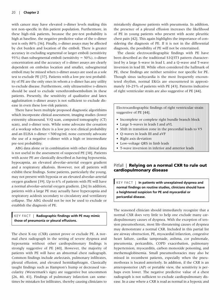
with cancer may have elevated D-dimer levels making thistest non-specific in this patient population. Furthermore, inthese high-risk patients, because the pre-test probability ishigh at baseline, the negative predictive value of the D-dimertest is only 80% [36]. Finally, D-dimer assays may be affectedby clot burden and location of the emboli. There is greateraccuracy in excluding segmental or larger emboli (sensitivity93%) than subsegmental emboli (sensitivity � 50%). D-dimerconcentration and the accuracy of D-dimer assays are clearlydependent on embolus location and smaller, subsegmentalemboli may be missed when D-dimer assays are used as a soletest to exclude PE [37]. Patients with a low pre-test probabil-ity of PE are the only ones in whom a D-dimer has any utilityto exclude disease. Furthermore, only ultrasensitive D-dimersshould be used to exclude venothromboembolism in thesepatients. Presently, the sensitivity of qualitative and latexagglutination D-dimer assays is not sufficient to exclude dis-ease in even these low-risk patients.
There have been multiple proposed diagnostic algorithimswhich incorporate clinical assessment, imaging studies (lowerextremity ultrasound, V/Q scan, computed tomography (CT)scan), and D-dimer tests. While some advocate the cessationof a workup when there is a low pre-test clinical probabilityand an ELISA D-dimer �500 ng/ml, none currently advocatethe use of a negative D-dimer with a high or intermediatepre-test probability.
ABG data alone or in combination with other clinical datais not useful in the assessment of suspected PE [38]. Patientswith acute PE are classically described as having hypoxemia,hypocapnia, an elevated alveolar–arterial oxygen gradientand a respiratory alkalosis. However, not all patients willexhibit these findings. Some patients, particularly the young,may not present with hypoxia or an elevated alveolar–arterialoxygen gradient [39]. Up to 6% of patients with PE will havea normal alveolar–arterial oxygen gradient. [26] In addition,patients with a large PE may actually have hypercapnia andrespiratory acidosis secondary to circulatory and ventilatorycollapse. The ABG should not be not be used to exclude orestablish the diagnosis of PE.
The chest X-ray (CXR) cannot prove or exclude PE. A nor-mal chest radiograph in the setting of severe dyspnea andhypoxemia without other cardiopulmonary findings isstrongly suggestive of PE [40]. However, the majority ofpatients with PE will have an abnormal chest radiograph.Common findings include atelectasis, pulmonary infiltrates,pleural effusion, and elevated hemidiaphragm. Classicallytaught findings such as Hampton’s hump or decreased vas-cularity (Westermark’s sign) are suggestive but uncommon[26, 40, 41]. Findings of pulmonary infarction can some-times be mistaken for infiltrates, thereby causing clinicians to
mistakenly diagnose patients with pneumonia. In addition,the presence of a pleural effusion increases the likelihood of PE in young patients who present with acute pleuriticchest pain [42]. This again highlights the importance of con-sidering the diagnosis of PE. If it is not in the differentialdiagnosis, the possibility of PE will not be entertained.
The classic electrocardiographic findings with PE havebeen described as the traditional S1Q3T3 pattern character-ized by a large S-wave in lead I, and a Q-wave and T-waveinversion in lead III. While often considered the hallmark ofPE, these findings are neither sensitive nor specific for PE.Though sinus tachycardia is the most frequently encoun-tered rhythm, normal EKGs are encountered in approxi-mately 10–25% of patients with PE [43]. Patterns indicativeof right ventricular strain are also suggestive of PE [44].
Pitfall | Relying on a normal CXR to rule outcardiopulmonary disease
The seasoned clinician should immediately recognize that anormal CXR does very little to help one exclude many car-diopulmonary causes of dyspnea. With the exception of ten-sion pneumothorax, most life-threatening causes of dyspneamay demonstrate a normal CXR. Included in this partial listare airway obstruction, PE, myocardial infarction, congestiveheart failure, cardiac tamponade, asthma, cor pulmonale,pneumonia, pericarditis, COPD exacerbation, pulmonaryhypertension, myocarditis, carbon monoxide poisoning, andmethemoglobinemia. Small pneumothoracies may also bemissed in recumbent patients, especially when the pneu-mothorax is located anteriorly. In addition, if the CXR is ananteroposterior (AP) or portable view, the sensitivity is per-haps even lower. The negative predictive value of a chestradiograph is not sufficient to exclude cardiopulmonary dis-ease. In a case where a CXR is read as normal in a hypoxic and
KEY FACT | In patients with unexplained dyspnea andnormal findings on routine studies, clinicians should havea heightened suspicion for PE and myocardial orpericardial disease.
KEY FACT | Radiographic findings with PE may mimicthose of pneumonia or pleural effusions.
20 | Chapter 2
Electrocardiographic findings of right ventricular strainsuggestive of PE [44].
• Incomplete or complete right bundle branch block• Large S-waves in leads I and aVL• Shift in transition zone in the precordial leads to V5• Q-waves in leads III and aVF• Right axis deviation• Low-voltage QRS in limb leads• T-wave inversion in inferior and anterior leads
dyspneic patient, the likelihood of cardiac and thromboem-bolic pathology increases. This warrants the further investi-gation of venothromboembolism and pericardial/ myocardialdisease states with the consideration of bedside echocardio-graphy, EKG, cardiac enzymes, and possibly chest CT. In fact,in one ED study, of 103 patients with unexplained dyspneawho then underwent bedside echocardiography, 14 (13.6%)had pericardial effusions [45]. Of these four were felt to belarge enough to explain the patients’ dyspnea.
Pitfall | Overly or underly aggressive oxygenadministration in patients with COPD
The traditional teaching has been that administration ofoxygen will cause the spontaneously breathing COPD patientin respiratory distress to “stop breathing.” It was traditionallytaught that suppression of the hypoxic ventilatory drive wouldlead to hypercapnia and subsequent hypercapnic respiratoryfailure. However, it appears as though our traditional under-standing of the pathophysiology was incorrect. Three mech-anisms have been shown to worsen hypercapnia whenoxygen is administered in the COPD patient:
1. Worsened V/Q matching due to a decrease in hypoxicpulmonary vasoconstriction (HPV);2. Decreased binding affinity of hemoglobin for CO2;3. Decreased minute ventilation [46, 47].
HPV is a physiologic response to alveolar hypoxia. Throughthis mechanism blood is diverted away from poorly ventilatedlung units. When hypoxia affects the entire lung (typicallyoccurs at PaO2 levels �60 mmHg), HPV occurs throughoutthe lung. This increases the pulmonary artery pressure andsubsequently recruits many previously unperfused pulmonarycapillaries, increasing the surface area available for gas diffu-sion, and improving the matching of ventilation and perfusion.If the hypoxia is resolved with supplemental oxygen, HPV is blunted, thus worsening V/Q matching. This essentiallyincreases the physiologic deadspace and shunt and subse-quently leads to elevations in PCO2 [46, 48].
The CO2 dissociation curve shifts to the right in the pres-ence of increased oxygen saturation. This is known as theHaldane effect. Subsequently, in the presence of increasedoxygen saturation from supplemental oxygen, oxyhemoglo-bin binds CO2 less avidly than deoxyhemoglobin, thusincreasing the amount of CO2 dissolved in blood. This inturn is reflected as an increased PaCO2 [49].
Administration of supplemental oxygen to a COPDpatient in respiratory distress has also been show to decreasethe patient’s minute ventilation. This is due to a smalldecrease in respiratory rate that will also increase PCO2 [47].
As a general rule, prevention of tissue hypoxia is moreimportant than CO2 retention concerns. Many patients withCOPD have a chronic compensated respiratory acidosis.While normal individuals may begin to experience depressed
level of consciousness at PaCO2 levels of 60–70 mmHg,COPD patients may not exhibit these symptoms until theirPaCO2 reaches a level in excess of 90–100 mmHg. COPDpatients are at greater risk from hypoxia than from hyper-capnia and oxygen should not be withheld from the hypoxicCOPD patient. Oxygen therapy should be emergently initiatedin all hypoxic COPD patients with an initial goal oxygen sat-uration of 88–92% unless there is evidence of active cardiacor cerebral ischemia.
The primary goal of oxygen therapy is to prevent tissuehypoxia. The American Thoracic Society/European RespiratorySociety recommends a SaO2 greater than 90% or a PaO2 of60–70 mmHg [50]. In addition, Kelly et al. reported that oxy-gen saturation by pulse oximetry may be an effective screen-ing test for systemic hypoxia, with the screening cutoff of92% having sensitivity for the detection of systemic hypoxiaof 100% with specificity of 86% [51]. Further increases inthe FiO2 above the level needed to achieve these goals do notadd significantly to the oxygen content of blood, but doincrease the potential for more severe secondary hypercapnia[50]. Venturi masks should be used when possible to permittight regulation of FiO2.
Finally, if CO2 retention occurs, monitor for acidemia. Ifacidemia occurs, consider a trial of non-invasive or invasivemechanical ventilation [50].
Pitfall | Not considering non-invasivepositive pressure ventilation; that is,CPAP/BiPAP as an alternative to intubationin selected patients
Non-invasive positive pressure ventilation (NPPV) has beenshown to reduce length of hospital stay, morbidity, mortal-ity, and need for invasive mechanical ventilation. The datasupporting the use of NPPV is strongest for COPD exacerba-tions; however, there is growing evidence to support its usein cardiogenic pulmonary edema [52–55], hypoxic respira-tory failure [56–60], pneumonia [56, 61], immunocompro-mised states [62, 63], and asthma [64, 65].
There is a wealth of information showing the benefit ofNPPV in acute exacerbations of COPD. This data includesrandomized controlled trials, systemic reviews, and meta-analysis [66–74]. NPPV has been shown to decrease intuba-tion rates (RR 0.42, 95% CI 0.31–0.59), decrease mortality(RR 0.41, 95% CI 0.26–0.64), decrease complications (RR0.32, 95% CI 0.18–0.56), and decrease hospital length ofstay (weight mean difference �3.24 days, 95% CI �4.42to �2.26) [68].
It is important to note that early institution of NPPV isvital. Conti et al. showed that if one waits for failure of con-ventional medical therapy, the benefits conferred by NPPV(length of ICU stay, number of days on mechanical ventila-tion, overall complications, ICU mortality, and hospital mor-tality) are eliminated [75]. As such, patients in respiratory
Management of the Dyspneic Patient in the ED | 21
distress, not requiring emergent intubation and withoutcontraindications should be considered for NPPV. TheAmerican Association for Respiratory Care recommends theearly use of NPPV when two or more of the following arepresent:
1. Respiratory distress with moderate to severe dyspnea.2. Arterial pH �7.35 with PaCO2 above 45 mmHg.3. Respiratory rate of �25/min [76].
Improvement should be seen within 30–60 min, and ifimprovement does not occur within 1–2 h, invasivemechanical ventilation should be initiated. One should alsobe aware that when using NPPV, frequent checks andadjustments are required. Failure to do so may lead to thepatient’s condition worsening and a delay in the institutionof invasive mechanical ventilation.
Pearls for Improving Patient Outcomes
• Do not rely solely on a normal physical examination to excludedisease.
• Patients with a normal pulse oximetry may still suffer from venti-latory failure.
• Shortness of breath should be attributed to anxiety only whenall other causes have been definitively excluded.
• Risk stratification of patients with suspected venous throm-boembolism utilizing clinical decision rules will aid in the diagno-sis of PE.
• A normal CXR does very little to exclude cardiopulmonarycauses of dyspnea.
• The negative predictive value of a normal ABG or CXR does notallow one to exclude PE.
• A D-dimer test should not be used to exclude PE in patients withmoderate or high clinic pre-test probability.
• Only high-sensitivity D-Dimer tests should be used to exclude PEin patients but should only be used in patients with a low pre-test probability.
• Do not withhold oxygen from a hypoxic COPD patient; however,be cautious with its use and follow PCO2 levels.
• Consider the use of NPPV in selected patients with respiratorydistress if they do not require immediate emergent intubation.This is especially true with patients in COPD exacerbation.
References
1. Dyspnea. Mechanisms, assessment, and management: a con-
sensus statement. American Thoracic Society. Am J Respir Crit
Care Med 1999; 159:321.
2. Banzett RB, Mulnier HE, Murphy K, Rosen SD, Wise RJ,
Adams L. Breathlessness in humans activates insular cortex.
NeuroReport 2000; 11(10):2117–20.
3. O’Connor GT, Anderson KM, Kannel WB, et al. Prevalence and
prognosis of dyspnea in the Farmingham Study. Chest 1987;
92:90S.
4. Hirshberg A, Thomson SR, Huizinga WK. Reliability of physical
examination in penetrating chest injuries. Injury 1988; 19(6):
407–9.
5. Chen SC, Chang KJ, Hsu CY. Accuracy of auscultation in the detec-
tion of haemopneumothorax. Eur J Surg 1998; 164(9):643–5.
6. Chen SC, Markmann JF, Kauder DR, Schwab CW.
Hemopneumothorax missed by auscultation in penetrating
chest injury. J Trauma 1997; 42(1):86–89.
7. Bokhari F, Brakenridge S, Nagy K, Roberts R, Smith R, Joseph
K, An G, Wiley D, Barrett J. Prospective evaluation of the sensi-
tivity of physical examination in chest trauma. J Trauma 2002;
53(6):1135–8.
8. Wipf JE, Lipsky BA, Hirschmann JV, Boyko EJ, Takasugi J,
Peugeot RL, Davis CL. Diagnosing pneumonia by physical
examination: relevant or relic? Arch Intern Med 1999; 159 (10):
1082–7.
9. Metlay JP, Kapoor WN, Fine MJ. Does this patient have
community-acquired pneumonia? Diagnosing pneumonia
by history and physical examination. J Am Med Assoc 1997;
278(17):1440–5.
10. Chakko S, Woska D, Martinez H, et al. Clinical, radiographic,
and hemodynamic correlations in chronic congestive heart fail-
ure: conflicting results may lead to inappropriate care. Am J Med
1991; 90:353–9.
11. Witting MD, Hsu S, Granja CA. The sensitivity of room-air
pulse oximetry in the detection of hypercapnia. Am J Emerg Med
2005; 23(4):497–500.
12. Witting MD, Lueck CH. The ability of pulse oximetry to screen
for hypoxemia and hypercapnia in patients breathing room air.
J Emerg Med 2001; 20(4):341–8.
13. Ayas N, Bergstrom LR, Schwab TR, Narr BJ. Unrecognized
severe postoperative hypercapnia: a case of apneic oxygena-
tion. Mayo Clin Proc 1998; 73(1):51–54.
14. Potkin RT, Swenson ER. Resuscitation from severe acute hyper-
capnia. Determinants of tolerance and survival. Chest 1992;
102(6):1742–5.
15. Stoneham MD. Uses and limitations of pulse oximetry. Br J
Hosp Med 1995; 54(1):35–41.
16. Hampson NB. Pulse oximetry in severe carbon monoxide poi-
soning. Chest 1998; 114(4):1036–41.
17. Miner JR, Heegaard W, Plummer D. End-tidal carbon dioxide
monitoring during procedural sedation. Acad Emerg Med 2002;
9(4):275–80.
18. Rohling R, Biro P. Clinical investigation of a new combined
pulse oximetry and carbon dioxide tension sensor in adult
anaesthesia. J Clin Monit Comput 1999; 15(1):23–27.
19. Senn O, Clarenbach CF, Kaplan V, Maggiorini M, Bloch KE.
Monitoring carbon dioxide tension and arterial oxygen satura-
tion by a single earlobe sensor in patients with critical illness or
sleep apnea. Chest 2005; 128(3):1291–6.
20. Chhajed PN, Kaegi B, Rajasekaran R, Tamm M. Detection of
hypoventilation during thoracoscopy: combined cutaneous car-
bon dioxide tension and oximetry monitoring with a new digi-
tal sensor. Chest 2005; 127(2):585–8.
21. Yohannes AM, Roomi J, Baldwin RC, Connolly MJ. Depression
in elderly outpatients with disabling chronic obstructive pul-
monary disease. Age Ageing 1998; 27(2):155–60.
22. Horton-Deutsch SL, Clark DC, Farran CJ. Chronic dyspnea and
suicide in elderly men. Hosp Commun Psychiatr 1992; 43(12):
1198–203.
22 | Chapter 2
23. Ben-Dov I. Fatal asthma: who, when, why and how to pre-
vent? Harefuah 2002; 141(6):524–6, 579, 578.
24. de Fockert JA. Clinical presentation of acute myocardial infarct
in the elderly. Tijdschr Gerontol Geriatr 1987; 18(6):305–8.
25. Laack TA, Goyal DG. Pulmonary embolism: an unsuspected
killer. Emerg Med Clin N Am 2004; 22(4):961–83.
26. Stein, PD, Terrin, ML, Hales, CA, et al. Clinical, laboratory,
roentgenographic, and electrocardiographic findings in patients
with acute pulmonary embolism and no pre-existing cardiac or
pulmonary disease. Chest 1991; 100:598.
27. Anderson Jr FA, Wheeler HB, Goldberg RJ, et al. A population-
based perspective of the hospital incidence and case-fatality
rates of deep vein thrombosis and pulmonary embolism: the
Worcester DVT Study. Arch Intern Med 1991; 151:933–8.
28. Silverstein MD, Heit JA, Mohr DN, Petterson TM, O’Fallon
WM, Melton III LJ. Trends in the incidence of deep vein throm-
bosis and pulmonary embolism: a 25-year population-based
study. Arch Intern Med 1998; 158:585–93.
29. Goldhaber SZ. Pulmonary embolism. New Engl J Med 1998;
339:93–104.
30. Carson JL, Kelley MA, Duff A, et al. The clinical course of pul-
monary embolism. New Engl J Med 1992; 326:1240–5.
31. Wolf SJ, McCubbin TR, Feldhaus KM, Faragher JP, Adcock DM.
Prospective validation of wells criteria in the evaluation of
patients with suspected pulmonary embolism. Ann Emerg Med
2004; 44(5):503–10.
32. Fedullo PF, Tapson VF. The evaluation of suspected pulmonary
embolism. New Engl J Med 2003; 349:1247–56.
33. Becker DM, Philbrick JT, Bachhuber TL, Humphries JE. D-dimer
testing and acute venous thromboembolism. A shortcut to
accurate diagnosis? Arch Intern Med 1996; 156(9):939–46.
34. Farrell S, Hayes T, Shaw M. A negative SimpliRED D-dimer
assay result does not exclude the diagnosis of deep vein throm-
bosis or pulmonary embolus in emergency department patients.
Ann Emerg Med 2000; 35(2):121–5.
35. Stein PD, Hull RD, Patel KC, Olson RE, Ghali WA, Brant R, Biel
RK, Bharadia V, Kalra NK. D-dimer for the exclusion of acute
venous thrombosis and pulmonary embolism: a systematic
review. Ann Intern Med 2004; 140(8):589–602.
36. Lee AY, Julian JA, Levine MN, Weitz JI, Kearon C, Wells PS,
Ginsberg JS. Clinical utility of a rapid whole-blood D-dimer
assay in patients with cancer who present with suspected acute
deep venous thrombosis. Ann Intern Med 1999; 131(6):417–23.
37. De Monye W, Sanson BJ, Mac Gillavry MR, Pattynama PM,
Buller HR, van den Berg-Huysmans AA, Huisman MV. Embolus
location affects the sensitivity of a rapid quantitative D-dimer
assay in the diagnosis of pulmonary embolism. Am J Respir Crit
Care Med 2002; 165(3):345–8.
38. Rodger MA, Carrier M, Jones GN, Rasuli P, Raymond F,
Djunaedi H, Wells PS. Diagnostic value of arterial blood gas
measurement in suspected pulmonary embolism. Am J Respir
Crit Care Med 2000; 162(6):2105–8.
39. Green RM, Meyer TJ, Dunn M, Glassroth J. Pulmonary
embolism in younger adults. Chest 1992; 101(6):1507–11.
40. ATS guidelines: the diagnostic approach to acute venous
thromboembolism. Am J Respir Crit Care Med 1999; 160:1043.
41. Worsley DF, Alavi A, Aronchick JM, Chen JT, Greenspan RH,
Ravin CE. Chest radiographic findings in patients with acute
pulmonary embolism: observations from the PIOPED study.
Radiology 1993; 189(1):133–6.
42. McNeill BJ, Hessel SJ, Branch WT, et al. Measures of clinical
efficacy: 3. The value of the lung scan in the evaluation of
young patients with pleuritic chest pain. J Nucl Med 1976;
17:163.
43. Ullman E, Brady WJ, Perron AD, Chan T, Mattu A. Electrocar-
diographic manifestations of pulmonary embolism. Am J Emerg
Med 2001; 19:514–9.
44. Sreeram N, Cheriex EC, Smeets JLRM, et al. Value of the 12-
lead electrocardiogram at hospital admission in the diagnosis of
pulmonary embolism. Am J Cardiol 1994; 73:298–303.
45. Blaivas M. Incidence of pericardial effusion in patients present-
ing to the emergency department with unexplained dyspnea.
Acad Emerg Med 2001; 8(12):1143–6.
46. Aubier M, Murciano D, Milic-Emili J, Touaty E, Daghfous J,
Pariente R, Derenne JP. Effects of the administration of O2 on
ventilation and blood gases in patients with chronic obstructive
pulmonary disease during acute respiratory failure. Am Rev
Respir Dis 1980; 122(5):747–54.
47. Aubier M, Murciano D, Fournier M, Milic-Emili J, Pariente R,
Derenne JP. Central respiratory drive in acute respiratory fail-
ure of patients with chronic obstructive pulmonary disease. Am
Rev Respir Dis 1980; 122(2):191–9.
48. Levitzky MG. Pulmonary Physiology, 6th edn. New York:
McGraw-Hill/Appleton & Lange, 2003, p. 107.
49. Levitzky MG. Pulmonary Physiology, 6th edn. New York:
McGraw-Hill/Appleton & Lange, 2003, pp. 158–9.
50. Standards for the diagnosis and care of patients with chronic
obstructive pulmonary disease. American Thoracic Society. Am
J Respir Crit Care Med 1995; 152:S77.
51. Kelly AM, McAlpine R, Kyle E. Respir Med 2001; 95(5):336–40.
52. Wigder HN, Hoffman P, Mazzolini D, et al. Pressure support
noninvasive positive pressure ventilation treatment of acute
cardiogenic pulmonary edema. Am J Emerg Med 2001; 19:
179–81.
53. Pang D, Keenan S, Cook D, et al. The effect of positive pressure
airway support on mortality and the need for intubation in car-
diogenic pulmonary edema. A systematic review. Chest 1998;
114(4):1185–92.
54. Bersten AD, Holt AW, Vedig AE, Skowronski GA, Baggoley CJ.
Treatment of severe cardiogenic pulmonary edema with con-
tinuous positive airway pressure delivered by face mask. New
Engl J Med 1991; 325(26):1825–30.
55. Nava S, Carbone G, DiBattista N, Bellone A, Baiardi P, Cosentini
R, Marenco M, Giostra F, Borasi G, Groff P. Noninvasive ventila-
tion in cardiogenic pulmonary edema: a multicenter randomized
trial. Am J Respir Crit Care Med 2003; 168(12):1432–7.
56. Ferrer M, Esquinas A, Leon M, Gonzalez G, Alarcon A, Torres
A. Noninvasive ventilation in severe hypoxemic respiratory
failure: a randomized clinical trial. Am J Respir Crit Care Med
2003; 168(12):1438–44.
57. Antonelli M, Conti G, Rocco M, Bufi M, De Blasi RA, Vivino G,
Gasparetto A, Meduri GU. A comparison of noninvasive posi-
tive-pressure ventilation and conventional mechanical ventila-
tion in patients with acute respiratory failure. New Engl J Med
1998; 339(7):429–35.
58. Martin TJ, Hovis JD, Costantino JP, Bierman MI, Donahoe MP,
Rogers RM, Kreit JW, Sciurba FC, Stiller RA, Sanders MH.
A randomized, prospective evaluation of noninvasive ventila-
tion for acute respiratory failure. Am J Respir Crit Care Med 2000;
161(3 Pt 1):807–13.
Management of the Dyspneic Patient in the ED | 23

59. Delclaux C, L’Her E, Alberti C, Mancebo J, Abroug F, Conti G,
Guerin C, Schortgen F, Lefort Y, Antonelli M, Lepage E,
Lemaire F, Brochard L. Treatment of acute hypoxemic nonhy-
percapnic respiratory insufficiency with continuous positive
airway pressure delivered by a face mask: a randomized con-
trolled trial. J Am Med Assoc 2000; 284(18):2352–60.
60. Keenan SP, Sinuff T, Cook DJ, Hill NS. Does noninvasive posi-
tive pressure ventilation improve outcome in acute hypoxemic
respiratory failure? A systematic review. Crit Care Med 2004;
32(12):2516–23.
61. Confalonieri M, Potena A, Carbone G, Porta RD, Tolley EA,
Umberto Meduri G. Acute respiratory failure in patients with
severe community-acquired pneumonia. A prospective ran-
domized evaluation of noninvasive ventilation. Am J Respir Crit
Care Med 1999; 160(5 Pt 1):1585–91.
62. Confalonieri M, Calderini E, Terraciano S, Chidini G, Celeste E,
Puccio G, Gregoretti C, Meduri GU. Noninvasive ventilation for
treating acute respiratory failure in AIDS patients with
Pneumocystis carinii pneumonia. Intens Care Med 2002;
28(9):1233–8.
63. Hilbert G, Gruson D, Vargas F, Valentino R, Gbikpi-Benissan G,
Dupon M, Reiffers J, Cardinaud JP. Noninvasive ventilation in
immunosuppressed patients with pulmonary infiltrates, fever,
and acute respiratory failure. New Engl J Med 2001;
344(7):481–7.
64. Soroksky A, Stav D, Shpirer I. A pilot prospective, randomized,
placebo-controlled trial of bilevel positive airway pressure in
acute asthmatic attack. Chest 2003; 123(4):1018–25.
65. Meduri GU, Cook TR, Turner RE, et al. Noninvasive positive
pressure ventilation in status asthmaticus. Chest 1996;
110(3):767–74.
66. Brochard L, Mancebo J, Wysocki M, Lofaso F, Conti G, Rauss A,
Simonneau G, Benito S, Gasparetto A, Lemaire F. Noninvasive
ventilation for acute exacerbations of chronic obstructive pul-
monary disease. New Engl J Med 1995; 333(13):817–22.
67. Wysocki M, Tric L, Wolff MA, Millet H, Herman B. Noninvasive
pressure support ventilation in patients with acute respiratory
failure. A randomized comparison with conventional therapy.
Chest 1995; 107(3):761–8.
68. Ram FS, Picot J, Lightowler J, Wedzicha JA. Non-invasive posi-
tive pressure ventilation for treatment of respiratory failure due
to exacerbations of chronic obstructive pulmonary disease.
Cochrane Database Syst Rev 2004; (3):CD004104.
69. Peter JV, Moran JL, Phillips-Hughes J, Warn D. Noninvasive
ventilation in acute respiratory failure—a meta-analysis
update. Crit Care Med 2002; 30(3):555–62.
70. Lightowler JV, Wedzicha JA, Elliott MW, Ram FS. Non-invasive
positive pressure ventilation to treat respiratory failure result-
ing from exacerbations of chronic obstructive pulmonary dis-
ease: cochrane systematic review and meta-analysis. Br Med J
2003; 326(7382):185.
71. Keenan SP, Sinuff T, Cook DJ, Hill NS. Which patients with
acute exacerbation of chronic obstructive pulmonary disease
benefit from noninvasive positive-pressure ventilation? A sys-
tematic review of the literature. Ann Intern Med 2003;
138(11):861–70.
72. Plant PK, Owen JL, Elliott MW. Early use of noninvasive venti-
lation for acute exacerbations of chronic obstructive pulmonary
disease on general respiratory wards: a multicentre randomised
controlled trial. Lancet 2000; 355:1931–5.
73. Keenan SP, Gregor J, Sibbald WJ, et al. Noninvasive positive
pressure ventilation in the setting of severe, acute exacerba-
tions of chronic obstructive pulmonary disease: more effective
and less expensive. Crit Care Med 2000; 28(6):2094–103.
74. Celikel T, Sungur M, Ceyhan B, et al. Comparisons of noninva-
sive positive pressure ventilation with standard medical therapy
in acute hypercapneic respiratory failure. Chest 1998;
114(6):1636–42.
75. Conti G, Antonelli M, Navalesi P, et al. Noninvasive vs. conven-
tional mechanical ventilation in patients with chronic obstruc-
tive pulmonary disease after failure of medical treatment in the
ward: a randomized trial. Intens Care Med 2002; 28(12):1701–7.
76. Bach, JR, Brougher, P, Hess, DR, et al. Consensus conference:
noninvasive positive pressure ventilation. Respir Care 1997;
42:364–9.
24 | Chapter 2
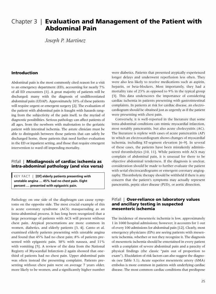
Introduction
Abdominal pain is the most commonly cited reason for a visitto an emergency department (ED), accounting for nearly 7%of all ED encounters [1]. A great majority of patients will bedischarged; many with the diagnosis of undifferentiatedabdominal pain (UDAP). Approximately 10% of these patientswill require urgent or emergent surgery [2]. The evaluation ofthe patient with abdominal pain is fraught with hazards rang-ing from the subjectivity of the pain itself, to the myriad ofdiagnostic possibilities. Serious pathology can affect patients ofall ages, from the newborn with malrotation to the geriatricpatient with intestinal ischemia. The astute clinician must beable to distinguish between those patients that can safely bedischarged home, those patients that need further evaluationin the ED or inpatient setting, and those that require emergentintervention to ward off impending mortality.
Pitfall | Misdiagnosis of cardiac ischemia asintra-abdominal pathology (and vice versa)
Pathology on one side of the diaphragm can cause symp-toms on the opposite side. The most crucial example of thisis acute coronary syndrome (ACS) masquerading as anintra-abdominal process. It has long been recognized that alarge percentage of patients with ACS will present withoutchest pain. Atypical presentations are more common inwomen, diabetics, and elderly patients [3, 4]. Canto et al.examined elderly patients presenting with unstable anginaand found that 45% had no chest pain; 8% of patients pre-sented with epigastric pain, 38% with nausea, and 11%with vomiting [5]. A review of the data from the NationalRegistry of Myocardial Infarction-2 again showed that one-third of patients had no chest pain. Upper abdominal painwas often instead the presenting complaint. Patients pre-senting without chest pain were on average 7 years older,more likely to be women, and a significantly higher number
were diabetics. Patients that presented atypically experiencedlonger delays and underwent reperfusion less often. Theywere also less likely to receive medications such as aspirin,heparin, or beta-blockers. Most importantly, they had amortality rate of 23% as opposed to 9% in the typical group[3]. This data underscores the importance of consideringcardiac ischemia in patients presenting with gastrointestinalcomplaints. In patients at risk for cardiac disease, an electro-cardiogram should be obtained just as urgently as if the patientwere presenting with chest pain.
Conversely, it is well-reported in the literature that someintra-abdominal conditions can mimic myocardial infarction,most notably pancreatitis, but also acute cholecystitis (AC).The literature is replete with cases of acute pancreatitis (AP)in which an electrocardiogram shows changes of myocardialischemia, including ST-segment elevation [6–9]. In severalof these cases, the patients have been mistakenly adminis-tered thrombolytics [10, 11]. While patients with ACS maycomplain of abdominal pain, it is unusual for there to beobjective abdominal tenderness. If the diagnosis is unclear,consideration should be made to further evaluate the patientwith serial electrocardiograms or emergent coronary angiog-raphy. Thrombolytic therapy should be withheld if there is anyconcern that the patient’s symptoms may actually representpancreatitis, peptic ulcer disease (PUD), or aortic dissection.
Pitfall | Over-reliance on laboratory valuesand ancillary testing in suspectedmesenteric ischemia
The incidence of mesenteric ischemia is low, approximately1 in 1000 hospital admissions; however, it accounts for 1 outof every 100 admissions for abdominal pain [12]. Clearly, mostemergency physicians (EPs) are seeing patients with mesen-teric ischemia, whether or not they recognize it. The diagnosisof mesenteric ischemia should be entertained in every patientwith a complaint of severe abdominal pain and a paucity ofphysical findings (the classic “pain out of proportion toexam”). Elucidation of risk factors can also suggest the diagno-sis (see Table 3.1). Acute superior mesenteric artery (SMA)embolus is more common in patients with underlying cardiacdisease. The most common cardiac conditions that predispose
KEY FACT | [Of] elderly patients presenting withunstable angina … 45% had no chest pain. Eightpercent … presented with epigastric pain.
Chapter 3 | Evaluation and Management of the Patient withAbdominal Pain
Joseph P. Martinez
25
to SMA embolus are atrial fibrillation, other arrhythmias,previous myocardial infarction with ventricular aneurysmformation, valvular heart disease, or dilated cardiomyopathy[13]. Physical findings of previous embolic disease such asprior stroke or amputated toes may increase suspicion. SMAthrombosis is seen in patients with classic cardiovascular riskfactors. These patients should be specifically interrogated forsymptoms of previous “intestinal ischemia” – abdominalpain provoked by meals. They may also demonstrate signifi-cant unintentional weight loss. One-half of patients withmesenteric venous thrombosis will have a personal or familyhistory of deep venous thrombosis or pulmonary embolus[14]. They should be interviewed as would any patient inwhom you suspect a thromboembolic process. Close atten-tion should be paid to clues that suggest inherited throm-bophilias – family history of thromboembolic events,multiple first trimester pregnancy losses, or previous per-sonal clot history. Acquired risks such as smoking or oralcontraceptive use should also be investigated. Non-occlusivemesenteric ischemia should be near the top of the differen-tial diagnosis list in any patient that exhibits a “low-flowstate” and presents with abdominal pain. This would includepatients that are septic, severely volume depleted, or with
end-stage congestive heart failure, and those that usecocaine, digitalis, or are on hemodialysis [15–17].
Mortality from mesenteric ischemia is nearly 80%. Thismortality rate has remained remarkably constant throughthe years. A landmark study by Boley et al. finally demon-strated improvement in mortality only through the aggres-sive use of early angiography. His group showed that withimmediate angiography, the mortality rate could be reducedto 54% [18]. These results have since been duplicated in anumber of other studies [19, 20]. Unfortunately, manyphysicians are unwilling to obtain angiography based solelyon the patient’s presentation and risk factors. They will waitfor collateral laboratory data, and obtain other, suboptimalimaging. This is akin to delaying reperfusion in ACS untilthe cardiac enzymes have peaked. While an elevated lactatelevel, leukocytosis, metabolic acidosis, and hyperamylasemia
KEY FACT | Mortality from mesenteric ischemia isnearly 80% … With immediate angiography, the mortalityrate could be reduced to 54%.
26 | Chapter 3
Table 3.1 Risk factors for mesenteric ischemia.
Type of mesenteric ischemia Risk factors Special notes
SMA embolus Cardiac disease One-third have a history of a previous embolic event• Atrial fibrillation or other arrhythmia• Valvular disease• Ventricular aneurysm• Cardiomyopathy
SMA thrombosis Vascular disease risks Acute event may be preceded by period of • Hypertension “intestinal angina” and prolonged period of • Hypercholesterolemia significant weight loss• Diabetes mellitus• Smoking
Mesenteric venous thrombosis Hypercoaguable state One-half have personal or family history of DVT/PE• Inherited (factor V Leiden Subacute presentation
mutation, etc.) Women � men• Acquired (malignancy, oral
contraceptives, etc.)
NOMI Low-flow states Often ICU patients• Sepsis• Heart failure• Volume depletionHemodialysisDrugs• Digitalis• Ergot derivatives• Cocaine• NorepinephrinePost-surgery
DVT: deep venous thrombosis; PE: pulmonary embolus; NOMI: non-occlusive mesenteric ischemia; ICU: intensive care unit.
support the diagnosis, none of them are sensitive enough torule it out completely. In addition, obtaining other imagingstudies merely delays the time required to obtain a definitiveangiographic diagnosis and start treatment. If the patient hasrisk factors for mesenteric ischemia, and the history andphysical examination suggest this diagnosis, arrangementsshould be made for immediate angiography and surgicalconsultation. The following caveats apply: (1) If mesentericvenous thrombosis is suspected, computed tomography (CT) is better than angiography [21], unless the angiogra-pher is specifically instructed to perform venous phaseangiography. (2) If there will be a long delay in obtainingangiography and the ED has access to a multi-detector rowCT scanner, a “mesenteric ischemia protocol,” and interpre-tation of the images in a timely fashion, CT-angiographyshows a great deal of promise as a screening modality (seeFigure 3.1) [22].
Pitfall | Failure to consider heterotopicpregnancy in women receivingreproductive assistance
Women of child-bearing age are another high-risk demo-graphic group when presenting with abdominal pain. MostEPs are well versed in the need to evaluate these patients forpossible ectopic pregnancy should they present while earlyin pregnancy. The standard practice for ruling out an ectopicpregnancy is to obtain an ultrasound, transabdominal orendovaginal depending on their gestational age at presenta-tion and/or quantitative ß-HCG count. The diagnosis ofectopic pregnancy is considered “ruled out” if an intrauter-ine pregnancy (IUP) with fetal heart rate is visualized [23].In general practice, this is a reasonable approach: hetero-topic pregnancy (simultaneous occurrence of two or moreimplantation sites, usually intrauterine and ectopic) is a rare event, with most estimates in the range of 1:30,000pregnancies [24].
With the advent of assisted reproductive technology (ART),this ratio has changed. Women receiving ART have rates ofheterotopic pregnancy as high as 1:100 pregnancies [25]. Thisappears to be independent of whether the patient received anovulation-inducing agent (e.g., clomiphene), an exogenousgonadotropin, or in vitro fertilization with embryo transfer[26–28]. Heterotopic pregnancy in natural conception cycles israre, though the incidence appears to be increasing [29].
Diagnosing a heterotopic pregnancy requires an extremelyhigh index of suspicion. Women have often already beendiagnosed with an IUP by ultrasound. The level of ß-HCGmay be increasing appropriately due to the presence of a
normal IUP. Adnexal masses and free fluid in the cul-de-sacmay be mistakenly attributed to ruptured corpus luteumcysts, an admittedly more common diagnosis. In fact, only10% of heterotopic gestations are diagnosed prior to operativeintervention [25].
A retrospective review of 66 cases of heterotopic preg-nancy elucidated 4 common signs and symptoms to helpsuggest the diagnosis: (1) abdominal pain, (2) adnexal mass,(3) peritoneal irritation, and (4) enlarged uterus [24]. If thediagnosis is made in the first trimester, an enlarged uterusmay be absent. More recently, it has been demonstrated that
KEY FACT | Women receiving ART have rates ofheterotopic pregnancy as high as 1:100 pregnancies.
Evaluation and Management of the Patient With Abdominal Pain | 27
A
B
Figure 3.1 Normal mesenteric CT angiogram.Sagittal reformatting of multi-detector row CT scan (CT angiography)demonstrating normal celiac axis (arrow A) and normal SMA (arrow B).
patients with an abnormal amount of free fluid in the cul-de-sac on ultrasound were five times as likely to have an ectopicpregnancy [30]. This finding should not be discounted in anypatient, even if a viable IUP is seen. Other findings that shouldraise a red flag for clinicians include abnormalities seen in theadnexa on ultrasound, especially an echogenic ring surround-ing a cystic structure, and relative bradycardia in the face ofhemorrhagic shock [31, 32].
Pitfall | Over-reliance on “classic”presentations and laboratory results inpopulations at high risk for atypicalpresentations of appendicitis
Appendicitis is a common surgical emergency in the generalpopulation with a lifetime risk of 7% [33]. Perforation ratesrange from 17% to 20% [34]. Once thought to be a diseaseof the young, it is now recognized that 10% of all cases ofappendicitis occur in patients over the age of 60 years. Whilethe overall mortality from appendicitis is less than 1%, inthe elderly population it ranges from 4% to 8%. This likelyreflects the fact that the incidence of perforation in elderlypatients is as high as 70% [35].
Dr. Alfredo Alvarado established a practical score known asMANTRELS to assist with the early diagnosis of appendicitis(see Table 3.2). The score is based on typical signs, symptoms,and laboratory values often seen in acute appendicitis, namely:Migration of pain, Anorexia (or ketonuria), Nausea and vomit-ing, Tenderness in the right lower quadrant, Rebound tender-ness, Elevated temperature (�100.4°F, �38°C), Leukocytosis(white blood cell (WBC) count �10,400 cells/ml), and Shift to
the left (�75% neutrophils). A point is assigned to each, withright lower quadrant tenderness and leukocytosis receiving 2points, for a total of 10 possible points. A score of 5 or 6 indi-cates possible appendicitis, 7 or 8 probable appendicitis, and 9or 10 is very probable appendicitis [36]. This scoring systemhas been found to be useful as a diagnostic tool in assisting cli-nicians. Depending on the practice setting, especially whenabdominal CT is not readily available for diagnostic purposes,the score can be used to risk stratify patients for discharge withearly follow-up, observation, or emergent consultation. If theMANTRELS score is used, it should be weighed in conjunctionwith physician judgement and should not be used as a meansto definitively rule out the possibility of acute appendicitis.Whether or not physicians formally utilize the MANTRELSscore, the findings that encompass it are thought to be highlyrepresentative of appendicitis.
Unfortunately, atypical presentations of appendicitis appearto be the norm in elderly patients. Only 20% of elderlypatients will have the classic findings of fever, anorexia, rightlower quadrant pain, and leukocytosis [37]. Fever is seen inless than one-third of elderly patients at the time of presenta-tion, right lower quadrant tenderness may be absent in 30%,and one-quarter will have a normal WBC count [38]. Thismay account for the staggering statistic that nearly one-quarterof elderly patients subsequently proven to have appendicitisare discharged home at the first ED visit [39].
It is no surprise then that the MANTRELS score underper-forms in elderly patients. A study in 2001 showed that in143 cases of surgically proven appendicitis, 12 cases wouldhave been missed using the MANTRELS scoring system (i.e.,MANTRELS score �5). Ten of the 12 cases were patientsbetween the age of 60 and 80 years [40].
A higher than normal index of suspicion is required toaccurately diagnose appendicitis in a timely fashion in elderly
KEY FACT | Only 20% of elderly patients will have theclassic findings of fever, anorexia, right lower quadrantpain, and leukocytosis.
28 | Chapter 3
Findings suspicious for heterotopic pregnancy.
• Relative bradycardia in the face of hypotension• Free fluid in cul-de-sac, despite normal IUP• Adnexal abnormality, especially echogenic ring
surrounding cystic structure• Abdominal pain or peritoneal irritation unexplained
by IUP
Points
Symptoms Migration 1Anorexia 1Nausea/vomiting 1
Signs Tenderness in right lower quadrant 2Rebound 1Elevated temperature (�100.4°F, �38.0°C) 1
Laboratory values Leukocytosis (WBC �10,400 cells/ml) 2Shift to the left (�75% neutrophils on differential) 2
Total 10
Score: 5–6: possible appendicitis; 7–8: probable appendicitis; 9–10: very probable appendicitis.
Table 3.2 MANTRELS (Alvarado) Score forsuspected acute appendicitis.
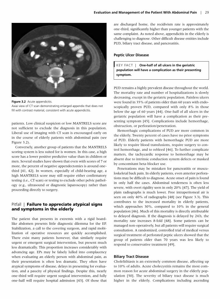
patients. Low clinical suspicion or low MANTRELS score arenot sufficient to exclude the diagnosis in this population.Liberal use of imaging with CT scan is encouraged early onin the course of elderly patients with abdominal pain (seeFigure 3.2).
Conversely, another group of patients that the MANTRELSscoring system is less suited for is women. In this case, a highscore has a lower positive predictive value than in children ormen. Several studies have shown that even with scores of 7 ormore, the percent of negative appendectomies is around one-third [41, 42]. In women, especially of child-bearing age, ahigh MANTRELS score may still require either confirmatorytesting (i.e., CT scan) or testing to exclude other pelvic pathol-ogy (e.g., ultrasound or diagnostic laparoscopy) rather thanproceeding directly to surgery.
Pitfall | Failure to appreciate atypical signsand symptoms in the elderly
The patient that presents in extremis with a rigid board-like abdomen presents little diagnostic dilemma for the EP.Stabilization, a call to the covering surgeon, and rapid mobi-lization of operative resources are quickly accomplished.There exist many patients however, that similarly requireurgent or emergent surgical intervention, but present muchless dramatically. This proportion increases considerably withadvancing age. EPs may be falsely lulled into complacencywhen evaluating an elderly person with abdominal pain, astheir presentation is often less dramatic. They often haveatypical symptoms of disease, delays in presenting for evalua-tion, and a paucity of physical findings. Despite this, nearlyone-third will require urgent surgical intervention, and fullyone-half will require hospital admission [43]. Of those that
are discharged home, the recidivism rate is approximatelyone-third, significantly higher than younger patients with thesame complaint. As noted above, appendicitis in the elderly ischallenging to diagnose. Other difficult disease entities includePUD, biliary tract disease, and pancreatitis.
Peptic Ulcer Disease
PUD remains a highly prevalent disease throughout the world.The mortality rate and number of hospitalizations is slowlydecreasing, except in the geriatric population. Painless ulcerswere found in 35% of patients older than 60 years with endo-scopically proven PUD, compared with only 8% in thosebelow the age of 60 years [44]. One-half of all ulcers in thegeriatric population will have a complication as their pre-senting symptom [45]. Complications include hemorrhage,obstruction, or perforation/penetration.
Hemorrhagic complications of PUD are more common inthe elderly. Twenty percent of cases have no prior symptomsof PUD. Elderly patients with hemorrhagic PUD are morelikely to require blood transfusions, require surgery to con-trol hemorrhage, and to rebleed [46]. To further complicatematters, the tachycardic response to hemorrhage may beabsent due to intrinsic conduction system defects or maskedby concomitant beta-blocker use.
Penetrations may be mistaken for pancreatitis or muscu-loskeletal back pain. In elderly patients, even anterior perfora-tions may be difficult to diagnose. Acute onset of pain is foundin only half the cases. Abdominal tenderness is often lesssevere, with overt rigidity seen in only 20% [47]. The yield ofplain radiographs is much lower. Free intraperitoneal air isseen on only 40% of radiographs [48] (see Figure 3.3). Thiscontributes to the increased mortality in elderly patients,which approaches 30%, compared to 10% in the generalpopulation [46]. Much of this mortality is directly attributableto delayed diagnosis. If the diagnosis is delayed by 24 h, themortality rate increases 8-fold [48]. Some patients can bemanaged non-operatively, but all patients will require surgicalconsultation. A randomized, controlled trial of medical versussurgical treatment of perforated peptic ulcers showed that thegroup of patients older than 70 years was less likely torespond to conservative treatment [49].
Biliary Tract DiseaseCholelithiasis is an extremely common disease, affecting upto 10% of adults. Acute cholecystitis remains the most com-mon reason for acute abdominal surgery in the elderly pop-ulation [50]. The severity of biliary tract disease is muchhigher in the elderly. Complications including ascending
KEY FACT | One-half of all ulcers in the geriatricpopulation will have a complication as their presentingsymptom.
Evaluation and Management of the Patient With Abdominal Pain | 29
Figure 3.2 Acute appendicitis.Axiai view of CT scan demonstrating enlarged appendix that does notfill with contrast material, consistent with acute appendicitis.
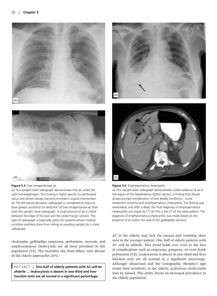
cholangitis, gallbladder empyema, perforation, necrosis, andemphysematous cholecystitis are all more prevalent in thispopulation [51]. The mortality rate from biliary tract diseasein the elderly approaches 20%.
AC in the elderly may lack the nausea and vomiting oftenseen in the younger patient. One-half of elderly patients withAC will be afebrile. This trend holds true even in the face of complications such as empyema, gangrene, or even frankperforation [52]. Leukocytosis is absent in one-third and liverfunction tests are all normal in a significant percentage.Although ultrasound and the sonographic Murphy’s signretain their sensitivity in the elderly, acalculous cholecystitismay be missed. This entity shows an increased prevalence inthe elderly population.
KEY FACT | One-half of elderly patients with AC will beafebrile … leukocytosis is absent in one-third and liverfunction tests are all normal in a significant percentage.
30 | Chapter 3
(a)
(b)
Figure 3.3 Free intraperitoneal air.(a) This upright chest radiograph demonstrates free air under theright hemidiaphragm. This finding is highly specific for perforated viscus and almost always warrants emergent surgical intervention.(b) The left lateral decubitus radiograph is considered by many tohave greater sensitivity for detection of free intraperitoneal air thaneven the upright chest radiograph. A small amount of air is notedbetween the edge of the liver and the costal margin (arrow). Thistype of radiograph is especially useful for patients whose medicalcondition prohibits them from sitting or standing upright for a chestradiograph.
(a)
(b)
Figure 3.4 Emphysematous cholecystitis.(a) This upright plain radiograph demonstrates subtle evidence of air inthe region of the hepatobiliary system (arrow), a finding that shouldalways prompt consideration of two deadly conditions – acutemesenteric ischemia and emphysematous cholecystitis. The finding wasoverlooked, and after a delay, the final diagnosis of emphysematouscholecystitis was made by CT. (b) This is the CT of the same patient. Thediagnosis of emphysematous cholecystitis was made based on thepresence of air within the wall of the gallbladder (arrows).
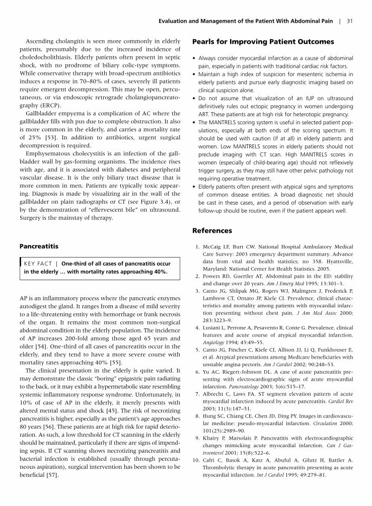
Ascending cholangitis is seen more commonly in elderlypatients, presumably due to the increased incidence ofcholedocholithiasis. Elderly patients often present in septicshock, with no prodrome of biliary colic-type symptoms.While conservative therapy with broad-spectrum antibioticsinduces a response in 70–80% of cases, severely ill patientsrequire emergent decompression. This may be open, percu-taneous, or via endoscopic retrograde cholangiopancreato-graphy (ERCP).
Gallbladder empyema is a complication of AC where thegallbladder fills with pus due to complete obstruction. It alsois more common in the elderly, and carries a mortality rateof 25% [53]. In addition to antibiotics, urgent surgicaldecompression is required.
Emphysematous cholecystitis is an infection of the gall-bladder wall by gas-forming organisms. The incidence riseswith age, and it is associated with diabetes and peripheralvascular disease. It is the only biliary tract disease that ismore common in men. Patients are typically toxic appear-ing. Diagnosis is made by visualizing air in the wall of thegallbladder on plain radiographs or CT (see Figure 3.4), orby the demonstration of “effervescent bile” on ultrasound.Surgery is the mainstay of therapy.
Pancreatitis
AP is an inflammatory process where the pancreatic enzymesautodigest the gland. It ranges from a disease of mild severityto a life-threatening entity with hemorrhage or frank necrosisof the organ. It remains the most common non-surgicalabdominal condition in the elderly population. The incidenceof AP increases 200-fold among those aged 65 years andolder [54]. One-third of all cases of pancreatitis occur in theelderly, and they tend to have a more severe course withmortality rates approaching 40% [55].
The clinical presentation in the elderly is quite varied. Itmay demonstrate the classic “boring” epigastric pain radiatingto the back, or it may exhibit a hypermetabolic state resemblingsystemic inflammatory response syndrome. Unfortunately, in10% of case of AP in the elderly, it merely presents withaltered mental status and shock [45]. The risk of necrotizingpancreatitis is higher, especially as the patient’s age approaches80 years [56]. These patients are at high risk for rapid deterio-ration. As such, a low threshold for CT scanning in the elderlyshould be maintained, particularly if there are signs of impend-ing sepsis. If CT scanning shows necrotizing pancreatitis andbacterial infection is established (usually through percuta-neous aspiration), surgical intervention has been shown to bebeneficial [57].
Pearls for Improving Patient Outcomes
• Always consider myocardial infarction as a cause of abdominalpain, especially in patients with traditional cardiac risk factors.
• Maintain a high index of suspicion for mesenteric ischemia inelderly patients and pursue early diagnostic imaging based onclinical suspicion alone.
• Do not assume that visualization of an IUP on ultrasound definitively rules out ectopic pregnancy in women undergoingART. These patients are at high risk for heterotopic pregnancy.
• The MANTRELS scoring system is useful in selected patient pop-ulations, especially at both ends of the scoring spectrum. Itshould be used with caution (if at all) in elderly patients andwomen. Low MANTRELS scores in elderly patients should notpreclude imaging with CT scan. High MANTRELS scores inwomen (especially of child-bearing age) should not reflexivelytrigger surgery, as they may still have other pelvic pathology notrequiring operative treatment.
• Elderly patients often present with atypical signs and symptomsof common disease entities. A broad diagnostic net should be cast in these cases, and a period of observation with early follow-up should be routine, even if the patient appears well.
References
1. McCaig LF, Burt CW. National Hospital Ambulatory Medical
Care Survey: 2003 emergency department summary. Advance
data from vital and health statistics; no 358. Hyattsville,
Maryland: National Center for Health Statistics. 2005.
2. Powers RD, Guertler AT. Abdominal pain in the ED: stability
and change over 20 years. Am J Emerg Med 1995; 13:301–3.
3. Canto JG, Shlipak MG, Rogers WJ, Malmgren J, Frederick P,
Lambrew CT, Ornato JP, Kiefe CI. Prevalence, clinical charac-
teristics and mortality among patients with myocardial infarc-
tion presenting without chest pain. J Am Med Assoc 2000;
283:3223–9.
4. Lusiani L, Perrone A, Pesavento R, Conte G. Prevalence, clinical
features and acute course of atypical myocardial infarction.
Angiology 1994; 45:49–55.
5. Canto JG, Fincher C, Kiefe CI, Allison JJ, Li Q, Funkhouser E,
et al. Atypical presentations among Medicare beneficiaries with
unstable angina pectoris. Am J Cardiol 2002; 90:248–53.
6. Yu AC, Riegert-Johnson DL. A case of acute pancreatitis pre-
senting with electrocardiographic signs of acute myocardial
infarction. Pancreatology 2003; 3(6):515–17.
7. Albrecht C, Laws FA. ST segment elevation pattern of acute
myocardial infarction induced by acute pancreatitis. Cardiol Rev
2003; 11(3):147–51.
8. Hung SC, Chiang CE, Chen JD, Ding PY. Images in cardiovascu-
lar medicine: pseudo-myocardial infarction. Circulation 2000;
101(25):2989–90.
9. Khairy P, Marsolais P. Pancreatitis with electrocardiographic
changes mimicking acute myocardial infarction. Can J Gas-
troenterol 2001; 15(8):522–6.
10. Cafri C, Basok A, Katz A, Abuful A, Gilutz H, Battler A.
Thrombolytic therapy in acute pancreatitis presenting as acute
myocardial infarction. Int J Cardiol 1995; 49:279–81.
KEY FACT | One-third of all cases of pancreatitis occurin the elderly … with mortality rates approaching 40%.
Evaluation and Management of the Patient With Abdominal Pain | 31
11. Wagner AM, Santolo M. A correct decision? Lancet 2002;
359(9301):157.
12. Stoney RJ, Cunningham CG. Acute mesenteric ischemia.
Surgery 1993; 114:489–90.
13. Ruotolo RA, Evans SRT. Mesenteric ischemia in the elderly. Clin
Geriatr Med 1999; 15:527–57.
14. Rhee RY, Gloviczki P, Mendonca CT, Petterson TM, Serry RD,
Sarr MG, et al. Mesenteric venous thrombosis: still a lethal dis-
ease in the 1990s. J Vasc Surg 1994; 20:688–97.
15. Diamond S, Emmett M, Henrich WL. Bowel infarction as a cause
of death in dialysis patients. J Am Med Assoc 1986; 256:2545.
16. Newman TS, Magnuson TH, Ahrendt SA, Smith-Meek MA,
Bender JS. The changing face of mesenteric infarction. Am Surg
1998; 64:611.
17. Sai-Sudhakar CB, Al-Hakeem M, MacArthur JD, Sumpio BE.
Mesenteric ischemia secondary to cocaine abuse: case reports
and literature review. Am J Gastroenterol 1997; 92:1053.
18. Boley SJ, Sprayregen S, Siegelman SJ, Veith FJ. Initial results
from an aggressive roentgenologic and surgical approach to
acute mesenteric ischemia. Surgery 1977; 82:848.
19. Clark RA, Gallant TE. Acute mesenteric ischemia: angiographic
spectrum. Am J Roentgenol 1984; 142:555.
20. Lobo Martinez E, Carvajosa E, Sacco O, Martinez Molina E.
Embolectomy in mesenteric ischemia. Rev Esp Enferm Dig 1993;
83:351.
21. Bradbury MS, Kavanaugh PV, Chen MY, Weber TM, Bechtold RE.
Noninvasive assessment of portomesenteric venous thrombosis:
current concepts and imaging strategies. J Comput Assist Tomogr
2002; 26:392–404.
22. Hellinger JC. Evaluating mesenteric ischemia with multidetector-
row CT angiography. Tech Vasc Interv Radiol 2004; 7(3):160–6.
23. Simon BC, Snoey ER. Ultrasound in emergency and ambula-
tory medicine. St. Louis: Mosby; 1997.
24. Reece EA, Petrie RH, Sirmans MF, Finster M, Todd WD.
Combined intrauterine and extrauterine gestations: a review.
Am J Obstet Gynecol 1983; 146:323–30.
25. Habana A, Dokras A, Giraldo J, et al. Cornual heterotopic preg-
nancy: contemporary management options. Am J Obstet Gynecol
2000; 182:1264–70.
26. Sotrel G, Rao R, Scommegna A. Heterotopic pregnancy follow-
ing clomid treatment. J Reprod Med 1976; 16:78–80.
27. Botta G, Fortunato N, Merlino G. Heterotopic pregnancy follow-
ing administration of human menopausal gonadotropin in vitro
fertilization and embryo transfer: two case reports and review of
the literature. Eur J Obstet Gynecol Reprod Biol 1995; 59:2112–15.
28. Sucov A, Deveau L, Feola P, et al. Heterotopic pregnancy after
in vitro fertilization. Am J Emerg Med 1995; 13:641–3.
29. Jerrard D, Tso E, Salik R, Barish RA. Unsuspected heterotopic
pregnancy in a woman without risk factors. Am J Emerg Med
1992; 10:58–60.
30. Dart R, McLean S, Dart L. Isolated fluid in the cul-de-sac: how
well does it predict ectopic pregnancy? Am J Emerg Med 2002;
20:1–4.
31. Breyer MJ, Costantino TG. Heterotopic gestation: Another pos-
sibility for the emergency bedside ultrasonographer to consider.
J Emerg Med 2004; 26(1):81–4.
32. Somers MP, Spears M, Maynard AS, Syverud SA. Ruptured het-
erotopic pregnancy presenting with relative bradycardia in a
woman not receiving reproductive assistance. Ann Emerg Med
2004; 43:382–5.
33. Irvin TT. Abdominal pain: a surgical audit of 1190 emergency
admissions. Br J Surg 1989; 76:1121–5.
34. Schwartz SI. Appendix. In: Schwartz SI (ed.), Principles of sur-
gery, 6th edn. New York: McGraw Hill, 1994, pp. 1307–18.
35. Yamini D, Hernan V, Bongard F, et al. Perforated appendicitis: is
it truly a surgical emergency? Am Surg 1995; 221:279–82.
36. Alvarado A. A practical score for the early diagnosis of acute
appendicitis. Ann Emerg Med 1986; 15:557–64.
37. Horattas MC, Guyton DP, Wu D. A reappraisal of appendicitis in
the elderly. Am J Surg 1990; 160:291–3.
38. Storm-Dickerson TL, Horattas MC. What have we learned over
the past 20 years about appendicitis in the elderly? Am J Surg
2003; 185:198–201.
39. Rogers J. Abdominal Pain: Forsight. Dallas: American College
of Emergency Physicians; Issue 3, Dec 1986.
40. Gwynn LK. The diagnosis of acute appendicitis: Clinical assess-
ment versus computed tomography evaluation. J Emerg Med
2001; 21(2):119–23.
41. Malik AA, Wani NA. Continuing diagnostic challenge of acute
appendicitis: evaluation through modified Alvarado score. Aust
NZ J Surg 1998; 68:504–5.
42. Kalan M, Talbot D, Cunliffe WJ, Rich AJ. Evaluation of the
modified Alvarado score in the diagnosis of acute appen-
dicitis: a prospective study. Ann Roy Coll Surg Engl 1994;
76:418–419.
43. Brewer RJ, Golden GT, Hitsch DC, et al. Abdominal pain: an
analysis of 1,000 consecutive cases in a university hospital
emergency room. Am J Surg 1976; 131:219–24.
44. Leverat M. Peptic ulcer disease in patients over 60: experience
in 287 cases. Am J Dig Dis 1966; 11:279–85.
45. Caesar R. Dangerous complaints: The acute geriatric abdomen.
Emerg Med Report 1994; 15:191–202.
46. Borum ML. Peptic-ulcer disease in the elderly. Clin Geriatr Med
1999; 15(3):457–71.
47. Fenyo G. Acute abdominal disease in the elderly: experience
from two series in Stockholm. Am J Surg 1982; 143:751–4.
48. Wakayama T. Risk factors influencing the short-term results of
gastroduodenal perforation. Surg Today 1994; 24(8):681–7.
49. Crofts TJ, Park KG, Steele RJ. A randomized trial of nonopera-
tive treatment for perforated peptic ulcer. New Engl J Med 1989;
320(15):970–3.
50. Rosenthal RA, Anderson DK: Surgery in the elderly: observa-
tions on the pathophysiology and treatment of cholelithiasis.
Exp Gerontol 1993; 28:459–72.
51. Bedirli A. Factors effecting the complications in the natural his-
tory of acute cholecystitis. Hepatogastroenterol 2001; 48(41):
1275–8.
52. Morrow DJ, Thompson J, Wilson SE. Acute cholecystitis in the
elderly. Arch Surg 1978; 113:1149–52.
53. Sen M, Williamson RCN. Acute cholecystitis: surgical manage-
ment. Balliere’s Clin Gastroenterol 1991; 5:817–37.
54. Martin SP, Ulrich CD. Pancreatic disease in the elderly. Clin
Geriatr Med 1999; 15(3):579–605.
55. Hoffman E, Perez E. Acute pancreatitis in the upper age groups.
Gastroenterol 1959; 36:675–85.
56. Paajanen H. AP in patients over 80 years. Eur J Surg 1996;
162(6):471–5.
57. Bradley EL III, Allen K. A prospective longitudinal study of
observation versus surgical intervention in the management of
necrotizing pancreatitis. Am J Surg 1991; 161:19–24.
32 | Chapter 3
Introduction
Up to 90% of adults will have at least one episode of backpain during their lifetime [1]. In the US alone, approxi-mately 15 million patients present each year to a physicianwith acute back pain [2]. Many of these patients arrive at local emergency departments (ED) seeking evaluationand symptom relief. In fact, back pain accounts for almost2% of all ED visits per year [3]. Thankfully, 90–95% ofpatients presenting with acute back pain have non-life-threatening etiologies [4]. Although most patients cannot begiven an exact diagnosis, nearly all recover within 4–6weeks [1, 5].
In the remaining 5–10% of patients, acute back pain is amanifestation of serious pathology. Thoracic aortic dissec-tion, abdominal aortic aneurysm, spinal epidural abscess,spinal epidural metastasis, and spinal cord compressive syn-dromes may all initially present with a complaint of acuteback pain. These “back pain emergencies” are missed in asignificant percentage of patients upon initial evaluation. Asa result of delays in diagnosis and treatment, patients withthese conditions continue to have unacceptably high mor-bidity and mortality. Although challenging, it is crucial thatemergency physicians identify patients with acute back painwith a potentially life-threatening disorder.
This chapter will focus on common pitfalls in the ED eval-uation of patients with acute back pain. With this informa-tion, the emergency physician can more effectively detectand treat patients with potentially catastrophic causes ofback pain.
Pitfall | Over-reliance on the “Classic” ClinicalPresentations of Aortic Dissection, RupturingAbdominal Aortic Aneurysm, Spinal EpiduralAbscess, and Pulmonary Embolism
In emergency medicine, it is well recognized that the textbookclinical presentations of disease are often the exception ratherthan the rule. This holds true for the clinical presentations ofseveral dangerous back pain emergencies [6]. The emergencyphysician must be able to diagnose these life-threatening disorders even in the absence of “classic” presentations.
The classic clinical description of aortic dissection is the sud-den onset of ripping chest pain that radiates to the inter-scapular region. In reality, this textbook description ispresent in less than one-third of patients with aortic dissec-tion [6]. Although the abrupt onset of pain is reported in85% of patients, many will not describe their pain as eitherripping or tearing [7, 8]. Patients with aortic dissection oftendescribe their pain as either sharp, pressure-like, pleuritic, oreven burning [7]. In addition to a variety of descriptions ofpain, patients with aortic dissection often report varyinglocations of pain. Isolated back pain, especially thoracic, iscommon in dissections of the descending aorta.
Textbook physical examination findings of aortic dissectioninclude hypertension, pulse deficits, blood pressure differen-tials, and a new diastolic murmur. These classic abnormalitiesare inconsistent and unreliable findings in patients with dis-section. Up to one-third of patients with an acute aortic dis-section are normotensive upon evaluation [9]. Althoughconsidered specific for aortic dissection, a pulse deficit occursin just 25–38% of cases [6, 9] and its absence cannot be usedto exclude the diagnosis of aortic dissection. Bilateral bloodpressure measurements are frequently obtained in cases ofsuspected aortic dissection. A difference of 20 mmHg or moreis considered significant. However, 20% of normal individualshave blood pressure differentials greater than 20 mmHg,thereby lowering the specificity of this finding [10–12]. Thesensitivity of a new diastolic murmur in the detection of aor-tic dissection is reportedly just 28% [6]. Thus, it is easy to seehow the clinical examination is inadequate for diagnosing orexcluding an acute aortic dissection [9]. For the emergencyphysician, suspicion for aortic dissection begins by identifyingrisk factors for aortic pathology. A thorough risk factor analy-sis for aortic disease must be performed in any patient presenting with acute back pain. Risk factor assessment hasbeen shown to increase the rate of diagnosis of aortic catas-trophes [10]. Thoracic aortic dissection must be considered in any patient with risk factors for the same (discussed below) who presents with the abrupt onset of severe thoracicback pain.
KEY FACT | The “classic” description of aortic dissectionpain is absent in 2/3 of cases.
Chapter 4 | Management of Patients with Acute Back Pain inthe ED
Michael E. Winters
33
The textbook triad of rupturing abdominal aortic aneurysmconsists of abdominal pain, hypotension, and a pulsatileabdominal mass. Unfortunately, less than 20% of patientswith a rupturing aneurysm present with this triad [13]. It isimportant for the emergency physician to recognize that arupturing abdominal aortic aneurysm can present with leftlower quadrant pain, flank pain, or isolated back pain. As aresult, many patients with a ruptured aneurysm are misdiag-nosed as having diverticulitis, nephrolithiasis, pyelonephritis,or musculoskeletal strain [13]. Misdiagnosis leads to signifi-cant morbidity and mortality. Mortality rates as high as 75%have been reported when patients with a rupturing abdomi-nal aortic aneurysm are initially misdiagnosed [10]. In addi-tion to recognizing back or flank pain as the initial presentingsymptom, the emergency physician must also understand thelimitations of physical examination.
The sensitivity of abdominal palpation for an aneurysmranges from 45% to 97% [13]. Sensitivity increases withincreasing sizes of aneurysm, however, nearly 25% of abdom-inal aneurysms greater than 5 cm cannot be palpated [13].Similar to aortic dissection, suspicion for an abdominal aor-tic aneurysm begins with a thorough risk factor assessment.A rupturing aneurysm must be considered in any elderlypatient with acute back, or flank pain, who has risk factorsfor an abdominal aortic aneurysm.
Spinal epidural abscess is classically associated with the triadof fever, back pain, and neurologic deficits. Although thesesymptoms are helpful if present, this triad occurs in as few as13% of patients [14]. Up to 75% of patients with a spinalepidural abscess will be afebrile and up to two-thirds ofpatients will have a normal initial neurologic exam [14]. As aresult, patients with spinal epidural abscess incur significantdiagnostic delays. Often, patients with spinal epidural abscesshave multiple ED visits before the diagnosis is made. Sincerapid neurologic deterioration can occur, it is imperative that
the emergency physician identify patients with a spinalepidural abscess. Rather than relying on the presence offever and back pain, the emergency physician should focuson identifying risk factors for spinal epidural abscess in anypatient with acute back pain. It is reported that risk factorassessment offers the highest sensitivity and negative predic-tive value in identifying patients with spinal epidural abscessbefore the onset of neurologic symptoms [14].
The clinical presentation of pulmonary embolism is classi-cally described as the acute onset of pleuritic chest pain associ-ated with dyspnea and hemoptysis. Atypical presentations ofpulmonary embolism, however, are common. Thoracic backpain is a recognized presentation of pulmonary embolism [15].In addition to aortic catastrophes and infectious etiologies, theemergency physician should include a risk factor assessmentfor venous thromboembolic disease when confronted with thepatient with acute thoracic back pain.
Pitfall | Failure to Perform a Risk FactorAssessment for Serious Etiologies ofBack Pain
Since the majority of patients with back pain have benignetiologies, identification of the patient in whom to pursuean extensive diagnostic workup is challenging. Suspicion forserious pathology begins with an assessment of patient riskfactors for disease. Unfortunately, many physicians fail toperform a risk factor profile in patients presenting with backpain. In fact, failure to inquire about risk factors is one of the most common reasons cited in cases of missed aortic dissection [10]. Thus, it is imperative that the emergencyphysician inquire about risk factors, perform a risk factorassessment, and guide the workup accordingly. In manycases, risk factors may be the only clue to serious underlyingpathology.
Risk factors for aortic catastrophes are well established andinclude male gender, older age, hypertension, connectivetissue disorders, chromosomal syndromes, smoking, andinflammatory conditions of the aorta [16]. Additional riskfactors for aortic dissection include a bicuspid aortic valve,coarctation, aortic instrumentation, decelerating trauma,and pregnancy [16]. In fact, over 50% of aortic dissectionsin women less than 40 years of age occur during pregnancy,most often during the third trimester and the initial stages oflabor [16]. Aortic dissection must be considered in the dif-ferential diagnosis of any pregnant patient presenting duringthe third trimester or labor with the abrupt onset of severeback pain.
KEY FACT | Pregnancy is an often overlooked riskfactor for aortic dissection.
KEY FACT | Because the vast majority of patients withspinal epidural abscesses present atypically, assessment ofpatients for risk factors for this disease is essential foridentifying it prior to the onset of irreversibleneurological sequelae.
KEY FACT | Abdominal palpation is insensitive fordetecting abdominal aortic aneurysm.
KEY FACT | Rupturing abdominal aortic aneurysm oftenpresents with symptoms mimicking renal colic,diverticulitis, and musculoskeletal pain.
34 | Chapter 4
Lastly, the emergency physician must always inquire aboutthe use of illicit substances in patients with back pain. Cocaineuse is an established risk factor for aortic dissection, particu-larly of the descending aorta, in otherwise healthy individuals[16]. Hypertensive patients presenting with the abrupt onsetof back pain in the setting of cocaine use should be imaged toexclude acute aortic dissection.
Although traditionally taught as a disease of older individuals,spinal epidural abscess can affect patients of any age. Risk fac-tors for spinal epidural abscess include diabetes mellitus, alco-holism, chronic renal failure, corticosteroid therapy, humanimmunodeficiency virus/acquired immunodeficiency syn-drome, and intravenous drug use [17]. Intravenous drug use isconsidered one of the primary risk factors for spinal epiduralabscess, accounting for over 50% of cases in some populations[18]. Any patient with back pain who uses intravenous drugsmust be considered to have a spinal epidural abscess untilproven otherwise. Regardless of the location of pain, thesepatients require imaging to exclude an epidural abscess. It isalso important for the emergency physician to inquire aboutrecent invasive procedures. Back pain that develops in the setting of a recent procedure, especially one known to have ahigh incidence of bacteremia, is highly suspicious for eithervertebral osteomyelitis or spinal epidural abscess [1].
Epidural spinal cord compression is a true medical emergencythat cannot be missed. Over 90% of cases are due to spinalepidural metastases. The primary risk factor for spinal epiduralmetastases is a history of malignancy. Although prostate,breast, and lung cancer most commonly cause bony metas-tases, it is important to realize that any systemic malignancycan metastasize to the spine. Lymphoma, renal cell cancer, gas-trointestinal malignancies, and multiple myeloma are fre-quently overlooked, yet account for a significant percentage ofcases. Spinal epidural metastases with resultant spinal cordcompression must be considered in any patient with back painand a history of malignancy. Additional etiologies for spinalcord compression include massive midline disc herniation,
spinal hematoma, epidural abscess, traumatic compression,and transverse myelitis [19].
Pitfall | Failure to Perform a CompleteNeurologic Exam
Arguably, the most important portion of the physical exam-ination in patients with back pain is the neurologic exami-nation. In many cases, subtle neurologic deficits are the onlyindication of serious pathology, namely spinal cord com-pression. Unfortunately, many physicians either fail to per-form a complete neurologic exam or fail to appropriatelydocument their findings. In a review of missed cases ofspinal epidural abscess, Davis et al. report that, in each case,minimal notation was made with regards to the neurologicexam [14]. Exams were documented as either “nonfocal” or“normal” without reference to the motor, sensory, deep ten-don reflex, rectal, or cerebellar components.
The importance of performing a complete neurologic examcannot be overstated. Any patient with acute back pain andneurologic deficits requires emergent diagnostic imaging. Incases of spinal cord compression, motor deficits are the mostcommon neurologic finding and are present in up to 85% of patients [20]. The emergency physician must pay closeattention to the motor examination of the lower extremi-ties. Appropriate examination should include an assessmentof hip flexion and extension, leg flexion and extension, ankledorsiflexion and inversion, and great toe dorsiflexion. Incases of thoracic spinal cord compression, the iliopsoas mus-cles are preferentially affected, producing weakness of theproximal lower extremities when testing hip flexion [20].Sensory abnormalities occur slightly less often than motordeficits, whereas bowel and/or bladder dysfunction is a latefinding in patients with epidural spinal cord compression[20]. Any patient with a report of fecal retention or inconti-nence and/or saddle anesthesia must have a rectal examina-tion performed and documented. Additional indications forrectal examination include severe pain and/or the presenceof any neurologic deficit. In addition to the rectal examina-tion, patients with suspected spinal cord compression shouldhave a post-void residual measurement. A post-void residualgreater than 100–200 ml is indicative of acute urinary reten-tion. Regardless of a history of prostatic abnormalities, anypatient with acute back pain and an abnormal post-voidresidual should be suspected of having spinal cord compres-sion until proven otherwise. Acute urinary retention isreported to have 90% sensitivity and 95% specificity forcauda equina syndrome [19].
KEY FACT | It is imperative to perform a completeneurologic examination including, when indicated, arectal examination and post-void residual measurement.
KEY FACT | Spinal metastases must be considered inpatients with back pain and a history of malignancy,regardless of the type.
KEY FACT | Patients with back pain using injectiondrugs must have diagnostic imaging to definitivelyexclude spinal epidural abscess.
KEY FACT | Aortic dissection must be excluded inhypertensive patients with back pain and a history ofrecent cocaine use.
Management of Patients with Acute Back Pain in the ED | 35
Pitfall | Failure to Recognize the Limitationsof Plain Radiography in Excluding a BackPain Emergency
Given their low cost and ready availability, plain radiographsare frequently obtained in the evaluation of patients withacute back pain. In the setting of trauma, plain films can beuseful to exclude fracture or dislocation. However, plain filmsare often erroneously utilized to exclude other potential backpain emergencies, such as spinal metastases or vertebralosteomyelitis. For vertebral metastases, plain radiographs arethe least sensitive of all imaging modalities [4]. The pooledsensitivity of plain radiographs for spinal metastases is just60% [4]. Furthermore, up to 17% of patients with epiduralspinal cord compression due to metastatic disease have nor-mal plain radiographs [20]. For the patient presenting withacute back pain and neurologic deficits, plain films should notbe relied upon as the sole imaging modality to exclude spinalepidural metastases or spinal cord compression.
Plain radiographs are equally insensitive in the diagnosis ofinfectious etiologies of back pain, namely osteomyelitis andspinal epidural abscess. Abnormalities that indicate a potentialinfectious etiology include endplate erosion, loss of vertebraldisc height, and bony lysis, but these abnormalities usuallytake several weeks to develop [4]. Thus, patients with infec-tious etiologies of back pain may have entirely normal plainfilms. For osteomyelitis, �33% of patients will have anabnormality on plain film during the first 7–10 days [21]. Theoverall sensitivity and specificity of plain radiographs forosteomyelitis or epidural abscess is 82% and 57%, respec-tively, and therefore cannot be used solely to rule out spinalinfection [4]. Given the potential for rapid neurologic deterio-ration, once these disease entities are suspected, definitivediagnostic imaging is indicated rather than false reassurancewith plain radiography.
Pitfall | Failure to Consider AorticCatastrophes in Patients with Acute BackPain and Neurologic Symptoms
The emergency physician must be knowledgeable regardingatypical presentations of aortic dissection and abdominal aortic aneurysm. It is imperative to recognize that aorticcatastrophes can present with back pain and focal neurologicabnormalities. In fact, neurologic deficits are found in18–36% of patients with aortic dissection [10]. Deficits seenin patients with aortic dissection range from acute stroke tounilateral lower extremity numbness. Unilateral lowerextremity deficits are caused by involvement of the greatradicular artery, a branch of the descending aorta. Thoracic
aortic dissection must be excluded in any patient with sud-den thoracic back pain and focal neurologic deficits.
For rupturing abdominal aortic aneurysm, up to 5% ofpatients will have associated neurologic deficits [10]. It isbelieved that an expanding retroperitoneal hematomacauses direct nerve compression. Since the femoral andobturator nerves are the most susceptible to compression,patients present with weakness of hip and knee flexion [10].The emergency physician must consider aortic catastrophesin the differential diagnosis of patients presenting with backpain and neurologic deficits.
Pitfall | Failure to Recognize the Limitationsof Laboratory Studies in the Evaluation ofPatients with Suspected Spinal EpiduralAbscess or Vertebral Osteomyelitis
When considering an infectious etiology of back pain, physi-cians frequently rely on the presence of a leukocytosis to guidefurther evaluation. This finding is variable in infectious causesof back pain. White blood cell counts above 12,000/mm3 areseen in just 40% of patients with spinal infection [22]. In addi-tion, the presence of a left shift has been shown to be unreli-able in the detection of an infectious etiology [17]. Emergencyphysicians should never exclude the possibility of a spinalinfection based upon a normal white blood cell count.
In contrast to the peripheral white blood cell count, inflam-matory markers, such as the erythrocyte sedimentation rateand C-reactive protein, have been shown to be useful in theidentification of patients with a spinal infection. These mark-ers are elevated in approximately 90% of patients with aninfectious cause of back pain [21]. Up to 10% of patients canhave normal levels, therefore, these markers should not beused as the sole criterion for excluding spinal infection.
Pitfall | Inadequate Radiographic Imaging inthe Diagnosis of Spinal Cord Compressionand Spinal Epidural Abscess
36 | Chapter 4
KEY FACT | There is virtually no indication for plainradiography in the evaluation of nontraumatic back pain.
KEY FACT | Hematoma surrounding an abdominal aorticaneurysm can cause weakness of hip and knee flexion.
KEY FACT | Lab studies cannot be used exclusively torule out an infectious cause of back pain.
KEY FACT | Magnetic resonance imaging is currentlythe only imaging modality that can be used to excludespinal cord compression or abscess.
Emergency physicians recognize that spinal cord compres-sion is a true emergency. Therefore, once the diagnosis is sus-pected, emergent imaging must be obtained. As discussed,plain films may be falsely negative in up to 17% of patientswith cord compression [20]. Bone scanning, computedtomography, and positron-emission tomography have allbeen used in the evaluation of patients with spinal cord com-pression. These modalities, however, have not been shownto be superior to magnetic resonance imaging. Magnetic res-onance imaging is the imaging modality of choice whendiagnosing or excluding spinal cord compression. The over-all diagnostic accuracy of magnetic resonance imaging forspinal cord compression is 95% [20]. For patients suspectedof cord compression due to metastatic disease, magnetic res-onance imaging of the entire spine is recommended, as com-pression can occur at multiple levels [23].
Definitive diagnostic imaging must also be obtained whenconsidering the diagnosis of spinal epidural abscess. Magneticresonance imaging is the most sensitive modality for identifyingand characterizing spinal epidural abscess. Magnetic resonanceimaging accurately defines the extent of an abscess, highlightsany surrounding inflammation, evaluates the degree of thecalsac compression, and aids in the planning of surgical drainage.Sensitivity and specificity of magnetic resonance imaging forspinal infection is 96% and 92%, respectively [4]. Computedtomography myelography remains an option for patientsunable to undergo magnetic resonance imaging; however, thisprocedure is invasive and can result in transmission of theinfection into the subarachnoid space. Currently, there is nodata on the accuracy of computed tomography without myel-ography in the diagnosis of osteomyelitis or spinal epiduralabscess [4]. Similar to spinal epidural metastases, infectious eti-ologies of back pain can occur at multiple spinal levels, espe-cially in the patient abusing intravenous drugs. A magneticresonance scan that images only isolated sections of the spinewill undoubtedly miss lesions at other spinal levels. Therefore,the entire spine should be imaged when evaluating patients forspinal epidural abscess.
Pitfall | Inadequate Antibiotic Treatment in Patients with Infectious Etiologies ofBack Pain
Although optimal management of spinal epidural abscessremains controversial, all patients with an infectious etiol-ogy of acute back pain require prompt antibiotic administra-tion. Ideally, antibiotics are given after tissue cultures areobtained. Because patients can develop rapid, irreversible
neurologic deterioration, it is crucial for the emergencyphysician to begin antibiotic therapy promptly.
Staphylococcal aureus is the most common isolate in cases of osteomyelitis and epidural abscess, but up to one-third ofcases are caused by gram-negative organisms [24, 25].Therefore, initial antibiotic coverage must be broad spectrumand effective against both gram-positive and gram-negativeorganisms. Acceptable regimens include a penicillinase-resist-ant penicillin plus a third-generation cephalosporin, or incases of suspected methicillin-resistant Staphylococcal aureus,vancomycin and a third-generation cephalosporin [21].
Pitfall | Failure to Initiate Prompt Treatmentin the Patient with Spinal Cord Compression
Definitive treatment of epidural spinal cord compressionrequires a multi-disciplinary approach. Once the diagnosis issuspected, appropriate consultation must be obtained. Often,this involves consultation with neurosurgery, orthopedicsurgery, and radiation oncology. While awaiting definitivetreatment, the emergency physician must provide support-ive care. Analgesics and corticosteroids are an importantcomponent of that supportive care. Corticosteroids havebeen shown to reduce edema, inhibit further inflammatorydamage, and delay the progression of neurologic deficits [20].Recent randomized trials have demonstrated that, in thecase of cord compression due to metastatic disease, patientswho received corticosteroids were more likely to be ambula-tory at long-term follow up [23, 26]. Dexamethasone is thecorticosteroid of choice given its low cost and relatively lowmineralocorticoid activity [23]. Currently, there is no con-census on the optimal dose. Dosing regimens in the litera-ture have ranged from 16 to 100 mg/day [20]. An acceptableregimen consists of an initial dose of 10 mg of dexametha-sone, followed by 6 mg every 4 h.
Pitfall | Failure to Promptly Refer Patientswith an Acute Motor Radiculopathy
Only 2–3% of patients presenting with acute low back painwill have evidence of a lumbar radiculopathy [1]. Althoughthere are many potential etiologies, the most common causeof a radiculopathy is an acute disc herniation. The size, level,
Management of Patients with Acute Back Pain in the ED | 37
KEY FACT | In suspected spinal infection, antibiotics suchas a third-generation cephalosporin plus a penicillinase-resistant penicillin or vancomycin should be administered assoon as possible after cultures are obtained.
KEY FACT | Parenteral pain medications anddexamethasone (10 mg followed by 6 mg every 4 h)should be administered to patients with suspected spinalcord compression.
KEY FACT | Patients with radiculopathy and motordeficits should have urgent neurosurgical referral.
and location of herniation determine a patient’s symptomsand physical examination findings. In contrast to the patientwith low back pain and a normal physical exam, those with alumbar motor radiculopathy may require early surgical treat-ment. The most important factor in determining whether anearly surgical procedure is needed is the presence of signifi-cant weakness [27]. If the weakness is substantial, delayingsurgery could result in a permanent deficit. Therefore, it isincumbent upon the emergency physician to ensure that thepatient with an acute lumbar motor radiculopathy is seenwithin seven days of their ED visit. Urgent magnetic reso-nance imaging, as well as neurosurgical referral, is needed toensure optimal outcomes.
Pearls for Improving Patient Outcomes
• “Classic” presentations are the exception rather than the rule inback pain emergencies and they often present with symptomsmimicking other disease.
• Risk factors assessment is crucial to help identify patients requir-ing emergent imaging.
• Pregnancy is a risk factor for aortic dissection.• A history of drug use should heighten suspicion for emergent
causes of back pain.• Patients with back pain require a careful neurological examina-
tion to identify those requiring emergent treatment.• Plain films of the back are almost never indicated for nontrau-
matic back pain.• Magnetic resonance imaging is currently the only test that can
exclude spinal cord compression or abscess.• For spinal infections, early administration of appropriate antibi-
otics is crucial.• Steroids are indicated for patients with spinal cord compression.• Patients with radicular symptoms and motor deficits should have
urgent neurosurgical referral.
References
1. Della-Giustina DA. Orthopedic emergencies: emergency
department evaluation and treatment of back pain. Emerg Med
Clin N Am 1999; 17(4):877–93.
2. Hart L, Deyo R, Churkin D. Physician office visits for low back
pain. Spine 1995; 20:11–19.
3. 1995 National Hospital Ambulatory Care Survey. National
Center for Health Statistics, Center for Disease Control and
Prevention, Hyattsville, Maryland.
4. Jarvik JG, Deyo RD. Diagnostic evaluation of low back pain
with emphasis on imaging. Ann Intern Med 2002; 137:586–97.
5. Bigos S, Bowyer O, Braen G, et al. Acute low back problems in
adults. In: Clinical Practice Guideline, Quick Reference Number
14, Rockville MD, U.S. Department of Health and Human
Services, Public Health Service, Agency for Health Care Policy
and Research. AHCPR Pub No. 95-0643. 1994.
6. Haro LH, Krajicek M, Lobl JK. Challenges, controversies, and
advances in aortic catastrophes. Emerg Med Clin N Am 2005;
23:1159–77.
7. Sullivan PR, Wolfson AB, Leckey RD, Burke JL. Diagnosis of
acute thoracic aortic dissection in the emergency department.
Am J Emerg Med 2000; 18:46–50.
8. Hagan PG, Nienaber CA, Isselbacher EM, et al. The International
Registry of Acute Aortic Dissection (IRAD): new insights into an
old disease. J Am Med Assoc 2000; 283;897–903.
9. Knaut AL, Cleveland JC. Aortic emergencies. Emerg Med Clin N
Am 2003; 21:817–45.
10. Rogers RL, McCormack R. Aortic disasters. Emerg Med Clin N Am
2004; 22:887–908.
11. Lane D, Beevers M, Barnes N, et al. Inter-arm differences in
blood pressure: when are they clinically relevant? J Hypertension
2002; 20:1089.
12. Pesola GR, Pesola HR, Lin M, et al. The normal difference in
bilateral blood indirect blood pressure recordings in hyperten-
sive individuals. Acad Emerg Med 2002; 9:342–5.
13. Carpenter CR. Abdominal palpation for the diagnosis of
abdominal aortic aneurysm. Ann Emerg Med 2005; 4:556–8.
14. Davis DP, Wold RM, Patel RJ, et al. The clinical presentation and
impact of diagnostic delays on emergency department patients
with spinal epidural abscess. J Emerg Med 2004; 26:285–91.
15. Laack TA, Goyal DG. Pulmonary embolism: an unsuspected
killer. Emerg Med Clin N Am 2004; 22:961–83.
16. Khan IA, Nair CK. Clinical, diagnostic, and management per-
spectives of aortic dissection. Chest 2002; 122:311–28.
17. Soehle M, Wallenfang T. Spinal epidural abscess: clinical mani-
festations, prognostic factors, and outcomes. Neurosurgery 2002;
51:79–87.
18. Calder KK, Severyn FA. Surgical emergencies in the intra-
venous drug user. Emerg Med Clin N Am 2003; 21:1089–116.
19. Small SA, Perron AD, Brady WJ. Orthopedic pitfalls: cauda
equina syndrome. Am J Emerg Med 2005; 23:159–63.
20. Prasad D, Schiff D. Malignant spinal-cord compression. Lancet
Oncol 2005; 6:15–24.
21. Perron AD, Brady WJ, Miller MD. Orthopedic pitfalls:
osteomyelitis. Am J Emerg Med 2003; 21:61–7.
22. Lurie JD, Gerber PD, Sox HC. A pain in the back. New Engl J
Med 2000; 343:723–6.
23. Schiff D. Spinal cord compression. Neurol Clin N Am 2003;
21:67–86.
24. Lu C, Chang W, Lui C, et al. Adult spinal epidural abscess: clin-
ical features and prognostic factors. Clin Neurol and Neurosurg
2002; 104:306–10.
25. Tunkel AR, Pradham SK. Central nervous system infections in
injection drug users. Infect Dis Clin N Am 2002; 16:589–605.
26. Sorensen PS, Helweg-Larsen S, Mouridsen H, et al. Effect of
high-dose dexamethasone in carcinomatous metastatic spinal
cord compression treated with radiotherapy: a randomized
trial. Eur J Cancer 1994; 30A:22–7.
27. Devereaux MW. Low back pain. Prim Care Clin Office Pract 2004;
31:33–51.
38 | Chapter 4
Introduction
In the course of a single year, 90% of people will experienceheadache [1]. Headaches account for fully 1–2% of emer-gency department (ED) visits, and the causes are myriad [2].The International Headache Society classification breaksheadaches into 13 categories. Still of those patients withheadache that find their way to the ED, perhaps as few as 1%will be found to have a serious underlying neurologic disorder[3]. In Emergency medicine (EM), identifying a specific causeof headache is less important than differentiating thoseheadaches with associated morbidity and treating themappropriately. The potential for encountering pitfalls is large.Headache is a common ED complaint and only a minority ofpatients have serious underlying pathology. The pitfalls aremany. It is easy to err in diagnosis and miss the opportunity totreat the one or two seriously ill patients from every hundredwho present with headache.
Pitfall | Failure to identify “red flag”features of headaches
Because headaches present frequently to the ED and themajority are benign, the emergency physician (EP) can easilybe lulled into complacency. Determining which headachesrequire neuroimaging can be a challenge. While not all seri-ous headaches will present with “red flags,” failure to elicithigh-risk features will increase the likelihood of missing seri-ous pathology.
The classic “do not miss” headaches are a simple, shortlist: meningitis, brain tumor or other mass lesion, andintracranial hemorrhage. The most effective and commonlyavailable initial diagnostic tests to evaluate for these fouritems are computed tomography (CT) and spinal fluid analy-sis after lumbar puncture (LP), but it is inefficient andwasteful to simply perform a CT and LP on everyone whopresents with headache. Downsides include time, discomfort,false positive results, radiation exposure, infection, and – inthe ultimate irony of LP for headache – post-dural punctureheadache. Determining who should receive diagnostic imaging and/or LP is crucial to the management of this complaint.
It is important to determine whether the presentingheadache represents a change from the typical headachepattern experienced by the patient. Likewise, headaches inpatients with cancer, immunosuppression, the elderly, orwith associated neurologic findings should raise concernand prompt further investigations.
It may be difficult to make the distinction between theneed for emergency scanning versus that which can wait afew days or even weeks for follow-up. For many, practicingin an environment where a head CT is readily available, andwhere follow-up may be more difficult, the distinctionbecomes less important. Though dependent on social con-siderations (such as the likelihood of follow-up and resourceavailability), headaches with associated “red flags” shouldhave as complete a workup in the ED as possible (Table 5.1).
Regarding meningitis, it’s worth pondering a 2004 Dutchstudy by van de Beek and colleagues [4]. In reviewing
KEY FACT | The classic triad of fever, neck stiffness, andaltered mental status occurs in only 44% of cases ofmeningitis.
KEY FACT | Headaches in the elderly, patients with ahistory of malignancy or immunosuppression, those thatrepresent a change from a patient’s typical headachepattern, or those that are associated with abnormalneurologic findings should prompt consideration forfurther diagnostic testing.
Chapter 5 | Headache Management
Stephen Schenkel
39
Table 5.1 “Red flag” indications for ED neuroimaging (CT or MRI) [3, 11].
• First or worst headache of life, especially if rapid onset, such as a“thunderclap” headache.
• Change in the nature of a headache, including increased frequency,worsening severity, or new associated symptoms.
• A new headache in a patient with cancer or HIV.• New-onset headache in a patient more than 50-year old.• Headache associated with seizures.• An abnormal neurologic examination including focal deficits or
altered mental status.
almost 700 cases of community-acquired bacterial meningi-tis, they found that the classic triad of fever, neck stiffness,and a change in mental status was relatively rare, occurringin only 44% of cases. On the other hand, at least two of foursigns – fever, neck stiffness, change in mental status, andheadache – occurred in 95% of cases; 4% of cases had onlyone of these symptoms, and 1% none.
Pitfall | Waiting for results before treatingsuspected bacterial meningitis
In medicine, order is greatly emphasized. The clinician gath-ers information, develops a differential, conducts tests,makes a diagnosis, then treats. In EM we frequently chal-lenge this doctrine and when faced with central nervoussystem (CNS) infections, it is imperative to do so. Two inde-pendent, retrospective studies demonstrated that delays inantibiotic therapy increased adverse outcomes and death inthe setting of bacterial meningitis [5, 6]. In the case of sus-pected meningitis, this delay can be deadly, especially whenthe time to antibiotics exceeds 6 h. The risk imparted by thedelay carries more risk than the potential adverse reactionfrom a single dose of antibiotics. The algorithm, therefore, isantibiotics, then CT and LP. This holds true even when hos-pital transfer is required to obtain the CT.
The argument that antibiotics will render the cere-brospinal fluid (CSF) non-diagnostic is also unfounded.Initial antibiotic therapy does not change CSF cell counts orprotein or glucose. Gram stain and culture may also remainpositive despite antibiotic administration. Blood cultures,drawn prior to antibiotic administration, may also reflect theresponsible bacterial agent [6, 7].
The 2002 study by de Gans and colleagues added anotherwrinkle to this series of actions [8]. It demonstrated theimportance of not only timely antibiotic therapy, but alsotimely and coordinated provision of corticosteroids in treat-ing adult bacterial meningitis. In this study of 301 adultpatients with meningitis, dexamethasone 10 mg intravenously15 min prior to antibiotic therapy substantially improvedresponse to therapy, reducing both unfavorable outcomesand death. For patients with suspected meningitis who meetthe inclusion criteria of the study – suspicion of meningitisbut no cerebrospinal shunt, no pregnancy, no recent antibiotic
therapy, no active tuberculosis or fungal infection, and norecent head trauma, neurosurgery, or peptic ulcer disease –both dexamethasone and antibiotics should be given prior tothe diagnostic procedures. Because the theoretical mechanismfor the steroid’s efficacy is to prevent immunologic responseto lysed bacterial proteins, the dexamethasone should begiven before or, at the latest, with the antibiotics [9]. If thepatient proves not to have meningitis, the therapy can bediscontinued, otherwise, it should be continued for 4 days.
Pitfall | Failing to recognize the limitationsof a non-contrast head CT
By the time we’ve completed our discussion of pitfall num-ber 1, and we’ve all looked for red flags in the diagnosis andtreatment of headache (Table 5.2), we are ordering non-contrast CTs on a large percentage of people who enter ourdepartments with headaches. This is not surprising, as thetechnology is available and common. But it can be decep-tively comforting, too, as not all diagnoses will be made onthe non-contrast CT. It is for this reason that magnetic reso-nance imaging (MRI) has become the most desirable test forheadache, though it is not as readily available in EM. Eventhe standard argument that CT scanning is to look for bleedand the MRI for everything else may be falling by the way-side. MRI may be equally as accurate as CT in seeking outacute intracerebral hemorrhage [10].
What might be missed on the non-contrast CT? The list islong, but falls into a few specific categories: vascular disease,neoplastic disease, posterior and cervicomedullary lesions,and infections [11]. As we seek to uncover the odd causes ofconcerning headaches, therefore, we try to bear in mindthat aneurysms themselves will not be visualized on CT, norwill arterial dissections, venous thromboses, early infarc-tions, or vasculitides. Neoplasms can hide on a non-contrastscan, as can brain abscesses. Posterior lesions and infarctionswill hide amidst the scatter of the posterior skull. Pituitarylesions may not be well visualized on the non-contrast CT.
Until we all have MRI available 24 hours daily, it will bebeneficial to recall when CT scanning with contrast might bebeneficial, improving on the sensitivity of the non-contrastscan. There are, as always, downsides to this approach.There are the risks of renal injury from contrast, along withthe risks of contrast reaction, and unintentional injection ofcontrast into the subcutaneous tissue. The advantage of con-trast lies in its ability to show where the blood–brain barrierhas broken down. The normal, healthy areas of the barrierwill not demonstrate uptake of contrast [12]. Therefore,tumors and inflammatory processes enhance. For the patientwith HIV, or in whom metastatic disease is suspected, con-trast may be particularly helpful.
In the patient with the worst headache ever, the “thun-derclap” headache, or the headache with nuchal rigidity, weoften ponder whether to head all the way down the path of
KEY FACT | Corticosteroids administered prior toantibiotics may improve outcomes in patients withmeningitis.
KEY FACT | In cases of suspected meningitis, antibioticsshould be administered as soon as possible and should notbe delayed pending results from diagnostic studies.
40 | Chapter 5
the CT and LP. A year 2000 review found three basic cate-gories of reasons for failure to diagnose a subarachnoidhemorrhage: failure to appreciate the spectrum of presenta-tions, failure to understand the limitations of head CT, andfailure to perform and interpret a LP [2]. To make mattersmore complicated, a 2003 review of 90 studies suggestedthat the only 10–43% of patients with subarachnoid hemor-rhage reported a prior “sentinel headache [13].” In otherwords, the sudden, severe headache, on its own, is not asensitive indicator for subarachnoid hemorrhage.
The limited sensitivity of CT for subarachnoid hemorrhagecontinues to be a matter of debate as our technology improves.Equally important, though, is recognizing that the sensitivitiesof the test varies with time. Within 12 h of a subarachnoidhemorrhage, a third-generation CT may identify up to 100%of bleeds. After 12 h however, the sensitivity may fall to 82%,and then continue to fall with time [2, 14]. On the other hand,CT scanners themselves improve with each generation ofmachines. At least one study suggests that with the advent ofnew generations of scanners, the LP will eventually becomeunnecessary in the effort to rule out subarachnoid hemor-rhage [15]. Until this is demonstrated consistently, however, anegative CT should never be used in isolation to rule out sub-arachnoid hemorrhage.
Pitfall | Trusting the laboratory evaluationof xanthochromia
Like the CT, CSF analysis is also time dependent.Hemoglobin metabolizes to pink oxyhemoglobin and yellowbilirubin, the colors that make CSF “xanthochromic.”Development of the discoloration, though, can take up to12 h. A traumatic LP may lead to a falsely positive finding ofxanthochromia, though more often, it will result in thepresence of erythrocytes in the CSF [16].
Ideally, the hospital laboratory will evaluate CSF with theuse of spectrophotometry. Few hospitals, though, actuallyhave appropriate equipment for this [17]. Visual evaluation ismuch less reliable. This is a small point, but a vital one. If yourhospital is not using spectrophotometry, the laboratory techni-cian’s estimation of “yellowing” of the spinal fluid is no differ-ent from your own. Simply hold the test tube up to the light.Unfortunately, visual inspection can miss xanthochromia upto 50% of the time [2].
Pitfall | Neglecting those sources ofheadache that lie outside the skull
Not all serious headaches derive from intracranial pathol-ogy. Unfortunately, “exogenous” causes of headache can bedifficult to recall and may present atypically. It is critical toconsider these to avoid significant morbidity.
Temporal ArteritisHeadache is the single most common symptom of temporalarteritis. Failure to diagnose and treat may result in perma-nent vision loss [18]. Almost all sufferers of temporal arteri-tis are aged 50 or older. The majority are women and thedisease is more common among whites than AfricanAmericans. While headache is the most common complaint,it is not necessarily a temporal headache. The most helpfulcorroborating features are jaw claudication and diplopia.Temporal artery findings – including a prominent, tender, orpulseless temporal artery – are the most helpful physicalexamination findings. Any elderly headache sufferer shouldbe considered at risk for temporal arteritis and suspicionshould lead one to obtain an erythrocyte sedimentation rate(ESR). There is no clear cut-off for what level of ESR neces-sitates therapy, but a value less than 50 is reassuring andmakes the disease unlikely. A value greater than 50 shouldprompt consideration of steroid treatment and urgent tempo-ral artery biopsy.
Acute Angle-Closure GlaucomaTypically, glaucoma occurs quietly and painlessly, but acuteangle-closure glaucoma represents 10% of cases and maypresent with a chief complaint of headache [19]. It appearsmore commonly in women, elderly patients, Asians, andthose with a family history. Typically patients will havesymptoms other than headache alone, potentially includingocular pain and redness, blurred vision, halos around lights,and vomiting. Physical examination may reveal a red eyeand a hazy cornea. Any suspicion for the disease shouldprompt evaluation of intra-ocular pressure.
Carbon Monoxide PoisoningHeadache is the most common presenting symptom amongpatients with carbon monoxide poisoning [20]. Unfortunately,
KEY FACT | Clusters of headaches in individuals with acommon exposure should prompt consideration of carbonmonoxide poisoning.
KEY FACT | Elderly patients with atypical headachesshould have an erythrocyte sedimentation rate checkedand steroids should strongly be considered for those withvalues greater than 50.
KEY FACT | Visual inspection of CSF for xanthochromiacan miss up to 50% of cases.
KEY FACT | The sensitivity of CT to identifysubarachnoid hemorrhage decreases with time.
Headache Management | 41
other acute effects of carbon monoxide poisoning are diffuseand non-specific. They include nausea, vomiting, weakness,dizziness, blurred vision, and confusion. The nature of theheadache itself offers little suggestion of the diagnosis, as theheadache associated with carbon monoxide poisoning has fewconsistent or specific characteristics [21]. With such vaguesymptoms, it is the history that must drive suspicion. Presen-tation in the early winter with newly introduced heating systems, new appliances, or multiple family members withsimilar presentations should prompt consideration of the diag-nosis and direct one toward obtaining a carboxyhemoglobinlevel and having carbon monoxide levels checked in the areaof potential exposure.
Pre-eclampsiaThe pregnant patient presenting with a headache deserves spe-cial consideration for the possibility of pre-eclampsia [22]. Thematernal complications of pre-eclampsia are extensive andmay include abruption placentae, renal failure, liver failure,preterm delivery, and development of the HELLP syndrome oreclampsia. The complaint of headache in a pregnant patientover 20-week gestation warrants extra attention to blood pres-sure measurement. If there is any question, patients should beevaluated for proteinuria, elevated liver function tests, andthrombolysis. While the physical symptoms of pre-eclampsiacan include blurred vision, epigastric pain, right upper quad-rant pain, and mental status changes, headache may be theearliest and most frequent presenting complaint [23].
Pitfall | Neglecting a few odd sources ofheadache that dwell inside the skull
A few unusual causes of headache remain among those discussed so far.
Pseudotumor CerebriThe classic patient with pseudotumor cerebri is an over-weight female between 15 and 44 who presents most typi-cally with a daily, retro-ocular headache that is made worse
with eye movement. Nausea, vomiting, photophobia, neck,and back pain, diplopia, and visual loss may also be associ-ated with the disorder. Physical examination findings mayinclude pipilledema and visual field constriction. The diag-nosis, though, is confirmed via LP by the finding of anintracranial pressure �250 mm of water. Treatment—withvolume CSF removal, carbonic anhydrase inhibitors, otherdiuretics, and short-term corticosteroids – may prevent per-manent visual loss [24].
Cerebral Venous ThrombosisCerebral venous thrombosis presents with severe headache inmore than 90% of patients [25]. Focal neurologic signs andseizures appear in approximately half of patients. The presenta-tion, however, is highly variable and – the challenge for us – theCT scan can be entirely normal. CT venography, if available,may be helpful. The patient at risk – and for whom the diag-nosis should be considered – is young or middle aged withunusual headache or stroke-like symptoms. Risk factorsinclude recent head injury, delivery, and oral contraceptiveuse. Treatments for the condition, including anti-coagulationand control of intracranial hypertension, remain unproven.
Viral EncephalitisTypical features of a viral encephalitis include headache,fever, seizures, and focal neurologic signs [26]. The respon-sible viruses may include Herpes viruses, adenoviruses,influenza viruses, and multiple others. In ED decision-making, if headache and fever are severe enough to warrantLP for bacterial meningitis, consideration should be given toempiric coverage with acyclovir for the possibility of a her-pes encephalitis, as untreated this disease carries a mortalityrate of over 70%.
Pitfall | Failure to pursue immediateneurosurgical intervention
It’s 2 a.m. An 18-year-old male appears on your doorstep. Heis unresponsive. His brother says, he sat down on the stoop,yelled, “I have a headache,” and collapsed. His CT shows anacute intra cerebral hemorrhage. Identifying the types ofhemorrhage that require neurosurgical intervention is crucialto optimizing outcomes.
It’s not always clear what intracerebral hemorrhages ben-efit from surgical therapy and which do not. Even in theneurosurgical literature, there is considerable debate regard-ing which cases would benefit from acute intervention.Comparative trials date back to the 1960s [27].
For hemorrhage that is neither aneurysmal nor caused byarteriovenous malformation, it is important to determinethe size and location of the bleed. In some settings, immedi-ate and aggressive medical care may be the most beneficialapproach, especially for patients with only mild disability. Forpatients with large hematomas, there may be some survival
42 | Chapter 5
Table 5.2 Headaches for the EP to find and treat.
Threatening headaches that respond to specific therapies
• Subarachnoid hemorrhage• Meningitis• Encephalitis• Cervico–cranial–artery dissection• Temporal arteritis• Acute angle-closure glaucoma• Hypertensive emergency• Carbon monoxide poisoning• Pseudotumor cerebri• Cerebral venous and dural sinus thrombosis• Acute stroke: either hemorrhagic or ischemic• Mass lesions including tumor, abscess, and hematoma
benefit to hematoma evacuation. For the patient with amid-size bleed and mid-level functional disability, the deci-sion to evacuate will depend on timing, location of bleed,and neurosurgical opinion. Consider surgical evacuation inyoung patients with moderate or large lobar hemorrhagesassociated with clinical deterioration and in patients withbasal ganglionic hemorrhages with larger volume, expand-ing volume, or progressive neurologic deterioration [28].
Infratentorial, or cerebellar bleeds, offer a much simpler per-spective. Even small bleeds can progress rapidly, but the cere-bellum can be approached with reduced risk of injury toother cortical structure. Generally any cerebellar bleed with adiameter �3 cm is a candidate for surgical evacuation [28].
The indications for surgery in the case of a rupturedaneurysm are complicated and depend on neurological stateof the patient and age. The key in this population is to makethe diagnosis of aneurysmal bleeding, as the patient withsignificant aneurysmal bleeding may benefit from rapid sur-gical intervention where an equally devastated patient witha non-aneurysmal intracerebral hemorrhage might not [29].
Pitfall | Not responding fully to the medicalmanagement of intracerebral hemorrhagewhile arranging for neurosurgicalevaluation
The effort to obtain neurosurgical consultation and determi-nation of operability can divert one’s attention from theintensive medical therapy that may, ultimately, be morebeneficial. The goal of medical therapy is to control intracra-nial pressure and maintain hemodynamic stability whilepreventing further bleeding from elevated blood pressure.The most common means of controlling intracranial pres-sure (ICP) is through control of its proxy, arterial blood pres-sure. Unfortunately, with impaired autoregulation, arterialblood pressure gives an inaccurate assessment of ICP [28].There is no clear answer as to what might be the “right”level of blood pressure control, though there is some sugges-tion that a mean arterial pressure �130 mHg should be con-trolled [30]. Labetalol and esmolol are appropriate agentsfor this. Hypotension should also be avoided, with systolicblood pressure kept above 90.
Intubation is warranted for patients who have lost theirairway reflexes. Avoid aggressive hyperventilation; thepCO2 should stay above 28 mmHg to avoid excessive cere-bral vasoconstriction. Mannitol is appropriate in the settingof impending or completed herniation. In this setting, the
head of the bed should be elevated to 30 degrees. Anti-convulsants reduce the risk of convulsive status epilepticus.Seizing patients should receive benzodiazepines and subse-quent phenytoin, valproic acid, or phenobarbital.
Corticosteroids have been used in both subarachnoid hem-orrhage and intracerebral hemorrhage in order to reduceswelling in the area of the bleed. There appears, however, tobe little evidence to either support or condemn this approach[31]. There may be benefit to treatment with RecombinantActivated Factor VIIA [32]. In any event, anti-coagulationshould be reversed in patients on warfarin.
Pitfall | Attributing headache to elevatedblood pressure
You need only read this particular pitfall if you are familiarwith this chief complaint from triage: “I always get aheadache when my blood pressure is up. I’d like you to checkmy blood pressure.” Is there any validity to this complaint?
While a 2004 abstract suggests that there might be an asso-ciation between blood pressure and daily headache in a largescreened population in Romania [33], this does not appear tobe the case in a large study in a US emergency department. APhiladelphia study evaluating 551 patients found no rela-tionship between blood pressure and symptoms, includingheadache [34]. A Norwegian study found an inverse rela-tionship between headache and hypertension [35].
A circular argument develops easily in these situations: isthe elevated blood pressure causing the headache, or is thepain of the headache leading to an elevated blood pressure?In either case, presuming that a patient’s headache arosepurely because of underlying hypertension – and is thereforebenign – is a dangerous approach.
Pitfall | Failure to consider a broadarmamentarium in the treatment ofheadache
Multiple therapies, specific and general, exist for the treat-ment of headache pain. Many medications have demon-strated efficacy, even though not specifically designed forheadache. Fortunately, EPs frequently seem to take advan-tage of the many options [36]. A review of the 1998 USbased National Hospital Ambulatory Medical Care Surveyrevealed over two-dozen oral and parenteral agents used totreat both migraine and unspecified headache [37]. Commonoral medications included acetaminophen, NSAIDS (mostcommonly ibuprofen and naproxen), and combination med-ications including hydrocodone/acetaminophen and oxy-codone/acetaminophen. Less common medications included
KEY FACT | Treatment of headache pain should beindividualized to the inciting cause.
KEY FACT | Patients with infratentorial or cerebellarhemorrhages require emergent neurosurgical consultationfor hematoma evacuation.
Headache Management | 43
tramadol, zolmitriptan, and prednisone. The most commonparenteral agents included the parenteral NSAID ketorolac,and parenteral prochlorperazine, metoclopramide, and chlor-promazine. Almost one-third of non-specific headache suffer-ers received opioid therapy. Use of anti-emetics was common,though mostly in combination with opioid therapy.
The anti-emetics, NSAIDS, and opiates all provide non-specific headache therapy. Each delivers its own side effects,as well. The anti-emetics, prochlorperazine, and promet-hazine, carry the risk of extrapyramidal symptoms, mostnotably a dystonia and akathisia. These symptoms reversewith diphenhydramine. Droperidol, another non-specificheadache treatment, though not alone in causing QT intervalprolongation, has earned a special warning in the US for thisside effect. The NSAIDS risk gastrointestinal upset and, inlong-term use, bleeding, and many patients have alreadytried them at home. The opiates tend to sedate patients, pre-vent them from safely driving home and carry with themthe potential for dependence.
Some headaches suggest specific, individualized therapy.Migraine headaches, for example, have their own armamen-tarium [38]. The specific pharmacologic migraine treatmentsinclude the ergotamines and triptans, though many migrainesufferers will have already tried one of these prior to coming tothe ED. With their mechanism of vasoconstriction, these med-ications are somewhat limited and even dangerous as a diag-nostic tool and are probably best avoided in the undefinedheadache. A 2005 meta-analysis suggests that dihydroergota-mine is less effective than sumatriptan or phenothiazines as asingle agent – suggesting the value of starting therapy, even for migraines, with a phenothiazine or anti-emetic [39]. In the same meta-analysis, however, combined treatment with dihydroergotamine and an anti-emetic – typically metoclo-pramide – outperformed treatment with meperidine, valproate,or ketorolac. The long-term non-pharmacologic therapies formigraine – exercise, sleep, regular diet, avoidance of dietarytriggers – are all tactics of which we may remind patients, butfor which the diligence of daily compliance relies much moreon regular primary and neurologic care.
Additional alternative remedies may be appropriate in anumber of settings. Oxygen effectively treats clusterheadaches. Injection of local anesthetic at occipital and tem-poral locations where applied pressure increases symptomsmay relieve recalcitrant headaches [40]. If muscle strain andtorticollis are the underlying cause, muscle relaxants may behelpful. There is some evidence that acupuncture may be auseful treatment for idiopathic headache [41].
Pearls for Improving Patient Outcomes
• Consider a more extensive search for serious pathology inpatients with headache who are elderly, immunosuppressed,have abnormal neurologic findinigs, or have a headache whichis atypical of their usual pattern of headache pain.
• Do not rely on the presence of the “classic” triad of fever, neckstiffness, and altered mental status to consider the diagnosis ofmeningitis.
• When meningitis is suspected, antibiotics should be adminis-tered immediately while waiting for diagnostic test results.
• CT cannot be used in isolation to rule out subarachnoid hemor-rhage.
• CSF should be analyzed spectophotometrically to assess for xan-thochromia to maximize diagnostic yield.
• Temporal arteritis, acute angle-closure glaucoma, pre-eclampsia,and carbon monoxide poisoning must be considered in at riskpatients.
• Infratentorial and cerebellar hematomas must prompt emergentneurosurgical consultation.
• Pharmacologic treatment of headache pain should be tailored tothe likely etiology.
References
1. Raskin NH. Headache. In: Kasper DL, et al. (eds), Harrison’s
Principles of Internal Medicine, 16th ed. New York: McGraw-Hill,
2005, pp. 85–94.
2. Edlow JA, Caplan LR. Avoiding pitfalls in the diagnosis of sub-
arachnoid hemorrhage. New Engl J Med 2000; 342:29–36.
3. American College of Emergency Physicians. Clinical Policy: criti-
cal issues in the evaluation and management of patients pre-
senting to the emergency department with acute headache.
Ann Emerg Med 2002; 39:108–22.
4 Van de Beek D, de Gans J, Spanjaard L, Weisfelt M, Reitsma JB,
Vermeulen M. Clinical features and prognostic factors in adults
with bacterial meningitis. New Engl J Med 2004; 351:1849–59.
5. Aronin SI, Peduzzi P, Quagliarello VJ. Community-acquired bac-
terial meningitis: risk stratification for adverse clinical outcome
and effect of antibiotic timing. Ann Intern Med 1998; 129:862–9.
6. Proulx N, Frechette D, Toye B, Chan J, Kravcik S. Delays in the
administration of antibiotics are associated with mortality from
adult acute bacterial meningitis. QJM 2005; 98:291–8.
7. Talan DA, Hoffman JR, Yoshikawa TT, Overturf GD. Role of
empiric antibiotics prior to lumbar puncture in suspected bacter-
ial meningitis: state of the art. Rev Infect Dis 1988; 10:365–76.
8. deGans J, Van de Beek D. Dexamethasone in adults with bacte-
rial meningitis. New Engl J Med 2002; 347:1549–56.
9. Van de Beek D, de Gans J, Tunkel AR, Wijdicks EFM.
Community-acquired bacterial meningitis in adults. New Engl J
Med 2006; 354:44–53.
10. Kidwell CS, Chalela JA, Saver JL, et al. Comparison of MRI and
CT for detection of acute intracerebral hemorrhage. J Am Med
Assoc 2004; 292:1823–30.
11. Silberstein SD, Lipton RB, Goadsby PJ. Diagnostic testing and
ominous causes of headache. In: Silberstein SD, Lipton RB,
Goadsby PJ (eds), Headache in Clinical Practice, 2nd edn. London:
Martin Dunitz Ltd, 2002, pp. 35–46.
12. Sklar EM, Ruiz A, Quencer RM, Falcone SF Structural neu-
roimaging. In: Bradley WG, Daroff RB, Fenichel GM, Jankovic J
(eds), Neurology in Clinical Practice: Principles of Diagnosis and
Management, 4th ed. Philadelphia: Butterworth Heinemann,
2004, pp. 521–97.
44 | Chapter 5
13. Polmear A. Sentinel headaches in aneurismal subarachnoid
haemorrhage: what is the true incidence? A systematic review.
Cephalgia 2003; 23:935–41.
14. Sidman R, Connolly E, Lemke T. Subarachnoid hemorrhage
diagnosis: lumbar puncture is still needed when the computed
tomography scan is normal. Acad Emerg Med 1996; 3:827–31.
15. Boesiger BM, Shiber JR. Subarachnoid hemorrhage diagnosis
by computed tomography and lumbar puncture: are fifth gen-
eration CT scanners better at identifying subarachnoid hemor-
rhage? J Emerg Med 2005; 29(1):23–27.
16. Graves P, Sidman R. Xanthochromia is not pathognomonic for
subarachnoid hemorrhage. Acad Emerg Med 2004; 11(2):131–35.
17. Edlow JA, Bruner KS, Horowitz GL. Xanthochromia: a survey
of laboratory methodology and its clinical implications. Arch
Pathol Lab Med 2002; 126:413–15.
18. Smetana GW, Schmerling RH. Does this patient have temporal
arteritis? J Am Med Assoc 2002; 287:92–101.
19. Lee DA, Higginbotham EJ. Glaucoma and its treatment: a review.
Am J Health-Syst Pharm 2005; 62:691–99.
20. Kao LW, Nanagas HA. Carbon monoxide poisoning. Med Clin N
Am 2005; 89:1161–94.
21. Hampson NB, Hampson LA. Characteristics of headache associ-
ated with acute carbon monoxide poisoning. Headache 2002;
42:220–23.
22. Sibai B, Dekker G, Kupferminc M. Pre-eclampsia. Lancet 2005;
365:785–99.
23. Sibai BM. Diagnosis, prevention, and management of eclamp-
sia. Obstet & Gynecol 2005; 105:402–10.
24. Friedman DI. Pseudotumor cerebri. Neurol Clin N Am 2004;
22:99–131.
25. Stam J. Thrombosis of the cerebral veins and sinuses. New Engl
J Med 2005; 352(17):1791–98.
26. Kennedy PGE. Viral encephalitis. J Neurol 2005; 252:268–72.
27. Singh RVP, Prusmack CJ, Morcos JJ. Spontaneous intracerebral
hemorrhage: non-arteriovenous malformation, nonaneurysm.
In: Winn HR (ed). Youmans Neurological Surgery, 5th ed.
Philadelphia: Saunders, 2004, pp. 1733–68.
28. Qureshi AI, Tuhrim S, Broderick JP, Batjer HH, Hondo H,
Hanley DF. Spontaneous intracerebral hemorrhage. New Engl J
Med 2001; 344:1450–60.
29. Roux PDL, Winn HR. Surgical decision making for the treat-
ment of cerebral aneurysms. In: Winn HR (ed). Youmans
Neurological Surgery, 5th ed. Philadelphia: Saunders, 2004,
pp. 1793–1812.
30. Mayer SA, Rincon F. Treatment of intracerebral haemorrhage.
Lancet Neurol 2005; 4:662–72.
31. Feigin VL, Anderson N, Rikel GJE, Algra A, van Gijn J, Bennett
DA. Corticosteroids for aneurysmal subarachnoid haemorrhage
and primary intracerebral haemorrhage. The Cochrane Database
of Systematic Reviews 2005; 3: CD004583.
32. Juvela S, Kase C. Advances in intracerebral hemorrhage man-
agement. Stroke 2006; 37(2):301–04.
33. Gordon PE, Chiang WK, Hexdall WK, Kovacs M, Levy P,
Compton S, Arafat R. Headache and hypertension – is there an
association? [Abstract] Acad Em Med 2004; 11(5)521.
34. Karras DJ, Ufberg JW, Harrigan RA, Wald DA, Botros MS,
McNamara RM. Lack of relationship between hypertension-
associated symptoms and blood pressure in hypertensive ED
patients. Am J Emerg Med 2005; 23:106–10.
35. Hagen K, Stovner LJ, Vatten L, et al. Blood pressure and risk
of headache: a prospective study of 22,685 adults in Norway.
J Neurol Neurosurg Psychiatry 2002; 72:463–66.
36. Vinson DR, Hurtado TR, Vandenberg JT, Banwart L. Variations
among emergency departments in the treatment of benign
headache. Ann Emerg Med 2003; 41:90–97.
37. Vinson DR. Treatment patterns of isolated benign headache in
US emergency departments. Ann Emerg Med 2002; 39:215–22.
38 Goadsby PJ, Lipton RB, Ferrari MD. Migraine – current under-
standing and treatment. New Engl J Med 2002; 346:257–70.
39. Colman I, Brown MD, Innes GD, Grafstein E, Roberts TE, Rowe
BH. Parenteral dihydroergotamine for acute migraine headache:
a systematic review of the literature. Ann Emerg Med 2005; 45:
393–401.
40. Brofeldt BT, Panacek EA. Pericranial injection of local anesthet-
ics for the management of resistant headaches. Acad Emerg Med
1998; 5:1224–29.
41. Melchart D, Linde K, Berman B, White A, Vickers A, Allais G,
Brinkhaus B. Acupuncture for idiopathic headache. The Cochrane
Database of Systematic Reviews 2001; 1: CD001218.
Headache Management | 45
Introduction
Neck pain can be a daunting problem in the emergency depart-ment, with etiologies ranging from benign to life threatening.The neck contains vascular, nerve, airway, digestive, andbony structures, any of which may be the source of pain. Inaddition, referred pain from other regions of the body maypresent with neck pain. Fascial planes connect compartmentsof the neck with the mediastinum and prevertebral spaces,posing a risk of spread of infection from the neck to theseregions. Pitfalls in the management of the patient with neckpain include failure to consider the diverse differential diagno-sis, diagnostic failures due to reliance on insensitive featuresof the history and exam, misunderstanding of the limitationsof diagnostic laboratory and imaging tests, and failure to treatwith specific therapy in a timely fashion. In this chapter we’llconsider some unusual but dangerous causes of neck painwhich are prone to pitfalls in diagnosis and treatment, includ-ing infections, vascular disasters, and spinal cord compres-sion syndromes. Pitfalls in the diagnosis and treatment ofcervical spine trauma are discussed separately in the chapterpertaining to the trauma.
Pitfall | Failure to consider a vascular causeof neck pain
Cervical artery dissection (CAD) is a rare condition. Failure torecognize it can lead to subsequent devastating neurologicimpairment from ischemic or thrombotic events. Unlike aor-tic dissection, CAD is a disease predominantly of the youngwith 70% occurring in people aged 20–40 years [1]. Internalcarotid artery dissection (ICAD) is the most common cause ofstroke in young adults. Headache and neck pain are promi-nent features and up to 25% of people experience neck painalone [1, 2]. Isolated neck pain is more common in ICADthan in vertebral artery dissection (VAD). The neck pain ofCAD is usually sudden, severe, and persistent pain.
CAD occurs after major blunt trauma, but also after trivialtrauma such as coughing, defecation, sexual activity, sporting
activities, and chiropractic manipulation. One study foundthat 16% of CAD occurred after chiropractic treatments [3].Apparently spontaneous dissections are also well documentedin the literature [3–10]. Other risk factors include history ofmigraines, hypertension, tobacco use, connective tissue dis-orders, and vasculitides.
Pitfall | Reliance on a normal physical examto rule out arterial dissection
The most common presentation of ICAD is headache and/orneck pain with ischemic symptoms that may not presentuntil several hours or even days after the onset of neck pain.36–58% of patients will have an incomplete Horner’s syn-drome (ptosis and miosis without anhydrosis) and �15% willhave lower cranial nerve palsies [1]. In addition to headacheor neck pain, VAD presents with posterior circulation strokeor cerebellar signs such as vertigo (57%), nausea/vomiting(53%), unilateral facial numbness (46%), unsteadiness/gaitdisturbance (42%), diplopia (23%), dysarthria (15%), dys-phagia (11%), or extremity paresthesias or weakness (11%)[2]. Symptoms can be transient and neurological deficits canbe subtle or absent at presentation.
When CAD is suspected based on history or physical exami-nation, radiologic imaging should be obtained to rule in or ruleout the diagnosis. Helical computed tomographic (CT) cervi-cal angiography is the diagnostic test of choice for CAD in the emergency department due to its excellent sensitivity and specificity (both approaching 100%), availability andrapidity of testing, ability to evaluate several alternativediagnoses, and the non-invasive nature of the test (seeFigure 6.1). Alternative diagnostic techniques include con-ventional cervical angiography (78–84% sensitive), vascularultrasound, or MRI/MRA of the head and neck whichis �95% sensitive. As with any testing technique, false posi-tive and false negative results may rarely occur with CTangiography, sometimes for technical reasons such as metal-lic streak artifacts in the image field or failure to include theentire artery in question. If significant suspicion for thisdiagnosis exists despite a normal diagnostic imaging exam,an additional exam using a different test modality should beconsidered [11].
KEY FACT | Internal carotid artery dissection is the mostcommon cause of stroke in young adults.
Chapter 6 | Evaluation and Management of the Patient withNeck Pain
Joshua Broder & Anita L’Italien
46
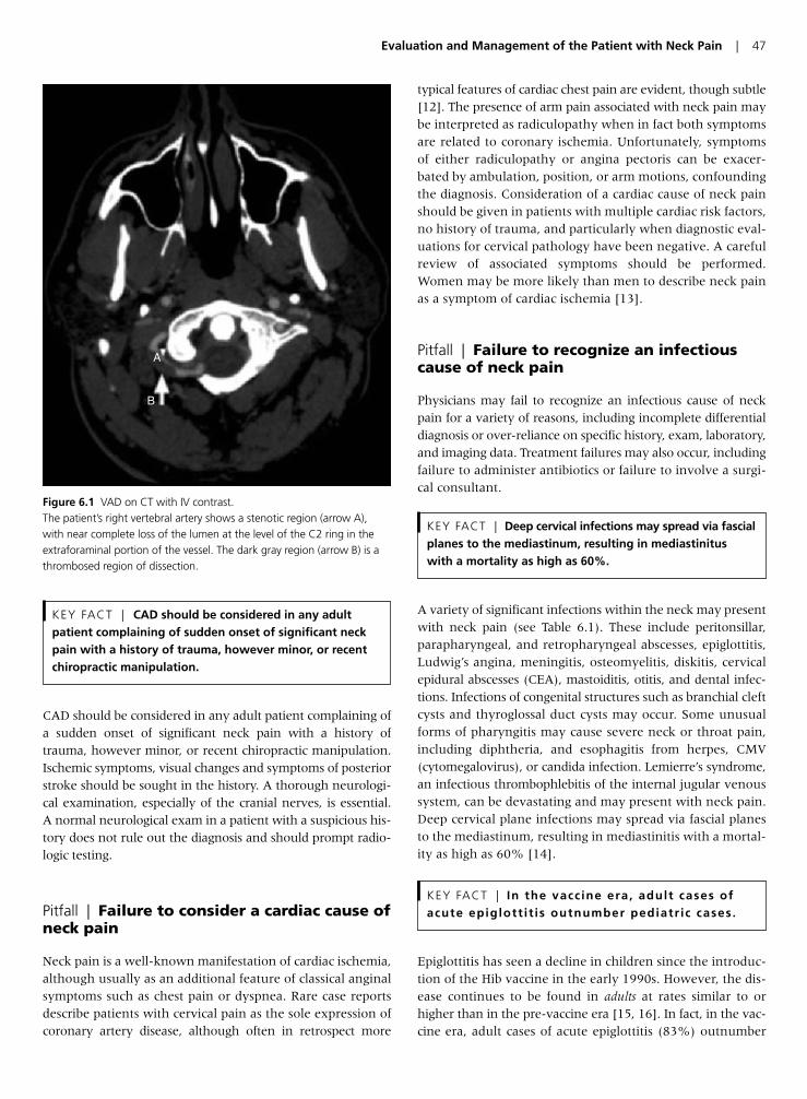
CAD should be considered in any adult patient complaining ofa sudden onset of significant neck pain with a history oftrauma, however minor, or recent chiropractic manipulation.Ischemic symptoms, visual changes and symptoms of posteriorstroke should be sought in the history. A thorough neurologi-cal examination, especially of the cranial nerves, is essential.A normal neurological exam in a patient with a suspicious his-tory does not rule out the diagnosis and should prompt radio-logic testing.
Pitfall | Failure to consider a cardiac cause ofneck pain
Neck pain is a well-known manifestation of cardiac ischemia,although usually as an additional feature of classical anginalsymptoms such as chest pain or dyspnea. Rare case reportsdescribe patients with cervical pain as the sole expression ofcoronary artery disease, although often in retrospect more
typical features of cardiac chest pain are evident, though subtle[12]. The presence of arm pain associated with neck pain maybe interpreted as radiculopathy when in fact both symptomsare related to coronary ischemia. Unfortunately, symptomsof either radiculopathy or angina pectoris can be exacer-bated by ambulation, position, or arm motions, confoundingthe diagnosis. Consideration of a cardiac cause of neck painshould be given in patients with multiple cardiac risk factors,no history of trauma, and particularly when diagnostic eval-uations for cervical pathology have been negative. A carefulreview of associated symptoms should be performed.Women may be more likely than men to describe neck painas a symptom of cardiac ischemia [13].
Pitfall | Failure to recognize an infectiouscause of neck pain
Physicians may fail to recognize an infectious cause of neckpain for a variety of reasons, including incomplete differentialdiagnosis or over-reliance on specific history, exam, laboratory,and imaging data. Treatment failures may also occur, includingfailure to administer antibiotics or failure to involve a surgi-cal consultant.
A variety of significant infections within the neck may presentwith neck pain (see Table 6.1). These include peritonsillar,parapharyngeal, and retropharyngeal abscesses, epiglottitis,Ludwig’s angina, meningitis, osteomyelitis, diskitis, cervicalepidural abscesses (CEA), mastoiditis, otitis, and dental infec-tions. Infections of congenital structures such as branchial cleftcysts and thyroglossal duct cysts may occur. Some unusualforms of pharyngitis may cause severe neck or throat pain,including diphtheria, and esophagitis from herpes, CMV(cytomegalovirus), or candida infection. Lemierre’s syndrome,an infectious thrombophlebitis of the internal jugular venoussystem, can be devastating and may present with neck pain.Deep cervical plane infections may spread via fascial planesto the mediastinum, resulting in mediastinitis with a mortal-ity as high as 60% [14].
Epiglottitis has seen a decline in children since the introduc-tion of the Hib vaccine in the early 1990s. However, the dis-ease continues to be found in adults at rates similar to orhigher than in the pre-vaccine era [15, 16]. In fact, in the vac-cine era, adult cases of acute epiglottitis (83%) outnumber
KEY FACT | In the vaccine era, adult cases ofacute epiglott i t is outnumber pediatr ic cases.
KEY FACT | Deep cervical infections may spread via fascialplanes to the mediastinum, resulting in mediastinitus with a mortality as high as 60%.
KEY FACT | CAD should be considered in any adultpatient complaining of sudden onset of significant neckpain with a history of trauma, however minor, or recentchiropractic manipulation.
Evaluation and Management of the Patient with Neck Pain | 47
A
B
Figure 6.1 VAD on CT with IV contrast.The patient’s right vertebral artery shows a stenotic region (arrow A),with near complete loss of the lumen at the level of the C2 ring in theextraforaminal portion of the vessel. The dark gray region (arrow B) is athrombosed region of dissection.
pediatric cases (17%) [17]. In addition, although rare,Haemophilus influenza-related epiglottitis can be found in fullyvaccinated children. A 10-year retrospective study in NewEngland found 19 cases of childhood epiglottitis in a singlechildren’s hospital, 6 positive for H. influenzae, and 5 of those6 in children with up-to-date immunizations [18].
Pitfall | Failure to elicit historical or examfindings suggesting infection
A history of injection drug use should prompt consideration ofvenous thrombosis or deep neck abscesses, including epiduralspinal abscess. Retained foreign bodies such as needle frag-ments may also provide a nidus for infection. In a retrospectivereview, patients failed to disclose a history of cervical injectionin 50% of cases [19, 20]. Cervical osteomyelitis has beenreported as a complication of cervical injection [21, 22].Swallowed foreign bodies such as bone fragments may alsocause deep space infection. A history of incomplete immuniza-tion or immigrant status may place the patient at risk of diph-theria infection. Immune compromise such as HIV/AIDSshould prompt consideration of infection with herpes, CMV,or candida. Neurological complaints may suggest CEA. Recentinfections, including distant sources such as UTI or skinabscesses, can lead to deep space infections, meningitis or CEAby hematogenous inoculation.
Patients with a “locus minoris resistantiae” of the cervical spinemay be at increased risk for hematogenous seeding. Examplesinclude spondylosis, degenerative joint disease, previouslaminectomy or non-penetrating trauma from a fall or motorvehicle collision. Some underlying diseases also predisposepatients to developing CEAs such as diabetes mellitus, alco-holism, cancer, chronic renal failure and those with compro-mised immune states. Any previous invasive spinal procedure,
even several months prior to presentation, should increasethe suspicion of CEA. Patients typically will not offer this infor-mation unless asked specifically; 18% percent of patients willhave no identifiable predisposing factors for CEA [23, 24].
Physical exam should be thorough, but normal findingsshould be incorporated into the overall picture, not used inisolation to exclude a disease entity. Careful attention shouldbe paid to temperature, heart rate, blood pressure, and respira-tory rate. The neck should be inspected for asymmetry orswelling. Palpation for fluctuance or adenopathy should beperformed. The thyroid should be examined. Any stridor orpooling of secretions should be noted. The oropharynx shouldbe inspected, looking for lesions or swelling. The dentitionand sublingual spaces should be examined. The uvula positionshould be noted, especially any deviation. Tonsils should beexamined for swelling or exudates. Trismus and spitting ofsecretions are also ominous signs. Brawny edema of the sub-mandibular region and elevation of the tongue are signs ofLudwig’s angina. A pseudomembrane should prompt con-sideration of diphtheria. A careful neurological examinationof the cranial nerves and upper extremities should be per-formed if suggested by history or symptoms.
Pitfall | Reliance on the absence of classicphysical exam findings to rule out infection
The classic physical exam findings of many dangerous neckinfections are not reliably present in patients with disease,and their absence should not be used to rule out pathology.Although neck stiffness is a common complaint in meningi-tis (88%) [25], classic exam signs of meningitis should not berelied upon due to poor sensitivity: a prospective study foundKernig’s sign and Brudzinski’s signs to be only 5% sensitivefor meningitis, and nuchal rigidity to be only 30% sensitive[26]. According to a retrospective review comparing physicalexam and symptoms with CT diagnosis of deep neck infection,the physical exam for deep cervical infections is relativelyinsensitive, underestimating the extent of infection in 70%of patients. Classic signs and symptoms of infection were pres-ent with the following frequency: fever (75.4%), pain (89.2%),odynophagia (63.1%), dysphagia (47.7%), neck swelling(84.6%), localized pain (76.9%), local erythema (66.7%),and localized increase in temperature (55.4%) [27]. Inepiglottitis, drooling is a presenting symptom in only 15.2%,and stridor or dyspnea in only 8.6%. Although “hot potato”voice may suggest epiglottitis [28], muffled voice is found injust over half of patients (52.1%) [29].
Pitfall | Over-reliance on fever as a sign ofinfection
KEY FACT | In patients with meningitis … absence ofa fever at presentation is associated with nearly 40-foldincreased mortality.
48 | Chapter 6
Table 6.1 Infectious causes of neck pain.
Peritonsillar abscessParapharyngeal abscessRetropharyngeal abscessEpiglottitisLudwig’s anginaMeningitisOsteomyelitisDiskitisCervical epidural abscessMastoiditisOtitisDental infectionsInfections of congenital structures (e.g. branchial cleft cysts,thyroglossal duct cysts)
PharyngitisEsophagitisLemierre’s syndromeDeep cervical plane infections
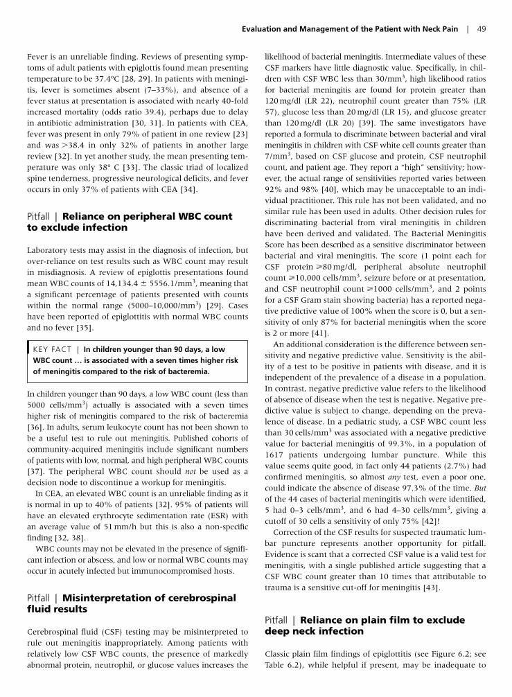
Evaluation and Management of the Patient with Neck Pain | 49
Fever is an unreliable finding. Reviews of presenting symp-toms of adult patients with epiglottis found mean presentingtemperature to be 37.4ºC [28, 29]. In patients with meningi-tis, fever is sometimes absent (7–33%), and absence of afever status at presentation is associated with nearly 40-foldincreased mortality (odds ratio 39.4), perhaps due to delayin antibiotic administration [30, 31]. In patients with CEA,fever was present in only 79% of patient in one review [23]and was �38.4 in only 32% of patients in another largereview [32]. In yet another study, the mean presenting tem-perature was only 38° C [33]. The classic triad of localizedspine tenderness, progressive neurological deficits, and feveroccurs in only 37% of patients with CEA [34].
Pitfall | Reliance on peripheral WBC count to exclude infection
Laboratory tests may assist in the diagnosis of infection, butover-reliance on test results such as WBC count may resultin misdiagnosis. A review of epiglottis presentations foundmean WBC counts of 14,134.4 5556.1/mm3, meaning thata significant percentage of patients presented with countswithin the normal range (5000–10,000/mm3) [29]. Caseshave been reported of epiglottitis with normal WBC countsand no fever [35].
In children younger than 90 days, a low WBC count (less than5000 cells/mm3) actually is associated with a seven timeshigher risk of meningitis compared to the risk of bacteremia[36]. In adults, serum leukocyte count has not been shown tobe a useful test to rule out meningitis. Published cohorts ofcommunity-acquired meningitis include significant numbersof patients with low, normal, and high peripheral WBC counts[37]. The peripheral WBC count should not be used as adecision node to discontinue a workup for meningitis.
In CEA, an elevated WBC count is an unreliable finding as itis normal in up to 40% of patients [32]. 95% of patients willhave an elevated erythrocyte sedimentation rate (ESR) withan average value of 51 mm/h but this is also a non-specificfinding [32, 38].
WBC counts may not be elevated in the presence of signifi-cant infection or abscess, and low or normal WBC counts mayoccur in acutely infected but immunocompromised hosts.
Pitfall | Misinterpretation of cerebrospinalfluid results
Cerebrospinal fluid (CSF) testing may be misinterpreted torule out meningitis inappropriately. Among patients withrelatively low CSF WBC counts, the presence of markedlyabnormal protein, neutrophil, or glucose values increases the
likelihood of bacterial meningitis. Intermediate values of theseCSF markers have little diagnostic value. Specifically, in chil-dren with CSF WBC less than 30/mm3, high likelihood ratiosfor bacterial meningitis are found for protein greater than120 mg/dl (LR 22), neutrophil count greater than 75% (LR57), glucose less than 20 mg/dl (LR 15), and glucose greaterthan 120 mg/dl (LR 20) [39]. The same investigators havereported a formula to discriminate between bacterial and viralmeningitis in children with CSF white cell counts greater than7/mm3, based on CSF glucose and protein, CSF neutrophilcount, and patient age. They report a “high” sensitivity; how-ever, the actual range of sensitivities reported varies between92% and 98% [40], which may be unacceptable to an indi-vidual practitioner. This rule has not been validated, and nosimilar rule has been used in adults. Other decision rules fordiscriminating bacterial from viral meningitis in childrenhave been derived and validated. The Bacterial MeningitisScore has been described as a sensitive discriminator betweenbacterial and viral meningitis. The score (1 point each forCSF protein �80 mg/dl, peripheral absolute neutrophilcount �10,000 cells/mm3, seizure before or at presentation,and CSF neutrophil count �1000 cells/mm3, and 2 pointsfor a CSF Gram stain showing bacteria) has a reported nega-tive predictive value of 100% when the score is 0, but a sen-sitivity of only 87% for bacterial meningitis when the scoreis 2 or more [41].
An additional consideration is the difference between sen-sitivity and negative predictive value. Sensitivity is the abil-ity of a test to be positive in patients with disease, and it isindependent of the prevalence of a disease in a population.In contrast, negative predictive value refers to the likelihoodof absence of disease when the test is negative. Negative pre-dictive value is subject to change, depending on the preva-lence of disease. In a pediatric study, a CSF WBC count lessthan 30 cells/mm3 was associated with a negative predictivevalue for bacterial meningitis of 99.3%, in a population of1617 patients undergoing lumbar puncture. While thisvalue seems quite good, in fact only 44 patients (2.7%) hadconfirmed meningitis, so almost any test, even a poor one,could indicate the absence of disease 97.3% of the time. Butof the 44 cases of bacterial meningitis which were identified,5 had 0–3 cells/mm3, and 6 had 4–30 cells/mm3, giving acutoff of 30 cells a sensitivity of only 75% [42]!
Correction of the CSF results for suspected traumatic lum-bar puncture represents another opportunity for pitfall.Evidence is scant that a corrected CSF value is a valid test formeningitis, with a single published article suggesting that aCSF WBC count greater than 10 times that attributable totrauma is a sensitive cut-off for meningitis [43].
Pitfall | Reliance on plain film to excludedeep neck infection
Classic plain film findings of epiglottitis (see Figure 6.2; seeTable 6.2), while helpful if present, may be inadequate to
KEY FACT | In children younger than 90 days, a lowWBC count … is associated with a seven times higher riskof meningitis compared to the risk of bacteremia.
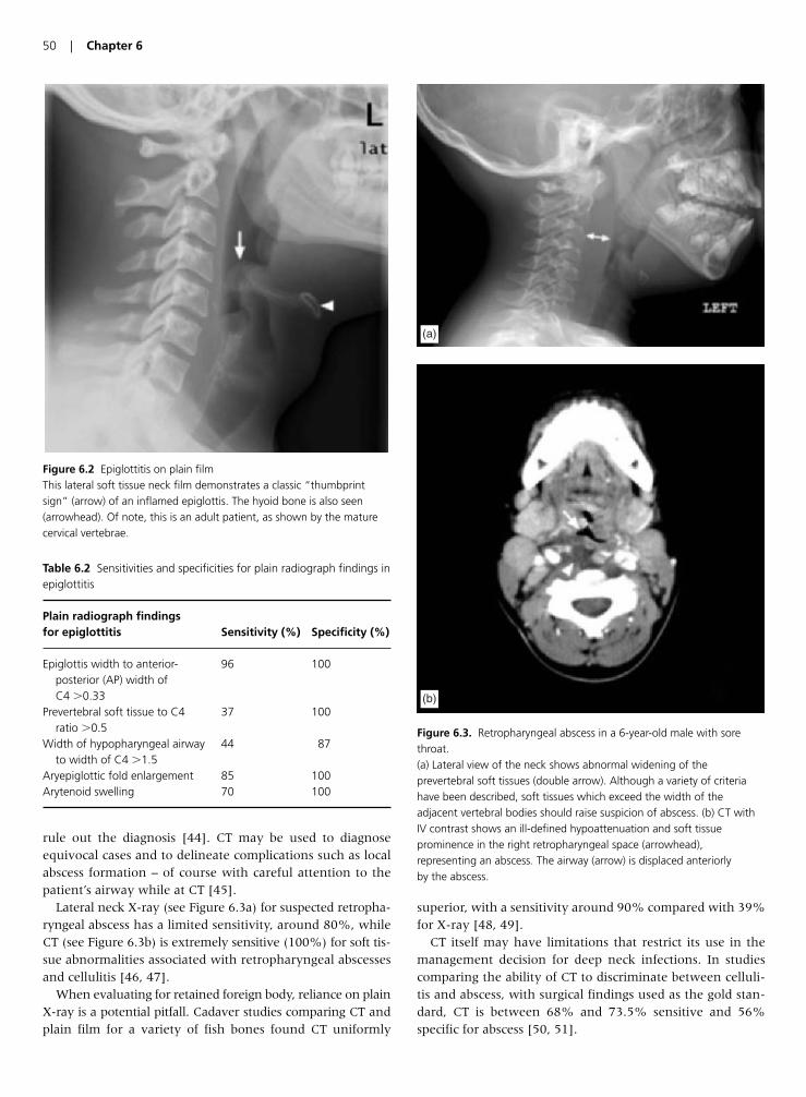
50 | Chapter 6
rule out the diagnosis [44]. CT may be used to diagnoseequivocal cases and to delineate complications such as localabscess formation – of course with careful attention to thepatient’s airway while at CT [45].
Lateral neck X-ray (see Figure 6.3a) for suspected retropha-ryngeal abscess has a limited sensitivity, around 80%, whileCT (see Figure 6.3b) is extremely sensitive (100%) for soft tis-sue abnormalities associated with retropharyngeal abscessesand cellulitis [46, 47].
When evaluating for retained foreign body, reliance on plainX-ray is a potential pitfall. Cadaver studies comparing CT andplain film for a variety of fish bones found CT uniformly
superior, with a sensitivity around 90% compared with 39%for X-ray [48, 49].
CT itself may have limitations that restrict its use in themanagement decision for deep neck infections. In studiescomparing the ability of CT to discriminate between celluli-tis and abscess, with surgical findings used as the gold stan-dard, CT is between 68% and 73.5% sensitive and 56%specific for abscess [50, 51].
Figure 6.2 Epiglottitis on plain filmThis lateral soft tissue neck film demonstrates a classic “thumbprintsign” (arrow) of an inflamed epiglottis. The hyoid bone is also seen(arrowhead). Of note, this is an adult patient, as shown by the maturecervical vertebrae.
Table 6.2 Sensitivities and specificities for plain radiograph findings inepiglottitis
Plain radiograph findings for epiglottitis Sensitivity (%) Specificity (%)
Epiglottis width to anterior- 96 100posterior (AP) width of C4 �0.33
Prevertebral soft tissue to C4 37 100ratio �0.5
Width of hypopharyngeal airway 44 87to width of C4 �1.5
Aryepiglottic fold enlargement 85 100Arytenoid swelling 70 100
(a)
(b)
Figure 6.3. Retropharyngeal abscess in a 6-year-old male with sorethroat.(a) Lateral view of the neck shows abnormal widening of theprevertebral soft tissues (double arrow). Although a variety of criteriahave been described, soft tissues which exceed the width of theadjacent vertebral bodies should raise suspicion of abscess. (b) CT withIV contrast shows an ill-defined hypoattenuation and soft tissueprominence in the right retropharyngeal space (arrowhead),representing an abscess. The airway (arrow) is displaced anteriorly by the abscess.
Pitfall | Delaying treatment of seriousinfections
Patients at risk for meningitis or diphtheria should immedi-ately undergo respiratory isolation and begin antibiotic ther-apy. Delay greater than 6 h in antibiotic therapy for meningitisis associated with a significant increase in mortality, with esti-mates from nearly 2-fold to perhaps 40-fold. Delay in diag-nosis due to CT scan preceding lumbar puncture increases therisk of delay in antibiotic administration by a factor of 5.6.
In the case of CEA, emergent neurosurgical consultationshould be obtained after the administration of intravenousantibiotics. Neurological function continues to deteriorate ina significant portion of patients after initiation of antibioticsand many experts believe that surgery is necessary even if noneurological deficits are present on diagnosis. About 50% ofpatients treated medically and admitted without neurologicdeficits will have neurological deficits on discharge com-pared to 20% of those treated surgically before the develop-ment of deficits [52]. Additionally, total paralysis can occurin a matter of a few hours. Once neurological symptomsdevelop, simple removal of the mass may not result in com-plete recovery because other processes likely occur, in addi-tion to the mechanical compression, that result in cordischemia such as venous thrombosis or focal septic emboli.Ultimate outcome is likely determined by a number of fac-tors including the duration of symptoms, the only factorwhere it is possible for emergency physicians to effect achange (by prompt diagnosis, antibiotic therapy, and consul-tation with a neurosurgeon). Once paralysis develops, it isusually irreversible if surgery is not performed within 24 hof onset [23]. In a review of 188 patients, treatment initiatedafter 36 h of weakness or paralysis resulted in completerecovery in only 39% of patients [53].
Pitfall | Failure to consider a neck mass
Neck pain may be due to the presence of a previously undiag-nosed cervical mass. These include malignant and benign neo-plasms and congenital structures such as branchial cleft cystsor thyroglossal duct cysts, which may be super-infected. Asso-ciated symptoms of weight loss, dysphagia or odynophagia,hoarseness, or neurological symptoms such as ptosis may sug-gest malignancy. Cervical neoplasms increase in incidencewith age, although some neoplasms, such as lymphomas,may occur in younger patients. In patients above the age of 40 years with solitary, benign-appearing cervical cysts, metasta-tic squamous cell carcinoma may be found in up to 23.5%[54]. Cigarette smoking and alcohol use are risk factors [55].
Congenital abnormalities may present with neck pain.These include thyroglossal duct cysts in the midline andbranchial cleft cysts in the lateral neck. Both thyroglossalduct cysts and branchial cleft cysts may be occult until infec-tion occurs, presenting then with pain and swelling.
Pitfall | Failure to evaluate the patient forevidence of malignancy
The physical exam should include inspection of the orophar-ynx for lesions, as oral cancers may spread locally or metasta-size within the neck. The neck should be inspected forasymmetry, and palpation should be performed, including thethyroid gland and anterior and posterior lymph nodes. Thevoice quality should be noted, and the patient should be askedabout changes in voice character. Although hoarseness maybe associated with a benign diagnosis such as laryngitis,hoarseness or change in voice could indicate local involve-ment of laryngeal structures or involvement of the recurrentlaryngeal nerve. A Horner’s syndrome (ptosis, miosis, anhy-drosis) can occur from compression of sympathetic nervesinnervating the face, so the patient’s eyes should be exam-ined for eyelid position and pupillary size and reactivity.Unfortunately, characteristics from exam and history, includ-ing location, number of lesions, bilaterality, size, and dura-tion, have poor positive and negative predictive values formalignancy, in the range of 60–80% [55].
Pitfall | Reliance on plain film to exclude amass
Due to the insensitivity of history and exam for masses,patients with unexplained neck pain may require imagingfor further diagnosis. Plain films of the neck are insensitiveand not generally useful. If a mass is considered, enhancedCT (CT with IV contrast) is the preferred test, with excellentsensitivity. MRI is helpful in patients with contraindicationsto iodinated contrast, and ultrasound plays a role in differ-entiating solid from cystic masses [56].
Pitfall | Failure to involve a consultant inpatients with mass lesions
Newly diagnosed cervical neoplasms require prompt follow-up,though admission may not be required if the airway is notthreatened, the patient is tolerating oral intake, and pain isadequately controlled. A firm outpatient follow-up plan shouldbe in place, with a clear understanding from the patient of thesignificance of the emergency department findings and poten-tial diagnosis. Otolaryngology referral is advisable for all cer-vical masses, even those that appear to be benign or localinfections, as malignancy may co-exist. Importantly, incision
KEY FACT | In patients above the age of 40 years withsolitary, benign-appearing cervical cysts, metastaticsquamous cell carcinoma may be found in up to 23.5%.
Evaluation and Management of the Patient with Neck Pain | 51
and drainage of cervical masses should rarely be performed byemergency physicians. Some cervical masses, including thy-roglossal duct lesions, require complete excision for cure [57].Emergency department incision and drainage may also becontraindicated because in rare cases these lesions contain thepatient’s only functional thyroid tissue. Moreover, incision ofan unsuspected malignancy may promote spread and com-promise chance for surgical cure, due to seeding of malignantcells in needle tracks [58, 59]. Missed diagnoses of laryngealmalignancy can have devastating medical consequences forpatients, with high rates of laryngectomy (63%) and mortal-ity (35%). Patients with misdiagnosed laryngeal carcinomapresent at an early age (mean 47 years), compared with thepeak incidence of the disease (age 70–74 years). Malpracticeawards occur in over half of cases [60].
Pitfall | Failure to consider spinal epiduralhematoma in patients with neck pain andupper extremity paresthesias or weakness
Cervical epidural hematomas (CEH) are rare, but life threat-ening. Mortality is 30% for CEH located above C5 [61]. CEHare almost uniformly fatal secondary to respiratory failure ifnot corrected [62]. Failure to recognize the diagnosis promptlyleads to a less favorable outcome and more severe permanentneurological deficits. Because they are so rare, can occur spon-taneously, and present with non-specific symptoms, theypresent a difficult diagnostic dilemma.
CEHs are caused by trauma, underlying hematological dis-orders or systemic disease, or as a complication of epiduralanesthesia [63]. However, there are many reports of sponta-neous spinal epidural hematomas (SSEH) in the literaturewhich are defined as occurring in the absence of one of theabove causes [61, 62, 64–69]. They occur in as many as40–50% of cases. SSEH has been reported to occur afterminor physical exertion, sneezing, coitus, straining, vomiting,micturition, coughing and bending over. There are several casereports of CEHs occurring after chiropractic manipulation[70–73]. Other risk factors include anticoagulation, priorthrombolytic therapy, pregnancy, alcoholism, liver disease,and rheumatologic disorders.
Pitfall | Reliance on a normal neurologicexam to exclude spinal epidural hematoma
The usual presentation is that of an acute onset of severe pos-terior neck pain that progresses to para- or tetraplegia in hoursor days. Cervical radicular pain is one of the earliest symptomsand a hallmark of cervical epidural hematoma [74]. Althoughmost neck pain due to trauma that presents with radicularsymptoms will be due to an injured cervical disk or nerveroot, the emergency physician should consider the diagnosis
of SSEH and perform a thorough neurological examination.The presence of other risk factors may necessitate imaging torule out a hematoma.
Pitfall | Failure to perform timely andappropriate imaging and consultationfor spinal epidural hematoma
Diagnosis requires MRI or CT myelography, and the diagnosismay be missed on non-contrast CT or CT angiography per-formed for suspected CAD. Treatment involves emergentdecompressive laminectomy with clot evacuation. It is alsoimperative to reverse any existing coagulopathy.
Early neurosurgical involvement is essential. The durationof symptoms affects the outcome as does the level andextent of the lesion and the severity of the neurologicaldeficits. Patients obtain a useful recovery �50% of the timewhen surgery is delayed for more than 36 h after symptomonset [75]. In one review, only 15/27 recovered completelywhen surgery was delayed for more than 36 h after symp-toms onset [76].
Pearls for Improving Patient Outcomes
• Consider CAD in patients complaining of sudden onset of signif-icant neck pain with history of trauma, however minor.Neurological symptoms may be absent at presentation.
• Assess for a cardiac cause of neck pain, especially in women, andevaluate for other symptoms or risk factors which may suggestcoronary artery disease.
• Suspect CEA in patients with significant neck pain and risk fac-tors such as injection drug use, diabetes, immunocompromised,chronic renal failure, cancer, alcoholism, recent distant or con-tiguous infections, or recent invasive spinal procedure.
• Never rule out meningitis using Kernig’s and Brudzinski’s signs,as they are poorly sensitive.
• Do not rely on the absence of fever or a normal WBC count aloneto rule out infection.
• Do not rule out meningitis using a corrected CSF value for trau-matic lumbar punctures.
• Do not rely on plain films to exclude deep neck infections.• Give immediate antibiotics for suspected meningitis, as delay in
antibiotic administration for CT or lumbar puncture significantlyincreases mortality.
• Arrange specific follow-up for patients with undifferentiatedneck masses.
• Exercise caution in incision and drainage of cervical masses inthe emergency department due to the possibility of malignancy.Consultation may be advisable.
• Suspect cervical epidural hematoma in a patient with neck painand radicular symptoms who has any of the following risk factors:anticoagulation, pregnancy, liver disease, rheumatologic disor-ders, preceding minor trauma or chiropractic manipulation.
52 | Chapter 6
Evaluation and Management of the Patient with Neck Pain | 53
References
1. Lee WW, Jensen ER. Bilateral internal carotid artery dissection
due to trivial trauma. J Emerg Med 2000; 19(1):35–41.
2. Saeed AB, Shuaib A, Al-Sulaiti G, Emery D. Vertebral artery
dissection: warning symptoms, clinical features and prognosis
in 26 patients. Can J Neurol Sci 2000; 27(4):292–6.
3. Dziewas R, Konrad C, Drager B, Evers S, Besselmann M,
Ludemann P, Kuhlenbaumer G, Stogbauer F, Ringelstein EB.
Cervical artery dissection – clinical features, risk factors, therapy
and outcome in 126 patients. J Neurol 2003; 250(10):1179–84.
4. Wessels T, Rottger C, Kaps M, Traupe H, Stolz E. Upper cranial
nerve palsy resulting from spontaneous carotid dissection. J
Neurol 2005; 252(4):453–6.
5. Chiche L, Praquin B, Koskas F, Kieffer E. Spontaneous dissection
of the extracranial vertebral artery: indications and long-term
outcome of surgical treatment. Ann Vasc Surg 2005; 19(1):5–10.
6. Wessels T, Sparing R, Neuschaefer-Rube C, Klotzsch C. Vocal
cord palsy resulting from spontaneous carotid dissection.
Laryngoscope 2003; 113(3):537–40.
7. Guidetti D, Pisanello A, Giovanardi F, Morandi C, Zuccoli G,
Troiso A. Spontaneous carotid dissection presenting lower cra-
nial nerve palsies. J Neurol Sci 2001; 184(2):203–7.
8. Rees JH, Valentine AR, Llewelyn JG. Spontaneous bilateral
carotid and vertebral artery dissection presenting as a Collet-
Sicard syndrome. Br J Radiol 1997; 70(836):856–8.
9. Silbert PL, Mokri B, Schievink WI. Headache and neck pain in
spontaneous internal carotid and vertebral artery dissections.
Neurology 1995; 45(8):1517–22.
10. Mokri B, Houser OW, Sandok BA, Piepgras DG. Spontaneous
dissections of the vertebral arteries. Neurology 1988; 38(6):
880–5.
11. Munera F, Cohn S, Rivas LA. Penetrating injuries of the neck:
use of helical computed tomographic angiography. J Trauma
2005; 58(2):413–18.
12. Lipetz JS, Ledon J, Silber J. Severe coronary artery disease pre-
senting with a chief complaint of cervical pain. Am J Phys Med
Rehabil 2003; 82(9):716–20.
13. Chen W, Woods SL, Puntillo KA. Gender differences in symp-
toms associated with acute myocardial infarction: a review of
the research. Heart Lung 2005; 34(4):240–7.
14. Cardenas-Malta KR, Cortes-Flores AO, Fuentes-Orozco C, Del
Carmen Martinez-Oropeza L, Lopez-Ramirez MK, Gonzalez-
Ojeda A. Necrotizing mediastinitis in deep neck infections. Cir
Cir 2005; 73(4):263–7. [Article in Spanish]
15. McVernon J, Trotter CL, Slack MP, Ramsay ME. Trends in
Haemophilus influenzae type b infections in adults in England and
Wales: surveillance study. Br Med J 2004; 329(7467):655–8.
16. Berger G, Landau T, Berger S, Finkelstein Y, Bernheim J, Ophir
D. The rising incidence of adult acute epiglottitis and epiglottic
abscess. Am J Otolaryngol 2003; 24(6):374–83.
17. Wood N, Menzies R, McIntyre P. Epiglottitis in Sydney before
and after the introduction of vaccination against Haemophilus
influenzae type b disease. Inter Med J 2005; 35(9):530–5.
18. Shah RK, Roberson DW, Jones DT. Epiglottitis in the
Hemophilus influenzae type B vaccine era: changing trends.
Laryngoscope 2004; 114(3):557–60.
19. Williams MF, Eisele DW, Wyatt SH. Neck needle foreign bodies
in intravenous drug abusers. Laryngoscope 1993; 103(1 Pt 1):
59–63.
20. Myers EM, Kirkland LS Jr, Mickey R. The head and neck seque-
lae of cervical intravenous drug abuse. Laryngoscope 1988;
98(2):213–18.
21. Smith MA, Trowers NR, Klein RS. Cervical osteomyelitis caused
by Pseudomonas cepacia in an intravenous-drug abuser. J Clin
Microbiol 1985; 21(3):445–6.
22. Sapico FL, Montgomerie JZ. Vertebral osteomyelitis in intra-
venous drug abusers: report of three cases and review of the lit-
erature. Rev Infect Dis 1980; 2(2):196–206.
23. Darouiche RO, Hamill RJ, Greenberg SB, Weathers SW,
Musher DM. Bacterial spinal epidural abscess. Review of 43 cases
and literature survey. Medicine (Baltimore) 1992; 71(6):369–85.
24. Ravicovitch MA, Spallone A. Spinal epidural abscesses: surgical
and parasurgical management. Eur Neurol 1982; 21:347–57.
25. Durand ML, Calderwood SB, Weber DJ, Miller SI, Southwick FS,
Caviness Jr. VS, Swartz MN. Acute bacterial meningitis in adults.
A review of 493 episodes.
26. Thomas KE, Hasbun R, Jekel J, Quagliarello VJ. The diagnostic
accuracy of Kernig’s sign, Brudzinski’s sign, and nuchal rigidity
in adults with suspected meningitis. Clin Infect Dis 2002;
35(1):46–52. Epub 2002 Jun 5.
27. Crespo AN, Chone CT, Fonseca AS, Montenegro MC, Pereira R,
Milani JA. Clinical versus computed tomography evaluation in
the diagnosis and management of deep neck infection. Sao
Paulo Med J 2004; 122(6):259–63. Epub 2005 Feb 2.
28. Shah RK, Roberson DW, Jones DT. Epiglottitis in the Hemophilus
influenzae type B vaccine era: changing trends. Laryngoscope
2004; 114(3):557–60.
29. Chang YL, Lo SH, Wang PC, Shu YH. Adult acute epiglottitis:
experiences in a Taiwanese setting. Otolaryngol Head Neck Surg
2005; 132(5):689–93.
30. Proulx N, Frechette D, Toye B, Chan J, Kravcik S. Delays in the
administration of antibiotics are associated with mortality from
adult acute bacterial meningitis. Q J Med 2005; 98(4):291–8.
Epub 2005 Mar 10.
31. van de Beek D, de Gans J, Spanjaard L, Weisfelt M, Reitsma JB,
Vermeulen M. Clinical features and prognostic factors in
adults with bacterial meningitis. N Engl J Med 2004; 351(18):
1849–59.
32. Rigamonti D, Liem L, Sampath P, Knoller N, Namaguchi Y,
Schreibman DL, Sloan MA, Wolf A, Zeidman S. Spinal epidural
abscess: contemporary trends in etiology, evaluation, and man-
agement. Surg Neurol 1999; 52(2):189–96.
33. Redekop GJ, Del Maestro RF. Diagnosis and management of
spinal epidural abscess. Can J Neurol Sci 1992; 19(2):180–7.
34. Ekbom DC, D-Elia J, Isaacson B, Lamarca F, Chepeha DB,
Bradford CR. Spinal epidural abscess after cervical pharyngoe-
sophageal dilation. Head Neck 2005; 27(6):543–8.
35. Jerrard DA, Olshaker J. Simultaneous uvulitis and epiglottitis
without fever or leukocytosis. Am J Emerg Med 1996; 14(6):551–2.
36. Bonsu BK, Harper MB. A low peripheral blood white blood cell
count in infants younger than 90 days increases the odds of
acute bacterial meningitis relative to bacteremia. Acad Emerg
Med 2004; 11(12):1297–301.
37. Aronin SI, Peduzzi P, Quagliarello VJ. Community-acquired
bacterial meningitis: risk stratification for adverse clinical out-
come and effect of antibiotic timing. Ann Intern Med 1998;
129(11):862–9.
38. Wong D, Raymond NJ. Spinal epidural abscess. NZ Med J 1998;
111:345–7.
54 | Chapter 6
39. Bonsu BK, Harper MB. Accuracy and test characteristics of
ancillary tests of cerebrospinal fluid for predicting acute bacter-
ial meningitis in children with low white blood cell counts in
cerebrospinal fluid. Acad Emerg Med 2005; 12(4):303–9.
40. Bonsu BK, Harper MB. Differentiating acute bacterial meningi-
tis from acute viral meningitis among children with cere-
brospinal fluid pleocytosis: a multivariable regression model.
Pediatr Infect Dis J 2004; 23(6):511–7.
41. Nigrovic LE, Kuppermann N, Malley R. Development and valida-
tion of a multivariable predictive model to distinguish bacterial
from aseptic meningitis in children in the post-Haemophilus
influenzae era. Pediatrics 2002; 110(4):712–19.
42. Freedman SB, Marrocco A, Pirie J, Dick PT. Predictors of bacte-
rial meningitis in the era after Haemophilus influenzae. Arch
Pediatr Adolesc Med 2001; 155(12):1301–6.
43. Mayefsky JH, Roghmann KJ. Determination of leukocytosis in
traumatic spinal tap specimens. Am J Med 1987; 82(6):1175–81.
44. Nemzek WR, Katzberg RW, Van Slyke MA, Bickley LS. A reap-
praisal of the radiologic findings of acute inflammation of the
epiglottis and supraglottic structures in adults. Am J Neuroradiol
1995; 16(3):495–502.
45. Smith MM, Mukherji SK, Thompson JE, Castillo M. CT in adult
supraglottitis. Am J Neuroradiol 1996; 17(7):1355–8.
46. Boucher C, Dorion D, Fisch C. Retropharyngeal abscesses: a clini-
cal and radiologic correlation. J Otolaryngol 1999; 28(3):134–7.
47. Nagy M, Backstrom J. Comparison of the sensitivity of lateral
neck radiographs and computed tomography scanning in pedi-
atric deep-neck infections. Laryngoscope 1999; 109(5):775–9.
48. Palme CE, Lowinger D, Petersen AJ. Fish bones at the cricopha-
ryngeus: a comparison of plain-film radiology and computed
tomography. Laryngoscope 1999; 109(12):1955–8.
49. Lue AJ, Fang WD, Manolidis S. Use of plain radiography and
computed tomography to identify fish bone foreign bodies.
Otolaryngol Head Neck Surg 2000; 123(4):435–8.
50. Vural C, Gungor A, Comerci S. Accuracy of computerized
tomography in deep neck infections in the pediatric population.
Am J Otolaryngol 2003; 24(3):143–8.
51. Stone ME, Walner DL, Koch BL, Egelhoff JC, Myer CM.
Correlation between computed tomography and surgical find-
ings in retropharyngeal inflammatory processes in children. Int
J Pediatr Otorhinolaryngol 1999; 49(2):121–5.
52. Curry WT, Hoh BL, Amin-Hanjani S, Eskandar EN. Spinal
epidural abscess: clinical presentation, management, and out-
come. Surg Neurol 2005; 63(4):364–71.
53. Williams JW, Powell T. Epidural abscess of the cervical spine:
case report and literature review. Br J Radiol 1990; 63(751):576–8.
54. Gourin CG, Johnson JT. Incidence of unsuspected metastases in
lateral cervical cysts. Laryngoscope 2000; 110(10 Pt 1):1637–41.
55. Bhattacharyya N. Predictive factors for neoplasia and malig-
nancy in a neck mass. Arch Otolaryngol Head Neck Surg 1999;
125(3):303–7.
56. Eskey CJ, Robson CD, Weber AL. Imaging of benign and malig-
nant soft tissue tumors of the neck. Radiol Clin N Am 2000;
38(5):1091–104.
57. Josephson GD, Spencer WR, Josephson JS. Thyroglossal duct
cyst: the New York Eye and Ear Infirmary experience and a litera-
ture review. Ear Nose Throat J 1998; 77(8):642–4, 646–7, 651.
58. Gritzmann N, Hollerweger A, Macheiner P, Rettenbacher T.
Sonography of soft tissue masses of the neck. J Clin Ultrasound
2001; 30(6):356–73.
59. Mighell AJ, High AS. Histological identification of carcinoma in
21 gauge needle tracks after fine needle aspiration biopsy of
head and neck carcinoma. J Clin Pathol 1998; 51(3):241–3.
60. Lydiatt DD. Medical malpractice and cancer of the larynx.
Laryngoscope 2002; 112(3):445–8.
61. Demierre B, Unger PF, Bongioanni F. Sudden cervical pain:
spontaneous cervical epidural hematoma. Am J Emerg Med
1991; 9(1):54–6.
62. Williams JM, Allegra JR. Spontaneous cervical epidural
hematoma. Ann Emerg Med 1994; 23(6):1368–70.
63. Stoll A, Sanchez M. Epidural hematoma after epidural block:
implications for its use in pain management. Surg Neurol 2002;
57(4):235–40.
64. Tender GC, Awasthi D. Spontaneous cervical spinal epidural
hematoma in a 12-year old girl: case report and review of the
literature. J La State Med Soc 2004; 156(4):196–8.
65. Steinmetz MP, Kalfas IH, Willis B, Chalavi A, Harlan RC.
Successful surgical management of a case of spontaneous
epidural hematoma of the spine during pregnancy. Spine J
2003; 3(6):539–42.
66. Ferry P, Reisner C, Ashpole R. A case of spontaneous cervical
extradural haematoma. Hosp Med 2001; 62(7):436–7.
67. Vaya A, Resureccion M, Ricart JM, Ortuno C, Ripoll F, Mira Y,
Aznar J. Clin Appl Thromb Hemost 2001; 7(2):166–8.
68. Lobitz B, Grate I. Acute epidural hematoma of the cervical spine:
an unusual cause of neck pain. South Med J 1995; 88(5):580–2.
69. Nagel MA, Taff IP, Cantos EL, Patel MP, Maytal J, Berman D.
Spontaneous spinal epidural hematoma in a 7-year-old girl.
Diagnostic value of magnetic resonance imaging. Clin Neurol
Neurosurg 1989; 91(2):157–60.
70. Lin IY. Diagnostic pitfall: nontraumatic spinal epidural hematoma
mimicking a brainstem stroke. Ann Emerg Med 2004; 44(2):183–4.
71. Saxler G, Barden B. Extensive spinal epidural hematoma—an
uncommon entity following cervical chiropractic manipulation.
Z Orthop Ihre Grenzgeb 2004; 142(1):79–82.
72. Segal DH, Lidov MW, Camins MB. Cervical epidural hematoma
after chiropractic manipulation in a healthy young woman:
case report. Neurosurgery 1996; 39(5):1043–5.
73. Zupruk GM, Mehta Z. Brown-sequard syndrome associated
with posttraumatic cervical epidural hematoma: case report
and review of the literature. Neurosurgery 1989; 25(2):278–80.
74. Sakamoto N, Yanaka K, Matsumaru Y, Nose T. Cervical epidural
hematoma causing hemiparesis. Arch Neurol 2003; 60(5):783.
75. McDonnell GV, Bell KE, Hawkins SA. A pain in the neck.
Postgrad Med J 2000; 76(891):229–30.
76. Patel H, Boaz JC, Phillips JP, Garg BP. Spontaneous spinal epidural
hematoma in children. Pediatr Neurol 1998; 19(4):302–7.
Introduction
Emergency medicine physicians (EPs) provide the initialtrauma care of most trauma patients. Although we oftenwork in concert with the trauma surgeons on the more seri-ously injured patients, it behooves us to be on the cuttingedge of trauma care for all of our injured patients. Severalpitfalls that frequently surface as areas of concern for manyEPs are addressed here.
Pitfall | Failure to implement surrogates to crystalloid during resuscitation
Crystalloid fluid resuscitation is crucial to maintain perfusionto vital organs. It is important, however, to frequently reassessthe patient between boluses to determine whether he/she is aresponder, partial responder, or non-responder to the fluids.This allows one to determine the need for blood products,surgical intervention, or other therapies. Therefore, infusingpredetermined boluses of fluid is essential to allowing one todetermine whether alternate therapies need be initiated.
Overzealous fluid administration can have several deleteri-ous physiologic effects. Elevating the blood pressure abovethe tamponade pressure of post-traumatic clot formation maycause clot disruption and worsen bleeding and thus shock.Also, hemodilution causing a progressive anemia decreasesthe oxygen carrying capacity of the intravascular space anddecreases the viscosity of the blood allowing it to more easilyelute around an incomplete thrombus [1]. By judiciously uti-lizing fluid resuscitation, selecting and implementing properblood products or surrogates when required (red blood cells(RBC), plasma, platelets, clotting factors, rFVIIa), and bettermonitoring the response to fluids, we should be able toimprove outcomes by limiting further hemorrhage and min-imizing the complications of hemodilution.
As optimal perfusion pressure must be maintained, elevationof the blood pressure above that of the tamponade pressureof the clot may be required. In doing so, a clinical trade off is
made and must be recognized. This allows for more aggres-sive management of the point of active bleeding, as well asresuscitation with oxygen carrying products to replace thelost hemoglobin. Administration of other perfusion adjunctscan minimize the volume of crystalloid necessary to main-tain perfusion. The rapidity of which definitive hemorrhagecontrol is established is paramount to the survival of thepatient. Likewise, identifying and controlling a bleeding sourceis critical to minimize ongoing blood loss.
Pitfall | Over-reliance on focused abdominal sonography in trauma
Focused abdominal sonography in trauma (FAST) examina-tions has rapidly replaced diagnostic peritoneal lavage (DPL)in the evaluation of unstable or stable trauma patients aspart of the primary or secondary survey. Multiple studieshave reported the sensitivity of the FAST examination fordetermining the presence of intra-abdominal fluid to be90%, with a specificity of 99%, and an accuracy of 98%[2–4]. As such, the FAST examination has its primary utilityin the unstable trauma patient who should not go to com-puted tomography (CT) scan, but rather the operating roomdue to increased suspicion for intra-abdominal injury as thecause of instability. In patients who are hypotensive afterblunt abdominal trauma and not hemodynamically stableenough to undergo diagnostic CT, negative US findings vir-tually exclude intra-abdominal surgical injury as the causeof hemodynamic instability, while positive US findings indi-cate surgical injury in approximately 70% of cases [5]. TheFAST examination also may be helpful in a multi-patienttrauma to triage critically ill patients when not all can get aCT scan in a timely manner.
In general, a positive FAST examination in the stable patientdoes not mandate surgical intervention as non-operativemanagement for a solid organ injury may be indicated basedon patient stability, age, physiologic reserve, and severity ofinjury based on CT grading. When indicated, CT imaging of
KEY FACT | In stable patients, a negative FASTexamination does not obviate the need for CT and apositive one does not mandate operative intervention.KEY FACT | Patients must be reassessed regularly after
boluses of predefined quantities of crystalloid. Only bydoing so can the need for other adjuncts be identified.
Chapter 7 | Trauma Management in the ED
David E. Manthey & Bret A. Nicks
55
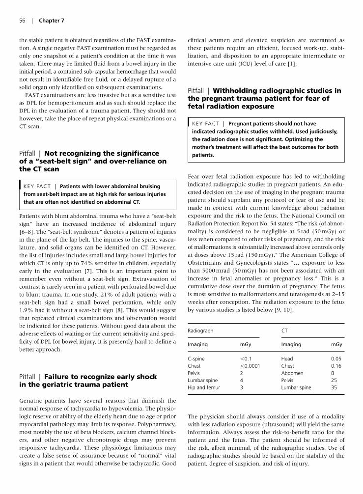
the stable patient is obtained regardless of the FAST examina-tion. A single negative FAST examination must be regarded asonly one snapshot of a patient’s condition at the time it wastaken. There may be limited fluid from a bowel injury in theinitial period, a contained sub-capsular hemorrhage that wouldnot result in identifiable free fluid, or a delayed rupture of asolid organ only identified on subsequent examinations.
FAST examinations are less invasive but as a sensitive testas DPL for hemoperitoneum and as such should replace theDPL in the evaluation of a trauma patient. They should nothowever, take the place of repeat physical examinations or aCT scan.
Pitfall | Not recognizing the significance of a “seat-belt sign” and over-reliance onthe CT scan
Patients with blunt abdominal trauma who have a “seat-beltsign” have an increased incidence of abdominal injury[6–8]. The “seat-belt syndrome” denotes a pattern of injuriesin the plane of the lap belt. The injuries to the spine, vascu-lature, and solid organs can be identified on CT. However,the list of injuries includes small and large bowel injuries forwhich CT is only up to 74% sensitive in children, especiallyearly in the evaluation [7]. This is an important point toremember even without a seat-belt sign. Extravasation ofcontrast is rarely seen in a patient with perforated bowel dueto blunt trauma. In one study, 21% of adult patients with aseat-belt sign had a small bowel perforation, while only1.9% had it without a seat-belt sign [8]. This would suggestthat repeated clinical examinations and observation wouldbe indicated for these patients. Without good data about theadverse effects of waiting or the current sensitivity and speci-ficity of DPL for bowel injury, it is presently hard to define abetter approach.
Pitfall | Failure to recognize early shock in the geriatric trauma patient
Geriatric patients have several reasons that diminish thenormal response of tachycardia to hypovolemia. The physio-logic reserve or ability of the elderly heart due to age or priormyocardial pathology may limit its response. Polypharmacy,most notably the use of beta blockers, calcium channel block-ers, and other negative chronotropic drugs may preventresponsive tachycardia. These physiologic limitations maycreate a false sense of assurance because of “normal” vitalsigns in a patient that would otherwise be tachycardic. Good
clinical acumen and elevated suspicion are warranted asthese patients require an efficient, focused work-up, stabi-lization, and disposition to an appropriate intermediate orintensive care unit (ICU) level of care [1].
Pitfall | Withholding radiographic studies inthe pregnant trauma patient for fear offetal radiation exposure
Fear over fetal radiation exposure has led to withholdingindicated radiographic studies in pregnant patients. An edu-cated decision on the use of imaging in the pregnant traumapatient should supplant any protocol or fear of use and bemade in context with current knowledge about radiationexposure and the risk to the fetus. The National Council onRadiation Protection Report No. 54 states: “The risk (of abnor-mality) is considered to be negligible at 5 rad (50 mGy) orless when compared to other risks of pregnancy, and the riskof malformations is substantially increased above controls onlyat doses above 15 rad (150 mGy).” The American College ofObstetricians and Gynecologists states “… exposure to lessthan 5000 mrad (50 mGy) has not been associated with anincrease in fetal anomalies or pregnancy loss.” This is acumulative dose over the duration of pregnancy. The fetus is most sensitive to malformations and teratogenesis at 2–15weeks after conception. The radiation exposure to the fetusby various studies is listed below [9, 10].
The physician should always consider if use of a modalitywith less radiation exposure (ultrasound) will yield the sameinformation. Always assess the risk-to-benefit ratio for thepatient and the fetus. The patient should be informed of the risk, albeit minimal, of the radiographic studies. Use ofradiographic studies should be based on the stability of thepatient, degree of suspicion, and risk of injury.
KEY FACT | Pregnant patients should not haveindicated radiographic studies withheld. Used judiciously,the radiation dose is not significant. Optimizing themother’s treatment will affect the best outcomes for bothpatients.
KEY FACT | Patients with lower abdominal bruisingfrom seat-belt impact are at high risk for serious injuriesthat are often not identified on abdominal CT.
56 | Chapter 7
Radiograph CT
Imaging mGy Imaging mGy
C-spine �0.1 Head 0.05Chest �0.0001 Chest 0.16Pelvis 2 Abdomen 8Lumbar spine 4 Pelvis 25Hip and femur 3 Lumbar spine 35
Pitfall | Inappropriate use of CTs and plainradiographs of the spine
Less than 50% of current academic emergency departmentscontinue to obtain additional lumbar and thoracic spine plainfilms after obtaining a CT of the chest and abdomen. Theseadditional radiographs occur at additional expense and radi-ation to the patient without additional diagnostic value. TheCT images can be reformatted to deliver images of the spinalcolumn with a sensitivity of 97% for thoracic spine fracturesand 95% for lumbar spine fractures. The CT may miss non-operative compression fractures. The sensitivity of plainradiographs is 62% for the thoracic spine and 86% for thelumbar spine [11]. Two recent reviews of more than 3000patients suggest that plain radiographs of the spine shouldno longer be utilized in the trauma patient as CT has sup-planted that radiographic modality [12]. The charges anddose of radiation must be taken into consideration withplain films charge about $145, and delivering 6.36 mSv ofradiation and CT charge about $880 and delivering19.42 mSv CT for imaging of the lumbar spine [13]. Due tothe additional radiation exposure and cost for CT, perhapsa more rational approach may be to reserve CT for thosein whom it is not possible to obtain adequate plain filmsor those with a high suspicion of injury, however, furtherresearch will be required to best identify a risk stratifyingapproach.
Pitfall | Failure to appropriately apply C-spine clearance rules
Two C-spine clearance rules have come to the forefront inrecent years, the NEXUS rule and the Canadian C-spinerule. Without getting into the discussion of which might bebetter, the authors would like to emphasize the limitationsof such rules in general. We can all recall that NEXUSrequires a patient to have normal level of alertness, but howmany can define what that really means. The NEXUS studydefines it as an altered level of alertness which can includeany of the following:
1. Glasgow Coma Scale (GCS) score of 14 or less.2. Disorientation to person, place, time, or events.3. Inability to remember three objects at 5 min.4. Delayed or inappropriate response to external stimuli.
This is not a downfall of the study, it is an issue with how weoften oversimplify a rule to make it easier to apply and
therefore utilize. But the NEXUS authors looked at their criteria and state it best: “Because each of the five low-riskcriteria was the only marker of non-low-risk status in at leasta few patients with significant cervical spine injury, modifica-tion of the overall NEXUS decision instrument by eliminatingany one of the criteria would markedly reduce sensitivityand make the instrument unacceptable for clinical use [14].”
Neither rule unambiguously defines distracting injuries. Arecent study suggests that proximity of injury due to the gat-ing theory that a noxious stimuli from the upper extremitymay provide enough counter-irritation through the spinalpain pathways that other pain signals may go unnoticed[15]. This may make upper extremity injuries distractingeven if they don’t fit the guidelines of either NEXUS or theCanadian C-spine rules.
The Canadian C-spine rule removes anyone over the ageof 64. The NEXUS rule does not, and has looked at the geri-atric population of the study citing a sensitivity of the NEXUScriteria for all cervical spine injuries in the geriatric group of98.5% (95% CI, 94.8–99.7%), and the sensitivity for clini-cally significant injury was 100% (95% CI, 97.1–100%).There are various case reports of elderly patients with injury,but they often cite inappropriate evaluation of the level ofalertness. The NEXUS group urged caution in using the crite-ria among children, especially those younger than 9 as therewas a limited number of patients and injuries in this group.The Canadian C-spine rule was for patients 16 years andolder.
The Canadian C-spine rule [16] included the presence of aconcerning mechanism (fall from 1 m or greater, axial loadto head, motorized recreational vehicles, bicycle collision, ormotor vehicle collision (MVC) with high speed, rollover, orejection). The NEXUS study recognized that it was not utiliz-ing a criterion for mechanism but all of these were certainlyincluded in the group that was studied.
The goal of both of these rules is to decrease the use ofunnecessary radiographs in the evaluation of the cervicalspine in blunt trauma. We doubt that either group wantedtheir algorithms to supplant good clinical acumen and judg-ment. Nevertheless, appropriate application of these rulescan identify patients at risk for cervical spine injury whilesignificantly reducing the number of cervical spine X-raysnecessary and should be encouraged. When doing so, how-ever, it is imperative that the clinician know how to applythe rules appropriately.
Pitfall | Failure to obtain CT imaging of thecervical spine in patients at high risk forinjury
KEY FACT | Because of its superior sensitivity, patientsin whom there is high suspicion for spinal injury shouldhave spinal CT.
KEY FACT | With newer CT scanners, spinal CTs can bereformatted from abdominal CTs. Because of its superiorsensitivity, patients in whom there is high suspicion forspinal injury should have spinal CT.
Trauma Management in the ED | 57
The three-view cervical series has anatomical and technicallimitations, particularly at the craniocervical and cervicotho-racic junctions, making 25–50% of films inadequate. This isparticularly evident in the unconscious polytrauma victimswhere image quality decreases secondary to tracheal tube orcollar artifact. Adequate plain radiographs have a sensitivityof only 52%, a specificity of 98%, a positive predictive valueof 81%, and a negative predictive value of 93%. Helical CThad a sensitivity and specificity of 98%, a positive predictivevalue of 89%, and a negative predictive value of �99%[17]. In patients with altered mental status, polytrauma, orin which there is a high index of suspicion for potentialinjury, CT scanning will provide better imaging than plainradiographs [18, 19].
The argument over additional costs of CT versus radi-ograph has been looked at based on institutional cost (insur-ance, legal action due to missed fractures and disability, etc.) with CT being cost-effective even if plain radiographswere 90% sensitive. In one study, the need for supple-mental CT imaging of the cervical spine in up to 62% oftrauma patients due to the inadequacy of plain radiographs[18]. Based on this data, especially in urban trauma centers,CT scan of the neck should be the imaging modality ofchoice in the aforementioned situations when imaging is needed. However, in patients with neurologic deficits and a negative CT scan, further evaluation often necessitatesan MRI.
Pitfall | Failure to screen for blunt carotidinjury in patients at risk
Data previously supported screening asymptomatic traumapatients for blunt cerebrovascular injury (BCVI) to preventassociated neurologic sequelae [20, 21]. Aggressive angio-graphic screening for BCVI based on a patient’s injury patternand symptoms allows for early diagnosis and treatment and iscost-effective because it prevents ischemic neurological events(INEs). Angiography, however is time-consuming and neces-sitates transfer of a potentially unstable patient to an angio-graphic suite. Duplex Doppler US has many features of apromising screening tool for BCVI: rapidity, mobility, cost-effectiveness, and non-invasiveness. Like focused abdominalsonography for trauma, it can be performed simultaneouslyin resuscitation, clearing of other torso injuries, and tempo-rary fixation of fractures. However, several studies haveshown US has inadequate sensitivity to help rule out this con-dition when compared with CT angiography [22]. Thenotable morbidity with missed dissections warrants routinecontrast-enhanced studies of the carotid and vertebral vesselswhen injury is suspected in blunt trauma patients with directinjury to the neck, evidence of ecchymosis or swelling overvascular structures, or neurologic deficits not otherwiseexplained.
Pitfall | Reliance on a dopplerable pulse torule out vascular injury
Current literature supports that there can be adequate flowfor a Doppler signal despite having an important arterialinjury proximally during the evaluation for occult arterialinjury from penetrating proximal extremity trauma. Dopplerpressure indices (Ankle–Brachial Index (ABI) or Brachial–Brachial Index (BBI)) have a sensitivity of 72.5%, a speci-ficity of 100%, a positive predictive value of 100%, and anegative predictive value of 96%. Depending on the level ofconcern, advancing to duplex sonography or arteriographymay be indicated. Careful and repeated clinical examinationand ABI/BBI indices are pivotal for early diagnosis of pres-ent or developing injury [23, 24]. Early diagnosis and timelytreatment of extremity vascular injuries are essential if limbsalvage and limb function are to be optimized.
Pitfall | Failure to intervene selectively onincidentally identified pneumothoraces
The number of patients undergoing abdominal CT during theirtrauma evaluation is increasing. CT is more accurate thanplain radiographs in the detection of pneumothoracies [25].With the increased sensitivity, it is not uncommon to identifyincidental small pneumothoraces that would otherwise havebeen undetected with plain radiography. CT-directed size clas-sification allows grading of occult pneumothoraces. Minisculerefers to air collections less than 1cm thick and on less thanfive contiguous 10-mm slices. Anterior pneumothoraces aredefined as air collections . 1cm thick, on five or more contigu-ous 10-mm slices, and not extending to the mid-coronal line.Anterolateral pneumothoraces extended to the mid-coronalline or beyond and therefore are larger than the defined ante-rior pneumothorax. [26].
Because many of these small pneumothoraces would likelyhave resorbed spontaneously and are therefore clinically
KEY FACT | As previously undetected pneumothoracesare being identified on CT scans, clinicians must identifythose that would benefit from intervention.
58 | Chapter 7
Classification of pneumothoraces by size
Miniscule Air collections less than 1 cm thick and that appearon less than five contiguous 10 mm slices
Anterior Air collections �1 cm thick and that appear on fiveor more contiguous slices but do not extend to themid-coronal line
Anterolateral Extend to the mid-coronal line or beyond and aretherefore larger than anterior pneumothoraces
insignificant, it is important to selectively intervene only onthose that are likely to benefit. The stability of the patient, thedegree of patient’s symptoms, the current size of the pneu-mothorax, the change in size over time, the etiology of thepneumothorax, and the degree of underlying lung diseasemust all be considered when deciding whether to intervene.
Treatment with tube thoracostomy should be consideredin occult pneumothoraces found in trauma patients if theyare more than anterior in size, the patient is undergoingpositive pressure ventilation, those associated with underly-ing lung disease, or if the patient is considered unstable. Astable patient has been described as one with a respiratoryrate of �24 bpm, a heart rate between 60 and 120 bpm, anormal blood pressure, room air saturations greater than90%, and can speak in full sentences between breaths [27].
Pitfall | Avoiding succinylcholine in theacutely burned or crushed patient
The concern in a burn or crush victim is the pathologichyperkalemic response associated succinylcholine. However,this response is caused by receptor up-regulation whichtakes about 5 days post injury to occur but lasts an indefin-able period of time. As well, there has been no correlationbetween the percent of body surface area burned and thelikelihood of this hyperkalemic response, especially in acuteinjuries. As most intubations in the ED occur well within the5-day window, it is safe and more appropriate to utilize suc-cinylcholine due to its rapidity of onset and short durationof action unmatched by any non-depolarizing agent. If, forsome reason, the patient arrives needing to be intubated afterseveral days, a non-depolarizing agent such as rocuroniumor vecuronium would be advised. Recall that the up-regulatedimmature nicotinic receptors are relatively refractory to non-depolarizing agents and therefore may require larger dosesfor appropriate rapid sequence intubation (RSI) response.
Pitfall | Failure to provide pelviccompression for open book pelvic fractures
Circumferential pelvic compression is a standard componentof the acute stabilization of patients with pelvic fractures.This is initiated in the pre-hospital setting, as well as in the
ED until stabilization and definitive management can beestablished. Recent compression devices have been intro-duced that provide consistent compression force better pelvicclosure and stabilization than standard sheet technique andat least one can be set to specific amount of torque. By decreas-ing the intra-pelvic volume the potential blood loss is reduced,minimizing subsequent hypovolemic shock [28]. Applicationof these simple corset devices is easy and rapid, and does notinterfere with subsequent radiographic imaging. The devicewill aid in controlling venous bleeding. However, if the patientremains unstable, an arteriogram to evaluate for and treatarterial bleeding may be indicated.
Pitfall | Discharging a mild head injurypatient with a normal head CT and GCSscore of 15 without close observation
The term “mild head injury” is used to denote amnesia or aloss of consciousness from a closed head injury. Current man-agement protocols for mild head injury include observation,helical CT, or frequently a combination of both [29]. Severalstudies addressing patients presenting to the emergencydepartment that have regained a normal level of conscious-ness, have no abnormal neurologic findings, lack additionalbody system injuries, and have a normal acute helical headCT state that home management is appropriate [30, 31]. Thekey to this approach is continued and appropriate observa-tion of the patient. Variables not completely addressed inthese studies were CT misinterpretations, and social factorslimiting the level of outpatient home care. Further consider-ation of available home monitoring, distance from the near-est care facility, and those on concurrent anticoagulation iscrucial prior to discharge.
Important Issues That Lack Formal Data
Pitfall | Inappropriate choice of vascularaccess
Vascular access can be accomplished via two large boreperipheral intravenous (i.v.) lines. The rate of flow througha 14 g antecubital is twice that of a 16 g central venous catheterdue to pressure and the catheter length. In the absence ofperipheral lines, the placement of an 8.5 French sheath intro-ducer centrally will allow for faster administration of resus-citative fluids and blood products than a triple lumen. As forthe location of the line, there is significant debate. The femoralaccess is logistically easiest because it is remote from any air-way and thoracic evaluation and resuscitative efforts (intuba-tion, chest tubes, etc.) as well as unhindered by the cervicalcollar. However, it carries a significantly higher rate of venousthrombosis and infection when compared to a subclavianline. Subclavian line thrombosis rate are between 1.5% and
KEY FACT | Unstable patients with pelvic fracturesshould have a pelvic compression device placed if theirbleeding source is not in their abdomen.
KEY FACT | Patients with significant burns are not atrisk for succinylcholine-induced hyperkalemia until 5 daysafter the injury.
Trauma Management in the ED | 59
12.5% whereas femoral line thrombosis rates reach up to25% [32]. Catheter-related clinical sepsis is only 1.5% in sub-clavian catheters, while it approaches 4.4% in femoralcatheters [33]. In the clinical situation of hemodynamic insta-bility with inability to obtain peripheral access, the femoralline should be utilized, but promptly replaced when the clini-cal situation improves.
Pitfall | Failure to remove patients frombackboards
Formal data gives us three significant points of information.First is that the backboard does not adequately stabilize thespine, nor maintain it in the appropriate position any betterthan lying flat on the gurney. Log rolling minimizes exces-sive movement of the patient’s spine. Finally, even inhealthy patients, increased duration of time on a backboardleads to increased discomfort and poor tissue oxygenation[34]. Backboards allow the convenient transport of a patientfrom the emergency medical services (EMS) stretcher to thegurney without detriment to the spine. Removal from thebackboard rapidly after transfer, CT, or radiographic imagingimproves patient comfort without detriment to spine care,and possibly decreases skin breakdown [35, 36].
Pitfall | Errors in transporting patients totertiary care facilities
The answer to this depends on the degree of certainty thatan injury has occurred, and the transferring facilities abilityto both diagnose and initiate treatment of the injury. Trans-porting all trauma patients to regional trauma centers isinefficient; however, the bypass of nearer, non-designatedhospitals in deference to regional trauma centers decreasesmortality in the severely injured [37–39]. In general, afterthe initial assessment, the transferring facility should initiatethe transfer as soon as possible. Appropriate stabilization of theABC’s of a trauma patient should always be undertaken andtherefore at least a CXR should be done to evaluate for apneumothorax (PTX). Appropriate resuscitation should con-tinue before, during, and after the transfer. Blood productscan be sent with the patient if they are available.
Radiographic studies are often repeated at the acceptinginstitution even if they can accompany the patient on CD orvia the Internet. Postponing a transfer in order to performthese studies delays the evaluation at the center that canaddress any life-threatening issues. Continuing the work-upwhile awaiting the transfer team and transferring all dataand images with them can quicken the involvement of the
appropriate subspecialty team. Sending the patients withouta full radiographic evaluation will increase the number whoarrive at tertiary care centers but do not require specialtyintervention, but that is the necessary and acceptable cost ofan aggressive stance in the treatment of trauma.
Pitfall | Over-Reliance on laboratory data intrauma
In most cases of trauma, laboratory data has minimal utility.A complete blood count can be misleading, as it only tellsyou the state of the intravascular concentration of RBC atthe time of the draw. Therefore it tells you the state of ane-mia, but not whether or not the patient bled a significantamount, or whether or not there is ongoing hemorrhage.A subsequent CBC to identify any change from the initialCBC is essential. An electrolyte panel is most commonlynormal or not relevant to the care of the patient unless aconcurrent co-morbid condition caused the traumatic event.Amylase is neither sensitive nor specific in the evaluation ofa pancreatic or viscous injury. A type and screen are importantif the use of blood products is being considered. However,point-of-care laboratory assessment, and more importantlythe trend in the patient’s laboratories associated with theirclinical course remain most valuable. In the hemodynami-cally unstable patient, the emphasis should be on resuscita-tion and blood draws for laboratories should not be allowedto interfere with this.
Pitfall | Intubation of trauma patients withan endotracheal tube (ETT) that is too small
When intubating a trauma patient, keep in mind that thepatient may need to undergo instrumentation of the endo-tracheal tube for cultures or bronchoscopy. Smaller tubeslimit the ability of these evaluations, and are more prone tomucopurulent plugging from airway secretions as early as24 hours post-intubation [40]. Placement of the largest tubepossible is beneficial to both future treatment and ventila-tion weaning of trauma or ICU patients.
Pitfall | Failure to provide adequateanalgesia to trauma patients
Under-treatment of pain in the emergency department hasreceived notable attention over the past years. Studies havedocumented an increase in the use of analgesics, but littleevidence supports whether the treatment provides adequateanalgesia. The i.v. morphine is considered the criterion stan-dard for the treatment of severe pain. Although morphinegiven in a single dose of 0.1 mg/kg is the appropriate weight-based dose, fear and overestimation of potential respiratory
KEY FACT | Patients should be removed frombackboards as soon as possible.
60 | Chapter 7
depression or hypotension lead to suboptimal doses amongstclinicians. The efficacy of 0.1 mg/kg of i.v. morphine todecrease the pain in half approaches only 33% – without anysignificant adverse effects [41]. Appropriate treatment, notinadequate analgesia, should remain the goal and may requirere-education of physicians and nurses.
Pearls for Improving Patient Outcomes
• Patients should be reassessed between crystalloid boluses todetermine the best resuscitation strategy.
• FAST examination do not replace CT in stable patients.• Up to 21% of patients with a seat-belt sign may have hollow vis-
cous injuries, for which CT is only 76% sensitive.• Elderly patients may not show tachycardia in the face of shock
due to medications and underlying physiology.• Indicated radiographic studies should be obtained on pregnant
patients.• C-spine clearance rules should be used appropriately and in the
proper population to obtain the same negative predictive value.• Doppler pulse alone in a higher-risk patient does not rule out
vascular injury in the trauma patient.• Occult pneumothoraces found on CT may not need intervention.• Succinylcholine is still paralytic of choice in acutely burned
patients.• Open book pelvic fractures should be treated with a compres-
sion device.• Despite a normal GCS and head CT, mild head injury patients
deserve continued monitoring, even in home settings.• Most trauma patients can be resuscitated with two large bore
peripheral i.v. access lines.• Trauma patients should be removed from backboards as soon as
possible for comfort and to prevent skin breakdown.• Critically ill trauma patients should be transferred to trauma cen-
ters after limited life-sustaining interventions without delay forfurther evaluation.
• Most laboratory data has limited utility in a trauma patient.• Utilization of a larger size ETT tube facilitates future airway man-
agement and evaluation.• Pain should be treated with adequate analgesia with relevant
regard to hemodynamic stability.
References
1. Scalea TM, Simon HM, Duncan AO, et al. Geriatric blunt multi-
ple trauma: improved survival with early invasive monitoring.
J Trauma 1990; 30(2):129–34; discussion 134–6.
2. Rozycki GS, Ochsner MG, Schmidt JA, et al. A prospective
study of surgeon-performed ultrasound as the primary adjunct
modality for injured patient assessment. J Trauma 1995;
39:492–500.
3. Ma OJ, Mateer JR, Ogata M, et al. Prospective analysis of a
rapid trauma ultrasound examination performed by emergency
physicians. J Trauma 1995; 38:879–85.
4. Hoffman R, Nerlich M, Muggia-Sullam M, et al. Blunt abdomi-
nal trauma in cases of multiple trauma evaluated by ultra-
sonography: a prospective analysis of 291 patients. J Trauma
1992; 32:452–8.
5. Farahmand N, Sirlin CB, Brown MA, et al. Hypotensive
patients with blunt abdominal trauma: performance of screen-
ing US. Radiology 235(2):436–43.
6. Sokolove PE, Kuppermann N, Holmes JF. Association between
the “seat belt sign” and intra-abdominal injury in children with
blunt torso trauma. Acad Emerg Med 2005; 12:808–13.
7. Albanese CT, Meza MP, Gardner MJ, et al. Is computed
tomography a useful adjunct to the clinical examination for the
diagnosis of pediatric gastrointestinal perforation from blunt
abdominal trauma in children? J Trauma 1996; 40(3):417–21.
8. Wotherspoon S, Chu K, Brown AF. Abdominal injury and the
seat-belt sign. Emerg Med 2001; 13(1):61–65.
9. Toppenberg KS, Hill DA, Miller DP. Safety of radiographic imaging
during pregnancy. Am Fam Physician 1999; 59(7):1813–8, 1820.
10. Parry RA, Glaze SA, Archer BR. The AAPM/RSNA physics tuto-
rial for residents. Typical patient radiation doses in diagnostic
radiology. Radiographics 1999; 19(5):1289–302.
11. Sheridan R, Peralta R, Rhea J, et al. Reformatted visceral protocol
helical computed tomographic scanning allows conventional
radiographs of the thoracic and lumbar spine to be eliminated in
the evaluation of blunt trauma patients. J Trauma 2003;
55(4):665–9.
12. Hauser CJ, Visvikis G, Hinrichs C, et al. Prospective validation
of computed tomographic screening of the thoracolumbar spine
in trauma. J Trauma 2003; 55(2):228–34.
13. Wintermark M, Mouhsine E, Theumann N, et al. Thoracolumbar
spine fractures in patients who have sustained severe trauma:
depiction with multi-detector row CT. Radiology 2003;
227(3):681–9.
14. Panacek EA, Mower WR, Holmes JF, et al. NEXUS Group. Test
performance of the individual NEXUS low-risk clinical screen-
ing criteria for cervical spine injury. Ann Emerg Med 2001;
38(1):22–25.
15. Heffernan DS, Schermer CR, Lu SW. What defines a distracting
injury in cervical spine assessment? J Trauma 2005; 59(6):
1396–9.
16. Bandiera G, Stiell IG, Wells GA, et al. Canadian C-Spine and CT
Head Study Group. The Canadian C-spine rule performs better
than unstructured physician judgment. Ann Emerg Med 2003;
42(3):395–402.
17. McCulloch PT, France J, Jones DL, et al. Helical computed
tomography alone compared with plain radiographs with
adjunct computed tomography to evaluate the cervical spine
after high-energy trauma. J Bone Joint Surg Am 2005; 87(11):
2388–94.
18. Gale SC, Gracias PH, Reilly PM, et al. The inefficiency of plain
radiography to evaluate the cervical spine after blunt trauma.
J Trauma 2005; 59(5):1121–5.
19. Holmes JF, Akkinepalli R. Computed tomography versus plain
radiography to screen for cervical spine injury: a meta-analysis.
J Trauma 2005; 58(5):902–5.
20. Fabian TC, Patton JH Jr, Croce MA, et al. Blunt carotid injury.
Importance of early diagnosis and anticoagulant therapy. Ann
Surg 1996; 223:513–25.
21. Miller PR, Fabian TC, Bee TK, et al. Blunt cerebrovascular
injuries: diagnosis and treatment. J Trauma 2001; 51:279–86.
Trauma Management in the ED | 61

22. Nederkoorn PJ, van der Graaf Y, HuninkMG. Duplex ultra-
sound and magnetic resonance angiography compared with
digital subtraction angiography in carotid artery stenosis: a sys-
tematic review. Stroke 2003; 34:1324–32.
23. Modrall JG, Weaver FA, Yellin AE. Diagnosis and management
of penetrating vascular trauma and the injured extremity.
Emerg Med Clin N Am 1998; 16(1):129–44.
24. Nassoura ZE, Ivatury RR, Simon RJ, et al. A reassessment of
Doppler pressure indices in the detection of arterial lesions in
proximity penetrating injuries of extremities: a prospective
study. Am J Emerg Med 1996; 14(2):151–6.
25. Bungay HK, Berger J, Traill ZC, et al. Pneumothorax post
CT-guided lung biopsy: a comparison between detection on
chest radiographs and CT. Br J Radiol 1999; 72(864):1160–3.
26. Wolfman NT, Myers WS, Glauser SJ, Meredith JW, Chen MY.
Validity of CT classification on management of occult
Pneumothorax: a prospective study. Am J Roentgenol 1998; 171:
1317–20.
27. Baumann MH, Strange C. The clinician’s perspective on pneu-
mothorax management. Chest 1997; 112(3):822–8.
28. Bottlang M, Simpson T, Sigg J, et al. Non-invasive reduction of
open book pelvic fractures by circumferential compression.
J Ortho Trauma 2002; 16(6):367–73.
29. Nagy KK, Joseph KT, Krosner SM, et al. The utility of head
computed tomography after minimal head injury. J Trauma
1999; 46(2):268–70.
30. Livingston DH, Lavery RF, Passannante MR, et al. Emergency
department discharge of patients with a negative cranial com-
puted tomography scan after minimal head injury. Ann Surg
2000; 232(1):126–32.
31. Geijerstam JL, Britton M. Mild head injury: reliability of early
computed tomographic findings in triage for admission. Emerg
Med J 2005; 22:103–7.
32. Timsit JF, Farkas JC, Boyer JM, et al. Central vein catheter-
related thrombosis in intensive care patients: incidence, risks
factors, and relationship with catheter-related sepsis. Chest
1998; 114:207–13.
33. Merrer J, DeJonghe B, Golliot F, et al. Complications of femoral
and subclavian venous catheterization in critically ill patients:
a randomized controlled trial. J Am Med Assoc 2001;
286(6):700–7.
34. Hauswald M, Hsu M, Stockoff C: Maximising comfort and min-
imizing ischemia: a comparison of four methods of spinal
immobilization. Prehosp Emerg Care 2000; 4(3):250–52.
35. Kwan I, Bunn F. Effects of prehospital spinal immobilization: a
systematic review of randomized trials on healthy subjects.
Prehosp Disast Med 2005; 20(1):47–53.
36. Black CA, Buderer NMF, Blaylock B, et al. Comparative study of
risk factors for skin breakdown with cervical orthotic devices:
Philadelphia and Aspen. J Trauma Nurs 1998; 5(3):62–66.
37. Kupas DF, Dula DJ, Pino BJ. Patient outcome using medical
protocol to limit “lights and siren” transport. Prehosp Disaster
Med 1994; 9(4):226–9.
38. Biggers WA, Zachariah BS, Pepe PE. Emergency medical vehicle
collisions in an urban system. Pre-hospital Disaster Med 1996;
11(3):195–201.
39. Nathens AB, Maier RV, Brundage SI, et al. The effect of interfacil-
ity transfer on outcome in an urban trauma system. J Trauma
2003; 55(3):444–9.
40. Boque MC, Gualis B, Sandiumenge A, et al. Endotracheal tube
intraluminal diameter narrowing after mechanical ventilation:
use of acoustic reflectometry. Intens Care Med 2004;
30(12):2204–9.
41. Bijur PE, Kenny MK, Gallagher EJ. Intravenous morphine at
0.1 mg/kg is not effective for controlling severe acute pain in the
majority of patients. Ann Emerg Med 2005; 46:362–67.
62 | Chapter 7
Introduction
The most common cause of death worldwide is infectious dis-ease. Infections kill millions every year around the world,especially in third-world countries. But even in the mostdeveloped countries, infections are still a leading killer.Researchers’ efforts to develop new ways to prevent infections,such as vaccination, and to treat infections, such as new antimi-crobials, are constantly met with new obstacles. Increasingantibiotic resistance leading to new strains of common organ-isms, as well as the development of completely new types ofinfections. Methicillin-resistant Staphylococcus aureus (MRSA)and sepsis have recently gained significant attention in aca-demic circles as well as the popular press; meanwhile, “old”infections such as pneumonia and meningitis continue to beleading causes of mortality in many populations. Emergencyphysicians must remain well informed about the trends andchanging nature of infectious disease, while at the same timenot ignoring the older diseases, in order to avoid the many pit-falls that are encountered in this field.
Pitfall | Failure to consider acute bacterialendocarditis in febrile patients
Infective endocarditis is relatively uncommon, but given themorbidity and mortality associated with a missed diagnosis itis important that emergency physicians consider the diagnosis to avoid the inappropriate discharge of patients with life-threatening infections. Furthermore, while infective endo-carditis generally cannot be diagnosed in the emergencydepartment (ED), the emergency physician often plays a criticalrole in obtaining initial blood cultures that are of vital impor-tance in determining management. Failure to obtain necessarycultures may result long-term therapy that is unnecessarilyprotracted or even inappropriate.
The most common signs and symptoms of bacterial endo-carditis are fever, malaise, night sweats, and a heart murmur,typically in a patient with underlying valvular heart disease[1]. The disease should be considered in any patient with anunexplained fever and a murmur. If evaluation for endocardi-tis is initiated in the ED or if hospitalization is warranted, threesets of blood cultures should be obtained from different sites
over 1–2 h before initiating antimicrobial therapy. Careful skinprepping should be performed before drawing cultures to min-imize the chances of contamination. Appropriately acquiredcultures obtained in the ED may be the only opportunity forthe pathogen and optimal therapy to be determined.
Pitfall | Over-reliance on exam findings inacute bacterial endocarditis
Fever is the most common manifestation of infective endo-carditis, and is noted 90% of cases [2]. However, fever is aubiquitous complaint among ED patients, and therefore isan exceedingly non-specific finding. Conversely, fever maybe absent in the elderly, uremic, or debilitated patient withendocarditis, and should not be considered an essentialcomponent to the diagnosis [1].
Cardiac murmurs are often the physical finding that promptsthe emergency physician to consider infectious endocarditis.Although the vast majority of patients with infectious endo-carditis will develop a new murmur or change in character ofa previously recognized murmur at some point in their illness,the finding will not necessarily be evident early in the courseof the illness. In one series a murmur on initial presentationwas only 35% sensitive for acute bacterial endocarditis [3].Some specific populations are even less likely to have mur-murs, including patients with right-sided valvular lesions and the elderly [4]. Absence of a cardiac murmur, therefore,should not cause the emergency physician to exclude thediagnosis of acute bacterial endocarditis if other features sug-gest the disease.
Other physical findings are associated with endocarditis.Roth spots are pale, ovoid retinal hemorrhages visible onfundoscopic exam. These are also seen in patients with anemiaand collagen vascular disease, and are present in only about5% patients with endocarditis [5]. Osler nodes are tender,indurated nodules on the palms of the hands and soles of
KEY FACT | A murmur on initial presentation was only35% sensitive for acute bacterial endocarditis … absenceof a cardiac murmur, therefore, should not cause theemergency physician to exclude the diagnosis.
Chapter 8 | Management of Infectious Diseases
David J. Karras, Wayne A. Satz & Jeffrey Barrett
63
the feet, and Janeway lesions are non-tender macules in thesame areas. These lesions are more specific to infectiveendocarditis and should strongly suggest the diagnosis, butare insensitive and noted in �20% of cases [4]. Splinterhemorrhages of the nailbeds and mucosal petechaie are sim-ilarly insensitive and also non-specific, being found inleukemia, lymphoma, and uremia [2]. These findings can bevery helpful when noted, but their absence should not dis-suade the emergency physician from considering infectiveendocarditis as a diagnostic possibility.
Pitfall | Failure to consider endocarditis inintravenous drug users with neurologicaldeficits
Neurologic complications occur in approximately one-thirdof cases of infective endocarditis and typically present as astroke syndrome. Valvular vegetations may embolize to thebrain and causes cerebral infarction, usually in a branch ofthe middle cerebral artery, causing contralateral motor orsensory symptoms. However, infarction can occur anywherein the cerebral circulation, producing a myriad of possibleneurologic symptoms [2, 5].
Embolized infectious vegetations can impact at branchpoints in the cerebral vascular tree, causing the formation ofmycotic aneurysms. These are noted in up to 2% of patientswith infective endocarditis, causing symptoms due to masseffect, vascular leakage, or sudden rupture [5]. This can leadto a broad spectrum of clinical presentations ranging fromsubarachnoid hemorrhage to meningitis to coma. Infectiveendocarditis is therefore an important consideration when ayoung person who has no risk factors for premature cere-brovascular disease presents with stroke symptoms, particu-larly when the patient has risk factors for the developmentof infective endocarditis, such as intravenous drug use orvalvular heart disease, or presents with a fever. Appropriateantimicrobial therapy, when promptly initiated, can reducethe rate of subsequent embolic events 10-fold [3, 5].
Pitfall | Misapplication of the pneumoniaseverity index
Community-acquired pneumonia (CAP) is very common,with over 5 million annual cases in the US. About 80% ofcases can be appropriately managed as outpatients, and emer-gency physicians play a pivotal role in the management ofthis disease. The majority of patients with CAP remain onthe antibiotic regimen started in the ED [6]. Furthermore,the emergency physician usually determines the treatment
setting – and will bear responsibility if a poor outcome ensuesin an individual with CAP who is discharged to home.
In 1997, Fine and colleagues published a rule to predictthe mortality associated with CAP in patients with specific co-morbidities, vital signs, and laboratory results [7]. Alter-nately known as the Pneumonia Severity Index (PSI), PatientOutcomes Research Trial (PORT) score, and the Fine rule,the algorithm is sometimes utilized to determine whetheroutpatient or inpatient therapy is appropriate for specificpatients with CAP. Information immediately available uponED presentation is used to determine whether patients withCAP meet Class I criteria (see Table 8.1): 50 years of age oryounger, no history of congestive heart failure or neoplastic,cerebrovascular, renal, or liver disease, normal mental status,pulse �125/min, respiratory rate �30/min, systolic bloodpressure �90 mmHg, and temperature between 35°C and40°C. When all these criteria were met, CAP-related mortalitywas predicted to be 0.1% and outpatient therapy was con-sidered appropriate.
Calculating CAP-related mortality in patients not meetingClass I criteria requires a complex point system based on20 demographic, vital sign, and laboratory characteristics.Outpatient therapy was presumed to be suitable for patientsassigned to Class II, who have a predicted mortality of 0.6%.A short-stay setting or home intravenous therapy might beappropriate for patients in Class III, with a predicted mortal-ity of 0.9%. Patients in higher risk classes have predictedmortality of at least 9% and were obvious candidates forhospitalization.
Initial enthusiasm regarding use of the PSI to determine apatient’s disposition, however, has been tempered by somebasic but important limitations in its design – the rule does notaccount for several factors that may have a significant impacton outcome. The index does not include immunocompro-mised individuals, and thus cannot be used in patients withHIV/AIDS, those undergoing treatment for malignancies, orothers who are immunosuppressed. The study also excluded
KEY FACT | Initial enthusiasm regarding use of the PSIto determine a patient’s disposition has been tempered bysome basic but important flaws in its design.
KEY FACT | Neurologic complications occur inapproximately one-third of cases of infective endocarditis.
64 | Chapter 8
Table 8.1 Low-risk (“Class I”) criteria for patients with CAP.
Age 50 yearsNo prior history of congestive heart failure, neoplasm, cerebrovascular
disease, renal disease, or liver diseaseNormal mental statusPulse �125/minRespiratory rate �30/minSystolic blood pressure �90 mmHgTemperature between 35°C and 40°C
children and patients who had recently been hospitalized, andthe rule cannot be applied to either of these groups.
The PSI algorithm also does not incorporate oxygen satu-ration. The PaO2 enters into the equation only when itsvalue is �60 mmHg. Most emergency physicians do not rou-tinely obtain an arterial blood gas in patients with CAPunless the pulse oximetry value is low. It would be wise,therefore, to consider obtaining an arterial blood gas on anypatient with CAP who is on the borderline between mortal-ity risk classes, and certainly in any patient with a low pulseoximetry value. The result may make an important differ-ence in disposition decision-making.
Another fundamental limitation of the PSI is its failure to incorporate social circumstances in the risk assessment.Homelessness and other unacceptable living arrangements,inability to obtain medication, absence of follow-up care, andinadequate social support may detrimentally affect the likeli-hood of successful outpatient therapy, but are not containedin the PSI algorithm. It is incumbent upon emergency physi-cian to assess the intended outpatient treatment setting and,should it appear unsuitable for successful therapy, attempt toremedy deficiencies, involve social service workers, or simplyoverride the PSI and admit the patient to the hospital.
In its revised treatment guidelines, the Infectious DiseaseSociety of America (IDSA) urges physicians to use clinicaljudgment and consider the patient’s overall health and socialenvironment when making disposition decisions, rather thanrelying on the PSI alone [8]. This is important acknowledg-ment of the pitfalls inherent in blind application of decisionrules. When the emergency physician rejects the PSI algo-rithm, medical decision-making should be documented tofacilitate quality assurance reviews, reimbursement, andsocial service consultation.
Pitfall | Use of antibiotics for uncomplicatedbronchitis
“Acute bronchitis” refers to a respiratory tract infectionof �2–3 weeks’ duration in which cough is a predominantfeature and pneumonia has been excluded. Acute bronchitisin patients without underlying lung disease, immuno-compromise, or co-morbidity is termed “acute uncomplicatedbronchitis” and in 90% of cases is viral in etiology [9].Placebo-controlled trials have failed to demonstrate a benefitto antibiotic therapy in the treatment of acute uncomplicatedbronchitis, and meta-analyses demonstrate no salutary effectof antibiotics on duration of illness or lost work days. Thereis no evidence that the color, volume, or consistency of sputumpredicts the presence of bacteria or need for antibiotics in
acute uncomplicated bronchitis. Furthermore, smokers with-out obstructive lung disease do not appear to benefit fromroutine administration of antibiotics for acute bronchitis.
Antimicrobials may be justified in patients with acute exac-erbations of chronic bronchitis (AECB), which is usually considered to be an increase in sputum purulence, increase insputum volume, or worsening dyspnea in a patient withchronic bronchitis. In contrast to acute uncomplicated bron-chitis, AECB is associated with a bacterial respiratory infectionin two-thirds of cases [10]. The presence of green sputumalone is a sensitive but fairly non-specific predictor of thepresence of a bacterial pathogen [11]. A meta-analysis of ninecomparable trials of antimicrobials in AECB favored antibiotictherapy by a small but statistically significant margin [12].Most authorities agree that antibiotic therapy is appropriatefor patients with moderate-to-severe cases of AECB and thosewith severe underlying lung disease [13]. There are multipleappropriate antibiotic options if therapy is warranted.
Pitfall | Inappropriate antibioticsprescription for sinusitis
Sinusitis is another heterogeneous disease with multiple eti-ologies and diagnostic uncertainties. In its broadest sense,sinusitis is an inflammation of the paranasal sinuses.Manifestations include nasal congestion, periorbital pain,purulent nasal discharge, maxillary toothache, fever, andpercussion tenderness of the infected sinus. Acute sinusitisis a frequent complication of the common cold. These infec-tions, representing the vast majority of acute sinusitis cases,are usually viral. Symptoms generally resolve within 10 daysof onset, and antimicrobial therapy has no benefit.
Bacterial sinusitis usually results from superinfection of apre-existing viral sinusitis. Unfortunately, no signs or symp-toms of acute sinusitis – either individually or in combination –can reliably distinguish bacterial from viral infections. The“classic” findings of purulent nasal discharge, facial pain onbending forward, sinus tenderness, and maxillary toothacheeach identify only about half of patients with acute bacterialsinusitis, and are almost as frequently seen in patients withviral infections [14]. Fever is noted in only about a quarter ofpatients with bacterial sinusitis, but is far more suggestive ofbacterial infection than the other signs. Plain sinus films havevery little diagnostic utility: there are no findings on com-puted tomography (CT) that distinguish viral from bacterialinfection, and sinus CT reveals a very high rate of abnormal-ities even in asymptomatic patients [15].
KEY FACT | … classic signs of acute (bacterial) sinusitis:fever, marked pain, tenderness, or swelling in the sinusregion, and molar pain without evidence of dentaletiology.
KEY FACT | There is no evidence that the color, volume,or consistency of sputum predicts the presence of bacteriaor need for antibiotics in acute uncomplicated bronchitis.
Management of Infectious Diseases | 65
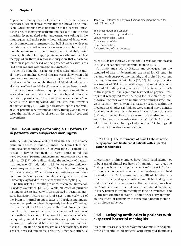
Appropriate management of patients with acute sinusitistherefore relies on clinical criteria that are known to be unre-liable. Most experts advise presuming that a bacterial infec-tion is present in patients with multiple “classic” signs of acutesinusitis: fever, marked pain, tenderness, or swelling in thesinus region, and molar pain without evidence of dental etiol-ogy. It is important to remember that half of patients with evenbacterial sinusitis will recover spontaneously within a week,though antimicrobial therapy may result in slightly fasterrecovery. It is therefore appropriate to prescribe antimicrobialtherapy when there is reasonable suspicion that a bacterialinfection is present based on the presence of “classic” signs[16] or in patients with protracted symptoms.
Patients lacking the “classic” signs of bacterial sinusitis usu-ally have uncomplicated viral sinusitis, particularly when coldsymptoms are present or patients complain of facial fullness,nasal obstruction, or cough. These individuals should gener-ally not be offered antibiotics. However, when patients thoughtto have viral sinusitis show no symptom improvement after aweek, it is reasonable to assume that they have developed abacterial superinfection. This scenario occurs in about 2% ofpatients with uncomplicated viral sinusitis, and warrantsantibiotic therapy [14]. Multiple treatment options are avail-able for patients who warrant antibiotic therapy, and in mostcases the antibiotic can be chosen on the basis of cost andconvenience.
Pitfall | Routinely performing a CT before LPin patients with suspected meningitis
With the widespread availability of CT in the US it has becomecommon practice to routinely image the brain before per-forming a lumbar puncture (LP) in evaluating ED patients sus-pected of having meningitis. A recent series found thatthree-fourths of patients with meningitis underwent a CT scanprior to LP [17]. More disturbingly, the majority of patientswho undergo CT study prior to LP do not receive antibioticsbefore imaging is performed [18]. Delays related to obtainingCT imaging prior to LP performance and antibiotic administra-tion result in 5-fold greater mortality among patients who areultimately diagnosed with acute bacterial meningitis [19].
The true risk of LP resulting in uncal or cerebral herniationis widely overstated [20–22]. While all cases of purulentmeningitis are associated with an increased intracranial pres-sure, herniation occurs in �1% of cases. Conversely, CT ofthe brain is normal in most cases of purulent meningitis,even among patients who subsequently herniate. CT findingsthat contraindicate LP are lateral shift of midline structures,loss of suprachiasmatic and basilar cisterns, obliteration ofthe fourth ventricle, or obliteration of the superior cerebellarand quadrigeminal plate cisterns with sparing of the ambientcisterns [23]. Abnormal findings that are not contraindica-tions to LP include a new mass, stroke, or hemorrhage, absentsigns of increased intracranial pressure. Using these criteria, a
recent study prospectively found that LP was contraindicatedin �10% of patients with bacterial meningitis [24].
An important study by Hasbun and colleagues sets thestandard of care in determining the need for CT study inpatients with suspected meningtitis, and is cited by currentmeningitis treatment guidelines [25, 26]. In this prospectiveassessment of 301 adults with suspected meningitis, only4% had CT findings that posed a risk of herniation, and eachof these patients had significant historical or physical find-ings that reliably indicated the need for CT (Table 8.2). Thehistorical findings were an immunocompromised state, pre-vious central nervous system disease, or seizure within theprevious week; physical findings were cranial nerve deficits,focal motor deficits, or a depressed level of consciousness(defined as the inability to answer two consecutive questionsand follow two consecutive commands). While 3 patientswith none of these findings had abnormal CT findings, allunderwent LP without complication.
Interestingly, multiple studies have found papilledema notto be a useful clinical predictor of herniation [22, 27]. Thefinding may be absent in patients at significant risk for her-niation, and conversely may be noted in those at minimalherniation risk. Papilledema may be difficult for the non-expert to detect, and appears to be an unreliable finding evenunder the best of circumstances. The takeaway points hereare 2-fold: (1) brain CT should not be considered mandatoryin every patient in whom meningitis is being evaluated, and(2) the performance of brain CT should never delay appropri-ate treatment of patients with suspected bacterial meningi-tis, as discussed below.
Pitfall | Delaying antibiotics in patients withsuspected bacterial meningitis
Infectious disease guidelines recommend administering appro-priate antibiotics to all patients with suspected meningitis
KEY FACT | The performance of brain CT should neverdelay appropriate treatment of patients with suspectedbacterial meningitis.
66 | Chapter 8
Table 8.2 Historical and physical findings predicting the need forbrain CT before LP.
Immunocompromised conditionPrior central nervous system diseaseSeizure within prior 1 weekCranial nerve deficitsFocal motor deficitsDepressed level of consciousness
within 30 min of presentation [25]. Despite these universalrecommendations, recent studies continue to report significantdelays in the administration of antibiotics [18, 25].Administration of antimicrobials in an inpatient unit, ratherthan in the ED, is associated with a 5-h delay in medicationdelivery and a 3-fold increase in mortality [28]. A recentstudy reported that a delay �6 h between ED presentationand administration of antibiotics independently increasedmortality in patients with bacterial meningitis by more than8-fold [19]. Furthermore, timing of antibiotic administrationwas incrementally and closely related to mortality. It is impor-tant to note that many studies relating antibiotic administra-tion to outcomes are confounded by factors that may havemade early diagnosis of meningitis difficult.
A common argument against immediate administration ofantibiotics is that the practice may impair the diagnosis ofbacterial meningitis on subsequent CSF testing. Antibioticadministration does not appear to cause changes in CSFwhite blood cell, protein, or glucose levels that would impedetheir diagnostic utility, and does not change CSF Gram stainfindings [29, 30]. Almost all patients with acute bacterialmeningitis still have diagnostic CSF abnormalities even if LPis delayed by 3 days. CSF cultures may be positive even afterone dose of antibiotic administration, although the yielddecreases with more fastiditious organisms such as meningic-occus [31]. Importantly, other modalities can reliably identifythe causative organism. Blood cultures obtained prior to anti-biotic administration reveal the causative organism in 86% ofpatients with bacterial meningitis. Given the enormous mor-tality benefit associated with early administration of antibi-otics, it is imperative to administer appropriate antimicrobialtherapy as soon as bacterial meningitis is suspected.
Pitfall | Failure to appreciate the range ofCSF findings associated with bacterialmeningitis
The CSF white blood cell (WBC) count, protein, and glucosevalues are often unreliable tools for differentiating betweenviral and bacterial meningitis. Commonly accepted indicatorsof bacterial meningitis are total WBC count �5 cells/mm3,polymorphonuclear WBC �75% of total WBC, CSF to serumglucose ratio �50%, and CSF protein �45 mg/dl [32]. WBCdifferentials may be misleading early in the course of menin-gitis, as more than 10% of patients with bacterial infection
will have an initial lymphocytic predominance, and viralmeningitis can be initially be dominated by neutrophils [30, 33, 34]. While each of these parameters is relativelyinsensitive, most authorities conclude that if any of theseindices are abnormal, the incidence of bacterial meningitisis high enough to warrant presumptive diagnosis and aggres-sive treatment until culture results are available [18, 25,30, 31, 35].
Pitfall | Over-reliance on physical findings indiagnosing bacterial meningitis
Failure to diagnose bacterial meningitis is in the top fivemalpractice claims in emergency medicine [36]. Virtually allpatients with bacterial meningitis will have a fever, neckstiffness, headache, or altered mental status, and thereforethe absence of all these findings can essentially exclude thediagnosis provided that the patient is capable of providing arelevant history [37]. Physical exam findings, however, arefar less reliable. The largest prospective study to date notedthe classic findings of fever, nuchal rigidity, and alteredmental status in less than half of patients with bacterialmeningitis, and a recent smaller study noted these classic find-ings in only 20% of cases [18].
Negative tests of nuchal rigidity, including the Kernig and Brudzinski signs, are commonly documented to indicateabsence of meningitis. The Brudzinski sign refers to thepatient’s spontaneous flexion of the hips during attempts topassively flex the neck. The Kernig sign refers to the patient’sreluctance to allow full extension of the knee when the hip isflexed 90°. Nuchal rigidity is more loosely defined, but is gen-erally described as inability or discomfort during neck flexion.Each of these signs were developed and tested in patients withsevere, late stage meningitis. While Kernig and Brudzinskisigns are each about 95% specific for meningitis, their sensitiv-ity is �10% [38]. Nuchal rigidity is slightly more sensitive, butless specific. Documenting the absence of meningeal signs,therefore, is not sufficient to exclude meningitis. Particularly inimmunocompromised hosts, the presentation of meningitis islikely to be subacute, and negative exam findings are evenmore likely.
Jolt accentuation of headache may be a more sensitivemaneuver for the diagnosis of meningitis. This test is per-formed by horizontally rotating the head two to three timesper second; increased headache severity is considered a pos-itive test. In a single small study, the maneuver displayed97% sensitivity but only 60% specificity for the presence ofmeningitis [39].
KEY FACT | The classic findings of fever, nuchal rigidity,and altered mental status [were found] in less than half ofpatients with bacterial meningitis.
KEY FACT | Antibiotic administration does not appearto cause changes in cerebrospinal fluid (CSF) white bloodcell, protein, or glucose levels … and does not change CSFGram stain findings.
Management of Infectious Diseases | 67
Pitfall | Failing to recognize methicillin-resistant Staphylococcus aureus as acommon pathogen
While MRSA emerged in the 1960s, until recently it hasbeen associated only with patients exposed to antibiotics forextended periods of time or quartered in places harboringresistant pathogens. Traditional risk factors for MRSAincluded recent surgery or hospitalization, residence in along-term care facility, prolonged antibiotic administration,hemodialysis, presence of indwelling devices or catheters,some chronic illnesses, and intravenous drug use. In 1998, apediatric inpatient study made that made it clear that MRSAwas relevant in patients with community-acquired infec-tions [40]. While 87% of children hospitalized with MRSAinfections between 1988 and 1990 had traditional risk factorsfor resistant infections, only 29% of patients with MRSAinfections between 1993 and 1995 had such risk factors. Theprevalence of MRSA infections in hospitalized children rose25-fold in the 5 years between the study periods.
Subsequent reports have confirmed the importance ofMRSA as a significant pathogen in a number of settings,including daycare, prisons, and among athletes [41–43]. A recent study of 1600 individuals with community-acquiredMRSA infections found that 87% had skin or wound infec-tions, but a small number had infections atypical for MRSA,including urinary tract infections, meningitis, osteomyelitis,sinusitis, and bacteremia [44]. Case reports have implicatedMRSA in fatal cases of pneumonia in immunocompetentadults and children [45, 46] and in necrotizing fasciitis, a dis-ease rarely associated with staphylococcal infections [47].
Community-acquired MRSA is now a common and seriousproblem among ED patients. In a recent multicenter studyof 422 ED patients with skin infections, 59% were found tohave MRSA [48]. All isolates were sensitive to trimethoprim-sulfamethoxizole, but only 90–95% were sensitive to clindamycin or tetracycline. A larger study of inpatients andoutpatients found that infections were sensitive to van-comycin and 97% to trimethoprim-sulfamethoxizole, but onlyabout 85% were sensitive to either clindamycin or tetracy-cline [44].
Despite the prevalence of MRSA skin lesions, antimicrobialtherapy is not necessary for all ED patients with abscesses.Uncomplicated abscesses generally only require incision anddrainage; antibiotics have not been shown to improve out-comes in these patients. Antibiotics are indicated for patientswith abscesses complicated by cellulitis or fever, however,
and the selected agent should be active against MRSA.Trimethoprim-sulfamethoxizole is an effective and inexpen-sive option. Patients allergic to sulfa may be offered clin-damycin or tetracycline, but it should be recognized that asignificant number of organisms have developed resistanceto these antibiotics. Linezolid is another option, but a courseof therapy costs over $1000. Some authorities advise addingrifampin to a course of trimethoprim-sulfamethoxizole toimprove efficacy [49]. Furthermore, it has been noted thatunlike abscesses, cellulitis is more commonly due to Strepto-coccus pyogenes than to Staphylococcus aureus, and trimethoprim-sulfamethoxizole may be less effective for these infections.Some experts therefore advise that patients who have cellulitis without abscess be treated with a combinationof a first-generation cephalosporin and trimethoprim-sulfamethoxizole, or with clindamycin alone [49].
Pitfall | Failure to aggressively treat sepsis
Emergency physicians frequently encounter sepsis, inwidely varying degrees of severity. Sepsis is broadly definedas the presence of at least two components of the systemicinflammatory response syndrome (SIRS) and a suspectedsource of infection (see Table 8.3). When associated withorgan dysfunction it is termed “severe sepsis,” while “septicshock” is associated with hypotension unresponsive to intra-venous fluid resuscitation without another cause forhypotension [50]. A recent analysis determined that thereare 751,000 cases of severe sepsis annually in the US, carry-ing an estimated mortality rate of 27% – killing as manypeople annually as acute myocardial infarction [51]. Althoughseptic shock is not usually a diagnostic dilemma, subtlemanifestations of organ dysfunction or tissue hypoxia can bedifficult to appreciate without frank hypotension.
In a 2001 study, Rivers et al. showed that early, goal-directed therapy (EGDT) of patients with severe sepsis andseptic shock significantly decreased in-hospital mortalityfrom 47% to 31% [52]. This approach to resuscitation tar-gets the normalization of central venous oxygen saturationinstead of the traditional target of blood pressure normaliza-tion. The goals of EGDT during the initial resuscitationperiod (first 6 h) include maintenance of the central venous
KEY FACT | Early, goal-directed therapy of patients …significantly decreased in-hospital mortality from 47% to 31%.
KEY FACT | Severe sepsis (carries) an estimatedmortality rate of 27% – killing as many people annually asacute myocardial infarction.
KEY FACT | Community-acquired MRSA is now acommon and serious problem among ED patients … in arecent study of ED patients with skin infections, 59%were found to have MRSA.
68 | Chapter 8
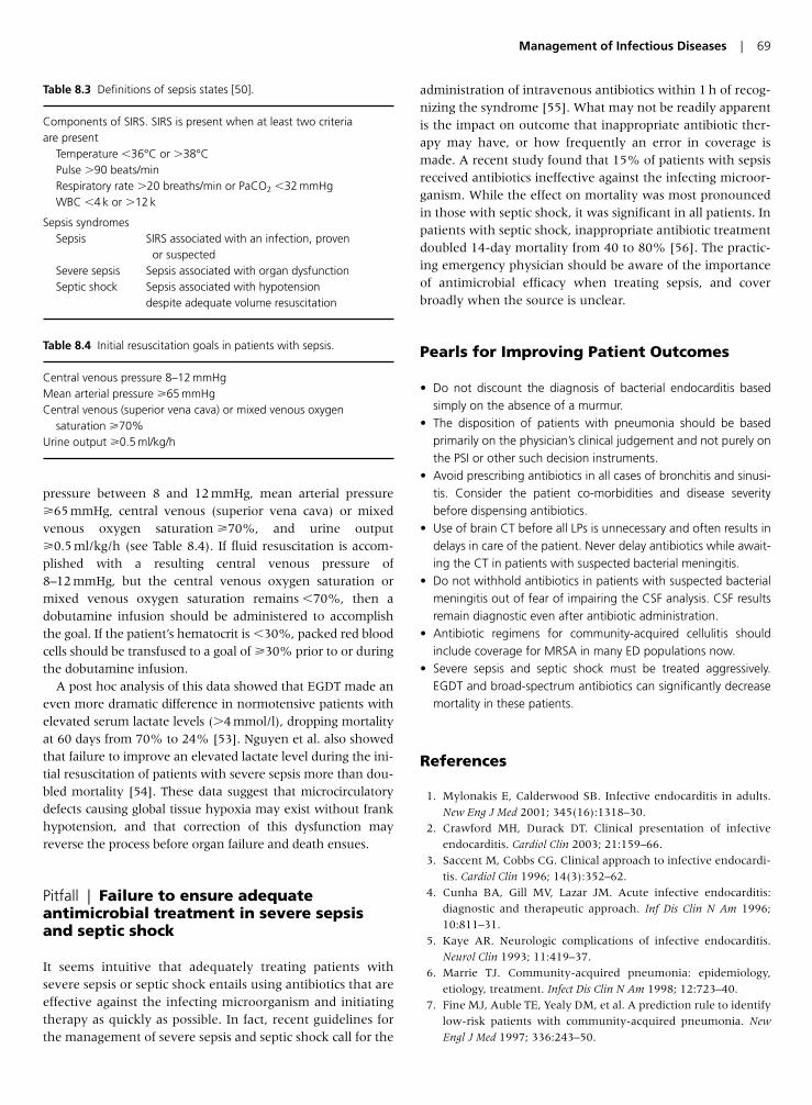
pressure between 8 and 12 mmHg, mean arterial pressure�65 mmHg, central venous (superior vena cava) or mixedvenous oxygen saturation �70%, and urine output�0.5 ml/kg/h (see Table 8.4). If fluid resuscitation is accom-plished with a resulting central venous pressure of8–12 mmHg, but the central venous oxygen saturation ormixed venous oxygen saturation remains �70%, then adobutamine infusion should be administered to accomplishthe goal. If the patient’s hematocrit is �30%, packed red bloodcells should be transfused to a goal of �30% prior to or duringthe dobutamine infusion.
A post hoc analysis of this data showed that EGDT made aneven more dramatic difference in normotensive patients withelevated serum lactate levels (�4 mmol/l), dropping mortalityat 60 days from 70% to 24% [53]. Nguyen et al. also showedthat failure to improve an elevated lactate level during the ini-tial resuscitation of patients with severe sepsis more than dou-bled mortality [54]. These data suggest that microcirculatorydefects causing global tissue hypoxia may exist without frankhypotension, and that correction of this dysfunction mayreverse the process before organ failure and death ensues.
Pitfall | Failure to ensure adequateantimicrobial treatment in severe sepsisand septic shock
It seems intuitive that adequately treating patients withsevere sepsis or septic shock entails using antibiotics that areeffective against the infecting microorganism and initiatingtherapy as quickly as possible. In fact, recent guidelines forthe management of severe sepsis and septic shock call for the
administration of intravenous antibiotics within 1 h of recog-nizing the syndrome [55]. What may not be readily apparentis the impact on outcome that inappropriate antibiotic ther-apy may have, or how frequently an error in coverage ismade. A recent study found that 15% of patients with sepsisreceived antibiotics ineffective against the infecting microor-ganism. While the effect on mortality was most pronouncedin those with septic shock, it was significant in all patients. Inpatients with septic shock, inappropriate antibiotic treatmentdoubled 14-day mortality from 40 to 80% [56]. The practic-ing emergency physician should be aware of the importanceof antimicrobial efficacy when treating sepsis, and coverbroadly when the source is unclear.
Pearls for Improving Patient Outcomes
• Do not discount the diagnosis of bacterial endocarditis basedsimply on the absence of a murmur.
• The disposition of patients with pneumonia should be basedprimarily on the physician’s clinical judgement and not purely onthe PSI or other such decision instruments.
• Avoid prescribing antibiotics in all cases of bronchitis and sinusi-tis. Consider the patient co-morbidities and disease severitybefore dispensing antibiotics.
• Use of brain CT before all LPs is unnecessary and often results indelays in care of the patient. Never delay antibiotics while await-ing the CT in patients with suspected bacterial meningitis.
• Do not withhold antibiotics in patients with suspected bacterialmeningitis out of fear of impairing the CSF analysis. CSF resultsremain diagnostic even after antibiotic administration.
• Antibiotic regimens for community-acquired cellulitis shouldinclude coverage for MRSA in many ED populations now.
• Severe sepsis and septic shock must be treated aggressively.EGDT and broad-spectrum antibiotics can significantly decreasemortality in these patients.
References
1. Mylonakis E, Calderwood SB. Infective endocarditis in adults.
New Eng J Med 2001; 345(16):1318–30.
2. Crawford MH, Durack DT. Clinical presentation of infective
endocarditis. Cardiol Clin 2003; 21:159–66.
3. Saccent M, Cobbs CG. Clinical approach to infective endocardi-
tis. Cardiol Clin 1996; 14(3):352–62.
4. Cunha BA, Gill MV, Lazar JM. Acute infective endocarditis:
diagnostic and therapeutic approach. Inf Dis Clin N Am 1996;
10:811–31.
5. Kaye AR. Neurologic complications of infective endocarditis.
Neurol Clin 1993; 11:419–37.
6. Marrie TJ. Community-acquired pneumonia: epidemiology,
etiology, treatment. Infect Dis Clin N Am 1998; 12:723–40.
7. Fine MJ, Auble TE, Yealy DM, et al. A prediction rule to identify
low-risk patients with community-acquired pneumonia. New
Engl J Med 1997; 336:243–50.
Management of Infectious Diseases | 69
Table 8.3 Definitions of sepsis states [50].
Components of SIRS. SIRS is present when at least two criteria are present
Temperature �36°C or �38°CPulse �90 beats/minRespiratory rate �20 breaths/min or PaCO2 �32 mmHgWBC �4 k or �12 k
Sepsis syndromesSepsis SIRS associated with an infection, proven
or suspectedSevere sepsis Sepsis associated with organ dysfunctionSeptic shock Sepsis associated with hypotension
despite adequate volume resuscitation
Table 8.4 Initial resuscitation goals in patients with sepsis.
Central venous pressure 8–12 mmHgMean arterial pressure �65 mmHgCentral venous (superior vena cava) or mixed venous oxygen
saturation �70%Urine output �0.5 ml/kg/h
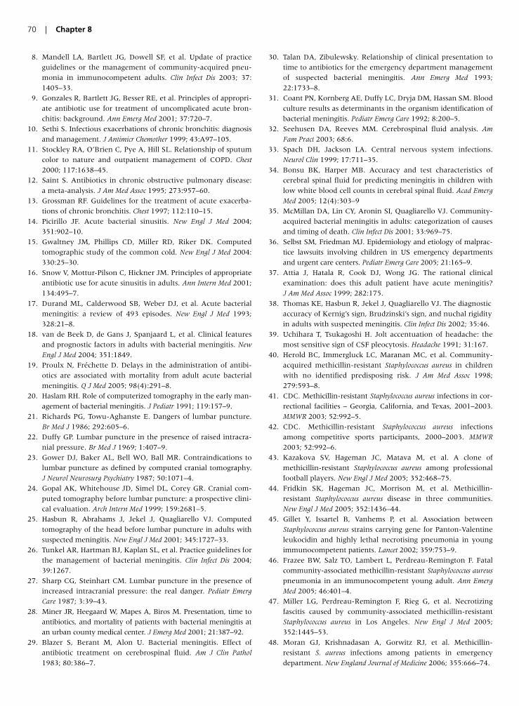
8. Mandell LA, Bartlett JG, Dowell SF, et al. Update of practice
guidelines or the management of community-acquired pneu-
monia in immunocompetent adults. Clin Infect Dis 2003; 37:
1405–33.
9. Gonzales R, Bartlett JG, Besser RE, et al. Principles of appropri-
ate antibiotic use for treatment of uncomplicated acute bron-
chitis: background. Ann Emerg Med 2001; 37:720–7.
10. Sethi S. Infectious exacerbations of chronic bronchitis: diagnosis
and management. J Antimicr Chemother 1999; 43:A97–105.
11. Stockley RA, O’Brien C, Pye A, Hill SL. Relationship of sputum
color to nature and outpatient management of COPD. Chest
2000; 117:1638–45.
12. Saint S. Antibiotics in chronic obstructive pulmonary disease:
a meta-analysis. J Am Med Assoc 1995; 273:957–60.
13. Grossman RF. Guidelines for the treatment of acute exacerba-
tions of chronic bronchitis. Chest 1997; 112:110–15.
14. Picirillo JF. Acute bacterial sinusitis. New Engl J Med 2004;
351:902–10.
15. Gwaltney JM, Phillips CD, Miller RD, Riker DK. Computed
tomographic study of the common cold. New Engl J Med 2004:
330:25–30.
16. Snow V, Mottur-Pilson C, Hickner JM. Principles of appropriate
antibiotic use for acute sinusitis in adults. Ann Intern Med 2001;
134:495–7.
17. Durand ML, Calderwood SB, Weber DJ, et al. Acute bacterial
meningitis: a review of 493 episodes. New Engl J Med 1993;
328:21–8.
18. van de Beek D, de Gans J, Spanjaard L, et al. Clinical features
and prognostic factors in adults with bacterial meningitis. New
Engl J Med 2004; 351:1849.
19. Proulx N, Fréchette D. Delays in the administration of antibi-
otics are associated with mortality from adult acute bacterial
meningitis. Q J Med 2005; 98(4):291–8.
20. Haslam RH. Role of computerized tomography in the early man-
agement of bacterial meningitis. J Pediatr 1991; 119:157–9.
21. Richards PG, Towu-Aghanste E. Dangers of lumbar puncture.
Br Med J 1986; 292:605–6.
22. Duffy GP. Lumbar puncture in the presence of raised intracra-
nial pressure. Br Med J 1969; 1:407–9.
23. Gower DJ, Baker AL, Bell WO, Ball MR. Contraindications to
lumbar puncture as defined by computed cranial tomography.
J Neurol Neurosurg Psychiatry 1987; 50:1071–4.
24. Gopal AK, Whitehouse JD, Simel DL, Corey GR. Cranial com-
puted tomography before lumbar puncture: a prospective clini-
cal evaluation. Arch Intern Med 1999; 159:2681–5.
25. Hasbun R, Abrahams J, Jekel J, Quagliarello VJ. Computed
tomography of the head before lumbar puncture in adults with
suspected meningitis. New Engl J Med 2001; 345:1727–33.
26. Tunkel AR, Hartman BJ, Kaplan SL, et al. Practice guidelines for
the management of bacterial meningitis. Clin Infect Dis 2004;
39:1267.
27. Sharp CG, Steinhart CM. Lumbar puncture in the presence of
increased intracranial pressure: the real danger. Pediatr Emerg
Care 1987; 3:39–43.
28. Miner JR, Heegaard W, Mapes A, Biros M. Presentation, time to
antibiotics, and mortality of patients with bacterial meningitis at
an urban county medical center. J Emerg Med 2001; 21:387–92.
29. Blazer S, Berant M, Alon U. Bacterial meningitis. Effect of
antibiotic treatment on cerebrospinal fluid. Am J Clin Pathol
1983; 80:386–7.
30. Talan DA, Zibulewsky. Relationship of clinical presentation to
time to antibiotics for the emergency department management
of suspected bacterial meningitis. Ann Emerg Med 1993;
22:1733–8.
31. Coant PN, Kornberg AE, Duffy LC, Dryja DM, Hassan SM. Blood
culture results as determinants in the organism identification of
bacterial meningitis. Pediatr Emerg Care 1992; 8:200–5.
32. Seehusen DA, Reeves MM. Cerebrospinal fluid analysis. Am
Fam Pract 2003; 68:6.
33. Spach DH, Jackson LA. Central nervous system infections.
Neurol Clin 1999; 17:711–35.
34. Bonsu BK, Harper MB. Accuracy and test characteristics of
cerebral spinal fluid for predicting meningitis in children with
low white blood cell counts in cerebral spinal fluid. Acad Emerg
Med 2005; 12(4):303–9
35. McMillan DA, Lin CY, Aronin SI, Quagliarello VJ. Community-
acquired bacterial meningitis in adults: categorization of causes
and timing of death. Clin Infect Dis 2001; 33:969–75.
36. Selbst SM, Friedman MJ. Epidemiology and etiology of malprac-
tice lawsuits involving children in US emergency departments
and urgent care centers. Pediatr Emerg Care 2005; 21:165–9.
37. Attia J, Hatala R, Cook DJ, Wong JG. The rational clinical
examination: does this adult patient have acute meningitis?
J Am Med Assoc 1999; 282:175.
38. Thomas KE, Hasbun R, Jekel J, Quagliarello VJ. The diagnostic
accuracy of Kernig’s sign, Brudzinski’s sign, and nuchal rigidity
in adults with suspected meningitis. Clin Infect Dis 2002; 35:46.
39. Uchihara T, Tsukagoshi H. Jolt accentuation of headache: the
most sensitive sign of CSF pleocytosis. Headache 1991; 31:167.
40. Herold BC, Immergluck LC, Maranan MC, et al. Community-
acquired methicillin-resistant Staphylococcus aureus in children
with no identified predisposing risk. J Am Med Assoc 1998;
279:593–8.
41. CDC. Methicillin-resistant Staphylococcus aureus infections in cor-
rectional facilities – Georgia, California, and Texas, 2001–2003.
MMWR 2003; 52:992–5.
42. CDC. Methicillin-resistant Staphylococcus aureus infections
among competitive sports participants, 2000–2003. MMWR
2003; 52:992–6.
43. Kazakova SV, Hageman JC, Matava M, et al. A clone of
methicillin-resistant Staphylococcus aureus among professional
football players. New Engl J Med 2005; 352:468–75.
44. Fridkin SK, Hageman JC, Morrison M, et al. Methicillin-
resistant Staphylococcus aureus disease in three communities.
New Engl J Med 2005; 352:1436–44.
45. Gillet Y, Issartel B, Vanhems P, et al. Association between
Staphylococcus aureus strains carrying gene for Panton-Valentine
leukocidin and highly lethal necrotising pneumonia in young
immunocompetent patients. Lancet 2002; 359:753–9.
46. Frazee BW, Salz TO, Lambert L, Perdreau-Remington F. Fatal
community-associated methicillin-resistant Staphylococcus aureus
pneumonia in an immunocompetent young adult. Ann Emerg
Med 2005; 46:401–4.
47. Miller LG, Perdreau-Remington F, Rieg G, et al. Necrotizing
fascitis caused by community-associated methicillin-resistant
Staphylococcus aureus in Los Angeles. New Engl J Med 2005;
352:1445–53.
48. Moran GJ, Krishnadasan A, Gorwitz RJ, et al. Methicillin-
resistant S. aureus infections among patients in emergency
department. New England Journal of Medicine 2006; 355:666–74.
70 | Chapter 8

49. Moran GJ, Talan DA. Community-associated methicillin-
resistant Staphylococcus aureus: is it in your community and should
it change practice? Ann Emerg Med 2005; 45:321–2.
50. Levy MM, Fink MP, Marschall JC, et al. 2001 SCCM/ ESICM/
ACCP/ATS/SIS International sepsis definitions conference. Crit
Care Med 2003; 31:1250–6.
51. Angus DC, Linde-Zwirble WT, Lidicker J, et al. Epidemiology of
severe sepsis in the United States: analysis of incidence, out-
come, and associated costs of care. Crit Care Med 2001;
29:1303–10.
52. Rivers E, Nguyen B, Havstad S, et al. Early goal-directed ther-
apy in the treatment of severe sepsis and septic shock. New Engl
J Med 2001; 345:1368–77.
53. Donnino MW, Nguyen B, Jacobsen G, et al. Cryptic septic
shock: a sub-analysis of early, goal-directed therapy. Chest 2003;
124(4):90S.
54. Nguyen HB, Rivers EP, Knoblich BP, et al. Early lactate clear-
ance is associated with improved outcome in severe sepsis and
septic shock. Crit Care Med 2004; 32:1637–42.
55. Dellinger RP, Carlet JM, Masur H, et al. Surviving sepsis cam-
paign guidelines for the management of severe sepsis and septic
shock. Crit Care Med 2004; 32:858–73.
56. Valles J, Rello J, Ochangavia A, et al. Community-acquired
bloodstream infection in critically Ill adult patients – impact of
shock and inappropriate antibiotic activity on survival. Chest
2003; 123:1615–24.
Management of Infectious Diseases | 71
Introduction
Emergency physicians are the primary providers of post-traumatic wound management. Over 11 million traumaticwounds are managed in emergency departments (ED) in theUS annually [1]. Of these, over 50% occur as a result of blunttrauma, while the remainder involve the penetration of sharpobjects, such as metal, glass, or wood. The majority of thesewounds occur on patients’ heads and upper extremities, bothhighly visible areas [2]. As a result, the cosmetic appearance ofhealed wounds is a priority for both patients and emergencyphysicians. According to a survey of ED patients, the top fourconcerns of patients with a laceration include the cosmeticappearance, preservation of function, decreasing the pain ofthe repair, and avoiding a wound infection [3]. As these keyoutcomes are closely related to the specific wound care tech-niques used during the initial ED encounter, it is of utmostimportance that emergency physicians stay abreast of thebiggest pitfalls in wound care in order to avoid negative out-comes. A national survey of wound care techniques revealedthat many practitioners manage wounds in ways that are contrary to the recommendations of published literature [4].Furthermore, between 5% and 20% of all emergency physi-cian malpractice claims involve issues of wound care, with upto 11% of all malpractice dollars being awarded for these cases[2]. These data further emphasize the importance of recogniz-ing and avoiding many of the common pitfalls of wound carereviewed in the subsequent sections.
Pitfall | Failure to properly prepare a woundprior to closure
Beyond anesthesia, inspection, and evaluation for foreignbody, proper preparation of a wound improves its outcome.Despite available evidence-based recommendations, manypractitioners continue to employ techniques and utilize prod-ucts that are unnecessary, and in some cases detrimental. In asurvey distributed to predominantly board certified emergencyphysicians, investigators found that despite the existence ofwound management guidelines, 90% of physicians treatedwounds based on personal preference. Thirty-eight percentsoaked wounds, 67% scrubbed wounds, 27% irrigatedwounds with inadequate technique, and 21% used concen-trated povidone–iodine solution or hydrogen peroxide [4].
Wound irrigation with high pressure and adequate volume hasbeen shown to have a dramatic impact on wound infectionrates. Simply soaking a wound provides no advantage andmay be detrimental to its ultimate outcome [2]. The necessarypressure for wound irrigation can be accomplished by pushingfluid in a 30–60 ml syringe through a 19-gauge catheter. This is a highly effective means of reducing potentially infectivematerial from a wound [5, 6]. The pressure generated throughthis technique cannot be reproduced by puncturing containersof irrigation fluid and squeezing with maximal hand pressure[1]. The data regarding the volume of irrigation fluid necessaryis limited. Most authorities recommend 50–100 ml of irrigationfluid per cm of laceration. However, these sources agree that the volume of irrigation should be adjusted to the woundcharacteristics and degree of contamination [2, 7]. For instance,a contaminated scalp wound may require less irrigation solu-tion than an extremity wound.
Irrigation of highly vascularized structures such as theface and scalp may not be as critical. Hollander et al. demon-strated that not irrigating clean-appearing, simple facial andscalp lacerations did not alter the rates of infection [8]. Inaddition, at very high irrigation pressures, research suggeststhat infection rates may actually increase due to tissue dam-age. Therefore, care must be taken not to use high-pressureirrigation in vascular wounds with loose alveolar tissue [7].
The choice of irrigation solution can have significant effectson wound healing. Antiseptic solutions such as chlorhexi-dine, povidone–iodine 10% solution, hydrogen peroxide,and detergents are toxic to tissues and impede wound heal-ing [9, 10]. Studies comparing diluted 1% povidone–iodineto normal saline have not shown any difference in infectionrates [11]. Furthermore, given the prevalence of iodine sen-sitivity, the use of diluted iodine irrigation is discouraged.More recently, published studies have concluded that there isno clinically significant difference in infection rates betweensimple wounds irrigated with potable tap water and sterilenormal saline [12, 13].
Another misguided practice which has been shown toincrease infection rates is shaving the area surrounding a
KEY FACT | The necessary pressure for woundirrigation can be accomplished by pushing fluid in a30–60 ml syringe through a 19-gauge catheter.
Chapter 9 | Wound Care in Emergency Medicine
Siamak Moayedi & Mercedes Torres
72
wound prior to repair [14, 15]. If hair removal is requiredfor adequate access to or visualization of a wound, it is rec-ommended that the hair is clipped, rather than shaved, atthe skin level [16]. One effective alternative is to use petro-leum products to comb hair away from the wound. Caremust be taken to remove small pieces of hair from thewound prior to closure.
Finally, many practitioners routinely use sterile gloves forthe repair of simple lacerations. Studies supporting this prac-tice are lacking. In a prospective study of 816 patients withsimple wounds randomized to wound repaired using sterileversus clean gloves, there was no difference in subsequentinfection rates [17].
Pitfall | Failure to detect the presence of aforeign body in a wound
When unrecognized, retained foreign bodies can cause sig-nificant morbidity, including local inflammation, infection,and compression of vital structures [18]. Emergency physi-cians have a variety of tools at their disposal to assist withthe detection of foreign bodies. The key to addressing a foreignbody is finding it.
Detection of foreign bodies by clinical examination alonehas proven unreliable. If the bottom of a wound is visible andno foreign body is seen, 7% of subsequent X-rays have beenshown to be positive for the presence of a foreign body (seeFigure 9.1). Furthermore, when the bottom of a wound is notvisualized, clinically undetected foreign bodies have beenfound on X-rays in 21% of cases [1]. There are clinical cluesthat can raise suspicion for the presence of a foreign body:
These include persistent pain, drainage, a palpable mass, sur-face discoloration, or a blood-stained tract. Obtaining a his-tory of glass breaking while in contact with the skin, blunttrauma with tooth fragments, clenched-fist injuries, puncture
wounds, motor vehicle accidents, and foreign body sensationshould raise suspicion for retained foreign bodies in a wound[18]. According to a study of wounds caused by glass, 15%had retained glass within the wound. Patients reporting for-eign body sensation had a positive predictive value of 31%and a negative predictive value of 89%. However, using patientperception alone to determine X-ray eligibility would havemissed 57% of wounds with retained glass [19].
One of the primary diagnostic modalities used for detec-tion of foreign bodies in wounds is X-ray. Legal precedentshave shown that practitioners who failed to X-ray woundscaused by glass were unsuccessful in defending themselvesin 60% of cases [1]. Radio opaque foreign bodies includeglass, metal, bone, teeth, pencil graphite, certain plastics,gravel, some aluminum, and some sand [2, 18]. X-rays candetect greater than 50% of glass with a single dimensionbetween 0.5 and 2.0 mm, and virtually all with a dimensiongreater than 2.0 mm [1, 7, 18]. Underpenetrated X-rays,highlighting the soft tissues are more useful for detectingretained foreign bodies not visualized with standard X-rays[18, 20]. In addition, it is possible to visualize a filling defecton a soft tissue X-ray created by a radiolucent foreign body[20]. One problem with X-rays is their two-dimensionalquality. If detected, it is often difficult to determine the exactlocation of the foreign body and its spatial relation to othervital structures. Obtaining multiple views and using markerssuch as paper clips or needles can assist with localization ofthe foreign body using X-ray alone [18].
When suspicion is high for a radiolucent foreign body, or aforeign body in close proximity to vital structures, a computedtomography (CT) scan is often the study of choice. CTs areuseful for detecting radiolucent objects such as wood, certainplastics, thorns, and spines. In addition, they can detect thepresence of vegetative material, which has a high rate of
KEY FACT | Missed retained foreign bodies are the fifthleading cause of malpractice claims against emergencyphysicians in the US [1].
Wound Care in Emergency Medicine | 73
Persistent painDrainage from woundPalpable mass at wound siteSurface discolorationBlood-stained wound tractHistory of contact with broken glassClenched-fist injuryPuncture woundsMotor vehicle accidentForeign body sensation by patient
Figure 9.1 Plain radiograph demonstrating metallic foreign body in thefoot.
infection and inflammatory complication. This benefit isoptimized within the first 48 hours because vegetative for-eign bodies absorb water within tissues, changing their den-sity to appear very similar to soft tissue, and therefore moredifficult to differentiate [1]. Regardless the age of a wound,CTs are still 100 times more sensitive for detecting variousdensities of objects as compared with X-ray. In addition,they provide a visualization of the location of foreign bodiesin relation to surrounding structures [18]. The ability tolocalize a foreign body assists the practitioner in decidingwhether it should be removed and if removal should be sur-gical. Magnetic resonance imaging (MRI) has also beenshown to be useful in identifying retained vegetative matter,but cannot be used if there is any suspicion for metal orgravel foreign bodies. The cost and “inconvenience” of MRIhas excluded this as a primary imaging modality for thedetection of retained wound foreign bodies [18].
Another diagnostic modality that has been shown to beefficient and less costly is bedside ultrasound. The value ofultrasound in detecting and localizing wound foreign bodiesis dependent upon the provider’s skill and the size of theobject. A 7.5 MHz linear probe is optimal for wound exami-nation, allowing approximately 3 cm of tissue penetration.The technique involves rotating the probe in a perpendicu-lar position to the skin in order to attempt to localize the for-eign body in cross section [21]. Increasing the frequency ofthe probe improves its ability to detect smaller foreign bod-ies [22]. One recent study examining the utility of bedsideultrasound in the detection of wood foreign bodies demon-strated a sensitivity of 95% and specificity of 89% [23]. Othershave cited ranges of sensitivity from 50% to 90%, and speci-ficity from 70% to 97%. Certain types of foreign bodies havecharacteristic appearances on ultrasound. Metal or glass isdescribed as having a comet-tail appearance, while wood,plastic, sand, and pebbles often demonstrate acoustic shad-owing similar to that seen with gallstones [21]. Unlike CTscans, ultrasound may be more helpful when patients pres-ent greater than 48 hours after suffering their wound, as theinflammation around a foreign body makes it easier to iden-tify its location [23]. However, the use of bedside ultrasoundis complicated by false positives. These include echo artifactsthat can be made by air pockets, calcifications, old scar tissue,blood, sutures, sesamoid bones, or purulence [18]. In addi-tion, the presence of multiple tissue planes can complicateultrasound guided localization [24]. Once a foreign body islocated, ultrasound can provide real-time visualization of itsremoval as well as identifying other smaller surroundingforeign bodies [25].
Pitfall | Failure to recognize the morbidityassociated with plantar puncture wounds
Plantar puncture wounds represent a unique challenge forphysicians. The soles of the feet are the most common site of
puncture wounds in the lower extremities. The force of apatient’s weight onto a foreign body and the relative small dis-tance from the skin to highly susceptible structures such asbones and joints make this injury especially prone to infectionand significant morbidity. Care of the patient presenting withan acute plantar puncture remains controversial. The twomost controversial areas involve the extent of wound explo-ration/debridement and the question of antibiotic prophylaxis.
Many authors have asserted that plantar punctures have ahigh predilection for retained foreign bodies. Schwab andPowers reported a 3% rate of retained foreign body after initial surface cleansing without wound exploration [26]. Alarger study of 887 patients also reported a 3% incidence offoreign body retention. Half of these were pieces of footwear, while the remainder included rust and dirt [27].However, because many patients with this injury do notpresent to the ED unless they experience continued pain orinfection, the true incidence of retained foreign body may belower [28].
There are no prospective, randomized trials that demon-strate the utility of prophylactic antibiotics in these injuries.One of the most quoted studies recommending antibioticprophylaxis was an uncontrolled, observational study, withoptional use of antibiotics [29]. Most authorities defer to theindividual clinician regarding the use of antibiotics with thecaveat that antibiotics do not compensate for inadequatewound care [18].
Uncomplicated puncture wounds without concern forretained foreign bodies may not require any wound explo-ration, but close follow up is advised [26]. Many aggressiveand time-consuming procedures, such as coring and woundextension, have been described for the evaluation of thesewounds. Coring refers to excision of a block of tissue downto the subcutaneous layer to allow visualization and accessto foreign bodies and contaminated tissue. There is no defin-itive study showing that routinely coring puncture woundsreduces the incidence of infection. Furthermore, extensivedebridement and coring may actually delay wound healingand cause undue pain [18]. Authors have argued that high-risk wounds which may require aggressive debridementinclude injuries through rubber-soled shoes, injuries overmetatarsal phalangeal joints, and injuries in patients withhigh-risk comorbidities, such as diabetes and neuropathy [2].
KEY FACT | The rate of osteomyelitis caused by plantarpuncture wounds is between 0.04% and 2%.
KEY FACT | High-risk wound which may requireaggressive debridement include injuries through rubber-soled shoes, injuries over metatarsal phalangeal joints,and injuries in patients with high-risk comorbidities.
74 | Chapter 9
A concerning clinical scenario involves a previous puncturewound presenting days later with evidence of infection.Delayed presentation is a significant marker for deep-seatedinfection [30]. These cases should prompt a thorough inves-tigation for a retained foreign body [18]. Referral to special-ists for removal of all but superficial foreign bodies andaggressive debridement is recommended. A high level ofconcern for osteomyelitis is raised when a wound relapses or fails to respond to the initial therapy. Overall, the rate ofosteomyelitis caused by plantar puncture wounds isbetween 0.04% and 2% [18]. Advanced imaging techniquessuch as MRI may be required for diagnosis.
Although gram-positive organisms remain the most com-mon cause of infection, several studies have demonstratedthe role of Pseudomonas aeruginosa when the puncture occursthrough rubber-soled foot wear [7]. It is presumed that themoist inner sole of the shoe provides a suitable environmentfor this bacterium [31]. When considering antibiotic pro-phylaxis, ciprofloxacin is still the antibiotic of choice. Thenewer quinolones, despite demonstrating better efficacyagainst gram-positive organisms, are less effective againstPseudomonas.
Pitfall | Indiscriminate use of prophylacticantibiotics for bite wounds
On a yearly basis, several million people are bitten by mam-mals in the USA. Almost one half of all children in the USAwill be bitten by a dog at some point during their childhood[32]. A small proportion of adults and children with bitewounds, somewhere between 1 and 2 million, seek treat-ment. Animal bites comprise 1% of all ED visits annually,with dog bites accounting for 80% of those visits [2, 33, 34].Although bite wounds are a commonly encountered com-plaint among emergency physicians, their managementremains controversial. They are thought to be at increasedrisk for infections and wound complications, leading to therecommended use of prophylactic antibiotics. However, stud-ies of infection rates after mammalian bites have failed todemonstrate any significant difference between those whoreceived prophylactic antibiotics and those who receivedplacebo except in cases of high-risk wounds [7, 32, 35, 36].
One of the primary concerns with any bite wound is possi-ble inoculation of the soft tissue with oral flora. The specificorganisms of concern vary based on the type of mammalwhich has bitten the patient. While over 80% of animal bitesreported are from dogs, only 4–25% of these become infected.
Cat bites, although less frequent, are more commonly compli-cated by infection rates that range between 30% and 80%,depending on the study referenced [34, 37]. Human bites are considered to have the highest risk of infection when compared with dogs and cats. Although most bite woundinfections contain mixed flora, pasturella is the primary cul-prit in both dog and cat bite wounds. Pasturella causes a rapidonset of signs and symptoms of infection, typically within 24hours of the initial injury [37]. Streptococci, Staphylococci,Moraxella, Corynebacterium, and Neisseria are the next mostcommon pathogens cultured from animal bite wounds.Infections attributed to Staphylococci and Streptococci typi-cally manifest as nonpurulent wounds with lymphangitis. Inaddition to the aforementioned flora, over 50% of bitewounds contain anaerobic organisms [37, 38]. Therefore,optimal prophylactic agents would include beta lactams witha beta lactamase inhibitor, second-generation cephalosporinswith anaerobic activity, or a combination of clindamycin anda fluoroquinolone. Traditional choices such as first-generationcephalosporins, ampicillin, or penicillin alone, have provenineffective and are not recommended [37].
Although there is evidence that bite wounds in generalhave higher infection rates as compared with other types ofwounds, the utility of prophylactic antibiotics for all bitewounds has been questioned. According to a 2005 CochraneDatabase review of eight randomized controlled trials, pro-phylactic antibiotics were associated with a statistically signif-icant reduction in the rate of infections after human bites, butnot after cat or dog bites [32]. However, other studies suggestthat even human bite wounds that involve minor injurieshave not been shown to benefit from prophylactic antibiotics[36]. Thus, physicians must consider more than just thespecies of the biting mammal when evaluating the benefit ofprophylactic antibiotics. It is safer and more cost-effective toreserve prophylactic antibiotics for high-risk wounds andhigh-risk patients (Table 9.1).
Focusing on the characteristics of the wound, deep punc-ture wounds, wounds with extensive crush injury or devital-ized tissue, and wounds involving underlying muscle ortendons are at higher risk of infection. In addition, any woundinvolving an underlying bone fracture, retained foreign body,or joint should be considered an increased infection risk [2].
KEY FACT | Studies of infection rates after mammalianbites have failed to demonstrate any significantdifference between those who received prophylacticantibiotics and those who received placebo except incases of high-risk wounds.
Wound Care in Emergency Medicine | 75
Table 9.1 Wound and patient characteristics that increase the risk ofwound infection.
Wound characteristicsDeep puncturesWounds with extensive crush injury or devitalized tissueUnderlying muscle or tendon involvement
Patient characteristicsDiabetesImmunocompromised stateHistory of endocarditisPresence of orthopedic prostheses
Bites on the hand, including fight bites, maintain a particu-larly high risk of infection and current evidence supports theuse of prophylactic antibiotics in these cases [32].
Patient-specific factors which would support the use of pro-phylactic antibiotics include a history of diabetes mellitus,immunocompromise, endocarditis, and orthopedic prosthe-ses. The time between the bite and presentation to the ED isanother important factor in determining the value of antibi-otic prophylaxis. Studies demonstrate that pasturella infec-tions typically manifest clinical signs and symptoms within24 hours. Infections caused by other organisms typicallydevelop within 72 hours [7, 38]. Therefore, patients whopresent greater than 72 hours from the initial bite with nosigns of infection do not require antibiotic prophylaxis.
In summary, recent literature supports the use of prophy-lactic antibiotics in nontrivial human bites, mammalian bitewounds with high-risk characteristics, and/or mammalianbite wounds suffered by high-risk patients. There is no sub-stitute for the value of adequate wound irrigation, cleansing,and debridement in the prevention of bite wound infectionsfor all bites. If a bite wound meets the aforementioned criteria of high infection risk, it is important to consider thetime since injury and the offending mammal in order toselect the most appropriate antibiotic. These practices willhelp minimize mammalian bite wound infection rates, whilepreventing unnecessary side effects and risks of antibioticprophylaxis.
Pitfall | Failure to provide appropriatewound after care instructions
There are several factors that affect the outcome of a wound. These include the choice of dressing placed on the wound and patient education regarding proper care ofthe wound. Experience demonstrates that there can be awide variation in outcome despite proper wound prepara-tion and suturing [39]. In order to optimize the cosmeticoutcome of a wound, the physician must be mindful of theeffects of UV light exposure and the choice of dressingapplied to the wound.
The data to support avoidance of sun light exposure to awound comes from plastic surgery and dermatology literature.In a retrospective study of patients undergoing dermabrasion,
the authors observed that wounds exposed to sunlight within6 months from the time of the injury can develop permanenthyperpigmentation [40]. Subsequent to this study, multipleanimal experiments have demonstrated that UV light expo-sure on laser- and knife-induced wounds significantly altersnormal skin structures and induces hyperpigmentation [41,42]. Although these findings have not been investigated inhumans, the recommendation is to use sunscreen products ona new wound for 6 months after epithelialization, which istypically complete within 48 hours. An optimal opportunity todiscuss this with a patient is during a wound check visit. Thepatient should also be cautioned about the risk of hypersensiti-zation to sunscreen products. It may be advantageous to rec-ommend hypoallergenic products which are readily availableover the counter.
The choice of dressing applied to a wound also impacts its cosmetic outcome. Dressings that provide moist, warmenvironments improve the rate of epithelialization [43].Furthermore, they prevent contamination, sunlight expo-sure, and repeated trauma. Petroleum ointment and topicalantibiotics such as bacitracin or triple antibiotic preparationsprovide the requisite moist environment for epithelializa-tion. The value of antibiotic over petroleum ointment is con-troversial. In one prospective, randomized study topicalantibiotics yielded lower infection rates [44]. However, asimilar study published the following year found essentiallyno difference between antibiotic and petroleum ointments[45]. Some authors have suggested the use of antibioticointments, since there is no disadvantage linked to their use[1]. More recently, academic dermatology literature haswarned against the routine use of bacitracin, citing that it is the seventh most common cause of contact dermatitis inNorth America. In addition to the adverse effects of contactdermatitis on cosmetic outcome, they cite more than 26cases of anaphylaxis related to bacitracin use [46]. Of note,both topical antibiotics and petroleum products cause dis-solving of cyanoacrylates and are not to be used on woundsclosed with skin glue preparations.
Pearls for Improving Patient Outcomes
• When cleansing a wound, avoid antiseptic solutions or prepara-tions, as they are tissue toxic. High-pressure irrigation with potabletap water is a safe and effective means of cleaning a wound.
• Clip or comb hair away from a wound rather than shaving at the skin.
• Wounds with high suspicion for retained foreign bodies shouldbe further investigated with radiographs. If concern for radiolu-cent foreign bodies exists, CT or ultrasound is recommended.
• Bedside ultrasound is an efficient and cost-effective tool forlocalizing and assisting with the removal of foreign bodies.
• Maintain a high clinical suspicion for the presence of retainedforeign bodies in plantar puncture wounds; especially if there isa delayed presentation.
KEY FACT | Wounds exposed to sunlight within 6 months from the time of the injury can developpermanent hyperpigmentation.
KEY FACT | Patients who present greater than 72 hoursafter the initial bite with no signs of infection do notrequire antibiotic prophylaxis.
76 | Chapter 9
Wound Care in Emergency Medicine | 77
• Universal prescription of prophylactic antibiotics for all bitewounds is not indicated. Clenched-fist human bite wounds areat highest risk for infection and mandate prophylactic antibiotictherapy.
• Optimal wound aftercare involves maintenance of a moist,warm environment until epithelialization occurs, followed byavoidance of UV light exposure for up to 6 months.
References
1. Wedmore IS. Wound care: modern evidence in the treatment of
man’s age-old injuries. Emerg Med Pract 2005; 7(3):1–24.
2. Pfaff JA, Moore GP. ED wound management: identifying and
reducing risk. ED Legal Lett 2005; 16(9):97–108.
3. Singer AJ, Mach C, Thode HC, et al. Patient priorities with trau-
matic lacerations. Am J Emerg Med 2000; 18(6):683–86.
4. Howell JM, Chisholm CD. Outpatient wound preparation and
care: a national survey. Ann Emerg Med 1992; 21(8):976–81.
5. Stevenson TR, Thacker JG, Rodeheaver GT, et al. Cleansing the
traumatic wound by high pressure syringe irrigation. JACEP
1976; 5(1):17–21.
6. Longmire AW, Broom LA, Burch J. Wound infection following
high-pressure syringe and needle irrigation. Am J Emerg Med
1987; 5(2):179–81.
7. Capellan O, Hollander JE. Management of lacerations in the
emergency department. Emerg Med Clin N Am 2003; 21:205–31.
8. Hollander JE, Richman PB, Werblud M, et al. Irrigation in facial
and scalp lacerations: does it alter outcome? Ann Emerg Med
1998; 31(1):73–77.
9. Oberg MS. Povidone–iodine solutions in traumatic wound
preparations. Am J Emerg Med 1987; 5(6):553–55.
10. Oberg MS, Lindsey D. Do not put hydrogen peroxide or povi-
done–iodine into wounds. Am J Dis Child 1987; 141(1):27–28.
11. Dire DJ, Welsh AP. A comparison of wound irrigation solutions
used in the emergency department. Ann Emerg Med 1990;
19(6):704–8.
12. Bansal BC, Wiebe RA, Perkins SD, et al. Tap water for irrigation
of lacerations. Am J Emerg Med 2002; 20:469–72.
13. Valente JH, Forti RJ, Zandieh SO, et al. Wound irrigation in
children: saline solution or tap water? Ann Emerg Med 2003;
41(5):609–16.
14. Tang K, Yeh JS, Sgouros S. The influence of hair shave on the
infection rate in neurosurgery. Ped Neurosurg 2001; 35(1):13–7.
15. Horgan MA, Piatt JH. Shaving of the scalp may increase the rate
of infection in CSF shunt surgery. Ped Neurosurg 1997;
26(4):180–4.
16. Seropian R, Reynolds BM. Wound infections after preoperative
depilatory versus razor preparation. Am J Surg 1971;
121(3):251–4.
17. Perelman VS, Francis GJ, Rutledge T, et al. Sterile versus non-
sterile gloves for repair of uncomplicated lacerations in the
emergency department: a randomized controlled trial. Ann
Emerg Med 2004; 43(3):362–70.
18. Singer AJ, Hollander JE (eds). Lacerations and acute wounds.
Philadelphia: F.A. Davis Company, 2003.
19. Steele MT, Tran LV, Watson WA, et al. Retained glass foreign
bodies in wounds: predictive value of wound characteristics,
patient perception, and wound exploration. Am J Emerg Med
1998; 16(7):627–30.
20. Lammers RL, Magill T. Detection and management of foreign
bodies in soft tissue. Emerg Med Clin N Am 1992; 10(4):767–81.
21. Schlager D. The use of ultrasound in the emergency depart-
ment. Emerg Med Clin N Am 1997; 15(4):896–913.
22. Hung YT, Hung LK, Griffith JF, et al. Ultrasound for the detec-
tion of vegetative foreign body in hand – a case report. Hand
Surg 2004; 9(1):83–87.
23. Graham DD. Ultrasound in the emergency department: detec-
tion of wooden foreign bodies in the soft tissues. J Emerg Med
2002; 22(1):75–79.
24. Dean AJ, Gronczewski CA, Constantino TG. Technique for
emergency medicine bedside ultrasound identification of a
radiolucent foreign body. J Emerg Med 2003; 24(3):303–08.
25. Dumarey A, De Maeseneer M, Ernst C. Large wooden foreign
body in the hand: recognition of occult fragments with ultra-
sound. Emerg Radiol 2004; 10:337–39.
26. Schwab RA, Powers RD. Conservative therapy of plantar punc-
ture wounds. J Emerg Med 1995; 13(3):291–5.
27. Fitzgerald RH, Cowan JDE. Puncture wounds of the foot.
Orthop Clin N Am 1975; 6(4):965–72.
28. Weber EJ. Plantar puncture wounds: A survey to determine the
incidence of infection. J Accid & Emerg Med 1996; 13(4):274–7.
29. Pennycook A, Makower R, O’Donnell AM. Puncture wounds of
the foot: can infective complications be avoided? J Roy Soc Med
1994; 87(10):581–3.
30. Eidelman M, Bialik V, Miller Y, et al. Plantar puncture wounds
in children: analysis of 80 hospitalized patients and late seque-
lae. Isr Med Assoc J 2003; 5(4):268–71.
31. Fisher MC, Goldsmith JF, Gilligan PH. Sneakers as a source of
Pseudomonas aeruginosa in children with osteomyelitis follow-
ing puncture wounds. J Ped 1985; 106(4):607–9.
32. Medeiros I, Saconato H. Antibiotic prophylaxis for mammalian
bites. Cochrane Database of Systematic Reviews 2005; 4.
33. Dire DJ. Emergency management of dog and cat bite wounds.
Emerg Med Clin N Am 1992; 10(4):719–36.
34. Taplitz RA. Managing bite wounds. Postgrad Med 2004; 116(2).
35. Smith MR, Walker A, Brenchley J. Barking up the wrong tree?
A survey of dog bite wound management. Emerg Med J 2003;
20:253–55.
36. Broder J, Jerrard D, Olshaker J, et al. Low risk of infection in
selected human bites treated without antibiotics. Am J Emerg
Med 2004; 22(1):10–13.
37. Talan DA, Citron DM, Abrahamian FM, et al. Bacteriologic
analysis of infected dog and cat bites. New Engl J Med 1999;
340(2):85–92.
38. Brook I. Management of human and animal bite wounds: an
overview. Adv Skin & Wound Care 2005; 18(4):197–203.
39. Hollander JE, Blasko B, Singer AJ, et al. Poor correlation of
short- and long-term appearance of repaired lacerations. Acad
Emerg Med 1995; 2(11):983–7.
40. Ship AG, Weiss PR. Pigmentation after dermabrasion: an avoid-
able complication. Plast Reconst Surg 1985; 75(4):528–32.
41. Haedersal M, Bech-Thomsen N, Poulsen T, et al. Ultraviolet
exposure influences laser-induced wounds, scars and hyperpig-
mentation: a murine study. J Am Soc Plast Surg 1998;
101(5):1315–22.
42. Davidson SF, Brantley SK, Das SK. The effects of ultraviolet radi-
ation on wound healing. Br J Plast Surg 1991; 44(3):210–4.

78 | Chapter 9
43. Pollack SV. Wound healing: environmental factors affecting
wound healing. J Derm Surg Oncol 1979; 5(6):477–81.
44. Dire DJ, Coppola M, Dwyer DA, et al. Prospective evaluation of
topical antibiotics for preventing infections in uncomplicated
soft-tissue wounds repaired in the ED. Acad Emerg Med 1995;
2(1):4–10.
45. Smack DP, Harrington AC, Dunn C, et al. Infection and allergy
incidence in ambulatory surgery patients using white petrola-
tum vs. bacitracin ointment. A randomized controlled trial.
J Am Med Assoc 1996; 276(12):972–7.
46. Jacob SE, James WD. Bacitracin after clean surgical procedures
may be risky. J Am Acad Derm 2004; 51(6):1036.
Introduction
Caring for a pregnant patient can evoke anxiety in even themost seasoned emergency physicians (EP). The responsibility ofensuring the health of both the mother and fetus depends onaccurate diagnosis and treatment of the mother without doingharm to the developing child. To make matters worse, preg-nancy produces physiologic changes that lead to atypical pre-sentations of illness and misinterpretation of diagnostic studies.This section includes some of the most common and mostdeadly pitfalls encountered in the care of a pregnant patient.
Pitfall | Failure to suspect pregnancy
While it seems obvious, the pearl here is to trust no one. Allwomen of childbearing age should be presumed to be preg-nant until proven otherwise. Every emergency department(ED) physician has a story of a patient who has a positivepregnancy test after being assured that there was “no chance”that she could be pregnant. In fact, one study found that 10%of women with abdominal pain or vaginal bleeding and apositive pregnancy test had denied the possibility of pregnancy[1]. Unless there is an operative report of a complete hys-terectomy and bilateral oophrectomy, obtain a pregnancytest regardless of the sexual and menstrual history.
Pitfall | Failure to detect ectopic due toheterotopic pregnancy
Heterotopic pregnancies are rare in the general population;however with the increasing incidence of pelvic inflamma-tory disease and in vitro fertilization (IVF), these cases arebecoming more frequent with some estimates as high as1:100 after assisted reproductive technology procedures [2].
Traditionally, ectopic pregnancy was ruled out once anintrauterine pregnancy (IUP) was identified. However, theincreasing rate of IVF and resultant increase in heterotopicpregnancy necessitates a more careful interpretation of theultrasound examination. The presence of a viable IUP doesnot rule out a heterotopic pregnancy unless the remainderof the exam is completely normal. Findings such as free fluidin the abdomen or abnormal appearing adnexae should beconsidered heterotopic pregnancy until proven otherwise,especially in patients who have conceived using assistedreproductive technology. Identification of an IUP in patientswho have conceived using IVF who have signs and symp-toms consistent with ectopic pregnancy should not be usedto definitively rule out ectopic pregnancy. If unstable, thesepatients should be treated as if they have a rupturing ectopicpregnancy. If stable and close follow-up is available, theycan be dismissed after consultation with their obstetrician.
Pitfall | Failure to correctly interpret vitalsigns and lab values in the setting ofpregnancy
The physiologic changes of pregnancy affect nearly all organsystems leading to changes in what are considered “normal”values for vital signs and many diagnostic studies. Misinter-pretation of these indices can lead to missed diagnoses. Recallthat values considered normal in the majority of the populationcan be a sign of significant pathology in the pregnant patient. Alist of clinically significant changes is provided in Table 10.1.For a complete review, please refer to the maternal physiology section in the 22nd edition of Williams Obstetrics, 2005 [3].
Pitfall | Failure to document Rh status andoffer alloimmunization prophylaxis topatients at risk for maternal–fetalhemorrhage
Screening for Rh status and offering anti-D immune globu-lin to pregnant patients who are at risk for maternal–fetalhemorrhage is critical in the ED. In order to be most effective,
KEY FACT | Pregnancy physiology must be consideredwhen interpreting vital signs and labs.
KEY FACT | Visualization of an intrauterine pregnancyin patients with risk factors for heterotopic pregnancydoes not exclude ectopic pregnancy.
KEY FACT | All women of childbearing age should beassumed to be pregnant until proven otherwise, regardlessof sexual and menstrual history.
Chapter 10 | Management of the Pregnant Patientin the ED
Kristine Thompson
79
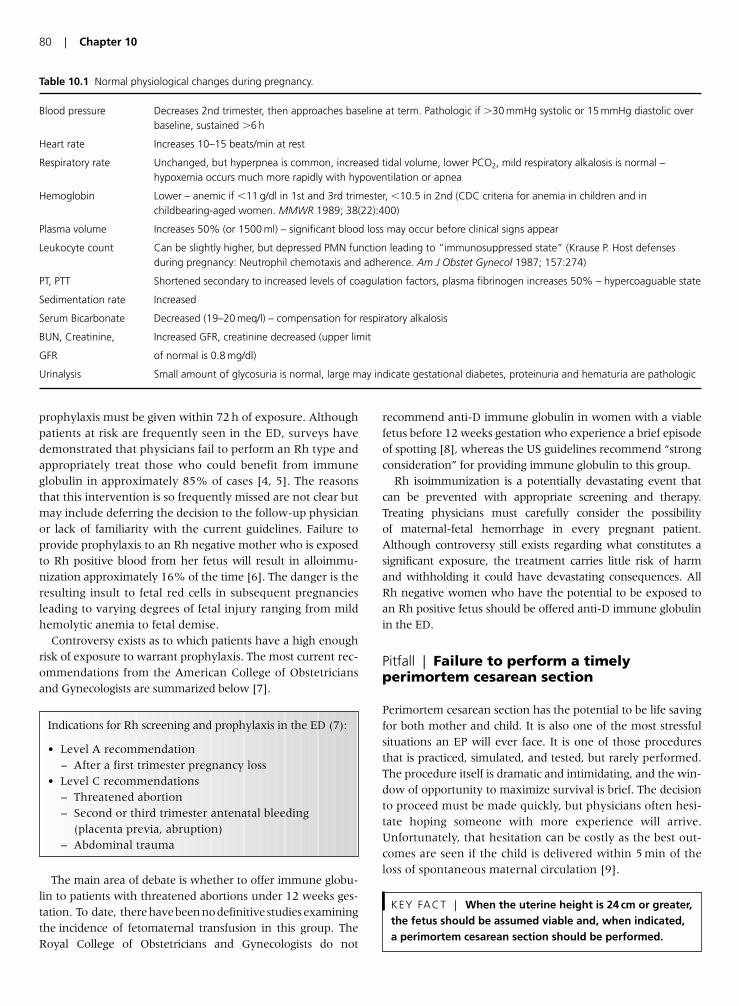
prophylaxis must be given within 72 h of exposure. Althoughpatients at risk are frequently seen in the ED, surveys havedemonstrated that physicians fail to perform an Rh type andappropriately treat those who could benefit from immuneglobulin in approximately 85% of cases [4, 5]. The reasonsthat this intervention is so frequently missed are not clear butmay include deferring the decision to the follow-up physicianor lack of familiarity with the current guidelines. Failure toprovide prophylaxis to an Rh negative mother who is exposedto Rh positive blood from her fetus will result in alloimmu-nization approximately 16% of the time [6]. The danger is theresulting insult to fetal red cells in subsequent pregnanciesleading to varying degrees of fetal injury ranging from mildhemolytic anemia to fetal demise.
Controversy exists as to which patients have a high enoughrisk of exposure to warrant prophylaxis. The most current rec-ommendations from the American College of Obstetriciansand Gynecologists are summarized below [7].
The main area of debate is whether to offer immune globu-lin to patients with threatened abortions under 12 weeks ges-tation. To date, therehavebeennodefinitivestudiesexaminingthe incidence of fetomaternal transfusion in this group. TheRoyal College of Obstetricians and Gynecologists do not
recommend anti-D immune globulin in women with a viablefetus before 12 weeks gestation who experience a brief episodeof spotting [8], whereas the US guidelines recommend “strongconsideration” for providing immune globulin to this group.
Rh isoimmunization is a potentially devastating event thatcan be prevented with appropriate screening and therapy.Treating physicians must carefully consider the possibilityof maternal-fetal hemorrhage in every pregnant patient.Although controversy still exists regarding what constitutes asignificant exposure, the treatment carries little risk of harmand withholding it could have devastating consequences. AllRh negative women who have the potential to be exposed toan Rh positive fetus should be offered anti-D immune globulinin the ED.
Pitfall | Failure to perform a timelyperimortem cesarean section
Perimortem cesarean section has the potential to be life savingfor both mother and child. It is also one of the most stressfulsituations an EP will ever face. It is one of those proceduresthat is practiced, simulated, and tested, but rarely performed.The procedure itself is dramatic and intimidating, and the win-dow of opportunity to maximize survival is brief. The decisionto proceed must be made quickly, but physicians often hesi-tate hoping someone with more experience will arrive.Unfortunately, that hesitation can be costly as the best out-comes are seen if the child is delivered within 5 min of theloss of spontaneous maternal circulation [9].
KEY FACT | When the uterine height is 24 cm or greater,the fetus should be assumed viable and, when indicated,a perimortem cesarean section should be performed.
80 | Chapter 10
Table 10.1 Normal physiological changes during pregnancy.
Blood pressure Decreases 2nd trimester, then approaches baseline at term. Pathologic if �30 mmHg systolic or 15 mmHg diastolic overbaseline, sustained �6 h
Heart rate Increases 10–15 beats/min at rest
Respiratory rate Unchanged, but hyperpnea is common, increased tidal volume, lower PCO2, mild respiratory alkalosis is normal – hypoxemia occurs much more rapidly with hypoventilation or apnea
Hemoglobin Lower – anemic if �11 g/dl in 1st and 3rd trimester, �10.5 in 2nd (CDC criteria for anemia in children and inchildbearing-aged women. MMWR 1989; 38(22):400)
Plasma volume Increases 50% (or 1500 ml) – significant blood loss may occur before clinical signs appear
Leukocyte count Can be slightly higher, but depressed PMN function leading to “immunosuppressed state” (Krause P. Host defenses during pregnancy: Neutrophil chemotaxis and adherence. Am J Obstet Gynecol 1987; 157:274)
PT, PTT Shortened secondary to increased levels of coagulation factors, plasma fibrinogen increases 50% – hypercoaguable state
Sedimentation rate Increased
Serum Bicarbonate Decreased (19–20 meq/l) – compensation for respiratory alkalosis
BUN, Creatinine, Increased GFR, creatinine decreased (upper limit
GFR of normal is 0.8 mg/dl)
Urinalysis Small amount of glycosuria is normal, large may indicate gestational diabetes, proteinuria and hematuria are pathologic
Indications for Rh screening and prophylaxis in the ED (7):
• Level A recommendation– After a first trimester pregnancy loss
• Level C recommendations– Threatened abortion– Second or third trimester antenatal bleeding
(placenta previa, abruption)– Abdominal trauma
The decision to proceed with perimortem cesarean sectionmust take into account both the benefits to the mother and tothe fetus. In cases where the mother has sustained a lethalinjury, the procedure is done to save the life of the child.However, maternal hemodynamic status is often improvedwhen the fetus is extracted [10]. Indications for the procedureinclude loss of spontaneous maternal circulation and evidenceof a viable fetus. As a history is rarely available, the assessmentof fetal viability must rely solely on physical examination. At20 weeks gestation, the fundus should be at the level of theumbilicus. After 20 weeks, the gestational age roughly corre-lates with the distance in centimeters from the pubic symph-ysis to the uterine fundus. If the fundal height is at least24 cm at the time of maternal cardiopulmonary arrest, peri-mortem cesarean delivery is indicated.
During the procedure itself, CPR should continue to maximizeplacental blood flow. Massive fluid resuscitation is continuedin lieu of pressors as placental vasoconstriction may compro-mise fetal outcome. Do not stop to prepare the abdomen asthis only delays delivery. Begin the procedure as soon as possi-ble, but no later than 4 min after loss of maternal pulseto ensure delivery by 5 min. A pre-assembled perimortemcesarean delivery kit can save time and frustration, however if there is no time for preparation, a large scalpel is all that isrequired. Perform a vertical midline incision from the epigas-trium to the symphysis pubis following the linea nigra if pres-ent. Rapidly cut through all layers of the abdomen until theuterus is exposed. Next, open the uterus through a midlineincision, thereby avoiding the uterine vessels which attach lat-erally. The safest practice is to start the incision at the funduswith a scalpel, then extend it with something more blunt suchas large scissors. Once the first incision is made into the uterus,place your hand inside between the fetus and the uterine wallto prevent injury to the child as you finish the incision. Deliverthe child head first if possible. Hold the child in a positionlower than the mother, suction the mouth, and then clampand cut the umbilical cord.
EPs must be prepared to act quickly and efficiently in orderto offer both patients the best chance of survival. Because thisskill is used infrequently and under very stressful circum-stances, it is critical that EPs are very familiar with both theindications and the technique. Practicing the scenario undersimulated conditions provides an excellent opportunity topractice both the cognitive decision to initiate the procedure,as well as the psychomotor skills for its performance. Therecent advances in simulator technology provide a greatopportunity for physicians to enhance their skills in the man-agement of this high risk, low incidence procedure.
Pitfall | Avoiding necessary radiologicimaging for fear of harm to the fetus
A common myth circulating in both the medical and lay com-munities is that routine X-rays are harmful and will lead tofetal anomalies if performed on a pregnant patient. In fact, therisk to the fetus for most imaging studies is minimal, whilethe risk of a missed diagnosis to both the mother and the fetusmay be substantial. There is no increased risk to the fetus ofanomalies, growth restriction, or abortion with radiationexposure of less than 5000 mrad [11]. There may be a slightlyhigher incidence of childhood leukemia, but only if the fetus isexposed to more than 1000–2000 mrad in the first trimester[12]. As demonstrated in Table 10.2, most emergency evalua-tions fall far short of the amount of radiation associated withharm to the fetus.
To date, there have been no adverse fetal effects reportedwith ultrasonography or MRI thus these are the diagnosticmodalities of choice in pregnant patients. Non-ionic oral andintravenous contrast agents are also felt to be safe. However,iodinated agents have been associated with neonatal hypothy-roidism and should be avoided [12].
The decision to use radiologic imaging in a pregnant patientmust take into account the risk to the fetus, the risk of harm tothe mother, and the risk to the mother and fetus of missing adiagnosis because of a reluctance to use available imaging.Although the health of the fetus must be considered, appro-priate diagnostic imaging should not be withheld from preg-nant patients.
Pitfall | Attributing dyspnea in pregnancy toa gravid uterus
Dyspnea is a very common complaint during the thirdtrimester of pregnancy. It is estimated that 75% of womenin their third trimester complain of shortness of breath. Thegravid uterus causes mass effect on the diaphragm limitingexcursion, progesterone induces a mild respiratory alkalosis,and oxygen consumption is increased all leading to the sensa-tion of dyspnea. However, the dyspnea of pregnancy tends to
KEY FACT | When performing a perimortem cesariansection, CPR should be continued, vasopressors should beavoided, and massive fluid resuscitation should becontinued.
Management of the Pregnant Patient in the ED | 81
Table 10.2 Radiation exposure with common ED diagnosticprocedures (values from multiple sources).
Study Fetal Radiation Exposure (mrad)
Chest X-ray (shielding abdomen) 0.02–0.07Extremity 1–2Abdomen (2 view) 250Pelvis 150–200VQ scan 100–500CT pulmonary angiogram 100–900CT abdomen and pelvis 3000–5000
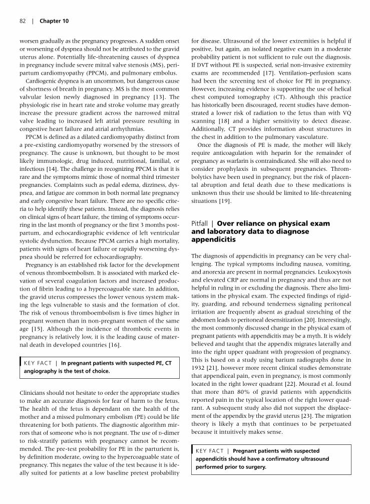
worsen gradually as the pregnancy progresses. A sudden onsetor worsening of dyspnea should not be attributed to the graviduterus alone. Potentially life-threatening causes of dyspneain pregnancy include severe mitral valve stenosis (MS), peri-partum cardiomyopathy (PPCM), and pulmonary embolus.
Cardiogenic dyspnea is an uncommon, but dangerous causeof shortness of breath in pregnancy. MS is the most commonvalvular lesion newly diagnosed in pregnancy [13]. Thephysiologic rise in heart rate and stroke volume may greatlyincrease the pressure gradient across the narrowed mitralvalve leading to increased left atrial pressure resulting incongestive heart failure and atrial arrhythmias.
PPCM is defined as a dilated cardiomyopathy distinct froma pre-existing cardiomyopathy worsened by the stressors ofpregnancy. The cause is unknown, but thought to be mostlikely immunologic, drug induced, nutritional, familial, orinfectious [14]. The challenge in recognizing PPCM is that it israre and the symptoms mimic those of normal third trimesterpregnancies. Complaints such as pedal edema, dizziness, dys-pnea, and fatigue are common in both normal late pregnancyand early congestive heart failure. There are no specific crite-ria to help identify these patients. Instead, the diagnosis relieson clinical signs of heart failure, the timing of symptoms occur-ring in the last month of pregnancy or the first 3 months post-partum, and echocardiographic evidence of left ventricularsystolic dysfunction. Because PPCM carries a high mortality,patients with signs of heart failure or rapidly worsening dys-pnea should be referred for echocardiography.
Pregnancy is an established risk factor for the developmentof venous thromboembolism. It is associated with marked ele-vation of several coagulation factors and increased produc-tion of fibrin leading to a hypercoaguable state. In addition,the gravid uterus compresses the lower venous system mak-ing the legs vulnerable to stasis and the formation of clot.The risk of venous thromboembolism is five times higher inpregnant women than in non-pregnant women of the sameage [15]. Although the incidence of thrombotic events inpregnancy is relatively low, it is the leading cause of mater-nal death in developed countries [16].
Clinicians should not hesitate to order the appropriate studiesto make an accurate diagnosis for fear of harm to the fetus.The health of the fetus is dependant on the health of themother and a missed pulmonary embolism (PE) could be lifethreatening for both patients. The diagnostic algorithm mir-rors that of someone who is not pregnant. The use of D-dimerto risk-stratify patients with pregnancy cannot be recom-mended. The pre-test probability for PE in the parturient is,by definition moderate, owing to the hypercoaguable state ofpregnancy. This negates the value of the test because it is ide-ally suited for patients at a low baseline pretest probability
for disease. Ultrasound of the lower extremities is helpful ifpositive, but again, an isolated negative exam in a moderateprobability patient is not sufficient to rule out the diagnosis.If DVT without PE is suspected, serial non-invasive extremityexams are recommended [17]. Ventilation–perfusion scanshad been the screening test of choice for PE in pregnancy.However, increasing evidence is supporting the use of helicalchest computed tomography (CT). Although this practicehas historically been discouraged, recent studies have demon-strated a lower risk of radiation to the fetus than with VQscanning [18] and a higher sensitivity to detect disease.Additionally, CT provides information about structures inthe chest in addition to the pulmonary vasculature.
Once the diagnosis of PE is made, the mother will likelyrequire anticoagulation with heparin for the remainder ofpregnancy as warfarin is contraindicated. She will also need toconsider prophylaxis in subsequent pregnancies. Throm-bolytics have been used in pregnancy, but the risk of placen-tal abruption and fetal death due to these medications isunknown thus their use should be limited to life-threateningsituations [19].
Pitfall | Over reliance on physical examand laboratory data to diagnoseappendicitis
The diagnosis of appendicitis in pregnancy can be very chal-lenging. The typical symptoms including nausea, vomiting,and anorexia are present in normal pregnancies. Leukocytosisand elevated CRP are normal in pregnancy and thus are nothelpful in ruling in or excluding the diagnosis. There also limi-tations in the physical exam. The expected findings of rigid-ity, guarding, and rebound tenderness signaling peritonealirritation are frequently absent as gradual stretching of theabdomen leads to peritoneal desensitization [20]. Interestingly,the most commonly discussed change in the physical exam ofpregnant patients with appendicitis may be a myth. It is widelybelieved and taught that the appendix migrates laterally andinto the right upper quadrant with progression of pregnancy.This is based on a study using barium radiographs done in1932 [21], however more recent clinical studies demonstratethat appendiceal pain, even in pregnancy, is most commonlylocated in the right lower quadrant [22]. Mourad et al. foundthat more than 80% of gravid patients with appendicitisreported pain in the typical location of the right lower quad-rant. A subsequent study also did not support the displace-ment of the appendix by the gravid uterus [23]. The migrationtheory is likely a myth that continues to be perpetuatedbecause it intuitively makes sense.
KEY FACT | Pregnant patients with suspectedappendicitis should have a confirmatory ultrasoundperformed prior to surgery.
KEY FACT | In pregnant patients with suspected PE, CTangiography is the test of choice.
82 | Chapter 10
A diagnosis of appendicitis in pregnancy should not be basedon exam and laboratory findings alone. Appendectomy duringpregnancy is associated with a fetal loss/miscarriage rate of5.6%. This climbs to 10% if the appendix is perforated [24].Because an unnecessary appendectomy could have devastatingconsequences for the pregnancy, radiologic imaging should beutilized to support the diagnosis prior to operative interven-tion. The diagnostic modality of choice is ultrasound which hasbeen shown to be around 84% sensitive. If it is a technicallydifficult study or the results are inconclusive, helical CT scan-ning is recommended. CT in non-pregnant patients has beendemonstrated to have a sensitivity of 98%, and a small caseseries in pregnant patients found CT to be 100% sensitive [25].Although the dose of radiation to the fetus is substantial atapproximately 3000 mrad, it does not reach the level of radia-tion believed to be harmful to the fetus (5000 mrad). As MRIbecomes more widely available, it may replace ultrasound asdiagnostic modality of choice as it combines little risk ofharm to the fetus with excellent anatomic visualization [26].
The diagnosis of appendicitis is often made by clinical examalone. This practice is not recommended in gravid patients asappendectomy in pregnancy carries a significant risk of mis-carriage and fetal loss. That being said, delay in diagnosis hasbeen associated with higher rates of perforation and fetal com-promise. A rapid and accurate diagnosis will ensure the bestoutcome for the mother and child. Although current surgicalpractice still includes diagnostic laparotomy based on highclinical suspicion, every effort should be made to avoid unnec-essary appendectomy during pregnancy.
Pitfall | Failure to monitor patients whohave sustained minor blunt trauma in thethird trimester
Trauma in pregnancy is not uncommon complicating at least 1 in 12 pregnancies [27]. Blunt trauma is more common thanpenetrating, with the most common mechanisms includingmotor vehicle accidents, assaults, and falls. Of these, car acci-dents occur most frequently accounting for nearly 60% ofblunt trauma in pregnancy [28]. Many of these accidents areminor and significant injury is not suspected. Unfortunately,what may seem like minimal injury to the mother can causelife-threatening consequences in the child. Without appro-priate monitoring, placental abruption and uterine rupturemay be missed.
Placental abruption is the most common cause of fetal demiseoutside of maternal death in blunt trauma. The shearing forcesexperienced in blunt trauma can lead to separation of therigid placenta from the elastic uterine wall. Approximately
2–4% of pregnant women experiencing minor traumaticinsults will suffer a placental abruption [29]. Clinical signsand symptoms such as vaginal bleeding, uterine or abdomi-nal tenderness, and abdominal pain are helpful, but are notalways present and cannot be relied upon. Ultrasound hasbeen proposed as an initial screening exam, however it has asensitivity of less than 50% for placental abruption [30]. Incontrast, uterine irritability has been strongly associated withabruption [31]. Cardiotocographic monitoring (CTM) hasemerged as the standard of care for all patients in the thirdtrimester who sustain blunt trauma regardless of the sever-ity of the mechanism of injury.
Ideally, an obstetrician or obstetrical nurse would be present toreview CTM data. However, EPs should be able to recognizesigns of fetal distress and uterine irritability. Fetal distress ismanifested by fetal tachycardia, bradycardia, loss of beat tobeat variability, or recurrent fetal heart rate decelerations. Thedefinition of uterine irritability after trauma varies in the lit-erature, but is most commonly referred to as more than onecontraction every 10–15 min. CMT monitoring should beinitiated as soon as possible in the ED and continued for a min-imum of 4 h. Those with uterine irritability require admis-sion for prolonged CMT monitoring. Any sign of fetal distressafter 24 weeks gestation is an indication for emergent cesareansection. In cases of minor trauma, patients without evidenceof fetal distress or uterine irritability during 4 h of monitor-ing may be safely discharged home.
Pearls for Improving Patient Outcomes
• Assume all women of childbearing age are pregnant until provenotherwise.
• Suspect heterotopic pregnancy in pregnant patients with abdomi-nal pain and a viable IUP in the setting of fertility-promoting tech-niques, or with free fluid or abnormal adnexae on ultrasound.
• Carefully interpret diagnostic studies and vital signs in pregnantpatients.
• Consider the potential for maternal-fetal hemorrhage in allpregnant patients presenting to the ED. Provide anti-D immuneglobulin to all women at risk for isoimmunization.
• Do not withhold appropriate diagnostic imaging from pregnantpatients at any period of gestation.
• Recognize the indications for and be prepared to perform anemergent cesarian section on the hemodynamically unstable preg-nant patient with potentially viable fetus.
• Patients with acute or significant progressive dyspnea in pregnancyshould be investigated for causes other than the gravid uterus.
KEY FACT | All patients in the third trimester ofpregnancy with any degree of blunt abdominal traumarequire CTM for a minimum of 4 hours.
KEY FACT | Abdominal pain, tenderness, and vaginalbleeding may be absent with placental abruption.
Management of the Pregnant Patient in the ED | 83
• Utilize all available data, including radiologic studies, to make anaccurate diagnosis of acute appendicitis and avoid unnecessaryappendectomy.
• Patients with viable gestations require at least 4 h of CTM moni-toring after even minor trauma.
References
1. Ramoska E. Reliability of patient history in determining the
possibility of pregnancy. Ann Emerg Med 1989; 18:48–50.
2. Habana A. Cornual heterotopic pregnancy: contemporary man-
agement options. Am J Obstet Gynecol 2000; 182:1264–70.
3. Cunninham et al. Maternal Physiology. Williams Obstetrics,
22nd edition, New York: McGraw-Hill, 2005; pp 121–50.
4. Grant J. Underutilization of Rh prophylaxis in the emergency
department: a retrospective survey. Ann Emerg Med 1991;
21:181–3.
5. Weinberg L. Use of anti-D immunoglobulin in the treatment of
threatened miscarriage in the accident and emergency depart-
ment. Emerg Med J 2001; 18:444–7.
6. Bowman J. Controversies in Rh prophylaxis. Am J obstet Gynecol
1985; 151:289–94.
7. ACOG practice bulletin. Prevention of Rh D alloimmunization.
Number 4, May 1999. Clinical management guidelines for
obstetrician–gynecologists. American College of Obstetrics and
Gynecology. Int J Gynecol Obstetr 1999; 66(1):63–70.
8. Royal College of Obstetricians and Gynaecologist. Green top
Guidelines. Anti-D immunoglobulin for Rh prophylaxis. London:
RCOG, 2002.
9. Katz VL. Perimortem cesarean delivery. Obstet gynecol 1986;
68:571–6.
10. Katz VL. Perimortem cesarean delivery: Were our assumptions
correct? Am J Obstet Gynecol 2005; 192:1916–21.
11. Brent R. The effect of embryonic and fetal exposure to X-ray,
microwaves, and ultrasound: counseling the pregnant and non-
pregnant patient about these risks. Semin Oncol 1989; 16:347–68.
12. Guidelines for diagnostic imaging during pregnancy. ACOG
Committee Opinion No. 299, American College of Obstetricians
and Gynecologists. Obstet Gynecol 2004; 104:647–51.
13. Elkayam et al. Valvular heart disease in pregnancy part I: native
valves. J Am Coll Cardiol 2005; 46(2):223–30.
14. Ro, A. Peripartum cardiomyopathy. Cardiology in Review 2006;
14(1):35–42.
15. National Institutes of Health Consensus Development Confer-
ence. Prevention of venous thrombosis and pulmonary
embolism. J Am Med Assoc 1986; 256: 744–9.
16. Berg C. et al. Pregnancy-related mortality in the United States,
1991–1997. Obstet Gynecol 2003; 101(2):289–96.
17. Toglia M. Venous thromboembolism during pregnancy. New
Engl J Med 1996; 335(2):108–14.
18. Winer-Muram H. Pulmonary embolism in pregnant patients:
fetal radiation dose with helical CT. Radiology 2002; 224:487–92.
19. Patel RK. Thrombolysis in pregnancy. Thromb Haemost 2003;
90:1216–17.
20. Martin C. Physiologic changes in pregnancy: surgical implica-
tions. Clin Obstet Gynecol 1994; 37:241–55.
21. Baer J. Appendicitis in pregnancy with changes in the position
and axis of the normal appendix in pregnancy. J Am Med Assoc
1932; 98:1359–64.
22. Mourad J. Appendicitis in pregnancy: New information that
contradicts long-held clinical beliefs. Am J Obstet Gynecol 2000;
182:1027–9.
23. Hodjati H. Location of the appendix in the gravid patient:
a re-evaluation of the established concept. Int J Gynaecol Obstetr
2003; 81(3):245–7.
24. Cohen-Kerem R. Pregnancy outcome following non-obstetric
surgical intervention. Am J Surg 2005; 190(3):467–3.
25. Castro M. The use of helical computed tomography in preg-
nancy for the diagnosis of acute appendicitis. Am J Obstet Gynecol
2001; 184:954–7.
26. Birchard K. MRI of acute abdominal and pelvic pain in preg-
nant patients.. Am J Roentgenology 2005; 184(2):452–8.
27. Rosenfeld J. Abdominal trauma in pregnancy: When is fetal
monitory necessary? Postgrad Med 1990; 88:89–91.
28. Shah A. Trauma in pregnancy. Emerg med Clin N Am 2003;
21:615–29.
29. Rothenberger D. Blunt maternal trauma: a review of 103 cases.
J Trauma 1978; 18(3):173–9.
30. Dahmus M. Blunt abdominal trauma: are there any predictive
factors for abruption placentae or maternal-fetal distress. Am J
Obstet Gynecol 1993; 169(4):1054–9.
31. Pearlman M. A prospective controlled study of outcome after
trauma during pregnancy. Am J Obstet Gynecol 1990;
162(6):1502–10.
84 | Chapter 10
Introduction
The care of children in the emergency department (ED) canbe quite challenging as the patient may be non-verbal,extremely anxious or present with a vague history. Severalhigh-risk areas have been identified in pediatric emergencymedicine. A recent retrospective study by Steve Selbst et al.discovered that the most common diagnoses involved in pedi-atric lawsuits in the US were meningitis, appendicitis, armfracture, and testicular torsion [1]. Other areas of potentialliability include medication errors, delayed diagnosis of intus-susception and inappropriate evaluation of fever in children.This chapter will discuss some of the more common pitfalls inthe acute care of children.
Pitfall | Overdiagnosis of otitis media
The American Academy of Pediatrics and the AmericanAcademy of Family Physicians formed a subcommittee onmanagement of acute otitis media (OM). Their publishedpractice guideline was designed to give recommendations formanagement of acute OM in children 2 months to 12 years of age (Table 11.1) [2]. In order to diagnose acute OM thefollowing must be present.
In addition, the subcommittee recommended an option towithhold antibiotics for 48 h in children over 6 months of agewho do not have severe infections or temperatures greaterthan 39°C. Children who have severe disease or infants�6 months of age with confirmed OM using insufflation,should be treated. High-dose amoxicillin (80–90 mg/kg) is stillthe first line drug of choice.
Pitfall | Failure to differentiate a simplefebrile seizure from other causes ofseizures
It is imperative that emergency providers know the definitionand age range for simple and complex febrile seizures. Thesecharacteristics are listed in Table 11.2. As an example of apotentially devastating error that can occur by misclassifying aseizure as being a simple febrile one, a 12-year-old girl pre-sented to an ED with the report of a fever and a generalizedseizure that lasted 10 min. The Emergency Physician (EP)noted that the patient was back to her baseline mental statusand no further workup was initiated. A diagnosis of a simplefebrile seizure was made and the patient was dischargedhome. She returned later that day with status epilepticus. Sheis now neurologically impaired as a result of encephalitis.
Therefore, a complex febrile seizure occurs when there is afocal seizure, more than one seizure in a 24-h period, orduration of seizure activity exceeding 15 min. The workupof a simple febrile seizure vastly differs from that which
KEY FACT | Though patients with simple febrileseizures require only an age-appropriate fever workup,patients with worrisome features should undergo lumbarpuncture.
KEY FACT | Antibiotics can be safely withheld for 48 hours in children older than 6 months of age withoutsevere disease.
Chapter 11 | Pediatric Care in the ED
Ghazala Sharieff
85
Table 11.1 Diagnostic Criteria for OM [2].
In order to diagnose acute OM the following must be present:Acute onset of symptomsSigns of middle ear infection(a) Bulging TM(b) Limited or absent mobility of the TM – middle ear effusion is best
confirmed by pneumatic otoscopy! (Pooled sensitivity of 94%and specificity of 80%.)
(c) Otorrhea(d) Air-fluid level behind the TMSigns of middle-ear inflammation(a) Distinct erythema of the TM or(b) Distinct otalgia that interferes with normal activity or sleep
Table 11.2 Characteristics of a simple febrile seizure.
To qualify as a simple febrile seizure, it must:Be a generalized seizureLast �15 minNot recur within a 24-h periodOccur in a patient aged 6 months to 5 yearsOccur in a patient with no evidence of meningitis on examination
should be performed in patients with complex febrileseizures. Trainer et al. [3] noted that patients with simplefebrile seizures had no higher incidence of serious bacterialillness than patients with febrile illnesses alone. Therefore,the authors concluded that patients with simple febrileseizures need only undergo an age-appropriate fever evalu-ation. Routine electrolyte testing, CT scans and EEG’s arenot necessary. Warden et al. [4] in a review article concludedthat routine lumbar punctures are not necessary in children�12 months of age with simple febrile seizures. However, afever evaluation should be performed that is appropriate forthe patient’s age. A lumbar puncture should be strongly con-sidered in patients �18 month of age who have: (1) a historyof irritability, decreased feeding, or lethargy; (2) a prolongedpostical period; (3) physical examination findings of meningitis such as a bulging fontanelle, photophobia, severeheadache, Kernig or Brudzinski signs; (4) an abnormalappearance or mental status findings on initial observationfollowing the postictal period; (5) any complex febrile seizurefeatures; or (6) pretreatment with antibiotics. If these factorsare not present and the child is well appearing a lumbarpuncture can be deferred. In children over 18 months of agewho have no signs of CNS infections, lumbar puncture canalso be safely deferred.
Pitfall | Over-reliance on the peripheralwhite blood cell (WBC) count to determinewhether a child needs to have a lumbarpuncture
For years the peripheral blood count has been used as amarker for identifying patients with serious bacterial illness.According to published fever policies, patients who have aperipheral WBC count between 5 and 15,000 cells/mm3
without a significant bandemia have a low risk of seriousbacterial illness [5, 6]. However, this theory has beenrecently challenged. In a study by Bonsu et al. [7], theperipheral WBC count was found to be an unreliable markerin patients with subsequent meningitis. The authors exam-ined the presenting WBC count in 22 patients who weresubsequently found to have bacterial meningitis based oncerebrospinal fluid (CSF) analysis. The median value of theperipheral WBC count for patients with bacterial meningitiswas 10,200 cells/mm3 versus a median peripheral WBCcount of 11,200 cells/mm3 for infants without bacterialmeningitis. Alarmingly, if a WBC count of 15 k was used asthe sole criteria to identify patients requiring evaluation formeningitis, 73% of patients would have erroneously beenmissed. If the cutoff was raised to 20 k, 96% of patients with
meningitis would not have been identified. Using peripheralWBC counts of 5–15 k as low-risk criteria would have failedto identify 41% of cases of bacterial meningitis. The sameauthors evaluated the utility of the WBC count to identifychildren at risk for bacteremia and again found this test to bean inaccurate screening tool [8].
A lumbar puncture should be performed if the patient hasclinical findings that are consistent with meningitis such asextreme irritability or lethargy, petechiae or purpura, bulgingfontanelle, prolonged seizure activity, or any other abnormalphysical examination findings. The reliance on the peripheralWBC count to guide the decision on whether or not to per-form a lumbar puncture or to obtain blood cultures will onlybe misleading and grant a false sense of security in thepatient’s differential diagnosis.
Pitfall | Misdosing medications for children
Furthermore, medication dosing errors are also frequent. Ina retrospective chart review of 1532 children treated at apediatric tertiary care hospital, prescribing errors werefound in 10.1% (154/1532) of the charts [9]. The followingvariables accounting for an increased incidence of errors:
1. Patients seen between 4 a.m. and 8 a.m.2. Patients with severe disease.3. Medication ordered by a trainee – higher incidence at thebeginning of the academic year.4. Patients seen on weekends.
The most common drugs involved in errors were: acetamin-ophen, antibiotics, asthma medications and antihistamines.There were two severe errors (drug error that could causedeath or that was ordered was not administered) includingone case in which a child with meningitis did not receiveantibiotic. The take-home message is that when dosingmedications for children, 47.5% of errors were ranked assignificant – a drug error that could cause a non-life-threat-ening consequence or less effective treatment (e.g. 10-foldlower dose of amoxicillin for OM).
A common cause of medication errors is the lack of stan-dardization of the units in which children are weighed.Medication dosing is typically determined by the weight ofthe individual in kilograms. If a child’s weight is mistakenlyreported in pounds and dosing is based on this number, seri-ous medication errors can occur. Alternatively, even if theweight is recognized as being in pounds, errors can arise bymiscalculating when converting to kilograms. As an exam-ple, a 10-day-old male presented to the ED with complaints
KEY FACT | The weight of children should only berecorded in kilograms and dosage calculations andconcentrations must be checked prior to administration.
KEY FACT | The peripheral WBC count should not beused to identify patients requiring a workup formeningitis or serious bacterial illness.
86 | Chapter 11
of poor feeding and vomiting. His weight was recorded as6 pounds. The attending physician calculated the newbornas weighing 13 kg (instead of 2.7 kg!) and ordered a 280 mlbolus of normal saline rather than the recommended bolusof 54 ml. The patient began to develop respiratory distressand seemed to have a delayed capillary refill after the boluswas administered. The baby was given a repeat bolus of280 ml of normal saline. The patient subsequently requiredintubation and transfer to a pediatric facility due to fluidoverload. It is imperative that children be weighed in kilo-grams and this should start from the very first measurement.Requiring an additional step to convert from pounds to kilo-grams is inherently fraught with error. The take-home mes-sage is that when dosing medications for children, cliniciansmust check and recheck the dosing to not only ensure thattheir calculations are correct but that the dose administeredis the dose ordered. This is particularly true in the setting ofthe high-risk variables listed above.
Pitfall | Diagnosing colic in an infantwithout ruling out other causes of crying
While colic is a common cause of persistent crying in infancy,other serious causes must be considered and excluded. Theclassic definition of colic follow a “rule of 3’s” first describedby Wessel [10]. A child with colic typically cries for 3 h/day,3 days/week for at least 3 weeks and this excessive cryingtypically resolves by age 3 months. The physical examinationwill help to determine the cause of crying in approximately41% of patients [11]. A systematic approach in imperativeand the infant should be fully undressed and examined. Hairtourniquet syndrome can easily be missed and there havebeen many case reports of infants with hair tourniquets notonly around the digits but also around the penis and clitoris.Corneal abrasions are also in the differential diagnosis andare readily diagnosed by flourescein examination. Intussus-ception also presents with crying and colicky abdominal painand therefore a high index of suspicion should be maintainedin patients who are described by the parents as “drawing uptheir legs” and then returning to normal baseline in betweenepisodes. “IT CRIES” is a useful mnemonic that has beendescribed to highlight common causes of unexplained cryingin infants (Table 11.3) [12]. It is important that life-threateningand readily treatable causes of unexplained crying are identi-fied and managed appropriately.
Pitfall | Relying on the physical appearanceof a newborn to determine the need for afull sepsis evaluation
Neonates are at high risk for serious infection and are typicallyinfected with virulent bacteria (such as group B streptococci,E. coli, and Listeria monocytogenes). Group B streptococci, is
associated with high rates of meningitis (39%), non-meningeal foci of infection (10%), and sepsis (7%) [13].
Several criteria have been tested in order to determinewhich infants are at risk for serious bacterial illness. Baskinet al. described the “Boston criteria” for febrile children�38.0°C between 1 and 3 months of age presenting to theED [14]. Infants were discharged after an injection of intra-muscular ceftriaxone (50 mg/kg) if the child was wellappearing and had no ear, soft tissue, joint or bone infec-tions on physical examination. These patients must havehad CSF with �10 WBC/high-power field (hpf), microscopicurinalysis with �10 WBC/hpf or urine dipstick negative forleukocyte esterase, peripheral WBC count �20,000/mm3,and a normal chest X-ray in patients in whom a chest X-raywas obtained.
Baker et al. developed the Philadelphia criteria to identifyneonates who were at low risk for serious bacterial illness[15]. Patients who looked well (as defined by an InfantObservation Score of 10 or less) and had a peripheral WBCcount �15,000/mm3, a band-to-neutrophil ratio of �0.2,uric acid with fewer than 10 WBC/hpf and few or no bacte-rial on a spun urine specimen, CSF with fewer than8 WBC/mm3 and a negative Gram stain, negative chestX-ray (obtained on all patients), and stool negative for bloodand few or no WBC on microscopy (sent on those patientswith watery diarrhea) were considered to have a negativescreen and were not treated with antibiotics. The neonatesthat were placed into the high-risk category had a higherincidence of bacterial disease (18.6%) but 4.6% of neonatesclassified as low risk had a serious bacterial infection. Kadishet al. found a similar rate of SBI in neonates that they cate-gorized as low risk when they retrospectively applied boththe Philadelphia criteria and the Boston criteria [16].
Another screening tool known as the “Rochester criteria”has been applied to infants 60-day old into high- and low-risk groups [17]. The children who met these criteria lookedwell, had been previously healthy, and had no evidence ofskin, soft tissue, bone, joint, or ear infection. Additionally,they had normal peripheral WBC counts (5–15,000/mm3),normal absolute band counts (1500/mm3), 10 WBC/hpfof spun urine sediment, and for those patients with diarrhea,5 WBC/hpf on stool smear. The low-risk group identifiedchildren who were unlikely to have serious bacterial infec-tion, with a negative predictive value of 98.9%.
Pediatric Care in the ED | 87
Table 11.3 Causes of crying in infants.
I–Infections: otitis media, urinary tract infection, meningitisT–Trauma: consider head CT, skeletal survey, focused radiographsC–Cardiac: Congestive heart failure, supraventricular tachycardiaR–Rectal fissure, reaction to medications (immunizations), refluxI–IntussusceptionE–Eye: corneal abrasion or ocular foreign bodyS–Surgical disease such as inguinal hernia, testicular torsionS–Strangulation: hair/fiber tourniquet syndrome
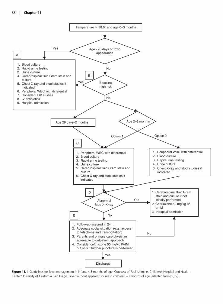
88 | Chapter 11
Temperature � 38.0° and age 0–3 months
Age <28 days or toxicappearance
1. Blood culture2. Rapid urine testing2. Urine culture4. Cerebrospinal fluid Gram stain and culture5. Chest X-ray and stool studies if indicated6. Peripheral WBC with differential7. Consider HSV studies8. IV antibiotics9. Hospital admission
Baselinehigh risk
1. Peripheral WBC with differential 2. Blood culture3. Rapid urine testing4. Urine culture6. Chest X-ray and stool studies if indicated
1. Peripheral WBC with differential2. Blood culture3. Rapid urine testing4. Urine culture5. Cerebrospinal fluid Gram stain and culture6. Chest X-ray and stool studies if indicated
Abnormallabs or X-ray
1. Cerebrospinal fluid Gram stain and culture if not initially performed2. Ceftriaxone 50 mg/kg IV or IM3. Hospital admission
1. Follow-up assured in 24 h.2. Adequate social situation (e.g., access to telephone and transportation)3. Parents and primary care physician agreeable to outpatient approach4. Consider ceftriaxone 50 mg/kg IV/IM but only if lumbar puncture is performed
Discharge
Age 29 days–2 months
A
B
C
D
Yes
No
Age 2–3 months
Option 1 Option 2
E
Yes
No
No
No
Yes
Yes
Figure 11.1 Guidelines for fever management in infants �3 months of age. Courtesy of Paul Ishimine. Children’s Hospital and HealthCenter/University of California, San Diego. Fever without apparent source in children 0–3 months of age (adapted from [5, 6]).
Despite the number of different criteria available, nonehave been shown to be as accurate in the evaluation of thefebrile neonate. Using the Rochester criteria, Jaskiewiczfound that 2 of 227 children �30-day old who met low-riskcriteria had serious bacterial illness (SBI) [18]. However,Ferrera et al. found a 6% incidence of serious bacterial illnessin neonates that were retrospectively classified as low risk by the Rochester criteria [19]).
Another recent study compared the Philadelphia criteriato the Rochester criteria [20]. Infants �56 days of age whopresented with fever were enrolled and all patients under-went a full sepsis evaluation (CBC, blood culture, urinalysis,urine culture and lumbar puncture). An investigator blindedto the final diagnosis assigned a risk category (low, interme-diate or high risk of serious bacterial illness) based on theinitial screening results. Ultimately the Rochester criteriawould have failed to identify 2/65 infants with serious bac-terial illness and the Philadelphia criteria would have missedone of these patients as well.
Because of inability to accurately predict serious infec-tions in the �2-month-old age group, recommendations forthese patients include obtaining blood cultures, urine forrapid urine testing, urine cultures, and lumbar puncture.Figures 11.1 and 11.2 show the current guidelines for feverevaluation in infants �3 months of age and infants and chil-dren 3–36 months of age [21].
Pitfall | Misdiagnosing children withvomiting with “gastroenteritis”
Vomiting is a non-specific symptom associated with manydifferent disease processes. While rotavirus and other causesof gastroenteritis can present with vomiting as the initialcomplaint, in the absence of diarrhea, the clinician mustmaintain a high index of suspicion for serious pathology.Though one should always use extreme caution when mak-ing the diagnosis of “gastroenteritis” even greater cautionshould be exercised in children, especially those withoutdiarrhea. It is also important to determine whether or notthe emesis is bilious in nature. Bilious emesis is typicallyseen in patients with a bowel obstruction below the ampullaof Vater. Table 11.4 lists some causes of vomiting and the fol-lowing case examples serve as a reminder to check for theunderlying cause of vomiting in a child.
An 8-month-old female presents with vomiting and low-grade fever of 100.4°F. She looks dehydrated on examina-tion but is not toxic appearing. Vitals signs show a heart rateof 160 beats/min, O2 saturation of 100% and a respiratoryrate of 20. The ED physician gives a bolus of IV fluids andsince the patient is now tolerating fluids, she is dischargedhome. No laboratory data was obtained. The patient returnedlater that day with a heart rate of 180 beats/min, a tempera-ture of 104°F, a systolic blood pressure of 60 mmHg and wasminimally responsive on examination. She was found to haveurosepsis. A common cause of vomiting is urinary tract infec-tion and this is easy to check for. Bag urinalysis is not accept-able and has a high rate of contamination. Al-Orifi discoveredthat the contamination rate for bag urines was 68% versus9% for catheterized specimens. A culture should also be sent,particularly in children �2 years of age as there is an approx-imate 10% incidence of bacteria without pyuria [22].
Another example of vomiting as a non-specific sign is thatof a 1 year-old male who presented with non-bilious emesisand lethargy. Vital signs were within normal limits except fora heart rate of 170 beats/min. The patient seemed to havesome mild abdominal discomfort but no focal tenderness. Hedid not have any diarrhea and was not febrile. The patient
KEY FACT | Vomiting is a non-specific symptom andshould not automatically presumed to be caused by a self-limited infection. Serious infections, abdominalcatastrophes, and inborn errors of metabolisms should beconsidered in all children presenting with this symptom.
Pediatric Care in the ED | 89
Figure 11.1 (Continued)
A. Urine testing can be accomplished by either microscopy, Gram stain or urine dipstick. Chest X-rays are indicated in patients with hypoxia,tachypnea, abnormal lung sounds, or respiratory distress. Stool studies are indicated in patients with diarrhea. Herpes simplex virus testing shouldbe considered in the presence of risk factors (see text for details). HSV testing is best accomplished by polymerase chain reaction or viral culture.Neonates should receive both ampicillin (50 mg/kg IV). Older children should receive ceftriaxone (50 mg/kg IV). A WBC count with differential maybe sent but the results should not dissuade the clinician from pursuing a full evaluation and treatment with antibiotics.B. Young patients who have increased underlying risk include children who were premature, had prolonged hospital stays after birth, those withunderlying medical conditions, patients with indwelling medical devices, fever �5 days, or patients already on antibiotics.C. Urine testing can be accomplished by either microscopy, Gram stain or urine dipstick. Chest X-rays are indicated in patients with hypoxia,tachypnea, abnormal lung sounds, or respiratory distress. Stool studies are indicated in patients with diarrhea.D. Abnormal labs: Peripheral WBC count: �5000/mm3 or band-to-enutrophil ration: �0.2; Urine testing: 5 WBC/hpf, bacteria on Gram stain, orpositive leukocyte esterase or nitrite; Cerebrospinal fluid: 8 WBC/mm3 or bacteria on Gram stain; Stool Specimen: 5 WBC/hpf; Chest X-ray: inflitrateon chest X-ray.E. Administering ceftriaxone (50 mg/kg IV or IM) is optional, but should only be considered in patients who have undergone lumbar puncture.Patients who have not undergone lumbar puncture should not get ceftriaxone.
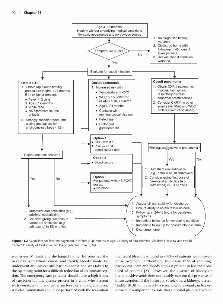
90 | Chapter 11
Age 3–36 months Healthy without underlying medical conditionsNontoxic appearance and no obvious source
1. No diagnostic testing required2. Discharge home with follow-up in 48 hours if fever persists3. Reevaluation if condition worsens
Evaluate for occult infection
Occult pneumonia
1. Obtain CXR if patient has hypoxia, tachypnea, respiratory distress, abnormal breath sounds2. Consider CXR if no other source identified and WBC
�20,000/mm (if obtained)
Occult UTI1. Obtain rapid urine testing and culture in girls �24 months if 1 risk factor present:
Fever � 2 daysAge �12 monthsWhite raceNo alternative sourceof fever
2. Strongly consider rapid urine testing and culture for uncircumcised boys �12 m
Rapid urine test positive?
1. Outpatient oral antibiotics (e.g., cefixime, cephalexin)2. Consider giving first dose of parenteral antibiotics (e.g., ceftriaxone) in ED or office
Findings suggestive of pneumonia?
1. Assess clinical stability for discharge2 Ensure ability to obtain follow-up care3. Follow-up in 24–48 hours for persistent symptoms4. Immediate follow-up for worsening condition5. Immediate follow-up for positive blood culture6. Discharge home
1. Outpatient oral antibiotics (e.g., amoxicillin, azithromycin)2. Consider giving first dose of parenteral antibiotics (e.g., ceftriaxone) in ED or office
Occult bacteremia 1. Increased risk with
Temperature � 40ºC
WBC � 15,000/mm3
or ANC �10,000/mm3
Age 6–24 months
Contacts withmeningococcal disease
Petechiae
Prolongedgastroenteritis
Temperature � 39°C
Yes No
Yes No
No
Yes
Option 2Blood culture
Option 3For patients with � 3 PCV7doses:
No blood
Option 1CBC with diffIf WBC >15k,blood culture and
Figure 11.2 Guidelines for fever management in infants 3–36 months of age. Courtesy of Paul Ishimine, Children’s Hospital and HealthCenter/University of California, San Diego (adapted from [5, 6] ).
was given IV fluids and discharged home. He returned thenext day with bilious emesis and frankly bloody stools. Heunderwent an unsuccessful barium enema and was taken tothe operating room for a difficult reduction of an intussuscep-tion. The emergency care provider should have a high indexof suspicion for this disease process in a child who presentwith vomiting only and either no fever or a low-grade fever.A rectal examination should be performed with the realization
that rectal bleeding is found in �40% of patients with provenintussusception. Furthermore, the classic triad of vomiting,paroxysmal pain and bloody stools is present in less than onethird of patients [23]. However, the absence of bloody orheme-positive stools does not reliably rule out the presence ofintussuception. If the history is suspicious, a kidneys, ureter,bladder (KUB) or preferably, a screening ultrasound can be per-formed. It is important to note that a normal plain radiograph
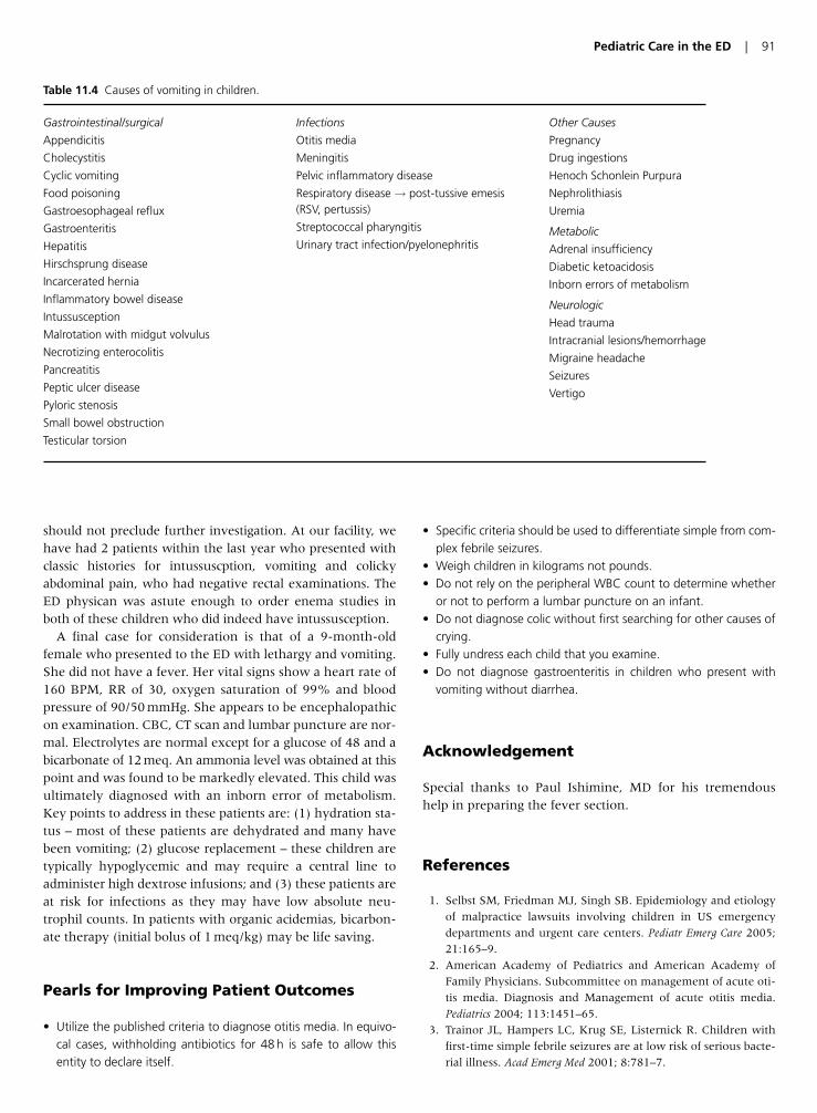
should not preclude further investigation. At our facility, wehave had 2 patients within the last year who presented withclassic histories for intussuscption, vomiting and colickyabdominal pain, who had negative rectal examinations. TheED physican was astute enough to order enema studies inboth of these children who did indeed have intussusception.
A final case for consideration is that of a 9-month-oldfemale who presented to the ED with lethargy and vomiting.She did not have a fever. Her vital signs show a heart rate of160 BPM, RR of 30, oxygen saturation of 99% and bloodpressure of 90/50 mmHg. She appears to be encephalopathicon examination. CBC, CT scan and lumbar puncture are nor-mal. Electrolytes are normal except for a glucose of 48 and abicarbonate of 12 meq. An ammonia level was obtained at thispoint and was found to be markedly elevated. This child wasultimately diagnosed with an inborn error of metabolism.Key points to address in these patients are: (1) hydration sta-tus – most of these patients are dehydrated and many havebeen vomiting; (2) glucose replacement – these children aretypically hypoglycemic and may require a central line toadminister high dextrose infusions; and (3) these patients areat risk for infections as they may have low absolute neu-trophil counts. In patients with organic acidemias, bicarbon-ate therapy (initial bolus of 1 meq/kg) may be life saving.
Pearls for Improving Patient Outcomes
• Utilize the published criteria to diagnose otitis media. In equivo-cal cases, withholding antibiotics for 48 h is safe to allow thisentity to declare itself.
• Specific criteria should be used to differentiate simple from com-plex febrile seizures.
• Weigh children in kilograms not pounds.• Do not rely on the peripheral WBC count to determine whether
or not to perform a lumbar puncture on an infant.• Do not diagnose colic without first searching for other causes of
crying.• Fully undress each child that you examine.• Do not diagnose gastroenteritis in children who present with
vomiting without diarrhea.
Acknowledgement
Special thanks to Paul Ishimine, MD for his tremendoushelp in preparing the fever section.
References
1. Selbst SM, Friedman MJ, Singh SB. Epidemiology and etiology
of malpractice lawsuits involving children in US emergency
departments and urgent care centers. Pediatr Emerg Care 2005;
21:165–9.
2. American Academy of Pediatrics and American Academy of
Family Physicians. Subcommittee on management of acute oti-
tis media. Diagnosis and Management of acute otitis media.
Pediatrics 2004; 113:1451–65.
3. Trainor JL, Hampers LC, Krug SE, Listernick R. Children with
first-time simple febrile seizures are at low risk of serious bacte-
rial illness. Acad Emerg Med 2001; 8:781–7.
Pediatric Care in the ED | 91
Gastrointestinal/surgical
Appendicitis
Cholecystitis
Cyclic vomiting
Food poisoning
Gastroesophageal reflux
Gastroenteritis
Hepatitis
Hirschsprung disease
Incarcerated hernia
Inflammatory bowel disease
Intussusception
Malrotation with midgut volvulus
Necrotizing enterocolitis
Pancreatitis
Peptic ulcer disease
Pyloric stenosis
Small bowel obstruction
Testicular torsion
Infections
Otitis media
Meningitis
Pelvic inflammatory disease
Respiratory disease→ post-tussive emesis(RSV, pertussis)
Streptococcal pharyngitis
Urinary tract infection/pyelonephritis
Other Causes
Pregnancy
Drug ingestions
Henoch Schonlein Purpura
Nephrolithiasis
Uremia
Metabolic
Adrenal insufficiency
Diabetic ketoacidosis
Inborn errors of metabolism
Neurologic
Head trauma
Intracranial lesions/hemorrhage
Migraine headache
Seizures
Vertigo
Table 11.4 Causes of vomiting in children.

4. Warden CR, Zibulewsky J, Mace S, Gold C, Gausche-Hill M.
Evaluation and management of febrile seizures in the out-of-
hospital and emergency department settings. Ann Emerg Med
2003; 41:215–22.
5. American College of Emergency Physicians Clinical Policies
Committee; American College of Emergency Physicians Clinical
Policies Subcommittee on Pediatric Fever. Clinical policy for chil-
dren younger than three years presenting to the emergency
department with fever. Ann Emerg Med 2003; 42:530–45.
6. Baraff LJ. Management of fever without source in infants and
children. Ann Emerg Med 2000 Dec; 36(6):602–14.
7. Bonsu B, Harper M. Utility of the peripheral blood white blood
cell count for identifying sick young infants who need lumbar
puncture. Ann Emerg Med 2003; 41:206–14.
8. Bonsu B, Harper M. Identifying febrile young infants with bac-
teremia: Is the peripheral white blood cell count an accurate
screen? Ann Emerg Med 2003; 42(2):216–25.
9. Kizer E, Scolnik D, Macpherson A, et al. Variables associated
with medication errors in pediatric emergency medicine.
Pediatrics 2002; 110;737–42.
10. Wessel MA, Cobb JC, Jackson EB, et al. Paroxysmal fussing in
infancy sometimes called colic. Pediatrics 1954; 14:421–34.
11. Poole SR. The infant with acute, unexplained, excessive crying.
Pediatrics 1991; 88:450–5.
12. Herman M, Nelson R. Critical Decision in emergency medicine
2006; 20:2–10.
13. Pena BM, Harper MB, Fleisher GR. Occult bacteremia with
group B streptococci in an outpatient setting. Pediatrics 1998;
102:67–72.
14. Baskin MN, O’Rourke EJ, Fleisher GR. Outpatient treatment of
febrile infants 28 to 89 days of age with intramuscular adminis-
tration of ceftriaxone. J Pediatr 1992; 120(1):22–7.
15. Baker MD, Bell LM, Avner JR. Outpatient management with-
out antibiotics of fever in selected infants. New Engl J Med 1993;
329(20):1437–41.
16. Kadish HA, Loveridge B, Tobey J, Bolte RG, Corneli HM.
Applying outpatient protocols in febrile infants 1–28 days of
age: can the threshold be lowered? Clin Pediatr 2000; 39:81–8.
17. Dagan R, Sofer S, Phillip M, Shachak E. Ambulatory care of
febrile infants younger than 2 months of age classified as being
at low risk for having serious bacterial infections. J Pediatr 1988;
112(3):355–60.
18. Jaskiewicz JA, McCarthy CA, Richardson AC, et al. Febrile
infants at low risk for serious bacterial infection – an appraisal
of the Rochester criteria and implications for management.
Febrile Infant Collaborative Study Group. Pediatrics 1994;
94(3):390–6.
19. Ferrera PC, Bartfield JM, Snyder HS. Neonatal fever: utility of
the Rochester criteria in determining low risk for serious bacte-
rial infections. Am J Emerg Med 1997; 15(3):299–302.
20. Garra G, Cunningham SJ, Crain EF. Reappraisal of criteria used
to predict serious bacterial illness in febrile illness in febrile
infants less than 8 weeks of age. Acad Emerg Med 2005;
12:921–5.
21. Ishimine P. Fever without source in children 0–36 months of
age. Pediatr Clin N Am 2006; 53:167–94.
22. Al-Orifi F, McGillivray D, Tange S, et al. Urine culture from bag
specimens in young children: are the risks too high?
J Pediatr 2000; 137(2):221–6.
23. Yamamoto LG, Morita SY, Boychuk RB, et al. Stool appearance
in intussusception: assessing the value of the term “currant
jelly”. Am J Emerg Med 1997; 15:293.
92 | Chapter 11
Introduction
The number of patients living with some form of hemato-logic or solid organ malignancy is rising. As treatment effi-cacy continues to improve, the number of patients withmalignancies is increasing. Emergency physicians (EP) canexpect to see increasing numbers of patients with these dis-eases presenting to emergency departments (EDs). Cancer iscurrently the second leading cause of death in the US, and itis estimated that one death in four is due to cancer [1].
Most of the oncologic urgencies and emergencies that pres-ent to EDs are actually, to some degree, amenable to therapy orintervention. Patients with these malignancies may presentwith a condition that, once treated, will allow them to lead rel-atively normal lives. Since hematologic and oncologic malig-nancies are so common, EPs must be experts in the recognitionand initial management of cancer-related emergencies.
From a practical standpoint, hematologic and oncologicemergencies can be divided into three main categories: infec-tious, mechanical, and metabolic. The most important infec-tious complication is fever in the setting of neutropenia.Mechanical emergencies include airway obstruction, superiorvena cava (SVC) syndrome, cardiac tamponade, and spinalcord compression. Metabolic emergencies make up the thirdgroup of emergencies and include entities such as the hyper-viscosity syndrome, hypercalcemia, syndrome of inappropriateantidiuretic hormone secretion (SIADH), and acute tumorlysis syndrome. Because of some of the unique characteristicsof cancer patients, it is essential that EPs seek out and treatthese emergencies to optimize the outcomes for their patients.
The purpose of the following review is to emphasize thesubtleties and atypical nature of some of the more commonhematologic and oncologic urgencies and emergencies, andto highlight important diagnostic and treatment errors thatoccur in daily clinical practice. By having an appreciation ofnon-classic disease presentations and the clinical errors thatoccur in these patients, the EP can truly have an impact onmorbidity and mortality in the cancer patient.
Pitfall | Failure to recognize and rapidlyinitiate appropriate antibiotic therapywhen treating neutropenic fever
Of all the emergencies in hematology and oncology, fever in the setting of neutropenia is one for which expeditious
recognition and management may significantly lessen mor-bidity and mortality. Rapid treatment of this condition isonly possible if the treating physician recognizes that a pre-existing hematologic or oncologic diagnosis exists or thatchemotherapy has been recently instituted. Absent of ahematologic malignancy treated by chemotherapy, otherconditions that lead to neutropenia, such as agranulocytosisare quite rare.
Neutropenia is said to exist when the total white blood cell(WBC) count is �500 cells/�l or a count of �1000 cells/�lwith an anticipated decrease to �500 cells/�l. Most patientswill have their nadir between 2 and 4 weeks afterchemotherapy is initiated. Thus, patients currently receivingchemotherapy with a total WBC count �1000/�l may be ontheir way to approaching 500 cells/�l, depending on thetime frame. In equivocal cases, the patient’s oncologistshould be contacted to collaboratively develop a treatmentplan with the EP. Rates of serious infection climb signifi-cantly when total WBC counts fall below 500 cells/�l [2].Fever in the neutropenic patient is present if the temperatureexceeds �38.3° (101°F) or if the temperature � 38.0°C(100.4°F) for �1 . Special care must be taken with patientswho complain of fever but who arrive afebrile. Patients whoreport a fever meeting the above criteria prior to arrival inthe ED should be treated as if they have a fever.
Patients with neutropenia may have few of the classicsigns and symptoms of bacterial infections because of aninadequate number of circulating WBCs. Specific examplesof altered presentations include the following:
1. A patient with a significant pneumonia may have nocough and no infiltrates on plain film.2. A patient with a urinary tract infection may present in a sub-tle fashion because WBCs do not deposit in the urinary tractand produce the typical symptoms of dysuria and frequency.3. Meningitis may present without clinical evidence ofmeningismus or cerebral spinal fluid pleocytosis.
Therefore, febrile patients with confirmed or suspected neu-tropenia should have cultures of blood and urine obtained
Chapter 12 | Management of Hematology/Oncology Patients in the ED
Robert L. Rogers
93
KEY FACT | Afebrile patients who report a history offever should be treated as if they are currently febrile.
and a chest X-ray performed. It is important to initiateempiric antibiotic therapy early and to provide broad cover-age since the source of infection is often unknown. There issome data that a chest computed tomography (CT) may bethe best initial choice to evaluate for pulmonary infection.Neutropenic patients with significant pulmonary infections,such as aspergillosis, may have a normal chest X-ray despitea remarkably abnormal chest CT [2]. If the patient has anindwelling central line in place, at least one set of blood cul-tures should be obtained through the line. Fever in the set-ting of neutropenia can rapidly deteriorate despite initiallybeing stable. A thorough physical examination shouldalways be performed to search for an underlying infection.
Progression of infection in neutropenic patients may be rapid,so empiric therapy should be started promptly at onset offever. Initial antibiotic therapy should be tailored to the sus-pected offending pathogens. In the not too distant past, neu-tropenic patients were empirically treated with twoantibiotics, typically a third-generation cephalosporin in com-bination with an aminoglycoside. Gram-negative bacilli,including Pseudomonas, Escherichia coli, and Klebsiella, havebeen the most common offending pathogens. Recently, how-ever, there has been an increase in the rate of infectionscaused by gram-positive bacteria, many of them being methi-cillin-resistant organisms [2]. Staphylococcus aureus and coagu-lase-negative staphylococci are the most common causes,particularly if the patient has an indwelling catheter forchemotherapy or transfusion. There is now evidence to sup-port single agent antimicrobial therapy. A third- or fourth-generation cephalosporin (ceftazidime or cefipime) or acarbapenem (imipenem–cilastatin or meropenem) can be usedas monotherapy [2]. Including vancomycin in the treatmentregimen would be indicated if a catheter-related infection issuspected, if the patient has known colonization with methi-cillin-resistant Staphylococcus aureus (MRSA), positive bloodculture results with gram-positive bacteria before an identifica-tion is available, or if hypotension or sepsis is present [2].
Pitfall | Failure to identify malignant spinalcord compressive lesions
Back pain is a common and generally benign complaintencountered in the ED. As such, it is easy to minimize theetiology of patients with this complaint. In patients with ahistory of malignancy, spinal metastases must be considered.What can EPs do to lessen their risk of missing this potentiallycatastrophic entity? And what therapies can be rapidly insti-tuted to lessen morbidity?
As many as 5% of all cancer patients will develop metas-tases to the spine and spinal cord at some point in the courseof their disease [3]. Epidural spinal cord compression is adevastating complication of many malignancies, includingbreast, lung, and prostate cancer. Epidural spinal cord com-pression may be the first clinical manifestation of malig-nancy. Patient outcomes have been shown to be related toearly diagnosis and rapid institution of therapy [4]. This mayoffer patients with advanced cancer the opportunity to max-imize their quality of life during their final days.
Often, patients with a known diagnosis of cancer (breast,lung, prostate, renal) present for evaluation of back pain ornew onset radiculopathy. EPs should maintain a high index of suspicion for potentially dangerous back pain entities, suchas cancer, cauda equina syndrome, and abdominal aorticaneurysm, Any patient with a known history of cancer, nomatter how remote, should be worked-up for this entity if theback pain is unexplained. Likewise, in patients with recentweight loss or other signs or symptoms of malignancy such asunexplained fever, an undiagnosed malignancy should beconsidered. There have been some case reports of recurrenceof a previously diagnosed and treated cancer presenting as anepidural metastasis with or without cord compression [5].
Patients with early spinal cord compression may present witha variety of clinical findings. Common complaints includenumbness, tingling, sensory loss, bowel or bladder inconti-nence, or abnormalities of proprioception, such as loss ofvibratory sense. A detailed motor, sensory, and deep tendonreflex examination should be performed in all patients withsuspected spinal cord compression, as well as observation ofgait and evaluation of sphincter tone. Back pain worsened byparticular maneuvers, such as coughing or lying in a supineposition, should be considered suspect, as should pain that isworse at night. Most mechanical back pain entities do notcause worsening of pain with these maneuvers. New-onsetradiculopathy from compression of a spinal root may also bethe first manifestation of tumor. Many cases of spinal cordmetastasis have been missed because of failure to considermetastasis to the epidural space and spine [5].
KEY FACT | Back pain that with neurological findings,atypical features, or in patients with symptomssuggestive of undiagnosed cancer should have urgentfollowup arranged.
94 | Chapter 12
KEY FACT | Appropriate initial empiric antibioticchoices include a third or fourth-generation cephalosporina carbapenum. Vancomycin should be added for patientsat risk for MRSA.
KEY FACT | Patients with suspected spinal malignanciesrequire magnetic resonance imaging (MRI) thought theurgency of the scan depends on the patients symptomsand findings. Patients with identified compressive lesionsshould receive corticosteroids and neurosurgicalconsultation.
For patients with suspected spinal lesions, MRI has becomethe modality of choice in most centers and should beobtained in all cases of suspected spinal cord compression. Thetiming of the study depends on the patients symptoms andfindings. Patients with new motor deficits or symptoms consis-tent with cauda equina syndrome should have definitiveimaging while in the ED. Those without findings should haveurgent follow-up arranged so the study can be performed andfollowed-up rapidly. Patients with cord compression requireneurosurgical consultation and high-dose corticosteroids.
Pitfall | Failure to consider the diagnosis ofSVC syndrome
Generally, SVC syndrome is an urgency rather than anemergency. Two situations in which this entity becomes anemergency are cases in which cerebral edema or in whichlaryngeal edema have developed. This can lead to life-threatening cerebral herniation and airway compromise,respectively. In the past, the most common causes of SVCsyndrome were tuberculosis, aortic aneurysm, and fibrosingmediastinitis. Today, lung carcinoma and other malignanciessuch as lymphoma are the most common causes. One par-ticular cause with an increasing in prevalence is catheter-related SVC thrombosis [6].
Clinical manifestations of SVC syndrome are protean anddepend on the degree of vena cava obstruction. Patientsmay be relatively asymptomatic and have only mild facialfullness when bending forward or may have florid symp-toms of facial swelling, headache, and airway compromise.The key to understanding the variability of the clinical pres-entation is the anatomy of the azygous vein. The azygousvein is a large vessel that enters the proximal SCV anddrains blood from the thorax. Obstruction above the level ofthe azygous entry point may lead to relatively few symp-toms due to the ability of the azygous to decompress theupper extremities, head, and neck. If a compressive lesion orthrombus obstructs the SVC below the entry point there isno mechanism for upper torso, head, and neck decompres-sion, and patients may present with marked venous collat-eral formation on the chest wall, neck, and shoulders andfacial swelling on examination [7].
A clue to the diagnosis is the presence of asymmetric neck,chest, or upper arm venous distension. It is often helpful tocompare the patient’s appearance to that of a picture of thepatient. If the patient does not have any photographs, a driv-ers license or other identification card picture can often behelpful to identify changes the patient’s appearance.
Traditional teaching is that most patients will have a sen-sation of facial fullness, facial swelling, cough, variable armswelling, and dilated neck and upper chest wall veins. SCVsyndrome would not be a difficult diagnosis to make in thisscenario. However, SNV syndrome may present with few ifany clinical findings. The one symptom reported by many
patients is a sensation of fullness when bending forward, or avague, chronic cough. However, if venous decompression isable to occur, there will be no prominent veins or facialswelling. Thus, the diagnosis may not even be considered.
Treatment is aimed at relieving congestive symptoms. Incases where suspected or confirmed lung cancer or lym-phoma is the cause, institution of radiation therapy mayhelp lessen venous congestion and reduce upper torso,head, and neck venous pressure. Though SVC syndromewas previously considered an indication for emergent radia-tion therapy, this treatment should be reserved for patientswith life-threatening laryngeal or cerebral edema. Patientswith a confirmed or suspected diagnosis of SVC syndromebased on clinical grounds should undergo CT scanning todefine the degree of SVC obstruction and to evaluate for thepossibility of thrombotic SVC occlusion [8]. If there are nolife-threatening features, this can be done as an outpatient if urgent follow-up can be arranged. Adjunctive treatmentssuch as steroids and diuretic therapy may be used, particu-larly if head and neck edema is a prominent feature.
Pitfall | Failure to identify malignantpericardial tamponade as a cause ofdyspnea
Pericardial effusions and tamponade are known complicationsof many malignancies including lung and breast cancer, lym-phoma, and leukemia. Patients with malignancy may presentfor evaluation with a stable pericardial effusion, or may pres-ent in extremis or in cardiac arrest secondary to hemodynami-cally significant cardiac tamponade. Because malignancies areoften identified late in their course, cardiac tamponade may bethe initial manifestation of malignancy. In a small study of 23cases of neoplastic cardiac tamponade, 8 cases were the initialpresentation of the underlying malignancy [9].
It is critical to remember that patients with tamponadephysiology may be relatively hemodynamically stable [10].Guberman et al. studied 56 cases of tamponade on a medicalservice and noted that most of the patients did not fit theclassic description. Importantly, most of the patients had anormal blood pressure and no jugular venous distension oninitial presentation [11]. Other studies have evaluated theclassic physical examination findings and have found that asmany as 30% of patients with tamponade lack classic find-ings [12]. More importantly, it is evident from the literaturethat patients with significant tamponade on the verge ofdecompensation and cardiac arrest may exhibit very fewclinical indicators suggesting the severity of the tamponade[11]. Many cases of neoplastic cardiac tamponade present
Management of Hematology/Oncology Patients in the ED | 95
KEY FACT | Patients with tamponade physiology areoften hemodynamically stable on initial presentation.
with dull or vague chest tightness, shortness of breath, andfatigue. Other clues to the diagnosis include low blood pres-sure, a narrowed pulse pressure, jugular venous distension,low voltage ECG, or cardiomegaly without evidence of heartfailure on chest radiography, however their absence shouldnot be used in isolation to exclude this process.
Treatment of neoplastic cardiac tamponade depends uponthe hemodynamic stability of the patient. Unstable patientsor patients who present in cardiac arrest (pulseless electricalactivity) should undergo needle pericardiocentesis. In a car-diac arrest situation this is commonly performed blindly. Forpatients who are hemodyamically stable pericardiocentesiscan also be performed by ECG guidance or under fluo-roscopy. With the increased use of bedside ultrasonographyin EDs, pericardial effusions can be more effectively local-ized and safely performed [13]. Consultation with a cardiol-ogist and a cardiothoracic surgeon should be undertakenbecause in some cases a definitive procedure such as a peri-cardial drain or window will need to be performed.
Pitfall | Failure to identify hyperviscositysyndrome as a cause for dyspnea or alteredmental status
Hyperviscosity syndrome and leukostasis syndrome may beone of the most difficult hematologic emergencies to detectin the ED. Hyperviscosity syndrome is caused by increasedblood viscosity while leukostasis syndrome is caused bymarked leukocytosis.
Signs and symptoms of hyperviscosity typically manifestwhen serum viscosity reaches five times the viscosity of water.Normal human serum is 1.4 to 1.8 times as viscous as water.Clinical manifestations are directly related to high viscosityand can be caused by multiple hematologic disorders, suchas Waldenstrom’s macroglobulinemia, multiple myeloma,acute leukemia with blast crisis, and polycythemia vera[14]. Patients with a known diagnosis of any one of thesedisorders who present with mental status changes, unex-plained dyspnea, or headache should be suspected of havinga hyperviscosity syndrome and should have appropriatetherapy instituted. Clinical presentations will be dictated byflow characteristics of different blood vessels and are directly
related to microcirculatory occlusion [15]. This may manifestas derangements in central nervous system (CNS) functionor hypoxemia and respiratory failure.
The important dilemma for EPs is in detecting this disorderde novo in a patient without a known hematologic diagnosis.One clinical pearl is to consider a hyperviscous state inpatients when the laboratory has trouble running their bloodthrough the laboratory equipment. Serum viscosity isreported in Centipoises. A normal serum viscosity is less than1.8, and clinical symptoms are rare unless serum viscosity isgreater than 4.0 Centipoises. In some hospitals the test can beperformed in an hour, whereas in others it is a send out labo-ratory test with a longer turnaround time. The EP should alsostrongly consider this diagnosis in cases of unexplained comaor unexplained shortness of breath [14]. Given that there isno known gold standard for diagnosis, EPs should have a highindex of suspicion for this disorder in patients with clinicaldeterioration with an underlying hematologic disease.
Treatment in the ED should be aimed at lowering serumviscosity. If a patient already has a diagnosis, this will almostalways involve consultation with the patient’s hematologist.Therapy is instituted to correct the causative factor of thehyperviscosity syndrome. Management of this conditionshould involve lowering of the cellular or protein componentsof blood [15]. In cases of multiple myeloma, this mightinvolve plasmapheresis to lower serum protein levels. Theprimary role of the EP is to assess the patient’s airway, breath-ing, and circulation, initiate resuscitative efforts, and to beginaggressive intravenous hydration. Phlebotomy of 2–3 units ofblood is also an effective temporizing treatment modality.
Leukostasis, or hyperleukocytosis, is a condition defined by tis-sue hypoperfusion secondary to elevated WBCs. This is seen insome cases of acute leukemia. This particular syndrome is typ-ically seen when total WBC counts approach 100,000/�l butcan be seen in certain types of monoblastic leukemias whenthe total WBC count is only 25–50,000/�l [16].
As with hyperviscosity syndrome, the central nervous andpulmonary systems are the most commonly involved inleukostasis syndrome. Patients may present with new onsetor unexplained shortness or breath, respiratory failure, anddepressed mental status. Infiltration of the gastrointestinal(GI) tract or liver may lead to GI bleeding or liver failure[16]. Diagnosis is typically made in the setting of organ dys-function and a markedly elevated peripheral WBC count.
96 | Chapter 12
KEY FACT | Treatment of malignant pericardial effusioninvolves drainage of the pericardial fluid. The techniqueand timing must be determined by the hemodynamicstability of the patient.
KEY FACT | Hyperviscosity syndrome must beconsidered in patients with mental status changes ordyspnea and a history of hematologic malignancy.
KEY FACT | Leukostasis syndrome is most commonlysymptomatic when leukocyte counts approach 100,000/�lbut can be seen with counts of 25000–50,000/�l
KEY FACT | Patients with leukostasis syndrome shouldreceive aggressive hydration and undergo whole bloodphlebotomomy in the ED.
Treatment requires a multifaceted approach. Aggressive intra-venous hydration should be rapidly instituted in the ED. Ahematologist should be contacted in order to plan definitivechemotherapy. In the ED, the key principles of treatmentinclude measures to rapidly lower peripheral WBC countsand to prepare for the inevitable development of the tumorlysis syndrome. This syndrome is composed of hyperkalemia,hyperuricemia, hyperhosphatemia, and renal failure andresults when leukemic blasts or lymphoma cells undergo celldeath with release of intracellular contents [17, 18].Treatments include oral administration of phosphate bindersand allopurinol along with aggressive intravenous hydration[19]. An initial dose of 600 mg of oral allopurinol can be givenin the ED. Alternatively, intravenous Rasbiricase, a recombi-nant urate oxidase, can be given in an initial dose of0.05–0.2 mg/kg intravenously. This drug has been shown tobe extremely effective at reducing serum urate levels and canprevent the development of acute renal failure [19]. The mostcommonly used phosphate binder is aluminum hydroxideand is generally given in a dose of 50–150 mg/kg orally daily.Leukapheresis or whole blood phlebotomy can be used tolower the white count until chemotherapy has begun. Thiscan be performed in the ED as a temporizing measure untildefinitive care is undertaken. Whole blood phlebotomy canbe performed by withdrawing 1–2 units of whole blood in aneffort to temporarily lower serum viscosity. In addition,hydroxyurea in a dosage of 50–100 mg/kg daily can be givenwhile awaiting chemotherapy.
Another important pitfall is transfusing red blood cells tothese patients. The combination of WBCs, red blood cells,platelets, and other serum constituents make up an individ-ual patient’s cytocrit. The cytocrit is related to the serum vis-cosity. Higher the cytocrit, higher is the viscosity. Sincemany, if not most, patients with leukostasis have leukemiaas the underlying diagnosis, almost all will be severely ane-mic secondary to bone marrow suppression. The temptationis to transfuse blood products. This should be performedwith extreme caution since even small elevations of thepatient’s cytocrit may cause worsening small vessel ischemiaand result in worsening hypoxia and CNS hemorrhage.
Pitfall | Not Considering the Diagnosis ofthe SIADH and Failure to AppropriatelyTreat
One of the more common oncologic entities seen in EDs isthe SIADH. This syndrome is caused by release of antidiuretichormone from ectopic sites such as a lung cancer or CNS
infection and leads to hyponatremia. When severe, hypona-tremia constitutes a true oncologic emergency [20].
Hyponatremia is the most frequent electrolyte disorder inclinical medicine. This frequently chronic and often stablecondition may become life threatening when serum sodiumconcentration continues to fall and patients develop CNSmanifestations such as coma or seizures. Symptoms mayalso include anorexia, weakness, confusion, and changes inmental status.
It is imperative that EPs know how fast to treat hypona-tremia. The most feared complication of excessively rapid cor-rection of hyponatremia is the osmotic demyelinationsyndrome (ODS), formerly known as central pontine myeli-nolysis (CPM), a devastating condition in which there isdestruction of neurons and edema of the pons and midbrain[21]. It is generally agreed that acute, symptomatic hypona-tremia (seizures or coma) should be treated aggressively toraise the serum sodium rapidly to a level above a theoreticalseizure threshold. The literature most often quotes a level ofsomewhere between 120 and 130 meq/dl [21]. A good rule ofthumb for dealing with hyponatremia in the ED is to correctchronic hyponatremia slowly and acute hyponatremia quickly.The problem with this traditional teaching is that most of thetime EPs are presented with an ill-appearing hyponatremicpatient with an unknown history and unknown duration ofhyponatremia. Frequently the decision to institute therapymust be done with little if any past medical history.
What is evident from the literature is that acute, sympto-matic hyponatremia (seizures and/or coma) should betreated rapidly with hypertonic (3%) saline [21]. Hypertonicsaline can be administered at a rate of approximately1–2 cc/kg/h in order to raise the serum sodium by 2.5 meq/h.In the ED, this can be safely accomplished by administering100–200 cc of 3% saline over a few hours. Thus, if a patientwith a sodium of 105 is actively seizing, hypertonic saline isgiven to correct the serum level to the seizure threshold,somewhere around 115–120 meq/l. The whole point of thistreatment is to raise the serum sodium by approximately10 meq/l, which in many cases will stop seizure activity. Thehypertonic saline should then be stopped. Frequent labora-tory assessments must be performed to assure that thedesired rate and level correction is not exceeded. It can alsobe determined precisely how much hypertonic saline toadminister and how fast according to the patient’s sodiumlevel on presentation and the goal sodium level. Since thiscan be tedious and time consuming for the practicing EP, it is
Management of Hematology/Oncology Patients in the ED | 97
KEY FACT | Blood transfusion should be avoided inpatients with leukostasis syndrome as it can worsen smallvessel ischemia.
KEY FACT | Rapidity of correction of hyponatremia isdetermined by the patient’s symptoms. Hypertonic salineshould be reserved for those actively seizing. Normalsaline or fluid restriction should be administered to thosewith milder disease.

not discussed here. As a rule of thumb, hyponatremia that ischronic in nature should be corrected slowly at a rate of nomore than 0.5 meq/h or 12 meq over a 24-h period.Treatment of mild hyponatremia secondary to SIADHincludes free water restriction (500 cc – 1 l/day) with closefollow-up by the patient’s oncologist or administration ofdemeclocycline, a tetracycline derivative, which producesnephrogenic diabetes insipidus. It is usually given in a doseof 250 mg four times a day and is usually started after dis-cussion with the patient’s oncologist.
Pearls for Improving Patient Outcomes
• All febrile patients with a history of cancer undergoing treat-ment with chemotherapy should be considered to be neu-tropenic until proven otherwise. They should receive broadspectrum antibiotics early.
• Spinal metastasis and cord compression may be the first symp-tom of a malignancy.
• Neoplastic cardiac tamponade may be the first clinical manifesta-tion of cancer. Cancer patients who present with unexplainedcardiopulmonary symptoms should have this diagnosis ruled out.
• The diagnosis of hyperviscosity syndrome should be consideredin all patients with hematologic malignancies who present witha constellation of unexplained signs and symptoms. Promptinstitution of measures to decrease serum viscosity should be initiated in the ED.
• Blood transfusion is to be avoided unless absolutely necessary inpatients with leukostasis syndrome.
• Hypertonic saline should be reserved for hyponatremic patientswho are actively seizing or have altered mental status.
References
1. Current Clinical Issues-Cancer chemotherapy: Teaching old
drugs new tricks. Annals of Int Med 2001; 135:1107–10.
2. Hughes WT, Armstrong D, Bodey GP, et al. Guidelines for the
use of antimicrobial agents in neutropenic patients with cancer-
IDSA guidelines. Clin Infect Dis 2002; 34:730–51.
3. Posner JB. Back pain and epidural spinal cord compression.
Med Clin N Am 1987; 71:185–204.
4. Kim RY, Spencer SA, Meridith RF, et al. Extradural spinal cord
compression: analysis of factors determining functional
progress, prospective study. Radiology 1990; 176:279–82.
5. Helweg-Larson S, Sorensen PS. Symptoms and signs in metasta-
tic spinal cord compression: a study of progression from first
symptoms until diagnosis in 153 patients. Eur J Cancer 1994;
3A:396–99.
6. Nieto AF, Doty DB. Superior vena cava obstruction: clinical
syndrome, etiology, and treatment. Curr Probl Cancer 1986;
10:441–84.
7. Maguire WM. Mechanical complications of cancer. Emerg Med
Clin N Am 1993; 11:421–30.
8. Schwartz EE, Goodman LR, Haskin ME. Role of CT scanning in
the superior vena cava syndrome. Am J Clin Oncol 1986;
9:71–78.
9. Haskel RJ, French WJ. Cardiac tamponade as the initial presen-
tation of malignancy. Chest 1985; 88(1):70–73.
10. Kralstein J, Fishman WH. Malignant pericardial disease: diag-
nosis and treatment. Cardiol Clin 1987; 5:583–89.
11. Guberman BA, Fowler NO, Gueron M, et al. Cardiac tampon-
ade in medical patients. Circulation 1981; 64(3):633–40.
12. Levine MJ, Lorell BH, Diver DJ, et al. Implications of
echocardiographically-assisted diagnosis of pericardial tampon-
ade in contemporary medical patients: detection before
hemodynamic embarrassment. J Am Coll Cardiol 1991; 17(1):
59–65.
13. Blavais M. Incidence of pericardial effusion in patients present-
ing to the emergency department with unexplained dyspnea.
Acad Emerg Med 2001; 8(12):1143–6.
14. Kwaan HC, Bongu A. The hyperviscosity syndromes. Semin
Thromb Hemost 1999; 25(2):199–208.
15. Coppell J. Consider ‘hyperviscosity syndrome’ in unexplained
breathlessness. Acta Haematol 2000; 104(1):52–53.
16. Markman M. Common complications and emergencies associ-
ated with cancer and its therapy. Cleve Clin J Med 1994;
12:105–14.
17. Arrambide K, Toto RD. Tumor lysis syndrome. Semin Nephrol
1993; 13:273–80.
18. Alkhuja S, Ulrich H. Acute renal failure from spontaneous
acute tumor lysis syndrome: a case report and review of the lit-
erature. Ren Fail 2002; 24(2):227–32.
19. Davidson MB, Thakkar S, Hix JK, et al. Pathophysiology, clini-
cal consequences, and treatment of tumor lysis syndrome. Am J
Med 2004; 116:546–54.
20. Lin M, Liu SJ, Lim IT. Disorders of water imbalance. Emerg Med
Clin N Am 2005; 23(3):749–70.
21. Adrogue HJ, Madias NE. Primary Care: Hyponatremia. New Eng
J Med 2000; 342:1581–89.
98 | Chapter 12
Introduction
Patients who are intoxicated, violent, or both often receivesuperficial or delayed medical evaluations in the emergencydepartment (ED). A number of reasons for this exist, as thesepatients may be unpleasant, disrespectful to staff, and oftenhave poor hygiene. Some staff may prefer that these patientsbe “quickly dispositioned,” as they may not be comfortablecaring for them, may not feel appreciated by them, or mayfeel their safety is threatened. Furthermore, these patients canbe disruptive to the ED and other patients. Often, intoxicatedor violent patients are given premature diagnoses based onstereotypes or presumptions, especially if they have had priorED visits. They may be kept in the waiting room or relegatedto corners of the department so that priority can be given toother patients.
Unfortunately, the management of intoxicated and vio-lent patients involves several high-risk medical and legalissues. Both groups may have serious underlying illnessesthat must be rapidly identified and addressed. At the sametime, emergency physicians (EPs) need to institute appropri-ate measures to protect themselves, other staff, the patient,and innocent third parties. Finally, EPs must be aware of andcomply with local, state, and federal regulations with respectto intoxicated and/or violent patients.
Pitfall | Failure to perform a comprehensivemedical evaluation
Intoxicated patients should be evaluated with the assump-tion that an underlying medical or traumatic condition existsuntil proven otherwise. Some patients may appear intoxi-cated despite an alternative diagnosis causing their symptoms.Behaviors associated with intoxication, such as slurredspeech, ataxia, sleepiness, or confusion may also be the resultof metabolic disorders (hypoglycemia, hypoxia, uremia,Wernicke’s encephalopathy, diabetic ketoacidosis, or elec-trolyte imbalances), neurological illnesses (trauma, ischemia,neoplasms, dementia, or postictal states), infections, or
endocrinopathies. One reason clinicians may be misled istheir over-reliance on a patient’s breath odor. Ethanol haslittle or no odor. The odor that physicians misidentify comesfrom aromatic substances in an alcoholic drink, which per-sist even after the ethanol has been metabolized. Correlationdoes not exist between the strength of odor on a patient’sbreath and his or her level of intoxication.
Even if a patient is intoxicated, it remains imperative toconsider and rule out co-existing illnesses and trauma. Somepatients become intoxicated to mask the pain of underlyinglife-threatening conditions. Doing so alters the presentationstypical of these conditions, leading to delayed or misseddiagnoses. Patients who drink alcohol chronically are espe-cially at increased risk for serious medical problems becauseof underlying liver disease, cardiomyopathies, and nutri-tional deficiencies. Furthermore, intoxicated patients aremore likely to fall, experience closed head injuries, and havesequelae related to bleeding diatheses.
Violent patients also run the risk of having underlyingmedical illnesses missed, as their behavior is often attributedto underlying psychiatric diagnoses. One study reported thatEPs documented “medically clear” in 80% of patients lateridentified to have medical disease [1]. Another studydemonstrated that 46% of consecutive newly hospitalizedpsychiatric patients had an unrecognized medical illness thateither caused or exacerbated their psychiatric illness [2].While several psychiatric illnesses may cause violent behav-ior, organic (medical) illnesses are more commonly respon-sible. One frequently cited study of 100 consecutive alertadult ED patients with new psychiatric symptoms reportedthat 63 had an organic etiology for their symptoms [3].
Alcohol or drug intoxication and withdrawal are amongthe most common causes for violent behavior. Medicationoverdoses and drug–drug interactions are also commonlyresponsible. Elderly patients and patients taking psychiatricmedications are at particular risk for drug toxicity due topolypharmacy, complicated dosing regimens, and narrowtherapeutic windows. Metabolic, neurologic, infectious, andendocrine disorders must also be considered as underlyingcauses of violent behavior.
KEY FACT | Psychiatric conditions rarely presentsuddenly or with visual, tactile, or olfactory hallucinations.
KEY FACT | Ethanol has little or no odor. Correlationdoes not exist between the strength of odor on apatient’s breath and his or her level of intoxication.
Chapter 13 | Management of Intoxicated/Violent Patients
Yesha Patel & Gus M. Garmel
99
Missing an organic condition increases the morbidity of apatient who has been declared “medically clear” and admittedto a psychiatric ward, as further medical evaluation may belimited or not occur. Given the prevalence of missed medicaldiagnoses, it is wise to assume that patients exhibiting violentbehavior have an associated organic illness until proven oth-erwise. Several screening criteria exist which may rapidly dif-ferentiate between medical and psychiatric etiologies (seeTable 13.1). Disorientation, fluctuating levels of conscious-ness, abnormal vital signs, and age greater than 40 withoutprevious psychiatric history strongly suggest organic illness[4]. In addition, psychiatric conditions rarely present sud-denly or with visual, tactile, or olfactory hallucinations [5].
Given that a history is often unobtainable in an intoxicatedor violent patient, a thorough physical examination is impor-tant. One study showed that failure to perform an appropriateexamination contributed to 43% of missed medical diagnosesin patients admitted from the ED to a psychiatric unit [6]. EPsshould take precautions to ensure that this evaluation is donecompletely and safely. The patient’s cervical spine should beimmobilized if the possibility of trauma exists. A complete setof vital signs should be measured and reviewed on everypatient, including the temperature. Fever in the presence ofnew psychiatric symptoms suggests an infectious etiology.Patients who abuse alcohol or certain stimulants are at partic-ular risk for hypothermia and hyperthermia, respectively. A bedside glucose measurement should be considered thesixth “vital sign.” Hypoglycemia is a readily correctable life-threatening cause of altered mental status in an apparentlyintoxicated or violent patient. Many chronic alcoholics havemalnutrition and depleted glycogen stores, placing them atincreased risk of hypoglycemia.
It is important that patients are disrobed prior to the phys-ical examination. Although this is often met with protest bythe patient and staff, EPs must aggressively search for cluesto medical illnesses or occult trauma. Disrobing patientsallows for weapon searches, which minimizes the possibilityof self-inflicted or staff injuries. Keys and wallets left in backpockets may cause skin lacerations and neurapraxias; theseitems should be removed. Without their clothing, keys, wal-lets, or shoes, patients are less likely to leave the ED beforetheir evaluation is completed.
The neurologic examination is particularly useful in cor-rectly identifying medical conditions in both intoxicated and
violent patients. Unfortunately, it is frequently neglected,incompletely or incorrectly performed, or not documented[1, 5]. In a random sample of 120 EPs from 1983, Zun foundthat few EPs performed an entire mental status examination(MSE); 72% took �5 min to perform one, using selected,unvalidated components [7]. In another study, failure toperform an adequate MSE was the single most importanterror contributing to the misdiagnosis of patients inappro-priately admitted to psychiatric units from EDs. In this study,none of the 64 misdiagnosed cases had an adequate MSEdocumented [6] (See appendices on page 106).
Serial reevaluations (including repeated vital signs) areprudent for every ED patient, especially those who areintoxicated. Intoxicated patients are at risk for delayed pre-sentations of bleeding or aspiration. Those involved inmotor vehicle collisions are five times more likely to havetraumatic injuries missed during their ED evaluation [8].Patients with chronic alcohol use can develop hypoglycemiaand/or withdrawal. Frequent reevaluation helps ensure thatillnesses with latent phases, such as toxic alcohol poisoning,are not missed. Although there is a great deal of variability,the mental status of most uncomplicated intoxicated patientsnormalizes 3–5 hr from ED admission [9]. Further work-up is warranted if the mental status deteriorates or fails toimprove, or if new signs or symptoms develop, such as pain,focal neurologic or abnormal vital signs, or abdominal guard-ing. A low threshold should exist for laboratory and radio-logic testing of patients with new psychiatric symptoms,medical comorbidities, and advanced age [7, 10].
Pitfall | Inappropriate use of blood alcohollevels during management and disposition
Ordering a serum blood alcohol level (BAL) on every sus-pected intoxicated patient is unnecessary. Such an approachis time–consuming, costly, may be invasive, and rarelychanges patient management. Being selective about whenand how to measure a BAL is recommended to preventerrors and delays in patient care, as well as to reduce costs.
One circumstance in which the BAL is important is whendifferentiating altered mental status secondary to alcoholfrom alternative or additional causes, such as head trauma,metabolic disturbances, seizures, psychosis, or multiple drugoverdose [11]. A BAL may also be useful when the diagno-sis is uncertain or an intoxicated patient’s mental status doesnot clear over time. One study of 918 consecutive traumapatients concluded that a patient’s GCS is not statisticallyaffected by alcohol until the BAL is 200 mg/dl or higher [12].
KEY FACT | Intoxicated patients with BALs�200–240 mg/dl and a Glasgow Coma Scale (GCS) of 13 orless should be evaluated for additional causes of alteredmental status.
100 | Chapter 13
Table 13.1 Factors favoring organic illness over psychiatric illness.*
Age �40 or �12, without previous history of psychiatric illnessAbnormal vital signsAbrupt onset of symptomsAbsent family history of psychiatric disease/diagnosisDisorientation or fluctuating levels of consciousnessPresence of visual, tactile, or olfactory hallucinations
*Overlap is possible; exceptions to these exist.
Another study evaluating GCS scores in intoxicated assaultpatients found that in most patients, a BAL �240 mg/dl did not reduce one’s GCS by more than 2–3 points [13].These studies suggest that intoxicated patients with BALs �200–240 mg/dl and a GCS of 13 or less should be evaluatedfor additional causes of altered mental status. For example, apatient with a GCS of 12 and a BAL of 80 mg/dl should befurther evaluated for an alternative diagnosis or diagnoses.On the other hand, if a patient’s BAL is consistent with thedegree of altered mental status, then supportive care withfrequent reevaluations is appropriate.
The results of a traditional serum BAL may take 30–120 min,which, in some circumstances, may cause dangerous delays inpatient care. Breath and saliva analyses allow correlation of apatient’s alcohol level with their clinical condition within 5 min.This rapid turnaround allows EPs to be more efficient if changesto patient management are necessary. These alternative meth-ods are less expensive, require minimal training, and are asaccurate as serum analysis if properly performed [14–17].
Once the patient’s alcohol level is known, repeat levels arerarely needed. Because alcohol is rapidly absorbed in thestomach, the level should be near its maximum when apatient arrives at the ED. After this, the rate of elimination ofalcohol follows zero-order kinetics: about 12 mg/dl/hr in non-drinkers, 15 mg/dl/hr in social drinkers, and 30 mg/dl/hr inchronic drinkers [11]. Furthermore, relying on a repeat alco-hol level to determine whether a patient is ready for dischargeis inappropriate. More important than an actual level is thepatient’s decisional capacity and ability for airway control.Chronic users can function with minimal mental or motordysfunction at levels as high as 400 mg/dl. In fact, waiting forthe BAL to decrease in these patients not only may take manyhours, but can also result in symptoms of withdrawal.Conversely, a patient with a BAL legally deemed safe to drivemay still have motor or decisional deficits due to a lack of tol-erance or an occult medical condition. Finally, using BALs forlegal purposes is state-dependent. Several states have manda-tory reporting laws or require EPs to disclose a BAL at policerequest. However, other states may require police to obtain acourt order before EPs can share patient information, includ-ing BAL results, without consent.
Pitfall | Failure to protect the patient, staff,and third parties from harm
The uncontrolled behavior of some patients can hinder theirevaluation and subsequent diagnosis of a serious medical
condition. Such behavior can also cause self-inflicted injuries,as well as injuries to ED staff or innocent third parties. Thechallenge for EPs is to ensure such patients receive a compre-hensive evaluation while protecting themselves and others.
Numerous studies show that ED personnel are at high riskfor assault by agitated patients. In a survey of 127 EDs at USteaching hospitals, 43% reported physical attacks against staffat least once a month. In the preceding 5 years, 80% had atleast one staff member injured by a violent patient, 57%reported at least one threat of violence with a weapon,including two hostage incidents, and 7% reported fatal violentacts. Eighteen percent of these EDs surveyed reported weaponthreats at least once a month [18].
Recognizing historical and physical predictors of impendingviolence may prevent such attacks (see Tables 13.2 and 13.3).The best predictor of a violent episode is a previous history ofviolence [19]. Patients brought in under arrest also representa high-risk group. Those individuals restrained by police priorto ED arrival will likely need restraints in the ED [20].Another risk factor for impending violence is a history of psy-chiatric illness. Organic disorders such as dementia, delirium,chemical intoxication with alcohol or stimulant drugs, andalcohol or chemical withdrawal also possess high potential forviolent behavior. Certain physical clues can help identifypotentially violent patients as well. Patients who are easilystartled, use pressured speech, pace, clench their fists, invadeother people’s personal space, or make abrupt body motionsare more likely to behave violently [21, 22]. Anotherextremely important predictor of impending violence is apatient who provokes anxiety or fear in the ED staff [19].These patients should always be presumed dangerous.
Once a potentially violent patient has been identified,most laws dictate that the least restrictive intervention is
KEY FACT | The rate of elimination of alcohol followszero-order kinetics: about 12 mg/dl/hr in non-drinkers,15 mg/dl/hr in social drinkers, and 30 mg/dl/hr in chronicdrinkers.
Management of Intoxicated/Violent Patients | 101
Table 13.2 Historical clues that predict violent behavior.
Prior history of violencePatient arrival to ED in police custody or in restraintsKnown history of psychiatric illnessHistory of substance abuse, including intoxication or withdrawal
Table 13.3 Clues that predict violent behavior.
Acute intoxication with (or withdrawal from) alcohol or other drugs, especially stimulants
Startles easilyPressured speechFrequent pacing in roomRepeated clenching of fistsInvasion of other people’s personal spaceAbrupt body motionsPhysically imposing individualClinical signs of prior altercations, such as scars, fight injuries, or
bruises
employed to control behavior. Thus, de-escalation strategiesshould be attempted first. Minimizing the time a patient iskept waiting decreases the potential for violence. He or sheshould be checked for weapons and placed in a quiet, pri-vate environment with minimal external stimuli. The roomshould be free of dangerous objects (needles, equipmentcords, laceration trays, pill bottles, etc.). Staff should beaware that many objects have been used to cause injury,such as IV poles, bedpans, or water basins. It is also impor-tant to separate patients from potentially violent familymembers and friends. Two recent studies established thatbetween 11% and 89% of assaults against ED staff camefrom the family or friends of an agitated patient [23, 24].Items to increase patient comfort and gain trust should beprovided, such as blankets and/or food. The proper position-ing of security may serve to de-escalate potentially violentsituations as well as provide protection. Although it maycompromise confidentiality, EPs should leave a potentiallyviolent patient’s door open during the evaluation, andremain between the patient and the door at all times [25].
It is important for EPs to appear calm, convey a sense ofcontrol, and express appropriate concern for the patient.Empathic verbal interventions and collaborative approachesare recommended by emergency psychiatrists and highlydesired by agitated patients, but are not frequently institutedby EPs [19, 22, 26, 27]. These include repeating phrasesback to the patient to demonstrate understanding (withoutnecessarily agreeing), sharing concern, and involving thepatient in treatment decisions when possible. The patientmust not feel rushed. Another staff member should sit andcalmly talk with the patient if the EP is too busy [22].Providers should avoid prolonged eye contact with thepatient, crossing their arms, or keeping their arms behindtheir backs, as these actions may threaten the patient andworsen agitation [19]. Other dangerous actions includeignoring a violent patient, arguing with him, ordering himto calm down, threatening to call security, or underestimat-ing the inherent risk of the situation [28].
If less restrictive methods fail to de-escalate the situation,the next step is a “show of force” (also called a “show ofnumbers”). Patients who are not psychotic will usually coop-erate on seeing several security personnel. If a patient con-tinues to be a danger to himself or others, and/or alife-threatening injury cannot be ruled out, the patient mayneed to be restrained. Restraint options include chemical,physical, or both. If the situation warrants, and it becomesnecessary to temporarily seclude a patient, extreme cautionshould be exercised. In 1982, the US Supreme Court madean historical decision that has served as legal precedent forseclusion and restraint use by EPs. In Youngberg v. Romeo, itwas held that reasons definitely exist to restrain patients “toprotect them as well as others from violence.” Furthermore,“courts must show deference to the judgment exercised by aqualified professional” [29]. Since then, courts have upheldcases requiring restraints and procedures, without patient
consent, to diagnosis and treat mentally incapacitatedpatients in emergency situations [30, 31]. Strict adherence tonational, state, and hospital guidelines must be maintainedto prevent or minimize untoward outcomes. Using seclusionor restraints to punish patients or for staff convenience isprohibited by the Joint Commission on Accreditation ofHealthcare Organizations (JCAHO), Medicare regulations,and most state laws.
Pitfall | Failure to consider the indicationsfor and adverse effects of chemical restraint
Many chemical restraints have similar efficacy and speed ofonset for reducing acute agitation, but differ greatly in termsof specific indications, available formulations, adverseeffects, cost, and patient preference. Medical outcomes andpatient satisfaction can improve if these factors are consid-ered prior to using chemical restraint.
Agitation from alcohol and benzodiazepine withdrawal,as well as cocaine and amphetamine hyperactivity, shouldbe treated with benzodiazepines. Lorazepam (Ativan) is theonly benzodiazepine absorbed well orally (PO), sublingually(SL), intramuscularly (IM), and intravenously (IV), and hasno active metabolites. The typical dose for each route is 0.5–2 mg. Midazolam (Versed) may have a more rapid onset ofaction and a shorter duration of action than lorazepam. Aneffective starting dose of midazolam appears to be 5 mg IM[10]. Regardless of the benzodiazepine chosen, lower dosesor alternative agents should be used in patients with alcoholintoxication, underlying chronic obstructive pulmonary dis-ease (COPD), or the elderly to avoid excessive sedation, res-piratory depression, and hypotension [32].
Agitation from alcohol intoxication or known psychiatric ill-ness can be effectively treated with haloperidol (Haldol), ahigh-potency butyrophenone or typical antipsychotic. Thestandard dose for younger patients is 5–10 mg PO/IM/IV.This dose is best reduced to 2 mg in the elderly or those with comorbidities. There is no risk of respiratory depressionor hypotension, but distressing extrapyramidal symptomscan occur. These can be avoided or treated with doses of2 mg benztropine (Cogentin), 25–50 mg diphenhydramine(Benadryl), or 2 mg lorazepam PO/IM/IV. Akathisia (the sen-sation of uneasiness and motor restlessness) should not beconfused with worsening agitation, and should not be treatedwith repeat doses of antipsychotics. Another potential compli-cation of most typical antipsychotics is QT-interval prolonga-tion, which predisposes to ventricular dysrhythmias. Patientsat greatest risk appear to have cardiac problems, electrolyte
KEY FACT | A potential complication of most typicalantipsychotics is QT-interval prolongation, whichpredisposes to ventricular dysrhythmias.
102 | Chapter 13
disturbances, or take medications with quinidine-like car-diac effects. The high potency butyrophenone droperidol(Inapsine), a favorite of many EPs for acute agitation, wasrecently taken off the market in Europe and given a BlackBox warning in the US due to concerns about QT-intervalprolongation leading to dysrhythmias. Many authors havedisputed these findings, and, despite these concerns, preferdroperidol 5 mg IM/IV for more rapid sedation compared tohaloperidol [10, 26, 32].
In 2006, the American College of Emergency Physicians(ACEP) issued a clinical policy statement giving a level Brecommendation for the use of benzodiazepines (lorazepamor midazolam) or typical antipsychotics (haloperidol ordroperidol) as monotherapy for undifferentiated causes ofacute agitation [10]. The combination of a benzodiazepineand an antipsychotic is felt to cause fewer extrapyramidalsymptoms, and allows less of each drug to be used [26, 32].One popular regimen for emergency control of acute agita-tion is 5 mg haloperidol and 2 mg lorazepam IM.
The use of atypical antipsychotics has recently beenreplacing the use of typical antipsychotics in the outpatientsetting, including psychiatric EDs. The atypical antipsy-chotics appear to be as effective as the typical antipsychotics,with less risk of extrapyramidal side effects. There alsoappears to be an overall decreased incidence of QT prolonga-tion. In its clinical policy, ACEP gives a level B recommenda-tion for the use of these agents as monotherapy for acutelyagitated patients with known psychiatric illness [10].Risperidone (Risperdal) is available as a liquid as well as anoral disintegrating tablet (ODT), which can be useful forcooperative patients who are moderately agitated. The typi-cal dose is 2 mg PO. However, risperidone’s utility is limitedin severely combative and uncooperative patients becausethere currently is no rapidly-acting IM preparation.Olanzapine (Zyprexa) can be used as an ODT for moderatelyagitated patients or IM for severely agitated patients. A doseof 10 mg IM appears to be safe and effective for mostpatients, with half that dose for the elderly or debilitated.Because of possible risks of bradycardia and hypotension,olanzapine should not be given within 1 hr of a benzodi-azepine. Therefore, it should be used with extreme cautionin patients with alcohol or sedative intoxication. Ziprasidone(Geodon) is a new agent for severely agitated or uncoopera-tive patients. The most effective dose appears to be 20 mgIM. There is concern over measurable prolongation of theQT interval, although these findings have been refuted bysome studies [26, 32, 33]. Research is ongoing to identifythe safest and most effective pharmacologic agent or combi-nation of agents for controlling all causes of acute agitation.
Consumer groups and consensus guidelines from emer-gency psychiatrists recommend that patients be asked towillingly take medications whenever possible prior to admin-istering them by force, as this increases patient satisfactionand improves therapeutic alliance. It is also recommended toask patients about a history of response to or adverse effects
from chemical restraints prior to selecting a particular medica-tion. Most patients prefer benzodiazepines to decrease acuteagitation, followed by atypical antipsychotics. Patients leastprefer conventional antipsychotics. Not surprisingly, patientsoverwhelmingly prefer the oral administration of any drug toinvoluntary IM administration [26, 27].
Regardless of the medication(s) chosen, it is important tobe aware that the half-life of some of these agents is severalhours. For patients who are being released (ideally to thecare of responsible adults) or transferred to psychiatric facil-ities, potential side effects of these medications must beanticipated and communicated.
Pitfall | Incomplete or dangerous physicalrestraint application
Physical restraints are often necessary in addition to chemi-cal restraints. On rare occasions, physical restraints may beused instead of chemical restraints. However, their use canbe dangerous not only from a medical perspective, but alsofrom a legal one. Because of their inherent risks, any use ofphysical restraints requires proper technique, frequentreevaluation, and meticulous documentation.
Many healthcare workers have been injured duringattempts to restrain combative patients. To reduce the likeli-hood of injury, no one should restrain a patient alone. Ideally,a six-person team should do this – one for each extremity, oneto control the head, and one leader to apply the restraints.Every team member should have pre-assigned roles, havingreceived education in and previously rehearsed restraint appli-cation. Team members should remove objects that could beused as weapons against them, such as stethoscopes, eye-glasses, neckties or scarves, pens, scissors, or lapel pins. Similarto a well-run cardiac resuscitation, only the team leader shouldgive orders. The team should advance to the patient from alldirections on the leader’s command. Prior to their application,the patient should have the reason for restraints explained.There should be no negotiation once the decision to restrainhas been made.
Patient-related complications from physical restraints areprobably under-reported in the literature. Lavoie’s surveyshowed that in the preceding 5 years, 13% of EDs hadcaused significant patient injuries from restraints; 16% weresued over restraining a patient, failing to restrain a patient,or injuries sustained by a patient during restraint applicationor while restrained [18]. Restraints must be secure enoughto prevent injury or escape, but loose enough to preventneurovascular complications. Leather restraints applied toboth ankles and wrists are the safest and most secure [21,22, 26]. One common error made by ED staff is incom-pletely restraining a patient, which allows the patient toharm himself, others, or release his or her restraints andelope. Patients can remove restraints applied only to the legsor only to one arm. Cloth or soft restraints should not be
Management of Intoxicated/Violent Patients | 103
used because they are less secure than leather restraints.Gauze bandages are discouraged because they risk neurovas-cular injury to the extremities. Other protective measuresinclude soft cervical collars to prevent head banging. A non-rebreather oxygen mask connected to high flow oxygen canprotect staff from spitting, as can placing a clear shield overthe patient’s face. Once restraints are applied, they shouldremain in place only as long as the reason for their useremains.
It is equally important that restraints are applied properly toprevent iatrogenic injuries. Skin tears, fractures, dislocations,and head injuries have been reported, as has dehydration. Inthe past 10 years, over 130 deaths have been reported fromrestraints in all settings [34]. The number one cause of thesedeaths is inadequate staff orientation to and training in theuse of restraints [35]. One ED active in research in this areareviewed its own restraint-related complications. It identifieddocumented complications only 7% of the time during theyear analyzed. Severe injuries and deaths related to restraintswere not reported [36]. A 2004 survey found that a largepercentage of EM residencies and pediatric EM fellowshipsdo not teach the proper application of physical restraints orthe appropriate situations in which restraints should be used[37]. These findings suggest that hospitals and training pro-grams should increase formal education on, including simu-lated exposure to, restraining patients.
The most common cause of restraint-related death isasphyxiation [38]. Patients restrained in the prone position,including those brought to the ED by police in hobblerestraints (“hog-tied”) may be at greatest risk. The proneposition can cause airway obstruction and restriction of thediaphragm and intercostal muscles. When prone-restrainedagitated patients struggle with police or ED staff, cate-cholamine surges can quickly result in unmet oxygendemands. Hypoxia, cardiac dysrhythmias, and sudden deathmay ensue [38, 39]. Other actions which cause asphyxiainclude putting a sheet over the patient’s head, “burying” thepatient’s head into the gurney, and inadvertently obstruct-ing the patient’s airway when pulling his arms across his neck [40].
Deaths while restrained have also been reported by stran-gulation. Patients with waist or vest restraints have strangledthemselves when trying to get out of confinement. Patientswho are altered and/or combative are also at high risk forentrapment of the head, neck, or thorax in the spaces createdby side rails. To prevent this, patients should be securelyrestrained to gurney rails; gaps no greater than 5 in. shouldbe present between the mattress and the side rails.
In response to restraint-related complications in varioussettings, the Centers for Medicare and Medicaid Services
(CMS) and JCAHO implemented regulations concerning theproper use of restraints. Only physicians or other licensedindependent practitioners (LIPs) can order restraints, whichmust be done in the least restrictive manner possible for theleast specified period of time. If an adult patient is placed inrestraints by other staff members, a physician or LIP mustevaluate the patient within 1 hr [42, 43]. Once restrained,patients must be continually monitored. This is importantbecause the second most common cause of restraint-relateddeaths is inadequate patient assessment [35]. Besides assess-ing for complications, patients must be evaluated for under-lying illnesses. One ED study reported that nearly 50% ofpatients restrained for behavioral reasons ultimately requiredmedical or surgical admissions [44].
Finally, documentation should be complete when usingchemical or physical restraints, or both. Meticulous docu-mentation is the best defense against potential future inves-tigations of battery and false imprisonment lawsuits. Themedical record should document the following:
1. The emergent indication for restraint (i.e., the patientwas a danger to himself or others, and a comprehensiveevaluation was impossible without restraints).
2. The need for restraints and treatment was explained tothe patient. If the patient lacked the mental capacity tounderstand the nature of his condition and the risks/benefits of treatment versus no treatment, this must bedocumented.
3. Less restrictive options had been tried and failed.4. The type of restraints used, and that no injuries were sus-
tained during their application (or any injuries thatoccurred).
5. Orders for and the results of frequent reassessments,position changes, and vital signs [21].
Pitfall | Failure to assess for and recognizesuicide and/or homicide risk
Many EPs ignore the suicidal statements of intoxicatedpatients, assuming they are “just drunk” or “don’t really meanit.” These are dangerous assumptions given that 40–60% ofpeople who commit suicide are intoxicated with alcohol atthe time of their death [45]. Alcohol intoxication increasessuicide risk in both chronic and occasional drinkers [46]. Itimpairs judgment, increases aggressiveness and impulsive-ness, and may potentiate or obscure the presence of danger-ous co-ingestants. Patients who are alcohol-dependent alsooften have additional independent risk factors for suicide,such as psychiatric illness or social and financial problems [45].
KEY FACT | Alcohol intoxication increases suicide risk.40–60% of people who commit suicide are intoxicatedwith alcohol at the time of their death.
KEY FACT | Restraints must be applied in the leastrestrictive manner possible for the shortest period of time.
104 | Chapter 13
Patients who express suicidal ideations should be carefullyobserved and restrained if necessary. Once patients are nolonger intoxicated, it is important to ascertain their suicidalintent prior to their release. A low threshold to consult apsychiatrist or psychologist trained in evaluating suicidalrisk should exist. Releasing intoxicated patients once they aresober without reassessment or intervention affords patientswho are suicidal “only when intoxicated” the opportunity to consider or attempt suicide the next time they becomeintoxicated [47].
Suicide and homicide are closely linked. People whothreaten homicide are more likely to commit suicide. It istherefore important to screen violent patients for both homi-cidal and suicidal ideations prior to disposition. Similarly, staffmust be aware of the potential for violence in suicidal patients[47]. Any patient who is a threat to himself or others can beinvoluntarily committed for a finite period of time. In addi-tion, if a specific threat of violence is made against an identifi-able third party, many jurisdictions have ruled that physicianshave the duty to break physician–patient confidentially andwarn law enforcement agencies and potential victim(s)[48–50].
Pitfall | Failure to provide briefinterventions and appropriate substanceabuse referrals to intoxicated patients
In the US, approximately 7.6 million ED visits annually arealcohol related, representing 7.9% of total ED visits [51].Between 20% and 30% of the patients seen in US hospitalEDs have underlying alcohol problems; as high as 38% arelegally intoxicated [52, 53]. This large prevalence of alcoholproblems costs the US $185 billion annually, with 20% ofthe total national expenditure for hospital care related toalcohol [53, 54]. These statistics are alarming given thatalcohol-related injuries and illnesses are preventable.
Many EPs underestimate the importance of providingbrief interventions to ED patients, despite numerous studiesdemonstrating the effectiveness that such interventions haveat decreasing future alcohol consumption [54–62]. A reviewof at least 12 randomized trials and 32 controlled studies in 14countries concluded that brief interventions are more effec-tive than no counseling, and, in many patients, as effective asmore extensive treatment [63]. As little as 5 min of advice hasbeen shown to be valuable to patients [60]. Interventionsshould focus on empathetically providing feedback about thedrinking, stressing patient responsibility and control, andnegotiating a strategy for change [64]. Conducting interven-tions in the ED capitalizes on the crucial teachable moment;patients are most motivated to change when suffering fromthe consequences of their drinking. In addition, brief inter-ventions require minimal training and can be performed by avariety of people: doctors, nurses, social workers, care coordi-nators, staff members, and even community workers [55, 59].
The benefits of screening patients and conducting briefinterventions are enormous. A 2005 cost–benefit analysis ofintoxicated accident victims demonstrated that the net costsavings of one intervention was $330 for each patient. Thebenefit in reduced health care expenditures was a savings of$3.81 for every $1.00 spent on screening and intervention.If interventions were routinely offered to eligible injuredadult patients nationwide, the potential net savings couldapproach $1.82 billion annually [65].
Because of these potential benefits, Healthy People 2010(the disease prevention agenda for the US) has listed amongits goals for this decade to increase the proportion of patientsreferred for follow-up care of alcohol or drug problems afterdiagnosis or treatment in hospital EDs [66]. To achieve thisobjective, the Society for Academic Emergency Medicine(SAEM) Substance Abuse Task Force recommends thatevery ED establish a referral system for substance abusers.Phone numbers and directions to local resources, such asdetoxification centers, community outpatient treatmentprograms, family counseling, Alcoholics Anonymous (AA)meetings, and AA sponsors should be available and providedto those in need [64].
Pitfall | Prematurely releasing anintoxicated or violent patient
The decision to discharge an intoxicated or violent patientfrom the ED must be made carefully. Extended liability lawscan hold a physician responsible if a patient is released andconsequently injures himself or a third party. Unfortunately,the premature release of intoxicated or violent patients is a common pitfall. Often, their loud and disruptive demandsfor discharge are enthusiastically and hastily met. Sometimes,a patient’s demands for premature discharge IS misguidedlyjustified by having HIM sign out against medical advice(AMA). Whatever the reason, it is crucial that patient safetyand the safety of third parties, as well as any legal require-ments, are considered prior to release. The most importantelement in the decision to comply with a patient’s demandsfor premature discharge is his decision-making capacity. Thepatient must be able to understand and compare the risks and benefits of various options. The patient’s choice shouldremain consistent over time and be clearly communicated[67]. Adults who are neither suicidal nor homicidal and havefull decisional capacity can refuse treatment, be discharged, orleave AMA if they have been fully informed about and com-prehend the risks and alternatives to their decision. If apatient’s mental status prevents him from having the neces-sary decision-making capacity, he cannot be discharged aloneor sign out AMA. Such a patient must be detained in the EDuntil his mental status has improved. Alternatively, it may beappropriate in certain situations to release a patient to thecare of a responsible adult who can drive safely and return orcall 911 if problems develop. It is important to remember that
Management of Intoxicated/Violent Patients | 105
alcohol consumption or an elevated BAL does not necessarilymean that a patient lacks decisional capacity.
It is not permissible to use a psychiatric hold in order todetain an intoxicated or aggressive patient unless very clearconditions are met (danger to self, danger to others, orgravely disabled). However, many EDs have what is com-monly referred to as an “ED Hold,” in which ED policy atthat institution allows security to prevent a patient fromleaving. Another alternative is contacting the police, whocan encourage patients to listen to instructions or behave ina more appropriate manner. It is advisable to have clearguidelines and be familiar with institutional policies, asthese situations occur commonly.
Appropriate documentation is crucial. Documentation of afinal reassessment, including vital signs, and a lack of suicidalor homicidal ideations should occur, with the date and time.Any patient who is released AMA should have documentationof full decisional capacity, and that he has been fully informedabout the risks and benefits of all options. For an intoxicatedpatient, outpatient referrals should be provided, and thepatient should be instructed not to drink and drive. Theseinstructions should be documented as well. If possible, docu-ment that the patient was discharged to the care of a responsi-ble adult driver to whom the discharge instructions wereprovided, explained, and understood. For a patient who elopesfrom the ED, efforts used to prevent elopement, attempts tofind or contact the patient, and notification of the appropriateauthorities and third parties should be documented.
Pearls for Improving Patient Outcomes
• A comprehensive medical evaluation is essential to rule outalternative or co-existing medical conditions resulting in or froman intoxicated or violent state.
• There are a few specific circumstances when a BAL is necessary;consider using rapid saliva or breath tests if these are available.
• Predictors of violent behavior should be recognized. Necessarymeasures to protect the patient, staff, and third parties, such as de-escalation techniques, seclusion, and restraints should be used.
• Chemical restraints should be administered based on suspecteddiagnosis and known adverse effects. Whenever possible,involve the patient in the selection of the medication and askhim/her to take it willingly.
• Physical restraint application should be rehearsed. Restraintsshould be applied to each extremity completely, safely, and withfrequent reevaluation.
• The potential for suicide and homicide in intoxicated or violentpatients must not be overlooked or minimized.
• The ED is a cost-effective and ideal place for providing briefinterventions and social services to individuals who abuse alco-hol or other substances.
• Do not allow patients who lack decision-making capacity toelope or be discharged AMA.
• Appropriate documentation is essential. Mental status examina-tions, frequent reevaluations, reasons for restraints, lack of suici-dality or homicidality, notification of appropriate authorities andpotential victims, and social service referrals should be clearlydocumented.
Appendix A
Mini-Mental State (MMS) examination, from Folstein MF, Folstein
SE, McHugh PR. “ Mini-Mental State:” A Practical Method for
Grading the Cognitive State of Patients for the Clinician.
J. Psych. Res. 1975; 12(3)189–198.
Appendix B
Confusion Assessment Method, from Inouye SK, van Dyck CH,
Alessi CA, et al. Clarifying Confusion. The Confusion Assess-
ment Method. Ann Intern Med 1990; 113:941–948.
References
1. Tintinalli JE, Peacock FW, Wright MA. Emergency medical
evaluation of psychiatric patients. Ann Emerg Med 1994; 23(4):
859–62.
2. Hall R, Gardner E, Stickney S, et al. Physical illness manifesting
as psychiatric disease. II. Analysis of a state hospital inpatient
population. Arch Gen Psych 1980; 37(9):989–995.
3. Henneman PL, Mendoza R, Lewis RS. Prospective evaluation
of emergency department medical clearance. Ann Emerg Med
1994; 24(4):672–7.
4. Dubin WR, Weiss KJ, Zeccardi JA. Organic brain syndrome. The
psychiatric imposter. J Am Med Assoc 1983; 249(1):60–2.
5. Lagomasino I, Daly R, Stoudemire A. Medical assessment of
patients presenting with psychiatric symptoms in the emer-
gency setting. Psych Clin N Am 1999; 22(4):819–50.
6. Reeves RR, Pendarvis EJ, Kimble R. Unrecognized medical
emergencies admitted to psychiatric units. Am J Emerg Med
2000; 18(4):390–3.
7. Zun LS. Evidence-based evaluation of psychiatric patients.
J Emerg Med 2005; 28(1):35–9.
8. Fabrri A, Marchesini G, Morselli-Labate A, et al. Blood alcohol
concentration and management of road trauma patients in the
emergency department. J Trauma 2001; 50(3):521–8.
9. Todd K, Berk WA, Welch RD, et al. Prospective analysis of men-
tal status progression in ethanol-intoxicated patients. Am J
Emerg Med 1992; 10(4):271–3.
10. Lukens TW, Wolf SJ, Edlow JA, et al. American College of
Emergency Physicians Clinical Policies Subcommittee (Writing
Committee) on critical issues in the diagnosis and management
KEY FACT | Any patient who is released AMA shouldhave documentation of full decisional capacity, and thathe has been fully informed about the risks and benefits ofall options.
106 | Chapter 13
of the adult psychiatric patient in the emergency department.
Ann Emerg Med 2006; 47(1):79–99.
11. Gibb K. Serum alcohol levels, toxicology screens, and use of the
breath alcohol analyzer. Ann Emerg Med 1986; 15(3):349–53.
12. Galbraith S, Murray W, Patel A, et al. The relationship between
alcohol and head injury and its effect on the conscious level.
Br J Surg 1976; 63(2):128–30.
13. Brickley M, Shepherd J. The relationship between alcohol intox-
ication, injury severity and Glasgow Coma Score in assault
patients. Injury 1995; 26(5):311–14.
14. Keim ME, Bartfield JM, Raccio-Robak N. Blood ethanol estima-
tion: a comparison of three methods. Acad Emerg Med 1996;
3(1):85–7.
15. Smolle K, Hofmann G, Kaufmann P, et al. QED alcohol test:
a simple and quick method to detect ethanol in saliva of
patients in emergency departments. Comparison with the con-
ventional determination in blood. Intens Care Med 1999; 25(5):
492–5.
16. Degutis L, Rabinovici R, Sabbaj A, et al. The saliva strip test is
an accurate method to determine blood alcohol concentration
in trauma patients. Acad Emerg Med 2004; 11(8):885–7.
17. Gibb K, Oak R, Yee A, et al. Accuracy and usefulness of a breath
alcohol analyzer. Ann Emerg Med 1984; 13(7):516–20.
18. Lavoie FW, Carter G, Danzi D, et al. Emergency department
violence in United States teaching hospitals. Ann Emerg Med
1998; 17(11):1227–33.
19. Hill S, Petit J. The violent patient. Emerg Med Clin N Am 2000;
18(2):301–15.
20. Blomhoff S, Seim S, Friis S. Can prediction of violence among
psychiatric inpatients be improved? Hosp Comm Psychiatr 1990;
41(7):771–5.
21. Salkin M. Use of restraints and seclusion in the ED. ED Legal
Letter 1998; 9(6):57–68.
22. Dubin W. Violent patients. In: Bosker, G. (ed.), The Emergency
Medicine Reports Textbook of Adult and Pediatric Emergency Medicine.
2nd edition. Atlanta, Georgia: American Health Consultants
Incorporated, 2002, pp. 2069–76.
23. Kowalenko T, Walters BL, Khare RK, et al. Workplace violence:
a survey of emergency physicians in the state of Michigan. Ann
Emerg Med 2005; 46(2):142–7.
24. Ayranci U. Violence toward health care workers in emer-
gency departments in west Turkey. J Emerg Med 2005; 28(3):
361–5.
25. Meyers T, Garmel GM. Abnormal behavior. In: Mahadevan SV,
Garmel GM (eds), An Introduction to Clinical Emergency Medicine:
Guide for Practitioners in the Emergency Department. Cambridge, UK:
Cambridge University Press, 2005, pp. 161–70.
26. Allen MH, Currier GW, Hughes DH, et al. Treatment of behav-
ioral emergencies. Post Grad Med May 2001: 1–88.
27. Allen M, Carpenter D, Sheets J, et al. What do consumers say
they want and need during a psychiatric emergency? J Psych
Practice 2003; 9(1):39–58.
28. Rice M, Moore G. Management of the violent patient.
Therapeutic and legal considerations. Emerg Med Clinics of N Am
1991; 9(1):13–30.
29. Youngberg v. Romeo, 457 US 307 (1982).
30. Miller v. Rhode Island Hospital, 625 A.2d 778, 785 (R.I. 1993).
31. Blackman v. Rifkin, 759 P.2d 54, 58 (Colo Ct. App 1988).
32. Battaglia J. Pharmacological management of acute agitation.
Drugs 2005; 65(9):1207–22.
33. Zimbroff DL, Allen MH, Battaglia J. Best clinical practice with
ziprasidone IM: update after 2 years of experience. CNS Spectrums
2005; 10(9):1–15.
34. Joint Commission for Accreditation of Healthcare Organizations.
Sentinel Event Statistics. September 30, 2005, www.jcaho.org
35. Joint Commission for Accreditation of Healthcare Organizations.
Root Causes of Restraint Deaths (1995–2004). May 13, 2002,
www.jcaho.org
36. Zun LS. A prospective study of the complication rate of use of
patient restraint in the emergency department. J Emerg Med
2003; 24(2):119–24.
37. Dorfman DH, Kastner B. The use of restraint for pediatric psy-
chiatric patients in emergency departments. Pediatr Emerg Care
2004; 20(3):151–6.
38. Mohr WK, Petti TA, Mohr BD. Adverse effects associated with
physical restraint. Can J Psych 2003; 48(5):330–7.
39. Abdon-Beckman D. An awkward position: restraints and sud-
den death. J Emerg Med Ser 1997; 22(3):88–94.
40. Joint Commission for Accreditation of Healthcare Organizations.
Sentinel Event Alert: Preventing Restraint Deaths. Issue 8, November
18, 1998. www.jcaho.org
41. FDA Safety Alert: Entrapment Hazards with Hospital Bed Side
Rails, 1995.
42. Joint Commission for Accreditation of Healthcare Organiza-
tions. PC.12.130 and PC.12.140. Revised April 1, 2005. www.
jcaho.org
43. Centers for Medicare & Medicaid Services. Conditions of
Participation for Hospitals. 482.13. Revised October 1, 1999.
www.cms.hhs.gov
44. Lavoie FW. Consent, involuntary treatment, and the use of force
in an urban emergency department. Ann Emerg Med 1992;
21(1):25–32.
45. Mental health and suicide facts. Department of Health and
Human Services. National Strategy for Suicide Prevention; 2004.
http://www.mentalhealth.org/suicideprevention/suicidefacts.asp
46. Hufford MR. Alcohol and suicide behavior. Clin Psych Rev 2001;
21(5):797–811.
47. Dwyer B. The suicidal patient. In: Bosker, G. (ed.), The Emergency
Medicine Reports Textbook of Adult and Pediatric Emergency Medicine.
2nd edition. Atlanta, Georgia: American Health Consultants
Incorporated, 2002, pp. 2053–60.
48. Tarasoff v. Regents of the University of California, 17 Cal. 3d 425,
131 Cal. Rptr. 14, 551 P.2d 334 (1976).
49. Thompson v. County of Alameda, 27 Cal. 3d 741, 167 Cal. Rptr. 70,
614 P.2d 728 (1980).
50. Brady v. Hopper, 570 F. Supp. 1333, 1338 D. Colo. 1983.
51. McDonald A, Wang N, Camargo C. US emergency department
visits for alcohol-related diseases and injuries between 1992
and 2000. Arch Intern Med 2004; 164(5):531–7.
52. Centers for Disease Control and Prevention. Injury Fact Book.
2002. www.cdc.gov/ncipc/fact_book/09_Alcohol_%20Injuries_
%20ED.htm
53. D’Onofrio G, Bernstein E, Bernstein J, et al. Patients with alco-
hol problems in the emergency department, Part 1: Improving
detection. SAEM Substance Abuse Task Force. Acad Emerg Med
1998; 5(12):1200–9.
54. D’Onofrio G, Degutis LC. Preventive care in the emergency
department: screening and brief intervention for alcohol prob-
lems in the emergency department: a systematic review. Acad
Emerg Med 2002; 9(6):627–38.
Management of Intoxicated/Violent Patients | 107

55. Bernstein E, Bernstein J, Levenson S. Project ASSERT: an
ED-based intervention to increase access to primary care, pre-
ventive services, and the substance abuse treatment system.
Ann Emerg Med 1997; 30(2):181–9.
56. Wright LM, Moran L, Meyrick M, et al. Intervention by an alco-
hol health worker in an accident and emergency department.
Alcohol 1998; 33:651–6.
57. Crawford M, Patton R, Touquet R, et al. Screening and referral
for brief intervention of alcohol-misusing patients in an emer-
gency department: a pragmatic randomized controlled trial.
Lancet 2004; 364(9442):1334–9.
58. Green M, Setchell J, Hames P, et al. Management of alcohol
abusing patients in accident and emergency departments. J R
Soc Med 1993; 86(7):393–5.
59. Gentilello LM, Rivara FP, Donovan DM, et al. Alcohol interven-
tions in a trauma center as a means of reducing the risk of
injury recurrence. Ann Surg 1999; 230(4):473–80.
60. Babor TF, Acuda W, Campillo C, et al. World Health
Organization Brief Intervention Study Group. A cross-national
trial of brief intervention with heavy drinkers. Am J Public
Health 1996; 86:948–55.
61. Wilk A, Jensen N, Havighurst T. Meta-analysis of randomized
control trials addressing brief interventions in heavy alcohol
drinkers. J Gen Intern Med 1997; 12(5):274–83.
62. Monti PM, Colby SM, Barnett NP, et al. Brief intervention for
harm reduction with alcohol-positive older adolescents in a
hospital emergency department. J Consult Clin Psych 1999;
67(6):989–94.
63. Bien TH, Miller WR, Tonigan JS. Brief interventions for alcohol
problems: a review. Addiction 1993; 88(3):315–35.
64. D’Onofrio G, Bernstein E, Bernstein J, et al. Patients with alcohol
problems in the emergency department, Part 2: Intervention and
referral. Acad Emerg Med 1998; 5(12):1210–17.
65. Gentilello LM, Ebel B, Wickizer T, et al. Alcohol interventions for
trauma patients treated in emergency departments and hospitals:
a cost benefit analysis. Ann Surg 2005; 241(4):541–50.
66. US Department of Health and Human Services. Healthy People
2010: Understanding and Improving Health. Washington, DC: US
Government Printing Office, 2000.
67. Marco C, Derse A. Leaving against medical advice: should you
take no for an answer? ED Legal Letter 2004; 15(11):121–32.
108 | Chapter 13

Abdominal pain, evaluation andmanagement
appendicitis presentation, 28–29heterotopic pregnancy, 27–28intra-abdominal pathologycardiac ischemia, 25mesenteric ischemia, 25–27signs and symptoms, in elderly, 29–31
Acute angle-closure glaucoma, 41–42Acute back pain management, 33–38
acute motor radiculopathy, 37–38antibiotic treatment, spinal infection, 37aortic catastrophes, 33, 34, 36aortic dissection, 33, 34, 35, 36corticosteroids, 37laboratory studies, spinal infection, 36MRI for spinal cord compression or
abscess, 36–37neurologic exam, 35plain radiography, 36pulmonary embolism, 34risk factor assessment, 34–35rupturing abdominal aortic aneurysm,
34, 36spinal epidural abscess, 34
Acute bacterial endocarditisexam findings, 63–64in febrile patients, 63
Acute bronchitis, 65Acute cholecystitis (AC), 25, 29, 30–31Acute coronary syndrome (ACS), 1, 25Acute exacerbations of chronic bronchitis
(AECB), 65Acute myocardial infarction, 1–2, 6, 11
anginal equivalents, 1risk factors, 1
Acute stroke, 8Alcohol, 99, 100–101, 104Amylase, 60Analgesia, 60–61Antibiotic therapy, 37, 40, 51, 65, 66,
69, 94Anticeptic solutions, 72Aortic dissection (AD), 7–8, 33, 34, 35, 36
cocaine, 35risk factor, 34
Appendicitis, 28–29, 82–83Arterial blood gas (ABG), 18, 19–20, 65Ascending cholangitis, 31Assisted reproductive technology (ART), 27
Ativan see lorazepamAuscultation, 10, 17
Bacitracin, 76Backboards, 60Bacterial meningitis, 40, 42, 49, 66–67, 86
CSF range, 67physical findings, 67
Bacterial Meningitis Score, 49Benzodiazepines see individual entriesBiliary tract disease, 29–31Bite wounds, 75–77Blunt cerebrovascular injury (BCVI), 58Boerhaave’s syndrome, 13Boston criteria, 87Brudzinski sign, 67
Calcium, 8Canadian C-spine rule, 57Carbon monoxide poisoning, 42Carboxyhemoglobin, 18, 42Cardiac markers see individual entriesCardiac murmur, 63Cardiac troponins, 6Cardiotocographic monitoring (CTM), 83Centers for Medicare and Medicaid Services
(CMS), 104Cephalosporin, 37, 68, 75, 94Cerebral venous thrombosis, 42Cerebrospinal fluid (CSF), 49, 67Cervical artery dissection (CAD), 46, 47Cervical epidural abcesses (CEA), 48, 49, 51Cervical epidural hematomas (CEH), 52Chemoreceptor, 17Chest syndromes, evaluation and
management, 1acute MI diagnosis, 1–2AD diagnosis, 7–8Boerhaave’s syndrome, 13cardiac ischemia , 2, 5–6chest wall tenderness, 2chest X-ray, 8, 12–13ECG findings, 9–10GI cocktail, 5herpes zoster, 13–14LBBB, 2–3negative cardiac enzymes, 6–7PE evaluation
pleuritic chest pain and dyspnea presence, 9
pericarditis differentiation, 10–12pneumothorax, 12–13VPR, 3–5
Chest wall tendernessmusculoskeletal pain, 2
Chest X-ray, 8–9, 12–13, 19–20, 94Cholelithiasis, 29Chronic obstructive pulmonary disease
(COPD), 11, 19, 21, 102CK-MB, 6–7Colic, 87Community-acquired pneumonia (CAP), 64Computed tomography (CT), 27, 42, 51,
74, 95Corticosteroids, 37, 40, 43, 95Crystalloid fluid resuscitation, 55C-spine clearance rules, 57CT angiography, 27, 46, 52, 58, 82Cytocrit, 97
D-dimer, 19–20, 82Doppler signal, 58Dyspenic patient management
ABG, 20cardiopulmonary disease, rule out of,
20–21CXR, 20D-dimer, 19–20dyspnea, definition, 17dyspneic for anxiety, confusion, 19EKG, 20NPPV use, 21–22oxygen administration in COPD
patients, 21PE, in differential diagnosis, 19physical examination, 17–18pulse oximetry measurement, 18–19
Early, goal-directed therapy (EGDT), 68ECG see electrocardiogramEchocardiography, 10, 19–20, 21, 82EKG see electrocardiogramElectrocardiogram, 2, 3, 5–6, 9–10, 11Emergency physicians (EP), 2, 25, 33–37,
63, 65, 72, 73, 85Emphysematous cholecystitis, 30, 31Endotracheal tube (ETT), 60End-tidal CO2 detectors, 18Enzyme-linked immunosorbent assay
(ELISA) test, 19, 20
Index
109

Epiglottitis, 47–48, 49–50in plain film, 50sensitivities and specificities, 50
Fetal distress, 83Fever management in infants, 88, 89, 90Focused abdominal sonography in trauma
(FAST), 55–56
Gallbladder empyema, 31GI cocktail, 5
Haemophilus influenza, 48Haldane effect, 21Headache management
bacterial meningitis, 40broad armamentarium, 43–44elevated blood pressure, 43skull
inside pain, 42outside pain, 41–42
intracerebral hemorrhage, 43limitations of CT, 40–41neurosurgical intervention, 42–43red flag features, 39–40xanthochromia, 41
Hematology/oncology patients managementhyperviscosity and leukostasis
syndromes, 96–97neutropenic fever treatment, 93–94pericardial effusions and cardiac
tamponade, 95–96SIADH diagnosis, 97–98spinal cord compression, 94–95SVC syndrome diagnosis, 95
hemoglobin, 18, 41, 80Herpes zoster, 13–14Heterotopic pregnancy, 27, 28, 79Horner’s syndrome, 8, 46, 51Hypercapnia, 17, 18, 20, 21Hyperviscosity syndrome, 96–97Hyponatremia, 97–98Hypoxia, 17, 18, 19, 20, 21, 69Hypoxic pulmonary vasoconstriction
(HPV), 21
In vitro fertilization (IVF), 27, 79Infectious Disease Society of America
(IDSA), 65Infectious diseases management
acute bacterial endocarditis, 63–64bacterial meningitis, 66–67intravenous drug users, with neurological
deficits, 64meningitis, CT before LP, 66–67MRSA, 68PSI, 64–65sepsis, treatment, 68–69septic shock, antimicrobial treatment, 69sinusitis, antibiotics prescription,
65–66uncomplicated bronchitis, antibiotics
use, 65Infective endocarditis, 63Internal carotid artery dissection (ICAD), 46Intestinal ischemia, 27
Intoxicated/violent patients management,99–106
blood alcohol levels, during managementand disposition, 100
chemical restraint, indication and adverseeffects, 102–103
comprehensive medical evaluation, 99, 106
intoxicated patientsinterventions and appropriate
substance abuse referrals, 105patient, staff, and third parties protection,
101–102physical restraint application, 103–104prematurely releasing, 105recognize suicide and/or homicide risk,
104–105Intra-abdominal pathology, 25Intrauterine pregnancy (IUP), 27, 79IT CRIESS, 87
JCAHO, 102, 104
Kernig’s sign, 67
latex agglutination tests, 19, 20Left bundle branch block (LBBB), 2–3Lemierre’s syndrome, 47Leukostasis syndrome, 96–97Lorazepam, 102, 103Lumbar puncture (LP), 39, 49, 51, 66,
86, 89
Magnetic resonance imaging (MRI), 37, 38,40, 41, 42, 52, 74, 75, 84, 96
MANTRELS, 28–29Maternal–fetal hemorrhage, 79, 80, 83Mechanoreceptor, 17Meningitis, 39–40, 40, 48, 49, 51, 86
CT before LP, 66see also bacterial meningitis
Mental status examination (MSE), 100Mesenteric ischemia, 25–27
risk factors, 26Methemoglobinemia, 18, 20Methicillin-resistant Staphylococcus aureus
(MRSA), 68, 94Midazolam, 102, 103Migraine headaches, 44Mild head injury, 59Misdosing medications, 86–87Myoglobin, 6, 7
Neck pain, evaluation and managementarterial dissection, rule out of, 46–47cardiac ischemia, 47classic physical exam, neck infection, 48CSF test, 49elicit history/exam findings, infection
suggestion, 48fever, infection sign, 48–49infectious cause recognition, 47–48malignancy evidence, 51mass lesion patients, 51–52neck mass consideration, 51peripheral WBC count, 49
plain film findings of epiglottitis, 49–50serious infection, delaying treatments, 51spinal epidural hematoma, 52
image appropriation and consultation,52
normal neurologic exam, 52vascular cause consideration, 46
Negative cardiac enzymes, 6–7Neurological deficits, 64Neutropenia, 93–94NEXUS study, 57Non-invasive positive pressure ventilation
(NPPV), 21–22Nuchal rigidity, 67
Olanzapine, 103Osteomyelitis, 37, 49, 75Otitis media (OM), 85Oxygen therapy, 21
Pancreatitis, 31Pediatric care, in ED
colic, 87misdosing medications, 86–87OM, 85peripheral WBC count, 86sepsis evaluation, 87–89simple febrile seizure, 85–86vomiting with gastroenteritis, 89–91
Peptic ulcer disease (PUD), 29, 40Pericardial effusions and cardiac tamponade
neoplastic cardiac tamponade, 95–96Pericarditis, 10–12Peripartum cardiomyopathy (PPCM), 82Peripheral WBC count, 36, 49, 86, 87, 96Phildadelphia criteria, 87, 89Placental abruption, 83Plain film, 36, 37, 49–50, 51, 57Plain radiography, 36, 50, 57, 58Pleuritic chest pain, 9Pneumonia severity index (PSI), 64–65Pneumothorax, 12–13, 17, 19, 20, 58–59Pre-eclampsia, 42Pregnant patient management
appendicitis, diagnosis of, 82–83, 84dyspnea in pregnancy, 81–82, 83heterotopic pregnancy, 79, 83maternal–fetal hemorrhage, 79–80minor blunt trauma, in third
trimester , 83perimortem cesarean section, 80–81pregnancy suspection, 79radiologic imaging, risk for fetus, 81, 83vital signs and lab values interpretation, 79
Pseudomonas aeruginosa, 75Pseudotumor cerebri, 42Pulmonary embolism (PE), 19, 20, 33,
34, 82pleuritic chest pain and dyspnea
presence, 9Pulmonary infarction, 9Pulse oximetry, 18–19, 21, 65
Radio opaque foreign bodies, 73Red flag features, of headaches, 39Retropharyngeal abscess, 50
110 | Index
Risperidal see risperidoneRisperidone, 103Rochester criteria, 87, 89Rupturing abdominal aortic aneurysm,
34, 36
Seat-belt syndrome, 56Sepsis, 68–69, 87
antimicrobial treatment, 69definitions of, 69resuscitation goals, 69evaluation, of newborn, 87–89
Boston criteria, 87Phildadelphia criteria, 87, 89Rochester criteria, 87, 89
Sgarbossa’s criteria, 10SIADH diagnosis, 97–98Simple febrile seizure, 85–86Sinusitis, 65–66Society for Academic Emergency Medicine
(SAEM), 105Spectrophotometry, 41Spinal cord compression, 35, 94–95Spinal epidural abscess, 34, 35, 36, 37
risk factor, 35Spinal epidural hematoma, 52Spontaneous spinal epidural hematomas
(SSEH), 52ST-segment elevation MI (STEMI), 2, 3
Sgarbossa’s criteria, 3Staphylococcal aureus, 37Subarachnoid hemorrhage, 41, 43Succinylcholine, 59
Superior mesenteric artery (SMA)embolus, 25–26thrombosis, 26
Superior vena cava (SVC) syndromediagnosis, 95
Supplemental oxygen, 18, 21
Temporal arteritis, 41Thromboembolism, 19, 82Thrombolytic therapy, 25, 52Toxidromes see individual entriesTrauma management, in ED
acutely burned or crushed patient,succinylcholineavoidance in, 59
analgesia, 60–61backboards, 60blunt carotid injury, 58cervical spine, CT imaging, 57–58crystalloid fluid resuscitation, 55C-spine clearance rules, 57Doppler signal, 58ETT, 60FAST examination, 55–56geriatric patient, 56laboratory data, 60mild head injury, 59pelvic compression, for pelvic fractures,
59pneumothoraces, 58–59pregnant patient, fetal radiation exposure
fear, 56seat-belt syndrome, 56
spine, CTs and plain radiographs of, 57tertiary care facilities, transporting
errors, 60vascular access, inappropriate choice,
59–60Trimethoprim-sulfamethoxizole, 68Troponin, 6, 10
Ultrasound, 27, 28, 30, 51, 74, 75, 79, 82, 83
Uncomplicated bronchitis, 65
Vascular access, 59–60Venous thromboembolism, 22, 82Ventricular paced rhythm (VPR), 3–5Vertebral artery dissection (VAD), 46, 47Viral encephalitis, 42Vomiting, causes of, 91
Wound careafter care instructions, 76foreign body detection, 73–74plantar puncture wounds, 74–75proper preparation, to closure, 72–73prophylactic antibiotics, for bite wounds,
75–76Wound infection, characteristic, 75Wound irrigation, 72
Xanthochromia, 41
Ziprasidone, 103Zyprexa see olanzapine
Index | 111