emerging therapies in atopic dermatitis · months to
TRANSCRIPT

Emerging Therapies
in Atopic DermatitisMICHAEL LIPP D.O. PGY3
DR. BRAD GLICK, DO, MPH, FAOCD, FAAD
LARKIN COMMUNITY HOSPITAL PALM SPRINGS CAMPUS-LECOMT/OPTI
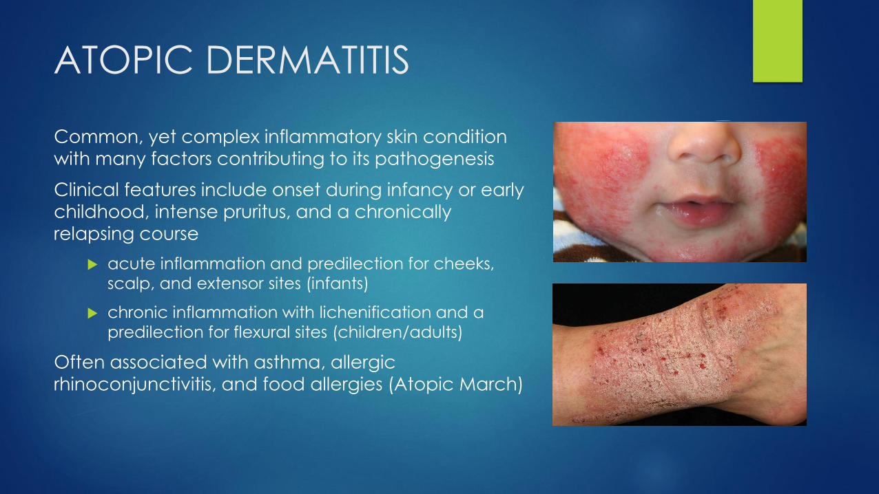
ATOPIC DERMATITIS
Common, yet complex inflammatory skin condition
with many factors contributing to its pathogenesis
Clinical features include onset during infancy or early
childhood, intense pruritus, and a chronically
relapsing course
acute inflammation and predilection for cheeks,
scalp, and extensor sites (infants)
chronic inflammation with lichenification and a
predilection for flexural sites (children/adults)
Often associated with asthma, allergic
rhinoconjunctivitis, and food allergies (Atopic March)

Pathogenesis
Divided into three major
categories
epidermal barrier
dysfunction
immune dysregulation
alteration of
the microbiome
Each of these can be
modulated by genetic
and environmental factors
Bolognia MD, Jean L.; Jorizzo MD, Joseph L.; Schaffer MD, Julie V.. Dermatology 4th edition, Elsevier. 2018

Treatment: General Approach
A “proactive approach” may modify the overall disease course and
prevent atopic comorbidities
Management includes
education
gentle skin care
moisturizer use
topical agents
Severe Disease
phototherapy
systemic medications

IS ATOPIC DERMATITIS THE NEW PSORIASIS?

NEW TARGETS
PDE4 inhibition
IL-4 antagonism
IL-13 antagonism
IL-31 antagonism
IL-22 antagonism
Janus Kinase inhibition
Neurokinin-1 Receptor inhibition
IL-12/23 antagonism
IL-17 antagonism
TSLP inhibition
Renert-Yuval Y, Guttman-Yassky E. Systemic therapies in atopic dermatitis: Thepipeline. Clin Dermatol. 2017 Jul - Aug;35(4):387-397


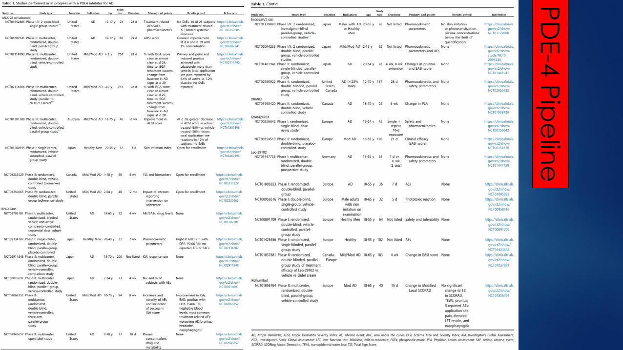
PD
E-4
Pip
elin
e

Topical Anti-Inflammatory Therapy:
Crisaborole 2% Crisaborole 2% ointment is a
phosphodiesterase-4 (PDE-4)
inhibitor FDA-approved for the treatment of mild-moderate AD in
patients ≥ 2yrs
PDE-4 inhibitor ↑ intercellular
cAMP→ ↓ production of
proinflammatory cytokines
Common side effect
stinging or burning (4.4%)


39.0%
20.9%

IL-4 and IL-13
Transgenetic mice with↑IL-4 in epidermis have 1
Atopic dermatitis-like lesions
Pruritus
Altered microbiome
↑ IgE levels
Key roles 2,3
IgE production
Eosinophil recruitment
Th2 differentiation (activation of IL-4Rα → STAT6) 4
Dupilumab (a monoclonal antibody that targets the IL-4Rα, is FDA-approved in adults for the treatment of AD)5
Bolognia MD, Jean L.; Jorizzo MD, Joseph L.; Schaffer MD, Julie V.. Dermatology 4th edition, Elsevier. 2018
1. LS Chan, N Robinson, Xu L: Expression of interleukin-4 in the epidermis of transgenic mice results in a pruritic inflammatory skin disease: an experimental animal model to study atopic
dermatitis.J Invest Dermatol. 117:977-983 2001 11676841
2. Chen L, Lin SX, L Overbergh, et al.: The disease progression in the keratin 14 IL-4-transgenic mouse model of atopic dermatitis parallels the up-regulation of B cell activation molecules,
proliferation and surface and serum IgE. Clin Exp Immunol.142:21-30 2005 16178852
3. GR Lee, RA Flavell: Transgenic mice which overproduce Th2 cytokines develop spontaneous atopic dermatitis and asthma. Int Immunol. 16:1155-1160 2004 15226271
4. K Shimoda, J van Deursen, MY Sangster, et al.: Lack of IL-4-induced Th2 response and IgE class switching in mice with disrupted Stat6 gene. Nature. 380:630-633 1996 8602264
5. LA Beck, D Thaçi, JD Hamilton, et al.: Dupilumab treatment in adults with moderate-to-severe atopic dermatitis. N Engl J Med.371:130-139 2014 25006719

≅ 80%
> 50%↓
subQ QW

https://www.dermnetnz.org/topics/easi-score/
WHAT IS THE EASI?

≅ 50%
QOW = QW
≅ 40%

Dosing: 600 mg (SubQ) initially; then 300 mg QOW
Main Side Effects:
• Injection site reactions 10-20% vs Placebo 7-8%
• Conjunctivitis 7-12% vs Placebo 2%

• Gene expression profiles of inflammatory
genes at baseline vs. after dupilumab therapy
• Inflammatory genes strongly downregulated
after dupilumab therapy
Red → Blue Transition
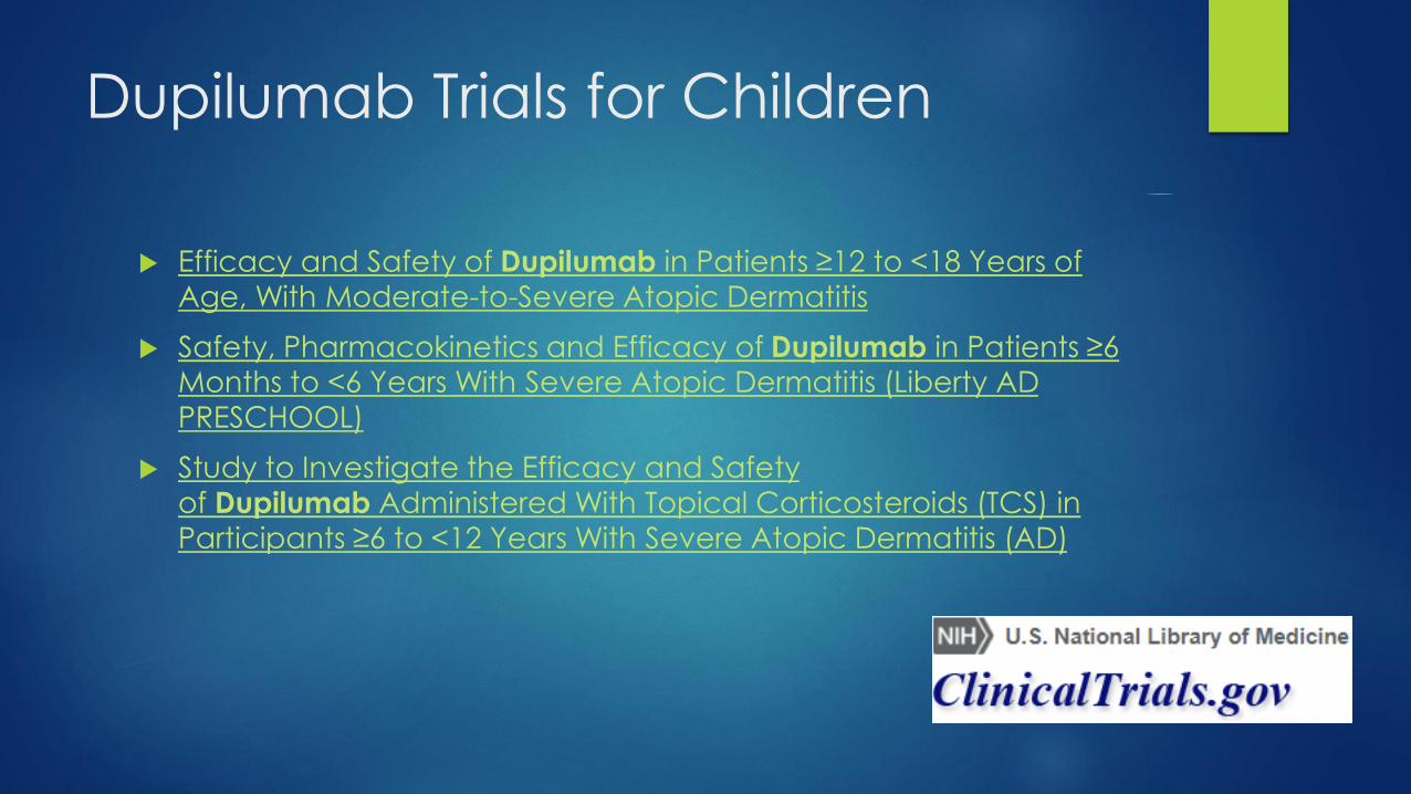
Dupilumab Trials for Children
Efficacy and Safety of Dupilumab in Patients ≥12 to <18 Years of
Age, With Moderate-to-Severe Atopic Dermatitis
Safety, Pharmacokinetics and Efficacy of Dupilumab in Patients ≥6 Months to <6 Years With Severe Atopic Dermatitis (Liberty AD
PRESCHOOL)
Study to Investigate the Efficacy and Safety
of Dupilumab Administered With Topical Corticosteroids (TCS) in
Participants ≥6 to <12 Years With Severe Atopic Dermatitis (AD)

OTHER TARGETS IN THE PIPELINE?

IL-31 IL-13
JAKs
STATs

TRALOKINUMAB:IL-13 Ab
NCT02347176, NCT03131648, NCT03160885, NCT03363854
• EASI scores at baseline (24.8
to 27.3)
• After 2 week period TCS, 300
mg of tralokinumab vs.
placebo given QOW
• The aEASI scores from
baseline
• -15.7 (tralokinumab)
• -10.8 (placebo) (P = .011)7
EASI-50

Results 209 patients received study drug
At Week 12, significantly more patients
achieved EASI-50 with lebrikizumab 125
mg Q4W (82.4%; p=0.026) versus placebo
(62.3%)
ConclusionLebrikizumab 125 mg Q4W led to significant
improvement in patients with moderate-to-
severe AD, when added to TCS, and was
well tolerated.
LEBRIKIZUMAB: IL-13 Ab

NEMOLIZUMAB: IL-31RA Ab
What we know about IL-31 Th2 cytokine highly expressed in lesions of AD
Staphylococcal superantigen rapidly induces IL-31 expression in AD pts
IL-31R is expressed by keratinocytes, eosinophils, activated macrophages, cutaneous C nerve fibers, and dorsal root ganglia1,2
Establishing a link between S. Aureus and pruritus
Nemolizumab (Phase 2, RCT) A humanized monoclonal antibody against the
IL-31RA which significantly reduces pruritus in pts with moderate to severe AD3
EASI score reduction from baseline was
−23.0±7.5% with 0.1 mg per kilogram
−42.3±7.3% with 0.5 mg per kilogram,
−40.9±7.5% with 2.0 mg per kilogram
−26.6±8.1% with placebo3
1. MM Neis, B Peters, A Dreuw, et al.: Enhanced expression levels of IL-31 correlate with IL-4 and IL-13 in atopic and allergic contact dermatitis. J Allergy Clin Immunol. 118:930-
937 2006 17030248
2. SR Dillon, C Sprecher, A Hammond, et al.: Interleukin 31, a cytokine produced by activated T cells, induces dermatitis in mice.Nat Immunol. 5:752-760 2004 15184896
3. T Ruzicka, JM Hanifin, M Furue, et al.: XCIMA Study Group. Anti-interleukin-31 receptor a antibody for atopic dermatitis. N Engl J Med. 376:826-835 2017 28249150

Pa
ller
AS, K
ab
ash
ima
K, B
ieb
er T. Th
era
pe
utic
pip
elin
e fo
r ato
pic
de
rma
titis: En
d o
f the
dro
ug
ht?
J A
llerg
y C
linIm
mu
no
l. 20
17
Se
p;1
40
(3):6
33
-64
3
Janus Kinase–Signal Transducer and
Activator of Transcription (JAK-STAT) pathway
is an intracellular signaling pathway in which
many different proinflammatory cytokines
(eg, IL-4, IL-5, IL-13, and IL-31) elicit their
pathophysiologic functions1
1. Villarino AV, Kanno Y, O’Shea JJ. Mechanisms and consequences of Jak-STAT signaling in the immune system. Nat Immunol 2017;18:374-84.
Baricitinib
Upadacitinib
PF-04965842
Ruxolitinib
Ruxolitinib
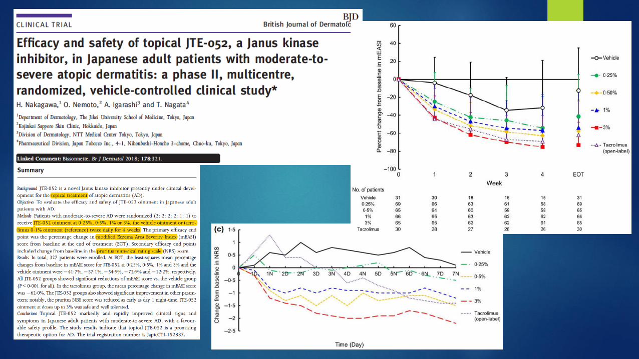
JTE-052
44% IGA 0 or 1
completed
completed
completed
200mg PO Daily
30mg PO Daily50% IGA 0 or 1

Baricitinib: Percentage of patients achieving EASI-50 (A) and percentage change from baseline in EASI score (B)
Guttman-Yassky E et al. Baricitinib in adult patients with moderate-to-severe atopicdermatitis: a phase 2 parallel, double-blinded, randomized placebo-controlledmultiple-dose study. J
Am Acad Dermatol. 2018 Feb 1. pii: S0190-9622(18)30129-4. doi: 10.1016/j.jaad.2018.01.018. [Epub ahead of print] PubMed PMID: 29410014.

TSLP is highly expressed in
acute and chronic lesions of AD,
but not in the nonlesional skin of
patients with AD or in
unaffected individuals1
1. SF Ziegler, D Artis: Sensing the outside world: TSLP regulates barrier immunity. Nat Immunol. 11:289-293 2010 20300138
OX40 is a member of the
TNF receptor superfamily. TSLP-
activated dendritic cells
express OX40L and are
activated in the lymph nodes
by OX40 → Th2 inflammatory
cytokine production

THE FUTURE IS BRIGHT
PDE4 inhibition
IL-4 antagonism
IL-13 antagonism
IL-31 antagonism
IL-22 antagonism
Janus Kinase inhibition
Neurokinin-1 Receptor inhibition
IL-12/23 antagonism
IL-17 antagonism
TSLP inhibition
AD AD

THANK YOU!

Alopecia Areata
A brief review and up-to-date
information on treatmentNADY HIN, DO, PGY-3
DR. BRAD GLICK, DO, MPH, FAOCD, FAAD
LARKIN COMMUNITY HOSPITAL PALM SPRINGS CAMPUS – LECOMT/OPTI

Introduction – Alopecia Areata
Non-scarring hair loss
Third most common form of hair loss
0.1-0.2% of US
Average lifetime risk of 1.7-2.1%
Males = Females
Onset: Mean Age 30
60% present by age 20
Spontaneous resolution rates : 8-68%
Tosti et Al(2006)
2/3 with <25% scalp involvement had complete resolution for mean of 17 yrs w/o
tx
34.6% of 51-75% hair loss recovered or
developed milder disease w/o tx

Likely Autoimmune, due to T-lymphocyte interaction with follicular antigens
Current thought:
Loss of immune privilege by Anagen bulb
Evidence for such:
Oligoclonal and autoreactive T-lymphocytes are present in peribulbar inflammatory infiltrate
Pathogenesis

Clinical – Presentations
Clinical presentations include:
Alopecia Areata - Patch
Alopecia Totalis
Alopecia Universalis
Ophiasis Pattern
Sisaipho Pattern
Acute Diffuse and Total Alopecia (ADTA)
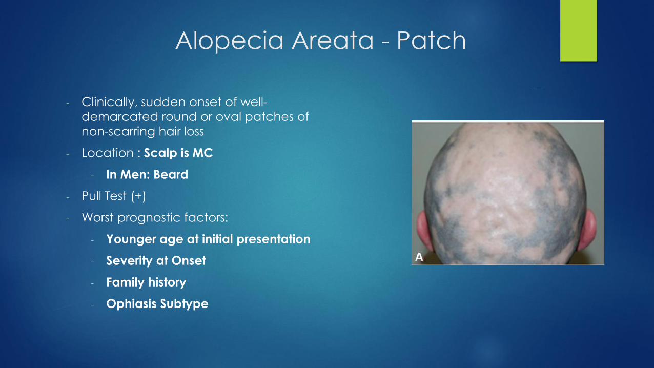
Alopecia Areata - Patch
- Clinically, sudden onset of well-
demarcated round or oval patches of
non-scarring hair loss
- Location : Scalp is MC
- In Men: Beard
- Pull Test (+)
- Worst prognostic factors:
- Younger age at initial presentation
- Severity at Onset
- Family history
- Ophiasis Subtype

Alopecia Totalis / Universalis
- Advanced forms of Alopecia
Areata
- 5% progression rate from Patch AA
- Alopecia Totalis
- Loss of all scalp hair
- Alopecia Universalis
- Loss of all scalp and
body hair

Alopecia Areata - Ophiasis
- Band-like alopecia
- Occipital hairline extending
towards temples
- Rarely can present at frontal hairline
- Can be confused with Frontal
Fibrosing Alopecia
- Worst Prognosis of all clinical
subtypes

Alopecia Areata - Sisaipho
- Opposite configuration of
Ophiasis subtype
- Hair loss centrally but sparing
hairs at margin of scalp
- Can be confused with
androgenetic alopecia

Acute Diffuse and Total Alopecia
(ADTA)
- More common in women
- Sudden and diffuse hair loss
that lasts around 3 months
followed by rapid regrowth
over 4-9 months
- Favorable Prognosis but it
may recur in future

Nail Changes
Nail Pitting (MC) Trachyonychia Longitudinal Ridging Red Lunulae

Comorbidities
Higher incidence noted in patients with:
Atopic Dermatitis (MC)
Higher risk of severe AA phenotype
Autoimmune Diseases (SLE, Thyroiditis, DM, Myasthenia Gravis, Vitiligo)
Patel et al(2017) conducted a retrospective analysis of 298 patients with AA
Thyroid abnormalities discovered in 20% of the pediatric patients
Screening should be done in those with thyroid symptomology
Vitamin D Levels
Tsai et al (2018)
Retrospective analysis showed association with severity
Meta-analysis of studies show association between VitD deficiency and AA
Diagnosis
Pull Test
Sign of active disease
Trichoscopy
See Next Slide
Biopsy
Peribulbar lymphocytic infiltrate

Trichoscopy
Yellow Dots
Infundibula with Sebum and Keratin
Exclamation Mark
Hairs
Broken hair with a thick pigmented tip
Black Dots
Destroyed hairs in hair follicle opening

Treatment
AA is often self limiting
Current first line treatments
Corticosteroids (Topical and Intralesional)
Minoxidil (5%)
Topical Immunotherapy
Newer Treatments
JAK Inhibitors
PRP
Others
Immunomodulators
Anti Inflammatories
Targeted Therapies
Devices (Lasers, Cryotherapy)

But Where Do I Start?

Current Treatments
Generally considered first-line
• Corticosteroids
• Topical and Intralesional
• Minoxidil 5%
• Contact Immunotherapy
• With Anthralin

Treatment – First Line
Corticosteroids
Intralesional - First line for limited disease
Chu et Al (2015)
Recommend: low concentration, higher volume
2.5mg/CC was as effective as 5-10mg/cc
Topical
Clobetasol vs Mometasone (for pediatric patients)
Tosti et al (2006)
Clobetasol foam in Double Blind RCT, greater regrowth in 89% vs 11%
Monitor for side effects such as skin atrophy
Minoxidil 5%
Insufficient as monotherapy
In long term studies, mild hair growth without statistical significant
Use for maintenance with other treatments
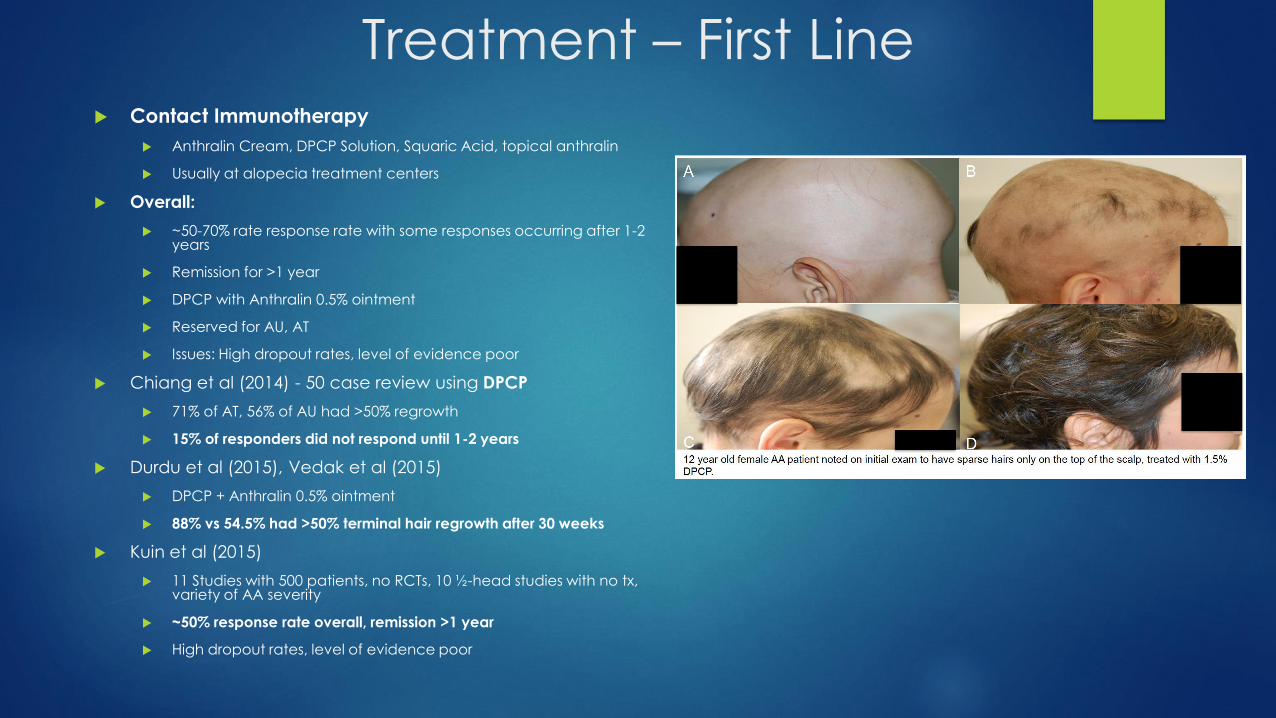
Treatment – First Line Contact Immunotherapy
Anthralin Cream, DPCP Solution, Squaric Acid, topical anthralin
Usually at alopecia treatment centers
Overall:
~50-70% rate response rate with some responses occurring after 1-2 years
Remission for >1 year
DPCP with Anthralin 0.5% ointment
Reserved for AU, AT
Issues: High dropout rates, level of evidence poor
Chiang et al (2014) - 50 case review using DPCP
71% of AT, 56% of AU had >50% regrowth
15% of responders did not respond until 1-2 years
Durdu et al (2015), Vedak et al (2015)
DPCP + Anthralin 0.5% ointment
88% vs 54.5% had >50% terminal hair regrowth after 30 weeks
Kuin et al (2015)
11 Studies with 500 patients, no RCTs, 10 ½-head studies with no tx, variety of AA severity
~50% response rate overall, remission >1 year
High dropout rates, level of evidence poor

Treatment – First Line
High Dropout Rates
Often due to expected SE
Patient compliance is a strong factor in decreased relapse rates (Duh)
Choe et al (2018)
Retrospective analysis, 159 pts
Modified DPCP treatment protocol with subclinical sensitization
Sensitized with 0.1% and tx with 0.01% QWeekly
Sensitization with an eczematous reaction may not be required for successful contact immunotherapy
46 (28.9%) complete response, 59 (37.1%) partial response

New and More Recent
Treatments
• JAK Inhibitors
• Platelet Rich Plasma (PRP)

Treatment – What’s New-Ish?
JAK Inhibitors
JAK – STAT Pathway
Cytokine binding
JAK receptors dimerize, phosphorylated, recruit STAT molecules to activate target gene transcription
Mediates downstream IL-15 signaling of T-cells
Baseline lab monitoring : CBC, CMP, Lipids, HIV, Quant-Gold, CXR
Avoid in : Hx of Malignancy, Tb, Hepatitis
Cost: $2000-$5000 per month

Treatment – JAK Inhibitors
- Tofacitinib (JAK1/3)
- Dose: 5-10mg BID, or 11mg ER QD
- Shapiro : Recommends 15mg QD + IntralesionalCorticosteroids
- Ruxolitinib (JAK1/2)
- 20mg BID
- Oclacitinib (JAK 1)
- Issues:
- Relapse once taken off medication
- Adverse Effects
- Higher doses have unknown safety profile
- Topical route safer but unknown benefit
- Long term likely necessary
- Longer duration and extent often has poorer response

Treatment – JAK Inhibitors

Treatment – JAK Inhibitors
Kennedy Crispin et al (2016) – Tofacitinib(5mg BID)
66 pts with AA, AT, AU
>66% showed regrowth by 3 months (32% had >50% SALT)
Relapse by 8.5 weeks
AE: 25% with Infxn (UTI/URI)
Liu et al (2016) – Tofacitinib (5-10mg BID) + Syst. Corticosteroids
90 pts with AA, AT, AU
Pulsed oral CST 300 mg monthly x 3 months
77% achieved clinical response (55% had >50% regrowth)
Liu et al (2018) – Topical 2% tofacitinibointment BID
10 patients, 24 weeks
3/10 experienced hair regrowth with Salt improvement of 34.6%

Treatment – JAK Inhibitors
(adolescents) Craiglow et al (2017) – Tofacitinib 5mg BID
10/14 pts with Salt 20-100%
Mean SALT improvement over 2-16 months of 88%
Mild AE, no treatment interruptions
Castelo-Soccio (2017) – Tofacitinib 5-10mg BID
8 patients age 12-19 with AU
All pts had >50% hair regrowth
1st 3 months – slow growth, rapid thereafter
No AE or infections noted
Bayart et al (2017) – Tofacitinib and Ruxolitinib 1% and 2% topical
6 patients, 3AU, 2AT, 1AA
Ruxolitinib (1 success, 1 fail)
75% eyelash regrowth
Tofacitinib (3 success, 1 fail)
20% medial eyebrow regrowth
20% 1 month, 80% 1 year
Fail with verabase cream, 95% regrowth with liposomal base of scalp

Mackay-Wiggan et al (2016) – Ruxolitinib
12 pts
20mg BID for 3-6 mos
9/12 pts with marked response
Average of 92% hair regrowth
Issue : Relapse over 3-6 months
Treatment – JAK Inhibitors

Treatment - PRP
Advantages:
Ability to induce longer disease remission
Regrow pigmented hairs from beginning of
hair regrowth
Safe – autologous material
No lab monitoring, drug interactions, side
effects
Issue : non standardized protocol
Trink et al (2013) - Double blind placebo, half
head x 3 months
Significant improvement monthly PRP(60%) vs
ILK(27%) vs placebo
Singh (2015) – Monthly x 6 months
19/20 with regrowth
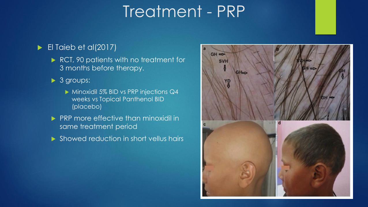
Treatment - PRP
El Taieb et al(2017)
RCT, 90 patients with no treatment for
3 months before therapy.
3 groups:
Minoxidil 5% BID vs PRP injections Q4
weeks vs Topical Panthenol BID
(placebo)
PRP more effective than minoxidil in
same treatment period
Showed reduction in short vellus hairs
Existing treatments with
possible utility
• Immunomodulators
• Systemic Corticosteroids
• Mycophenolate Mofetil
• Methotrexate
• Cyclosporine
• Sulfasalazine
• Azathioprine
• Prostaglandin Analogs
• Anti-Inflammatories
• Simvastatin/Ezetimibe
• Anti-histamines (Fexofenadine)
• Low Dose Naltrexone
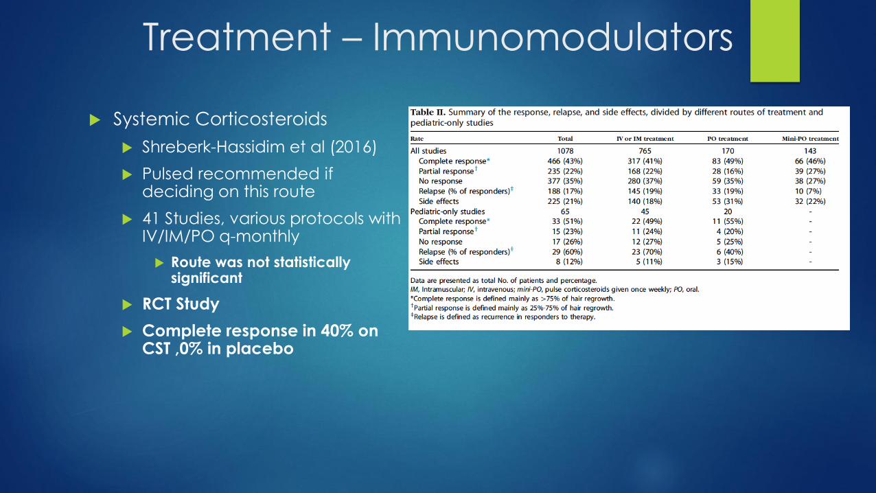
Treatment – Immunomodulators
Systemic Corticosteroids
Shreberk-Hassidim et al (2016)
Pulsed recommended if deciding on this route
41 Studies, various protocols with IV/IM/PO q-monthly
Route was not statistically significant
RCT Study
Complete response in 40% on CST ,0% in placebo

Treatment – Immunomodulators
Mycophenolate Mofetil
Systemic : 500mg BID – 1500mg BID
Topical : 2% Cream
Methotrexate
Comparative Study, MTX + Prednisone vs Prednisone alone
5/14 pts had >50% hair growth with combo MTX + Prednisone
Cyclosporine
Ranges from 25-76.7% success rate >50% regrowth
One uncontrolled study – 45.4% of 25 pts showed sig. regrowth
Sulfasalazine
Pilot study
43% of 14 pts showed complete regrowth
66% showed no signs of relapse after treatment discontinuation
33% relapsed after 2.5 months
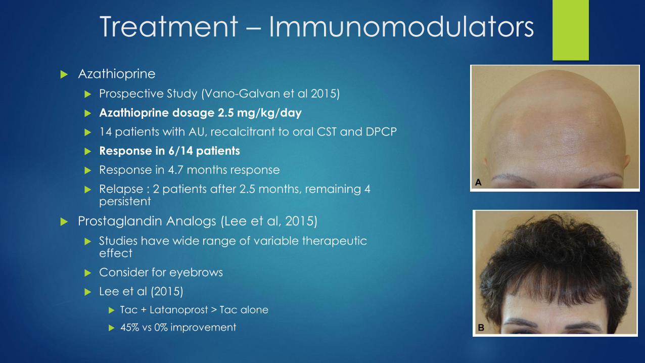
Azathioprine
Prospective Study (Vano-Galvan et al 2015)
Azathioprine dosage 2.5 mg/kg/day
14 patients with AU, recalcitrant to oral CST and DPCP
Response in 6/14 patients
Response in 4.7 months response
Relapse : 2 patients after 2.5 months, remaining 4 persistent
Prostaglandin Analogs (Lee et al, 2015)
Studies have wide range of variable therapeutic effect
Consider for eyebrows
Lee et al (2015)
Tac + Latanoprost > Tac alone
45% vs 0% improvement
Treatment – Immunomodulators
Treatment – Anti-Inflammatories
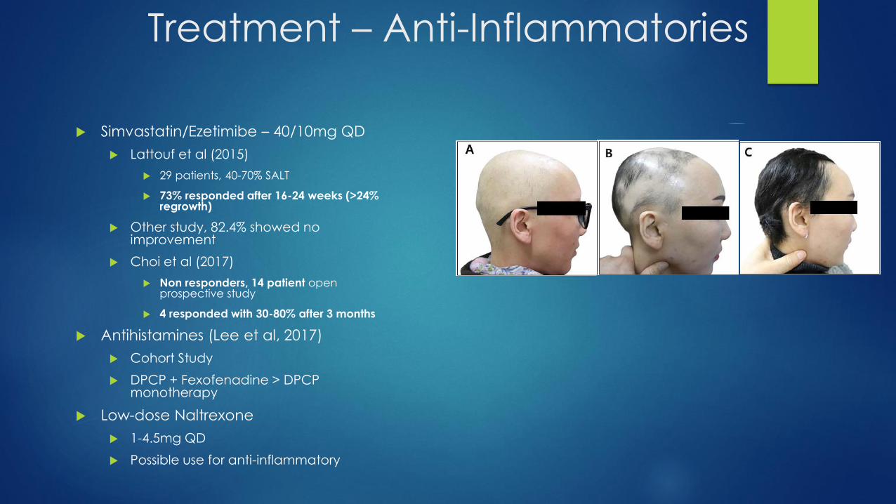
Simvastatin/Ezetimibe – 40/10mg QD
Lattouf et al (2015)
29 patients, 40-70% SALT
73% responded after 16-24 weeks (>24% regrowth)
Other study, 82.4% showed no improvement
Choi et al (2017)
Non responders, 14 patient open prospective study
4 responded with 30-80% after 3 months
Antihistamines (Lee et al, 2017)
Cohort Study
DPCP + Fexofenadine > DPCP monotherapy
Low-dose Naltrexone
1-4.5mg QD
Possible use for anti-inflammatory

Targeted Therapies
• Ustekinumab
• Apremilast
• Secukinumab
• Abatacept

Treatment - Targeted Therapy
Ustekinumab – 90mg Q12 weeks
Guttman-Yassky E Et Al (2016)
3/9 pts with complete response after 12 months, 1 had AU
Apremilast – 30mg BID x 3-6 months (mean
4.2mos)
Liu et Al (2017)
9 patients (1 AA, 8AU)
Duration of disease 23.3 years
None showed improvement over 3-6 months
Secukinumab
RCT was terminated in 2017 due to low
enrollment

Treatment - Targeted Therapy
Abatacept (CTLA4 Agonist) – 125mg SC
weekly
Guttman-Yassky E et al (2016), Mackay-Wiggan
J et al (2015), Keren a et al (2015)
SALT 30-100% (3/15 improved)
1/15 pts with 98% regrowth after 6 months
2/15 with 23% regrowth
Devices
• Superficial Cryotherapy
• Carboxytherapy
• Excimer Laser
• Fractional Photolasers
• Fecal Transplant.. Device?
Treatment - Devices
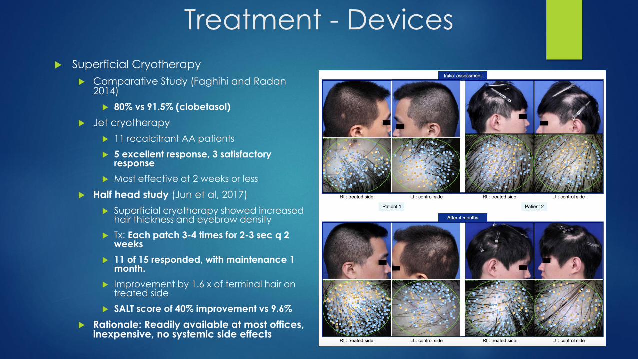
Superficial Cryotherapy
Comparative Study (Faghihi and Radan2014)
80% vs 91.5% (clobetasol)
Jet cryotherapy
11 recalcitrant AA patients
5 excellent response, 3 satisfactory response
Most effective at 2 weeks or less
Half head study (Jun et al, 2017)
Superficial cryotherapy showed increased hair thickness and eyebrow density
Tx: Each patch 3-4 times for 2-3 sec q 2 weeks
11 of 15 responded, with maintenance 1 month.
Improvement by 1.6 x of terminal hair on treated side
SALT score of 40% improvement vs 9.6%
Rationale: Readily available at most offices, inexpensive, no systemic side effects

Treatment - Devices
Carboxytherapy
Doghaim et al (2018)
80 pts (40 AA, 40 AGA), 4 groups (1a, 1b, 2a, 2b)
Placebo was intradermal distilled water
Injection: 30g Needle, 2mL CO2 per injection site
Significant improvement
3 months after last session
SALT from 9 -> 5.7
Control group : 12.5 -> 16.0
Before, after 6 sessions, then 3 months after last session
Rationale: Inexpensive

Excimer Laser
308nm Excimer
Pilot study
42 recalcitrant patches in 18 patients
Twice per week for max of 24 sessions
50mJ/cm2 less than MED
Complete regrowth in 13/42 lesions, excellent in 5/42
Presence of Atopic Diathesis had an unfavorable prognosis
Rationale
Minimal side effects, ideal for pediatric patients
Treatment - Devices

Treatment - Devices
Fractional Photothermolysis
Yalici-Armagan et al (2016)
Controlled clinical trial
32 subjects, 21pAA, 2AT, 1AU, 8 ophiasis
3 patches on each subject
Control patch, Nd:YAG patch(2-3 sessions with 2-8 week intervals), fractional CO2 patch (3-6 sessions with 2-4 week intervals)
No significant difference between baseline and final hair counts between treated patches and the control patch
But other studies have reported some improvement
Cho et al (2013)
17 patients with 10,600 nm Co2
30-50mJ, spot 150 spots/cm2, 8-22 sessions
12/17 reported clinical response

Treatment - Devices
Fecal Microbiota Transplant
Rebello et al (2017)
Pt A
38yoM with recalcitrant AU(Dx at 28yo), p/w C.Diff and tx with FMT. 8 weeks later, patchy hair growth on beard, arms, scalp, face.
Pt B
20yoM with Pmhx of Crohn’s and recalcitrant AU(Dx at 18yo)
Pt previously tx with ILK, Topical CST, Laser, Squaric Acid with no improvement
C. Diff tx with FMT at 20yo
Improved from AU to 25-49% Hair loss with body hair regrowth as well
But what does this mean?
Microbiota and the immune system
Conclusion
Alopecia Areata can and almost always will self resolve… eventually
Initial treatment should follow an algorithmic approach with corticosteroids, minoxidil, immunotherapy
Widespread and recalcitrant cases
JAK inhibitors, PRP, immunomodulators, and various devices
Be aware of the comorbidities of AA
Thyroid Disorders
Vitamin D deficiency
Atopic Diathesis
Anemia
References Al-Mutairi N et al. 308-n Excimer laser for the treatment of alopecia areata. Dermatologic Surgery. 2007; 33(12)1483-1487.
Batalla A. Methotrexate in Alopecia Areata: A report of three cases. Int J Trichology. 2016 Oct-Dec; 8(4): 188-190
Bavart et al. Topical Janus Kinase Inhibitors for the treatment of pediatric alopecia areata. J Am Acad Dermatol. 2017 Jul; 77(1):167-170.
Castelo-Soccio L. Experience with oral tofacitinib in 8 adolescent patients with alopecia universalis. J Am Acad Dermatol. 2017. 76(4)754-755.
Chiang et Al. Clinical efficacy of diphenylcyclopropenone in alopecia areata: Retrospective data analysis of 50 patients. Journal of American Academy of Dermatology. 2014; 71: 595-597
Choe et al. Subclinical sensitization with diphenylcyclopropenone is sufficient for the treatment of alopecia areata: Retrospective analysis of 159 cases. J Am Acad Derm. 2018. 78(3) 515-521.
Choi et al. Simvastatin/Ezetimibe Therapy for Recalcitrant Alopecia Areata : An Open Prospective Study of 14 patients. Ann Dermatol. 29(6) 755-760, 2017.
Chu TW, AlJasser M, Alharbi A, Abahussein O, McElwee K, Shapiro J. Benefit of different concentrations of intralesional triamcinolone acetonide in alopecia areata: an intrasubjectpilot study. J Am Acad Dermatol. 2015;73:338-340.
Craiglow BG, Liu LY, King BA. Tofacitinib for the treatment of alopecia areata in adolescents. J Am Acad Dermatol. 2017; 76(1):29-32.
Doghaim N et al. Study of the efficacy of carboxytherapy in alopecia. J Cosmet Dermatol. 2018:1-11
El Taieb MA, Ibrahim H, Nada EA, and Seif Al-Din M. Platelets rich plasma versus minoxidil 5% in treatment of alopecia areata: A trichoscopic evaluation. Dermatologic Therapy 2017;30:e12437. doi:10.1111/dth.12437.
Faghihi et al. Jet cryotherapy vs clobetasol proprionate lotion in alopecia areata. Skinmed. 2014 Jul-Aug;12(4):209-11.
Guttman-Yassky E, Ungar B, Noda S, Suprun M, Shroff A, Dutt R, et al. Extensive alopecia areata is reversed by IL-12/IL-23p40 cytokine antagonism. J Allergy Clin Immunol2016;137:301-4.
Guttman-Yassky E, Ungar B, Noda S, Suprun M, Shroff A, Dutt R, et al. Extensive alopecia areata is reversed by IL-12/IL-23p40 cytokine antagonism. J Allergy Clin Immunol2016;137:301-4.
Jun M et al. Therapeutic effect of superficial cryotherapy on Alopecia areata: A Prospective, split-scalp study in Patients with Multiple Alopecia Patches. Ann Dermatol. 2017; 29(6)722-727
Kennedy Crispin M, Ko JM, Craiglow BG, et al. Safety and efficacy of the JAK inhibitor tofacitinib citrate in patients with alopecia areata. JCI Insight. 2016;1(15): e89776.
Keren A, Shemer A, Ullmann Y, Paus R, Gilhar A. The PDE4 inhibitor, apremilast, suppresses experimentally induced alopecia areata in human skin in vivo. J Dermatol Sci 2015;77:74-6.
LC Strazzulla et al. An overview of the biology of platelet-rich plasma and microneedling as potential treatments for Alopecia Areata. Journal of Investigative Dermatology Symposium Proceedings. 2018; 19. 521-524.

References Lee et Al. Algorithmic Approach to management of AA. Journal of Dermatology. 2017. 44: 1199-1211.
Liu LY, Craiglow BG, Dai F, King BA. Tofacitinib for the treatment of severe alopecia areata and variants: a study of 90 patients. J Am Acad Dermatol. 2017;76:22-28.
Liu et al. Lack of Efficacy of apremilast in 9 patients with severe alopecia areata. J Am Acad Dermatol. 2017. 77(4)773-774.
Mackay-Wiggan J, Jabbari A, Nguyen N, Cerise JE, Clark C, Ulerio G, et al. Oral ruxolitinib induces hair regrowth in patients with moderate-to-severe alopecia areata. JCI Insight 2016;1:e89790.
Nonomura Y et al. Case of intractable ophiasis type alopecia areata presumably improved by fexofenadine. The Journal of dermatology 2012; 39: 1-2.
Patel et al. Screening Guidelines for Thyroid Function in Children With Alopecia Areata. JAMA Dermatol. 2017; 153(12): 1307-1310.
Rebello D et al. Hair growth in two alopecia patients after fecal microbiota transplant. ACG Case Reports Journal. 2017(4)1-3.
Shapiro, Jerry. Treatment and follow up of non scarring alopecias. American Academy of Dermatology. San Diego, California. 2018.
Shreberk-Hassidim et al. A systematic Review of pulse steroid therapy for alopecia areata. J Am Acad Dermatol. 2016; 74(2) 372-374
Singh, S. (2015). Role of platelet-rich plasma in chronic alopecia areata. Indian Journal of Plastic Surgery, 48, 57–59.
Strazzulla et al. Alopecia Areata An appraisal of new treatment approaches and overview of current therapies. Journal of American Academy of Dermatology. 2018; 78:15-24
Tosti A, Iorizzo M, Botta GL, Milani M. Efficacy and safety of a new clobetasol propionate 0.05% foam in alopecia areata: a randomized, double-blind placebo-controlled trial. J EurAcad Dermatol Venereol. 2006;20:1243-1247.
Trink, A., Sorbellini, E., Bezzola, P., Rodella, L., Rezzani, R., Ramot, Y., &
Rinaldi, F. (2013). A randomized, double-blind, placebo- and active controlled half-head study to evaluate the effects of platelet-rich plasma on alopecia areata. British Journal of Dermatology, 169(3), 690–694.
Tosti et al. Alopecia Areata: a long term follow-up study of 191 patients. J Am Acad Dermatol. 2006 Sep; 55(3):438-41.
Tsai et al. Vitamin D Deficiency in patients with Alopecia Areata: A systemic review and meta-analysis. J Am Acad Dermatol. 2018; 78(1): 207-209.
Vano-Galvan S. et al. Treatment of recalcitrant adult alopecia areata universalis with oral azathioprine. Journal of American Academy of Dermatology. 2015. 74; 1007-1008.

Thank you!

Granuloma Annulare:
A Brief Review and Up-to-date
Information on Treatment
RACHEL WHITE, DO, PGY-3
DR. BRAD GLICK, DO, MPH, FAOCD, FAAD
LARKIN COMMUNITY HOSPITAL PALM SPRINGS CAMPUS – LECOMT/OPTI
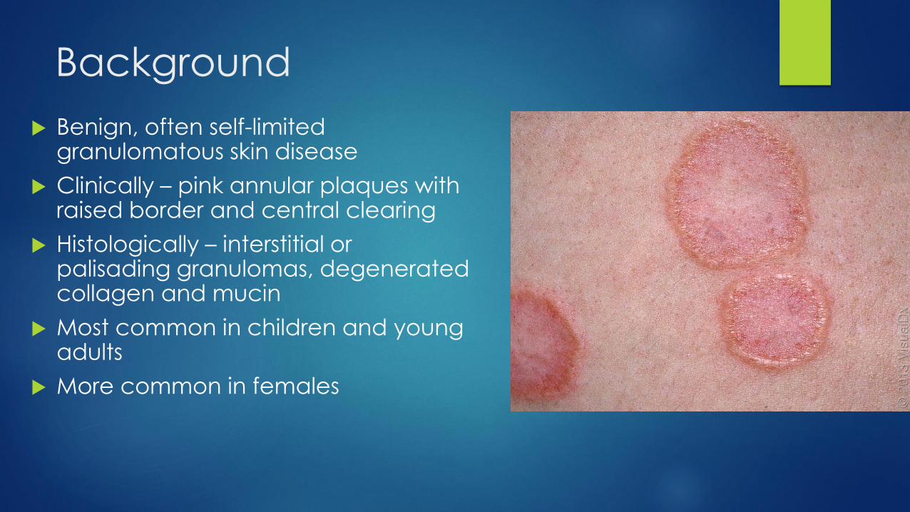
Background
Benign, often self-limited granulomatous skin disease
Clinically – pink annular plaques with raised border and central clearing
Histologically – interstitial or palisading granulomas, degenerated collagen and mucin
Most common in children and young adults
More common in females

Pathogenesis – Mechanisms
Unknown mechanism; theories originate from
histologic findings
Original theory – immune-mediated type III hypersensitivity reaction vasculitis
Recent theory – cell-mediated delayed-type IV
hypersensitivity reaction to unknown antigen
Sensitized Th1 lymphocytes macrophages
proinflammatory cytokines & collagen-degrading enzymes tissue injury
Other theories – injury to dermal elastic fibers

Pathogenesis – Inciting Factors
Trauma/foreign body – insect bite, tuberculin skin testing, vaccinations,
subcutaneous immunotherapy for allergies, tattoo, isomorphic response
Infectious – viruses (Hep B, Hep C, EBV, HIV); Borrelia species
Drugs – TNF-⍺ inhibitors, allopurinol, topiramate, gold therapy
Genetic – familial cases including identical twins, HLA-Bw35 (generalized
GA)
Associated Disorders
Diabetes
Definitive evidence lacking and conflicting data
Dyslipidemia
Evidence shows link with adult GA
Malignancy
No causative relationship
Seen in atypical GA variants
Most common malignancy is lymphoma
Thyroid disease – autoimmune
HIV – atypical variants

Clinical – Localized GA
Most common form
Skin-colored to pink erythematous annular or
arcuate plaques with raised border and central
clearing
Discrete papules at periphery
Location – wrists, ankles, dorsal hands and feet
Asymptomatic
Onset – children, young adults
~50% patients have >1 lesion

Clinical –
Disseminated/Generalized GA
Widespread skin-colored to
pink erythematous papules
and plaques of varying sizes
Location – trunk and
extremities
Asymptomatic or pruritic
Onset – adulthood
Associated with HLA-B35

Clinical – Deep/Subcutaneous GA
Large skin-colored nodules, overlying skin uninvolved
Location – scalp, buttocks, extremities
Painless
Onset – children <6 yo
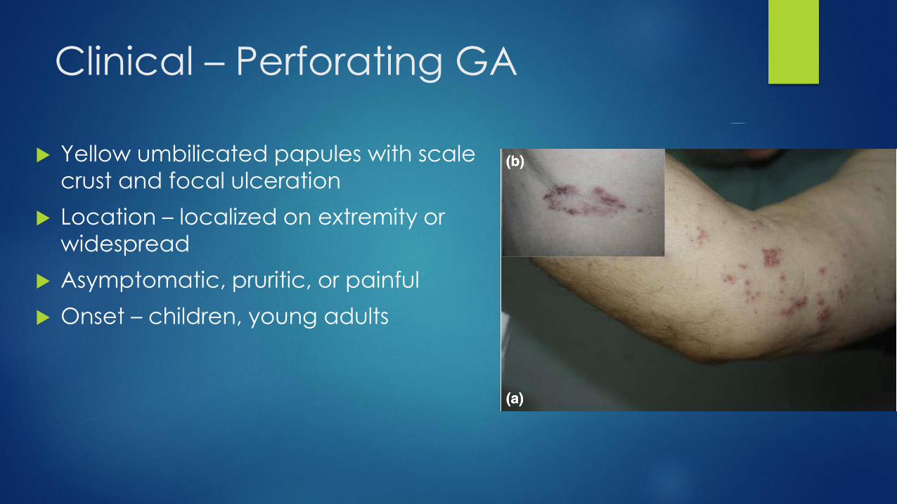
Clinical – Perforating GA
Yellow umbilicated papules with scale
crust and focal ulceration
Location – localized on extremity or
widespread
Asymptomatic, pruritic, or painful
Onset – children, young adults

Clinical – Patch GA
Symmetric annular patches
Location – proximal extremities, dorsal
feet
Onset – adults
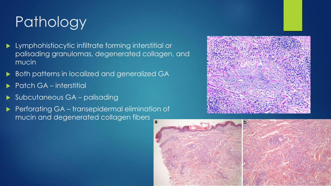
Pathology
Lymphohistiocytic infiltrate forming interstitial or
palisading granulomas, degenerated collagen, and mucin
Both patterns in localized and generalized GA
Patch GA – interstitial
Subcutaneous GA – palisading
Perforating GA – transepidermal elimination of
mucin and degenerated collagen fibers

Differential Diagnoses –
Annular Lesions
Annular elastolytic giant cell granuloma (actinic
granuloma)
Interstitial granulomatous dermatitis
Tinea corporis
Annular lichen planus
Erythema annulare centrifugum
Sarcoidosis
Nodular tertiary syphilis
Mycosis fungoides
Borderline leprosy

Differential Diagnoses
Generalized GA
Arthropod assault
ID reaction
Interstitial granulomatous
dermatitis
Secondary syphilis
Eruptive xanthomas
Eruptive syringomas
Histiocytomas
Subcutaneous GA
Rheumatoid nodules
Epithelioid sarcoma
Sarcoidosis
Deep fungal infection
Tendinous xanthomas
Perforating GA
Reactive perforating
collagenosis
Perforating folliculitis
Elastosis perforans
serpiginosa
Calcinosis cutis
Perforating gout
Sarcoidosis
Molluscum contagiosum

Diagnosis and Work Up
Clinical diagnosis
Punch biopsy with H&E for atypical presentations
Lipid panel in adults
Review signs/symptoms/risk factors for diabetes, HIV
Age-appropriate cancer screening in elderly patients with atypical
presentations
Treatments – Overview
No treatment necessary – often self-limited
50% localized GA resolve within 2 years
Generalized GA more persistent – 25% courses >5 years
Resolves without scar
Treatment dependent on type, symptoms, cosmesis

Treatment – Localized GA
First-line
High-potency corticosteroids topical +/- intralesional
Clobetasol 0.05% cream BID x 2-4 w
Triamcinolone acetonide 2.5-10 mg/cc q 6-8 w
Others (limited evidence)
Cryotherapy
Topical calcineurin inhibitors – tacrolimus, pimecrolimus
Phototherapy – PUVA, UVA1, NB-UVB, PDT
Topical dapsone
Intralesional IFN-γ
Imiquimod

Treatments – Generalized GA
First-line
High potency topical/intralesional corticosteroids
Topical calcineurin inhibitors
Tacrolimus 0.1% ointment BID x 6 w
Pimecrolimus 1% cream
Phototherapy
UVA1 – high cumulative doses most effective = 1770 – 1840 J/cm2
PUVA – oral or bath PUVA with cumulative dose 60.4 J/cm2
Narrow-band UVB – cumulative dose 47.7 J/cm2 54% complete/partial
response
Photodynamic therapy

Treatments – Generalized GA
Systemic treatment
Antimalarials – first line
Hydroxychloroquine – 3 – 6 mg/kg/d
Chloroquine – 3 mg/kg/d
TNF-⍺ inhibitors
Adalimumab – 80 mg at week 0 40 mg every other week SQ
Infliximab – 5 mg/kg at weeks 0, 2, 6 every month IV
Isotretinoin – 0.5-1 mg/kg/d
Dapsone – 100 mg/d
Pentoxifylline – 400 mg TID
Nicotinamide – 500 mg TID
Cyclosporine – 3-4 mg/kg/d
ROM (rifampin, ofloxacin, minocycline)
Vitamin E oral – 400-600 IU daily
Fumaric acid esters – used in Europe
Other case reports: doxycycline, clofazimine, allopurinol, methotrexate, hydroxyurea, alkylating agents (chlorambucil), oral calcitriol, defibrotide, etretinate

Treatments – Lasers
Pulsed dye laser
Localized or generalized GA
~1/3 no improvement; ~1/3 some improvement;
~1/3 >50% improvement
Fractional photothermolysis
Case reports – significant improvement
height and diameter
Excimer laser – complete remission ¾ patients
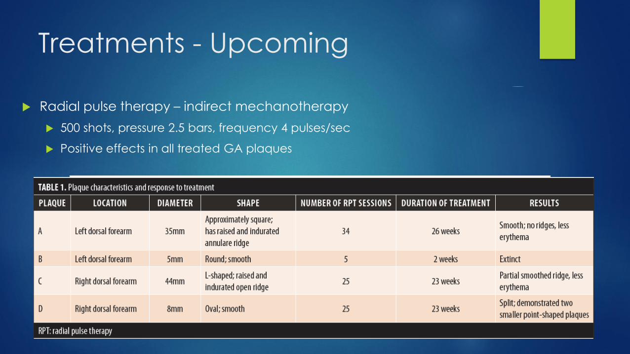
Treatments - Upcoming
Radial pulse therapy – indirect mechanotherapy
500 shots, pressure 2.5 bars, frequency 4 pulses/sec
Positive effects in all treated GA plaques

Treatments – Subcutaneous GA
Treatment not indicated
Surgical excision – recurrence common
Local hyperthermia – case report
44° C for 30 min, improvement after 10 treatments

Conclusion
Benign, often self-limited disease
Work up may include history/labs for diabetes, dyslipidemia,
malignancy, and HIV
Pathogenesis involves possible cell-mediated delayed type IV
hypersensitivity reaction to unknown antigen
Many treatments available from topical and light-based
therapy to systemic and biologic medications
Prospective double blind randomized control trials need to be
performed for improved evidence-based treatment
References Bolognia JL, Jorizzo JL, Schaffer JV. Dermatology. 3rd ed. Philadelphia, PA: Saunders Elsevier; 2012.
Brodell RT. Granuloma Annulare. UpToDate. https://www-uptodate-com.ezproxy.pcom.edu/contents/granuloma-annulare?source=history_widget. Updated July 17, 2017. Accessed February 25, 2018.
Successful treatment of disseminated granuloma annulare with topical tacrolimus.AUJain S, Stephens CJ SOBr J Dermatol. 2004;150(5):1042.
Mickel M, Kunstfeld R, Crevenna R. Granuloma Annulare and Radial Pulse Therapy: Preliminary Findings. J Clin Aesthet Dermatol. 2018 Jan;11(1):32-4.
Min MS, Lebwohl M. Treatment of recalcitrant granuloma annulare (GA) with adalimumab: A single-center, observational study. J Am Acad Dermatol. 2016 Jan;74(1):127-33.
Muhlemann MF, Williams DR. Localized granuloma annulare is associated with insulin-dependent diabetes mellitus. Br J Dermatol. 1984;111(3):325.
Nambiar KG, Jagadeesan S, Balasubramanian P, et al. Successful Treatment of Generalized Granuloma Annulare with Pentoxifylline. Indian Dermatol Online J. 2017 May-Jun;8(3):218-20.
Nebesio CL, Lewis C, Chuang TY. Lack of an association between granuloma annulare and type 2 diabetes mellitus. Br J Dermatol. 2002;146(1):122.
Pavlovsky M, Samuelov L, Sprecher E, et al. NB-UVB phototherapy for generalized granuloma annulare. Dermatol Ther. 2016 May;29(3):152-4.
Piette EW, Rosenbach M. Granuloma annulare: Pathogenesis, disease associations and triggers, and therapeutic options. J Am Acad Dermatol. 2016 Sep;75(3):467-9.
Satta R, Biondi G, Puggioni GM, et al. Malignancy-associated generalized perforating granuloma annulare. Clin Exp Dermatol. 2018 Mar;43(2):219-21.
Strazzula L, Wong V, Burgin S, et al. Granuloma annulare. Visual Dx. https://www-visualdx-com.ezproxy.pcom.edu/visualdx/diagnosis/granuloma%20annulare?moduleId=101&diagnosisId=51632#top. Updated May 25, 2017. Accessed February 25, 2018.
Vaalamo M, Kariniemi AL, Shapiro SD, Saarialho-Kere U. Enhanced expression of human metalloelastase (MMP-12) in cutaneous granulomas and macrophage migration. J Invest Dermatol. 1999;112:499-505.
Verne SH, Kennedy J, Falto-Aizpurua LA, et al. Laser treatment of granuloma annulare: a review. Int J Dermatol. 2016 Apr;55(4):376-81.
Wallet-Faber N, Farhi D, Gorin I, et al. Outcome of granuloma annulare: shorter duration is associated with younger age and recent onset. J Eur Acad Dermatol Venereol. 2010;24:103-104.
Wang J, Khachemoune A. Granuloma Annulare: A Focused Review of Therapeutic Options. Am J Clin Dermatol. 2017 Dec 11. doi: 10.1007/s40257-017-0334-5. [Epub ahead of print]
Yang Y, Zheng S, Li XD, et al. Case of successful treatment of subcutaneous granuloma annulare with local hyperthermia. J Dermatol. 2017 Oct;44(10):e246-e247.
Yong A, Chong WS, Pan JY. Disseminated granuloma annulare responding to narrowband UVB phototherapy. Photodermatol Photoimmunol Photomed. 2016 Mar;32(2):107-9.