emerging transporters of clinical importance: an update ... · zurich open repository and archive...
TRANSCRIPT
Zurich Open Repository andArchiveUniversity of ZurichMain LibraryStrickhofstrasse 39CH-8057 Zurichwww.zora.uzh.ch
Year: 2013
Emerging transporters of clinical importance: an update from theInternational Transporter Consortium
Hillgren, K M ; Keppler, D ; Zur, A A ; Giacomini, K M ; Stieger, B ; Cass, C E ; Zhang, L
Abstract: The International Transporter Consortium (ITC) has recently described seven transportersof particular relevance to drug development. Based on the second ITC transporter workshop in 2012,we have identified additional transporters of emerging importance in pharmacokinetics, interference ofdrugs with transport of endogenous compounds, and drug-drug interactions (DDIs) in humans. Themultidrug and toxin extrusion proteins (MATEs, gene symbol SLC47A) mediate excretion of organiccations into bile and urine. MATEs are important in renal DDIs. Multidrug resistance proteins (MRPsor ABCCs) are drug and conjugate efflux pumps, and impaired activity of MRP2 results in conjugatedhyperbilirubinemia. The bile salt export pump (BSEP or ABCB11) prevents accumulation of toxic bilesalt concentrations in hepatocytes, and BSEP inhibition or deficiency may cause cholestasis and liverinjury. In addition, examples are presented on the roles of nucleoside and peptide transporters in drugtargeting and disposition.
DOI: https://doi.org/10.1038/clpt.2013.74
Posted at the Zurich Open Repository and Archive, University of ZurichZORA URL: https://doi.org/10.5167/uzh-93550Journal ArticleAccepted Version
Originally published at:Hillgren, K M; Keppler, D; Zur, A A; Giacomini, K M; Stieger, B; Cass, C E; Zhang, L (2013). Emergingtransporters of clinical importance: an update from the International Transporter Consortium. ClinicalPharmacology and Therapeutics, 94(1):52-63.DOI: https://doi.org/10.1038/clpt.2013.74

Accep
ted m
anusc
ript
ACCEPTED ARTICLE PREVIEW
© 2013 American Society for Clinical Pharmacology and Therapeutics. All rights reserved
Emerging Transporters of Clinical Importance: An Update from the International Transporter
Consortium
Kathleen M. Hillgren, Dietrich Keppler, Arik Zur, Kathleen M. Giacomini, Bruno Stieger,
Carol E. Cass, and Lei Zhang
Cite this article as: Kathleen M. Hillgren, Dietrich Keppler, Arik Zur, Kathleen M. Giacomini,
Bruno Stieger, Carol E. Cass, and Lei Zhang, Emerging Transporters of Clinical Importance: An
Update from the International Transporter Consortium, Clinical Pharmacology & Therapeutics
accepted article preview online 8 April 2013; doi:10.1038/clpt.2013.74
This is a PDF file of an unedited peer-reviewed manuscript that has been accepted for publication.
NPG is providing this early version of the manuscript as a service to our customers. The manuscript
will undergo copyediting, typesetting and a proof review before it is published in its final form.
Please note that during the production process errors may be discovered which could affect the
content, and all legal disclaimers apply.
Received 6 March 2013; accepted 1 April 2013; Accepted article preview online 8 April 2013
Accep
ted m
anusc
ript
ACCEPTED ARTICLE PREVIEW
© 2013 American Society for Clinical Pharmacology and Therapeutics. All rights reserved
Emerging Transporters of Clinical Importance: An Update from the
International Transporter Consortium
Kathleen M. Hillgren1*, Dietrich Keppler
2*, Arik Zur
3, Kathleen M. Giacomini
4, Bruno Stieger
5,
Carol E. Cass6, and Lei Zhang
7*
(on behalf of the International Transporter Consortium)
1
Drug Disposition, Lilly Research Laboratories, Indianapolis, IN 46285, USA; Email:
[email protected]; Tel: 1-317-433-6678 2 German Cancer Research Center (DKFZ), 69120 Heidelberg, Germany; Email:
[email protected]; Tel: 49-6221-422400 3 Department of Bioengineering and Therapeutic Sciences, University of California, San
Francisco, San Francisco, CA, USA; Email: [email protected]; Tel: 415-476-1936 4 Department of Bioengineering and Therapeutic Sciences, University of California, San
Francisco, San Francisco, CA, USA; Email:[email protected]; Tel: 415-476-1936 5 University Hospital, Department of Clinical Pharmacology and Toxicology, 8091 Zurich,
Switzerland; Email: [email protected]; Tel: 41-44-634-3169 6 Department of Oncology, University of Alberta, Cross Cancer Institute, 11560 University Ave,
Edmonton, AB, T6G2H7, Canada; Email: [email protected]; Tel: 780-436-4911 7 Office of Clinical Pharmacology, Office of Translational Sciences, CDER, FDA, 10903 New
Hampshire Ave, Building 51, Room 3196, Silver Spring, MD, USA; Email:
[email protected]; Tel: 301-797-1635
* Corresponding authors
*Kathleen M. Hillgren, Ph.D.
Department of Drug Disposition
Lilly Research Laboratories
Indianapolis, IN 46285, USA
Tel: 317-433-6678
Email: [email protected]
*Prof. Dr. Dietrich Keppler
German Cancer Research Center (DKFZ)
Im Neuenheimer Feld 280
D-69120 Heidelberg, Germany
Tel: 49-6221-422400
Fax: 49-6221-422402
Email: [email protected]
*Lei Zhang, Ph.D.

Accep
ted m
anusc
ript
ACCEPTED ARTICLE PREVIEW
© 2013 American Society for Clinical Pharmacology and Therapeutics. All rights reserved
Office of Clinical Pharmacology
Office of Translational Sciences
CDER, FDA
10903 New Hampshire Ave
Building 51, Room 3196
Silver Spring, MD 20993-0002, USA
Tel: 301-797-1635
Email: [email protected]
Format: Invited State of the Art
Introduction: 150 words
Text: 7523 (excluding introduction, references, tables, and figures)
Figure: 3
Table: 2
Reference: 75
Running Title: Emerging Transporters of Clinical Importance
Number of Supplementary Files for Supporting Information on line: 1

Accep
ted m
anusc
ript
ACCEPTED ARTICLE PREVIEW
© 2013 American Society for Clinical Pharmacology and Therapeutics. All rights reserved
Abstract (150 words)
The International Transporter Consortium (ITC) has described recently seven transporters of
particular relevance for drug development (Giacomini et al., Nat. Rev. Drug Discov. 9: 215-236,
2010). Based on the second ITC transporter workshop in 2012, we have identified additional
transporters of emerging importance in pharmacokinetics, interference of drugs with transport of
endogenous compounds and drug-drug interactions in humans. The multidrug and toxin
extrusion proteins (MATEs, gene symbol SLC47A) mediate excretion of organic cations into bile
and urine. MATEs are important in renal drug-drug interactions. Multidrug resistance proteins
(MRPs or ABCCs) are drug and conjugate efflux pumps, and impaired activity of MRP2 results
in conjugated hyperbilirubinemia. The bile salt export pump (BSEP or ABCB11) prevents
accumulation of toxic bile salt concentrations in hepatocytes, and BSEP inhibition or deficiency
may cause cholestasis and liver injury. Additionally, examples are presented on the roles of
nucleoside and peptide transporters in drug targeting and disposition.
INTRODUCTION
Transporters are membrane bound proteins that control access of endogenous substances
and xenobiotics (drugs) to various sites of the human body (1, 2). In contrast to drug
metabolizing enzymes, which are largely concentrated in the liver and intestine, transporters are
present in all tissues in the body and play important roles in drug absorption, distribution, tissue-
specific drug targeting, and elimination, thus influencing drug pharmacokinetics (PK) and
pharmacodynamics (PD). Transporters can also work in concert with metabolizing enzymes in
affecting a drug’s PK and PD. Similar to metabolizing enzymes, transporters have substrate

Accep
ted m
anusc
ript
ACCEPTED ARTICLE PREVIEW
© 2013 American Society for Clinical Pharmacology and Therapeutics. All rights reserved
binding sites that are saturable and can be inhibited (3). Moreover, some transporters are
inducible, or can change their localization by endocytic retrieval and exocytic insertion (3).
The human genome project has identified more than 400 transporters that belong to one
of two superfamilies: ATP-binding Cassette (ABC) or Solute Carrier (SLC). Changes in
transporter expression or activity either via genetic factors or drug-drug interactions (DDIs) can
contribute to variability in drug exposure and response. The advancement of molecular cloning
of cDNAs encoding various transporters and in vitro cell expression systems to study
interactions of drugs with transporters has enabled researchers to examine the underlying
mechanisms of DDIs mediated by transporters. This improved understanding has provided the
foundation to predict in vivo DDIs based on in vitro assays. For example, it was found that many
statin drugs are substrates of the organic anion transporting polypeptide (OATP1B1, gene
symbol SLCO1B1); their interactions with cyclosporine (a broad transporter and enzyme
inhibitor) are therefore “anticipated”. In addition, recent findings that many HIV protease
inhibitors are OATP1B1/OATP1B3 inhibitors are critical in the design of clinical DDI studies to
manage a myriad of potential drug interactions between HIV protease inhibitors and other
concomitantly administered drugs, including statins (4).
In 2007, the International Transporter Consortium (ITC), which includes members from
academia, industry and the Food and Drug Administration (FDA), was formed with the goal of
determining transporters that are of emerging importance in clinical DDIs, establishing standards
for in vitro evaluation of transporter-based interactions that may reduce the need for in vivo
studies, and achieving, where possible, a consensus on current knowledge of transporters in drug

Accep
ted m
anusc
ript
ACCEPTED ARTICLE PREVIEW
© 2013 American Society for Clinical Pharmacology and Therapeutics. All rights reserved
development (5). The ITC organized an FDA critical path initiative-funded transporter workshop
in October 2008 and authored a transporter whitepaper that was published in Nature Reviews
Drug Discovery in March of 2010 (2). The publication shared experiences, stimulated further
discussion, and provided strategic directions in the following scientific areas: key transporters
with clinical implications, in vitro methodologies, and decision trees for key transporters as to
when to conduct in vitro and in vivo DDI evaluations (2).
The ITC whitepaper identified seven transporters as having compelling clinical evidence
of involvement in clinical DDIs (Figure 1). These were: P-gp (MDR1, gene symbol ABCB1),
breast cancer resistance protein (BCRP, ABCG2), OATP1B1, OATP1B3 (gene symbols
SLCO1B1 and SLCO1B3), organic cation transporter 2 (OCT2, SLC22A2) and organic anion
transporters 1 and 3 (OAT1/OAT3, SLC22A6/SLC22A7) (2). The whitepaper recommended that
these transporters should be studied in vitro to determine the potential of clinical DDIs and
proposed decision trees to determine if clinical studies should be conducted to evaluate the
propensity for clinically relevant DDIs.
The FDA’s 2012 draft drug interaction guidance (6) included recommendations on when
to evaluate transporter-based DDIs. The European Medicines Agency (EMA) also included
recommendations on transporters in their recently published drug interaction guidelines (7).
In addition to contributing to DDIs, transporters may contribute to drug tissue toxicity or
serve as treatment targets. In March 2012, the ITC organized a second transporter workshop that
included discussions of emerging transporters involved in DDIs, efficacy, and drug induced
Accep
ted m
anusc
ript
ACCEPTED ARTICLE PREVIEW
© 2013 American Society for Clinical Pharmacology and Therapeutics. All rights reserved
toxicity, including multidrug and toxin extrusion transporters (MATEs, gene symbol SLC47A),
multidrug resistance proteins (MRPs, ABCCs, gene symbols ABCCs) and the bile salt export
pump (BSEP, ABCB11, gene symbol ABCB11) (Figure 1) (8). In addition, clinically important
examples involving nucleoside transporters and peptide transporters were also presented (8).
The goal of this whitepaper is to discuss the research and clinical observations available
for emerging transporters of clinical importance. The clinical relevance and importance of these
transporters and recommendations on how to study them during drug development and what
criteria may be used to assess their role in DDIs and toxicity are discussed.
The emerging transporters MATE1 and MATE2K
General description
The multidrug and toxin extrusion proteins (MATEs) include three major functional
solute carriers: MATE1 (SLC47A1), MATE2 (SLC47A2) and the splice variant MATE2K. The
three functional isoforms are expressed abundantly in the apical membrane of the renal proximal
tubule and play roles in the secretion of cations and zwitterions into urine (1, 9, 10). They
function as cation/H+ antiporters in tandem with organic cation transporters (e.g., OCT2)
localized at the basolateral membrane of proximal tubule cells. In addition, MATE1 is highly
expressed on the canalicular membrane of hepatocytes where it appears to cooperate with OCT1
in mediating biliary excretion. The PK, efficacy, safety and/or tissue levels of a substrate of
MATEs may be altered by changes in transporter function and/or level of expression caused by
genetic polymorphisms or DDIs. For more detail, see recent reviews that describe molecular

Accep
ted m
anusc
ript
ACCEPTED ARTICLE PREVIEW
© 2013 American Society for Clinical Pharmacology and Therapeutics. All rights reserved
characteristics, expression and function of MATEs (9, 10). Current evidence is strongest for
MATE1 and MATE2K and accordingly this consortium update focuses on them.
Structure
The human SLC47A1 (MATE1) and SLC47A2 (MATE2) genes are located in tandem on
chromosome 17p11.2 and encode membrane proteins of 570 and 602 amino acids, respectively
(11). MATE2K, which lacks part of exon 7 due to alternative splicing, is a 566 amino acid
protein. Various structural studies suggest that human MATE1 topology includes a 13
transmembrane helix with an extracellular COOH terminus (12). Homology models of human
MATEs based on the available bacterial X-ray structure (i.e., NorM) are emerging but have not
yet been used effectively in structure based screens of large drug databases (13). Site directed
mutagenesis studies of human MATEs have identified several structural motifs with importance
for transport activity (a detailed list of residues can be found in the supplementary material).
Genetic variants
Polymorphisms found in the SLC47A1 and SLC47A2 genes include both regulatory
single nucleotide polymorphisms (rSNPs) and non-synonymous and synonymous SNPs in the
coding region (cSNPs) (9). Seven rSNPs in MATE1/2 have been characterized in reporter
assays, and fifteen cSNPs have been characterized in transporter assays (14). A detailed list of
the reported polymorphisms in the two genes is summarized in Supplementary Table 1. The
clinical significance of some of these SNPs has been linked to clinical effects in metformin
treated subjects. Separate studies have shown that the MATE1 intronic variants rs2289669 and

Accep
ted m
anusc
ript
ACCEPTED ARTICLE PREVIEW
© 2013 American Society for Clinical Pharmacology and Therapeutics. All rights reserved
rs8065082 have significant effects on metformin response. Similarly a common promoter region
variant in MATE2K, rs12943590 (-130G>A), was associated with reduced metformin response
(15).
Methodology for evaluating function
Under physiological conditions, proximal tubule cells express MATE1 and MATE2K in
the brush border membrane, where sodium/proton exchangers maintain a proton gradient in
which the extracellular fluids of the tubule lumen are slightly more acidic than the cytosol. For in
vitro studies, cell lines expressing MATE1 and MATE2 or MATE2K are recommended.
Currently, there is little information on differences in substrate or inhibitor specificity between
MATE2 and MATE2K, therefore, cell lines expressing MATE2K are generally used as models
of both MATE2 isoforms. Several in vitro assays have been designed to mimic physiologic
parameters with epithelial cell lines and used to test MATE1 or MATE2K function. Human
Embryonic Kidney cells (HEK), Chinese Hamster Ovary cells (CHO) or Madin-Darby Canine
Kidney cells (MDCK) are the most commonly used cell lines and are transfected with a MATE
recombinant ortholog or vector control and used in adherent culture. Ammonia pre-pulse is
commonly used to acidify the cytosol and reverse transport direction. Then a substrate is applied
and taken into the cell against the flux of protons from the cytosol. Although these assays
measure substrate influx, MATEs exhibit reversible transport, and the assays can be easily used
to identify MATE ligands. Variations to the experimental setting have been reported and include
extracellular pH changes to avoid preloading of cells, and use of polarized cells and double
transfected cells (MATE1/2K and OCT2) for measurement of transport through a monolayer of
differentiated cells (16). These in vitro assays are instrumental when assessing whether a new

Accep
ted m
anusc
ript
ACCEPTED ARTICLE PREVIEW
© 2013 American Society for Clinical Pharmacology and Therapeutics. All rights reserved
molecular entity (NME) is a MATE substrate or inhibitor (17). Mouse is the preferred animal
model to study Mate1, but not Mate2 because mice do not express either Mate2 or Mate2K in
their kidneys (18). Mice have two functional genomic variants for Slc47a1 (i.e., mSlc471a and
mSlc471b).(9, 10). mMate1 knockout mice have been instrumental in elucidating the systemic
distribution and clearance of endogenous compounds and xenobiotics in vivo. The physiologic
and pharmacological relevance of mMate1 has been demonstrated for metformin, cephalexin,
cisplatin, paraquat and other molecules, and usually correlates well with the human ortholog.
However, differences in ligand affinity between orthologs as well as general limitations of
interpretation of rodent PK data (e.g., Cyp450 activity) need to be considered (Supplementary
Table 2).
Substrate and inhibitor selectivity
To date, over 900 compounds have been tested as substrates and inhibitors of MATEs,
and several structure activity relationship models have been proposed. Typical substrates for
MATEs, which overlap with OCT substrates, are hydrophilic, low molecular weight organic
cations such as metformin and 1-methyl-4-phenylpyridinium (MPP+). However, in contrast to
OCTs, MATEs appear to transport a wider range of chemical structures including anionic
compounds (e.g., acyclovir, gancyclovir and estrone sulfate) and zwitterions (e.g., cephalexine
and cephradine). Supplementary Table 2 lists various ligands reported in the literature and their
relative selectivities for MATEs or OCTs. For transepithelial flux, it is thought that MATEs
partner with OCTs for cationic substrates and with organic anion transporters (OATs) for
zwitterionic and anionic substrates. The substrate specificities of MATE1 and MATE2K are
highly similar, but not identical, as is underscored by the MATE2K preferred substrate,
Accep
ted m
anusc
ript
ACCEPTED ARTICLE PREVIEW
© 2013 American Society for Clinical Pharmacology and Therapeutics. All rights reserved
oxaliplatin, which interacts poorly with MATE1. Physicochemical properties of inhibitors of
MATE1 and MATE2K include a positive charge at pH 7.4, a high LogP value and a large
molecular weight (16, 17). Selectivity for MATEs over OCT2 is exhibited by cimetidine and
pyrimethamine, both of which can interact with MATEs at clinically relevant concentrations.
Clinical significance and drug development recommendations
Two lines of evidence suggest that MATEs are important determinants of both PK and
PD in humans. First, as noted above, studies of genetic polymorphisms in both SLC47A1 and
SLC47A2 suggest that these transporters play important roles in the renal elimination and
pharmacologic effects of metformin. In some cases measurement of PD endpoints seem more
sensitive for DDI identification than apparent changes in plasma concentrations. Second, DDI
studies with selective inhibitors of MATEs such as cimetidine and pyrimethamine (e.g., >10–
fold more potent in comparison to OCT2) have demonstrated substantial effects of MATE
inhibitors on renal drug clearance and drug concentrations in plasma (AUC and Cmax) (Table 1).
Previous studies had suggested that cimetidine causes DDIs by inhibiting OCT2; however, in
light of the in vitro studies showing that cimetidine interacts more potently with MATEs than
with OCT2, these DDIs are now believed to be mediated through inhibition of MATE1 and/or
MATE2K.
In view of the strong in vitro and in vivo evidence that MATEs are sites of renal DDIs,
the ITC recommends that clinical decision trees for evaluating the interactions of MATEs with
NMEs be adopted and included along with OCT2 and OAT1/OAT3 (Figures 2 and 3). The

Accep
ted m
anusc
ript
ACCEPTED ARTICLE PREVIEW
© 2013 American Society for Clinical Pharmacology and Therapeutics. All rights reserved
first decision tree, is targeted to NMEs for which renal secretion is an important route of
drug elimination. For such compounds, in vitro uptake studies in MATE transfected cells are
recommended. If the NME is found to be a substrate of MATEs (or OCT2), a clinical study is
recommended to be considered with cimetidine or pyrimethamine as inhibitors. When evaluating
the potential of a NME to cause a DDI due to inhibition of MATEs, the decision tree in Figure 3,
which is based on previously published ITC recommended cutoffs, suggests an empirical
approach (4). That is, therapeutic maximum unbound concentrations of the NME that are greater
than or equal to 1/10th of its in vitro IC50 or Ki value suggest a DDI potential with drugs that are
substrates for MATEs or OCT2. For most clinical studies of MATE substrates or inhibitors,
creatinine clearance, which is often used as a measure of filtration clearance, may be replaced by
other methods to evaluate filtration clearance since creatinine is a substrate of MATEs (19, 20).
Multidrug Resistance Proteins (MRPs, ABCCs): The drug and conjugate efflux pumps
MRP2, MRP3, and MRP4
General description
Among the nine multidrug resistance proteins (MRPs) encoded in the human genome,
MRP2 (ABCC2), MRP3 (ABCC3), and MRP4 (ABCC4) have received particular attention
because of their roles in the disposition of drugs and conjugates (21). MRPs are unidirectional,
ATP-dependent efflux pumps in the plasma membrane of human cells and many other organisms
(22). Human MRP2, formerly also termed canalicular multispecific organic anion transporter
(cMOAT), was the second member of the MRP subfamily to be identified by molecular cloning
of its cDNA, and is localized to the apical membrane of hepatocytes, intestinal epithelia, and
proximal tubules of the kidney (23). Accordingly, it is essential for hepatobiliary and renal
Accep
ted m
anusc
ript
ACCEPTED ARTICLE PREVIEW
© 2013 American Society for Clinical Pharmacology and Therapeutics. All rights reserved
elimination of many anionic substrates, including drugs and conjugates such as bilirubin
glucuronides. Conjugated hyperbilirubinemia in patients suggests MRP2 dysfunction. MRP2 of
intestinal epithelia pumps its substrates into the intestinal lumen. In hepatocytes, MRP3 and
MRP4 are localized to the basolateral (sinusoidal) membrane and mediate efflux of substrates,
particularly glucuronides and glutathione conjugates, into sinusoidal blood. Thus, they play a
compensatory role under conditions of cholestasis and when MRP2 function is impaired. MRP4
has a number of additional localizations important for drug elimination and action: it is localized
to the apical (luminal) membrane of kidney proximal tubules and epithelia of the blood-brain
barrier (24) and to membranes of blood platelets (25).
Structure and function
Human MRP2 is an integral plasma membrane glycoprotein composed of 1,545 amino
acids. The total number of amino acids for MRP3 and MRP4 is 1,527 and 1,325, respectively,
and the amino acid identities of MRP2 compared to MRP3 and MRP4 are 48% and 39%,
respectively (22, 23). All MRPs contain two ATP-binding domains. The ABCC genes encoding
the MRP transport proteins considered here are located on different chromosomes, namely 10q24
for MRP2, 17q22 for MRP3, and 13q32 for MRP4 (22, 23). At present, MRP2 orthologs have
been cloned from more than 20 organisms, ranging from cellular slime molds, plants, bony fishes,
and birds to many mammalian species, including human, rat, mouse, dog, rabbit, and monkey.
Thus, MRP2 is a phylogenetically ancient efflux pump involved in detoxification. Genetic
variants in mammals leading to an inactive MRP2 transporter are usually well compensated by
alternative efflux pathways, including basolateral hepatocellular MRP3 and apical MRP4 in
kidney proximal tubule cells.
Accep
ted m
anusc
ript
ACCEPTED ARTICLE PREVIEW
© 2013 American Society for Clinical Pharmacology and Therapeutics. All rights reserved
The members of the MRP subfamily exhibit an overlap in substrate selectivity, but differ
in cell and tissue distribution and in domain-specific cellular localization (21). MRP2 is the only
member that is localized exclusively to the apical membrane domain of polarized cells. Specific
antibodies serve to localize the MRP2 protein in the apical domain of various polarized cell types,
including hepatocytes, kidney proximal tubule cells, human small intestine, colon, gall bladder,
segments of bronchi, and placenta. MRP2 protein is absent or below current detection limits in
several other normal human cell types and tissues, including endothelial cells of the blood-brain
barrier.
Typical substrates for which both human MRP2 and rat Mrp2 have high affinities include
many glutathione conjugates of drugs and endogenous compounds, notably the arachidonic acid-
derived glutathione conjugate termed leukotriene C4 , as well as glucuronic acid conjugates,
including bilirubin monoglucuronide and bilirubin bisglucuronide (23). Synthetic substrates
include bromosulfophthalein, cholecystokinin octapeptide (CCK-8), cholyl-L-lysyl-fluorescein,
fluo-3, carboxy-2’,7’-dichlorofluorescein (CDF), and methotrexate. Major differences in the
substrate specificities of recombinant human and rat MRP2/Mrp2 have not yet been detected
(23).
Important substrates for human MRP3 include glucuronic acid conjugates of drugs and
endogenous substances (26), particularly bilirubin monoglucuronide and bilirubin bisglucuronide
(21). MRP3 plays a clinically important role in jaundice with conjugated hyperbilirubinemia. It
is the interaction between canalicular MRP2 and sinusoidal (basolateral) MRP3 with efflux of

Accep
ted m
anusc
ript
ACCEPTED ARTICLE PREVIEW
© 2013 American Society for Clinical Pharmacology and Therapeutics. All rights reserved
bilirubin glucuronides from hepatocytes into blood that explains the conjugated
hyperbilirubinemia in cholestasis and under conditions of impaired function of MRP2 .
Subsequently, bilirubin glucuronides can be eliminated via the kidneys into urine. In intestinal
epithelia, basolateral MRP3 may also function in transport of glucuronides, formed
intracellularly, into blood. Detailed studies in knock-out mice lacking Mrp3 support the role of
this basolateral efflux pump in the transport of bilirubin glucuronides from cells into blood (27).
The substrate specificity of MRP4 is relatively broad compared to other MRP subfamily
members (21, 24, 28). This is due in part to the broadening of the substrate spectrum in the
presence of GSH, which is normally present in living cells at millimolar concentrations, as
exemplified by the MRP4-mediated ATP-dependent transport of bile acids (28) and leukotriene
B4, which occur only in the presence of GSH. Additional substrates of MRP4 include
leukotriene C4, prostanoids, cyclic nucleotides, ADP, urate, dehydroepiandrosterone 3-sulfate,
and the synthetic nucleoside phosphonate analog 9-(2-phosphonylmethoxyethyl) adenine
(PMEA) (24, 26).
Inhibitors of high selectivity are not yet available for MRP2, MRP3, or MRP4. Useful
nonselective inhibitors, however, include several quinoline derivatives, developed as leukotriene
D4 receptor antagonists such as MK-571. MK-571 was originally identified as a potent inhibitor
of MRP1-mediated transport and is now widely used as a nonspecific in vitro inhibitor of the
transport of many organic anions mediated by MRP1, MRP2, MRP3, and MRP4 (21). However,
it should be noted that, in addition to the action of MK-571 on MRP subfamily members, this
anionic quinoline derivative is a potent inhibitor of the hepatocellular uptake transporters
Accep
ted m
anusc
ript
ACCEPTED ARTICLE PREVIEW
© 2013 American Society for Clinical Pharmacology and Therapeutics. All rights reserved
OATP1B1, OATP1B3, and OATP2B1 (29). Cyclosporin A acts as a good but nonselective
inhibitor of MRP2, in addition to its more potent inhibition of MDR1, BSEP (30), and the uptake
transporters OATP1B1 and OATP1B3 (29). Corresponding inhibitory actions on uptake as well
as efflux have been described for rifampicin and rifamycin SV. Thus, for studies in vivo and with
intact cells, the lack of selectivity with respect to MRP subfamily members as well as the action
of the inhibitors on uptake transporters must be considered.
Genetic variants
A large number of genetic variants in the ABCC2 gene encoding MRP2 have been
identified in various ethnic groups, but only some of these variants lead to loss of the MRP2
protein or to loss of functionally active MRP2 in apical plasma membranes (23). A complete loss
of functionally active MRP2 in the hepatocyte canalicular membrane has been recognized as the
molecular basis of Dubin-Johnson syndrome (31), which is associated with mild, predominantly
conjugated hyperbilirubinemia, and upregulation of basolateral MRP3 in human hepatocytes and
Mrp3 in Mrp2-deficient rats (21). Under this condition, MRP3/Mrp3 compensate for the
impairment of MRP2/Mrp2 function by efflux into blood of conjugated bilirubin and other
endogenous and xenobiotic anionic substrates of MRP2 and MRP3.
Multiple genetic polymorphisms exist in the ABCC3 gene (32). However, so far no major
PK consequences have been documented. The ABCC4 gene is highly polymorphic, and large
variability of ABCC4/MRP4 mRNA (38-fold) and protein (45-fold) expression was found in
human liver, and significant upregulation was observed in livers of patients with cholestasis (33).
A common single-nucleotide polymorphism (rs3765534; >18% in the Japanese population),

Accep
ted m
anusc
ript
ACCEPTED ARTICLE PREVIEW
© 2013 American Society for Clinical Pharmacology and Therapeutics. All rights reserved
which reduces MRP4 function, seems to be associated with thiopurine-induced hematopoietic
toxicity (34).
Methodology for evaluating function
The exact substrate specificity of the ATP-dependent efflux pumps of the MRP subfamily,
MRP2, MRP3, and MRP4, may be defined in inside-out-oriented membrane vesicles for which
substrate concentrations can be adjusted and metabolism or complex formation, as it may occur
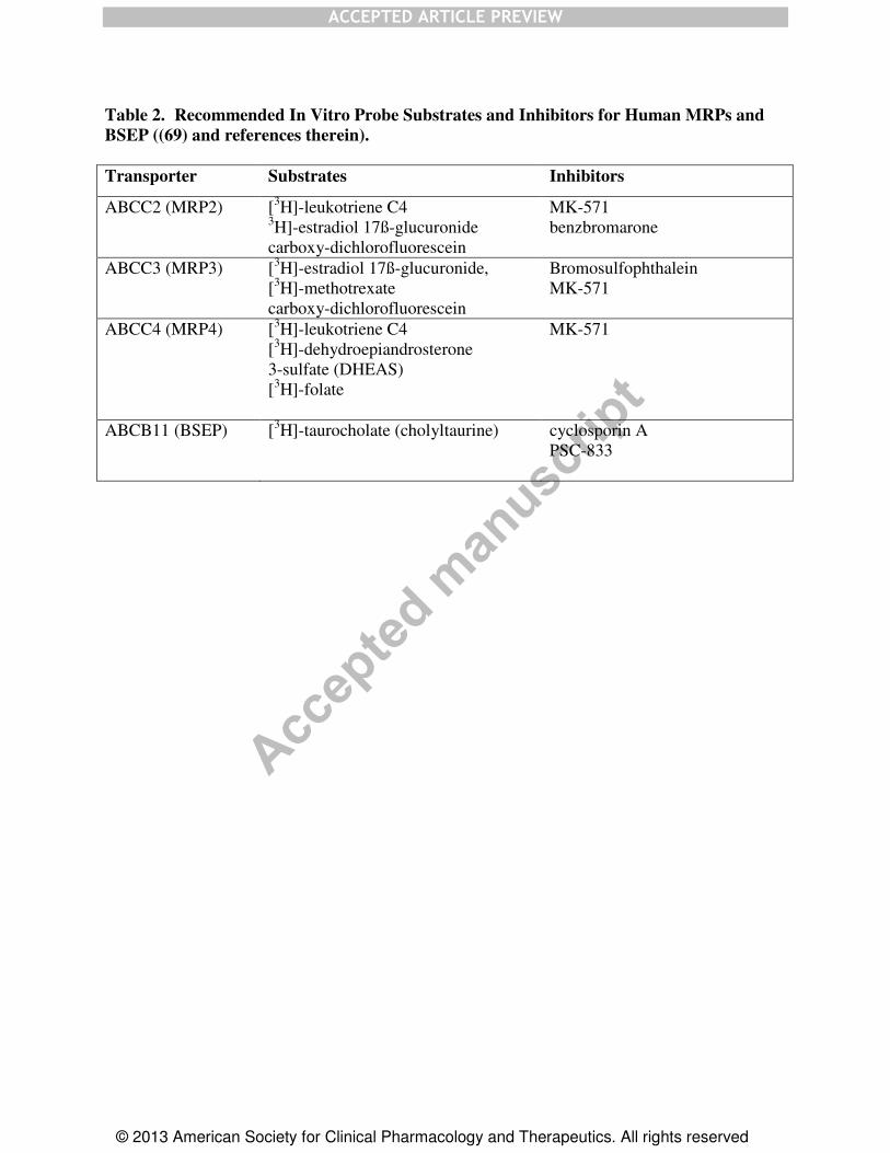
in intact cells and in vivo, can be controlled. Moreover, Table 2 presents some recommended
probe substrates and inhibitors. It should be noted that many MRP substrates are only generated
intracellularly in Phase II reactions forming the respective glutathione, sulfate, and glucuronide
conjugates, as illustrated by S-(2,4-dinitrophenyl)glutathione and glucuronosyl bilirubin.
An extensive analysis of the prediction and identification of in vitro drug interactions
with human MRP2 was published by Pedersen et al. (35) using inside-out-oriented membrane
vesicles from Sf9 cells and estradiol-17β-D-glucuronide as the substrate. This study with a large
set of structurally diverse drugs and drug-like compounds identified many inhibitors,
noninhibitors, and even stimulators of MRP2-mediated transport (35). The stimulators exhibited
similar charge distributions as MRP2 substrates, with at least one negative charge at physiologic
pH. As pointed out above, such a study gains additional physiological relevance when performed
in the presence and absence of GSH which is present in living cells at millimolar concentrations.
Moreover, for drugs and drug candidates that undergo predominantly hepatobiliary elimination,
it is essential to determine if glucuronate or glutathione conjugates are formed intracellularly and
represent inhibitors or substrates of MRP2, MRP3, or MRP4.

Accep
ted m
anusc
ript
ACCEPTED ARTICLE PREVIEW
© 2013 American Society for Clinical Pharmacology and Therapeutics. All rights reserved
For a NME, it is important to examine MRP2 inhibition if drug-induced conjugated
hyperbilirubinemia is observed in patients or in preclinical species. To date no classical PK DDI
has been attributed to MRP2 and there is no evidence that MRP3 or MRP4 should be examined
or that there is a likelihood of DDIs or toxicity due to being a substrate of MRP2. It is plausible,
but not proven, that hepatotoxicity may result, however, from compounds or metabolites that
potently inhibit MRP2 as well as MRP3 and MRP4 and thus cannot exit from hepatocytes via
alternative pathways.
The bile salt export pump (BSEP) and its role in hepatotoxicity
General Description
In addition to elimination of drugs and xenobiotics, bile formation is another key function
of the liver. Bile has two major physiologic functions: it provides a vehicle for excretion of
poorly water soluble substances (e.g., cholesterol) and is essential for digestion and absorption of
fat and fat soluble vitamins in the intestine. Bile is mainly composed of small ions, organic
anions, lipids (e.g., phosphatidylcholine) and bile salts (36). Bile salts are amphipathic molecules,
have detergent properties and are highly concentrated in bile (36) where they form mixed
micelles with phospholipids, which act as carriers for poorly water soluble substances (36). Due
to their detergent properties, bile salts can be highly toxic to hepatocytes if they accumulate
intracellularly (37).
Bile salts are taken up from the sinusoidal blood plasma by several transporters into
hepatocytes (38), where they mix with newly synthesized bile salts. Bile salts are secreted by the

Accep
ted m
anusc
ript
ACCEPTED ARTICLE PREVIEW
© 2013 American Society for Clinical Pharmacology and Therapeutics. All rights reserved
BSEP into canaliculi, which constitute the starting points of the biliary tree (39, 40). The BSEP
mediates the rate-limiting step of bile salt transport across hepatocytes for which there is not a
backup system in the canalicular membrane. Consequently, any impairment of BSEP function
can potentially lead to accumulation of cytotoxic bile salts in hepatocytes.
Function
BSEP has a narrow substrate specificity and transports conjugated monovalent bile salts
in addition to taurolitocholyl 3-sulfate (39, 41). Data on transport of unconjugated bile acids such
as cholic acid are conflicting (40). For taurocholate, Km values ranging from 5 to 22 µM have
been reported by different groups in different expression systems (40) and other bile salts have
Km values in this range (40). The intrinsic clearances for human, rat and mouse BSEP have the
same rank order (40, 42), indicating similar transport properties of BSEPs from these three
species. In addition to bile salts, pravastatin has been reported to be a BSEP substrate, albeit with
low affinity (40), and indirect evidence from sandwich-cultured hepatocytes suggests that the
antifungal micafungin may be transported by BSEP (43). Taking together the evidence obtained
so far, it seems unlikely that BSEP plays a major role in drug export from hepatocytes into bile
and consequently in drug disposition.
Genetic variants
Much has been learned about the physiologic role of BSEP from patients with inherited
liver diseases due to impaired BSEP function. ABCB11 encoding BSEP was identified by
positional cloning in families with progressive familial intrahepatic cholestasis (PFIC) type 2
(44). Four mutations identified in this study predict premature truncation of BSEP, while the

Accep
ted m
anusc
ript
ACCEPTED ARTICLE PREVIEW
© 2013 American Society for Clinical Pharmacology and Therapeutics. All rights reserved
other mutations are missense mutations. Such patients develop progressive cholestatic liver
disease with low biliary bile salt concentrations (45). Basolateral efflux via MRP4 and the
organic solute transporter OSTalpha/OSTbeta is not sufficient to compensate for genetic variants
resulting in the absence of BSEP protein, clearly demonstrating the absence of a functional
backup system in the canalicular membrane for secretion of bile salts. Some mutations in
ABCB11 lead to less severe forms of cholestatic liver disease called benign recurrent intrahepatic
cholestasis type 2 (BRIC2). The clinical spectrum of ABCB11 mutations covering BRIC2 to
PFIC2 is also known as BSEP deficiency syndrome (45).
It can be inferred from patients with benign BSEP deficiency syndrome that in symptom-
free intervals the residual BSEP transport capacity is sufficient for a seemingly normal liver
function. Consequently, inhibition of BSEP activity by endogenous or exogenous substances
below (a yet unknown) threshold function can lead to cholestasis. Cyclosporine is a drug leading
to acquired cholestasis and consequently to drug induced liver injury (DILI) in some patients
(41). Using rat Bsep as a model, competitive inhibition of Bsep by drugs known to be associated
with drug-induced cholestasis was demonstrated (46). This finding was later extended to human
BSEP (42, 47). To date, a large variety of drugs and xenobiotics have been reported to interfere
with BSEP function in vitro, the information for which is summarized in (40, 41, 48, 49).
Drug-induced cholestasis is a rare but often serious side effect of drugs (50).
Consequently, investigations for identification of susceptibility factors are ongoing, but have so
far proved difficult (50). With respect to factors affecting BSEP, information is scarce. The most
common polymorphism in ABCB11 is c.1331T>C (p.V444A) and is ethnicity dependent with a

Accep
ted m
anusc
ript
ACCEPTED ARTICLE PREVIEW
© 2013 American Society for Clinical Pharmacology and Therapeutics. All rights reserved
frequency around 60% in Caucasian populations (51). The c.1331C variant leads to lower BSEP
protein levels (52) and is overrepresented in patients with drug induced cholestasis as well as in
patients with intrahepatic cholestasis of pregnancy (50, 51). As the two BSEP variants have
indistinguishable kinetic properties and both variants are almost equally abundant in some
populations, the role of this polymorphism as a susceptibility factor for drug- induced cholestasis
is, as yet, unknown.
Methodology for evaluating function
Bile salts are negatively charged and can barely cross membranes. Therefore, studies with
isolated membrane vesicles are the best technical approach to investigate transport mediated by
BSEP. ATP-dependent taurocholate transport into canalicular plasma membrane vesicles is
electrogenic (40). Inhibition studies with NMEs and BSEP are preferentially done at taurocholate
concentrations of 5 to 10 µM in the presence of a permeant anion (for compensation of charge
movement). The suggested taurocholate concentration is in the range of the Km value of BSEP
(40) and allows an estimation of Ki values from IC50 values. If a NME inhibits BSEP with a low
Ki value, additional experiments may be warranted. One additional test system for a NME is
human hepatocytes cultured in a sandwich configuration (53). This system offers the advantage
that it is metabolically competent and to some extent includes the potential impact of metabolites
on BSEP. In addition, experiments in a preclinical species, e.g. rats, may be warranted. If the
NME is also an inhibitor of rat Bsep, application of the NME in a repeat dose study for several
weeks to animals at a dose leading to at least the expected human serum concentrations will be
helpful. It is recommended that this test includes sampling of serum on a daily basis followed by
measurements of bile salt concentration, alkaline phosphatase, and transaminases. A course of

Accep
ted m
anusc
ript
ACCEPTED ARTICLE PREVIEW
© 2013 American Society for Clinical Pharmacology and Therapeutics. All rights reserved
serum parameters showing a steady increase of bile salts, even if before elevation of
transaminases, points to a likely Bsep interaction. At the end of the experiment, bile flow may be
determined in control and treated animals, as any reduction of bile flow is a very sensitive
indicator for cholestasis. If a NME showing a Bsep interaction in rodents is given to humans,
serum bile salt levels as well as serum markers may be monitored from the very beginning. For
humans, the revised standards for drug-induced liver injury may serve as a guideline (54).
Clinical significance and drug development recommendations
The decision whether or not to investigate a NME for BSEP interaction may depend on
various factors. As BSEP is an unlikely player in disposition of NMEs, concerns may focus on
potential DILI related to BSEP. Here, inhibition of BSEP will lead to intracellular accumulation
of bile salts followed by cytotoxic events.
Currently, it is impossible to define a value for a BSEP inhibition constant that will
realistically predict significant BSEP-mediated DILI. The reasons are: 1) While a trend between
low IC50 values of BSEP inhibition and DILI was described (48, 49), no correlation between
Cmax, unbound with the potency of BSEP inhibition and with DILI was observed (48), showing that
the uptake mechanism of drugs into hepatocytes is a major contributor to intracellular drug
concentrations. At present, the exact transport mechanism and in particular the energetics of the
OATPs, which are the major drug uptake systems into hepatocytes, are poorly understood. It
should also be noted that cyclosporin A is a very potent BSEP inhibitor although it is widely
used in solid organ transplantation despite the occurrence of cholestatic episodes. In addition,
there is large interindividual variability of BSEP protein levels in normal human liver

Accep
ted m
anusc
ript
ACCEPTED ARTICLE PREVIEW
© 2013 American Society for Clinical Pharmacology and Therapeutics. All rights reserved
contributing to adverse actions (52). 2) Many drugs form an array of metabolites, some of which
may be even more potent BSEP inhibitors than the parent drug and act synergistically. Examples
are troglitazone and bosentan (41). The bosentan interaction is discussed in its EU label. 3)
Some drugs may in addition to interference with BSEP also impair mitochondrial function (e.g.,
troglitazone (41)) forming reactive intermediates. Such a situation may lead to a synergism in
toxicity, as elevated intracellular bile salts may aggravate mitochondrial toxicity of a metabolite
(41). And 4) There are BSEP inhibitors that require parallel transport activity of BSEP and
MRP2 in the same membrane. This was first demonstrated for estradiol 17β-glucuronide (46)
and later for drug metabolites (50). Drugs have been identified that are BSEP inhibitors but are
not associated with DILI. In conclusion, prospective BSEP testing cannot be endorsed at this
moment without a strategy to assess clinical relevance of such inhibition, but in vitro
characterization of BSEP-drug interactions are certainly warranted after the appearance of
cholestatic issues in clinical trials or safety studies. However, the EMA DDI guideline states that
investigating BSEP inhibitory potential should be considered. If inhibition is indicated, adequate
biochemical monitoring including serum bile salts is recommended during drug development.
Other Transporters of Clinical Importance
The transporters described above and in the previous ITC white paper have the potential
to interact with a broad range of drugs. However, it is important to keep in mind that for certain
classes of drugs other transport mechanisms may be important. Two such examples are given
below: Human equilibrative nucleoside transporter 1 (hENT1) which is important in the delivery

Accep
ted m
anusc
ript
ACCEPTED ARTICLE PREVIEW
© 2013 American Society for Clinical Pharmacology and Therapeutics. All rights reserved
of certain drugs to tumors and the human intestinal Peptide Transporter which is important in the
oral delivery of many peptide-like drugs.
Human equilibrative nucleoside transporter 1 (hENT1)
General description
Nucleosides have diverse roles in many processes, including cellular metabolism,
signaling and proliferation. Many nucleoside analogs are used clinically for treatment of cancer
and viral diseases. Since most nucleosides are hydrophilic, their cellular uptake and release is
dependent on the activity of membrane transport proteins. Two evolutionarily unrelated protein
families are responsible for nucleoside transport in humans: the Solute Carrier (SLC)28 and
SLC29 families of nucleoside transporters (NTs), whose members are known, respectively, as
the human (h) concentrative and equilibrative NTs (hCNTs; hENTs)(for recent reviews see (55-
57)). Three isoforms in the hCNT family and four in the hENT family have been identified and
functionally characterized by molecular cloning and expression of their cDNAs. These isoforms,
the genes encoding them and their chromosomal locations are: hCNT1, SLC28A1, 15q25.3;
hCNT2, SLC28A2, 15q15; hCNT3, SLC28A3, 9q22.2; hENT1, SLC29A1, 6p21,1; hENT2,
SLC29A2, 11q13; hENT3, SLC29A3, 10q22.1; hENT4, SLC29A4, 7p22.1. The focus in this
whitepaper will be on hENT family members and their roles in delivery of drugs to tumors.
Current understanding of the distribution of hENTs in intestine, kidney, liver and brain (57) is
summarized in Figure 1 .
Among members of the hENT family, the properties of hENT1 and hENT2 are well
established (56). Both translocate nucleosides bidirectionally down their concentration gradients,

Accep
ted m
anusc
ript
ACCEPTED ARTICLE PREVIEW
© 2013 American Society for Clinical Pharmacology and Therapeutics. All rights reserved
have broad substrate selectivities, and are widely distributed among tissues. They are
functionally distinguished by differences in their sensitivities to transport inhibitors. hENT1 is
potently inhibited by nitrobenzylmercaptopurine ribonucleoside (NBMPR) and its structural
analogs with Ki values in the nM range whereas hENT2 is relatively insensitive with Ki values in
the µM range. Because NBMPR and its structural analogs are highly specific for hENT1, they
have been used extensively as molecular probes for identification and isolation of hENT1 in cells
and tissues. hENT1 and hENT2 are integral membrane proteins with 11 transmembrane helices
and regions important for inhibitor and substrate interactions have been identified for both
proteins (57).
hENT1 and hENT2 both transport a wide range of purine and pyrimidine nucleosides, and
hENT1 has higher apparent affinities for its substrates than hENT2. Both also have the capacity
to transport nucleobases (hENT2 > hENT1), although the physiological importance of these
activities is unknown (56, 57). In general, nucleoside analog drugs are poorer substrates for
hENT1 and hENT2 than their physiological counterparts. Among clinical anticancer nucleosides,
fludarabine, cladribine, clofarabine, 5-fluorouridine, 2'-deoxy-5-fluorouridine, azacytidine and
decitabine are substrates of both, whereas gemcitabine and cytarabine are preferred by hENT1.
Among clinical antiviral nucleosides, hENT2 appears to be more important than hENT1 in
cellular uptake of zidovudine, zalcitabine and didanosine.
Transport assays
Because most human cell types contain multiple hNT types, the most reliable functional
studies of hNTs are those conducted with (i) recombinant proteins produced individually in

Accep
ted m
anusc
ript
ACCEPTED ARTICLE PREVIEW
© 2013 American Society for Clinical Pharmacology and Therapeutics. All rights reserved
otherwise NT-deficient cells, primarily the yeast Saccharomyces cerevisiae (engineered to lack
NT activity), oocytes of Xenopus laevis (lack endogenous NT activity) and human cell lines
(mutants that lack NT activity) and (ii) native proteins in cells that either produce a single
transporter type (e.g., hENT1-containing human erythrocytes) or that can be manipulated
pharmacologically by treatment with selective NT inhibitors such that only a single transporter
type is functional (e.g., treatment of hENT1/2-containing cells with NBMPR). For
methodological details see (58, 59). Studies of radioisotope fluxes can be undertaken with these
systems to identify substrates and inhibitors, which for some hNT and cell combinations is
difficult because of rapid equilibration of intra and extracellular nucleosides.
Clinical implications: hENT1 in anticancer therapies with nucleoside drugs
Although the potential role of hENT1 in the clinical activity of nucleoside drugs has been
recognized for decades (reviewed in (60)), evidence of its importance has only recently been
obtained. The development of monoclonal antibodies against hENT1 (61) provided the tools for
analysis of the distribution of hENT1 in human tissues, including immunohistochemistry studies
of human cancer samples, which revealed marked variations in its abundance in a variety of
cancers, giving rise to the hypothesis that low levels of hENT1 in target tumors predicts
resistance to nucleoside drugs. In a retrospective study of pancreatic adenocarcinoma samples
from deceased patients who received gemcitabine mono-chemotherapy for palliation, patients
whose tumor cells all exhibited hENT1 immunostaining had longer survival times than those
whose tumors contained cells that lacked hENT1 immunostaining (13 vs 4 months), indicating a
relationship between hENT1 abundance and response to gemcitabine (62). A subsequent study of
hENT1 abundance and gemcitabine therapy in a larger group of pancreatic adenocarcinoma
Accep
ted m
anusc
ript
ACCEPTED ARTICLE PREVIEW
© 2013 American Society for Clinical Pharmacology and Therapeutics. All rights reserved
patients yielded similar findings (63). It has been proposed that resistance to nucleoside drugs
because of low hNT abundance in target tumor cells can be circumvented by the development of
nucleoside drugs that enter cells by other routes (e.g., passive diffusion). Since pharmacologic
resistance to anticancer nucleoside drugs is likely if other drugs administered simultaneously
inhibit cellular uptake, NMEs with nucleoside-like structures may be assessed for their potential
to inhibit hENT1 if they are likely to be administered with ENT substrates.
Peptide transport
Another class of transporter that will handle selected targeted drugs is the Peptide
Transporter Family (gene symbol SLC15) (64). There are four transporters in the SLC15 family
(SLC15A1-4). The proteins encoded and their chromosomal location are PEPT1 (SLC15A1,
13q33-q34 ), PEPT2 (SLC15A2, 3q13.3-q21), PHT1 (SLC15A4, 12q24.32) and PHT2 (SLC15A3,
11q12.2). PHT1 and 2 transport histidine and peptides and have not been shown clinically to
play a major role in the disposition of drugs. PEPT1 and PEPT2 are the most studied
transporters in the family and are key in the disposition of peptide-like drugs (e.g., beta lactam
antibiotics, ACE and renin inhibitors) and in peptide prodrugs (valacyclovir)(65). PEPT1 is
apically located in the intestine and to a lesser extent in the kidney. PEPT1 is responsible for the
uptake of di- and tri- peptides from the intestinal lumen into the blood and in the kidney for the
scavenging of peptides from the urine back into the renal proximal tubule cells. PEPT2 is
located in the kidney, but is also found at the blood cerebrospinal fluid barrier and the lung.
PEPT2 is the primary mechanism in the proximal tubule cells for the reabsorption of di- and tri-

Accep
ted m
anusc
ript
ACCEPTED ARTICLE PREVIEW
© 2013 American Society for Clinical Pharmacology and Therapeutics. All rights reserved
peptides from the urine. In the brain, PEPT2 is used to clear di- and tri-peptides from the
cerebrospinal fluid (CSF) back into the blood and in the lung PEPT2 absorbs peptides.
Because of PEPT1’s location and absorptive function in the intestine, it is a useful
mechanism to increase the bioavailability of small, hydrophilic, charged molecules (64). It has
been utilized to increase the bioavailability of amino acid- and nucleoside-like drugs. It is also
known to transport some antibiotics. It transports molecules via a proton dependent mechanism
taking advantage of the acidic micro-environment of the intestine to co-transport a substrate
along with a proton(65). PEPT1 is considered a low affinity high capacity transporter. The
affinity (Km) for natural substrates is in the high micromolar range and for drugs such as beta
lactam antibiotics the Km values are in the low millimolar range. However, since it has high
capacity and is expressed along the length of the gut, it efficiently scavenges its substrates’
uptake into intestinal enterococytes where they are eventually shunted into the blood.
The genetics of PEPT1 have been studied in vitro and the most frequent coding region
SNP (S117N) does not lead to functional change in vitro (66). Two SNPs that do lead to
functional changes in vitro (P586L and F28Y) have very low frequency in all populations studied
to date (65). To date no clinical consequences for any PEPT1 variants have been found.
Functional studies for PEPT1 have been carried out in a number of heterologous
expression systems. The transporter is easily expressed transiently in mammalian cell lines such
as HEK, COS or HeLa or it can also be stably expressed (64). Many laboratories have also used
oocyte expression to study peptide transport. PEPT1 is also expressed in intestinal cell lines
Accep
ted m
anusc
ript
ACCEPTED ARTICLE PREVIEW
© 2013 American Society for Clinical Pharmacology and Therapeutics. All rights reserved
such as CACO-2. However, the expression level is low and requires 14 to 21 days in culture for
maximal expression. The role of PEPT1 in absorption can also be studied in knockout mice (67).
Although the knock-out strains have been characterized and used to study probe substrates, they
are not currently commercially available.
Clinical significance and drug development recommendations
PEPT1 works well as a mechanism for oral delivery of small charged amino acid, peptide
or nucleoside like drugs (65). The transporter appears not to be easily saturated and can deliver
relatively large doses of drugs. For example, the cephalosporin antibiotics are given at gram
doses. Review of labels for drugs (e.g., cephalosporins, valacyclovir, enalapril) that utilize
peptide transporters for absorption shows little concern for interactions during absorption as
there are no consistent food interactions or interactions with other drugs transported by PEPT1.
It is of note that PEPT1 appears to be upregulated in inflammatory bowel disease leading to
expression of PEPT1 in the colon which is believed to be part of the proinflammatory response
(68). Thus, although PEPT1 is normally thought of as a delivery mechanism, it may also be a
pharmacological target for inflammatory bowel disease. There have also been studies as to its
role in various cancers as a possible delivery mechanism (64, 65).
SUMMARY AND FUTURE PERSPECTIVES
Membrane transporters of emerging importance in pharmacokinetics, drug-drug
interactions, and interference of drugs with the transport of endogenous compounds are
discussed in this paper. We focus here on human transporters involved in the uptake and efflux

Accep
ted m
anusc
ript
ACCEPTED ARTICLE PREVIEW
© 2013 American Society for Clinical Pharmacology and Therapeutics. All rights reserved
of drugs that were not considered in detail in our previous ITC whitepaper on membrane
transporters (2).
The multidrug and toxin extrusion proteins (MATEs) were discovered only in 2005 (11)
and include MATE1, MATE2 and the splice variant MATE2K. They are expressed abundantly
in the apical membrane of the renal proximal tubule and play a role in the secretion of cations
and zwitterions into the urine. MATE1 is also highly expressed in the canalicular membrane of
hepatocytes where it contributes to biliary excretion of cationic compounds. Many clinically
used drugs, including metformin, cimetidine, and acyclovir, were identified as substrates for
MATEs. There is compelling in vitro and in vivo evidence that MATEs are major sites of renal
drug-drug interactions. ITC recommends that NMEs be evaluated as potential substrates or
inhibitors for MATEs along with OCT2 and OAT1/OAT3, and decision trees for evaluating the
interactions of MATEs with NMEs are presented. These decision trees will need to be tested in
the clinic and revised if necessary as more data becomes available.
Within the subfamily of multidrug resistance proteins (MRPs, ABCCs), the ATP-driven
efflux pumps MRP2, MRP3, and MRP4 have received particular attention because of their role
in the disposition of anionic drugs and anionic conjugates. Human MRP2 is localized to the
apical membrane of hepatocytes, intestinal epithelia, and proximal tubules of the kidney.
Accordingly, it is essential for the hepatobiliary and renal elimination of many anionic substrates,
including drugs and conjugates such as bilirubin glucuronides. In hepatocytes, MRP3 and MRP4
are localized to the basolateral (sinusoidal) membrane and mediate efflux of substrates,
particularly glucuronides and glutathione conjugates, into sinusoidal blood and play a

Accep
ted m
anusc
ript
ACCEPTED ARTICLE PREVIEW
© 2013 American Society for Clinical Pharmacology and Therapeutics. All rights reserved
compensatory role in cholestasis. Human MRP4 has a number of localizations important for drug
elimination and action, including its presence in the apical (luminal) membrane of the kidney
proximal tubule, the epithelium of the blood-brain barrier and membranes of the blood platelet.
Drug-induced conjugated hyperbilirubinemia, in patients or in preclinical species, points to an
inhibition of MRP2. Thus, under many conditions, conjugated hyperbilirubinemia may
necessitate in vitro analyses using, for instance, inside-out-oriented membrane vesicles to
elucidate the interaction of a compound with MRP2.
The bile salt export pump (BSEP) is exclusively localized in the hepatocyte canalicular
membrane and prevents accumulation of toxic concentrations of bile salts in hepatocytes by
unidirectional ATP-dependent transport into the biliary tree. BSEP has no functional backup and
impairment of BSEP function may lead to liver disease as evidenced by increased plasma bile
salt concentrations and liver enzymes. NMEs leading to cholestasis with increased plasma bile
salt concentrations in preclinical species or in humans may be examined for their inhibitory
potential using inside-out membrane vesicles containing recombinant human BSEP in ATP-
dependent transport assays with taurocholate (cholyltaurine) as a probe substrate. Future work is
needed to identify susceptibility factors and/or genes leading to drug-induced cholestasis due to
BSEP impariment. Such information may be obtained from retrospective analysis of clinical data,
but will subsequently need to be verified in prospective multicenter trials.
Other transporters have shown importance in drug delivery. In this whitepaper, we
presented two examples, NTs and PEPTs.
Accep
ted m
anusc
ript
ACCEPTED ARTICLE PREVIEW
© 2013 American Society for Clinical Pharmacology and Therapeutics. All rights reserved
NTs (e.g., hENT1) are important for the uptake of nucleosides and their analogs into cells
and are widely distributed among cells and tissues in humans. Many nucleoside analogs are used
clinically for treatment of cancer and viral diseases. Recent data showed a correlation between
abundance of hENT1 and survival of pancreatic cancer patients treated with gemcitabine.
Peptide transporters, as exemplified by PEPT1 in the apical membrane of small intestine,
are not only important for nutritional absorption of di- and tri-peptides but also for delivery of
amino acid-like or dipeptide-like drugs and prodrugs. Drug substrates include many ß-lactam
antibiotics and prodrugs such as valaciclovir. Transporter assays (e.g., using transfected PEPT1-
expressing HeLa cells) serve not only in studies on the substrate properties of new chemical
entities but also in the in vitro analysis of drug-drug interactions.
Consistent methods for studying transporters are still under development. However,
recommendations and a detailed discussion of determination of kinetic parameters of transporter
substrates are given in an accompanying article in this issue (69). In brief, transport studies with
transfected cells are recommended for uptake transport systems while transport studies with
inside-out vesicles isolated from transfected cells are recommended for efflux transporters.
Kinetic parameters have to be determined using conditions of initial uptake rates. For inhibition
experiments Ki values obtained from Dixon plots are preferred over IC50 values.
In summary, NMEs that are actively renally secreted are recommended for investigation
as a MATE1 or MATE2K substrates in addition to being investigated as OAT1, OAT3 or OCT2

Accep
ted m
anusc
ript
ACCEPTED ARTICLE PREVIEW
© 2013 American Society for Clinical Pharmacology and Therapeutics. All rights reserved
substrates according to Figure 2. It is recommended that the potential of all NMEs to inhibit
MATE1 and MATE2k be determined in vitro. Figure 3 may be followed to determine if a
clinical interaction study is necessary. The inhibition of both BSEP and MRPs has the potential
to cause cholestasis and if signs of cholestasis are seen, retrospective analysis of inhibition of
the BSEP and MRPs can help determine the mechanism of toxicity. Because there are no known
cut-offs for the inhibition potential (IC50 or Ki values) of these transporters that indicate
increased risk of hepatotoxicity, screening of their inhibition on a routine basis is not yet
warrented. Three other transporters that were highlighted in this review, hENT1, hENT2 and
PEPT1, although examples of transporters that do not need to be routinely screened, are also
known to distribute specific type of drugs and thus are important determinants for efficacy and
distribution. The hENTs transport nucleoside drugs and PEPT1 transports a variety of drugs
with peptide-like properties.
Disclaimer:
The opinions expressed in this paper reflect the views of the authors and should not be construed
to represent FDA’s views or policies.
Lei Zhang has no conflict of interest to report.
Carol Cass is a paid consultant for Clavis Pharma. The University of Alberta licensed antibodies
against hENT1 produced in Cass's research laboratory to Clavis Pharma (Oslo, Norway) for use
in diagnostic assays to predict response of cancer patients to nucleoside derivatives produced by
Clavis that enter cells independently of nucleoside transport systems.

Accep
ted m
anusc
ript
ACCEPTED ARTICLE PREVIEW
© 2013 American Society for Clinical Pharmacology and Therapeutics. All rights reserved
Figure Legends
Figure-1: Proposed expansion of transporters for evaluation during drug development by
ITC.
Proposed expansion of transporters for evaluation during drug development by ITC.
Transporters were highlighted on the basis of evidence of clinical drug interactions and relevance
to toxicity or efficacy, as well as availability of in vitro assays, substrates and inhibitors.
Transporters recommended for evaluation in the 2012 FDA Draft Drug Interaction Guidance are
marked with red circles. MATEs (green circles) are being proposed for prospective investigation
in drug development as discussed at the ITC Second Workshop. In addition MRP2 and BSEP
inhibition are recommended for retrospective studies based on preclinical and clinical
observations (yellow circles.) Examples given in this paper (ENTs and PEPTs) are in light blue
circles. Modified from ref. 8 by Sook Wah Yee, University of California, San Francisco.
Figure 2: Decision tree for renally cleared substrates proposed by ITC a. For simplicity organic anion transporters, OAT1 and OAT3 are not included; however, if
secretory clearance is an important route of elimination, NMEs should also be studied as
substrates of OAT1 and OAT3.
a-Percent (%) active renal secretion was estimated from (CLr–fu*GFR)/CLTotal; fu is the
unbound fraction in plasma (6).
b-The ratio of the investigational drug uptake in the cells expressing the transporter versus the
control (or empty vector) cells should be greater than 2. It is important that uptake into the
transfected cells be significantly greater than background in a control cell line and be inhibited
by a known inhibitor of the transporter. Michaelis–Menten studies may be conducted in the
transfected cells to determine the kinetic parameters of the investigational drug. A positive
control should be included. In an acceptable cell system, the positive control should show a ≥ 2
fold increase in uptake compared to vector-transfected cells. An uptake ratio (transporter
transfected vs. empty vector transfected cells) other than 2 may be used if a ratio of 2 is deemed
non-discriminative as supported by prior experience with the cell system used.
c-If the NME interacts with OCT2 and MATE1/2K, suggested inhibitors to use for a clinical
DDI study are pyrimethamine or cimetidine, both of which are clinically validated. If the NME
is a substrate of OAT1 or OAT3 and MATE1/2K, probenecid should be considered for use as an
inhibitor.
Figure-3: Decision tree for renal transporter inhibitors proposed by ITC a-First step might be uptake of model substrate decreases with increasing concentration of NME,
then full IC50 curve (modified from Ref 6).
b-For NME that is a MATE1, MATE2K or OCT2 inhibitor, metformin may be used as the
substrate for the clinical drug interaction study.

Accep
ted m
anusc
ript
ACCEPTED ARTICLE PREVIEW
© 2013 American Society for Clinical Pharmacology and Therapeutics. All rights reserved
Table 1: Selected putative MATE-mediated clinical drug-drug interactions
Interacting drug Affected drug Clinical effect on affected drug Ref.
Fexofenadine 39% decrease in CLr of fexofenadine (70)
Metformin 50% decrease in CLr of metformin
and 27% increase in AUC
(71)
Dofetilide 33% decrease in CLr of dofetilide,
48% and 29% increase in AUC and
Cmax respectively
(72)
Chepalexin 33% decrease in CLr of cephexin (73)
Cimetidine (400-
800mg, PO)
Procainamide 44% decrease in CLr of
procainamide and 35% increase in
AUC
(74)
Chepalexin
(500mg, PO)
Metformin 14% decrease in CLr of metformin,
24% and 34% increase in AUC and
Cmax respectively
(75)
Metformin 35% decrease in CLr of metformin,
42% and 39% increase in Cmax and
AUC respectively
(20) Pyrimethamine
(50mg, PO)
Creatinine 20% decrease in CLr of creatinine (20)
Legend:
Percent change refers to the difference in the presence or absence of interacting drug
normalized to the value in the absence of interacting drug.
Accep
ted m
anusc
ript
ACCEPTED ARTICLE PREVIEW
© 2013 American Society for Clinical Pharmacology and Therapeutics. All rights reserved
Table 2. Recommended In Vitro Probe Substrates and Inhibitors for Human MRPs and
BSEP ((69) and references therein).
Transporter Substrates Inhibitors
ABCC2 (MRP2) [3H]-leukotriene C4
3H]-estradiol 17ß-glucuronide
carboxy-dichlorofluorescein
MK-571
benzbromarone
ABCC3 (MRP3) [3H]-estradiol 17ß-glucuronide,
[3H]-methotrexate
carboxy-dichlorofluorescein
Bromosulfophthalein
MK-571
ABCC4 (MRP4) [3H]-leukotriene C4
[3H]-dehydroepiandrosterone
3-sulfate (DHEAS)
[3H]-folate
MK-571
ABCB11 (BSEP) [3H]-taurocholate (cholyltaurine) cyclosporin A
PSC-833

Accep
ted m
anusc
ript
ACCEPTED ARTICLE PREVIEW
© 2013 American Society for Clinical Pharmacology and Therapeutics. All rights reserved
References
1. K.M.Giacomini and Y.Sugiyama. Membrane Transporters and Drug Response. In
L.L.Brunton, B.C.Chabner, and B.A.Knollman (eds.), Goodman & Gilman's. The
Pharmaceutical Basis of Therapeutics, McGraw-Hill Education, 2010.
2. K.M.Giacomini, S.M.Huang, D.J.Tweedie, L.Z.Benet, K.L.Brouwer, X.Chu,
A.Dahlin, R.Evers, V.Fischer, K.M.Hillgren, K.A.Hoffmaster, T.Ishikawa, D.Keppler, R.B.Kim,
C.A.Lee, M.Niemi, J.W.Polli, Y.Sugiyama, P.W.Swaan, J.A.Ware, S.H.Wright, S.W.Yee,
M.J.Zamek-Gliszczynski, and L.Zhang. Membrane transporters in drug development. Nat Rev
Drug Discov. 9:215-236 (2010).
3. R.G.Tirona. Molecular mechanisms of drug transporter regulation. Handb Exp
Pharmacol:373-402 (2011).
4. FDA Drug Safety Communication: Interactions between certain HIV or hepatitis
C drugs and cholesterol-lowering statin drugs can increase the risk of muscle injury.
http://www.fda.gov/Drugs/DrugSafety/ucm293877.htm (accessed 7/15/12).
5. S.M.Huang, L.Zhang, and K.M.Giacomini. The International Transporter
Consortium: a collaborative group of scientists from academia, industry, and the FDA. Clin
Pharmacol Ther. 87:32-36 (2010).
6. Guidance for Industry (Draft): Drug Interaction Studies -Study Design, Data
Analysis, Implications for Dosing, and Labeling Recommendations.
http://www.fda.gov/Drugs/GuidanceComplianceRegulatoryInformation/Guidances/ucm064982.h
tm (accessed 2/18/12).
7. . European Medicines Agency's Guideline on the Investigation of Drug
Interactions (Final). In 2012.
8. M.J.Zamek-Gliszczynski, K.A.Hoffmaster, D.J.Tweedie, K.M.Giacomini, and
K.M.Hillgren. Highlights from the International Transporter Consortium second workshop. Clin
Pharmacol Ther. 92:553-556 (2012).
9. K.Damme, A.T.Nies, E.Schaeffeler, and M.Schwab. Mammalian MATE
(SLC47A) transport proteins: impact on efflux of endogenous substrates and xenobiotics. Drug
Metab Rev. 43:499-523 (2011).
10. A.Yonezawa and K.Inui. Importance of the multidrug and toxin extrusion
MATE/SLC47A family to pharmacokinetics, pharmacodynamics/toxicodynamics and
pharmacogenomics. Br J Pharmacol. 164:1817-1825 (2011).
11. M.Otsuka, T.Matsumoto, R.Morimoto, S.Arioka, H.Omote, and Y.Moriyama. A
human transporter protein that mediates the final excretion step for toxic organic cations. Proc
Natl Acad Sci U S A. 102:17923-17928 (2005).
Accep
ted m
anusc
ript
ACCEPTED ARTICLE PREVIEW
© 2013 American Society for Clinical Pharmacology and Therapeutics. All rights reserved
12. X.Zhang and S.H.Wright. MATE1 has an external COOH terminus, consistent
with a 13-helix topology. Am J Physiol Renal Physiol. 297:F263-F271 (2009).
13. M.Lu, J.Symersky, M.Radchenko, A.Koide, Y.Guo, R.Nie, and S.Koide.
Structures of a Na+-coupled, substrate-bound MATE multidrug transporter. Proc Natl Acad Sci
U S A. 110:2099-2104 (2013).
14. J.H.Choi, S.W.Yee, A.H.Ramirez, K.M.Morrissey, G.H.Jang, P.J.Joski,
J.A.Mefford, S.E.Hesselson, A.Schlessinger, G.Jenkins, R.A.Castro, S.J.Johns, D.Stryke, A.Sali,
T.E.Ferrin, J.S.Witte, P.Y.Kwok, D.M.Roden, R.A.Wilke, C.A.McCarty, R.L.Davis, and
K.M.Giacomini. A common 5'-UTR variant in MATE2-K is associated with poor response to
metformin. Clin Pharmacol Ther. 90:674-684 (2011).
15. J.Ha Choi, S.Wah Yee, M.J.Kim, L.Nguyen, J.Ho Lee, J.O.Kang, S.Hesselson,
R.A.Castro, D.Stryke, S.J.Johns, P.Y.Kwok, T.E.Ferrin, M.Goo Lee, B.L.Black, N.Ahituv, and
K.M.Giacomini. Identification and characterization of novel polymorphisms in the basal
promoter of the human transporter, MATE1. Pharmacogenet Genomics. 19:770-780 (2009).
16. B.Astorga, S.Ekins, M.Morales, and S.H.Wright. Molecular determinants of
ligand selectivity for the human multidrug and toxin extruder proteins MATE1 and MATE2-K. J
Pharmacol Exp Ther. 341:743-755 (2012).
17. M.B.Wittwer, A.A.Zur, N.Khuri, Y.Kido, A.Kosaka, X.Zhang, K.M.Morrissey,
A.Sali, Y.Huang, and K.M.Giacomini. Discovery of Potent, Selective Multidrug and Toxin
Extrusion Transporter 1 (MATE1, SLC47A1) Inhibitors Through Prescription Drug Profiling
and Computational Modeling. J Med Chem. 56:781-795 (2013).
18. T.Terada, S.Masuda, J.Asaka, M.Tsuda, T.Katsura, and K.Inui. Molecular cloning,
functional characterization and tissue distribution of rat H+/organic cation antiporter MATE1.
Pharm Res. 23:1696-1701 (2006).
19. M.L.Becker, L.E.Visser, R.H.van Schaik, A.Hofman, A.G.Uitterlinden, and
B.H.Stricker. Genetic variation in the multidrug and toxin extrusion 1 transporter protein
influences the glucose-lowering effect of metformin in patients with diabetes: a preliminary
study. Diabetes. 58:745-749 (2009).
20. H.Kusuhara, S.Ito, Y.Kumagai, M.Jiang, T.Shiroshita, Y.Moriyama, K.Inoue,
H.Yuasa, and Y.Sugiyama. Effects of a MATE protein inhibitor, pyrimethamine, on the renal
elimination of metformin at oral microdose and at therapeutic dose in healthy subjects. Clin
Pharmacol Ther. 89:837-844 (2011).
21. D.Keppler. Multidrug resistance proteins (MRPs, ABCCs): importance for
pathophysiology and drug therapy. Handb Exp Pharmacol:299-323 (2011).
22. R.G.Deeley, C.Westlake, and S.P.Cole. Transmembrane transport of endo- and
xenobiotics by mammalian ATP-binding cassette multidrug resistance proteins. Physiol Rev.
86:849-899 (2006).

Accep
ted m
anusc
ript
ACCEPTED ARTICLE PREVIEW
© 2013 American Society for Clinical Pharmacology and Therapeutics. All rights reserved
23. A.T.Nies and D.Keppler. The apical conjugate efflux pump ABCC2 (MRP2).
Pflugers Arch. 453:643-659 (2007).
24. F.G.Russel, J.B.Koenderink, and R.Masereeuw. Multidrug resistance protein 4
(MRP4/ABCC4): a versatile efflux transporter for drugs and signalling molecules. Trends
Pharmacol Sci. 29:200-207 (2008).
25. G.Jedlitschky, A.Greinacher, and H.K.Kroemer. Transporters in human platelets:
physiologic function and impact for pharmacotherapy. Blood. 119:3394-3402 (2012).
26. P.Borst, C.de Wolf, and K.van de Wetering. Multidrug resistance-associated
proteins 3, 4, and 5. Pflugers Arch. 453:661-673 (2007).
27. E.van de Steeg, V.Stranecky, H.Hartmannova, L.Noskova, M.Hrebicek,
E.Wagenaar, E.A.van, D.R.de Waart, R.P.Oude Elferink, K.E.Kenworthy, E.Sticova, M.al-
Edreesi, A.S.Knisely, S.Kmoch, M.Jirsa, and A.H.Schinkel. Complete OATP1B1 and OATP1B3
deficiency causes human Rotor syndrome by interrupting conjugated bilirubin reuptake into the
liver. J Clin Invest. 122:519-528 (2012).
28. M.Rius, J.Hummel-Eisenbeiss, A.F.Hofmann, and D.Keppler. Substrate
specificity of human ABCC4 (MRP4)-mediated cotransport of bile acids and reduced glutathione.
Am J Physiol Gastrointest Liver Physiol. 290:G640-G649 (2006).
29. K.Letschert, H.Faulstich, D.Keller, and D.Keppler. Molecular characterization
and inhibition of amanitin uptake into human hepatocytes. Toxicol Sci. 91:140-149 (2006).
30. J.A.Byrne, S.S.Strautnieks, G.Mieli-Vergani, C.F.Higgins, K.J.Linton, and
R.J.Thompson. The human bile salt export pump: characterization of substrate specificity and
identification of inhibitors. Gastroenterology. 123:1649-1658 (2002).
31. J.Kartenbeck, U.Leuschner, R.Mayer, and D.Keppler. Absence of the canalicular
isoform of the MRP gene-encoded conjugate export pump from the hepatocytes in Dubin-
Johnson syndrome. Hepatology. 23:1061-1066 (1996).
32. T.Lang, M.Hitzl, O.Burk, E.Mornhinweg, A.Keil, R.Kerb, K.Klein, U.M.Zanger,
M.Eichelbaum, and M.F.Fromm. Genetic polymorphisms in the multidrug resistance-associated
protein 3 (ABCC3, MRP3) gene and relationship to its mRNA and protein expression in human
liver. Pharmacogenetics. 14:155-164 (2004).
33. U.Gradhand, T.Lang, E.Schaeffeler, H.Glaeser, H.Tegude, K.Klein, P.Fritz,
G.Jedlitschky, H.K.Kroemer, I.Bachmakov, B.Anwald, R.Kerb, U.M.Zanger, M.Eichelbaum,
M.Schwab, and M.F.Fromm. Variability in human hepatic MRP4 expression: influence of
cholestasis and genotype. Pharmacogenomics J. 8:42-52 (2008).
34. P.Krishnamurthy, M.Schwab, K.Takenaka, D.Nachagari, J.Morgan, M.Leslie,
W.Du, K.Boyd, M.Cheok, H.Nakauchi, C.Marzolini, R.B.Kim, B.Poonkuzhali, E.Schuetz,
W.Evans, M.Relling, and J.D.Schuetz. Transporter-mediated protection against thiopurine-
induced hematopoietic toxicity. Cancer Res. 68:4983-4989 (2008).
Accep
ted m
anusc
ript
ACCEPTED ARTICLE PREVIEW
© 2013 American Society for Clinical Pharmacology and Therapeutics. All rights reserved
35. J.M.Pedersen, P.Matsson, C.A.Bergstrom, U.Norinder, J.Hoogstraate, and
P.Artursson. Prediction and identification of drug interactions with the human ATP-binding
cassette transporter multidrug-resistance associated protein 2 (MRP2; ABCC2). J Med Chem.
51:3275-3287 (2008).
36. A.Esteller. Physiology of bile secretion. World J Gastroenterol. 14:5641-5649
(2008).
37. R.J.Sokol, M.Devereaux, R.Dahl, and E.Gumpricht. "Let there be bile"--
understanding hepatic injury in cholestasis. J Pediatr Gastroenterol Nutr. 43 Suppl 1:S4-S9
(2006).
38. P.J.Meier and B.Stieger. Bile salt transporters. Annu Rev Physiol. 64:635-661
(2002).
39. B.Stieger, Y.Meier, and P.J.Meier. The bile salt export pump. Pflugers Arch.
453:611-620 (2007).
40. B.Stieger. The role of the sodium-taurocholate cotransporting polypeptide
(NTCP) and of the bile salt export pump (BSEP) in physiology and pathophysiology of bile
formation. Handb Exp Pharmacol. 201:205-259 (2011).
41. B.Stieger. Role of the bile salt export pump, BSEP, in acquired forms of
cholestasis. Drug Metab Rev. 42:437-445 (2010).
42. J.Noe, B.Stieger, and P.J.Meier. Functional expression of the canalicular bile salt
export pump of human liver. Gastroenterology. 123:1659-1666 (2002).
43. S.B.Yanni, P.F.Augustijns, D.K.Benjamin, Jr., K.L.Brouwer, D.R.Thakker, and
P.P.Annaert. In vitro investigation of the hepatobiliary disposition mechanisms of the antifungal
agent micafungin in humans and rats. Drug Metab Dispos. 38:1848-1856 (2010).
44. S.S.Strautnieks, L.N.Bull, A.S.Knisely, S.A.Kocoshis, N.Dahl, H.Arnell, E.Sokal,
K.Dahan, S.Childs, V.Ling, M.S.Tanner, A.F.Kagalwalla, A.Nemeth, J.Pawlowska, A.Baker,
G.Mieli-Vergani, N.B.Freimer, R.M.Gardiner, and R.J.Thompson. A gene encoding a liver-
specific ABC transporter is mutated in progressive familial intrahepatic cholestasis. Nat Genet.
20:233-238 (1998).
45. R.Kubitz, C.Droge, J.Stindt, K.Weissenberger, and D.Haussinger. The bile salt
export pump (BSEP) in health and disease. Clin Res Hepatol Gastroenterol. 36:536-553 (2012).
46. B.Stieger, K.Fattinger, J.Madon, G.A.Kullak Ublick, and P.J.Meier. Drug- and
estrogen-induced cholestasis through inhibition of the hepatocellular bile salt export pump
(Bsep) of rat liver. Gastroenterology. 118:422-430 (2000).
47. J.A.Byrne, S.S.Strautnieks, G.Mieli-Vergani, C.F.Higgins, K.J.Linton, and
R.J.Thompson. The human bile salt export pump: characterization of substrate specificity and
identification of inhibitors. Gastroenterology. 123:1649-1658 (2002).
Accep
ted m
anusc
ript
ACCEPTED ARTICLE PREVIEW
© 2013 American Society for Clinical Pharmacology and Therapeutics. All rights reserved
48. S.Dawson, S.Stahl, N.Paul, J.Barber, and J.G.Kenna. In Vitro Inhibition of the
Bile Salt Export Pump Correlates with Risk of Cholestatic Drug Induced Liver Injury in Man.
Drug Metab Dispos. 2011/10/04:130-138 (2012).
49. R.E.Morgan, M.Trauner, C.J.van Staden, P.H.Lee, B.Ramachandran,
M.Eschenberg, C.Afshari, C.W.Qualls, Jr., R.Lightfoot-Dunn, and H.K.Hamadeh. Interference
with Bile Salt Export Pump Function is a Susceptibility Factor for Human Liver Injury in Drug
Development. Toxicol Sci. 118:485-500 (2010).
50. C.Pauli-Magnus, P.J.Meier, and B.Stieger. Genetic determinants of drug-induced
cholestasis and intrahepatic cholestasis of pregnancy. Semin Liver Dis. 30:147-159 (2010).
51. B.Stieger and A.Geier. Genetic variations of bile salt transporters as predisposing
factors for drug-induced cholestasis, intrahepatic cholestasis of pregnancy and therapeutic
response of viral hepatitis. Expert Opin Drug Metab Toxicol. 7:411-425 (2011).
52. Y.Meier, C.Pauli-Magnus, U.M.Zanger, K.Klein, E.Schaeffeler, A.K.Nussler,
N.Nussler, M.Eichelbaum, P.J.Meier, and B.Stieger. Interindividual variability of canalicular
ATP-binding-cassette (ABC)-transporter expression in human liver. Hepatology. 44:62-74
(2006).
53. B.Swift, N.D.Pfeifer, and K.L.Brouwer. Sandwich-cultured hepatocytes: an in
vitro model to evaluate hepatobiliary transporter-based drug interactions and hepatotoxicity.
Drug Metab Rev. 42:446-471 (2010).
54. G.P.Aithal, P.B.Watkins, R.J.Andrade, D.Larrey, M.Molokhia, H.Takikawa,
C.M.Hunt, R.A.Wilke, M.Avigan, N.Kaplowitz, E.Bjornsson, and A.K.Daly. Case definition and
phenotype standardization in drug-induced liver injury. Clin Pharmacol Ther. 89:806-815 (2011).
55. M.Pastor-Anglada, P.Cano-Soldado, E.Errasti-Murugarren, and F.J.Casado.
SLC28 genes and concentrative nucleoside transporter (CNT) proteins. Xenobiotica. 38:972-994
(2008).
56. J.D.Young, S.Y.Yao, L.Sun, C.E.Cass, and S.A.Baldwin. Human equilibrative
nucleoside transporter (ENT) family of nucleoside and nucleobase transporter proteins.
Xenobiotica. 38:995-1021 (2008).
57. J.D.Young, S.Y.M.Yao, J.M.Baldwin, C.E.Cass, and S.Baldwin. The human
concentrative and equilibrative nucleoside transporter families, SLC28 and SLC29. Mol Aspects
Med. 34:529-547 (2013).
58. M.L.Clarke, V.L.Damaraju, J.Zhang, D.Mowles, T.Tackaberry, T.Lang,
K.M.Smith, J.D.Young, B.Tomkinson, and C.E.Cass. The role of human nucleoside transporters
in cellular uptake of 4'-thio-beta-D-arabinofuranosylcytosine and beta-D-arabinosylcytosine.
Mol Pharmacol. 70:303-310 (2006).
59. K.M.King, V.L.Damaraju, M.F.Vickers, S.Y.Yao, T.Lang, T.E.Tackaberry,
D.A.Mowles, A.M.Ng, J.D.Young, and C.E.Cass. A comparison of the transportability, and its

Accep
ted m
anusc
ript
ACCEPTED ARTICLE PREVIEW
© 2013 American Society for Clinical Pharmacology and Therapeutics. All rights reserved
role in cytotoxicity, of clofarabine, cladribine, and fludarabine by recombinant human nucleoside
transporters produced in three model expression systems. Mol Pharmacol. 69:346-353 (2006).
60. V.L.Damaraju, S.Damaraju, J.D.Young, S.A.Baldwin, J.Mackey, M.B.Sawyer,
and C.E.Cass. Nucleoside anticancer drugs: the role of nucleoside transporters in resistance to
cancer chemotherapy. Oncogene. 22:7524-7536 (2003).
61. L.L.Jennings, C.Hao, M.A.Cabrita, M.F.Vickers, S.A.Baldwin, J.D.Young, and
C.E.Cass. Distinct regional distribution of human equilibrative nucleoside transporter proteins 1
and 2 (hENT1 and hENT2) in the central nervous system. Neuropharmacology. 40:722-731
(2001).
62. J.Spratlin, R.Sangha, D.Glubrecht, L.Dabbagh, J.D.Young, C.Dumontet, C.Cass,
R.Lai, and J.R.Mackey. The absence of human equilibrative nucleoside transporter 1 is
associated with reduced survival in patients with gemcitabine-treated pancreas adenocarcinoma.
Clin Cancer Res. 10:6956-6961 (2004).
63. J.J.Farrell, H.Elsaleh, M.Garcia, R.Lai, A.Ammar, W.F.Regine, R.Abrams,
A.B.Benson, J.Macdonald, C.E.Cass, A.P.Dicker, and J.R.Mackey. Human equilibrative
nucleoside transporter 1 levels predict response to gemcitabine in patients with pancreatic cancer.
Gastroenterology. 136:187-195 (2009).
64. M.Brandsch, I.Knutter, and E.Bosse-Doenecke. Pharmaceutical and
pharmacological importance of peptide transporters. J Pharm Pharmacol. 60:543-585 (2008).
65. D.E.Smith, B.Clemencon, and M.A.Hediger. Proton-coupled oligopeptide
transporter family SLC15: Physiological, pharmacological and pathological implications. Mol
Aspects Med. 34:323-336 (2013).
66. E.Y.Zhang, D.J.Fu, Y.A.Pak, T.Stewart, N.Mukhopadhyay, S.A.Wrighton, and
K.M.Hillgren. Genetic polymorphisms in human proton-dependent dipeptide transporter PEPT1:
implications for the functional role of Pro586. J Pharmacol Exp Ther. 310:437-445 (2004).
67. Y.Hu, D.E.Smith, K.Ma, D.Jappar, W.Thomas, and K.M.Hillgren. Targeted
disruption of peptide transporter Pept1 gene in mice significantly reduces dipeptide absorption in
intestine. Mol Pharm. 5:1122-1130 (2008).
68. S.A.Ingersoll, S.Ayyadurai, M.A.Charania, H.Laroui, Y.Yan, and D.Merlin. The
role and pathophysiological relevance of membrane transporter PepT1 in intestinal inflammation
and inflammatory bowel disease. Am J Physiol Gastrointest Liver Physiol. 302:G484-G492
(2012).
69. K.L.Brouwer, D.Keppler, Hoffmaster KA, Bow DA, Chen Y, Y.Lai, Palm JE,
B.Stieger, and R.Evers. In Vitro Methods to Support Transporter Evaluation in Drug Discovery
and Development. In 2013.

Accep
ted m
anusc
ript
ACCEPTED ARTICLE PREVIEW
© 2013 American Society for Clinical Pharmacology and Therapeutics. All rights reserved
70. N.Yasui-Furukori, T.Uno, K.Sugawara, and T.Tateishi. Different effects of three
transporting inhibitors, verapamil, cimetidine, and probenecid, on fexofenadine
pharmacokinetics. Clin Pharmacol Ther. 77:17-23 (2005).
71. A.Somogyi, C.Stockley, J.Keal, P.Rolan, and F.Bochner. Reduction of metformin
renal tubular secretion by cimetidine in man. Br J Clin Pharmacol. 23:545-551 (1987).
72. S.Abel, D.J.Nichols, C.J.Brearley, and M.D.Eve. Effect of cimetidine and
ranitidine on pharmacokinetics and pharmacodynamics of a single dose of dofetilide. Br J Clin
Pharmacol. 49:64-71 (2000).
73. J.van Crugten, F.Bochner, J.Keal, and A.Somogyi. Selectivity of the cimetidine-
induced alterations in the renal handling of organic substrates in humans. Studies with anionic,
cationic and zwitterionic drugs. J Pharmacol Exp Ther. 236:481-487 (1986).
74. A.Somogyi, A.McLean, and B.Heinzow. Cimetidine-procainamide
pharmacokinetic interaction in man: evidence of competition for tubular secretion of basic drugs.
Eur J Clin Pharmacol. 25:339-345 (1983).
75. G.Jayasagar, M.Krishna Kumar, K.Chandrasekhar, C.Madhusudan Rao, and
Y.Madhusudan Rao. Effect of cephalexin on the pharmacokinetics of metformin in healthy
human volunteers. Drug Metabol Drug Interact. 19:41-48 (2002).

ACCEPTED ARTICLE PREVIEW
© 2013 American Society for Clinical Pharmacology and Therapeutics. All rights reserved
Figure 1

ACCEPTED ARTICLE PREVIEW
© 2013 American Society for Clinical Pharmacology and Therapeutics. All rights reserved
Figure 2

ACCEPTED ARTICLE PREVIEW
© 2013 American Society for Clinical Pharmacology and Therapeutics. All rights reserved
Figure 3