emodin reduces breast cancer lung metastasis by suppressing … · 1 emodin reduces breast cancer...
TRANSCRIPT
1
Emodin reduces breast cancer lung metastasis by suppressing macrophage-induced breast cancer
cell epithelial mesenchymal transition and cancer stem cell formation
Qing Liu1, Johnie Hodge1, Junfeng Wang1, Yuzhen Wang1, Lianming Wang2, Udai Singh3, Yong Li1,
Yongzhong Yao4, Dawei Wang5, Walden Ai6, Prakash Nagarkatti3, Hexin Chen7, Peisheng Xu8, E.
Angela Murphy3, and Daping Fan1*
1Department of Cell Biology and Anatomy, University of South Carolina School of Medicine, Columbia,
SC 29209
2Department of Statistics, University of South Carolina, Columbia, SC 29209
3Department of Pathology, Microbiology and Immunology, University of South Carolina School of
Medicine, Columbia, SC 29209
4Department of General Surgery, Nanjing Drum Tower Hospital, the Affiliated Hospital of Nanjing
University Medicine School, Nanjing, China, 210008
5Guangdong Provincial Hospital of Chinese Medicine, The 2nd Clinical School of Medicine, Guangzhou
University of Chinese Medicine, Guangzhou, 510120, China
6Department of Biology and Environmental Health Science, Benedict College, Columbia, SC 29204
7Department of Biological Sciences, University of South Carolina, Columbia, SC 29209
8Department of Drug Discovery and Biomedical Sciences, University of South Carolina, College of
Pharmacy, Columbia, SC 29208
*To whom correspondence should be addressed: Daping Fan, Department of Cell Biology and
Anatomy, University of South Carolina School of Medicine, 6439 Garners Ferry Road, Columbia, SC
29208. Phone: 803-216-3806; Fax: 803-216-3846; E-mail: [email protected]
2
Abstract
Our previous studies demonstrated that the natural compound emodin blocks the tumor-promoting
feedforward interactions between cancer cells and macrophages, and thus ameliorates the
immunosuppressive state of the tumor microenvironment. Since tumor-associated macrophages (TAMs)
also affect epithelial mesenchymal transition (EMT) and cancer stem cell (CSC) formation, here we
aimed to test if emodin as a neoadjuvant therapy halts breast cancer metastasis by attenuating TAM-
induced EMT and CSC formation of breast cancer cells.
Methods: Bioinformatical analysis was performed to examine the correlation between macrophage
abundance and EMT/CSC markers in human breast tumors. Cell culture and co-culture studies were
performed to test if emodin suppresses TGF-β1 or macrophage-induced EMT and CSC formation of
breast cancer cells, as well as suppresses breast cancer cell migration and invasion. Using mouse models,
we tested if short-term administration of emodin before surgical removal of breast tumors halts breast
cancer post-surgery metastatic recurrence in the lungs. The effects of emodin on TGF-β1 signaling
pathways in breast cancer cells were examined by western blots and immunofluorescent imaging.
Results: Macrophage abundance positively correlates with EMT and CSC markers in human breast
tumors. Emodin suppresses TGF-β1 production in breast cancer cells and macrophages and attenuates
TGF-β1 or macrophage-induced EMT and CSC formation of breast cancer cells. Short-term
administration of emodin before surgery halts breast cancer post-surgery metastatic recurrence in the
lungs by reducing tumor-promoting macrophages and suppressing EMT and CSC formation in the
primary tumors. Mechanistic studies reveal that emodin inhibits both canonical and noncanonical TGF-β1
signaling pathways in breast cancer cells and suppresses transcription factors key to EMT and CSC.
Conclusion: Natural compound suppresses EMT and CSC formation of breast cancer cells by blocking
TGF-β1-mediated crosstalk between tumor-associated macrophages and breast cancer cells. Our study
provides evidence suggesting that emodin harbors the potential for clinical development as a new
effective and safe agent to halt metastatic recurrence of breast cancer.
Key words: Breast cancer, Emodin, Macrophage, Epithelial mesenchymal transition, Cancer stem cell
3
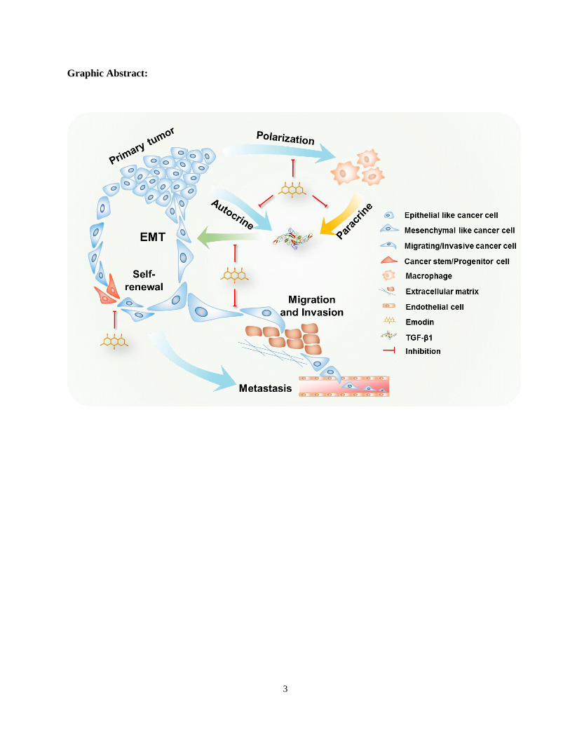
Graphic Abstract:
4
Introduction
Most breast cancer mortality results from metastatic recurrence after initial success of surgery and/or
other therapies [1, 2]. Neoadjuvant and adjuvant therapies have been increasingly used, particularly for
triple-negative breast cancer (TNBC) and HER2-positive breast cancer [3], in order to prevent local or
distant tumor recurrence after surgery. However, the metastatic rate observed after surgery with these
therapies is still substantial [4], and chemotherapy-associated side effects compromise quality of life.
Therefore, more effective and safer neoadjuvant or adjuvant treatments are urgently needed. Natural
compounds are an eminent source of drug development; many blockbuster drugs, such as paclitaxel and
artemisinin, are plant extracts [5, 6]. We aim to develop a pleiotropic natural compound, emodin (1,3,8-
trihydroxy-6-methylanthraquinone), as an effective and safe agent to halt breast cancer post-surgery
metastatic recurrence.
Epithelial-mesenchymal transition (EMT) contributes to cancer progression, and particularly,
metastasis [7]. Reprogramming of gene expression during EMT is initiated and controlled by various
signaling pathways in response to extracellular cues, among which the transforming growth factor-β
(TGF-β) signaling plays a predominant role [8]. The self-renewing cancer stem cells (CSCs) and
progenitor cells constitute a minor portion of neoplastic cells within the tumor and are collectively
defined as tumor-initiating cells (TIC) [9]. The TIC population is the key source of metastatic lesions in
breast cancer [10, 11]. The relationship between EMT and CSC is well documented; EMT confers cancer
cell mesenchymal traits and an ability to enter the CSC state [12-14]. Furthermore, EMT promotes tumor
cell invasion and dissemination; it also enables CSCs to clonally expand in distant organs [8], leading to
cancer metastasis.
Emodin is an anthraquinone derivative isolated from many Chinese herbs including Rheum palmatum
L. and Polygonum cuspidatum. Our previous studies have shown that emodin blocks the tumor-promoting
feedforward interactions between cancer cells and macrophages, reduces recruitment of macrophages to
the tumor and their subsequent M2-like polarization, and thus ameliorates the immunosuppressive state of
the tumor microenvironment (TME) [15-17]. When emodin was administered to mice soon after the
5
tumor cells were inoculated, it inhibited breast tumor growth [16]; while when emodin treatment began
after tumors were well established, it had no effects on the growth of the primary tumor but significantly
reduced lung metastasis [17]. Because tumor-associated macrophages (TAMs) also promote EMT of
cancer cells and the generation of CSCs, contributing to cancer invasion and metastasis [18-20], we
hypothesize that emodin inhibits breast cancer cell EMT and reduce CSC through acting on both
macrophages and cancer cells, thus halts breast cancer post-surgery metastatic recurrence if it is
administered as a neoadjuvant therapy.
Methods
Mice
Mice including C57BL/6, BALB/c, and NOD-SCID mice were purchased from Jackson Laboratories.
MMTV-PyMT mice generated on an FVB background [21] were crossed to the C57BL/6 background in
Dr. Zena Werb’s laboratory at UCSF and further in our lab for over 10 generations. All mice were housed
in the University of South Carolina’s Department of Laboratory Animal Research. Animal care
procedures and experimental methods were approved by the Institutional Animal Care and Use
Committee of the University of South Carolina according to National Institutes of Health guidelines.
Cell culture
The breast cancer cell lines EO771, 4T1, MCF7, and MDA-MB-231 were obtained from the
American Type Culture Collection. The cell line authentication was described in our recent study [22].
Cells were cultured in high glucose Dulbecco's modified Eagle medium (DMEM, Invitrogen) with 10%
FBS (Invitrogen) and penicillin/streptomycin at 37°C in a humidified 5% CO2 atmosphere.
Primary cell isolation
To obtain primary MMTV-PyMT cells, mouse mammary tumors were cut into small fragments (<3
mm) and digested in dissociation solution (DMEM supplemented with 10% FBS, Collagenase type IV
(5320 U), DNase I (319 U) and hyaluronidase (500 U)) for 60 min in a 37°C water bath with shaker.
After digestion and filtering, erythrocytes were lysed with red blood cell lysing buffer (Sigma). Cell
6
suspensions were passed through 70-μm cell strainers, then washed and cultured in complete medium for
further experimentation.
Collection of cell conditioned medium
To obtain tumor cell conditioned medium (TCCM) or peritoneal macrophage conditioned medium
(PMCM), the tumor cells (4T1 or EO771) were cultured to 90% confluence in complete medium, and
mouse peritoneal macrophages were isolated from mice as described previously [22] and cultured in the
indicated medium overnight, and then the medium was replaced with serum-free DMEM. After 24 h, the
medium was collected and filtered through a 0.22-μm filter.
Coculture of cancer cells with macrophages
The indirect contact coculture was performed in 24-well plates with 8-μm polyethylene terephthalate
membrane filters (Corning) separating the lower and upper chambers. After the pretreatment with
corresponding TCCM with or without emodin at indicated concentrations, macrophages from syngeneic
mice were seeded in the upper chambers, while EO771 cells or 4T1 cells were seeded in the lower
chamber; 48 h later, cancer cells in the lower chamber were collected for analysis.
Wound-healing migration assay and invasion assay
The cell migration margins before and after stimulation were marked and the distances were
calculated by image processing software Image-Pro Plus 6.0. The invasion of breast cancer cells was
examined using 24-well Matrigel-coated invasion chambers with 8-μm pore size inserts (Corning). Cell
migration was allowed for 24 h. Non-invading cells on the upper surface of the inserts were removed
from the chambers and cells that reached the lower surface were fixed and stained by DAPI. The invaded
cells were counted and quantified with the image processing software Image-Pro Plus 6.0.
Aldefluor assay
The Aldefluor assays were conducted using the Aldefluor assay kit following the manufacturer's
instructions (STEMCELL Tech). Briefly, Aldefluor reagents were added to the cell suspension after
indicated treatments. Cell plates were incubated at 37°C for 30 min. After washing, flow cytometry
7
analysis was conducted to quantify ALDHbr cells. Cells treated by an ALDH inhibitor, diethylamino
benzaldehyde (DEAB) were used as a negative control.
Tumor mammosphere formation assay
A primary mammosphere formation assay was performed in ultra-low attachment 96-well plates
(Corning Costar). Dissociated breast cancer cells (EO771, 4T1, MCF7, or MDA-MB-231) at indicated
numbers (3000, 1000, 300, 100 and 30 cells) were cultured in serum-free DMEM with 20 ng/ml EGF, 20
ng/ml bFGF and 1 × B27 supplement. On Day 7 of culture, the numbers of primary mammospheres with
a diameter larger than 50 μm were counted, and cells were serially passaged for secondary mammosphere
formation for another 7 days.
In vivo limiting dilution analysis
Cancer cells pretreated with emodin or DMSO for 48 h were injected into the 4th pair of mammary fat
pads in mice. Tumor cells in each group were implanted with various cell numbers in 20 μl PBS.
Approximately four weeks after cell implantation, the tumor numbers were recorded to calculate tumor-
initiating frequency and statistically analyzed using the ELDA software (WEHI).
Tumor cell inoculation
For orthotopic inoculation of breast cancer cells, 2 × 10⁵ cells in 20 μl PBS were injected into each of
the mammary fat pads of mice. For the intravenous injection of breast cancer cells, 1 × 10⁶ cells in 100 μl
PBS were injected into the tail vein. To monitor the primary tumor growth, tumors were measured using a
caliper at indicated time points, and the tumor volume was calculated using formula: length × width2/2.
Quantitative real-time PCR (qPCR)
Total RNA was extracted using the TRIzol reagent (Invitrogen) and reverse transcribed using iScript
cDNA Synthesis Kit (Bio-Rad, Life Science). qPCR was conducted on a CFX96 system (Bio-Rad) using
iQ SYBR Green Supermix (Bio-Rad). All primers used for qPCR analysis were synthesized by Integrated
DNA Technologies and the primer sequences are listed in the Supplemental Table S1. The relative
amount of target mRNA was determined using the comparative threshold (Ct) method by normalizing
8
target mRNA Ct values to those of internal control RNA. PCR thermal cycling conditions were 3 min at
95°C, and 45 cycles of 15 s at 95°C and 58 s at 60°C.
Western blot
Cells were lysed in RIPA buffer (Pierce) supplemented with protease inhibitor cocktail and
phosphatase inhibitor cocktail (Sigma). The total cellular extract was separated in 10% SDS-PAGE
precast gels (Bio-Rad) and transferred onto nitrocellulose membranes (Millipore). Membranes were first
probed with indicated primary antibodies, followed by the corresponding secondary antibodies which
were conjugated with horseradish peroxidase (Millipore). Protein detection was conducted using ECL
substrate (Pierce) and the signal intensities were quantified by Image-Pro Plus 6.0.
Flow cytometry
A single cell suspension was made from cultured cells or mouse tissue after enzyme digestion. For the
staining of cell surface markers, cells were blocked in Fc blocker antibody and then stained with indicated
antibodies conjugated with a fluorescent dye in staining buffer (PBS containing 2% FBS) for 30 min on
ice and in the dark. Samples were washed twice with staining buffer, and then analyzed by a FACS Aria
flow cytometer (BD).
Immunofluorescence staining
For cell slides, cells were fixed in 4% paraformaldehyde and washed with PBS. After blocking, anti-
mouse or anti-human primary antibody (1:200, Abcam) was applied to corresponding breast cancer cell
slides overnight at 4°C. Then the slides were washed and incubated with secondary antibody in 1% BSA
for 1 h at room temperature in the dark, followed by washing, mounting and sealing with a coverslip
before observation under a fluorescence microscope (Nikon). A series of images from each sample were
taken at the same photography setting, and the mean fluorescence intensity (MFI) was measured by
Image-Pro Plus 6.0. For breast tumor tissues from patients, de-identified formalin-fixed paraffin
embedded tissues were collected from mastectomy surgery with ethical approval by Nanjing Drum Tower
Hospital in 2015. Sections were cut (4-μm thick), transferred to a warm water bath, and placed on a glass
9
slide. Immunofluorescence staining was performed using anti-human antibodies: p-Smad2 (1:300, Cell
Signaling Tech.) and CD68 (1:300, Abcam).
Database mining
The cancer genome atlas data repository was used as the primary source of samples for the analysis.
Dimensionality reduction was performed with PCA. Three representative samples in each of the CD68lo
and CD68hi groups were used for a heatmap plot of the differentially expressed genes. Correlations
between the expression levels of CD68 and the indicated genes were calculated by the spearman
correlation coefficient and statistical significance was shown as –log (P value). For the prognostic
analysis of TGFβR1 and Smad2, the breast cancer patient survival data of TCGA was obtained from the
Human Protein Atlas database (https://www.proteinatlas.org). Based on the median value (FPKM) of
each gene, patients were classified into two groups and association between survival rate and gene
expression was examined. The survival curve was estimated using Kaplan-Meier analysis, and the P-
values were calculated with the log-rank (Mantel-Cox) test.
Statistical analysis
Data were presented as mean ± SEM. Statistical significance was calculated using the Student t test
(two-tailed, for two-group comparison) or one-way ANOVA followed by post Dunnett’s test (for multi-
group comparison) using the GraphPad Prism statistical program. Survival was analyzed using the Log-
rank (Mantel-Cox) test. P < 0.05 was considered as statistically significant.
Results
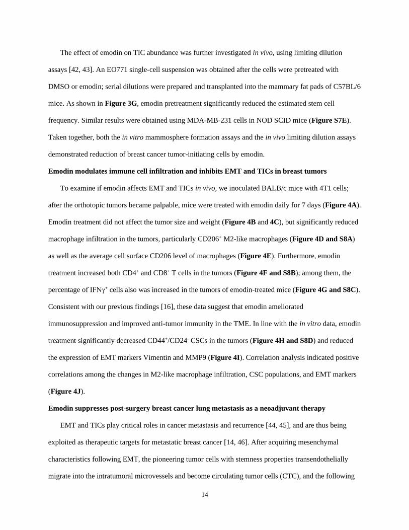
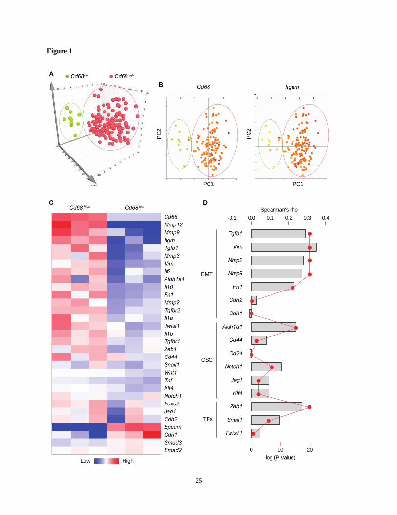
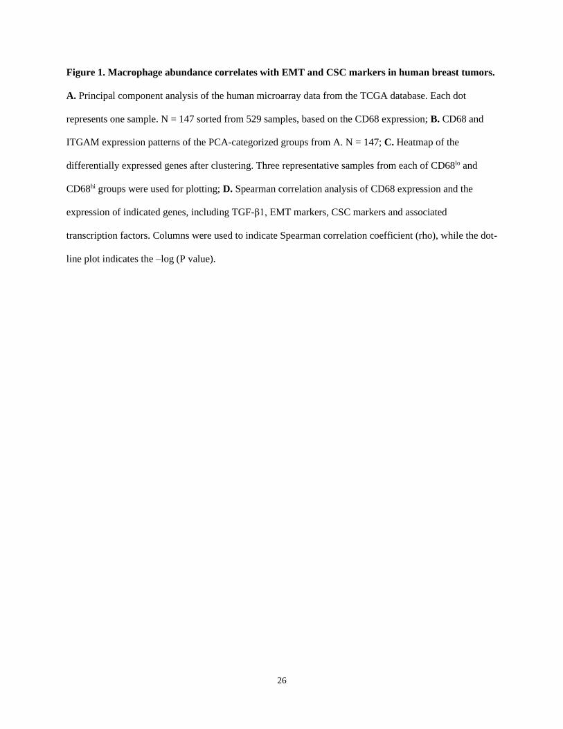
Macrophage abundance correlates with EMT and CSC markers in human breast tumors
Macrophages, the most abundant leukocytes in mammary tumors, play critical roles in cancer
progression [23]. TAMs exhibit a high plasticity and remodel the TME in response to various signals
including those from tumor cells [24]. To examine the relationship between macrophage abundance and
other tumor properties in breast cancer, data from 1,105 samples obtained from 1,098 breast cancer
patients in the cancer genome atlas were examined. Among them, microarray data are available for 529
10
samples. Using the expression level of the pan-macrophage marker CD68 as an indicator of macrophage
abundance, we sorted the CD68lo and CD68hi samples (cutoff, fold change > 1.5; n = 147). Dimensionality
reduction with the principal component analysis (PCA) clearly categorized CD68lo and CD68hi samples as
two separate groups (Figure 1A). The expression pattern of another common marker of macrophages,
ITGAM (CD11b), in those samples is very similar to that of CD68 (Figure 1B). Using three
representative samples from each of the CD68lo and CD68hi groups, visualization of heatmap of
differentially expressed mRNAs strongly demonstrated that CD68lo and CD68hi groups displayed distinct
mRNA expression patterns (Figure 1C). We examined the overall expression pattern of these genes in all
of the sorted samples according to PCA-categorized CD68lo and CD68hi groups, and found that the
CD68hi group was associated with pro-tumorigenic markers, including TGF-β1, EMT markers, stemness
markers, and related transcription factors (TFs) (Figure S1). Further Spearman correlation analysis
confirmed the correlation between CD68 expression and the expression of TGF-β1, EMT and CSC
markers, as well as the associated TFs (Figure 1D). These results suggest that intratumoral macrophages
likely play an essential role in EMT and CSC generation. And indeed, a study showed that liposomal
simvastatin could suppress cancer cell EMT via repolarizing TAMs [25]. Since our previous studies
showed that emodin inhibits breast cancer growth and metastasis by blocking the interactions between
cancer cells and TAMs [15-17], these bioinformatics data prompted us to investigate if emodin affects
EMT and CSC through acting on TAMs in the TME.
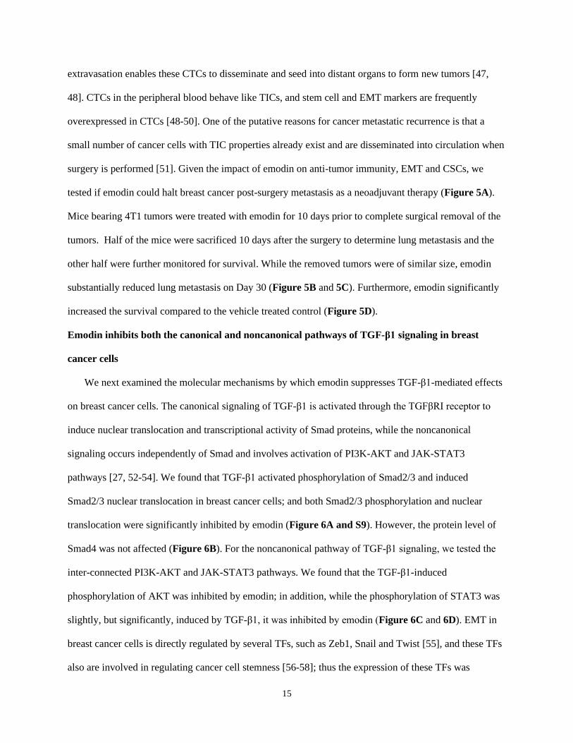
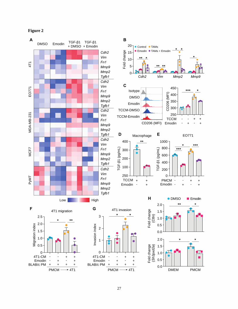
Emodin inhibits TGF-β1 and macrophage-induced EMT in breast cancer cells
First, we used the TGF-β1-induced EMT model to assess the effects of emodin on breast cancer cell
EMT. It was found that TGF-β1 induced 4T1 cells to acquire a fibroblast-like, mesenchymal cell
morphology (Figure S2A), accompanied with increased expression of signature genes of EMT (Figure
S2B). The inhibitory effect of emodin on EMT was tested using various breast cancer cell lines (Figure
S2C-G). In all tested breast cancer cell lines, upregulation of EMT genes by TGF-β1, including TGF-β1
itself, was diminished by emodin (Figure 2A). Loss of the epithelial marker E-cadherin and acquisition of
the mesenchymal marker N-cadherin are key features of the EMT process [26]. Western blotting showed

11
that TGF-β1 treatment increased N-cadherin and decreased E-cadherin protein levels in various breast
cancer cell lines, while emodin reversed these changes (Figure S3A). Immunofluorescence staining
confirmed that the TGF-β1-induced loss of E-cadherin was reversed by emodin (Figure S3B-C).
Cancer cells and TAMs at the invasive front of breast tumors interact to enable cancer cell invasion
[27]. To determine whether emodin affects macrophage-induced EMT of breast cancer cells, we isolated
primary macrophages from mice and pretreated them with tumor cell conditioned medium (TCCM) to
generate TAM-like macrophages, and then added them to breast cancer cells (Figure S4A). Examination
of the expression of EMT markers indicated that emodin significantly attenuated TAM-like macrophage-
induced N-cadherin, Vimentin and MMP expression in breast cancer cells (Figure 2B and S4B). Based
on our previous findings [16], we hypothesized that emodin may suppress TCCM-induced macrophage
M2-like polarization and M2-like macrophage-induced EMT of breast cancer cells by blocking the TGF-
β1-mediated reciprocal interaction between macrophages and cancer cells. As expected, TCCM-
pretreated macrophages exhibited an increased expression of CD206, a marker of M2 macrophages,
which was attenuated by emodin (Figure 2C). ELISA showed that TGF-β1 production in macrophages
after TCCM treatment was significantly decreased by emodin (Figure 2D). In addition, while TGF-β1
production in breast cancer cells was elevated after peritoneal macrophage conditioned medium (PMCM)
treatment, emodin significantly reduced baseline and PMCM-induced TGF-β1 production in cancer cells
(Figure 2E). Moreover, we confirmed by qPCR that emodin suppressed both baseline and TGF-β1
induced Arg-1 expression in macrophages (Figure S4C); Arg-1 expression has been shown to define
immunosuppressive subsets of TAMs [28]. Taken together, these data suggest that emodin 1) suppresses
cancer cell-induced macrophage M2-like polarization and thus TGF-β1 production, 2) inhibits polarized
macrophage-induced TGF-β1 production in breast cancer cells, and 3) blocks EMT of breast cancer cells
induced by TGF-β1 from both macrophages and cancer cells.
Emodin inhibits TGF-β1 and macrophage-induced migration and invasion of breast cancer cells
EMT enables cancer cells to migrate and invade [29, 30]; we thus examined if emodin affects the
migration and invasion of breast cancer cells. First, a wound-healing migration assay showed that TGF-β1
12
enhanced 4T1 breast cancer cell migration by 2-fold, and this effect was abolished by emodin (Figure
S5A). Second, a matrigel invasion assay demonstrated that TGF-β1 increased 4T1 cell invasion by >5-
fold, and this effect was significantly diminished by emodin (Figure S5B). Similar results were obtained
using MDA-MB-231 cells (Figure S5C and S5D). TAMs aggregate to induce migration, invasion and
dissociation of cancer cells through the local production of TGF-β1 and MMPs [31, 32]. We thus tested if
emodin affects the cancer cell migration and invasion stimulated by conditioned medium of cultured
peritoneal macrophages (PMCM). PMCM was collected from macrophages treated with 4T1 conditioned
medium with or without emodin. 4T1 cells were then stimulated by PMCM for 16 h, and migration and
invasion were measured. 4T1 cells treated with PMCM from macrophages that were treated with 4T1
conditioned medium without emodin exhibited more aggressive migration and invasion compared with
those 4T1 cells treated with PMCM from macrophages treated with 4T1 conditioned medium with
emodin (Figure 2F and 2G; Figure S5E and S5F).
During breast cancer cell migration and invasion, pioneer cancer cells at the edge of the tumor
degrade the extracellular matrix by developing specialized actin-rich membrane protrusion structures
called invadopodia [33]; and macrophages promote breast cancer cell migration through Notch1-initiated
invadopodia formation [34]. We thus examined the expression of Cortactin and Tks5, markers of
invadopodia formation, following DMEM (with or without emodin) or PMCM treatment. As shown in
Figure 2H, while emodin itself did not affect Cortactin or Tks5 expression in breast cancer cells, PMCM
from macrophages treated with EO771 conditioned medium significantly increased the expression of
these two genes; moreover, upregulation of these genes was abolished if the macrophages were treated
with emodin in addition to EO771 conditioned medium.
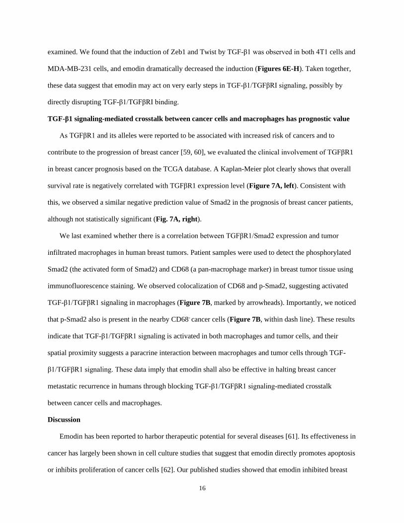
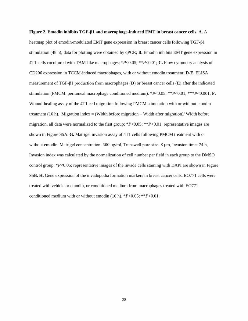
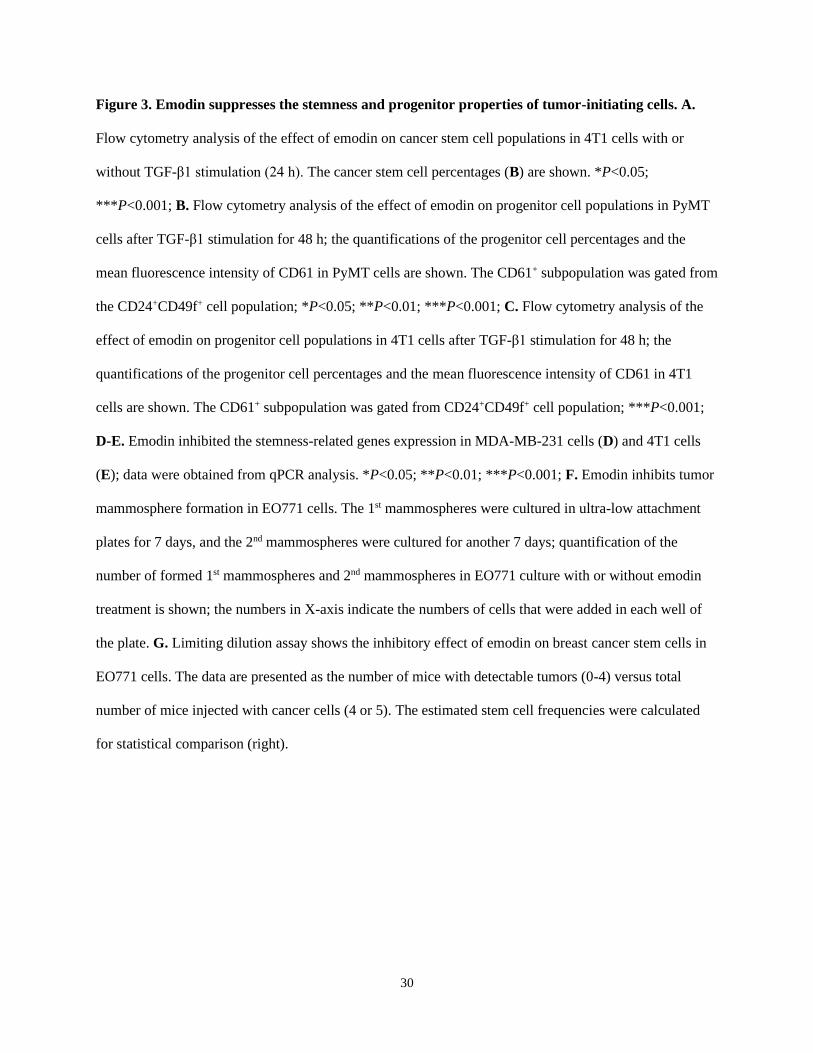
Emodin suppresses the stemness and progenitor properties of breast cancer cells
We hypothesized that emodin also could affect EMT-promoted CSC generation and maintenance.
First, we found that emodin reduced baseline levels of CD44hi/+/CD24lo/- cells in various breast cancer cell
lines (Figure S6A); CD44hi/+ and CD24lo/- are recognized CSC markers. Measurement of aldehyde
dehydrogenase activity (ALDH), another marker of both normal and malignant mammary stem cells [35],
13
confirmed the suppressive effects of emodin on the CSC population in MDA-MB-231 cells (Figure S6B).
Induction of EMT by TGF-β1 increases breast cancer stem cells [36, 37]. We showed that TGF-β1
increased CSCs by 50% in 4T1 cells, while emodin abolished this effect (Figure 3A and S6C).
Adult stem cells can generate multipotent progenitors that further develop into specialized cells [38].
However, the heterogenic niche in the TME provides WNT and EGF signals that not only help maintain
resident stem cells, but also instruct progenitor cells to revert to a stem cell state, contributing to tumor
regeneration and therapy resistance [39]. We thus further examined if emodin could affect the progenitor
population in breast cancer cells using the markers CD24, CD49f and CD61. We found that emodin
suppressed the progenitor population in both EO771 and MDA-MB-231 cells (Figure S6D and S6E). For
the primary PyMT tumor cells, emodin suppressed the percentage of progenitor cells and the average
CD61 expression levels, with or without TGF-β1 stimulation (Figure 3B and S6F). Similarly, TGF-β1
enhanced the percentage of progenitor population and the average CD61 expression level in 4T1 cells,
which was halved by emodin (Figure 3C and S6G). Additionally, expression levels of transcription
factors Oct4, KLF4 and Nanog, and other TIC-associated genes FoxC2 and Jagged1, were determined.
We found that the baseline expression of FoxC2, Nanog, Oct4 and KLF4 was downregulated by emodin
in MDA-MB-231 cells (Figure 3D), and the TGF-β1-induced expression of Jagged1, KLF4 and FoxC2
was decreased by emodin in 4T1 cells (Figure 3E).
Breast Cancer TICs can be propagated in vitro as nonadherent spheres; these spherical clusters of
self-replicating cells from suspension cultures are called mammospheres [40, 41]. We tested whether
emodin affects mammosphere formation. Cultured in the ultra-low attachment plates for 7 days, EO771
cells formed typical mammospheres (primary mammospheres); re-culturing of the cells enzymatically
dissociated from the primary mammospheres under the same conditions yielded secondary
mammospheres. Emodin significantly reduced both primary and secondary mammosphere formation
(Figure 3F and Figure S7A). Similar results were obtained using 4T1 (Figure S7B), MCF7 (Figure
S7C), and MDA-MB-231 cells (Figure S7D).
14
The effect of emodin on TIC abundance was further investigated in vivo, using limiting dilution
assays [42, 43]. An EO771 single-cell suspension was obtained after the cells were pretreated with
DMSO or emodin; serial dilutions were prepared and transplanted into the mammary fat pads of C57BL/6
mice. As shown in Figure 3G, emodin pretreatment significantly reduced the estimated stem cell
frequency. Similar results were obtained using MDA-MB-231 cells in NOD SCID mice (Figure S7E).
Taken together, both the in vitro mammosphere formation assays and the in vivo limiting dilution assays
demonstrated reduction of breast cancer tumor-initiating cells by emodin.
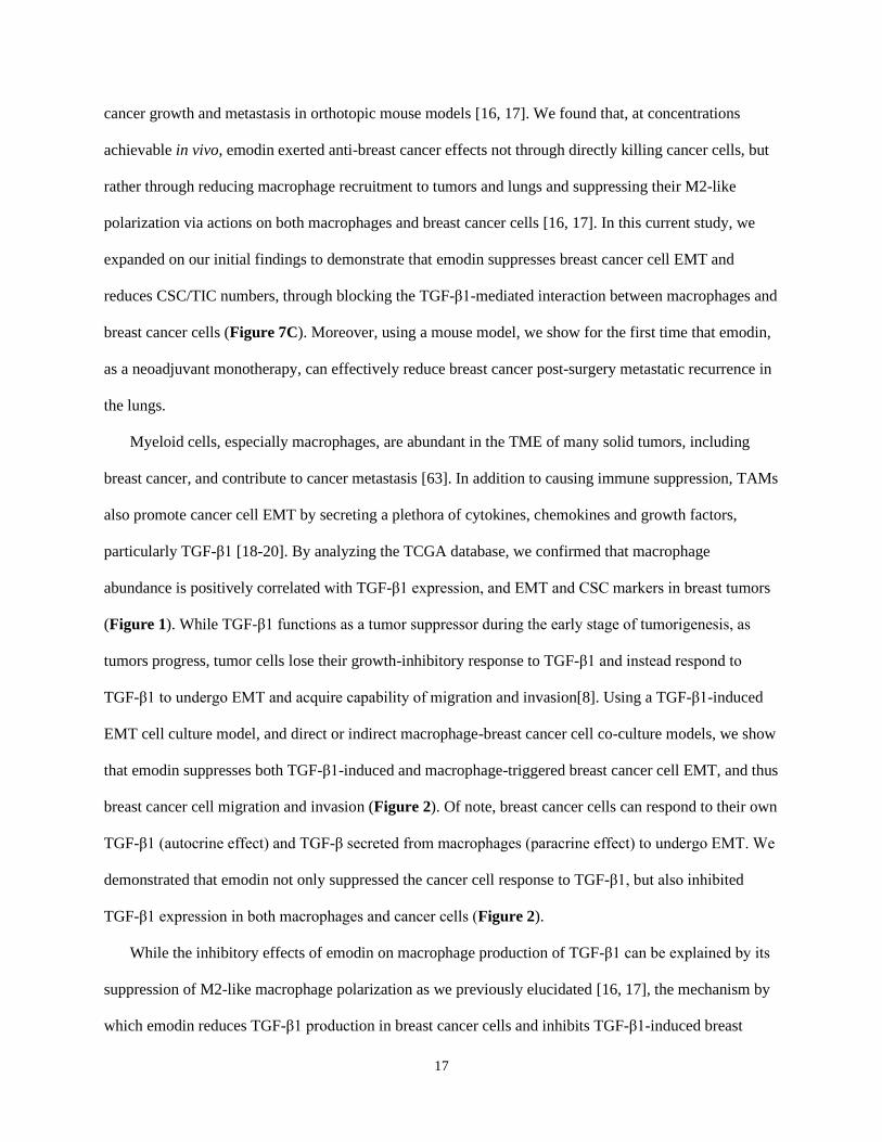
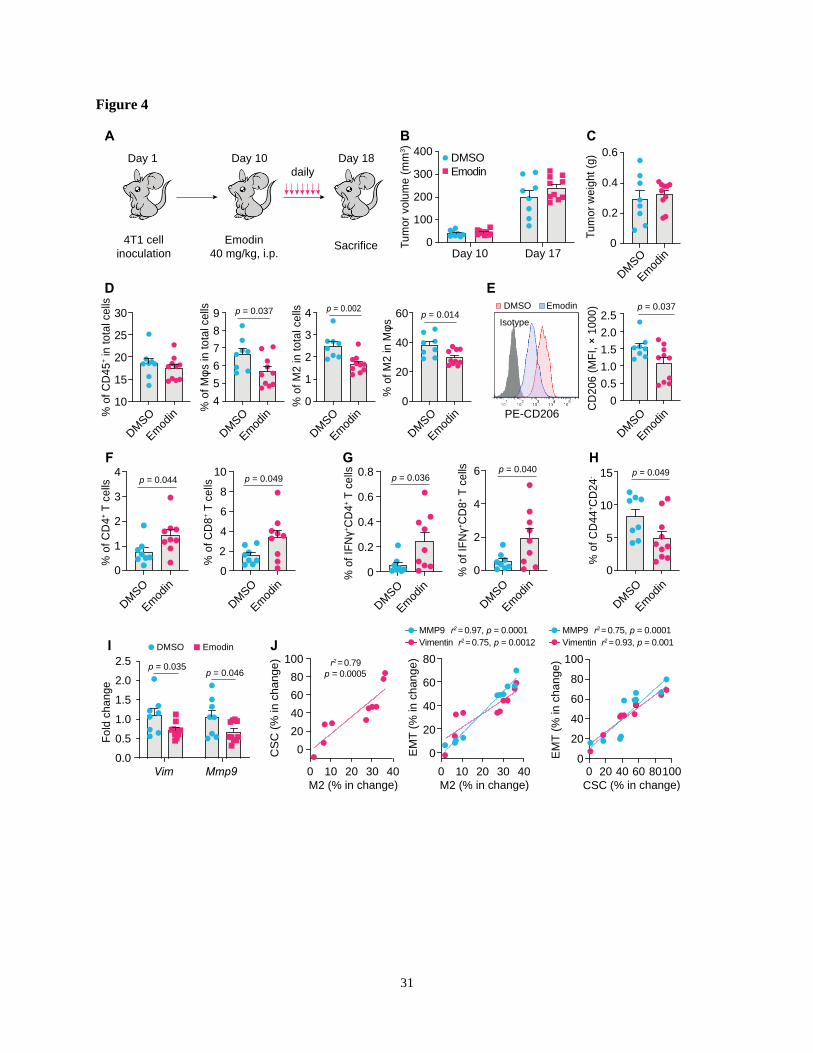
Emodin modulates immune cell infiltration and inhibits EMT and TICs in breast tumors
To examine if emodin affects EMT and TICs in vivo, we inoculated BALB/c mice with 4T1 cells;
after the orthotopic tumors became palpable, mice were treated with emodin daily for 7 days (Figure 4A).
Emodin treatment did not affect the tumor size and weight (Figure 4B and 4C), but significantly reduced
macrophage infiltration in the tumors, particularly CD206+ M2-like macrophages (Figure 4D and S8A)
as well as the average cell surface CD206 level of macrophages (Figure 4E). Furthermore, emodin
treatment increased both CD4+ and CD8+ T cells in the tumors (Figure 4F and S8B); among them, the
percentage of IFNγ+ cells also was increased in the tumors of emodin-treated mice (Figure 4G and S8C).
Consistent with our previous findings [16], these data suggest that emodin ameliorated
immunosuppression and improved anti-tumor immunity in the TME. In line with the in vitro data, emodin
treatment significantly decreased CD44+/CD24- CSCs in the tumors (Figure 4H and S8D) and reduced
the expression of EMT markers Vimentin and MMP9 (Figure 4I). Correlation analysis indicated positive
correlations among the changes in M2-like macrophage infiltration, CSC populations, and EMT markers
(Figure 4J).
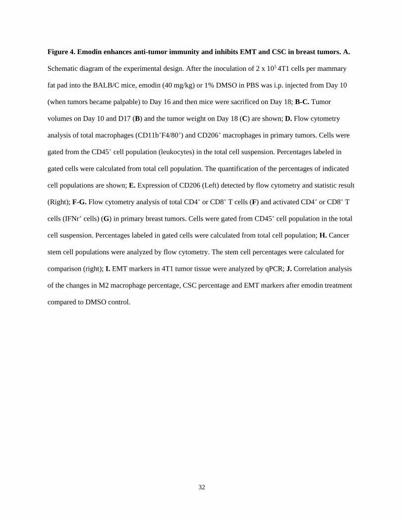
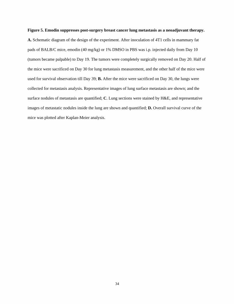
Emodin suppresses post-surgery breast cancer lung metastasis as a neoadjuvant therapy
EMT and TICs play critical roles in cancer metastasis and recurrence [44, 45], and are thus being
exploited as therapeutic targets for metastatic breast cancer [14, 46]. After acquiring mesenchymal
characteristics following EMT, the pioneering tumor cells with stemness properties transendothelially
migrate into the intratumoral microvessels and become circulating tumor cells (CTC), and the following
15
extravasation enables these CTCs to disseminate and seed into distant organs to form new tumors [47,
48]. CTCs in the peripheral blood behave like TICs, and stem cell and EMT markers are frequently
overexpressed in CTCs [48-50]. One of the putative reasons for cancer metastatic recurrence is that a
small number of cancer cells with TIC properties already exist and are disseminated into circulation when
surgery is performed [51]. Given the impact of emodin on anti-tumor immunity, EMT and CSCs, we
tested if emodin could halt breast cancer post-surgery metastasis as a neoadjuvant therapy (Figure 5A).
Mice bearing 4T1 tumors were treated with emodin for 10 days prior to complete surgical removal of the
tumors. Half of the mice were sacrificed 10 days after the surgery to determine lung metastasis and the
other half were further monitored for survival. While the removed tumors were of similar size, emodin
substantially reduced lung metastasis on Day 30 (Figure 5B and 5C). Furthermore, emodin significantly
increased the survival compared to the vehicle treated control (Figure 5D).
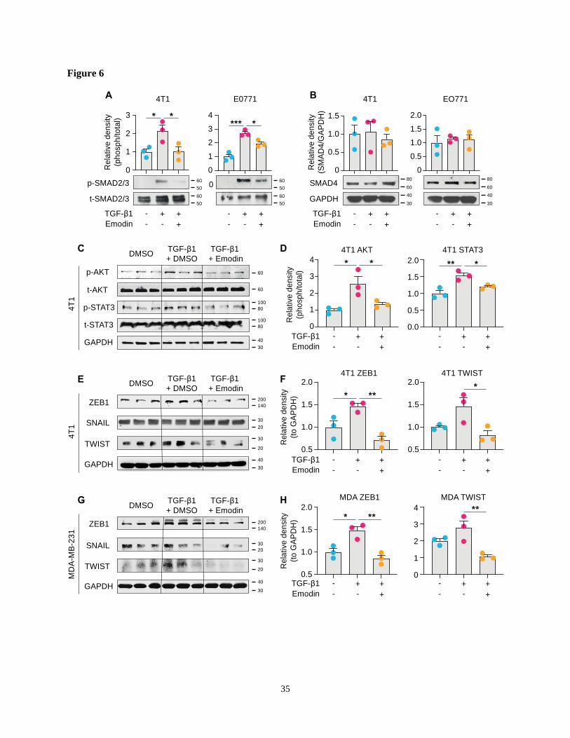
Emodin inhibits both the canonical and noncanonical pathways of TGF-β1 signaling in breast
cancer cells
We next examined the molecular mechanisms by which emodin suppresses TGF-β1-mediated effects
on breast cancer cells. The canonical signaling of TGF-β1 is activated through the TGFβRI receptor to
induce nuclear translocation and transcriptional activity of Smad proteins, while the noncanonical
signaling occurs independently of Smad and involves activation of PI3K-AKT and JAK-STAT3
pathways [27, 52-54]. We found that TGF-β1 activated phosphorylation of Smad2/3 and induced
Smad2/3 nuclear translocation in breast cancer cells; and both Smad2/3 phosphorylation and nuclear
translocation were significantly inhibited by emodin (Figure 6A and S9). However, the protein level of
Smad4 was not affected (Figure 6B). For the noncanonical pathway of TGF-β1 signaling, we tested the
inter-connected PI3K-AKT and JAK-STAT3 pathways. We found that the TGF-β1-induced
phosphorylation of AKT was inhibited by emodin; in addition, while the phosphorylation of STAT3 was
slightly, but significantly, induced by TGF-β1, it was inhibited by emodin (Figure 6C and 6D). EMT in
breast cancer cells is directly regulated by several TFs, such as Zeb1, Snail and Twist [55], and these TFs
also are involved in regulating cancer cell stemness [56-58]; thus the expression of these TFs was
16
examined. We found that the induction of Zeb1 and Twist by TGF-β1 was observed in both 4T1 cells and
MDA-MB-231 cells, and emodin dramatically decreased the induction (Figures 6E-H). Taken together,
these data suggest that emodin may act on very early steps in TGF-β1/TGFβRI signaling, possibly by
directly disrupting TGF-β1/TGFβRI binding.
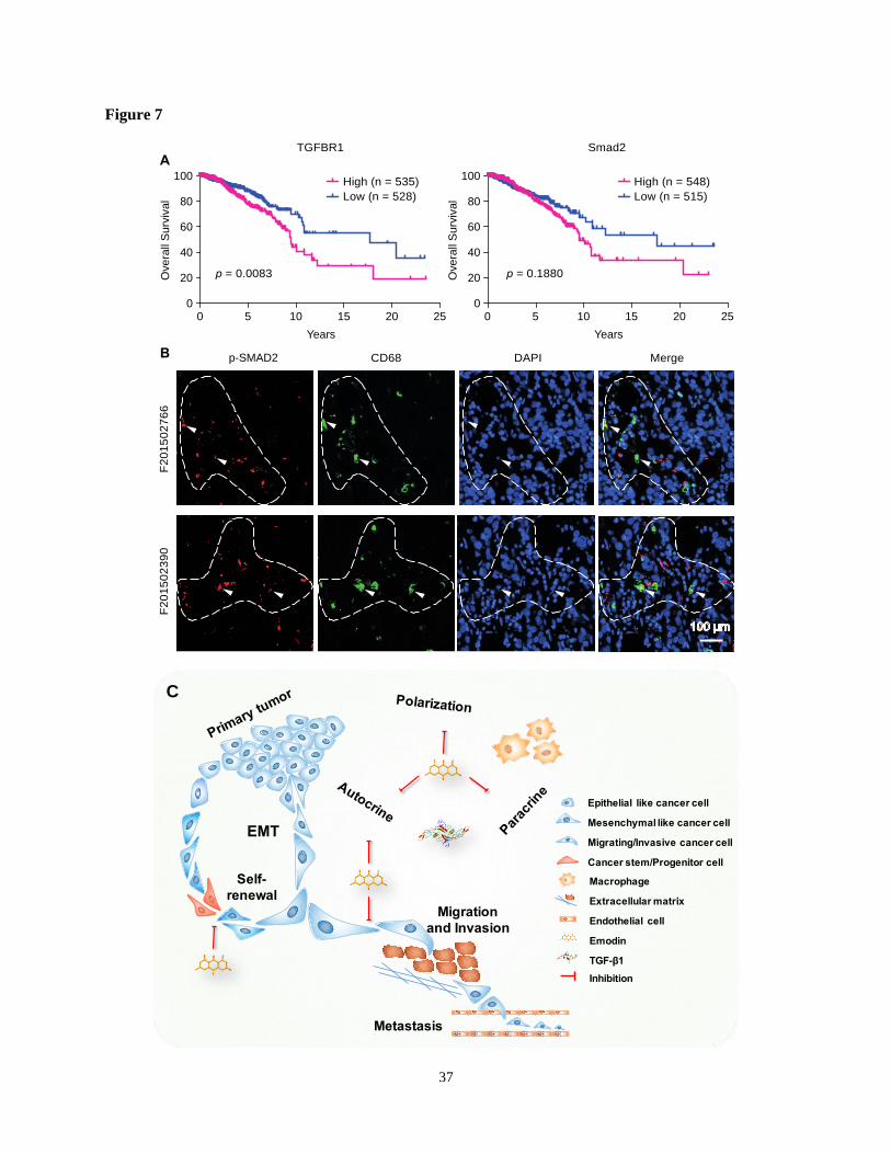
TGF-β1 signaling-mediated crosstalk between cancer cells and macrophages has prognostic value
As TGFβR1 and its alleles were reported to be associated with increased risk of cancers and to
contribute to the progression of breast cancer [59, 60], we evaluated the clinical involvement of TGFβR1
in breast cancer prognosis based on the TCGA database. A Kaplan-Meier plot clearly shows that overall
survival rate is negatively correlated with TGFβR1 expression level (Figure 7A, left). Consistent with
this, we observed a similar negative prediction value of Smad2 in the prognosis of breast cancer patients,
although not statistically significant (Fig. 7A, right).
We last examined whether there is a correlation between TGFβR1/Smad2 expression and tumor
infiltrated macrophages in human breast tumors. Patient samples were used to detect the phosphorylated
Smad2 (the activated form of Smad2) and CD68 (a pan-macrophage marker) in breast tumor tissue using
immunofluorescence staining. We observed colocalization of CD68 and p-Smad2, suggesting activated
TGF-β1/TGFβR1 signaling in macrophages (Figure 7B, marked by arrowheads). Importantly, we noticed
that p-Smad2 also is present in the nearby CD68- cancer cells (Figure 7B, within dash line). These results
indicate that TGF-β1/TGFβR1 signaling is activated in both macrophages and tumor cells, and their
spatial proximity suggests a paracrine interaction between macrophages and tumor cells through TGF-
β1/TGFβR1 signaling. These data imply that emodin shall also be effective in halting breast cancer
metastatic recurrence in humans through blocking TGF-β1/TGFβR1 signaling-mediated crosstalk
between cancer cells and macrophages.
Discussion
Emodin has been reported to harbor therapeutic potential for several diseases [61]. Its effectiveness in
cancer has largely been shown in cell culture studies that suggest that emodin directly promotes apoptosis
or inhibits proliferation of cancer cells [62]. Our published studies showed that emodin inhibited breast
17
cancer growth and metastasis in orthotopic mouse models [16, 17]. We found that, at concentrations
achievable in vivo, emodin exerted anti-breast cancer effects not through directly killing cancer cells, but
rather through reducing macrophage recruitment to tumors and lungs and suppressing their M2-like
polarization via actions on both macrophages and breast cancer cells [16, 17]. In this current study, we
expanded on our initial findings to demonstrate that emodin suppresses breast cancer cell EMT and
reduces CSC/TIC numbers, through blocking the TGF-β1-mediated interaction between macrophages and
breast cancer cells (Figure 7C). Moreover, using a mouse model, we show for the first time that emodin,
as a neoadjuvant monotherapy, can effectively reduce breast cancer post-surgery metastatic recurrence in
the lungs.
Myeloid cells, especially macrophages, are abundant in the TME of many solid tumors, including
breast cancer, and contribute to cancer metastasis [63]. In addition to causing immune suppression, TAMs
also promote cancer cell EMT by secreting a plethora of cytokines, chemokines and growth factors,
particularly TGF-β1 [18-20]. By analyzing the TCGA database, we confirmed that macrophage
abundance is positively correlated with TGF-β1 expression, and EMT and CSC markers in breast tumors
(Figure 1). While TGF-β1 functions as a tumor suppressor during the early stage of tumorigenesis, as
tumors progress, tumor cells lose their growth-inhibitory response to TGF-β1 and instead respond to
TGF-β1 to undergo EMT and acquire capability of migration and invasion[8]. Using a TGF-β1-induced
EMT cell culture model, and direct or indirect macrophage-breast cancer cell co-culture models, we show
that emodin suppresses both TGF-β1-induced and macrophage-triggered breast cancer cell EMT, and thus
breast cancer cell migration and invasion (Figure 2). Of note, breast cancer cells can respond to their own
TGF-β1 (autocrine effect) and TGF-β secreted from macrophages (paracrine effect) to undergo EMT. We
demonstrated that emodin not only suppressed the cancer cell response to TGF-β1, but also inhibited
TGF-β1 expression in both macrophages and cancer cells (Figure 2).
While the inhibitory effects of emodin on macrophage production of TGF-β1 can be explained by its
suppression of M2-like macrophage polarization as we previously elucidated [16, 17], the mechanism by
which emodin reduces TGF-β1 production in breast cancer cells and inhibits TGF-β1-induced breast
18
cancer cell EMT likely involves blockage of TGFβR1 signaling. Our data show that emodin suppresses
both canonical and noncanonical pathways of TGF-β1 signaling (Figure 6), suggesting that emodin
interferes the early events in TGF-β1/TGFβRI signaling likely by blocking TGF-β1/TGFβRI binding.
Several direct targets of emodin have been identified, including protein tyrosine kinase p65lck [64],
casein kinase 2 (CK2) [65], and β-hydroxyacyl-acyl carrier protein dehydratase [66]; among them, CK2 is
the best characterized one; emodin binds to it with high affinity [65, 67]. However, inhibition of these
proteins cannot fully explain the effects of emodin we observed in this study.
The EMT process generates cells with properties of stem cells [13]. TICs, including CSCs and cancer
progenitor cells, have emerged as promising targets for cancer therapy due to their roles in cancer
metastasis and therapy-resistance [68]. Therefore, we examined whether the EMT-facilitated TIC
population generation and maintenance could be inhibited by emodin. Indeed, we found that emodin
could effectively reduce TGF-β1-induced expression of CSC markers in multiple breast cancer cell lines
and diminish their ability to form mammospheres in vitro (mammosphere formation assays) and to
generate tumors in vivo (limited dilution assays) (Figure 3). We further demonstrated that emodin
suppressed TGF-β1-induced expression of several transcription factors that are critical to CSC generation
and maintenance, such as FoxC2, Nanog, Oct4, Jagged1, and KLF4 (Figure 3).
The major significance of this study lies in the finding that emodin substantially halts post-surgery
breast cancer metastasis in mouse models (Figure 4 and 5, Figure S8). First, we showed that 7 daily
emodin treatments in mice with established orthotopic breast tumors alleviated tumor immunosuppression
of the TME, improved anti-tumor immunity, and suppressed EMT and CSCs independent of any change
in tumor size (Figure 4), demonstrating that the identified in vitro actions of emodin all operate in vivo.
Second, we showed that emodin as a neoadjuvant therapy (10 daily treatments in mice with established
orthotopic breast tumors prior to surgical removal of the tumors) significantly reduced post-surgery breast
cancer lung metastasis and improved mouse survival (Figure 5). These experiments demonstrated that
emodin, as a neoadjuvant can significantly reduce breast cancer post-surgery metastasis.
19
The effectiveness of emodin in blocking the TGF-β1-mediated crosstalk between macrophages and
breast cancer cells has evident clinical implications. As we showed, analysis of the TCGA database
revealed a prognostic role of TGFβR1 and p-Smad2 in the overall survival of breast cancer patients, and
examination of breast cancer tissue samples indicated the proximity of macrophage marker CD68 and p-
Smad2 in breast cancer cells (Figure 7). One may argue that specific TGF-β1 signaling inhibitors such as
SB431542 could be better choices than emodin in clinical applications. However, clinical use of TGF-β1
signaling inhibitors has been hindered by their severe side effects due to the many vital physiological
roles of TGF-β1. We believe that the natural compound emodin has intrinsic advantages compared to
those specific TGF-β1 signaling inhibitors in that 1) with the relatively mild nature of its TGF-β1
signaling inhibition, emodin may only suppress excessive TGF-β1 signaling in the tumors while sparing
its physiological activities; and 2) in addition to suppression of TGF-β1 signaling, emodin also acts on
macrophage-breast cancer cell interactions through other mechanisms as we showed previously [16, 17].
In summary, while more work is still needed to confirm if emodin indeed directly blocks the
interaction between TGF-β1 and TGFβR1, this study provides convincing pre-clinical evidence
suggesting that emodin harbors the potential for clinical development as a new effective and safe agent to
halt metastatic recurrence of breast cancer, either as a monotherapy, or in combination with other
neoadjuvant or adjuvant therapies.
Abbreviations
ALDH: aldehyde dehydrogenase activity; CSC: cancer stem cells; CTC: circulating tumor cell; EMT:
epithelial-mesenchymal transition; ERK: extracellular signal regulated kinase; MAPK: mitogen-activated
protein kinase; MET: mesenchymal-epithelial transition; MFI: median fluorescent intensity; MMTV:
mouse mammary tumor virus; PC: progenitor cells; PI3K: phosphoinositide 3-kinase; PMCM: peritoneal
macrophage conditioned medium; PyMT: Polyoma virus middle T antigen; STAT: signal transducer and
activator of transcription; TAM: tumor-associated macrophages; TCCM: tumor cell conditioned medium;
20
TF: transcription factor; TGF-β: transforming growth factor-β; TIC: tumor initiating cells; TNBC: triple-
negative breast cancer; TβRI/II: TGF-β receptor type I/II.
Acknowledgement
This work was supported by NIH grants R01CA218578 (to DF and EAM), R21CA216230 (to DF), and
P01AT003961 (to PN and DF). Johnie Hodge has been supported by the Susan G. Komen trainee
program (GTDR17500160 to EAM).
Author contributions
Q.L. and J.H. performed most of the experiments and analyzed the data. Y.W., and Y.L. assisted in
animal experiments; U.S. assisted inflow cytometry analysis; Y.Y. provided human tissues; D.F. designed
the study; H.C. and E.A.M. assisted in designing the study; L.W. assisted in statistical analysis; Q.L. J.W.
and D.F. wrote the manuscript; W.A., P.N., D.W., H.C., P.X. and E.A.M. helped in data interpretation
and manuscript writing.
Competing interests
There are no competing interests to be disclosed
21
References
1. Chambers AF, Groom AC, MacDonald IC. Dissemination and growth of cancer cells in
metastatic sites. Nat Rev Cancer. 2002; 2: 563-72.
2. Weigelt B, Peterse JL, van 't Veer LJ. Breast cancer metastasis: markers and models. Nat Rev
Cancer. 2005; 5: 591-602.
3. Cain H, Macpherson IR, Beresford M, Pinder SE, Pong J, Dixon JM. Neoadjuvant Therapy in
Early Breast Cancer: Treatment Considerations and Common Debates in Practice. Clin Oncol (R Coll
Radiol). 2017; 29: 642-52.
4. Karagiannis GS, Pastoriza JM, Wang Y, Harney AS, Entenberg D, Pignatelli J, et al. Neoadjuvant
chemotherapy induces breast cancer metastasis through a TMEM-mediated mechanism. Sci Transl Med.
2017; 9(397):eaan0026.
5. Cragg GM, Newman DJ. Natural products: a continuing source of novel drug leads. Biochim
Biophys Acta. 2013; 1830: 3670-95.
6. Chaturvedi D, Goswami A, Saikia PP, Barua NC, Rao PG. Artemisinin and its derivatives: a
novel class of anti-malarial and anti-cancer agents. Chem Soc Rev. 2010; 39: 435-54.
7. Nieto MA, Huang RY, Jackson RA, Thiery JP. EMT: 2016. Cell. 2016; 166: 21-45.
8. Lamouille S, Xu J, Derynck R. Molecular mechanisms of epithelial-mesenchymal transition. Nat
Rev Mol Cell Biol. 2014; 15: 178-96.
9. de Souza VB, Schenka AA. Cancer Stem and Progenitor-Like Cells as Pharmacological Targets
in Breast Cancer Treatment. Breast Cancer (Auckl). 2015; 9: 45-55.
10. Velasco-Velazquez MA, Popov VM, Lisanti MP, Pestell RG. The role of breast cancer stem cells
in metastasis and therapeutic implications. Am J Pathol. 2011; 179: 2-11.
11. Liu H, Patel MR, Prescher JA, Patsialou A, Qian D, Lin J, et al. Cancer stem cells from human
breast tumors are involved in spontaneous metastases in orthotopic mouse models. Proc Natl Acad Sci U
S A. 2010; 107: 18115-20.
12. Lu H, Clauser KR, Tam WL, Frose J, Ye X, Eaton EN, et al. A breast cancer stem cell niche
supported by juxtacrine signalling from monocytes and macrophages. Nat cell biol. 2014; 16: 1105-17.
13. Mani SA, Guo W, Liao MJ, Eaton EN, Ayyanan A, Zhou AY, et al. The epithelial-mesenchymal
transition generates cells with properties of stem cells. Cell. 2008; 133: 704-15.
14. Brown RL, Reinke LM, Damerow MS, Perez D, Chodosh LA, Yang J, et al. CD44 splice isoform
switching in human and mouse epithelium is essential for epithelial-mesenchymal transition and breast
cancer progression. J Clin Invest. 2011; 121: 1064-74.
15. Iwanowycz S, Wang J, Altomare D, Hui Y, Fan D. Emodin Bidirectionally Modulates
Macrophage Polarization and Epigenetically Regulates Macrophage Memory. J Biol Chem. 2016; 291:
11491-503.
16. Iwanowycz S, Wang J, Hodge J, Wang Y, Yu F, Fan D. Emodin Inhibits Breast Cancer Growth
by Blocking the Tumor-Promoting Feedforward Loop between Cancer Cells and Macrophages. Mol
Cancer Ther. 2016; 15: 1931-42.
17. Jia X, Yu F, Wang J, Iwanowycz S, Saaoud F, Wang Y, et al. Emodin suppresses pulmonary
metastasis of breast cancer accompanied with decreased macrophage recruitment and M2 polarization in
the lungs. Breast Cancer Res Treat. 2014; 148: 291-302.
18. Kitamura T, Qian BZ, Pollard JW. Immune cell promotion of metastasis. Nat Rev Immunol. 2015;
15: 73-86.
19. Quatromoni JG, Eruslanov E. Tumor-associated macrophages: function, phenotype, and link to
prognosis in human lung cancer. Am J Transl Res. 2012; 4: 376-89.
20. Quail DF, Joyce JA. Microenvironmental regulation of tumor progression and metastasis. Nat
Med. 2013; 19: 1423-37.
22
21. Guy CT, Cardiff RD, Muller WJ. Induction of mammary tumors by expression of polyomavirus
middle T oncogene: a transgenic mouse model for metastatic disease. Mol Cell Biol. 1992; 12: 954-61.
22. Fang L, Hodge J, Saaoud F, Wang J, Iwanowycz S, Wang Y, et al. Transcriptional factor EB
regulates macrophage polarization in the tumor microenvironment. Oncoimmunology. 2017; 6: e1312042.
23. Williams CB, Yeh ES, Soloff AC. Tumor-associated macrophages: unwitting accomplices in
breast cancer malignancy. NPJ Breast Cancer. 2016; 2:15025
24. Sousa S, Brion R, Lintunen M, Kronqvist P, Sandholm J, Monkkonen J, et al. Human breast
cancer cells educate macrophages toward the M2 activation status. Breast Cancer Res. 2015; 17: 101.
25. Jin H, He Y, Zhao P, Hu Y, Tao J, Chen J, et al. Targeting lipid metabolism to overcome EMT-
associated drug resistance via integrin beta3/FAK pathway and tumor-associated macrophage
repolarization using legumain-activatable delivery. Theranostics. 2019; 9: 265-78.
26. Pastushenko I, Brisebarre A, Sifrim A, Fioramonti M, Revenco T, Boumahdi S, et al.
Identification of the tumour transition states occurring during EMT. Nature. 2018; 556: 463-8.
27. Su S, Liu Q, Chen J, Chen J, Chen F, He C, et al. A positive feedback loop between
mesenchymal-like cancer cells and macrophages is essential to breast cancer metastasis. Cancer Cell.
2014; 25: 605-20.
28. Arlauckas SP, Garren SB, Garris CS, Kohler RH, Oh J, Pittet MJ, et al. Arg1 expression defines
immunosuppressive subsets of tumor-associated macrophages. Theranostics. 2018; 8: 5842-54.
29. Pickup M, Novitskiy S, Moses HL. The roles of TGFbeta in the tumour microenvironment. Nat
Rev Cancer. 2013; 13: 788-99.
30. Scheel C, Eaton EN, Li SH, Chaffer CL, Reinhardt F, Kah KJ, et al. Paracrine and autocrine
signals induce and maintain mesenchymal and stem cell states in the breast. Cell. 2011; 145: 926-40.
31. Wang D, Li YJ, Ding N, Wang JY, Yang Q, Yang YR, et al. [Molecular networks and
mechanisms of epithelial-mesenchymal transition regulated by miRNAs in the malignant melanoma cell
line]. Yi chuan = Hereditas. 2015; 37: 673-82.
32. Ager EI, Kozin SV, Kirkpatrick ND, Seano G, Kodack DP, Askoxylakis V, et al. Blockade of
MMP14 activity in murine breast carcinomas: implications for macrophages, vessels, and radiotherapy. J
Natl Cancer Inst. 2015; 107(4):djv017.
33. Goicoechea SM, Zinn A, Awadia SS, Snyder K, Garcia-Mata R. A RhoG-mediated signaling
pathway that modulates invadopodia dynamics in breast cancer cells. J Cell Sci. 2017; 130: 1064-77.
34. Pignatelli J, Bravo-Cordero JJ, Roh-Johnson M, Gandhi SJ, Wang Y, Chen X, et al. Macrophage-
dependent tumor cell transendothelial migration is mediated by Notch1/Mena(INV)-initiated
invadopodium formation. Sci Rep. 2016; 6: 37874.
35. Ginestier C, Hur MH, Charafe-Jauffret E, Monville F, Dutcher J, Brown M, et al. ALDH1 is a
marker of normal and malignant human mammary stem cells and a predictor of poor clinical outcome.
Cell Stem Cell. 2007; 1: 555-67.
36. Asiedu MK, Ingle JN, Behrens MD, Radisky DC, Knutson KL. TGFbeta/TNF(alpha)-mediated
epithelial-mesenchymal transition generates breast cancer stem cells with a claudin-low phenotype.
Cancer Res. 2011; 71: 4707-19.
37. Wang H, Wu J, Zhang Y, Xue X, Tang D, Yuan Z, et al. Transforming growth factor beta-
induced epithelial-mesenchymal transition increases cancer stem-like cells in the PANC-1 cell line. Oncol
Lett. 2012; 3: 229-33.
38. Tang DG. Understanding cancer stem cell heterogeneity and plasticity. Cell Res. 2012; 22: 457-
72.
39. Batlle E, Clevers H. Cancer stem cells revisited. Nat Med. 2017; 23: 1124-34.
40. Ponti D, Costa A, Zaffaroni N, Pratesi G, Petrangolini G, Coradini D, et al. Isolation and in vitro
propagation of tumorigenic breast cancer cells with stem/progenitor cell properties. Cancer Res. 2005; 65:
5506-11.
41. Yu F, Yao H, Zhu P, Zhang X, Pan Q, Gong C, et al. let-7 regulates self renewal and
tumorigenicity of breast cancer cells. Cell. 2007; 131: 1109-23.
23
42. Eirew P, Stingl J, Raouf A, Turashvili G, Aparicio S, Emerman JT, et al. A method for
quantifying normal human mammary epithelial stem cells with in vivo regenerative ability. Nat Med.
2008; 14: 1384-9.
43. Illa-Bochaca I, Fernandez-Gonzalez R, Shelton DN, Welm BE, Ortiz-de-Solorzano C, Barcellos-
Hoff MH. Limiting-dilution transplantation assays in mammary stem cell studies. Methods Mol Biol.
2010; 621: 29-47.
44. Ye X, Brabletz T, Kang Y, Longmore GD, Nieto MA, Stanger BZ, et al. Upholding a role for
EMT in breast cancer metastasis. Nature. 2017; 547: E1-E3.
45. Malanchi I, Santamaria-Martinez A, Susanto E, Peng H, Lehr HA, Delaloye JF, et al. Interactions
between cancer stem cells and their niche govern metastatic colonization. Nature. 2011; 481: 85-9.
46. Sun Q, Lesperance J, Wettersten H, Luterstein E, DeRose YS, Welm A, et al. Proapoptotic
PUMA targets stem-like breast cancer cells to suppress metastasis. J Clin Invest. 2018; 128: 531-44.
47. McAlhany SJ, Ressler SJ, Larsen M, Tuxhorn JA, Yang F, Dang TD, et al. Promotion of
angiogenesis by ps20 in the differential reactive stroma prostate cancer xenograft model. Cancer Res.
2003; 63: 5859-65.
48. Aktas B, Tewes M, Fehm T, Hauch S, Kimmig R, Kasimir-Bauer S. Stem cell and epithelial-
mesenchymal transition markers are frequently overexpressed in circulating tumor cells of metastatic
breast cancer patients. Breast Cancer Res. 2009; 11: R46.
49. Kasimir-Bauer S, Hoffmann O, Wallwiener D, Kimmig R, Fehm T. Expression of stem cell and
epithelial-mesenchymal transition markers in primary breast cancer patients with circulating tumor cells.
Breast Cancer Res. 2012; 14: R15.
50. Ning Y, Zhang W, Hanna DL, Yang D, Okazaki S, Berger MD, et al. Clinical relevance of EMT
and stem-like gene expression in circulating tumor cells of metastatic colorectal cancer patients.
Pharmacogenomics J. 2018; 18: 29-34.
51. Pantel K, Brakenhoff RH, Brandt B. Detection, clinical relevance and specific biological
properties of disseminating tumour cells. Nat Rev Cancer. 2008; 8: 329-40.
52. Shi Y, Massague J. Mechanisms of TGF-beta signaling from cell membrane to the nucleus. Cell.
2003; 113: 685-700.
53. Liu A, Luo J, Zhang JH. [Emodin combined gemcitabine inhibited the growth of pancreatic
cancer in vitro and in vivo and its mechanisms study]. Zhongguo Zhong xi yi jie he za zhi Zhongguo
Zhongxiyi jiehe zazhi = Chinese journal of integrated traditional and Western medicine / Zhongguo
Zhong xi yi jie he xue hui, Zhongguo Zhong yi yan jiu yuan zhu ban. 2012; 32: 652-6.
54. Zhang L, Zhou F, Drabsch Y, Gao R, Snaar-Jagalska BE, Mickanin C, et al. USP4 is regulated by
AKT phosphorylation and directly deubiquitylates TGF-beta type I receptor. Nat Cell Biol. 2012; 14:
717-26.
55. Soini Y, Tuhkanen H, Sironen R, Virtanen I, Kataja V, Auvinen P, et al. Transcription factors
zeb1, twist and snai1 in breast carcinoma. BMC Cancer. 2011; 11: 73.
56. Preca BT, Bajdak K, Mock K, Sundararajan V, Pfannstiel J, Maurer J, et al. A self-enforcing
CD44s/ZEB1 feedback loop maintains EMT and stemness properties in cancer cells. Int J Cancer. 2015;
137: 2566-77.
57. Mladinich M, Ruan D, Chan CH. Tackling Cancer Stem Cells via Inhibition of EMT
Transcription Factors. Stem Cells Int. 2016; 2016: 5285892.
58. Chuang HY, Jiang JK, Yang MH, Wang HW, Li MC, Tsai CY, et al. Aminopeptidase A initiates
tumorigenesis and enhances tumor cell stemness via TWIST1 upregulation in colorectal cancer.
Oncotarget. 2017; 8: 21266-80.
59. Pasche B, Knobloch TJ, Bian Y, Liu J, Phukan S, Rosman D, et al. Somatic acquisition and
signaling of TGFBR1*6A in cancer. JAMA. 2005; 294: 1634-46.
60. Rosman DS, Phukan S, Huang CC, Pasche B. TGFBR1*6A enhances the migration and invasion
of MCF-7 breast cancer cells through RhoA activation. Cancer Res. 2008; 68: 1319-28.
61. Srinivas G, Babykutty S, Sathiadevan PP, Srinivas P. Molecular mechanism of emodin action:
transition from laxative ingredient to an antitumor agent. Med Res Rev. 2007; 27: 591-608.
24
62. Wei WT, Lin SZ, Liu DL, Wang ZH. The distinct mechanisms of the antitumor activity of
emodin in different types of cancer (Review). Oncol Rep. 2013; 30: 2555-62.
63. Gajewski TF, Schreiber H, Fu YX. Innate and adaptive immune cells in the tumor
microenvironment. Nat Immunol. 2013; 14: 1014-22.
64. Jayasuriya H, Koonchanok NM, Geahlen RL, McLaughlin JL, Chang CJ. Emodin, a protein
tyrosine kinase inhibitor from Polygonum cuspidatum. J Nat Prod. 1992; 55: 696-8.
65. Battistutta R, Sarno S, De Moliner E, Papinutto E, Zanotti G, Pinna LA. The replacement of ATP
by the competitive inhibitor emodin induces conformational modifications in the catalytic site of protein
kinase CK2. J Biol Chem. 2000; 275: 29618-22.
66. Chen J, Zhang L, Zhang Y, Zhang H, Du J, Ding J, et al. Emodin targets the beta-hydroxyacyl-
acyl carrier protein dehydratase from Helicobacter pylori: enzymatic inhibition assay with crystal
structural and thermodynamic characterization. BMC Microbiol. 2009; 9: 91.
67. Battistutta R, De Moliner E, Sarno S, Zanotti G, Pinna LA. Structural features underlying
selective inhibition of protein kinase CK2 by ATP site-directed tetrabromo-2-benzotriazole. Protein Sci.
2001; 10: 2200-6.
68. Pattabiraman DR, Bierie B, Kober KI, Thiru P, Krall JA, Zill C, et al. Activation of PKA leads to
mesenchymal-to-epithelial transition and loss of tumor-initiating ability. Science. 2016; 351: aad3680.
25
Figure 1
Cd68 low
Cd68
Mmp12
Mmp9
Itgm
Tgfb1
Mmp3
Vim
Aldh1a1
Il6
Fn1
Il10
Mmp2
Tgfbr2
Il1a
Il1b
Twist1
Tgfbr1
Zeb1
Cd44
Snail1
Wnt1
Klf4
Notch1
Foxc2
Tnf
Jag1
Cdh2
Epcam
Cdh1
Smad3
Smad2
Cd68 high
Low High
A
C D
PC1
B Cd68 Itgam
PC
2
PC1
PC
2
-0.1 0.0 0.1 0.2 0.3 0.4
Spearman's rho
Tgfb1
Vim
Mmp2
Mmp9
Fn1
Cdh2
Cdh1
Aldh1a1
Cd44
Cd24
Notch1
Jag1
Klf4
Zeb1
Snail1
Twist1
EMT
CSC
TFs
0 10 20
-log (P value)
Cd68highCd68low
26
Figure 1. Macrophage abundance correlates with EMT and CSC markers in human breast tumors.
A. Principal component analysis of the human microarray data from the TCGA database. Each dot
represents one sample. N = 147 sorted from 529 samples, based on the CD68 expression; B. CD68 and
ITGAM expression patterns of the PCA-categorized groups from A. N = 147; C. Heatmap of the
differentially expressed genes after clustering. Three representative samples from each of CD68lo and
CD68hi groups were used for plotting; D. Spearman correlation analysis of CD68 expression and the
expression of indicated genes, including TGF-β1, EMT markers, CSC markers and associated
transcription factors. Columns were used to indicate Spearman correlation coefficient (rho), while the dot-
line plot indicates the –log (P value).
27
Figure 2
0
10
5
15
20
Isotype
Control TAMs
TAMs + EmodinEmodin
Cdh2 Vim Mmp2 Mmp9
CD206 (MFI)
DMSO
Emodin
TCCM-DMSO
TCCM-Emodin
250
200
100
300
400
TG
F-β
1 (
pg
/mL
)
+ +
+
Macrophage
Emodin
TCCM
0
400
200
800
600
1000
TG
F-β
1 (
pg
/mL
)
EO771
PMCMEmodin
+ + - - -
- -
+ +
0
2
1
3
4T1 invasion
0
1.0
0.5
1.5
2.0
2.5
4T1 migration
Low High
Mig
ratio
n in
de
x
Inva
sio
n in
de
x
Fo
ld c
ha
ng
e
(Ctt
n)
Fo
ld c
ha
ng
e
4T1-CM
BLAB/c PMEmodin
+ + + +
+ -
- - -
+ + +
PMCM 4T1
4T1-CM
BLAB/c PMEmodin
+ + + +
+ -
- - -
+ + +
PMCM 4T1
0.0
0.5
1.0
1.5
2.0
Fo
ld c
han
ge
(Sh
3p
xd
2a
)
DMEM PMCM0.0
0.5
1.0
1.5
2.0
*
* *
250
350
300
400
450
CD
20
6 (
MF
I)
TCCM
Emodin
+ +
- -
- -
+ +
*** *
** *
*
*
*
** **
*******
**
**
** *
* *
TGF-β1
+ DMSO
TGF-β1
+ EmodinDMSO
A
F G
H
B
C
D E
Emodin
Cdh2
Tgfb1
Mmp2
Mmp9
Fn1
Vim
Cdh2
Tgfb1
Mmp2
Mmp9
Fn1
Vim
Cdh2
Tgfb1
Mmp2
Mmp9
Fn1
Vim
Cdh2
Tgfb1
Mmp2
Mmp9
Fn1
Vim
Cdh2
Tgfb1
Mmp2
Mmp9
Fn1
Vim
4T
1E
O77
1M
DA
-MB
-23
1M
CF
7P
yM
T
DMSO Emodin
28
Figure 2. Emodin inhibits TGF-β1 and macrophage-induced EMT in breast cancer cells. A. A
heatmap plot of emodin-modulated EMT gene expression in breast cancer cells following TGF-β1
stimulation (48 h); data for plotting were obtained by qPCR; B. Emodin inhibits EMT gene expression in
4T1 cells cocultured with TAM-like macrophages; *P<0.05; **P<0.01; C. Flow cytometry analysis of
CD206 expression in TCCM-induced macrophages, with or without emodin treatment; D-E. ELISA
measurement of TGF-β1 production from macrophages (D) or breast cancer cells (E) after the indicated
stimulation (PMCM: peritoneal macrophage conditioned medium). *P<0.05; **P<0.01; ***P<0.001; F.
Wound-healing assay of the 4T1 cell migration following PMCM stimulation with or without emodin
treatment (16 h). Migration index = (Width before migration – Width after migration)/ Width before
migration, all data were normalized to the first group; *P<0.05; **P<0.01; representative images are
shown in Figure S5A. G. Matrigel invasion assay of 4T1 cells following PMCM treatment with or
without emodin. Matrigel concentration: 300 μg/ml, Transwell pore size: 8 μm, Invasion time: 24 h,
Invasion index was calculated by the normalization of cell number per field in each group to the DMSO
control group. *P<0.05; representative images of the invade cells staining with DAPI are shown in Figure
S5B. H. Gene expression of the invadopodia formation markers in breast cancer cells. EO771 cells were
treated with vehicle or emodin, or conditioned medium from macrophages treated with EO771
conditioned medium with or without emodin (16 h). *P<0.05; **P<0.01.
29
Figure 3
0
10
20
30
40
CD
44
+C
D2
4- (%
)
0
2
4
6
8
CD
24
+C
D49
f+C
D6
1+
(%
)
CD
61
(M
FI,
× 1
00
0)
4T1A
D
F
G
E
B C4T1 4T1PyMT PyMT
Fo
ld c
ha
ng
eM
am
mo
sp
he
re n
um
be
rs
MDA-MB-231 4T1
Foxc2 Nanog Oct4 Klf4 Jagged1 Klf4 Foxc2
DMSO Emodin
0
1
2
3
4
CD
24
+C
D4
9f+
CD
61
+ (%
)
CD
61
(M
FI,
× 1
00
0)
0
5
10
15
20
DMSO
EO771 cells
injected
4/4 3/4 1/5 0/5
1 × 10⁵ 1 × 10⁴ 1 × 10³ 1 × 10²
3/4 2/4 0/5 0/5 1 in 47142 (16102 - 138021) * Emodin
Tumor incidence/injections in mice
* p = 0.0107 vs DMSO group.
1 in 6582 (2262 - 19156)
Estimated stem cell frequencey
(with 95% confidence intervals)
1st mammosphere 2nd mammosphere
DMSO
1000 300 100 30 1000 300 100 30
Emodin
*
* ***
**
* *
*
*
**
** **
**
0
2
4
6
8******
***
***
***
*
***
***
**
**
***
******
0
0.5
1.0
1.5
2.0
Fo
ld c
ha
ng
e
0
54321
10
15
20
25 Control
TGF-β1 + EmodinEmodin
TGF-β1
TGF-β1
DMSO
Emodin
+ + +
+ +
++
+
- -
- -
TGF-β1
DMSO
Emodin
+ + +
+ +
++
+
- -
- -
+ + +
+ +
++
+
- -
- -
TGF-β1
DMSO
Emodin
+ + +
+ +
++
+
- -
- -
+ + +
+ +
++
+
- -
- -
0
5
10
15
0
50
100
15020
30
Figure 3. Emodin suppresses the stemness and progenitor properties of tumor-initiating cells. A.
Flow cytometry analysis of the effect of emodin on cancer stem cell populations in 4T1 cells with or
without TGF-β1 stimulation (24 h). The cancer stem cell percentages (B) are shown. *P<0.05;
***P<0.001; B. Flow cytometry analysis of the effect of emodin on progenitor cell populations in PyMT
cells after TGF-β1 stimulation for 48 h; the quantifications of the progenitor cell percentages and the
mean fluorescence intensity of CD61 in PyMT cells are shown. The CD61+ subpopulation was gated from
the CD24+CD49f+ cell population; *P<0.05; **P<0.01; ***P<0.001; C. Flow cytometry analysis of the
effect of emodin on progenitor cell populations in 4T1 cells after TGF-β1 stimulation for 48 h; the
quantifications of the progenitor cell percentages and the mean fluorescence intensity of CD61 in 4T1
cells are shown. The CD61+ subpopulation was gated from CD24+CD49f+ cell population; ***P<0.001;
D-E. Emodin inhibited the stemness-related genes expression in MDA-MB-231 cells (D) and 4T1 cells
(E); data were obtained from qPCR analysis. *P<0.05; **P<0.01; ***P<0.001; F. Emodin inhibits tumor
mammosphere formation in EO771 cells. The 1st mammospheres were cultured in ultra-low attachment
plates for 7 days, and the 2nd mammospheres were cultured for another 7 days; quantification of the
number of formed 1st mammospheres and 2nd mammospheres in EO771 culture with or without emodin
treatment is shown; the numbers in X-axis indicate the numbers of cells that were added in each well of
the plate. G. Limiting dilution assay shows the inhibitory effect of emodin on breast cancer stem cells in
EO771 cells. The data are presented as the number of mice with detectable tumors (0-4) versus total
number of mice injected with cancer cells (4 or 5). The estimated stem cell frequencies were calculated
for statistical comparison (right).
31
Figure 4
Day 10 Day 17
DM
SO
Em
odin
DM
SO
Em
odin
DM
SO
Em
odin
DM
SO
Em
odin
DM
SO
Em
odin
DM
SO
Em
odin
DM
SO
Em
odin
DM
SO
Em
odin
DM
SO
Em
odin
DM
SO
Em
odin
10
15
30
0
100
200
300
400
% o
f C
D4
5+ in
to
tal ce
lls
20
25
4
5
9
% o
f Mφ
s in
to
tal ce
lls
Tu
mo
r vo
lum
e (
mm
3)
0
0.2
0.4
0.6
Tu
mo
r w
eig
ht
(g)
6
7
8
Day 1
A
D
F
I J
G H
E
B C
4T1 cell
inoculationSacrifice
Day 10 Day 18
0
1
4
% o
f C
D4
+ T
ce
lls
2
3
0
2
10
% o
f C
D8
+ T
ce
lls
4
6
8
0.0
Vim Mmp9
0.5
2.5
EmodinDMSO
1.0
1.5
2.0
Fo
ld c
ha
ng
e
DMSO
Emodin
CSC (% in change)
EM
T (
% in
ch
an
ge
)
0
20
40
60
80
0 20 40 60 80100
p = 0.035 p = 0.046
DM
SO
Em
odin
0
0.2
0.8
% o
f IF
Nγ
+C
D4
+ T
ce
lls
0.4
0.6
p = 0.036
0
6%
of
IFNγ
+C
D8
+ T
ce
lls
2
4
p = 0.040 p = 0.044
p = 0.037
0
20
60
% o
f M
2 in
Mφ
s
40
p = 0.014
0
1
4
% o
f M
2 in
to
tal ce
lls
2
3
p = 0.002
p = 0.049
MMP9 r2 = 0.97, p = 0.0001
r2 = 0.75, p = 0.0012 Vimentin
MMP9 r2 = 0.75, p = 0.0001
r2 = 0.93, p = 0.001 Vimentin
Emodin
40 mg/kg, i.p.
daily
PE-CD206
Isotype
DMSO Emodin
0
1.0
0.5
1.5
2.5
CD
20
6 (
MF
I, ×
10
00
)
2.0
p = 0.037
0
15
% o
f C
D4
4+C
D2
4-
5
10
p = 0.049
CS
C (
% in
cha
ng
e)
M2 (% in change)
0
0
20
40
60
80
100
10 20 30 40
M2 (% in change)
0 10 20 30 40
r2 = 0.79
p = 0.0005
EM
T (
% in
ch
an
ge)
0
20
40
60
100
80
32
Figure 4. Emodin enhances anti-tumor immunity and inhibits EMT and CSC in breast tumors. A.
Schematic diagram of the experimental design. After the inoculation of 2 x 10⁵ 4T1 cells per mammary
fat pad into the BALB/C mice, emodin (40 mg/kg) or 1% DMSO in PBS was i.p. injected from Day 10
(when tumors became palpable) to Day 16 and then mice were sacrificed on Day 18; B-C. Tumor
volumes on Day 10 and D17 (B) and the tumor weight on Day 18 (C) are shown; D. Flow cytometry
analysis of total macrophages (CD11b+F4/80+) and CD206+ macrophages in primary tumors. Cells were
gated from the CD45+ cell population (leukocytes) in the total cell suspension. Percentages labeled in
gated cells were calculated from total cell population. The quantification of the percentages of indicated
cell populations are shown; E. Expression of CD206 (Left) detected by flow cytometry and statistic result
(Right); F-G. Flow cytometry analysis of total CD4+ or CD8+ T cells (F) and activated CD4+ or CD8+ T
cells (IFNr+ cells) (G) in primary breast tumors. Cells were gated from CD45+ cell population in the total
cell suspension. Percentages labeled in gated cells were calculated from total cell population; H. Cancer
stem cell populations were analyzed by flow cytometry. The stem cell percentages were calculated for
comparison (right); I. EMT markers in 4T1 tumor tissue were analyzed by qPCR; J. Correlation analysis
of the changes in M2 macrophage percentage, CSC percentage and EMT markers after emodin treatment
compared to DMSO control.
33
Figure 5
Day 1
DMSO
A
B
C
D
DMSO
Emodin
Emodin
Day 10 Day 20 Day 30
Metastasis analysis
4T1 cell
inoculation
Emodin
40 mg/kg, i.p.
Tumor
removal
Survival analysis
Day 32-39
50
Nu
mb
er
of
lun
g n
od
ule
s
20
10
30
40
0 0
80
Me
tasta
tic a
rea
(%
)20
40
60
Pe
rce
nt
su
rviv
al
DMSO
Emodin
Days post surgery
0 200
20
40
60
80
100
5 10 15
p = 0.046
p = 0.0008
DM
SO
Em
odin
DM
SO
Em
odin
DM
SO
Em
odin
50
Nu
mb
er
of
lun
g n
od
ule
s
20
10
0
30
40
daily for 10 days
p = 0.046 p = 0.043
1 cm
3 mm
1 cm
34
Figure 5. Emodin suppresses post-surgery breast cancer lung metastasis as a neoadjuvant therapy.
A. Schematic diagram of the design of the experiment. After inoculation of 4T1 cells in mammary fat
pads of BALB/C mice, emodin (40 mg/kg) or 1% DMSO in PBS was i.p. injected daily from Day 10
(tumors became palpable) to Day 19. The tumors were completely surgically removed on Day 20. Half of
the mice were sacrificed on Day 30 for lung metastasis measurement, and the other half of the mice were
used for survival observation till Day 39; B. After the mice were sacrificed on Day 30, the lungs were
collected for metastasis analysis. Representative images of lung surface metastasis are shown; and the
surface nodules of metastasis are quantified; C. Lung sections were stained by H&E, and representative
images of metastatic nodules inside the lung are shown and quantified; D. Overall survival curve of the
mice was plotted after Kaplan-Meier analysis.
35
Figure 6
p-AKT
DMSOTGF-β1
+ DMSO
TGF-β1
+ Emodin
DMSOTGF-β1
+ DMSO
TGF-β1
+ Emodin
DMSOTGF-β1
+ DMSO
TGF-β1
+ Emodin
4T
1
t-AKT
p-STAT3
t-STAT3
GAPDH
4T
1M
DA
-MB
-23
1
ZEB1
SNAIL
TWIST
GAPDH
ZEB1
SNAIL
TWIST
GAPDH
p-SMAD2/3
E0771
t-SMAD2/3
TGF-β1 - + +
Emodin - - +
TGF-β1 - + +
Emodin - - +
- + +
- - +
TGF-β1 - + +
Emodin - - +
- + +
- - +
- + +
- - +
TGF-β1 - + +
Emodin - - +
- + +
- - +
SMAD4
GAPDH
0
1
4
3
2
4T1
0
0.5
1.5
1.0
0
0.5
2.0
1.5
1.0
Re
lative
de
nsity
(SM
AD
4/G
AP
DH
)
EO771 4T1 A
C
E
G
D
F
H
B
0
1
3
2
Re
lative
de
nsity
(ph
osp
h/t
ota
l)
Re
lative
de
nsity
(ph
osp
h/t
ota
l)
Re
lative
de
nsity
(to
GA
PD
H)
TGF-β1 - + +
Emodin - - +
- + +
- - +
Re
lative
de
nsity
(to
GA
PD
H)
4T1 AKT
0
1
4
3
2
0.0
0.5
2.0
1.5
1.0
4T1 STAT3
MDA ZEB1
0.5
1.0
2.0
1.5
MDA TWIST
0
1
4
2
3
4T1 ZEB1
0.5
1.0
2.0
1.5
4T1 TWIST
0.5
1.0
2.0
1.5
* *
* *
* **
* **
** *
*
**
*** *
60
50
0
60
50
60
60
80
100
80
100
30
40
30
40
30
40
140
200
140
200
20
30
20
30
20
30
20
30
60
50
60
50
40
30
40
30
80
60
80
60
36
Figure 6. Emodin suppresses both the canonical and noncanonical pathways of TGF-β1 signaling in
breast cancer cells. A. 4T1 and EO771 cells were stimulated by TGF-β1 with or without emodin for 1 h.
Western blot was used to detect the phosphorylation and total protein of smad2/3; and the band densities
were quantified using three samples in each group; B. Total protein of smad4 was detected by western
blot and quantified using three samples in each group; C-D. The phosphorylation and total protein levels
of STAT3 and AKT were detected in 4T1 cells after 1 h treatment as indicated (C), and quantifications of
the band densities are shown (D); E-H. The transcription factors for EMT were detected in 4T1 and
MDA-MB-231 cells using western blot (E,G), and the quantifications of the relative band densities are
shown (F,H). *P<0.05; **P<0.01; ***P<0.001.
37
Figure 7
p-SMAD2 CD68
100 μm
F2
01
50
27
66
F2
01
50
23
90
DAPI Merge
A
B
TGFBR1
p = 0.0083
High (n = 535)
Low (n = 528)
Smad2
Ove
rall
Surv
ival
00
20
5 10 15
Years
20 25
100
80
60
40
p = 0.1880
High (n = 548)
Low (n = 515)
Ove
rall
Surv
ival
00
20
5 10 15
Years
20 25
100
80
60
40
Epithelial like cancer cell
Mesenchymal like cancer cell
Migrating/Invasive cancer cell
Cancer stem/Progenitor cell
Inhibition
Macrophage
Extracellular matrix
Endothelial cell
Emodin
TGF-β1
EMT
Self-
renewal
Migration
and Invasion
Metastasis
C
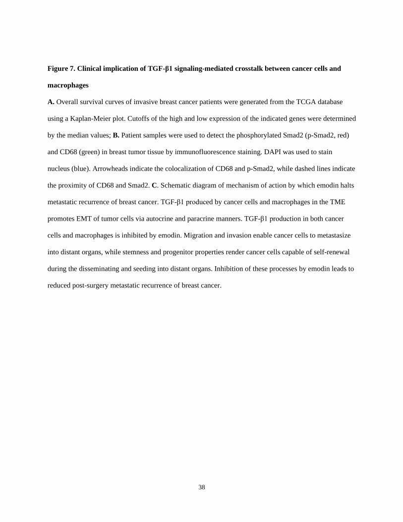
38
Figure 7. Clinical implication of TGF-β1 signaling-mediated crosstalk between cancer cells and
macrophages
A. Overall survival curves of invasive breast cancer patients were generated from the TCGA database
using a Kaplan-Meier plot. Cutoffs of the high and low expression of the indicated genes were determined
by the median values; B. Patient samples were used to detect the phosphorylated Smad2 (p-Smad2, red)
and CD68 (green) in breast tumor tissue by immunofluorescence staining. DAPI was used to stain
nucleus (blue). Arrowheads indicate the colocalization of CD68 and p-Smad2, while dashed lines indicate
the proximity of CD68 and Smad2. C. Schematic diagram of mechanism of action by which emodin halts
metastatic recurrence of breast cancer. TGF-β1 produced by cancer cells and macrophages in the TME
promotes EMT of tumor cells via autocrine and paracrine manners. TGF-β1 production in both cancer
cells and macrophages is inhibited by emodin. Migration and invasion enable cancer cells to metastasize
into distant organs, while stemness and progenitor properties render cancer cells capable of self-renewal
during the disseminating and seeding into distant organs. Inhibition of these processes by emodin leads to
reduced post-surgery metastatic recurrence of breast cancer.