encap msc protocol for nih - keck.rutgers.edu · • hemocytometer • centrifuge • water bath...
TRANSCRIPT
Encap MSC Protocol for NIH
1
Rutgers University Materials and Methods for MSC Growth & Encapsulation Page
A. Protocol for Cell Growth and Passage 2
A1. Preparation of MSC Media Composition 2
A2. Protocol for Thawing MSCs 3
A3. Protocol for Growth & Passage of MSC 4
B. Protocol to Encapsulate MSC into Alginate Microspheres 5
B1. Preparation of Prepare 2.2% Alginate Solution 5
B2. Preparation of Cross-linking Solutions 5 B3. Preparation of MSC Suspension for Alginate Encapsulation 6
B4. Protocol For MSC Encapsulation Using NISCO 7-8
B5. Protocol For MSC Encapsulation Using Spraybase Profector 9-10
C. Rapid Capsule Recovery for Capsules During Cell Encapsulation 11-12
D. Viability Assay: Live/Dead staining 12
E. Spraybase Encapsulator 13
F. NISCO Encapsulator 14

Encap MSC Protocol for NIH
2
Encap MSC Protocol for NIH
3
A. Protocol for Cell Growth and Passage A1. MSC Media Composition
Safety Materials:
• Lab coat • Regular gloves
Equipment Needed:
• Biosafety Cabinet • 50 mL conical tubes • Aspirator • Hemocytometer • Centrifuge • Water bath (37° C)
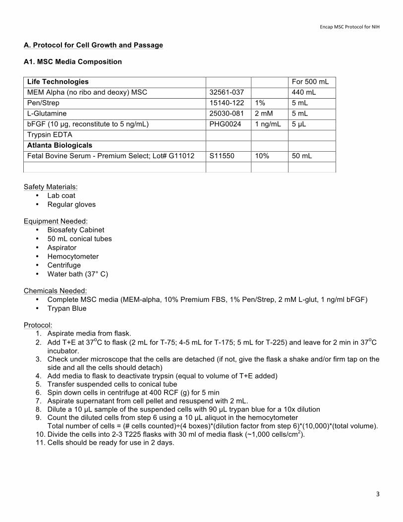
Chemicals Needed: • Complete MSC media (MEM-alpha, 10% Premium FBS, 1% Pen/Strep, 2 mM L-glut, 1 ng/ml bFGF) • Trypan Blue
Protocol:
1. Aspirate media from flask. 2. Add T+E at 37oC to flask (2 mL for T-75; 4-5 mL for T-175; 5 mL for T-225) and leave for 2 min in 37oC
incubator. 3. Check under microscope that the cells are detached (if not, give the flask a shake and/or firm tap on the
side and all the cells should detach) 4. Add media to flask to deactivate trypsin (equal to volume of T+E added) 5. Transfer suspended cells to conical tube 6. Spin down cells in centrifuge at 400 RCF (g) for 5 min 7. Aspirate supernatant from cell pellet and resuspend with 2 mL. 8. Dilute a 10 µL sample of the suspended cells with 90 µL trypan blue for a 10x dilution 9. Count the diluted cells from step 6 using a 10 µL aliquot in the hemocytometer
Total number of cells = (# cells counted)÷(4 boxes)*(dilution factor from step 6)*(10,000)*(total volume). 10. Divide the cells into 2-3 T225 flasks with 30 ml of media flask (~1,000 cells/cm2). 11. Cells should be ready for use in 2 days.
Life Technologies For 500 mL MEM Alpha (no ribo and deoxy) MSC 32561-037 440 mL Pen/Strep 15140-122 1% 5 mL L-Glutamine 25030-081 2 mM 5 mL bFGF (10 µg, reconstitute to 5 ng/mL) PHG0024 1 ng/mL 5 µL Trypsin EDTA Atlanta Biologicals Fetal Bovine Serum - Premium Select; Lot# G11012 S11550 10% 50 mL
Encap MSC Protocol for NIH
4
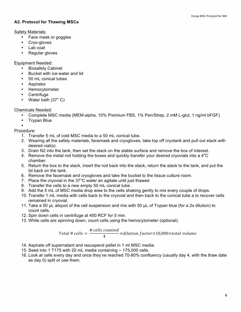
A2. Protocol for Thawing MSCs Safety Materials:
• Face mask or goggles • Cryo-gloves • Lab coat • Regular gloves
Equipment Needed:
• Biosafety Cabinet • Bucket with ice-water and lid • 50 mL conical tubes • Aspirator • Hemocytometer • Centrifuge • Water bath (37° C)
Chemicals Needed: • Complete MSC media (MEM-alpha, 10% Premium FBS, 1% Pen/Strep, 2 mM L-glut, 1 ng/ml bFGF) • Trypan Blue •
Procedure: 1. Transfer 5 mL of cold MSC media to a 50 mL conical tube. 2. Wearing all the safety materials, facemask and cryogloves, take top off cryotank and pull out stack with
desired vial(s). 3. Drain N2 into the tank, then set the stack on the stable surface and remove the box of interest. 4. Remove the metal rod holding the boxes and quickly transfer your desired cryovials into a 4oC
chamber. 5. Return the box to the stack, insert the rod back into the stack, return the stack to the tank, and put the
lid back on the tank. 6. Remove the facemask and cryogloves and take the bucket to the tissue culture room. 7. Place the cryovial in the 37°C water an agitate until just thawed 8. Transfer the cells to a new empty 50 mL conical tube. 9. Add the 5 mL of MSC media drop wise to the cells shaking gently to mix every couple of drops. 10. Transfer 1 mL media with cells back to the cryovial and then back to the conical tube a to recover cells
remained in cryovial. 11. Take a 50 µL aliquot of the cell suspension and mix with 50 µL of Trypan blue (for a 2x dilution) to
count cells. 12. Spin down cells in centrifuge at 400 RCF for 5 min. 13. While cells are spinning down, count cells using the hemocytometer (optional).
𝑇𝑜𝑡𝑎𝑙 # 𝑐𝑒𝑙𝑙𝑠 = # 𝑐𝑒𝑙𝑙𝑠 𝑐𝑜𝑢𝑛𝑡𝑒𝑑
4×𝑑𝑖𝑙𝑢𝑡𝑖𝑜𝑛 𝑓𝑎𝑐𝑡𝑜𝑟×10,000×𝑡𝑜𝑡𝑎𝑙 𝑣𝑜𝑙𝑢𝑚𝑒
14. Aspirate off supernatant and resuspend pellet in 1 ml MSC media. 15. Seed into 1 T175 with 20 mL media containing ~ 175,000 cells. 16. Look at cells every day and once they’ve reached 70-80% confluency (usually day 4, with the thaw date
as day 0) split or use them.
Encap MSC Protocol for NIH
5
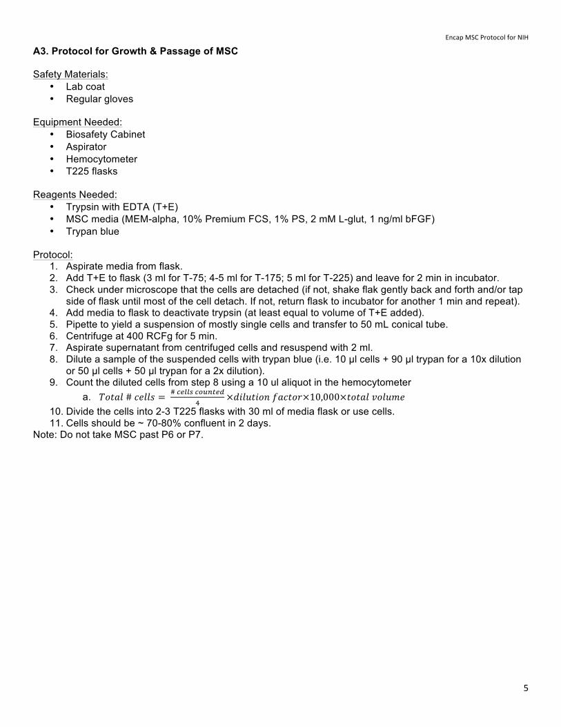
A3. Protocol for Growth & Passage of MSC Safety Materials:
• Lab coat • Regular gloves
Equipment Needed:
• Biosafety Cabinet • Aspirator • Hemocytometer • T225 flasks
Reagents Needed:
• Trypsin with EDTA (T+E) • MSC media (MEM-alpha, 10% Premium FCS, 1% PS, 2 mM L-glut, 1 ng/ml bFGF) • Trypan blue
Protocol:
1. Aspirate media from flask. 2. Add T+E to flask (3 ml for T-75; 4-5 ml for T-175; 5 ml for T-225) and leave for 2 min in incubator. 3. Check under microscope that the cells are detached (if not, shake flak gently back and forth and/or tap
side of flask until most of the cell detach. If not, return flask to incubator for another 1 min and repeat). 4. Add media to flask to deactivate trypsin (at least equal to volume of T+E added). 5. Pipette to yield a suspension of mostly single cells and transfer to 50 mL conical tube. 6. Centrifuge at 400 RCFg for 5 min. 7. Aspirate supernatant from centrifuged cells and resuspend with 2 ml. 8. Dilute a sample of the suspended cells with trypan blue (i.e. 10 µl cells + 90 µl trypan for a 10x dilution
or 50 µl cells + 50 µl trypan for a 2x dilution). 9. Count the diluted cells from step 8 using a 10 ul aliquot in the hemocytometer
a. 𝑇𝑜𝑡𝑎𝑙 # 𝑐𝑒𝑙𝑙𝑠 = # !"##$ !"#$%&'!
×𝑑𝑖𝑙𝑢𝑡𝑖𝑜𝑛 𝑓𝑎𝑐𝑡𝑜𝑟×10,000×𝑡𝑜𝑡𝑎𝑙 𝑣𝑜𝑙𝑢𝑚𝑒 10. Divide the cells into 2-3 T225 flasks with 30 ml of media flask or use cells. 11. Cells should be ~ 70-80% confluent in 2 days.
Note: Do not take MSC past P6 or P7.
Encap MSC Protocol for NIH
6
B. Protocol to Encapsulate MSC into Alginate Microspheres B1. Preparation of 2.2% Alginate Solution Safety Materials:
• Lab coat • Regular gloves
Equipment Needed:
• Biosafety Cabinet • Heated Stirrer & Stir bar • 0.22 µm filters • 3 ml syringes
Reagents Needed:
• Alginate (PRONOVA sodium alginate, FMC BioPolymer AS) • 50ml sterile jar • 0.55g of alginate • 25ml of Ca2+ free DMEM
Procedure:
1. Take 25ml of Ca2+ free DMEM and put it on 50ml jar. 2. Put it on a hot stirring plate at 60°C. 3. Dissolve the alginate slowly into the DMEM. With periods of fast and slow stirring. 4. Mix for ~1hr or more at max speed. 5. Sterilize by filtration with .22µm filter and store at 4oC.
B2. Preparation of Cross-linking Solutions Equipment Needed:
• 500ml sterile bottle • 1000 ml graduated cylinder • Stirring bar • pH meter • Vacuum filter
Reagents Needed:
• 500ml DI water • 2.5 g of glucose • 5.549 g of CaCl2 , or 10.35 g of BaCl2 • 4.2369g of NaCl • 1.0465g MOPS
Procedure:
• Add all reagents into 500ml of DI water and stir until well mixed. • Adjust pH to ~7.00. • Filter into a sterile bottle. Store at 4oC.
Encap MSC Protocol for NIH
7
B3. Preparation MSC Suspension for Alginate Encapsulation Safety Materials:
• Lab coat • Regular gloves
Equipment Needed:
• Biosafety Cabinet • Encapsulator (NISCO or Spraybase) • Aspirator • Hemocytometer
Reagents Needed:
• Trypsin with EDTA (T+E) • MSC media (MEM-alpha, 10% Premium FCS, 1% PS, 2 mM L-glut, 1 ng/ml bFGF) • Trypan blue
Procedure:
1. Aspirate media from T225 flasks with MSC at ~70-80% confluence. 2. Add 5 ml of T+E to each T225 flask and leave for 2 min in incubator. 3. Check under microscope that the cells are detached (if not, shake flask gently back and forth, and/or
tap on the side until most of the cells detach. If not, return flask to incubator for another 1 min and repeat).
4. Add media to flask to deactivate trypsin (at least equal to volume of T+E added). 5. Pipette to yield a suspension of mostly single cells and transfer all the cells into one 50 mL conical tube. 6. Dilute a sample of the suspended cells with trypan blue (i.e. 100 µl cells + 100 µl trypan for a 2x
dilution). 7. Count the diluted cells from step 6 using a 10 ul aliquot in the hemocytometer
a. 𝑇𝑜𝑡𝑎𝑙 # 𝑐𝑒𝑙𝑙𝑠 = # !"##$ !"#$%&'!
×𝑑𝑖𝑙𝑢𝑡𝑖𝑜𝑛 𝑓𝑎𝑐𝑡𝑜𝑟×10,000×𝑡𝑜𝑡𝑎𝑙 𝑣𝑜𝑙𝑢𝑚𝑒 8. Calculate total number of cells, centrifuge at 400 RCFg for 5 min. 9. Aspirate as much of the supernatant as possible from centrifuged cells and resuspend to yield a final
concentration of 6-10 X 107 cells/ml.
Encap MSC Protocol for NIH
8
B4. Protocol For MSC Encapsulation Using NISCO
Equipment Needed: Autoclaved
• Single arm and hose • Needle • Forceps • Medium Sized Stir Bar • Scissors
Not Autoclaved • Biosafety Cabinet containing NISCO Encapsulator • 80 by 40mm Petri Dish • 1mL, 3mL, and 10mL Syringe
Reagents Needed: • MSC media (MEM-alpha, 10% Premium FCS, 1% PS, 2 mM L-glut, 1 ng/ml bFGF) • Trypan blue • Hepes buffered saline • Poly-L-Lysine (1 mg/ml) for use only with Ca++ cross-linked capsules
Procedure (Steps 1-9 must be done prior to Step 10 to minimize time of cells after harvest):
1. Turn on Encapsulator and Pump and Set Parameters a. 5m/hr b. 3mL Syringe
2. Adjust syringe holder on pump so that the wings are in the holder; adjust for syringe placement 3. Attach Hose and Side Arm 4. Wash needle to check flow with 1ml PBS once. (If flow is not smooth, choose another needle or clean
needle by sonication for 30 minute in 0.1 mM EDTA). Flush needle with to dry. 5. Attach needle tightly to side arm 6. Add 100 mL crosslinking solution and stir bar into 80 by 40 mm Petri Dish and place it in Encapsulator.
(avoid splashing needle) 7. Open Encapsulator lower arm to 5.5 cm above level of crosslinking solution. 8. Lower positive electrode to enter the cross-link solution but do not lower to much to avoid electrode
hitting capsules. 9. Harvest cells as described in B3. Preparation MSC Suspension for Alginate Encapsulation 10. Determine volume of alginate to add to cells. Add 9 X 2.2 % alginate solution so than final cell
concentration is 6-10 million cells/mL. This will cell dilute the alginate to 2.0%.The alginate is highly viscous, add small volumes ~100 µl to gently mix with the cell suspension by gentle pipetting using a swirling motion to mix.
11. Using a cut 1 mL pipette, load the cell-alginate mixture into syringe keeping it vertical so that the mixture settles to the bottom of syringe.
12. Attach syringe to hose and place into pump 13. Plug electrode into side arm. 14. Avoid bubbles, which can interrupt the run by clogging needle. If you see any bubbles coming out from
nozzle of the needle, aspirate without touching nozzle 15. Turn on power to electrodes; voltage should be 6.4 kV, otherwise adjust to this voltage. 16. Switch pump on and watch run to make sure needle does not clog. 17. After encapsulation, turn off pump and electrodes. Run times should be 20-30 minutes. 18. Leave capsules to crosslink for and additional 10 minutes
Encap MSC Protocol for NIH
9
19. During this 10 minute incubation, remove needle from encapsulator and wash with distilled water 3 each with 10mL. Flush syringe with air 3 times. Place syringe in 15mL tube and add enough Sodium Citrate or EDTA to cover needle and sonicate for 30 minutes.
20. After 10 minutes, aspirate the surface of the crosslink to remove floating capsules; they are imperfect. 21. Remove stir bar with forceps and place into 10% bleach. 22. Collect capsules into a 2.2 µm cell strainer by repeated pipetting to collect all capsules form the 100 mL
cross-link solution. 23. Wash Capsules with 20 mL of Hepes buffered saline. 24. For additional treatments to stabilize the capsules (e.g. PLL or divalent cation solution), place cell
strainer in a 6-well plate, and wash rapidly by dipping cell strainer 3 X through adjacent wells with 5 mL Hepes buffered saline.
25. Transfer cell strainer in a well with 5mL of alpha complete MEM. 26. Resuspend capsules by repeated pipetting, transfer in media into T75-Flask, a put into 37oC incubator.
Minimize the time of cells with alginate as extended incubation may decrease viability.
Encap MSC Protocol for NIH
10
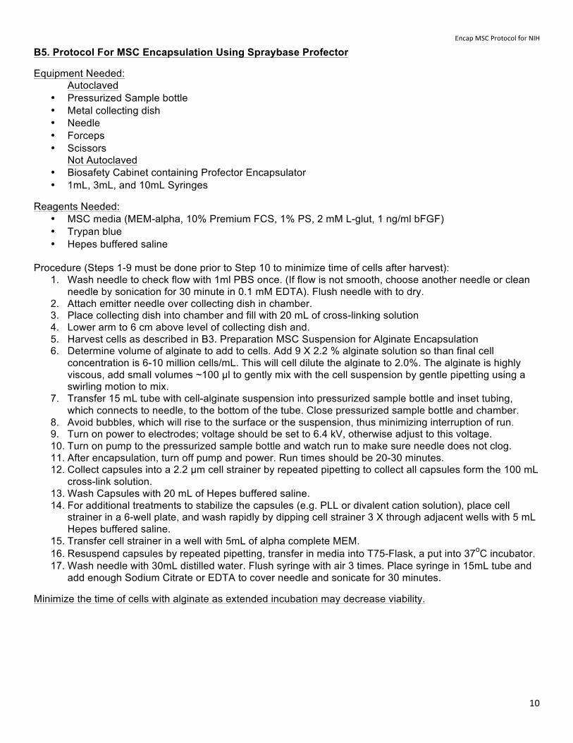
B5. Protocol For MSC Encapsulation Using Spraybase Profector
Equipment Needed: Autoclaved
• Pressurized Sample bottle • Metal collecting dish • Needle • Forceps • Scissors
Not Autoclaved • Biosafety Cabinet containing Profector Encapsulator • 1mL, 3mL, and 10mL Syringes
Reagents Needed: • MSC media (MEM-alpha, 10% Premium FCS, 1% PS, 2 mM L-glut, 1 ng/ml bFGF) • Trypan blue • Hepes buffered saline
Procedure (Steps 1-9 must be done prior to Step 10 to minimize time of cells after harvest):
1. Wash needle to check flow with 1ml PBS once. (If flow is not smooth, choose another needle or clean needle by sonication for 30 minute in 0.1 mM EDTA). Flush needle with to dry.
2. Attach emitter needle over collecting dish in chamber. 3. Place collecting dish into chamber and fill with 20 mL of cross-linking solution 4. Lower arm to 6 cm above level of collecting dish and. 5. Harvest cells as described in B3. Preparation MSC Suspension for Alginate Encapsulation 6. Determine volume of alginate to add to cells. Add 9 X 2.2 % alginate solution so than final cell
concentration is 6-10 million cells/mL. This will cell dilute the alginate to 2.0%. The alginate is highly viscous, add small volumes ~100 µl to gently mix with the cell suspension by gentle pipetting using a swirling motion to mix.
7. Transfer 15 mL tube with cell-alginate suspension into pressurized sample bottle and inset tubing, which connects to needle, to the bottom of the tube. Close pressurized sample bottle and chamber.
8. Avoid bubbles, which will rise to the surface or the suspension, thus minimizing interruption of run. 9. Turn on power to electrodes; voltage should be set to 6.4 kV, otherwise adjust to this voltage. 10. Turn on pump to the pressurized sample bottle and watch run to make sure needle does not clog. 11. After encapsulation, turn off pump and power. Run times should be 20-30 minutes. 12. Collect capsules into a 2.2 µm cell strainer by repeated pipetting to collect all capsules form the 100 mL
cross-link solution. 13. Wash Capsules with 20 mL of Hepes buffered saline. 14. For additional treatments to stabilize the capsules (e.g. PLL or divalent cation solution), place cell
strainer in a 6-well plate, and wash rapidly by dipping cell strainer 3 X through adjacent wells with 5 mL Hepes buffered saline.
15. Transfer cell strainer in a well with 5mL of alpha complete MEM. 16. Resuspend capsules by repeated pipetting, transfer in media into T75-Flask, a put into 37oC incubator. 17. Wash needle with 30mL distilled water. Flush syringe with air 3 times. Place syringe in 15mL tube and
add enough Sodium Citrate or EDTA to cover needle and sonicate for 30 minutes.
Minimize the time of cells with alginate as extended incubation may decrease viability.
Encap MSC Protocol for NIH
11
C. Rapid Capsule Recovery System for Collection of Capsules During Cell Encapsulation:
Introduction
Mesenchymal stem cells are used clinically to modulate uncontrolled inflammation. However, large doses are injected and they survive transiently limiting long-term efficacy. Encapsulation of cells prolongs cell survival and requires dramatically reduced doses for efficacy. However, methods for scaled-up yields of encapsulated cells have not been described and are not available. The current invention enables scaled-up production of capsules and controls encapsulation parameters.
Problems with Current Cell Encapsulation Systems & Scaled-Up Production:
1. Encapsulation systems use a batch vessel for crosslinking of alginate microspheres that does not allow removal of the resulting capsules during the encapsulation process.
2. Long encapsulation runs to increase yields result in large differences in crosslinking times from entry of the first to the last capsules into the cross-linking bath. Thus, there are considerable differences in cross-linking times. Short times decrease capsule stability and long times reduce permeability and decreases efficacy.
3. Cell encapsulation systems in use do not yield sufficient amounts for clinical studies. 4. Advantages of the Rapid Capsules Recovery System:
1. Capsules are recovered at desired times without disrupting the encapsulation process. 2. Enables tighter control of crosslinking times yielding more uniform preparations. 3. Enables scaled-up cell encapsulation to yield much larger amounts of encapsulated cells.
Rapid Capsule Recovery System
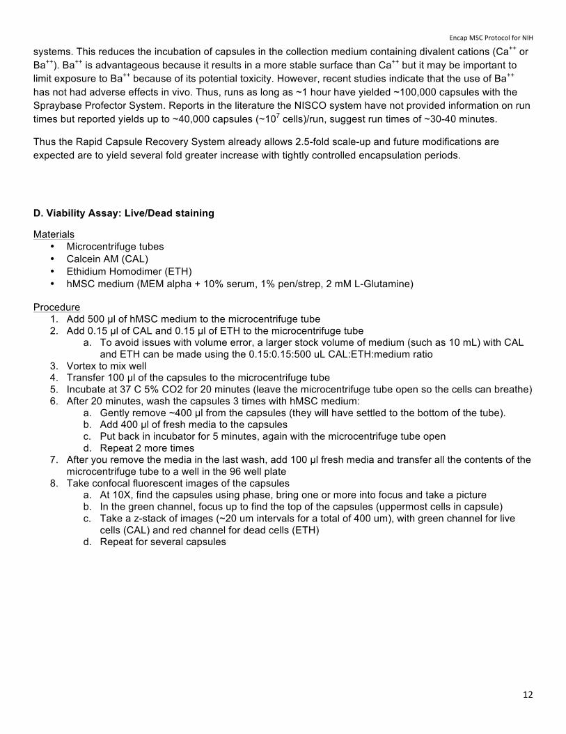
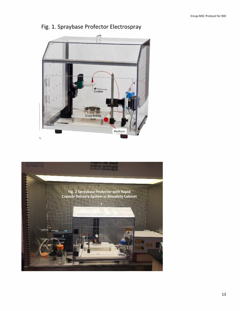
The Rapid Capsule Recovery System comprises input and output flow ports to collect capsules at selected times while allowing the encapsulation process to proceed without interruption. No existing system can do this. Microspheres of cells in alginate are propelled from an emitter into a collector dish containing crosslinking solution where they form microcapsules of polymerized alginate (The Profector Spraybase is shown in Fig.1). Capsule collection can be performed in <1 minute by controlled flows of crosslinking solution out of and into the metal collector dish (Fig. 2-4) without disrupting the encapsulation process. Rapid capsule collection on filters allows more precise control of incubation times in crosslinking buffer as well as rapid washing into media to terminate crosslinking and protect encapsulated cells. Detailed protocols will be posted after a patent application is submitted.
Comparison to Current State-of-the Art Cell Encapsulation Systems
The most widely used electrospray system for cell encapsulation is manufactured by NISCO. Alginate capsules can only be recovered after all are cross-linked in a batch vessel. A typical 20-minute run yields ~20,000 capsules and is followed by an additional incubation for 10 minutes in cross-linking buffer. Thus the entire crosslinking periods range from 10-30 minutes. Longer encapsulation runs to increase yields will result in longer exposure times of ells to alginate, which may reduce viability, and will result in greater variability in crosslinking times using the batch vessels.
The Rapid Capsule Recovery System allows for collection of capsules in <1 minute during encapsulation process and can be performed repeatedly at desired intervals without interrupting a run. Collections can be performed every 10 minutes, which enables more rapid washing by comparison to existing
Encap MSC Protocol for NIH
12
systems. This reduces the incubation of capsules in the collection medium containing divalent cations (Ca++ or Ba++). Ba++ is advantageous because it results in a more stable surface than Ca++ but it may be important to limit exposure to Ba++ because of its potential toxicity. However, recent studies indicate that the use of Ba++ has not had adverse effects in vivo. Thus, runs as long as ~1 hour have yielded ~100,000 capsules with the Spraybase Profector System. Reports in the literature the NISCO system have not provided information on run times but reported yields up to ~40,000 capsules (~107 cells)/run, suggest run times of ~30-40 minutes.
Thus the Rapid Capsule Recovery System already allows 2.5-fold scale-up and future modifications are expected are to yield several fold greater increase with tightly controlled encapsulation periods.
D. Viability Assay: Live/Dead staining
Materials • Microcentrifuge tubes • Calcein AM (CAL) • Ethidium Homodimer (ETH) • hMSC medium (MEM alpha + 10% serum, 1% pen/strep, 2 mM L-Glutamine)
Procedure
1. Add 500 µl of hMSC medium to the microcentrifuge tube 2. Add 0.15 µl of CAL and 0.15 µl of ETH to the microcentrifuge tube
a. To avoid issues with volume error, a larger stock volume of medium (such as 10 mL) with CAL and ETH can be made using the 0.15:0.15:500 uL CAL:ETH:medium ratio
3. Vortex to mix well 4. Transfer 100 µl of the capsules to the microcentrifuge tube 5. Incubate at 37 C 5% CO2 for 20 minutes (leave the microcentrifuge tube open so the cells can breathe) 6. After 20 minutes, wash the capsules 3 times with hMSC medium:
a. Gently remove ~400 µl from the capsules (they will have settled to the bottom of the tube). b. Add 400 µl of fresh media to the capsules c. Put back in incubator for 5 minutes, again with the microcentrifuge tube open d. Repeat 2 more times
7. After you remove the media in the last wash, add 100 µl fresh media and transfer all the contents of the microcentrifuge tube to a well in the 96 well plate
8. Take confocal fluorescent images of the capsules a. At 10X, find the capsules using phase, bring one or more into focus and take a picture b. In the green channel, focus up to find the top of the capsules (uppermost cells in capsule) c. Take a z-stack of images (~20 um intervals for a total of 400 um), with green channel for live
cells (CAL) and red channel for dead cells (ETH) d. Repeat for several capsules
Encap MSC Protocol for NIH
13
Fig.%2%Spraybase%Profector%with%Rapid%Capsule%Delivery%System%in%Biosafety%Cabinet%
Encap MSC Protocol for NIH
14
nisco
nisco Nisco Engineering AG Dufourstrasse 110 CH-8008 Zurich Tel: +41 44 380 06 30 Fax: +41 44 380 06 31 Issue 02.10.2009
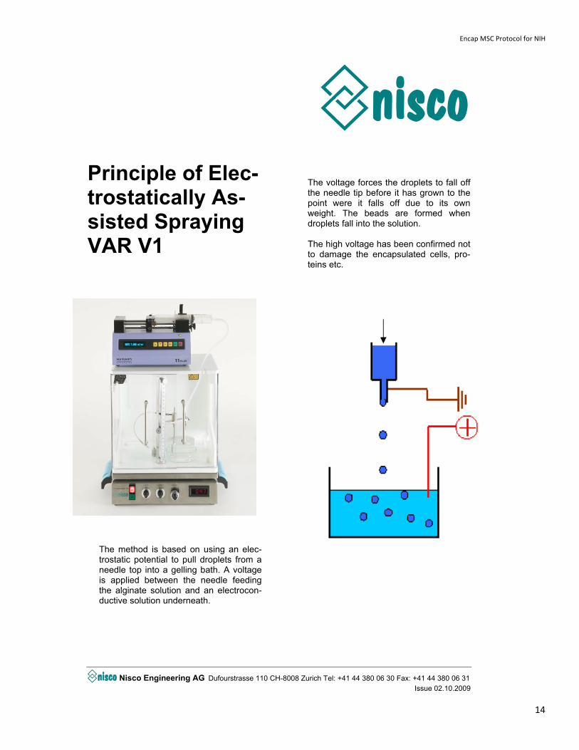
Principle of Elec-trostatically As-sisted Spraying VAR V1
The method is based on using an elec-trostatic potential to pull droplets from a needle top into a gelling bath. A voltage is applied between the needle feeding the alginate solution and an electrocon-ductive solution underneath.
The voltage forces the droplets to fall off the needle tip before it has grown to the point were it falls off due to its own weight. The beads are formed when droplets fall into the solution. The high voltage has been confirmed not to damage the encapsulated cells, pro-teins etc.