endo optiks® - beaver-visitec international inc. brochure provides a high level overview of the ......
TRANSCRIPT
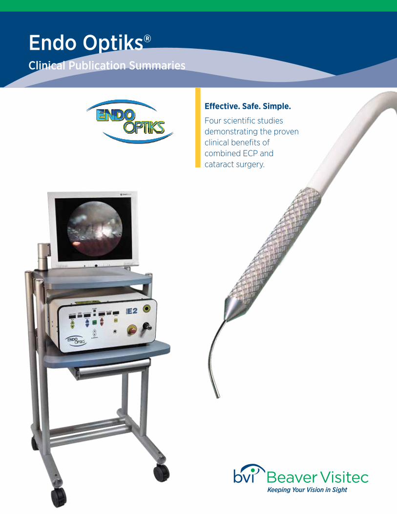
Endo Optiks®
Clinical Publication Summaries
Effective. Safe. Simple.
Four scientific studies demonstrating the proven clinical benefits of combined ECP and cataract surgery.
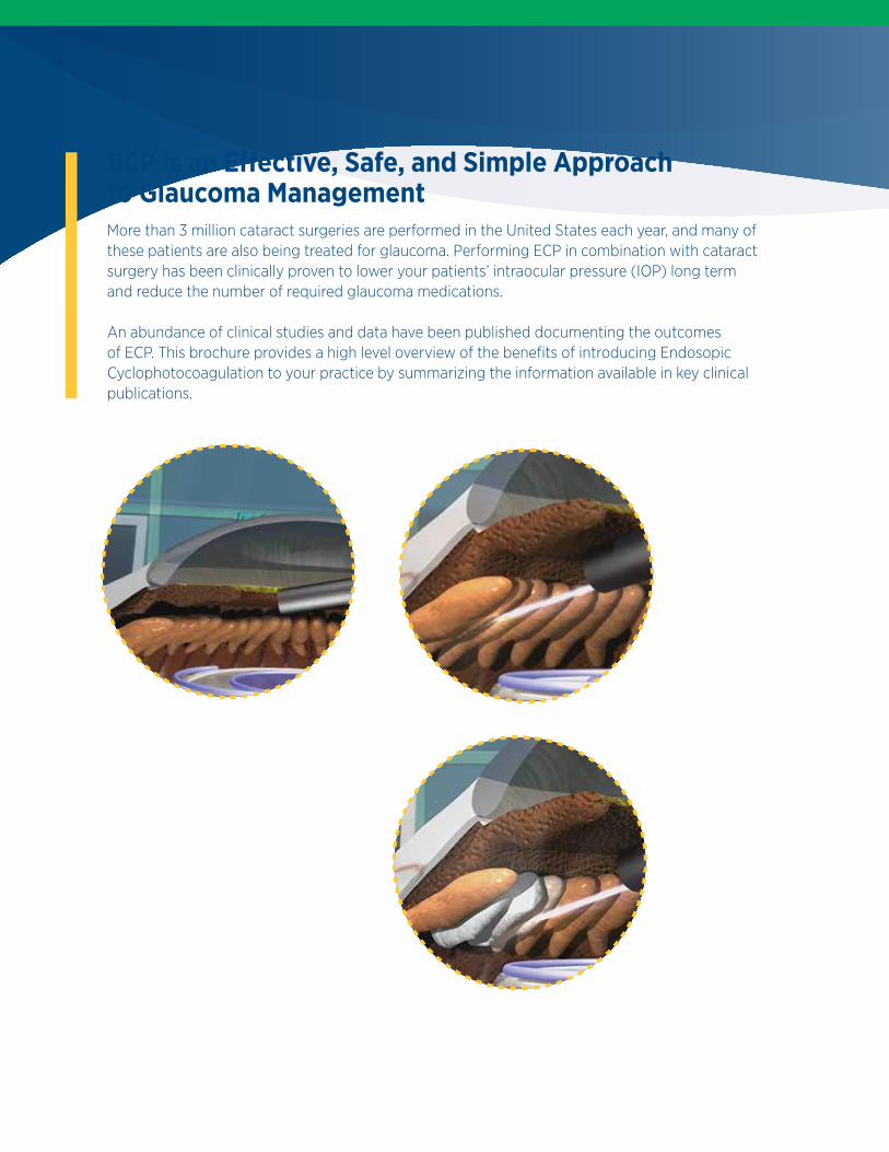
ECP is an Effective, Safe, and Simple Approach to Glaucoma ManagementMore than 3 million cataract surgeries are performed in the United States each year, and many of these patients are also being treated for glaucoma. Performing ECP in combination with cataract surgery has been clinically proven to lower your patients’ intraocular pressure (IOP) long term and reduce the number of required glaucoma medications.
An abundance of clinical studies and data have been published documenting the outcomes of ECP. This brochure provides a high level overview of the benefits of introducing Endosopic Cyclophotocoagulation to your practice by summarizing the information available in key clinical publications.

Endoscopic Cyclophotocoagulation Combined With Phacoemulsification Versus Phacoemulsification Alone in Medically Controlled Glaucoma .................................................................................................................... 5
Brian A. Francis, MD, MS, Stanley J. Berke, MD, Laurie Dustin, MS, Robert Noecker, MDJournal of Cataract & Refractive Surgery, Volume 40, Number 8, August 2014
‘Phaco-ECP’: Combined Endoscopic CyclophotocoagulationAnd Cataract Surgery to Augment Medical Control Of Glaucoma.............................................................................................................. 7
Dan Lindfield, MDRobert W. Ritchie, MDMichael FP Griffiths, MDBMJ Open 2012:2, April 2012
One-site Versus Two-site Endoscopic Cyclophotocoagulation ................................................................................................................... 9
Malik Y. Kahook, MD, Kira L. Lathrop, MAMSRobert J. Noecker, MDJournal of Glaucoma, Volume 16, Number 6, September 2007
Combined ECP and Cataract Surgery ........................................................................................................................................................................ 11
Stanley J. Berke, MD, FACSCataract & Refractive Surgery Today Europe, Cover Story Page 1, October 2011
Table of Contents
4
“Combined ECP-cataract extraction resulted in lower intraocular pressure & greater reduction in glaucoma medications than cataract extraction alone in medically controlled open angle glaucoma patients with visually significant cataract.”
— Brian A. Francis, MD, MS
Journal of Cataract & Refractive Surgery, Volume 40, Number 8, August 2014
Brian A. Francis, MD, MS Stanley J. Berke, MDLaurie Dustin, MS Robert Noecker, MD
5
Purpose
To compare the outcomes of endoscopic cyclophotocoagulation (ECP) and phacoemulsification cataract extraction versus cataract extraction alone in eyes with medically controlled open-angle glaucoma (OAG) and visually significant cataract.
Setting
Clinical practices of glaucoma specialists and comprehensive ophthalmologists.
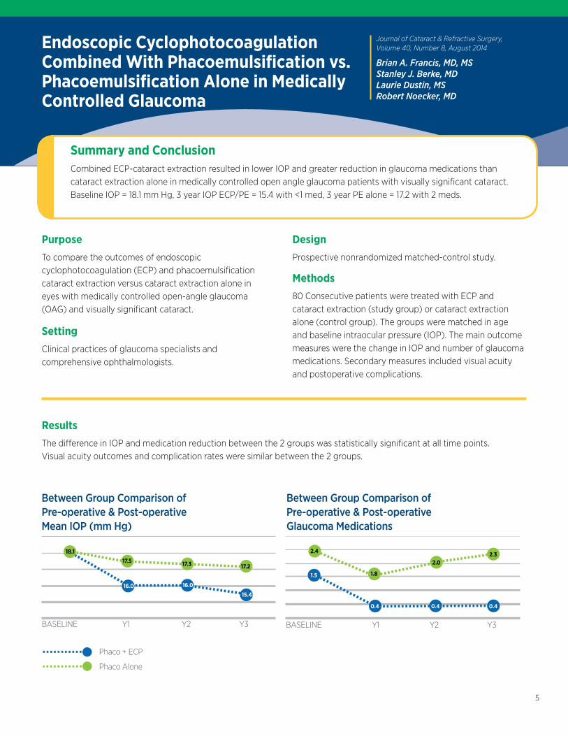
Summary and ConclusionCombined ECP-cataract extraction resulted in lower IOP and greater reduction in glaucoma medications than cataract extraction alone in medically controlled open angle glaucoma patients with visually significant cataract. Baseline IOP = 18.1 mm Hg, 3 year IOP ECP/PE = 15.4 with <1 med, 3 year PE alone = 17.2 with 2 meds.
Endoscopic Cyclophotocoagulation Combined With Phacoemulsification vs. Phacoemulsification Alone in Medically Controlled Glaucoma
Results
The difference in IOP and medication reduction between the 2 groups was statistically significant at all time points. Visual acuity outcomes and complication rates were similar between the 2 groups.
Design
Prospective nonrandomized matched-control study.
Methods
80 Consecutive patients were treated with ECP and cataract extraction (study group) or cataract extraction alone (control group). The groups were matched in age and baseline intraocular pressure (IOP). The main outcome measures were the change in IOP and number of glaucoma medications. Secondary measures included visual acuity and postoperative complications.
BASELINE Y1 Y2 Y3
Between Group Comparison of Pre-operative & Post-operative Mean IOP (mm Hg)
BASELINE Y1 Y2 Y3
Between Group Comparison of Pre-operative & Post-operative Glaucoma Medications
18.117.5 17.3 17.2
16.0 16.0
15.4
2.4
1.8
2.02.3
0.4 0.4 0.4
1.5
Phaco + ECP
Phaco Alone
6
“ECP combines almost symbiotically with cataract surgery. Glaucoma, like cataract is predominantly a disease of the older population and is often concurrent.”
“The procedure adds 5 minutes to the on-table time.”
“ECP can now be performed through standard phaco incisions.”
“This study confirms the safety of Phaco-ECP. In this case series, the IOP lowering effect was significant at all time points.”
— Dan Lindfield, MD
BMJ Open 2012:2, April 2012
Dan Lindfield, MDRobert W. Ritchie, MDMichael FP Griffiths, MD
7
Purpose
To determine if Phaco-ECP reduces intraocular pressure (IOP) and if Phaco-ECP is safe.
Setting
Single District General Hospital Ophthalmology Department within the UK.
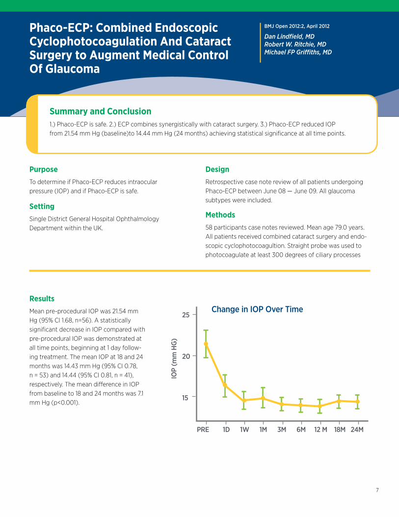
Summary and Conclusion1.) Phaco-ECP is safe. 2.) ECP combines synergistically with cataract surgery. 3.) Phaco-ECP reduced IOP from 21.54 mm Hg (baseline)to 14.44 mm Hg (24 months) achieving statistical significance at all time points.
Phaco-ECP: Combined Endoscopic Cyclophotocoagulation And Cataract Surgery to Augment Medical Control Of Glaucoma
Results
Mean pre-procedural IOP was 21.54 mm Hg (95% CI 1.68, n=56). A statistically significant decrease in IOP compared with pre-procedural IOP was demonstrated at all time points, beginning at 1 day follow-ing treatment. The mean IOP at 18 and 24 months was 14.43 mm Hg (95% CI 0.78, n = 53) and 14.44 (95% CI 0.81, n = 41), respectively. The mean difference in IOP from baseline to 18 and 24 months was 7.1 mm Hg (p<0.001).
Design
Retrospective case note review of all patients undergoing Phaco-ECP between June 08 — June 09. All glaucoma subtypes were included.
Methods
58 participants case notes reviewed. Mean age 79.0 years. All patients received combined cataract surgery and endo-scopic cyclophotocoagultion. Straight probe was used to photocoagulate at least 300 degrees of ciliary processes
Change in IOP Over Time
PRE 1D 1W 1M 3M 6M 12 M 18M 24M
25
20
15
IOP
(mm
HG
)

8
“The safety and efficacy of ECP has been demonstrated in treating various forms of glaucoma when performed alone or in combination with phacoemulsification.”
“Combined Phaco-ECO is commonly practiced with ECP performed, in most cases, through a single clear cornea incision allowing approximately 240 – 300 degrees of treatment.”
— Robert J. Noecker, MD
Journal of Glaucoma, Volume 16, Number 6, September 2007
Malik Y. Kahook, MDKira L. Lathrop, MAMSRobert J. Noecker, MD
9
Purpose
To report the intraocular pressure (IOP) lowering effect of ECP treatment through 1 vs. 2 corneal incisions
Design
Retrospective nonrandomized study comparing data from consecutive patients who underwent 1-site (group 1) vs. 2-site (group 2) PE-ECP. Patients were selected for each group, equivalent in age, baseline IOP and number of medications.
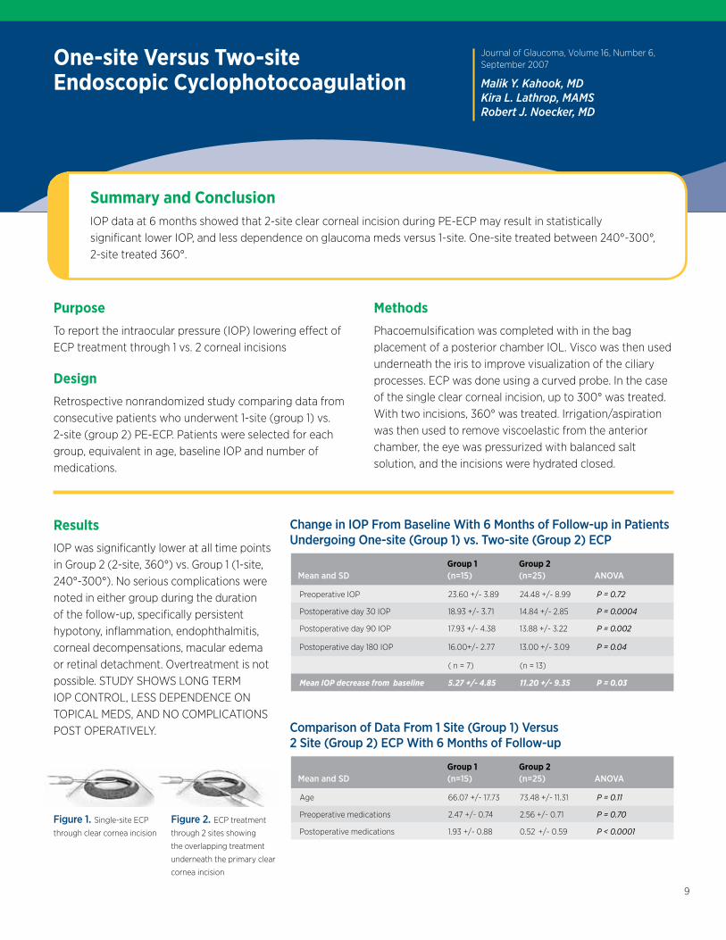
Summary and ConclusionIOP data at 6 months showed that 2-site clear corneal incision during PE-ECP may result in statistically significant lower IOP, and less dependence on glaucoma meds versus 1-site. One-site treated between 240°-300°, 2-site treated 360°.
One-site Versus Two-site Endoscopic Cyclophotocoagulation
Results
IOP was significantly lower at all time points in Group 2 (2-site, 360°) vs. Group 1 (1-site, 240°-300°). No serious complications were noted in either group during the duration of the follow-up, specifically persistent hypotony, inflammation, endophthalmitis, corneal decompensations, macular edema or retinal detachment. Overtreatment is not possible. STUDY SHOWS LONG TERM IOP CONTROL, LESS DEPENDENCE ON TOPICAL MEDS, AND NO COMPLICATIONS POST OPERATIVELY.
Methods
Phacoemulsification was completed with in the bag placement of a posterior chamber IOL. Visco was then used underneath the iris to improve visualization of the ciliary processes. ECP was done using a curved probe. In the case of the single clear corneal incision, up to 300° was treated. With two incisions, 360° was treated. Irrigation/aspiration was then used to remove viscoelastic from the anterior chamber, the eye was pressurized with balanced salt solution, and the incisions were hydrated closed.
Change in IOP From Baseline With 6 Months of Follow-up in Patients Undergoing One-site (Group 1) vs. Two-site (Group 2) ECP
Group 1 Group 2 Mean and SD (n=15) (n=25) ANOVA
Preoperative IOP 23.60 +/- 3.89 24.48 +/- 8.99 P = 0.72
Postoperative day 30 IOP 18.93 +/- 3.71 14.84 +/- 2.85 P = 0.0004
Postoperative day 90 IOP 17.93 +/- 4.38 13.88 +/- 3.22 P = 0.002
Postoperative day 180 IOP 16.00+/- 2.77 13.00 +/- 3.09 P = 0.04
( n = 7) (n = 13)
Mean IOP decrease from baseline 5.27 +/- 4.85 11.20 +/- 9.35 P = 0.03
Figure 1. Single-site ECP
through clear cornea incision
Figure 2. ECP treatment
through 2 sites showing
the overlapping treatment
underneath the primary clear
cornea incision
Comparison of Data From 1 Site (Group 1) Versus2 Site (Group 2) ECP With 6 Months of Follow-up
Group 1 Group 2 Mean and SD (n=15) (n=25) ANOVA
Age 66.07 +/- 17.73 73.48 +/- 11.31 P = 0.11
Preoperative medications 2.47 +/- 0.74 2.56 +/- 0.71 P = 0.70
Postoperative medications 1.93 +/- 0.88 0.52 +/- 0.59 P < 0.0001
10
10: EASY TO DO Adds only 2-4 minutes to the procedure.
9: TITRATABLE No reports of hypotony or phthisis as a primary procedure with more than 1,000 cases with 5 years’ follow-up and more than 50,000 cases worldwide in the past 10 years.
8: REPEATABLE Can treat full 360° because the tips of the ciliary processes are treated, sparing the valleys in between.
7: VALUE ADDED FOR PATIENTS Phaco-ECP is proven to further decrease IOP and the number of meds than phaco alone.
6: NO ADDITIONAL PATIENT FOLLOW UPS Patients only need to be seen the same as phaco procedures alone post operatively, 1d, 1wk, 1m.
“ECP is fun and interesting — The technology and the views are amazing.”
“ECP provides the advantages of a direct view of ciliary process photocoagulation and avoids the complications associated with transscleral cyclodestructive procedures.”
“Phaco-ECP resulted in a mean decrease in IOP and a reduction in glaucoma medications.”
— Stanley J. Berke, MD, FACS
5: NO LONG TERM COMPLICATIONS No incidents of hypotony, macular edema, or retinal detachment.
4: CONJUCTIVA IS LEFT UNDISTURBED If necessary, selective laser trabeculoplasty, repeat ECP, trabeculectomy or glaucoma drainage devices can be done subsequently.
3: NO EARLY OR LATE COMPLICATIONS Unlike trabeculectomy or shunt procedures.
2: REIMBURSEABLE In the US, CPT Code 06711 more than covers costs associated with the procedure for both surgeon and facility.
1: ECP IS FUN AND INTERESTING! A new view for surgeons, revealing a different look at cataracts, pathologic ciliary processes, zonules, capsular defects, etc.
Top Ten Reasons to Perform Combined Phaco-ECP
Cataract & Refractive Surgery Today Europe, Cover Story Page 1, October 2011
Stanley J. Berke, MD, FACS
11
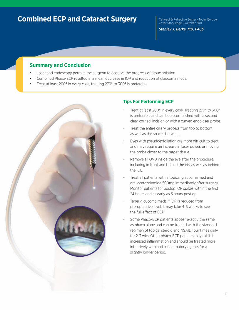
Summary and Conclusion• Laser and endoscopy permits the surgeon to observe the progress of tissue ablation.• Combined Phaco-ECP resulted in a mean decrease in IOP and reduction of glaucoma meds.• Treat at least 200° in every case, treating 270° to 300° is preferable.
Combined ECP and Cataract Surgery
Tips For Performing ECP
• Treat at least 200° in every case. Treating 270° to 300° is preferable and can be accomplished with a second clear corneal incision or with a curved endolaser probe.
• Treat the entire ciliary process from top to bottom, as well as the spaces between.
• Eyes with pseudoexfoliation are more difficult to treat and may require an increase in laser power, or moving the probe closer to the target tissue.
• Remove all OVD inside the eye after the procedure, including in front and behind the iris, as well as behind the IOL.
• Treat all patients with a topical glaucoma med and oral acetazolamide 500mg immediately after surgery. Monitor patients for postop IOP spikes within the first 24 hours and as early as 3 hours post op.
• Taper glaucoma meds if IOP is reduced from pre-operative level. It may take 4-6 weeks to see the full effect of ECP.
• Some Phaco-ECP patients appear exactly the same as phaco alone and can be treated with the standard regimen of topical steroid and NSAID four times daily for 2-3 wks. Other phaco-ECP patients may exhibit increased inflammation and should be treated more intensively with anti-inflammatory agents for a slightly longer period.

BVI, BVI Logo and all other trademarks are property of Beaver-Visitec International (BVI) © 2015 BVI
Beaver-Visitec International, Inc.411 Waverley Oaks Road, Waltham, MA 02452 Tel: 1.866.906.8080 Fax: 1.866.906.4304 www.beaver-visitec.com
Beaver-Visitec International, Sales Limited85c Park Drive, Milton Park,Abingdon, Oxfordshire, OX14 4RY, UKTel: 44.1865.601.256 Fax: 44.1865.595.761
06711-0815