endodontic treatment of immature teeth...
TRANSCRIPT
Semmelweis UniversityPedodontic and Orthodontic Clinic
Endodontic treatment of immature teethApexification
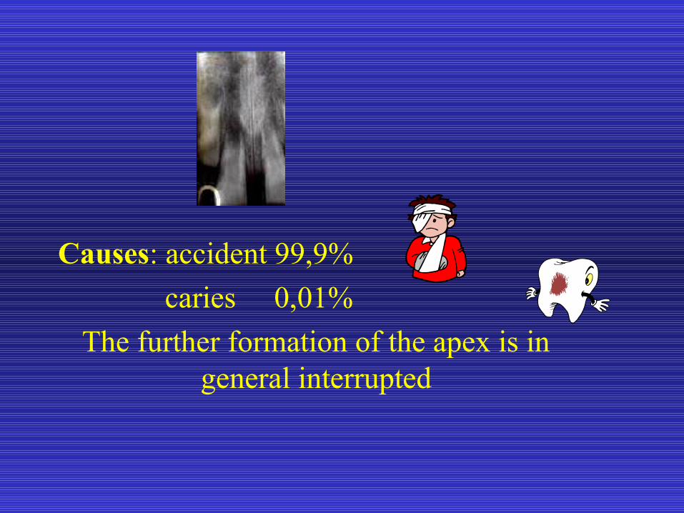
Causes: accident 99,9% caries 0,01%
The further formation of the apex is in general interrupted
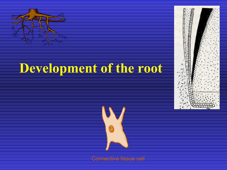
Development of the root

Initial root formation ¼ half ¾ of root length is achieved
Root length complete with apical foramen wide open
Half closed Root is complete
Root formation can be classified in seven stages by Moorrees
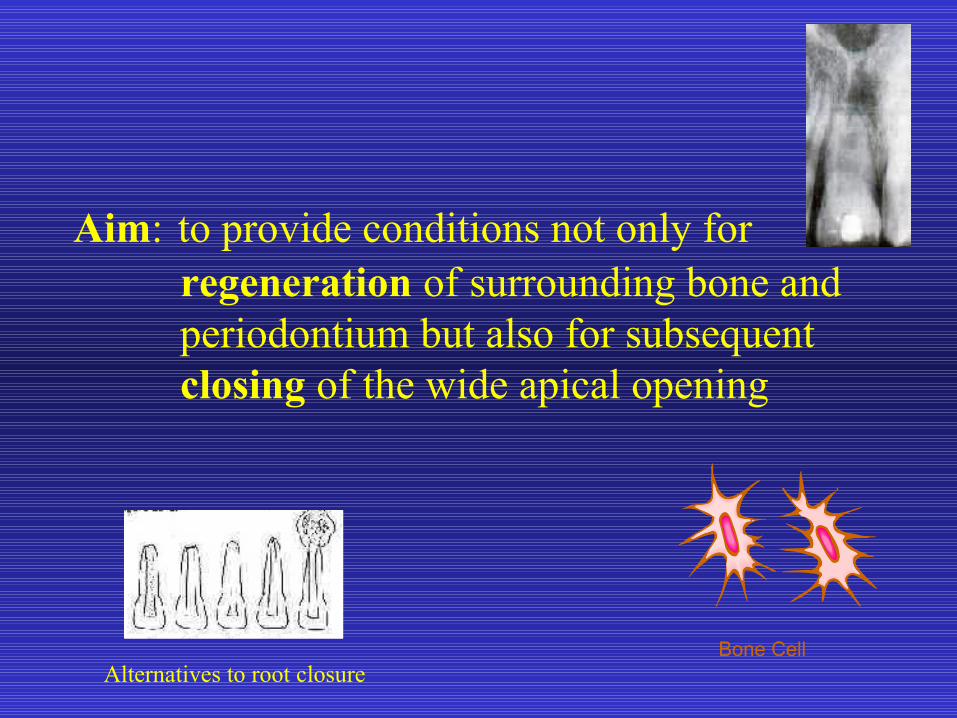
Aim: to provide conditions not only for regeneration of surrounding bone and periodontium but also for subsequent closing of the wide apical opening
Alternatives to root closure

It is very important to choose the adequat therapy to reach the desired aim. This depends on that tissue which fills in the apex at the beginning of the treatment.
vital restpulp odontogenesisdentin tissue
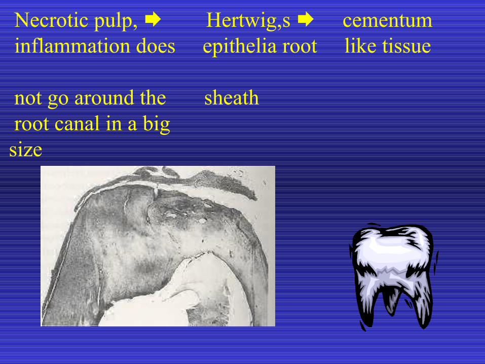
Necrotic pulp, Hertwig,s cementum inflammation does epithelia root like tissue not go around the sheath root canal in a bigsize

Special case:
infected pulp granulation tissue reparativeinflammation spread + blood clot granulationover into bone tissue tissuemesenchimal cells calcified tissue differentiate into irregular dentin formatively active immature bone ones cementum like tissue
Technical procedure
• X–ray photo to diagnose the developing
stadium of root canal and the size of periapical lesion
Technical procedure
• Isolation, sterile appliances trepanation
• mechanical cleansing careful manipulation, methodical filing
of the dentinal walls, causing minimal damage to the wide apical opening flushing with O,5% Na0Cl
Special case:• strong bleeding is to be induced from periapical region by careful manipulation with a Kerr needle 12. Bleeding removes necrotic remnants apex periapical destroyed cavity
• irrigating with saline • drying

• Drying sterile cotton point

Ca(OH) paste₂ , or
Ca(OH) point₂ (deposit preparation, which releases calcium hydroxide from gutta–percha matrix)one size smallerensures moist milieu

•Repetition of treatmentsuppuration spontaneous} cease bleeding
Temporary canal filling
Ca(OH) paste₂ periapical healing
• antibacterial effect• leaching effect neutralizes necrotic residue• mineralisation of organic matrix
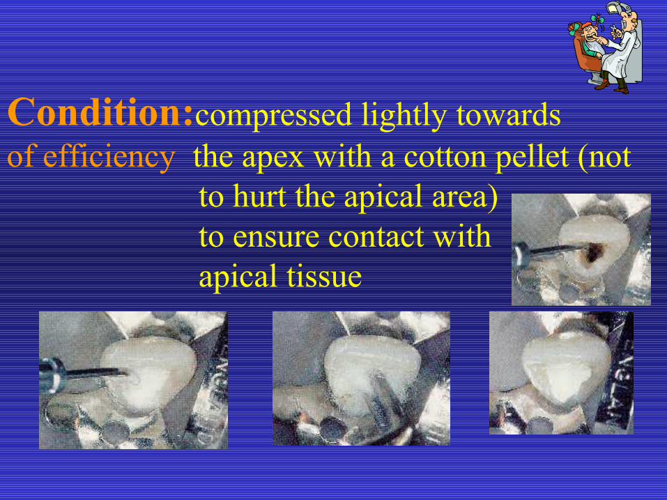
Condition:compressed lightly towards of efficiency the apex with a cotton pellet (not to hurt the apical area) to ensure contact with apical tissue
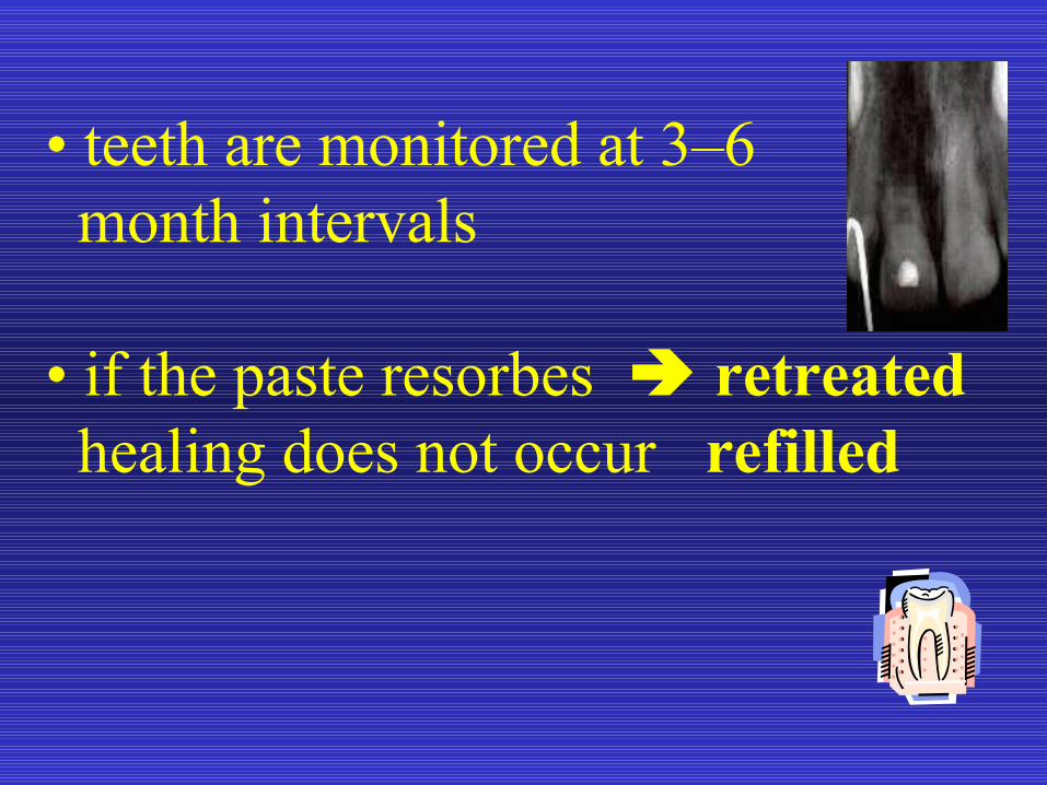
• teeth are monitored at 3–6 month intervals
• if the paste resorbes retreated healing does not occur refilled
Permanent obturation
Condition: • apex is closed by calcified tissue • regeneration of surrounding bone tissue and periodontium

Selection of a suitable thickest point

The tip of the point is warmed by passing once through an alcohol
flame

The slightly softened point is pressed steadily but gently against the apical
hard tissue barrier

• Lateral condensation

• Completed filling

Mineral Trioxid Aggregate
• Developed by Torabinejad at Loma Linda University in 1993
Mixture of tricalcium silcate, tricalcium oxide, tricalcium aluminate with other oxides. (Portland cement with bismuthoxide).
pH:12,5 and biological, histological properties can be compared to those of Ca(OH)2.
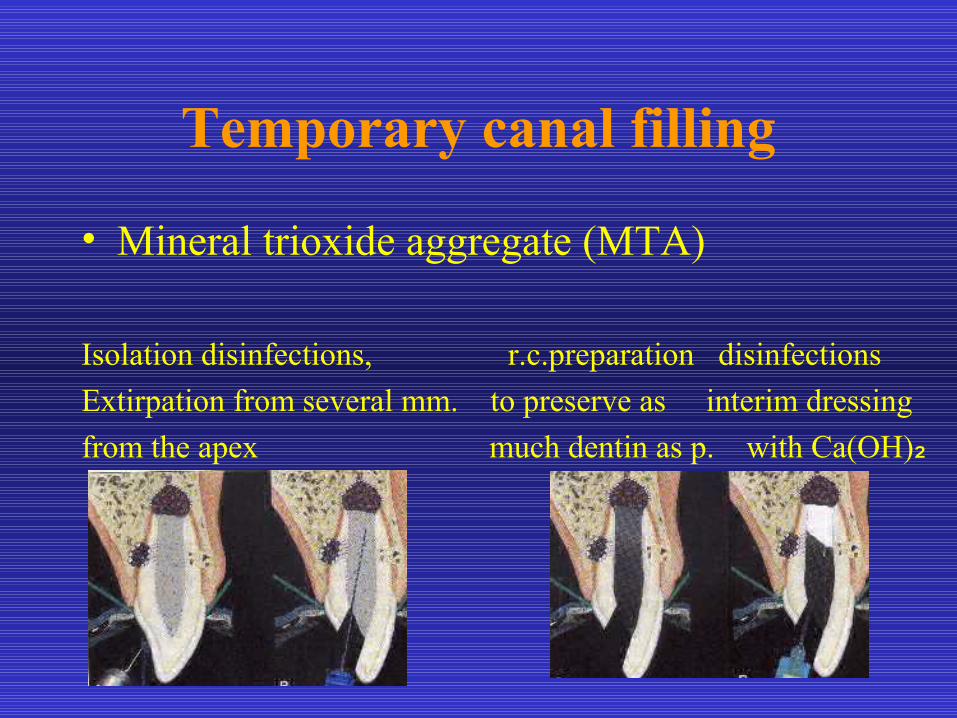
Temporary canal filling
• Mineral trioxide aggregate (MTA)
Isolation disinfections, r.c.preparation disinfectionsExtirpation from several mm. to preserve as interim dressingfrom the apex much dentin as p. with Ca(OH)₂

Root end filling
• Mineral trioxide aggregate (MTA)
irrigation with saline MTA moistened plug is checkedmixture introduced, gently cotton pellet irrigation,condensed apical MTA plug 4 mm thick temporary filling drying, filling
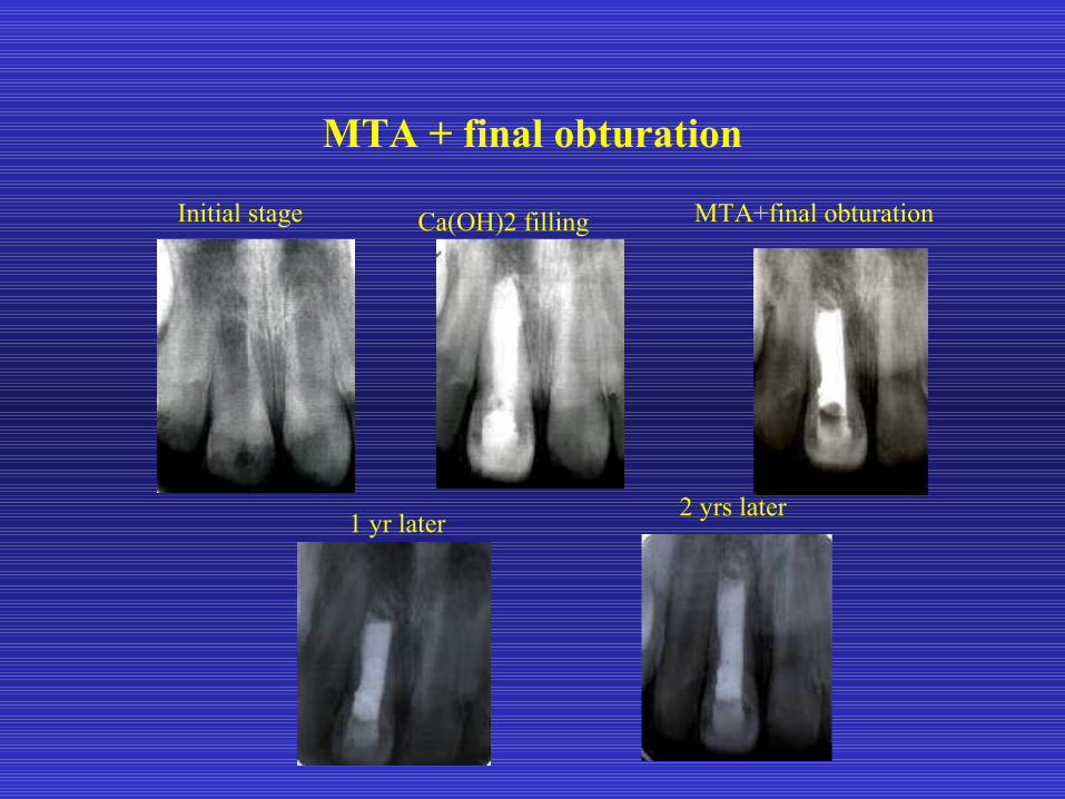
MTA + final obturation
Initial stage Ca(OH)2 filling MTA+final obturation
1 yr later 2 yrs later

Initial stage 7 months later
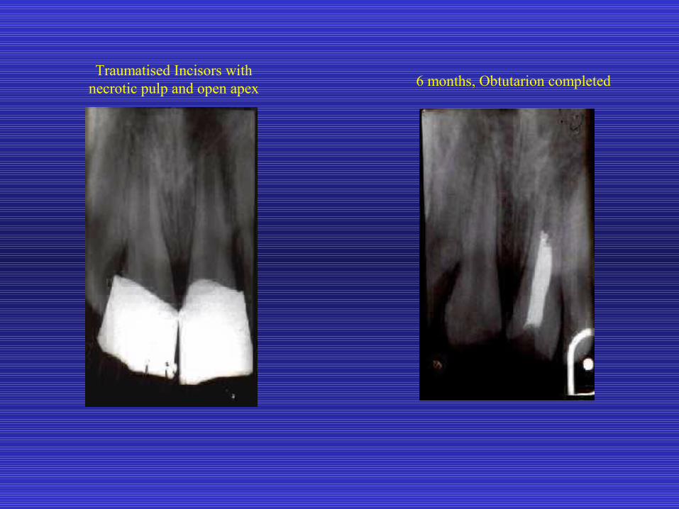
Traumatised Incisors with necrotic pulp and open apex 6 months, Obtutarion completed
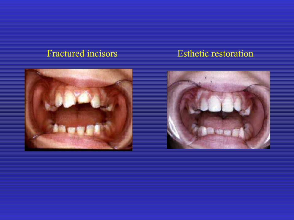
Fractured incisors Esthetic restoration

Open apex with periapical lesion
Formation of calcific barrier at the apex

Permanent obturation
4 yrs later 5 yrs later
Traumatised incisorsCa(OH)2 paste
placed in the canal
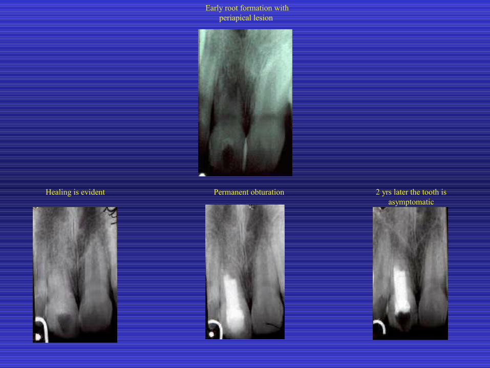
Early root formation with periapical lesion
Healing is evident Permanent obturation 2 yrs later the tooth is asymptomatic
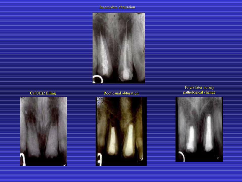
Incomplete obturation
Ca(OH)2 filling Root canal obturation10 yrs later no any
pathological change
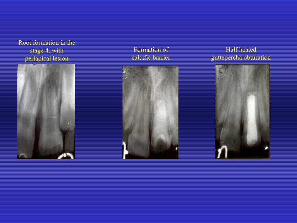
Root formation in the stage 4, with
periapical lesionFormation of
calcific barrierHalf heated
guttepercha obturation
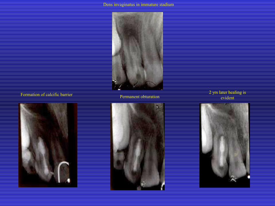
Dens invaginatus in immature stadium
Formation of calcific barrier Permanent obturation2 yrs later healing is
evident

Thank you for your kind attention