endodontics - suffolk root canalssuffolkrootcanal.co.uk/wp-content/uploads/2015/04/...endodontics...
TRANSCRIPT

endodontics Editor: MILTON SISKIN, D.D.S. College of Dentistry The University of Tennessee 847 Monroe Avenue Memphis, Tennessee 38 163
Immunopathogenesis of chronic periapical lesions
A review
Mahmoud Torabinejad, D.M.D., M.S.D., * and Leif K. Bakland, D.D.S., ** Loma Linda, Calif.
SCHOOL OF DENTISTRY, LOMA LINDA UNIVERSITY
The pathogenesis of periapical lesions has not been fully elucidated. Currently, the possibility of its being an immunologic phenomenon is receiving much attention. This article presents a review of the literature concerning immunologic reactions which may involve periapical lesions. It appears that antigen-antibody complexes and IgE-mediated reactions can initiate preliminary changes in periapical tissues. It is also likely that delayed hypersensitivity participates in the perpetuation and progression of periapical disease.
C hronic periapical lesions of pulpal origin are areas of inflammatory response to the content of the root canal system; the noxious agents that accumulate within this system may be viable and dead bacteria, bacterial fragments, bacterial toxins, the proteolytic products subsequent to deteriorating pulp tissue, and altered host tissue. Clinical, radio- graphic, histopathologic, and microbiologic studies of human periapical pathosis have been described extensively in the dental literature. l--l9 Most microbiologic studies indicate that microorganisms are not present in chronic periapical lesions. It therefore appears that egress of bacterial products and deteriorated pulp tissue from the root canal system may be as important as the actual presence of microorganisms. How these irritants initiate and propagate bone loss in periapical areas has been the topic of much discussion and specula- tion. Currently, the possibility of the immunologic phenomenon playing a role is receiving much attention. It has been shown that bacteria and altered host tissue substances are potential antigens capable of initiating immunologic reactions.20-23
*Associate Professor, Department of Endodontics. **Associate Professor and Chairman of Endodontics.
0030-4220/78/l 10685+15$01.50/O 0 1978 The C. V. Mosby Co 685
686 Torabinejad and Bakland Oral Surg. November, 1978
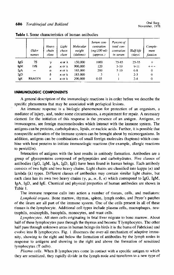
Table I. Some characteristics of human antibodies
Serum con- Percent of Heavy Light Molecular centration total con- Comple-
Older chain chain weight (mgl100 ml) centration Half-life ment names class class (daltons) (awox. I in serum (days) Jixation
W 7s Y KOT h 150,000 loo0 75-85 25-35 +
W 19s P K or x 900,000 120 5- 10 9-11 +++ IgA - a K or A 165,000 200 5-10 6-8 0 I@ 6 K or A 183,COO 3 I 2-3 0 I@ REAGIN E K or A 200,cQo 0.05 I 2-4 0
IMMUNOLOGIC COMPONENTS
A general description of the immunologic reactions is in order before we describe the specific phenomena that may be associated with periapical lesions.
An immune response is a biologic phenomenon for protection of an organism, a mediator of injury, and, under some circumstances, a requirement for repair. A necessary element for the initiation of this response is the presence of an antigen. Antigens, or immunogens, are foreign macromolecules which interact with the immune system. The antigens can be proteins, carbohydrates, lipids, or nucleic acids. Further, it is possible that nonspecific activation of the immune system can be brought about by microorganisms. In addition, antigens can be combinations of small foreign molecules (haptens) which com- bine with host proteins to initiate immunologic reactions (for example, allergic reactions to penicillin).
Interaction of antigens with the host results in antibody formation. Antibodies are a group of glycoproteins composed of polypeptides and carbohydrates. Five classes of antibodies (IgG, IgM, IgA, IgD, IgE) have been found in human beings. Each antibody consists of two light and two heavy chains. Light chains are classified into kappa (K) and lambda (A) types. Different classes of antibodies may contain similar light chains, but each class has its own two heavy chains (y, p, CX, 6, 6) which correspond to IgG, IgM, IgA, IgD, and IgE. Chemical and physical properties of human antibodies are shown in Table I.
The immune response calls into action a number of tissues, cells, and mediators: Lymphoid organs. Bone marrow, thymus, spleen, lymph nodes, and Peyer’s patches
of the ileum are all part of the immune system. One of the cells present in all of these tissues is the lymphocyte. Additional cell types include plasma cells, macrophages, neu- trophils, eosinophils, basophils, monocytes, and mast cells.
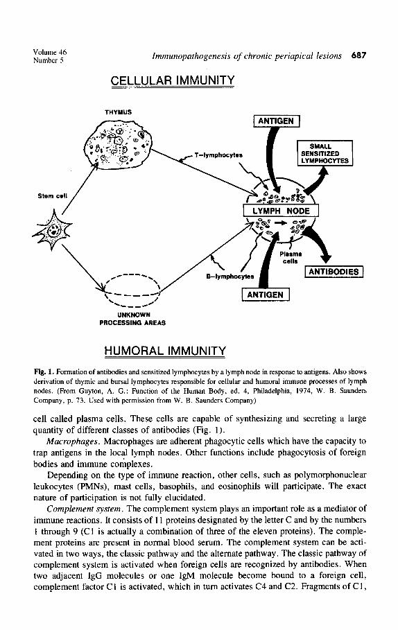
Lymphocytes. All stem cells originating in fetal liver migrate to bone marrow. About half of these lymphocytes pass through the thymus and become T lymphocytes. The other half pass through unknown areas in human beings (in birds it is the bursa of Fabricius) and evolve into B lymphocytes. Fig. 1 illustrates the over-all mechanism of adaptive immu- nity, showing to the right and below the formation of antibodies by the lymph nodes in response to antigens and showing to the right and above the formation of sensitized lymphocytes (T cells).
Plasma cells. When B lymphocytes come in contact with a specific antigen to which they are sensitized, they rapidly divide in the lymph node and transform to a new type of
Volume 46 Number 5 Immunopathogenesis of chronic periapical lesions 687
CELLULAR IMMUNITY
THYMUS
UNKNOWN PROCESSING AREAS
HUMORAL IMMUNITY
Fig. 1. Formation of antibodies and sensitized lymphocytes by a lymph node in response to antigens. Also shows derivation of thymic and bursal lymphocytes responsible for cellular and humoral immune processes of lymph nodes. (From Guyton, A. G.: Function of the Human Body, ed. 4, Philadelphia, 1974, W. B. Saunders Company, p. 73. Used with permission from W. B. Saunders Company)
cell called plasma cells. These cells are capable of synthesizing and secreting a large quantity of different classes of antibodies (Fig. 1).
Macrophages. Macrophages are adherent phagocytic cells which have the capacity to trap antigens in the local lymph nodes. Other functions include phagocytosis of foreign bodies and immune complexes.
Depending on the type of immune reaction, other cells, such as polymorphonuclear leukocytes (PMNs), mast cells, basophils, and eosinophils will participate. The exact nature of participation is not fully elucidated.
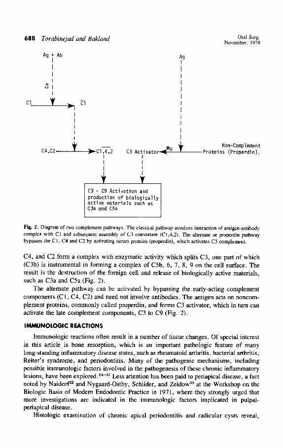
Complement system. The complement system plays an important role as a mediator of immune reactions. It consists of 1 I proteins designated by the letter C and by the numbers I through 9 (Cl is actually a combination of three of the eleven proteins). The comple- ment proteins are present in normal blood serum. The complement system can be acti- vated in two ways, the classic pathway and the alternate pathway. The classic pathway of complement system is activated when foreign cells are recognized by antibodies. When two adjacent IgG molecules or one IgM molecule become bound to a foreign cell, complement factor C I is activated, which in turn activates C4 and C2. Fragments of C 1,
688 Torabinejad and Bakland
Ag i Ab
Oral Surg. November, 1978
4” I
I
Fig. 2. Diagram of two complement pathways. The classical pathway involves interaction of antigen-antibody
complex with C I and subsequent assembly of C3 convertase (Cl ,4,2). The alternate or properdin pathway bypasses the C I, C4 and C2 by activating serum proteins (properdin), which activates C3 complement.
C4, and C2 form a complex with enzymatic activity which splits C3, one part of which (C3b) is instrumental in forming a complex of C5b, 6, 7, 8, 9 on the cell surface. The result is the destruction of the foreign cell and release of biologically active materials, such as C3a and C5a (Fig. 2).
The alternate pathway can be activated by bypassing the early-acting complement components (C 1, C4, C2) and need not involve antibodies. The antigen acts on noncom- plement proteins, commonly called properdin, and forms C3 activator, which in turn can activate the late complement components, C3 to C9 (Fig. 2).
lMMUNOLOGlC REACTIONS
Immunologic reactions often result in a number of tissue changes. Of special interest in this article is bone resorption, which is an important pathologic feature of many long-standing inflammatory disease states, such as rheumatoid arthritis, bacterial arthritis, Reiter’s syndrome, and periodontitis. Many of the pathogenic mechanisms, including possible immunologic factors involved in the pathogenesis of these chronic inflammatory lesions, have been explored. 24-31 Less attention has been paid to periapical disease, a fact noted by Naidorfa2 and Nygaard-Ostby, Schilder, and Zeldow33 at the Workshop on the Biologic Basis of Modem Endodontic Practice in 1971, where they strongly urged that more investigations are indicated in the immunologic factors implicated in pulpal- periapical disease.
Histologic examination of chronic apical periodontitis and radicular cysts reveal,
Volume 46 Number 5 lmmunopathogenesis of chronic periapical lesions 689
among other cells, the presence of macrophages, lymphocytes, plasma cells, PMNs, and mast cells. All of the above cells have been implicated in various types of immune response, and their presence in the periapical lesions would lend support to the contention that egress of antigens from the root canal system is quite likely. There have been a number of investigations into the possibility of the root canal being a source or route for antigens responsible for periapical immune reactions and systemic humoral antibodies. In 1957 Kennedy, Hamilton, and Sylverton 2o showed that the presence of hemolytic strep- tococci within the root canal system was capable of producing an increased antistreptoly- sin 0 titer. A similar result was obtained by Rosengren21 when he introduced the same organism into the root canals of cats. The root canal as a pathway for introducing antigens was used by Barnes and Langeland. 34 They deposited bovine serum albumin into the pulp spaces of monkey teeth and observed the formation of systemic antibodies. Further evi- dence of the feasibility of the root canal system being a source of sensitization was provided by investigations involving bacitracin-neomycin,35 polyantibiotics,36 horse se- rum,22 and paraformaldehyde.23
Since cells that are commonly associated with immunologic reactions have been observed in periapical lesions, it would seem reasonable to expect that immunoglobulins also should be found. Indeed, a number of recent investigations have demonstrated differ- ent classes of antibodies in such lesions. Naidof17 and others,38-44 using different tech- niques, showed that IgG, IgM, IgA, IgE, and C3 complement fragments were present in chronic periapical lesions (except scar tissue).
The evidence from this review of the literature supports the assumption that metabolic and breakdown products of microorganisms and altered host tissue products can penetrate beyond the apical foramen to induce host sensitization. The continuous egress of these antigenic materials into the periapical tissues of such a sensitized host can lead to im- munologic reactions.
Immunologic reactions are commonly divided into two types, humoral and cellular. For convenience, we have classified immunopathologic reactions involved in periapical disease as follows:
Type I-Humoral (A) Antigen-antibody complex or immune complex reactions (Arthus-like reac-
tions) (B) IgE-mediated reactions (anaphylactic reactions)
Type II-Cellular
ANTIGEN-ANTIBODY COMPLEX OR IMMUNE COMPLEX REACTIONS
In 1903 Arthus% injected equine serum subcutaneously in sensitized rabbits and pro- duced a local reaction at the injection site. This phenomenon was later studied by Sher- wood46 who described it as a hemorrhagic, necrotizing inflammation, accompanied by edema and PMN infiltration. These reactions are initiated when immune complexes be- tween antigens and antibodies are formed in moderate antigen excess.47 A local antigen excess may occur when a relatively large amount of antigen is deposited in the tissues of a subject who is in the process of antibody production. 48, 4g Local deposition of antigen- antibody complexes can elicite an Arthuslike reaction.50-52 Ward and co-workers53, 54 have shown that antigen-antibody complexes fix complement and consequently attract PMNs. The immune complexes are normally phagocytosed by PMNs, an activity which
690 Torabinejad and Bakland Oral Surg. November, 1978
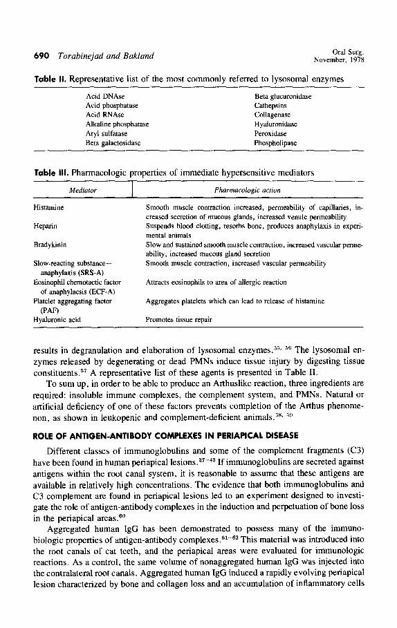
Table II. Representative list of the most commonly referred to lysosomal enzymes
Acid DNAse Acid phosphatase Acid RNAse Alkaline phosphatase Aryl sulfatase Beta galactosidase
Beta glucuronidase Cathepsins Collagenase Hyaluronidase Peroxidase Phospholipase
Table Ill. Pharmacologic properties of immediate hypersensitive mediators
Mediator Pharmacologic action
Histamine
Heparin
Bradykinin
Slow-reacting substance- anaphylaxis (SRS-A)
Eosinophil chemotactic factor of anaphylacsis (ECF-A)
Platelet aggregating factor PAF)
Hyahrronic acid
Smooth muscle contraction increased, permeability of capillaries, in- creased secretion of mucous glands, increased venule permeability Suspends blood clotting, resorbs bone, produces anaphylaxis in experi- mental animals Slow and sustained smooth muscle contraction, increased vascular perme- ability, increased mucous gland secretion Smooth muscle contraction, increased vascular permeability
Attracts eosinophils to area of allergic reaction
Aggregates platelets which can lead to release of histamine
Promotes tissue repair
results in degranulation and elaboration of lysosomal enzymes.55, j6 The lysosomal en- zymes released by degenerating or dead PMNs induce tissue injury by digesting tissue constituents.57 A representative list of these agents is presented in Table II.
To sum up, in order to be able to produce an Arthuslike reaction, three ingredients are required: insoluble immune complexes, the complement system, and PMNs. Natural or artificial deficiency of one of these factors prevents completion of the Arthus phenome- non, as shown in leukopenic and complement-deficient animals.“8’ jg
ROLE OF ANTIGEN-ANTIBODY COMPLEXES IN PERIAPICAL DISEASE
Different classes of immunoglobulins and some of the complement fragments (C3) have been found in human periapical lesions. 37-43 If immunoglobulins are secreted against antigens within the root canal system, it is reasonable to assume that these antigens are available in relatively high concentrations. The evidence that both immunoglobulins and C3 complement are found in periapical lesions led to an experiment designed to investi- gate the role of antigen-antibody complexes in the induction and perpetuation of bone loss in the periapical areas.60
Aggregated human IgG has been demonstrated to possess many of the immuno- biologic properties of antigen-antibody complexes. 61-63 This material was introduced into the root canals of cat teeth, and the periapical areas were evaluated for immunologic reactions. As a control, the same volume of nonaggregated human IgG was injected into the contralateral root canals. Aggregated human IgG induced a rapidly evolving periapical lesion characterized by bone and collagen loss and an accumulation of inflammatory cells
Volume 46 Number 5 Immunopathogenesis of chronic periapical lesions 691
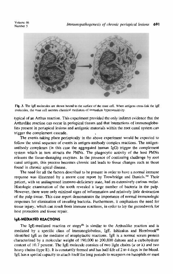
Fig. 3. The IgE molecules are shown bound to the surface of the mast cell. When antigens cross-link the I@ molecules, the mast cell secretes chemical mediators of immediate hypersensitivity.
typical of an Arthus reaction. This experiment provided the only indirect evidence that the Arthuslike reaction can occur in periapical tissues and that interactions of immunoglobu- lins present in periapical lesions and antigenic materials within the root canal system can trigger the complement cascade.
The events taking place periapically in the above experiment would be expected to follow the usual sequence of events in antigen-antibody complex reactions. The antigen- antibody complexes (in this case the aggregated human IgG) trigger the complement system which in turn attracts the PMNs. The phagocytic activity of the host PMNs releases the tissue-damaging enzymes. In the presence of continuing challenge by root canal antigens, this process becomes chronic and leads to tissue changes such as those found in chronic apical disease.
The need for all the factors described to be present in order to have a normal immune response was illustrated by a recent case report by Trowbridge and Daniels.64 Their patient, with an undiagnosed immuno-deficiency state, had an extensively carious molar. Histologic examination of the tooth revealed a large number of bacteria in the pulp. However, there were only minimal signs of inflammation and relatively little destruction of the pulp tissue. This case report demonstrates the importance of normal immunologic responses for elimination of invading bacteria. Furthermore, it emphasizes the need for tissue injury, which can result from immune reactions, in order to lay the groundwork for host protection and tissue repair.
IgE-MEMATED REACTIONS
The IgE-mediated reaction or atopy65 is similar to the Arthuslike reaction and is mediated by a specific class of immunoglobulins, IgE. Ishizakas and Hornbrook@ identified IgE as the mediator of anaphylactic reactions. IgE is a normal serum protein characterized by a molecular weight of 190,000 to 200,000 daltons and a carbohydrate content of 10.7 percent. The IgE molecule consists of two light chains (K or X) and two heavy chains (type E). It is constantly formed and has half-life of 2 to 4 days in the blood. IgE has a special capacity to attach itself for long periods to receptors on basophils or mast
692 Torabinejad and Bakland Oral Surg. November, 1978
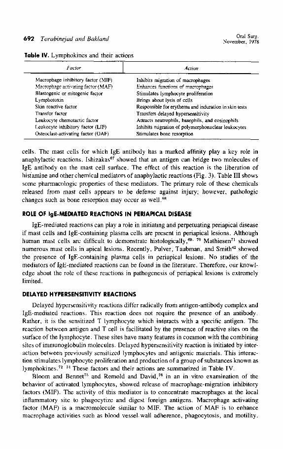
Table IV. Lymphokines and their actions
Factor I Action
Macrophage inhibitory factor (MIF) Macrophage activating factor (MAF) Blastogenic or mitogenic factor Lymphotoxin Skin reactive factor Transfer factor Leukocyte chemotactic factor Leukocyte inhibitory factor (LIF) Osteoclast-activating factor (OAF)
Inhibits migration of macrophages Enhances functions of macrophages Stimulates lymphocyte proliferation Brings about lysis of cells Responsible for erythema and induration in skin tests Transfers delayed hypersensitivity Attracts neutrophils, basophils, and eosinophils Inhibits migration of polymorphonuclear leukocytes Stimulates bone resorption
cells. The mast cells for which IgE antibody has a marked affinity play a key role in anaphylactic reactions. Ishizakas 67 showed that an antigen can bridge two molecules of IgE antibody on the mast cell surface. The effect of this reaction is the liberation of histamine and other chemical mediators of anaphylactic reactions (Fig. 3). Table III shows some pharmacologic properties of these mediators. The primary role of these chemicals released from mast cells appears to be defense against injury; however, pathologic changes such as bone resorption may occur as we11.68
ROLE OF IgE-MEDIATED REACTIONS IN PERIAPICAL DISEASE
IgE-mediated reactions can play a role in initiating and perpetuating periapical disease if mast cells and IgE-containing plasma cells are present in periapical lesions. Although human mast cells are difficult to demonstrate histologically,6s, 7o Mathiesen7i showed numerous mast cells in apical lesions. Recently, Pulver, Taubman, and Smith4’ showed the presence of IgE-containing plasma cells in periapical lesions. No studies of the mediators of &E-mediated reactions can be found in the literature. Therefore, our knowl- edge about the role of these reactions in pathogenesis of periapical lesions is extremely limited.
DELAYED HYPERSENSITIVITY REACTIONS
Delayed hypersensitivity reactions differ radically from antigen-antibody complex and IgE-mediated reactions. This reaction does not require the presence of an antibody. Rather, it is the sensitized T lymphocyte which interacts with a specific antigen. The reaction between antigen and T cell is facilitated by the presence of reactive sites on the surface of the lymphocyte. These sites have many features in common with the combining sites of immunoglobulin molecules. Delayed hypersensitivity reaction is initiated by inter- action between previously sensitized lymphocytes and antigenic materials. This interac- tion stimulates lymphocyte proliferation and production of a group of substances known as lymphokines.72-74 These factors and their actions are summarized in Table IV.
Bloom and Bennet’” and Remold and David,76 in an in vitro examination of the behavior of activated lymphocytes, showed release of macrophage-migration inhibitory factors (MIF). The activity of this mediator is to concentrate macrophages at the local inflammatory site to phagocytize and digest foreign antigens. Macrophage activating factor (MAF) is a macromolecule similar to MIF. The action of MAF is to enhance macrophage activities such as blood vessel. wall adherence, phagocytosis, and motility.
Volume 46 Number 5 tmmunopathogenesis of chronic periapical lesions 693
The production of both MIF and MAF is a protective mechanism; other lymphokines appear to be destructive to the host cells. One of the latter is lymphotoxin,“, ‘* an enzyme released by lymphocytes to kill invading foreign cells but which, however, has the potential to destroy host cells.
Horton and associates7g have detected a new soluble lymphokine (osteoclast activating factor) which is capable of activating osteoclasts to resorb bone. It is of interest also to note that macrophages and neutrophils attracted to the area of injury release the enzyme collagenase which enhances the dissolution of collagen fibers.80-82
These observations point to the fact that delayed hypersensitivity is another effective way to cope with foreign antigens.
ROLE OF DELAYED HYPERSENSITIVITY IN PERlAPlCAL DISEASE
The proportions of various types of lymphoid cells to be found in chronic periapical disease have not been established. Histopathologic examinations of chronic periapical lesions show that there are often more lymphocytes than plasma or plasmalike cells. These former cells are either T lymphocytes capable of mediating delayed hypersensitivity, an expanded subpopulation of immature B lymphocytes, or a combination of both. However, the cellular pattern is similar to the histopathologic response seen in delayed hypersen- sitivity reactions. Dietz,83 in 1952, showed that filtrates prepared from the contents of root canals caused an immunologic reaction when injected subcutaneously. This is only indi- rect evidence that delayed hypersensitivity reactions can occur in periapical lesions. Polliak and associates, 84 Lin, Cooper, and Wortis,85 and Sullivan and associates86 claim that they are able to differentiate B and T lymphocytes. On the basis of Polliak’s study, Farber*’ recently investigated the cellular composition of the periapical lesions with a scanning electron microscope. His findings indicate that most lymphocytes appeared to be T cells. He also found a large number of macrophages in these lesions. Although identification of T and B lymphocytes on the basis of surface architecture is not accurate and not generally accepted, **+ the presence of macrophages in these lesions may be interpreted as another indication that delayed hypersensitivity can play a role in perpetuat- ing periapical lesions.
When a sensitized lymphocyte comes in contact with the antigen to which it is sensitive, it may proliferate. This phenomenon is called lymphocyte transformation and has been cited as an in vitro correlate of delayed hypersensitivity reactions.g2* g3 However, this idea has been disputed by others. s4* gs Lymphocyte proliferation has been used to examine the role of cellular immunity in periodontal disease.g6* g7 Eleazer, Farber, and Seltze?” employed the same phenomenon to determine if pulpal tissues or the soft tissues of periapical lesions from pulpless teeth evoke a delayed hypersensitivity reaction. Their results showed no evidence of lymphocyte transformation. Several factors ought to be considered in interpreting the results of this study:
1. Lymphokines are not only secreted by sensitized T cells. Immune complexes are also capable of activating C3,complement and B cells to produce lymphokines.gs-lO1
2. In vitro T lymphocyte transformation does not always occur with delayed hyper- sensitivity.
3. The concentration of the toxic materials used and the period of incubation in the tissue culture in order to elicit lymphocyte transformation are important factors in such investigations.
694 Torabinejad and Bakland Oral Surg. November. 1978
4. The lack of response of the T lymphocytes may be related to restricted diffusion of the antigen in the culture medium. Therefore, in spite of the results obtained in the above-mentioned study, delayed hyper- sensitivity reaction may in part play a role in the pathogenesis of periapical disease. This protective phenomenon may start as a response to root canal antigens. However, damage to adjacent tissues is apparently inevitable. There is no known mechanism to shut down a delayed hypersensitivity reaction, once it is initiated, and it continues until the antigens are removed. This emphasizes the concept that complete cleaning and debridement of the root canal system is the most important aspect of root canal therapy.102-104 The problem of persistent periapical lesions has not been studied from the standpoint of failure to elimi- nate phagocytosed material by macrophages. However, it may well provide fruitful areas for future studies.
THE ROLE OF ENDOTOXINS IN PERIAPICAL DISEASE
It appears that host reactions to bacterial endotoxins do not fit specifically into the above categories of immunologic reactions. Since they cannot currently be so classified, they will be discussed separately. Endotoxins are derived from the cell walls of gram- negative bacteria and have toxic and pyrogenic effects when injected in vivo. Hook, Snyderman, and Mergenhagen lo5 found that administration of endotoxin in vivo resulted in rapid degranulation of mast cells, which are abundant sources of histamine and heparin. Several investigators have also shown that bacterial endotoxins can activate the alternate pathway of the complement system. 106-113 Therefore, endotoxins can bypass the early- acting complement components (C 1,4,2) and elicit the formation of biologic products of the late-acting complement components (Fig. 3). Cleavage of C5 and release of C5a produce vascular permeability and attract PMNs, mononuclear leukocytes, and macro- phagesln-“’ Release of abundant amounts of lysosomal enzymes by these cells leads to soft-and hard-tissue destruction. ‘l* Furthermore, bacterial endotoxins have been found to stimulate bone resorption, inhibit bone growth in tissue cultures, and attract osteoclasts to bone 119-123
Endotoxins have been found in root canals of pulpless teeth.rZ4 Theoretically, egress of these substances from the root canal system into the periapical area can cause periapical lesions. Studies ate currently in progress at Loma Linda University to determine the role of endotoxins in periapical disease.
SUMMARY
The data presented here indicate that the process of bone resorption in human periapi- cal disease is multifactorial. Viable bacteria, bacterial products, and altered host tissues can initiate and propagate periapical disease. This reaction to persistent antigenic stimuli from the root canal system can take two forms: antibody-mediated and cell-mediated forms of immunity. The antigen-antibody complex and IgE-mediated reactions could very well initiate the preliminary changes in the periapical tissues. The delayed hypersensitivity (cell-mediated immunity) is likely to join in the process and participate in the perpetuation and progression of periapical disease. Although immune responses are important for localization and destruction of antigenic materials emerging from the root canal system, they often lead to a local destruction of the periapical tissues. It appears quite possible that periapical disease may be a by-product of a successful endeavor by the immune system to
Volume 46 Number 5 Zmmunopathogenesis of chronic periapical lesions 695
protect the host from noxious agents within the root canal system, causing the periapical tissues to undergo progressive chronic destruction.
On the basis of the above data, the significance of the role of the immune system in periapical disease becomes quite apparent. It is also very apparent that more investigations are still needed to answer the many remaining questions.
REFERENCES
1. Freeman, N.: Histopathological Investigation of the Dental Granuloma, J. Dent. Res. 11: 175-200, 1931. 2. Burket, L.: Recent Studies Relating to Periapical Infection, Including Data Obtained From Human Nec-
ropsy Studies, J. Am. Dent. Assoc. 25: 260-272, 1938. 3. Fish, E. W.: Bone Infection, J. Am. Dent. Assoc. 26: 691-712, 1939. 4. Kronfeld, R.: Histopathology of the Teeth and Their Surrounding Structures, ed. 2, Philadelphia, 1939, Lea
& Febiger, pp. 209-2 I 1. 5. Hedman, W. J.: An Investigation Into Residual Periapical Infection After Pulp Canal Therapy, ORAL SURG.
4: 1173-1179, 1951. 6. Grossman, L. I., Bacteriologic Status of Periapical Tissue in 150 Cases of Infected Pulpless Teeth, J. Dent.
Res. 38: 101-104, 1959. 7. Lineberg, W. B., Waldron, C. A., and DeLaune, G. F., Jr.: A Clinical, Roentgenographic and His-
tophatologic Evaluation of Periapical Lesions, ORAL SURG. 17: 467-472, 1964. 8. Moller, A. J.: Microbiological Examination of Root Canals and Periapical Tissues of Human Teeth, Odont.
74(Suppl): I-380, 1966. 9. Melville, T. H., and Birch, R. H.: Root Canal and Periapical Floras of Infected Teeth, ORAL SURG. 23:
93-98, 1967. IO. Lyons, D. C., and others.: The Histopathologic Variations of Chronic Dental Granuloma, J. Oral Med. 25:
46-30, 1970. I I. Seltzer, S.: Endodontology; Biologic Considerations in Endodontic Procedures, ed. I, New York, I97 I,
McGraw-Hill Book Co., pp. 197-227. 12. Andreason, J. O., and Rud, J.: A Histobacteriologic Study of Dental and Periapical Structures After
Endodontic Surgery, Int. J. Oral Surg. 1: 272-281, 1972. 13. Winkler, T. F., III., Mitchell, D. F., and Healey, H. J.: A Bacterial Study of Human Periapical Pathosis
Employing a Modified Gram Tissue Stain, ORAL SURG. 34: 109-I 16, 1972. 14. Blechman, H.: Infections of the Pulp and Periapical Tissues. In Nolte, W. A. (editor): Oral Microbiology.
ed. 2, St. Louis, 1973, The C. V. Mosby Company, pp. 237-239. 15. Spangberg, L., Engstrom, B., and Langeland, K.: Biologic Effect of Dental Materials, ORAL SURG. 36:
856-871, 1973. 16. Grossman, L. I.: Endodontic Practice, ed. 8, Philadelphia, 1974. Lea & Febiger, p. 86. 17. Shafer, W. B., Hine, M. K., and Levy, B. M.: A Textbook of Oral Pathology, ed. 3, Philadelphia, 1974,
W. B. Saunders Co., pp. 440-462. 18. Block, R. M., Bushell, A., Rodrigues, H., and Langeland, K.: A Histopathological, Histobacteriologic and
Radiographic Study of Periapical Endodontic Surgical Specimens, ORAL SURG. 42: 656-678, 1976. 19. Langeland, K., and Block, R. M.: A Histopathologic and Histobacteriologic Study of 35 Periapical En-
dodontic Surgical Specimens, J. Endod. 3: 8-23, 1977. 20. Kennedy, D. R., Hamilton, T. R., and Syverton, J. T.: Effects of Monkeys of Introduction of Hemolytic
Streptococci Into Root Canals, J. Dent. Res. 36: 496-506, 1957. 2 I. Rosengren, L.: The Antibody Response to Experimental Streptococci Infection (S84) of the Dental Pulp of
the Cat, Odontol. Tidskr. 70: 261-360, 1962. 22. Okada, H., Aono, M., Yoshida, M., Munemoto, K., Nishida, 0.. and Yokomizo. I.: Experimental Study
on Focal Infection in Rabbits by Prolonged Sensitization Through Dental Pulp Canals, Arch. Oral Biol. 12: 1017-1034, 1967.
23. Block, R. M., Lewis, R. D., Sheats, J. B., and Burke, S. H.: Antibody Formation to Dog Pulp Tissue Altered by NZ-Type Paste Within the Root Canal, J. Endod. 3: 309-315. 1977.
24. Hammerman, D. L., Sandson, J., and Schubert. M.: Biochemical Events in Joint Disease. J. Chron. Dis. 16: 835-852, 1963.
25. Berens, D. L., and Lin. R.: Roentgen Diagnosis of Rheumatoid Arthritis. Springfield, Ill. 1969. Charles C Thomas, Publisher.
26. Kelly, P. J.. Martin, W. J., and Coventry, M. B.: Bacterial (Suppurative) Arthritis in the Adult. J. Bone Joint Surg. 52A: 1595-1602, 1970.
27. McEwen, C.. Ditata. D.. Lingg. C.. Parinig, F.. Geirly. A.. and Rankin. T.: Ankylosing Spondylitis and
696 Torabinejad and Bakland Oral Surg. November, 1978
28. 29.
30.
31.
32.
33.
34.
35.
36.
37.
38.
39.
40. 41. 42.
43.
44.
45: 46. 41.
48.
49.
50.
51.
52.
53.
54.
55.
56.
57. 58.
Spondylitis Accompanying Ulcerative Colitis, Regional Enteritis, Psoriasis and Reiter’s Disease: A Com- parative Study, Arthritis Rheum. 14: 291-318, 1971. Karten, I. : Septic Arthritis Complicating Rheumatoid Arthritis, Ann. Intern, Med. 70: l l47- 1158, 1969. Page, R., and Schroeder, H. E.: Pathogenesis of Inflammatory Periodontal Disease. A Summary of Current Work, Lab. Invest. 34: 235-249, 1976. Goldhaber, P., Rabadjija, L., Beyer, W. R., and Kornhauser, A.: Bone Resorption in Tissue Culture and Its Relevance to Periodontal Disease, J. Am. Dent. Assoc. 87: 1027-1033, 1973. Rizzo, A. A.: Histologic and Immunologic Evaluation of Antigen Penetration Into Oral Tissues After Topical Application, J. Periodontol. 41: 210-213, 1970. Naidorf, I. J.: Inflammation and Infection of the Pulp and Periapical Tissues. In Siskin, M. (editor): The Biology of the Human Pulp, St. Louis, 1973, The C. V. Mosby Co., pp. 391402. Nygaard-Ostby, B., SchiIder, H., and Zeldow, B. J.: Inflammation and Infection of the Pulp and Periapical Tissues. In Siskin, M. (editor): The Biology of the Human Pulp, St. Louis, 1973, The C. V. Mosby Co., pp. 403406. Barnes, G. W., and Langeland, K.: Antibody Formation in Primates Following Introduction of Antigens Into the Root Canal, J. Dent. Res. 45: I I1 l-l 114, 1966. Pirila, V., and Rantanen, A. V.: Root Canal Treatment with Bacitracin-Neomycin as a Cause of Flare up of Allergic Eczema, ORAL SURG. 13: 589-593, 1960. Fox, J., and Moodnick, R. M.: Systemic Reaction of Polyantibiotic Root Canal Dressing, N. Y. State Dent, J. 30: 282-283, 1964. Naidorf, I. J.: Immunoglobulins in Periapical Granulomas: A Preliminary Report, J. Endod. 1: 15-18, 1975. Morse, D. R., Lasater, D. R., and White, D.: Presence of Immunoglobulin Producing Cells in Periapical Lesions, J. Endod. 1: 338-343, 1975. Mahnsbom, M.: Immunoglobulin Classes of IgG, IgM, IgA and complement Components C3 in Dental Periapical Lesions of Patients with Rheumatoid Disease, Stand. J. Rheumatol. 4: 57-64, 1975. Toller, P. A.: Immunological Factors in Cysts of the Jaws, Proc. Roy. Sot. Med. 64: 555-559, 1971. Toller, P. A.: Newer Concepts of Odontogenic Cysts, Int. J. Oral Surg. 1: 3-16, 1972. Pulver, W. H., Taubman, M. A., and Smith, D. J.: Immune Components in Normal and Inflamed Human Dental Pulp. Read before the American Association of Endodontists, Annual session, Hollywood, Fla., 1976. Kuntz, D. D., Genco, R. J., Guttuso, J., and Natiella, J. R.: Localization of Immunoglobulins and the Third Component of Complement in Dental Periapical Lesions, J. Endod. 3: 68-73, 1977. Morton, T. H., Clagett, J. A., and Yavorsky, J. D.: Role of Immune Complexes in Human Periapical Periodontitis, J. Endod. 3: 261-268, 1977. Terrier, Cornelia: Arthus Reaction in the Oral Cavity of Laboratory Animals, Periodontics 3: 18-22, 1965. Sherwood, N. P.: Immunology, ed. 3, St. Louis, 1951, The C. V. Mosby Company, pp. 551-586. Weigle, W. 0.: Fate and Biological Action of Antigen-Antibody Complexes. In Advances in Immunology, ed. I, New York and London, 1961, American Press, pp. 283-315. Gell, P. G. H., and Coombs, R. R. A.: Clinical Aspects of Immunology, ed. 2, Philadelphia, 1975, F. A. Davis Company, p. 172. Ward, P. A., Remold, H. G., and David, J. R.: Leukotactic Factors Produced by Sensitized Lymphocytes, Science 163: 1079- 1081, 1969. Deshazo, C. V., Henson, P. M., and Cochrane, C. G.: Acute Immunologic Arthritis in Rabbits, J. Clin. Investigation 51: 50-57, 1972. Rawson, A. J., and Torralba, T. P.: Induction of Proliferative Synovitis in Rabbits by Intra-Articular Injection of Immune Complexes, Arthritis Rheum. 10: 44-52, 1967. Cochrane, C. G., and Koffler, D.: Immune Complex Disease in Experimental Animals and Man, In Kunkeland and Dixon (editors): Advance in Immunology, New York, 1973, Academic Press Inc., 16: 86-264. Ward, P. A., Cochrane, C. G., and Muller-Eberhard, H. J.: The Role of Serum Complement in Chemotaxis of Leukocytes in Vitro, J. Exp. Med. 122: 327-346, 1965. Ward, P. A., Cochrane, C. G., and Muller-Eberhard, H. J.: Further Studies on the Chemotactic Factors of Complement and its Formation in Vivo, Immunol. 11: l41- 153, 1966. Henson, P. M.: The Immunologic Release of Constituents From the Neutrophil Leukocytes, J. Immunol. 107: 1547-1557, 1971. Golub, E. S., and Spitznagel, J. K.: The Role of Lysosomes in Hypersensitivity by Reactions: Tissue Damage by Polymorphonuclear Neutrophil Lysosomes, J. Immunol. 95: 1060-1066, 1965. Henson, P. M.: Pathologic Mechanisms in Neutrophil-Mediated Injury, Am. J. Path. 68: 593-610, 1972. Stetson, C. A., Jr.: Studies on the Mechanism of the Shwartzman Phenomenon: Certain Factors Involved in the Production of the Local Hemorrhagic Necrosis, J. Exp. Med. 93: 489-504, I95 I.
Volume 46 Number 5 Immunopathogenesis of chronic periapical lesions 697
59.
60.
61.
62.
63.
64.
65.
66.
67.
68.
69.
70. 71.
72.
73. 74. 75.
76.
77.
78. 79.
80.
81.
82.
83.
84.
8.5.
86.
87. 88.
89.
Ward, P. A., and Cochrane, C. G.: Bound Complement and Immunologic Injury of Blood Vessels, J. Exp. Med. 121: 215-234, 1965. Torabinejad, M., Clagett, J. A., and Engel, L. D.: Aggregated Immunoglobulin-Induced Bone Loss: A Model System, Calcif. Tissue Res. (In press.) Christian, C. L.: Studies of Aggregated y-globulin. I. Sedimentation, Electrophoretic and Anticomplemen- tary Properties, J. Immunol. 84: 112-I 16, 1960. Ishizaka, K.: Gamma Globulin and Molecular Mechanisms of Hypersensitivity Reactions, Prog. Allergy 7: 32-106, 1963. Theofilopoulos, A. N., Dixon, F. J., and Bokisch, V. N.: Binding of Soluble Immune Complexes to Human Lymphoblastoid Cells. I. Characterization of Receptors for IgG F, and Complement and Descrip- tion of the Binding Mechanism, J. Exp. Med. 14th 877-894, 1974. Trowbridge, H., and Daniels, T.: Abnormal Immune Response to Infection of the Dental Pulp, ORAL SURG. 43: 902-909, 1977. Coca, A. F., and Cooke, R. A.: On the Classification of Phenomena of Hypersensitiveness, J. Immunol. 8: 163- 182, 1923. Ishizaka, K., Ishizaka, T., and Hornbrook, M. M.: Physiochemical Properties of Reaginic Antibody, IV. Presence of a Unique Immunoglobulin as a Carrier of Reaginic Activity, J. Immunol. 9275-85, 1966. Ishizaka, K., and Ishizaka, T.: Mechanisms of Reaginic Hypersensitivity: A Review, Clin. Allergy 1: 9-24, 1971. Goldhaber, P.: Heparin Enhancement of Factors Stimulating Bone Resorption in Tissue Culture, Science 147: 407-409, 1965. Eda, S., and Langeland, K.: The Alteration of Mast Cell Staining Due to Various Fixatives and De- mineralizing Agents, Stand. J. Dent. Res. 78: 217-231, 1970. Zachrisson, B. U.: Mast Cells in Human Dental Pulp, Arch. Oral Biol. 16: 555-5.56, 1971. Mathiesen, Age: Preservation and Demonstration of Mast Cells in Human Apical Granulomas and Radicular Cysts, Stand. J. Dent. Res. 81: 218-299, 1973. Pick, E., and Turk, J. L.: The Biological Activities of Soluble Lymphocyte Products, Clin. Exp. Immunol. lo: l-23, 1972. David, J. R.: Lymphocyte Mediators and Cellular Hypersensitivity, New Eng. J. Med. 288: 143-149, 1973. David, J. R.: Mediators Produced by Sensitized Lymphocytes, Fed. Proc. 30: 1730- 1735, 1971, Bloom, B. R., and Bennet, B.: Mechanism of a Reaction in Vitro Associated With Delayed-Type- Hypersensitivity, Science 153: SO-82,1966. Remold, H. G., and David, J. R. : Cellular Hypersensitivity: Characterization of Migration Inhibitory Factor (MIF) by Enzymatic Treatment, Fed. Proc. 29: 305-Ab339, 1970. Ruddle, N. H., and Waksman, B. H.: Cytotoxicity Mediated by Soluble Antigen and Lymphocytes in Delayed Hypersensitivity, J. Exp. Med. 128: 1237- 1254, 1968. Granger, G., and Kolb, W. P.: Lymphocyte in Vitro Cytotoxicity, J. Immunol. 101: I1 I-120, 1968. Horton, J. E., Raisz, L. G., Simmon, S. H. A., Oppenheim, J. J., and Mergenhagen, S. E.: Bone Resorbing Activity in Supernatant Fluid From Cultured Peripheral Blood Leukocytes, Science 177: 793- 795, 1972. Robertson, P. G., Shyu, K. W., Vail, M. S., Taylor, R. E., and Fullmer, H. M.: Collagenase Demonstra- tion in Rabbit Macrophages, J. Dent. Res. SZ(Specia1 Issue): 189 (Abst. 522), 1973. Lazarus, G. S., Brown, R. S., Daniels, J. R., and Fullmer, H. M.: Human Granulocyte Collagenase, Science 159: 1483-1485, 1968. Wahl, L. M., Wahl, S. M., and Mergenhagen, S. E.: Collagenase Production by Lymphokine-Activated Macrophages, Science 187: 261-263, 1975. Dietz, V. H.: Intracutaneous Tests Using Filtrates Prepared From Pathology Pulps of Human Teeth, With Special Reference to Rheumatoid Arthritis, ORAL SURG. 5: 59-73; 199-209; 315-324; 418-433; 536-548; 646-652; 752-761; 877-883, 1952. Pollick, A., and others: Identification of Human B and T Lymphocytes by Scanning Electron Microscopy, J. Exp. Med. 138: 607-624, 1973. Lin, P. S., Cooper, A. G., and Wortis, H. H. : Scanning Electron Microscopy of Human T-cell and B-cell Rosettes, N. Engl. J. Med. 289: 548-551, 1973. Sullivan, A. K., Adams, L. S., Silke, I., and Jerry, L. M.: “Hairy” B Cells and “Smooth” T Cells, N. Engl. J. Med. 290: 689-690, 1974. Farber, P. A.: Scanning Electron Microscopy of Cells from Periapical Lesion, J. Endod. 1: 291-294, 1975. Linthicum, D. S., Sell, S., and Wagner, R. M. : Scanning Immunoelectron Microscopy of Mouse B and T Lymphocytes, Nature 252: 173- 175 (Nov. S), 1974. Lin, P. S., and Hoelzl Wallach, D. F.: Surface Modification of T-lymphocytes Observed During Rosetting, Science 184: I 300- 1301, 1974.
698 Torabinejad and Bakland Oral Surg. November, 1978
90.
91.
92.
93.
94.
95.
96.
97.
98.
99.
100.
101.
102. 103.
104.
105.
106.
107.
108.
109.
110.
III.
112.
113.
114.
115.
116.
Alexander, E. L.: Human Lymphocytes: Similarity of B and T Cell Surface Morphology, Science 188: 732-734, 1975. Baur, P. S., Thurman, G. B., and Goldstein, A. L.: Reappraisal of Lymphocyte Classification by Means of Surface Morphology, J. Immunol 115: 1375-1380, 1975. Oppenheim, J. J.: Relationship of in Vitro Lymphocyte Transformation to Delayed Hypersensitivity in Guinea Pig and Man, Fed. Proc. 27: 21-28, 1968. Oppenheim, J. J.: Immunological Relevance of Antigen-Antibody Complex Induced Lymphocyte Trans- formation, Ann. Allergy 27: 305-315, 1969. Kiger, R. D., Wright, W. H., and Creamer, H. R.: The Significance of Lymphocyte Transformation Responses to Various Microbial Stimulants, J. Periodontal. 45: 780-785, 1974. Curtis, J. E., and Hersh, E. M.: Cellular Immunity in Man: Correlation of Leukocyte Migration Inhibition Factor Formation and Delayed Hypersensitivity, Cell Immunol. 8: 55-61, 1973. Ivanyi, L., and Lehner. T.: Lymphocyte Transformation by Sonicates of Dental Plaque in Human Peri- odontal Disease, Arch. Oral Biol. 16: 1117-l 121, 1971. Horton, J. E., Leikin, S., and Oppenheim, J. J.: Human Lymphoproliferative Reaction to Saliva and Dental Plaque-Deposits, J. Periodontol. 43: 522-527, 1972. Eleazer, P. D., Farber, P. A., and Seltzer. S.: Lack of Lymphocyte Stimulation by Root Canal Products, J. Endod. 1: 388-394, 1975. Mackler, B. F., Altman, L. C., Wahl, S., Rosenstreich, D. I.. Oppenheim, J. J., and Mergenhagen, S. E.: Lymphokine Synthesis by T and B Lymphocytes From Patients With Periodontal Disease, Infect. Immu- nol. 10: 844-850, 1974. Mackler, B. F., Altman, L. C., Rosensteich, D. L., and Oppenheim, J. J.: Induction of Lymphokine Production by EAC and of Blastogenesis by Soluble Mitogens During Human B Cell Activation, Nature 249: 834-837, 1974. Altman, L. C., Chassy, B., and Mackler, B. F.: Physiolochemical Characterization of Chemotactic Lymphokines Produced by Human T and B Lymphocytes, J. Immunol. 115: 18-21, 1975. Grossman, L. I.: Endodontic Practice, ed. 8, Philadelphia, 1974, Lea & Febiger, p. 188. Ingle, J. I., and Beveridge, E. E.: Endodontics, ed. 2, Philadelphia, 1976, Lea & Febiger, p. 101. Schilder, H.: Canal De’bridement and Disinfection, In Cohen, S., and Bums, R. C.: Pathways of the Pulp, St. Louis, 1976, The C. V. Mosby Company, p. I I I. Hook, W. A., Snyderman, R., and Mergenhagen, S. E.: Histamine Releasing Factor Generated by the Interaction of Endotoxin With Hamster Serum, Infect. Immun. 2: 462-467, 1970. Pillemer, L., Blum, L., Lepow, I. H., Todd, E. W., Ross, 0. A., and Wardlaw, A. C.: The Properdin System and Immunity. I. Demonstration and Isolation of a New Serum Protein, Properdin, and its Role in Immune Phenomena, Science 120: 279-285, 1954. Pillemer, L., Schoenberg, M. D., Blum, L., and Worz, L.: The Properdin System and Immunity. II. Interaction of the Properdin System With Polysaccharides, Science 122: 545-549, 1955. Pensky, J., Hinz, C. F., Jr., Todd, E. W., Wedgwood, R. J., Boyer, J. T., and Lepow, I. J.: Properties of Highly Purified Human Properdin, J. Immunol. 100: 142-158, 1968. Mergenhagen, S. E., Snyderman, R., Gewurz, H., and Shin, H. S.: Significance of Complement to the Mechanism of Action of Endotoxin, Curr. Top. Microbial. Immunol. 50: 37-78, 1969. Alper, C. A., Abramson, N., Johnston, R. B. Jr., Jandl, J. H., and Rosen, F. S.: Increased Susceptibility to Infection Associated With Abnormalities of Complement-Mediated Functions and of the Third Compo- nent of Complement (C3), N. Engl. J. Med. 282: 349-353, 1970. Marcus, R. L., Shin, H. S., and Mayer, M. M.: An Alternate Complement Pathway: Evidence for C3 Convertase Activity Other Than C4, 2 on Serum Treated Endotoxic Lipopolysaccharide, Fed. Proc. 29: 304, 1970. Goodkofsky, I., and Lepow, I. H.: Functional relationship of Factor B in the Properdin System to C3 Proactivator of Human Serum, J. Immunol. 107: 1200- 1204, 197 I. Gotze, O., and Muller-Eberhard, H. J.: The C3-Activator System: An Alternate Pathway of Complement Activation, J. Exp. Med. 134: 905- 1085, 1971. Lichtenstein, L. M., Gewurz, H., Adkinson, N. F., Shin, H. S., and Mergenhagen, S. E.: Interaction of the Complement System Witb Endotoxic Lipopolysaccharide: The Generation of an Anaphylotoxin, Immunology 16: 327-336, 1969. Snyderman, R. H., Gewerz, H., and Mergenhagen, S. E.: Interactions of the Complement System With Endotoxic Lipopolysaccharide: Generation of a Factor Chemotactic for Polymorphonuclear Leukocytes, J. Exp. Med. 128: 259-275, 1968. Shin, H. S., Snyderman, R., Friedman, E., Mellors, A., and Mayer, M. M.: Chemotactic and Anaphylatoxic Fragment Cleaved From the Fifth Component of Complement, Science 162: 361-363, 1968.
Volume 46 Number 5 Immunopathogenesis of chronic periapical lesions 699
117. Snyderman, R., Shin, H. S., and Hausman, M. S.: A Chemotactic Factor for Mononuclear Leukocytes, F’roc. Sot. Exp. Biol. 138: 387-391, 1971.
118. Cochrane, C. G.: Immunologic Tissue Injury Mediated by Neutrophilic Leukocytes, Adv. Immunol. 9: 97-162, 1969.
I 19. Fell, H. B., Coombs, R. R., and Dingle, J. T.: The Breakdown of Embryonic (Chick) Cartilage and Bone Cultivated in the Presence of Complement Sufficient Antiserum, Int. Arch. Allerg. 30: 146-176, 1966.
120. Hausman, E., Weinfeld, N., and Miller, N.: Effects of Lipopolysaccharides on Bone Resorption in Tissue Culture, Calcif. Tissue Res. 9: 272-282, 1972.
121. Hausman, E., Raisz, L. G., and Miller, W. A.: Endotoxin Stimulation of Bone Resorption in Tissue Culture, Science 168: 862-864, 1970.
122. Norton, L. A., Proffitt, W. R., and Moore, R. R.: In Vitro Bone Growth Inhibition in the Presence of Histamine and Endotoxins, J. Periodontol. 41: l53- 157, 1970.
123. Rizzo, A. A., and Mergenhagen, S. S.: Histopathologic Effects of Endotoxin Injected Into Rabbit Oral Mucosa, Arch. Oral Biol. 9: 659-670, 1964.
124. Schein, B., and Schilder, H.: Endotoxin Content in Endodontically Involved Teeth, J. Endod. 1: 19-21, 1975.
Reprint requests to: Dr. Leif K. Bakland Associate Professor and Chairman of Dept. of Endodontics School of Dentistry Loma Linda University Loma Linda, Calif. 92354