endoscopic mucosal resection and endoscopic submucosal ... · endoscopic mucosal resection and...
TRANSCRIPT
Gut and Liver, Vol. 1, No. 1, June 2007, pp. 12-21
Endoscopic Mucosal Resection and Endoscopic Submucosal Dissection as Treatments for Early Gastrointestinal Cancers in Western Countries
Sergio Coda, Sun-Young Lee*, and Takuji Gotoda†
Operative Unit of Diagnostic and Therapeutic Endoscopy, Department of General and Specialized Surgery and Organ Transplantation "Paride Stefanini", University of Rome "La Sapienza", Rome, Italy, *Department of Internal Medicine, Konkuk University School of Medicine, Seoul, Korea, and †Endoscopy Division, National Cancer Center Hospital, Tokyo, Japan
Correspondence to: Takuji GotodaEndoscopy Division, National Cancer Center Hospital, 5-1-1, Tsukiji, Chuo-ku, Tokyo, JapanTel: +81-3-3542-2511, Fax: +81-3-3542-3815, E-mail: [email protected]
Received on May 14, 2007. Accepted on June 7, 2007.
Early gastrointestinal cancers are defined as lesions limited to the mucosa or submucosa without invading the muscularis propria, regardless of the presence of lymph node metastases. Although the natural history of these diseases is basically alike worldwide, its management is quite different between the East and West; aggressive surgery is frequently adopted by Western surgeons, while less invasive techniques are adopted by Asian colleagues. These techniques in-clude endoscopic mucosal resection and endoscopic submucosal dissection which are now accepted as treatments for early gastrointestinal cancers in se-lected cases. Recent advances in endoscopic de-tection and treatment techniques, especially in Japan and Korea, have prompted Western endoscopists to learn these techniques. This review addresses recent advances regarding endoscopic resections of early gastrointestinal cancers, which promoted its use in Western countries. In addition, prospective studies on endoscopic resection in Western countries are also described. (Gut and Liver 2007;1:12-21)
Key Words: Endoscopic resection; Gastrointestinal can-cer; West
INTRODUCTION
Therapeutic endoscopy plays a major role in the man-agement of gastrointestinal (GI) neoplasia. Its indications
can be generalized into four broad categories; (i) to re-move or obliterate neoplastic lesion, (ii) to palliate malig-nant obstruction, (iii) to treat bleeding, and (iv) the others.1,2 Only endoscopic resection allows complete his-tological staging of the cancer, which is critical as it al-lows stratification and refinement for further treatment. Although other endoscopic techniques such as ablation therapy may also cure early GI cancer, they can not pro-vide a definitive pathological specimen. Early stage lesions reveal low frequency of lymph node metastasis which allows for less invasive treatments and thereby improving the quality of life when compared to surgery.3 Endoscopic mucosal resection (EMR) and sub-mucosal dissection (ESD) are now accepted worldwide as treatment modalities for early cancers of the GI tract.1-3 Recent advances in endoscopic detection and treatment techniques in Japan and Korea have prompted Western endoscopists to use EMR and ESD as elective treatment modalities in selected cases. However, they are still chal-lenging techniques and require considerable experience. Following possible explanations are proposed why EMR and ESD are less often performed in West; (i) the screen-ing system for these diseases, (ii) the relative infrequency of gastric and esophageal cancers and their premalignant precursors, (iii) the differences in the judging criteria be-tween Japanese and Western pathologists, and (iv) the risk perception of the procedures.4 The purpose of this review was to examine the recent advances in the endo-scopic resection of early GI cancers in Western countries.
Review
Coda S, et al: EMR and ESD in the West 13
Table 1. The Endoscopic Classification of Early Gastroin-testinal Cancers
Shape Classification
Protruding Pedunculated 0-Ip Sessile 0-IsNeither protruding nor excavated Slightly elevated 0-IIa Completely flat 0-IIb Slightly depressed 0-IIc Elevated and depressed 0-IIc+IIaExcavated Ulcerated 0-III Excavated and depressed 0-IIc+III or 0-III+IIc
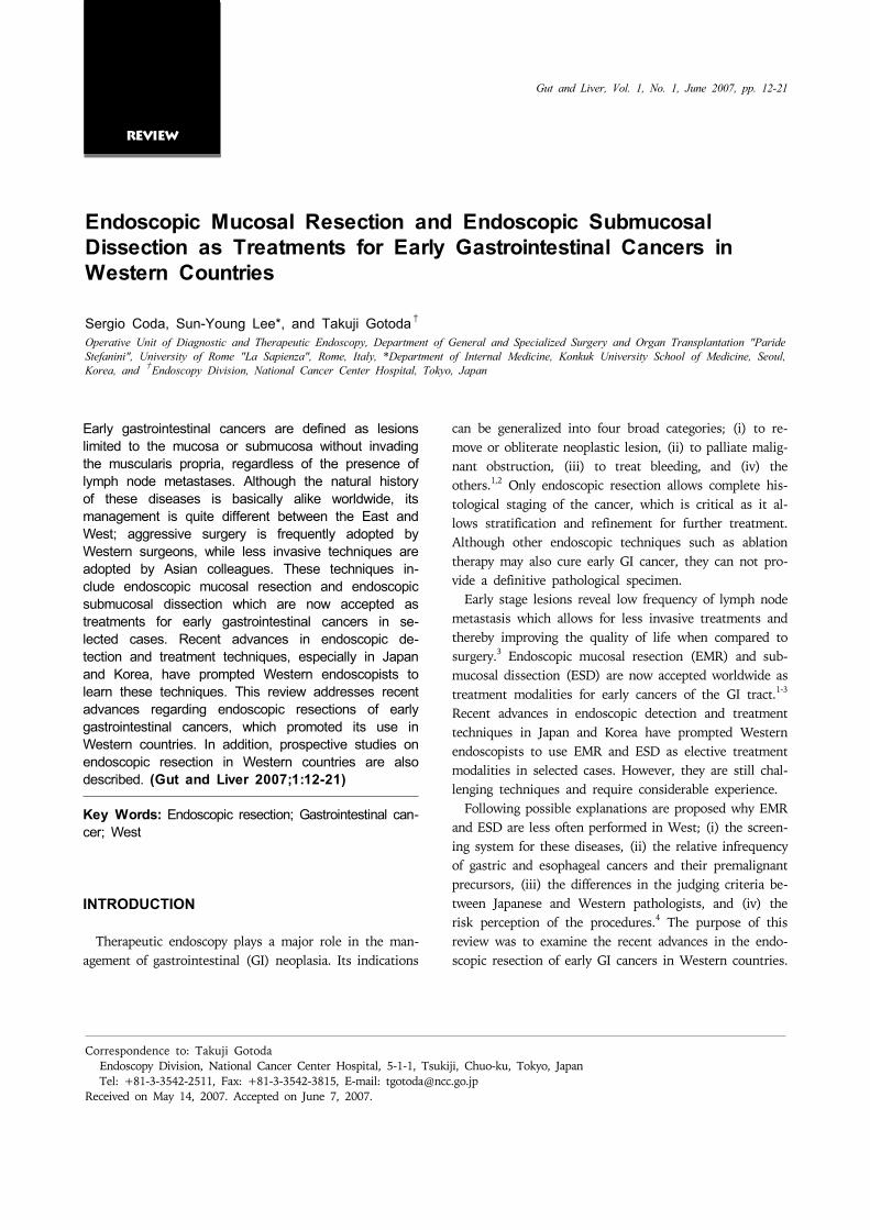
Fig. 1. Histological diagnosis of EGC. (A) A small depressed lesion is noticed on the posterior wall of upper gastric body. (B) Lesion without invasive finding is noticed on the biopsy, which is classified as dysplasia (Vienna classification 4.1.) in the West. Incontrast, this lesion is classified as well differentiated adenocarcinoma (Vienna classification 4.2.) in Japan. (C) After the resection, submucosally invasive adenocarcinoma with lymphatic involvement (Vienna classification 5.2.) is diagnosed.
DETECTION OF EARLY GASTROINTESTINAL CANCERS
Early GI cancers (except the esophagus) are defined as being limited to the mucosa or submucosa but not invad-ing the muscularis propria, regardless of the presence of lymph node metastases. A macroscopic classification of these lesions was first established by Japanese endo-scopists (Table 1). It was ratified at the Paris workshop in 2002 and has now been accepted worldwide.5 Usually, early cancers are detected as 0-I and 0-III types in most of the world, but widespread use of chromoendoscopy, anti-foaming agents, and newer imaging methods have en-abled identification of superficial lesions such as faint mucosal irregularity or discoloration, which had pre-viously overlooked. Over 50% of cancers diagnosed in Japan are early cancers while its frequency is only 10% in the West.6
1. Endoscopic detection
Detection and diagnosis of early GI cancer can be diffi-cult because of the less well defined subtle findings. These may include slight differences in color (i.e., more red or pale), loss of vascularity, nodularity, thickening, or depression.6 Thus, apart from searching for protrusion, erosion, ulceration, and depression, examination for early GI cancer requires continuous search for mucosa with fea-tures distinct from those of its surrounding.5 Therefore, the subtleties of endoscopic findings of early GI cancer require patient preparation to be most optimal. Effective methods to increase visualization of the mucosa of the upper gastrointestinal tract (UGI) include the usage of pronase, a proteolytic enzyme solution, to remove mucus. Chromoendoscopy is an important adjunct technique to enhance visualization of superficial early cancers and to define their borders. Indigo carmine solution (0.2%), a contrast dye, is commonly used in the stomach to high-light the contours and topography of the lesion by enter-ing mucosal depressions and crevices thus enabling the biopsy of the minute lesions. Recently, narrow band im-age (NBI) and autofluorescence images (AFI) are in-troduced as a virtual chromoendoscopy.
2. Histological diagnosis
Apart from the diagnostic difficulty of biopsy as a rea-son for the low incidence of early GI cancer in the West, different histological criteria between the West and Japan would be another reason.7 For example, most mucosal cancer of the intestinal type in Japan is not diagnosed as cancer in the West (Fig. 1). There are large discrepancies in the diagnostic criterion between the Western and Japanese pathologist. These differences have caused con-siderable problems in the interpretation of Japanese can-
14 Gut and Liver, Vol. 1, No. 1, June 2007
Table 2. Vienna Classification of Gastrointestinal Epithelial Neoplasia
Category Treatment
1 - Negative for neoplasia/dysplasia No treatment 2 - Indefinite for neoplasia/dysplasia Follow up3 - Noninvasive low grade dysplasia Follow up (low grade adenoma/dysplasia)4 - Noninvasive high grade dysplasia Endoscopic 4.1 - High grade adenoma resection, 4.2 - Noninvasive carcinoma surgery (carcinoma in situ) 4.3 - Suspicion of invasive carcinoma5 - Invasive neoplasia Surgery 5.1 - Intramucosal carcinoma (recently ESD) 5.2 - Submucosal carcinoma or beyond
ESD, endoscopic submucosal dissection.
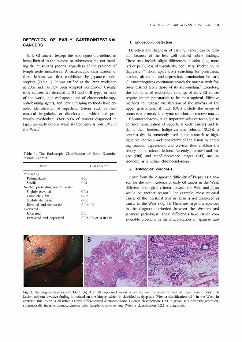
Fig. 2. Standard EMR methods. (A) Snare polypectomy, (B) strip biopsy, (C) EMR with cap technique, and (D) EMR with ligation technique.
cer research by Western clinicians and researchers. Carci-noma is defined as invasion with submucosal layer, mus-cularis mucosa, or at least lamia propria in the West. However, in Japan, cellular atypia and structural atypia is also considered as GI neoplasia regardless of invasive findings. The differences between the Western and Japanese his-tological classifications of GI epithelial neoplastic lesions have been largely resolved by adopting the Vienna classi-fication (Table 2).7 Non-invasive neoplasia include low- and high-grade dysplasia (LGD and HGD) in which the basement membrane is not infiltrated. Invasive lesions in-clude intramucosal neoplasia infiltrating the submucosa. To quantify the risk of lymph node metastases, mucosal (m) and submucosal (sm) layers have each been divided into three sections: m1 (epithelium), m2 (lamina prop-ria), and m3 (muscularis mucosae) and sm1, sm2, and sm3. In addition, sm1 is subclassified into a, b, and c ac-cording to the degree of horizontal extension. It is the depth that mainly determines the necessity of inter-vention. The depth of invasion is measured microscopically and the risk of lymph node metastasis is known to be related with a defined micrometric cutoff. In squamous cell carci-noma (SCC) of the esophagus, when infiltration is less than 200μm, the risk of nodal metastases is low.8 For early adenocarcinoma in Barrett's esophagus (BE) and ear-ly gastric cancer (EGC), a submucosal infiltration micro-metric cutoff of 500μm has been proposed, as the risk of nodal metastases appears low.9 In contrast, a 20-25% risk of node involvement with submucosal infiltration in Barrett's cancer has been reported in the West.10 In the colorectum, the risk of lymph node metastasis is negli-gible when the tumor invasion is less than 1,000μm.11
Other factors, such as the degree of differentiation and the presence of vascular or lymphatic invasion are also re-lated to the risk of lymph node metastasis.
ENDOSCOPIC TREATMENT OF EARLY GAST-ROINTESTINAL CANCERS IN THE WEST
The treatment of early GI cancers in the West is still mainly an operation. However, many Western endo-scopists are now learning from the Japanese experiences and increasingly adopting endoscopic resection (Fig. 2). In contrast to Japan and Korea, there has so far been only limited experience in Europe as well as in US. Here, we report the latest and largest prospective series so far in Western countries with early GI cancers, treated by EMR or ESD.
1. Esophagus
Multiple synchronous lesions as well as metachronous lesions have been reported up to 31% of patients in esophageal SCC.12 Five-year survival rate is up to 95% af-ter EMR in patients with superficial SCC without lymph node metastasis in m1 and m2 SCC of the esophagus.13 Recently, an expanded indications for EMR in patients with superficial esophageal carcinoma m3 or sm1 has been proposed.14 A study from Germany, consisted of 12 HGD and 53 mucosal SCC of the esophagus, reported on the treatment using a ligation EMR technique.15 This was the first Western study which showed similar results to those of the Japan in early esophageal SCC. They reported that
Coda S, et al: EMR and ESD in the West 15
Table 3. Indications for Endoscopic Mucosal Resection / Endoscopic Submucosal Dissection in Early Gastrointestinal Cancers
Esophagus Stomach Colorectum
• Well or moderately differentiated • Well and/or moderately differentiated AC and/or • Well or moderately SCC-AC papillary carcinoma differentiated AC• IIa, IIb, IIc <20 mm • Cancer confined to mucosa or IMC IIa <20 mm • Cancer confined to• m1 or m2 cancers • Cancer confined to mucosa or IMC IIb, IIc <10 mm mucosa or IMC IIa <20 mm• Without evidence of venous or • Without evidence of venous or lymphatic involvement • Cancer confined to mucosa lymphatic involvement • Regardless of tumor size without ulcer finding or IMC IIb, IIc <10 mm• Involvement of <1/3 of the (expanded criteria) • Superficially invading the circumference (stricture formation) • ≤30 mm in size with ulcer finding (expanded criteria) submucosa (<500μ from • HGD and IMC in • Undifferentiated IMC, no lymphovascular the muscularis mucosa) Barrett's esophagus invasion, without ulcer findings, ≤20 mm • Laterally spreading • Minute submucosal cancer (sm1) ≤30 mm tumors in size (expanded criteria)
SCC, squamous cell carcinoma; AC, adenocarcinoma; HGD, high grade dysplasia; IMC, intramucosal cancer.
complete resection was achieved in 11/12 patients of HGD and 51/53 patients of mucosal SCC. Although re-currence was observed in 16 patients after EMR, the le-sions were completely resected after further endoscopic treatment. Complications occurred in 15/65 patients all being esophageal strictures which were successfully man-aged by endoscopy. Seven year survival rate was 77%.
2. Barrett's esophagus
Endoscopic therapy aims to remove the dysplastic Barrett's epithelium allowing restoration of squamous epithelium. EMR could be a therapeutic alternative to surgical esophagectomy which carries substantial morbid-ity and mortality.16 When HGD is diagnosed in short seg-ment BE (BE shorter than 30 mm in length), EMR would be considered to remove all the metaplastic epithelium. A study from US reported that visible areas of HGD in long segment BE (BE longer than 30 mm in length) may be removed by EMR, followed by photodynamic therapy to destroy invisible foci.17 Although strictures occurred in 30% of patients, 17 superficial esophageal cancers were removed by combining EMR and photodynamic therapy. A study from Germany reported that circumferential EMR was carried out by using a simple snare technique without a cap in 12 patients with BE containing multi-focal high-grade intraepithelial neoplasia or intramucosal cancer.18 Five had multifocal lesions while two developed strictures that required bougienage. There was no re-currence during the median follow-up of 9 months. Similar study from France reported on circumferential EMR performed in 21 patients with HGD or mucosal cancer.19 Three patients needed additional therapy such as surgery or chemotherapy due to residual disease after the endoscopic resection. In addition, two local recurrences were retreated by EMR.
A study from Netherlands, consisted of 77 esophagec-tomy specimens containing HGD or T1 adenocarcinoma, reported that lymph node metastasis occurred in 23% sm2 and 69% sm3 tumors, but not in m1, m2, m3, and sm1 lesions.20 They concluded that m1, m2, m3, and sm1 lesions could be treated endoscopically if the lesions are less than 30 mm, well differentiate type adenocarcinoma, and without lymphangitic invasion. However, care must be taken with the intitial diagnosis of endoscopic biopsy since significant changes in diagnosis occur after EMR such as downgrading from HGD to BE without dysplasia or being reclassified from benign to malignant diagnosis.21 A study from Germany reported that suck-and-cut EMR technique was performed in 100 consecutive patients with low-risk adenocarcinoma of the esophagus arising from BE, and complete local remission was achieved in 99/100 patients.22 During the mean follow-up period of 36.7 months, recurrent or metachronous carcinomas were found in 11% of the patients, but were successfully treat-ed by repeated EMR. Five-year survival rate was 98%.
3. Stomach
EMR and ESD are established as an alternative treat-ment to surgery for EGC in Japan.23 Criteria for endo-scopic therapy are; (i) elevated type intramucosal cancer (0 to IIa) less than 20 mm, (ii) depressed type mucosal cancer without ulceration (0 to IIb, 0 to IIc) less than 10 mm, and (iii) well differentiated or moderately differ-entiated intestinal type adenocarcinoma. Recently, an ex-tended indication for EMR in EGC has been proposed; (i) well differentiated cancers less than 30 mm without ulcer or ulcer scar, (ii) mucosal cancers less than 20 mm with ulcer or ulcer scar, (iii) sm1 cancer less than 20 mm without ulcer or ulcer scar, and (iv) poorly differentiated cancer less than 10 mm.24 The risk of node metastasis is
16 Gut and Liver, Vol. 1, No. 1, June 2007
about 0.4% for differentiated cancers while it is about 4% in undifferentiated mucosal cancers.25 In another report, poorly differentiated and signet ring cell carcinomas less than 5 mm in size would be considered for EMR or ESD.26 In addition, the risk of lymph node metastasis in additional groups of patients with EGC was defined in Japan by using a large database involving more than 5,000 patients who underwent gastrectomy with R2 level lymph node dissection.25 The results of this study led to an expanded list of candidates suitable for endoscopic re-section including lesions larger than 21 mm and ulcer-ative lesions which were originally resected by surgery (Table 3). Although the need for gastrectomy in EGC can be reduced by expanding the criteria for EMR, it is diffi-cult to resect large and ulcerative lesions by conventional EMR. Therefore, substantial experience has been gained and ESD using the IT knife (Fig. 3) became a standard practice in Japan for the treatment of EGC (Fig. 4).27 In Europe, there is a report from Germany on ESD us-ing a new double-channel endoscope (the R-scope).28 After the resection, surgery was performed in 4/10 pa-tients because of perforation, incomplete resection or deep submucosal tumor infiltration.
4. Duodenum
EMR has been used for ampullary and peri-ampullary ne-oplasias and sub-epithelial lesions including stromal cell tumors, cysts, and neuroendocrine tumors. Although en-
doscopic resection can provide a wide tumor resection with a negative resection margin, it is not yet recom-mended as a curative therapy for early stage ampulla of Vater cancer because of the high lymphovascular invasion rate.29 A study from US reported a higher risk of bleeding (33%) among 27 duodenal EMR afer complete resec-tion.30
5. Submucosal tumor
EMR technique may also be applied to submucosal tu-mors in order to achieve histologic diagnosis and to ach-ieve complete removal. In Germany, complete resection was achieved in 19/20 patients with submucosal esoph-ageal tumors using a rubber band or a simple snare.31 Bleeding occurred in 40% of the cases and was success-fully managed by endoscopic hemostasis. Rosch et al at-tempted endoscopic en bloc resection of mucosal and submucosal tumors of the UGI tract, using the IT knife.32 In this pilot series, complete removal was achievied in 25% of the mucosal and 36% of the submucosal lesions of 37 lesions; 13 in esophagus, 24 in stomach and 1 in duodenum. Perforation occurred in one case, and was managed conservatively with endoscopic clipping. In US, variously located superficial neoplastic or sub-mucosal lesions were treated by EMR using strip biopsy or suction cap techniques after the evaluation with high-frequency (20 MHz) ultrasonography.33 Wide variety of tumors including flat colonic adenomas, gastric and
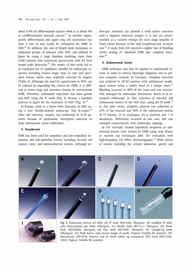
Fig. 3. Endoscopic devices for ESD. (A) IT knife (KD-610L, Olympus), (B) modified IT knife with three-pointed star blade (Olympus), (C) Needle knife (KD-1L-1, Olympus), (D) Hook knife (KD-620LR, Olympus), (E) Flex knife (KD-630L, Olympus), (F) Triangle-tip knife (Olympus), (G) Flash knives with several length of needle (Fujinon Toshiba ES systems), (H) Mucosectom (DP-2518, Pentax), and (I) Small caliber tip transparent (ST) hood (DH-15GR, 15CR, Fujinon Toshiba ES systems).
Coda S, et al: EMR and ESD in the West 17
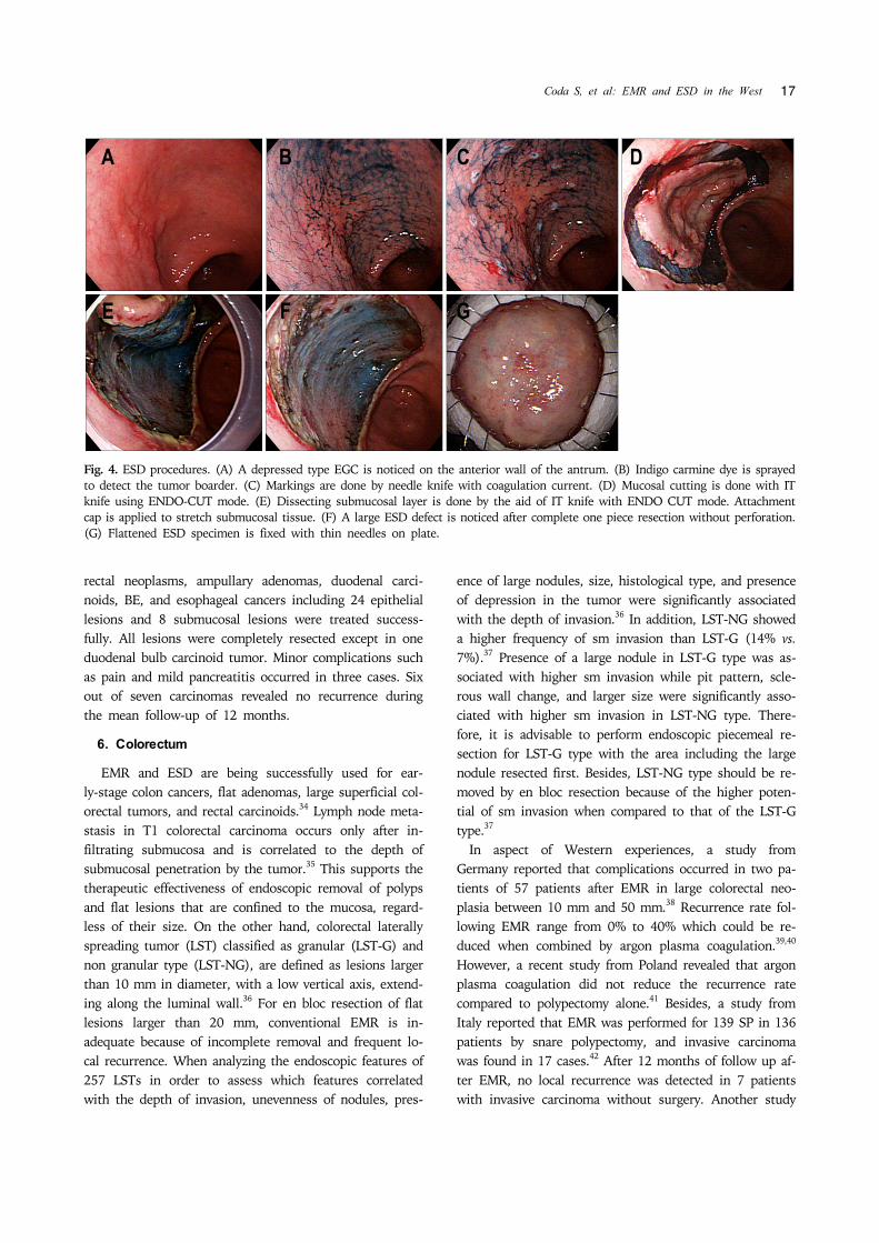
Fig. 4. ESD procedures. (A) A depressed type EGC is noticed on the anterior wall of the antrum. (B) Indigo carmine dye is sprayed to detect the tumor boarder. (C) Markings are done by needle knife with coagulation current. (D) Mucosal cutting is done with ITknife using ENDO-CUT mode. (E) Dissecting submucosal layer is done by the aid of IT knife with ENDO CUT mode. Attachmentcap is applied to stretch submucosal tissue. (F) A large ESD defect is noticed after complete one piece resection without perforation.(G) Flattened ESD specimen is fixed with thin needles on plate.
rectal neoplasms, ampullary adenomas, duodenal carci-noids, BE, and esophageal cancers including 24 epithelial lesions and 8 submucosal lesions were treated success-fully. All lesions were completely resected except in one duodenal bulb carcinoid tumor. Minor complications such as pain and mild pancreatitis occurred in three cases. Six out of seven carcinomas revealed no recurrence during the mean follow-up of 12 months.
6. Colorectum
EMR and ESD are being successfully used for ear-ly-stage colon cancers, flat adenomas, large superficial col-orectal tumors, and rectal carcinoids.34 Lymph node meta-stasis in T1 colorectal carcinoma occurs only after in-filtrating submucosa and is correlated to the depth of submucosal penetration by the tumor.35 This supports the therapeutic effectiveness of endoscopic removal of polyps and flat lesions that are confined to the mucosa, regard-less of their size. On the other hand, colorectal laterally spreading tumor (LST) classified as granular (LST-G) and non granular type (LST-NG), are defined as lesions larger than 10 mm in diameter, with a low vertical axis, extend-ing along the luminal wall.36 For en bloc resection of flat lesions larger than 20 mm, conventional EMR is in-adequate because of incomplete removal and frequent lo-cal recurrence. When analyzing the endoscopic features of 257 LSTs in order to assess which features correlated with the depth of invasion, unevenness of nodules, pres-
ence of large nodules, size, histological type, and presence of depression in the tumor were significantly associated with the depth of invasion.36 In addition, LST-NG showed a higher frequency of sm invasion than LST-G (14% vs. 7%).37 Presence of a large nodule in LST-G type was as-sociated with higher sm invasion while pit pattern, scle-rous wall change, and larger size were significantly asso-ciated with higher sm invasion in LST-NG type. There-fore, it is advisable to perform endoscopic piecemeal re-section for LST-G type with the area including the large nodule resected first. Besides, LST-NG type should be re-moved by en bloc resection because of the higher poten-tial of sm invasion when compared to that of the LST-G type.37 In aspect of Western experiences, a study from Germany reported that complications occurred in two pa-tients of 57 patients after EMR in large colorectal neo-plasia between 10 mm and 50 mm.38 Recurrence rate fol-lowing EMR range from 0% to 40% which could be re-duced when combined by argon plasma coagulation.39,40 However, a recent study from Poland revealed that argon plasma coagulation did not reduce the recurrence rate compared to polypectomy alone.41 Besides, a study from Italy reported that EMR was performed for 139 SP in 136 patients by snare polypectomy, and invasive carcinoma was found in 17 cases.42 After 12 months of follow up af-ter EMR, no local recurrence was detected in 7 patients with invasive carcinoma without surgery. Another study
18 Gut and Liver, Vol. 1, No. 1, June 2007
from UK reported on 30 large colorectal polyps which were treated by en bloc resection in 22 cases and by piecemeal resection in 8 cases.43 Histologically, the le-sions were predominantly adenomatous polyps, but 7 cas-es revealed incidental focus of adenocarcinoma. Although bleeding occurred in two cases, there was no bowel perforation. There was no evidence of recurrence during the median follow-up of 21 months. In a prospective cohort study in Italy, IT knife was used for EMR of large colorectal polyps larger than 3 cm which are unsuitable for standard polypectomy.44 The re-sults showed the likelihood of complete en bloc resection of mucosal lesions improved by new approach with IT knife when compared with previous studies on colonic EMR, even for lesions located in difficult positions or larger than 30 mm. En bloc resection was achieved only in 55.1% of the lesions and piecemeal resection was per-formed in the rest of the cases. Although complete tumor removal was achieved in 19 patients, 13 had LGD, 15 had HGD, and one had a tumor invading the submucosa. Complications occurred in four patients which were all managed conservatively. Local recurrences were detected in five patients and were treated by argon plasma coagu-lation and snare polypectomy. There was no recurrence during the median follow-up period of 15.7 months.44
MANAGEMENT AFTER ENDOSCOPIC RESEC-TION
After the retrieval, resected specimen is flattened and fixed on the board (Fig. 4). Additional resections and re-constructions are seldom necessary as long as the mark-ing dots are completely included in en bloc resection.
Accurate evaluation of the resected specimen should be achieved immediately after the removal before being im-mersed in formaldehyde. Orientation of the specimen is accomplished by fixing its periphery with thin needles in-serted into an underlying plate of rubber or wood. The submucosa side of the specimen is apposed to the plate. After the fixation, the specimen should be sectioned se-rially in 2 mm intervals parallel to a line that includes the closest resection margin of the specimen so that both lateral and vertical margins could be assessed. The depth of tumor invasion (T) is then evaluated along with the degree of differentiation and lymphatic or vascular in-volvement.
1. Complications
The complications of endoscopic resection for EGC in-clude pain, bleeding, and perforation.45 Pain after re-section is typically mild. Standard dose of proton-pump inhibitor is administrated for 4-8 weeks to prevent post-operative bleeding and to promote ulcer healing.46 For the first 24 hours after the procedure, the patient is kept nil by mouth, followed by clear liquid on the second day, and soft diet for another three days. ESD-induced ulcer is reported to heal within 8 weeks after resection under ant-acid treatment.47
Bleeding is the most common complication occurring in up to 7-8% of patients undergoing standard EMR or ESD.48 Immediate bleeding appears more common with resections of tumors located on the upper third of the stomach. During ESD, immediate minor bleeding is not uncommon, but could be successfully treated by grasping the bleeding vessels using a hot biopsy forcep with 80W soft mode coagulation of ICC 200 (ERBE, Tubingen,
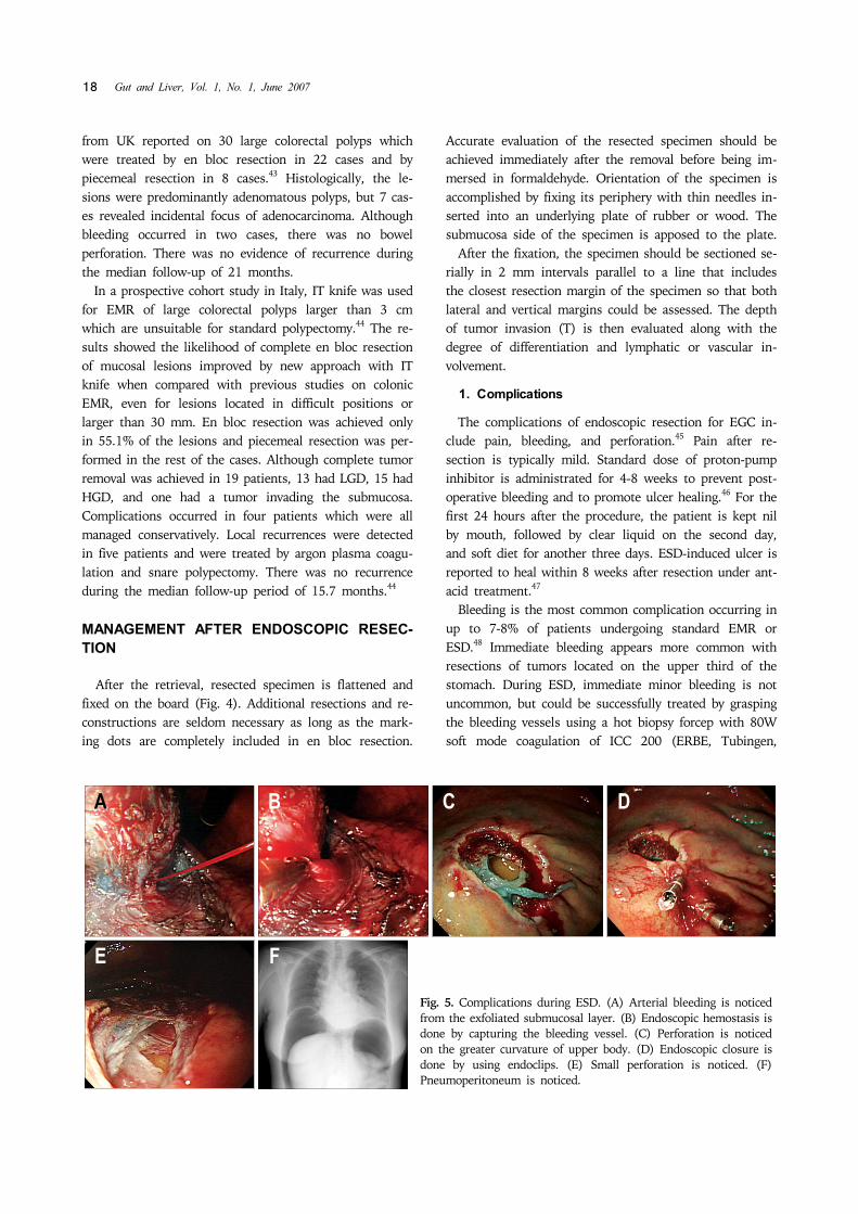
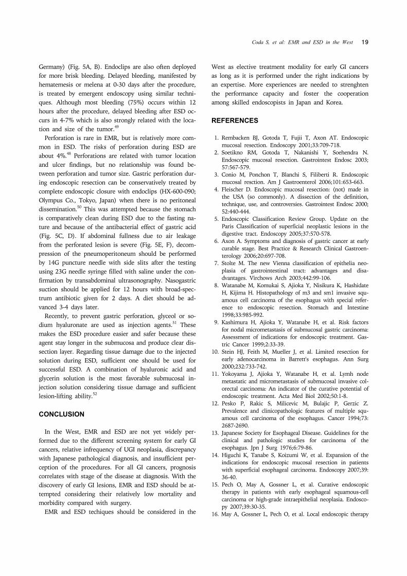
Fig. 5. Complications during ESD. (A) Arterial bleeding is noticed from the exfoliated submucosal layer. (B) Endoscopic hemostasis is done by capturing the bleeding vessel. (C) Perforation is noticed on the greater curvature of upper body. (D) Endoscopic closure is done by using endoclips. (E) Small perforation is noticed. (F) Pneumoperitoneum is noticed.
Coda S, et al: EMR and ESD in the West 19
Germany) (Fig. 5A, B). Endoclips are also often deployed for more brisk bleeding. Delayed bleeding, manifested by hematemesis or melena at 0-30 days after the procedure, is treated by emergent endoscopy using similar techni-ques. Although most bleeding (75%) occurs within 12 hours after the procedure, delayed bleeding after ESD oc-curs in 4-7% which is also strongly related with the loca-tion and size of the tumor.49
Perforation is rare in EMR, but is relatively more com-mon in ESD. The risks of perforation during ESD are about 4%.48 Perforations are related with tumor location and ulcer findings, but no relationship was found be-tween perforation and tumor size. Gastric perforation dur-ing endoscopic resection can be conservatively treated by complete endoscopic closure with endoclips (HX-600-090; Olympus Co., Tokyo, Japan) when there is no peritoneal dissemination.50 This was attempted because the stomach is comparatively clean during ESD due to the fasting na-ture and because of the antibacterial effect of gastric acid (Fig. 5C, D). If abdominal fullness due to air leakage from the perforated lesion is severe (Fig. 5E, F), decom-pression of the pneumoperitoneum should be performed by 14G puncture needle with side slits after the testing using 23G needle syringe filled with saline under the con-firmation by transabdominal ultrasonography. Nasogastric suction should be applied for 12 hours with broad-spec-trum antibiotic given for 2 days. A diet should be ad-vanced 3-4 days later. Recently, to prevent gastric perforation, glyceol or so-dium hyaluronate are used as injection agents.51 These makes the ESD procedure easier and safer because these agent stay longer in the submucosa and produce clear dis-section layer. Regarding tissue damage due to the injected solution during ESD, sufficient one should be used for successful ESD. A combination of hyaluronic acid and glycerin solution is the most favorable submucosal in-jection solution considering tissue damage and sufficient lesion-lifting ability.52
CONCLUSION
In the West, EMR and ESD are not yet widely per-formed due to the different screening system for early GI cancers, relative infrequency of UGI neoplasia, discrepancy with Japanese pathological diagnosis, and insufficient per-ception of the procedures. For all GI cancers, prognosis correlates with stage of the disease at diagnosis. With the discovery of early GI lesions, EMR and ESD should be at-tempted considering their relatively low mortality and morbidity compared with surgery. EMR and ESD techiques should be considered in the
West as elective treatment modality for early GI cancers as long as it is performed under the right indications by an expertise. More experiences are needed to strenghten the performance capacity and foster the cooperation among skilled endoscopists in Japan and Korea.
REFERENCES
1. Rembacken BJ, Gotoda T, Fujii T, Axon AT. Endoscopic mucosal resection. Endoscopy 2001;33:709-718.
2. Soetikno RM, Gotoda T, Nakanishi Y, Soehendra N. Endoscopic mucosal resection. Gastrointest Endosc 2003; 57:567-579.
3. Conio M, Ponchon T, Blanchi S, Filiberti R. Endoscopic mucosal resction. Am J Gastroenterol 2006;101:653-663.
4. Fleischer D. Endoscopic mucosal resection: (not) made in the USA (so commonly). A dissection of the definition, technique, use, and controversies. Gastrointest Endosc 2000; 52:440-444.
5. Endoscopic Classification Review Group. Update on the Paris Classification of superficial neoplastic lesions in the digestive tract. Endoscopy 2005;37:570-578.
6. Axon A. Symptoms and diagnosis of gastric cancer at early curable stage. Best Practice & Research Clinical Gastroen-terology 2006;20:697-708.
7. Stolte M. The new Vienna classification of epithelia neo-plasia of gastrointestinal tract: advantages and disa-dvantages. Virchows Arch 2003;442:99-106.
8. Watanabe M, Komukai S, Ajioka Y, Nisikura K, Hashidate H, Kijima H. Histopathology of m3 and sm1 invasive squ-amous cell carcinoma of the esophagus with special refer-ence to endoscopic resection. Stomach and Intestine 1998;33:985-992.
9. Kashimura H, Ajioka Y, Watanabe H, et al. Risk factors for nodal micrometastasis of submucosal gastric carcinoma: Assessment of indications for endoscopic treatment. Gas-tric Cancer 1999;2:33-39.
10. Stein HJ, Feith M, Mueller J, et al. Limited resection for early adenocarcinoma in Barrett's esophagus. Ann Surg 2000;232:733-742.
11. Yokoyama J, Ajioka Y, Watanabe H, et al. Lymh node metastatic and micrometastasis of submucosal invasive col-orectal carcinoma: An indicator of the curative potential of endoscopic treatment. Acta Med Biol 2002;50:1-8.
12. Pesko P, Rakic S, Milicevic M, Bulajic P, Gerzic Z. Prevalence and clinicopathologic features of multiple squ-amous cell carcinoma of the esophagus. Cancer 1994;73: 2687-2690.
13. Japanese Society for Esophageal Disease. Guidelines for the clinical and pathologic studies for carcinoma of the esophagus. Jpn J Surg 1976;6:79-86.
14. Higuchi K, Tanabe S, Koizumi W, et al. Expansion of the indications for endoscopic mucosal resection in patients with superficial esophageal carcinoma. Endoscopy 2007;39: 36-40.
15. Pech O, May A, Gossner L, et al. Curative endoscopic therapy in patients with early esophageal squamous-cell carcinoma or high-grade intraepithelial neoplasia. Endosco-py 2007;39:30-35.
16. May A, Gossner L, Pech O, et al. Local endoscopic therapy
20 Gut and Liver, Vol. 1, No. 1, June 2007
for intraepithelial high-grade neoplasia and early ad-enocarcinoma in Barrett's oesophagus: acute-phase and in-termediate results of a new treatment approach. Eur J Gastroenterol Hepatol 2002;14:1085-1091.
17. Buttar NS, Wang KK, Lutzke LS, Krishnadath KK, Ander-son MA. Combined endoscopic mucosal resection and pho-todynamic therapy for esophageal neoplasia within Barre-tt's esophagus. Gastrointest Endosc 2001;54:682-688.
18. Seewald S, Akaraviputh T, Seitz U, et al. Circumferential EMR and complete removal of Barrett's epithelium: a new approach to management of Barrett's esophagus containing high-grade intraepithelial neoplasia and intramucosal carcinoma. Gastrointest Endosc 2003;57:854-859.
19. Giovannini M, Bories E, Pesenti C, et al. Circumferential endoscopic mucosal resection in Barrett's esophagus with high-grade intraepithelial neoplasia or mucosal cancer. Preliminary results in 21 patients. Endoscopy 2004;36: 782-787.
20. Buskens CJ, Westerterp M, Lagarde SM, Bergman JJ, ten Kate FJ, van Lanschot JJ. Prediction of appropriateness of local endoscopic treatment for high-grade dysplasia and early adenocarcinoma by EUS and histopathologic features. Gastrointest Endosc 2004;60:703-710.
21. Nijhawan PK, Wang KK. Endoscopic mucosal resection for lesions with endoscopic features suggestive of malignancy and high-grade dysplasia within Barrett's esophagus. Gastrointest Endosc 2000;52:328-332.
22. Ell C, May A, Pech O, et al. Curative endoscopic resection of early esophageal adenocarcinomas (Barrett's cancer). Gastrointest Endosc 2007;65:3-10.
23. Gotoda T. Endoscopic resection of early gastric cancer. Gastric Cancer 2007;10:1-11.
24. Amano Y, Ishihara S, Amano K, et al. An assessment of local curability of endoscopic surgery in early gastric can-cer without satisfaction of current therapeutic indications. Endoscopy 1998;30:548-552.
25. Gotoda T, Yanagisawa A, Sasako M, et al. Incidence of lymph node metastasis from early gastric cancer: estima-tion with a large number of cases at two large centers. Gastric Cancer 2000;3:219-225.
26. Makuuchi H, Kise Y, Shimada H, Chino O, Tanaka H. Endoscopic mucosal resection for early gastric cancer. Semin. Surg Oncol 1999;17:108-116.
27. Gotoda T, Kondo H, Ono H, et al. A new endoscopic mu-cosal resection procedure using an insulation-tipped elec-ctrosurgical knife for rectal flat lesions. Gastrointest Endosc 1999;50:560-563.
28. Neuhaus H, Costamagna G, Deviere J, Fockens P, Ponchon T, Rosch T; ARCADE Group. Endoscopic submucosal dis-section (ESD) of early neoplastic gastric lesions using a new double-channel endoscope (the "R-scope"). Endoscopy 2006;38:1016-1023.
29. Lee SY, Jang KT, Lee KT, et al. Can endoscopic resection be applied for early stage ampulla of Vater cancer? Gastrointest Endosc 2006;63:783-788.
30. Ahmad NA, Kochman ML, Long WB, Furth EE, Ginsberg GG. Efficacy, safety, and clinical outcomes of endoscopic mucosal resection: a study of 101 cases. Gastrointest Endosc 2002;55:390-396.
31. Wehrmann T, Martchenko K, Nakamura M, Riphaus A, Stergiou N. Endoscopic resection of submucosal esoph-
ageal tumors: a prospective case series. Endoscopy 2004; 36:802-807.
32. Rosch T, Sarbia M, Schumacher B, et al. Attempted endo-scopic en bloc resection of mucosal and submucosal tu-mors using insulated-tip knives: a pilot series. Endoscopy 2004;36:788-801.
33. Waxman I, Saitoh Y. Clinical outcome of endoscopic mu-cosal resection for superficial GI lesions and the role of high-frequency US probe sonography in an American population. Gastrointest Endosc 2000;52:322-327.
34. Saito Y, Uraoka T, Matsuda T, et al. Endoscopic treatment of large superficial colorectal tumors: a case series of 200 endoscopic submucosal dissections (with video). Gastroint-est Endosc 2007;in press.
35. Yamamoto S, Watanabe M, Hasegawa H, et al. The risk of lymph node metastasis in T1 colorectal carcinoma. Hepatogastroenterology 2004;51:998-1000.
36. Saito Y, Fujii T, Kondo H, et al. Endoscopic treatment for laterally spreading tumors in the colon. Endoscopy 2001; 33:682-686.
37. Uraoka T, Saito Y, Matsuda T, et al. Endoscopic in-dications for endoscopic mucosal resection of laterally spreading tumours in the colorectum. Gut 2006;55: 1592-1597.
38. Bergmann U, Beger HG. Endoscopic mucosal resection for advanced non-polypoid colorectal adenoma and early stage carcinoma. Surg Endosc 2003;17:475-479.
39. Zlatanic J, Waye JD, Kim PS, Baiocco PJ, Gleim GW. Large sessile colonic adenomas: use of argon plasma coagulator to supplement piecemeal snare polypectomy. Gastrointest Endosc 1999;49:731-735.
40. Brooker JC, Saunders BP, Shah SG, Thapar CJ, Suzuki N, Williams CB. Treatment with argon plasma coagulation re-duces recurrence after piecemeal resection of large sessile colonic polyps: a randomized trial and recommendations. Gastrointest Endosc 2002;55:371-375.
41. Regula J, Wronska E, Polkowski M, et al. Argon plasma coagulation after piecemeal polypectomy of sessile color-ectal adenomas: long-term follow-up study. Endoscopy 2003;35:212-218.
42. Conio M, Repici A, Demarquay JF, Blanchi S, Dumas R, Filiberti R. EMR of large sessile colorectal polyps. Gastrointest Endosc 2004;60:234-241.
43. Jameel JK, Pillinger SH, Moncur P, Tsai HH, Duthie GS. Endoscopic mucosal resection (EMR) in the management of large colorectal polyps. Colorectal Dis 2006;8:497-500.
44. Repici A, Conio M, De Angelis C, et al. Insulated-tip knife endoscopic mucosal resection of large colorectal polyps un-suitable for standard polypectomy. Am J Gastroenterol 2007;in press.
45. Kaneko E, Hanada H, Kasugai T, Ogoshi K, Niwa K. The survey of gastrointestinal endoscopic complications in Japan. Gastroenterol Endosc (In Japanese) 2000;42:308- 313.
46. Lee SY, Kim JJ, Lee JH, et al. Healing rate of EMR-induced ulcer in relation to the duration of treatment with omeprazole. Gastrointest Endosc 2004;60:213-217.
47. Kakushima N, Yahagi N, Fujishiro M, et al. The healing process of gastric artificial ulcers after endoscopic sub-mucosal dissection. Dig Endosc 2004;16:327-331.
48. Oda I, Gotoda T, Hamanaka H, et al. Endoscopic sub-
Coda S, et al: EMR and ESD in the West 21
mucosal dissection for early gastric cancer: Technical feasi-bility, operation time and complications from large consec-utive cases. Dig Endosc 2005;17:54-58.
49. Shiba M, Higuchi K, Kadouchi K, et al. Risk factors for bleeding after endoscopic mucosal resection. World J Gastroenterol 2005;14:7335-7339.
50. Minami S, Gotoda T, Ono H, Oda I, Hamanaka H. Complete endoscopic closure using endoclips for gastric perforation during endoscopic resection for early gastric cancer can avoid emergent surgery. Gastrointest Endosc
2006;63:596-601.51. Fujishiro M, Yahagi N, Nakamura M, et al. Successful out-
comes of a novel endoscopic treatment for GI tumors: en-doscopic submucosal dissection with a mixture of high-molecular-weight hyaluronic acid, glycerin, and sugar. Gastrointest Endosc 2006;63:243-249.
52. Fujishiro M, Yahagi N, Kashimura K, et al. Tissue damage of different submucosal injection solutions for EMR. Gastrointest Endosc 2005;62:933-942.