endoscopy skills 2 2-2015
TRANSCRIPT

INTRODUCTION TO ENDOSCOPIC SURGERY
Dr. Mohamed Hesham AnwarProf. Obstetrics & Gynecology
AL AZHAR UNIVERSITY
GYNECOLOGICAL ENDOSCOPY…..
LAPAROSCOPY HYSTEROSCOPY

Gynecological Endoscopy
• Endoscopy in obstetrics and gynaecology has many branches:
» Laparoscopy» Hysteroscopy. » Colposcopy» Falloposcopy» Fetoscopy
• Endoscopy in obstetrics and gynaecology has many branches:
» Laparoscopy» Hysteroscopy. » Colposcopy» Falloposcopy» Fetoscopy

Operative LaparoscopyOperative Laparoscopy
Successful operative laparoscopy requires 3 essential ingredients:Successful operative laparoscopy requires 3 essential ingredients:
1. Surgical skill
2. Well designed & equipped Operating Room
3. Surgical team

LAPAROSCOPY
• Outline:
• Definition• Instruments • The Procedures• Indications and contraindications• Complications
• Outline:
• Definition• Instruments • The Procedures• Indications and contraindications• Complications

LAPAROSCOPY
• Definition:
It is a technique which allows viewing (Diagnostic) and surgical maneuvers (Therapeutic) to be performed in abdominal organs through a surgical incision of < 1cm with help of pneumoperitoneum.

Instruments
1. Verres needle:
used to inflate air to the peritoneal cavity (pneumoperitoneum) through the umbilicus where there is the thinnest abdominal wall.

2. Electronic laparoflator: INSUFFLATOR
– Used to insufflate through the verres needle.
– Maintains constant intra-abdominal pressure without exceeding the safety limit.
– Some types have heating system to prevent lowering the patient body temperature.

3. Trocars:
– Permit access to the intraperitoneal cavity in which other instruments can pass.
– The trocar used should be adapted to the diameter of the telescope selected.

4. Telescope:
– There are different sizes and angels, each with a different use.
– They are used to visualize the peritoneal cavity.

5. Camera equipment.
6. Light source.

There are two types:- Disposable - Reusable
They can be either atraumatic or grasping foreceps.
7. Forceps and scissors:7. Forceps and scissors:


8. Bipolar elecrtosurgey.9. Unipolar electrosurgery.10. Laser.11. Ultrasound system.12. Suction and irrigation system.13. Suture.14. Laparoscopic bag.15. Tissue morcellator: used to remove large specimens
like myomas or an entire uterus in small pieces.16. Uterine manipulator: used to mobilize or stabilize
the uterus and adnexa.
InstrumentsInstruments

1. Preparation of the patient:
– Inform the patient about the therapeutic benefits and potential risks (informed consent).
– Intestinal preparation: Simple intestinal emptying, for better viewing and preventing injuries.
– Place the patient in the dorsolithotomy position.
ProcedureProcedure

a. The abdominal wall is lifted by hand or by grasping forcepsb. Pnemoperitoneum is created by verres needle introduced to the
umbilical area (less subcutaneous and preperitoneul tissue). c. The needle is inserted in an oblique angle toward the uterine
fundusd. The negative pressure will allow the underlying structures to fall
away.e. After making sure that the needle is in correct position, air flow
can be increased to 2.5 liters per minute till a pressure of 15mmHg
2. Creation of pneumoperitoneum:2. Creation of pneumoperitoneum:

a. Once the intra-abdominal pressure reaches 15 mmHg the main trocar is introduced after removal of veress needle.
b. The position of the trocar must be verified by inserting the laparoscope and viewing the pelvic cavity.
3. Trocar introduction3. Trocar introduction

A. The omentum, bowel and bifurcation of pelvic vessels should be evaluated to avoid injuries caused during the introduction of Verres needle or trocar.
B. The site of introduction of other trocars should be verified by finger palpation and transillumination of abdominal wall to avoid injury to epigastric vessels.
C. Identify if there is any bleeding
4. Viewing the peritoneal cavity:4. Viewing the peritoneal cavity:

LapSim Basic Skills
http://surgical-science.com
camera & instrument navigation; coordination; grasping; lifting, grasping & transfer; cutting, clip applying; suturing; precision & speed

After the procedure CO2 gas must be evacuated completely to reduce post-operative pain
In operative procedures:- 1 or 2 bottles of Ringer’s lactate are used to wash the peritoneal cavity after laparoscopy.- Leave 500/1000 cc of ringer’s lactate to reduce the incidence of post operative pain.

Used as a diagnostic tool
– Infertility: status of the fallopian tube (morphology and functionality) and any pathological condition e.g. adhesions.
– Ovarian cysts or tumors.– Ectopic pregnancy.– PID: tubal abscess or adhesions.– Endometriosis: define the sites of implants and
endometrial cysts.
IndicationsIndications

Diagnostic Laparoscopyfor Gynecological Pelvic Disorders

Gynecologic Disorders: Chronic pelvic pain Ectopic pregnancy Pelvic inflammatory disease Endometriosis Adhesions Ovary: cysts, torsion Fallopian tube: torsion,
salpingitis Uterus: fibroid, leiomyomata Pelvic congestion syndrome Infertility

The role of laparoscopy as a diagnostic tool in chronic pelvic pain. Fred M Howard MS, MDBaillieÁ re's Clinical Obstetrics and Gynaecology Vol. 14, No. 3, pp. 467-494, 2000.
> 40% of gynaecological diagnostic laparoscopies are done for CPP
Combining the results of published series of laparoscopies for CPP shows that :
*No visible pathology is detected in 35% (range . 3±92%) of patients *Endometriosis is diagnosed in 33% (range . 2±80%) *Adhesive disease is found in 24% (range . 0± 52%)
A negative laparoscopy is not synonymous with no diagnosis or no disease
A meticulously performed negative laparoscopy means that a woman does not have endometriosis-associated or adhesion-associated pain
Chronic Pelvic Pain

Ectopic pregnancy
Photograph courtesy of Dr. Syed
Laparoscopy should be regarded as a therapeutic rather than diagnostic tool for suspected ectopic pregnancy.
Transvaginal ultrasound has replaced laparoscopy for diagnosis of ectopic pregnancy.
Laparoscopy has diagnostic role if probable tubal pregnancy or diagnosis in doubt.
Large uncontrolled studies have demonstrated that > 80% of ectopic pregnancies can be managed laparoscopically
The most commonly used procedures at laparoscopy are salpingectomy and salpingotomy.
The role of laparoscopy in the management of ectopic pregnancy. Martin Christopher Sowter, MD, MRCOGa, Jonathan Frappell, FRCS, FRCOG Reviews in Gynaecological Practice #2 (2002) 73-82

Pelvic Inflammatory DiseaseClinical diagnosis of PID is often difficult especially when symptoms are mild, as frequently when the primary organism is C. trachomatis.
Laparoscopy is the gold standard for the diagnosis of PID – should be used when diagnosis is uncertain, especially in young women for whom the preservation of fertility is important.
Sellors et al. reported that only by resorting to diagnostic laparoscopy were they able to demonstrate that PID was the cause of acute pelvic pain in 46% of a group of 95 women.
Laparoscopy should be considered for patients who have not responded to antibiotic therapy within 48 to 72 hours.
The role of laparoscopy in the management of pelvic pain in women of reproductive age. Maria Grazia Porpora, M.D. FERTILITY AND STERILITY Vol. 6M, No. 5, November 1997.

Tubo-ovarian Abscess
Most commonly isolated pathogens from a tubo-ovarian abscess are C. trachomatis and peptostreptococci.
At laparoscopy the peritoneal cavity (pelvis and abdomen) is inspected carefully.
The surgical steps include adhesiolysis, aspiration of the abscess cavity, dissection and excision of necrotic tissue, tubal lavage, and irrigation of the peritoneal cavity before completion of the procedure.
Laparoscopic surgery combined with adequate broad-spectrum antibiotic therapy has proven successful in the treatment of more than 95% of patients.

Endometriosis
Endometriosis is a histologically-defined disease :“the presence of ectopic tissue which possesses the histological structure and function of the uterine mucosa”. ( Sampson 1921)
Laparoscopy has largely replaced laparotomy as the diagnostic procedure for any patient suspected of having endometriosis.
Based on visual appearance endometriosis are classified as atypical (red, yellow, white or clear) or typical (black-brown, black or puckered black stellate) lesions.
The role of laparoscopy as a diagnostic tool in chronic pelvic pain. Fred M Howard MS, MD BaillieÁ re's Clinical Obstetrics and Gynaecology Vol. 14, No. 3, pp. 467-494, 2000.

The role of laparoscopy as a diagnostic tool in chronic pelvic pain. Fred M Howard MS, MD BaillieÁ re's Clinical Obstetrics and Gynaecology Vol. 14, No. 3, pp. 467-494, 2000.
Brownish lesion on the ovaryWhite fibrotic lesion on the uterosacral ligament
Peritoneal pocket of endometriosis Red stellate lesions in the cul-de-sac
Endometriosis presents with a variety of appearances that may make visual diagnosis difficult and inaccurate.

The role of laparoscopy as a diagnostic tool in chronic pelvic pain. Fred M Howard MS, MD BaillieÁ re's Clinical Obstetrics and Gynaecology Vol. 14, No. 3, pp. 467-494, 2000.
Visually misdiagnosed as endometriosis: Haemangiomas, old suture, ovarian
carcinoma, residual carbon deposits from prior surgery, ectopic pregnancy, …
Histological confirmation of visually diagnosed endometriosis ranges from 9-90%, depending on characteristics of the lesions
Apparent classic endometriotic lesion that was actually a suture granuloma.
Endometriosis

The role of laparoscopy as a diagnostic tool in chronic pelvic pain. Fred M Howard MS, MD BaillieÁ re's Clinical Obstetrics and Gynaecology Vol. 14, No. 3, pp. 467-494, 2000.
Laparoscopic evaluation requires detailed visualization of all sites of endometriosis:
A study of 716 women with endometriosis found these anatomical distributions: cul-de-sac and uterosacrals ( 69%) ovaries ( 45%) ovarian fossae ( 33%)and uterovesical fold ( 24% ) Chocolate fluid from needle puncture
of a suspected occult endometrioma.
Endometriosis
ovaries (all surfaces) ovarian fossae pelvic peritoneum (cul-de-sac, periureteral, bladder peritoneum)
uterine ligaments sigmoid colon appendix fallopian tubes rectovaginal septum
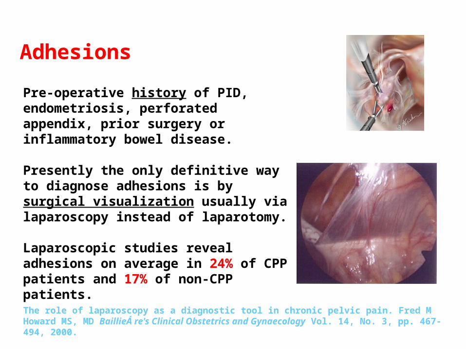
Adhesions
Pre-operative history of PID, endometriosis, perforated appendix, prior surgery or inflammatory bowel disease.
Presently the only definitive way to diagnose adhesions is by surgical visualization usually via laparoscopy instead of laparotomy.
Laparoscopic studies reveal adhesions on average in 24% of CPP patients and 17% of non-CPP patients.
The role of laparoscopy as a diagnostic tool in chronic pelvic pain. Fred M Howard MS, MD BaillieÁ re's Clinical Obstetrics and Gynaecology Vol. 14, No. 3, pp. 467-494, 2000.

Adenomyosis
Endometrial cells penetrate the myometrium causing either localized (adenomyoma) or diffuse overgrowth.
Adenomyomas that penetrate the uterine cavity become submucosal tumors.
An enlarged uterus from adenomyosis is often misdiagnosed as being from fibroids

The role of laparoscopy as a diagnostic tool in chronic pelvic pain. Fred M Howard MS, MD BaillieÁ re's Clinical Obstetrics and Gynaecology Vol. 14, No. 3, pp. 467-494, 2000.
Most ovarian cysts are haemorrhagic corpora lutea or follicle cysts.
They are usually asymptomatic and when they cause pain it is almost always acute.
Laparoscopic evaluations of patients with CPP reveal ovarian cysts on average in only 3% of all cases.
Paratubal cyst
Ovarian Cysts

The role of laparoscopy as a diagnostic tool in chronic pelvic pain. Fred M Howard MS, MD BaillieÁ re's Clinical Obstetrics and Gynaecology Vol. 14, No. 3, pp. 467-494, 2000.
Even when the surgeon is ‘certain’ that the ovary is benign, it is essential that tissue be sent for histological evaluation.
Open the cyst and inspect the lining for papillary structures or excrescences.
If these are noted, then a laparotomy should be done .
Ovarian Cysts
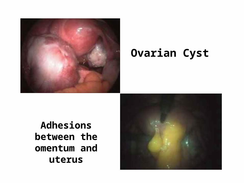
Ovarian Cyst
Adhesions between the
omentum and uterus

Ovarian CystsThe nature of the fluid is characteristically diagnostic : chocolate (usually endometrioma or haemorrhagic corpus luteum) sebaceous (teratoma), or mucinous (mucinous cystoma).
Mucinous cyst Teratoma
Dermoid cyst

Adnexal Torsion
A rare gynecologic emergency that nearly always occurs unilaterally.
Common causes are benign ovarian tumors and cysts; malignant processes are rare.
Relapse or bilateral adnexal torsion can cause sterility interfering with fertility.
In 30% of the patients, there is torsion of a normal adnexa, while the majority of the cases are associated with ovarian pathology.
Adnexal torsion in very young girls: diagnostic pitfalls. Marieke Emontsa, Heleen Doornewaardb, J.(Co’tje) F. Admiraala,* European Journal of Obstetrics & Gynecology and Reproductive Biology 116 (2004) 207-210.
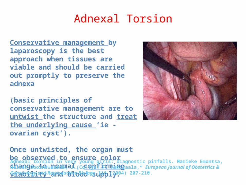
Adnexal Torsion
Conservative management by laparoscopy is the best approach when tissues are viable and should be carried out promptly to preserve the adnexa
(basic principles of conservative management are to untwist the structure and treat the underlying cause ‘ie - ovarian cyst’). Once untwisted, the organ must be observed to ensure color change to normal, confirming viability and blood supply.
Adnexal torsion in very young girls: diagnostic pitfalls. Marieke Emontsa, Heleen Doornewaardb, J.(Co’tje) F. Admiraala,* European Journal of Obstetrics & Gynecology and Reproductive Biology 116 (2004) 207-210.

Endosalpingiosis
Endosalpingiosis is the presence of fallopian tubal glandular epithelium in an ectopic location. Visually it appears as white to yellow, opaque or translucent, punctate, cystic lesions.
Endosalpingiosis is generally not recognized by gynaecologists at the time of laparoscopic evaluation or is misdiagnosed as endometriosis.
The role of laparoscopy as a diagnostic tool in chronic pelvic pain. Fred M Howard MS, MD BaillieÁ re's Clinical Obstetrics and Gynaecology Vol. 14, No. 3, pp. 467-494, 2000

Leiomyomata (fibroids)

Infertility

As a therapeutic tool- Management of ovarian cyst
by: Drainage. Ovarian cystectomy. Ovarian drilling of the cortex
and stroma to decrease androgens in the ovaries
Correcting ovarian torsion. As a treatment of
endometriosis By removal of the endometrial cyst, cauterization of endometrial spots and adhesiolysis
- Management of infertility:
Adhesiolysis
Treat the cause (endometriosis, PCOS)
- Myomectomy for fibroids: used for subserosal and intramural fibroids only, not used for submucosal fibroids.
- Management of PID: by draining tubal abscess and adhesiolysis.

AdhesiolysisAdhesiolysis
Myomectomy

Salpingotomy
– Used to preserve the tubes for desired reproductivity.
– Done if the patient is hemodynamicaly stable
– If size < 5 cm– Location must be ampullary, infundibular
or isthmic. – Contralateral tube either normal or
absent.
Management of Ectopic Pregnancy:Management of Ectopic Pregnancy:

Salpingectomy (it is the standard for ectopic pregnancy)
- Ruptured tube
- Multiple recurrence of ectopic pregnancy.
- Size of ectopic > 5 cm

Tubal sterilization by:
- Bipolar coagulation.
- Clips (filshie clips) and rings
- Before doing this you should consult the patient about 3 things
- Chance of irreversibility
- Failure rate 1/200
- Bleeding may occur and we may shift to laparatomy.
Ring sterilization
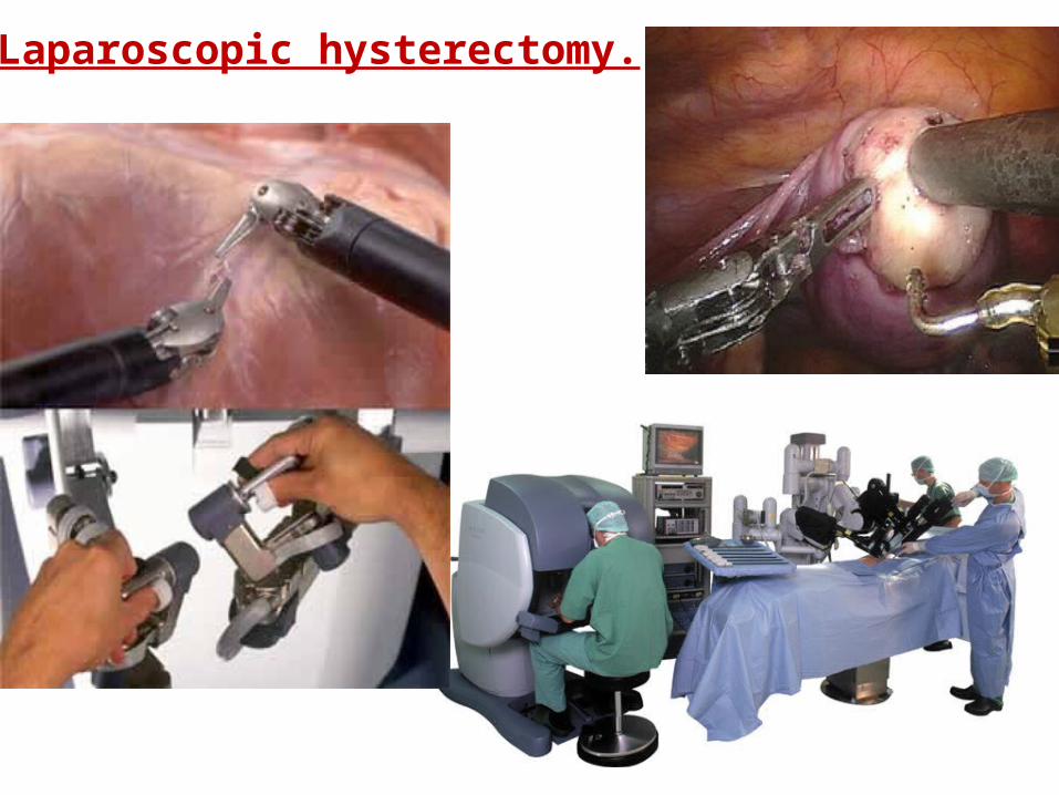
Laparoscopic hysterectomy.

Contraindications
1. Generalized peritonitis
2. Hypovolemic shock
3. Severe cardiac disease
4. Hemoglobin less than 7 g/dL
5. Uterine size > 12 wks.
6. Multiple previous abdominal procedures
7. Extreme body weight

- Pneumoperitoneum:- Extraperitonel emphysema due to failure of
introducing verres needle correctly into the peritoneal cavity and not checking the negative pressure on the machine.
- Gas may extend to the mediastinum and compromise cardiac function
- Pneumoomentum: and put the patient on the trendlenberg
- Injury to abdominal organs- GI: if the intestine is distended or adherent to the
abdominal wall (prevented by good intestinal preparation) and putting the patient on the telendelenburg position.
- Bladder injury: prevented by emptying the bladder.
ComplicationsComplications

Blood vessel injury:
- Pelvic, omental and mesentric- Prevented by introducing the verres needle in
an angle.- In obese patients you can insert the needle in
straight manner because of the thick fatty layer.
ComplicationsComplications

Conclusion:
Laparoscopy provides a vital tool for diagnosing pelvic pain – it provides first hand visual comprehension of the problem as well as an immediate opportunity to continue with therapeutic surgical correction.

HYSTEROSCOPY
• Definition• Instruments • The Procedures• Indications and contraindications• Complications
• Definition• Instruments • The Procedures• Indications and contraindications• Complications

Hysteroscopy
• Definition:
– It is a technique which allows viewing and surgical maneuvers to be performed in the uterine cavity.
– It has many advantages that made it wide spread and fundamental diagnostic method in daily gynecological
practice.

Instruments
1. Distention media of the uterine cavity (RL / CO2 distention)
2. Light source.xenon light source gives the best image quality

3. Camera Equipment
4. Endoscopeflexible: high cost and fragile cannot be autoclaved.rigid: gives different direction of the view.
- 0°, 12°, 30° (best for diagnostic purpose).

5. Hysteroscope:
There are 2 types of hysteroscopes:DiagnosticTherapeutic

Hysteroscopy Trainer –3rd generation system (2003)
Tasks • cannulation Skills • advance (endoscope) through endocervical canal
• exploration (Visual & • navigate, visualize endometrial cavity,Haptic) identify & palpate lesion
• resection (myoma) • hold endoscope proximally, extend loop distally, contact lesion, activate diathermy, retract loop to excise: repeat –
Metrics– % of the myoma resected– # perforations mechanical electrosurgical– timesec
Metrics– % of the myoma resected– # perforations mechanical electrosurgical– timesec

1. Preparation of the patient:– Detailed history and complete physical examination– It is preferable to do the procedure in the first part of the menstrual
cycle, because there is less mucus (better viewing) and no chance of encountering early pregnancy
– Informed consent– Patient is placed in lithotomy position– Accurate bimanual examination to asses the uterine (position,
morphology, volume).
Procedure
2. Technique:– Clean cervix with antiseptics– Cervical forceps is placed on the front labia– Light source & CO2 gas supply are connected to the instrument– Insert hysteroscope into the cervical canal, which dilates from
the gas pressure.
2. Technique:– Clean cervix with antiseptics– Cervical forceps is placed on the front labia– Light source & CO2 gas supply are connected to the instrument– Insert hysteroscope into the cervical canal, which dilates from
the gas pressure.

Used as a diagnostic tool:
- Abnormal uterine bleeding caused by: - submucous and intramural myoma. - endometrial polyps. - endometrial atrophy.
- Endometrial tumors.- Infertility related to:
- Intrauterine adhesions (Asherman’s syndrome)- Submucous fibroids. - Endometrial polyps.
- Uterine malformation (it cannot differentiate between sepatate and bicorneate uterus)<- this can be done by laparoscopy.
IndicationsIndications

Used as a therapeutic toolEndometrial ablation (using laser):• Abnormal uterine bleeding but we should role out
cancerous or pre cancerous cause of bleeding.• Also used in patients with high risk for hysterectomy
or the patient does not want to do the surgery.steroscopic Surgeries and Endometrial Polypectomy
IndicationsIndications

– Correct uterine malformation like septate uterus by resection of the septum. (bicorneate uterus is corrected by laparotomy using metroplasty).
– Polypectomy.– Intrauterine adhesions.– Myomectomy: The main indication for hysteroscopic
myomectomy is AUB caused by submucous myomas in infertile patients
IndicationsIndications

Hysteroscopic Surgeries and Endometrial Polypectomy
Hysteroscopic Surgeries and Endometrial Polypectomy

Used as a therapeutic tool- Removal of foreign bodies and IUCD.- Fallopian tube catheterization
- to canalize the tube.- to place intra tubal device for
reversible sterilization.
IndicationsIndications

Uterine polyp
Uterine anomaly

Intrauterine Adhesions

Endometrial Ca.

Contraindications
• Pregnancy.• Current or recent pelvic infection.• Current vaginitis, cervicitis and
endometritis.• Recent uterine perforation.• Active Bleeding.

Complications related to distention media:due to CO2 insufflation:
- Cardiac arrhythmia due to excessive absorption.- Gas embolism.- use hysteroflator that insufflate pressure of 100-120 mmHg constantly without
exceeding the safety limit.due to fluid:- HMW (dextran)- Anaphylactic reaction- Pulmonary edema- Adult RDS
ComplicationsComplications
- LMW (saline)- Fluid overload: prevented by keeping the operating time to minimum.- Avoid entering vascular channels.- Close monitoring of fluid balance. - If you exceed 1000 ml of infused fluid stop the procedure.
- Intraoperative complications:- Uterine perforation (<1%)- Hemorrhage either from: - Perforation - Tenaculum used to hold the
cervix.-Trauma.- Thermal damage.

- Late onset Complications:- Infections: like acute PID, so we give prophylactic antibiotics.- Vaginal discharge: common after ablative procedures and it is self limiting.
- Adhesion formation:- Common after myomectomy when 2 fibroids are located opposite to each
other in the uterine wall.- To prevent the adhesions it is better to remove the fibroids in stages, and
give estrogen (to build up the endometrial) therapy directly after resection. And also we can use IUCD.

• Asherman Syndrome:• It is defined as intrauterine adhesions • Cause can be iatrogenic (after hysteroscopic
myomectomy) and can due to infection.• It can be treated by hysteroscopic adhesiolysis
followed by inserting IUCD to make the uterine walls apart from each other. Also estrogen use after adhesiolysis cause the emdometrium to build up and prevent adhesions to reoccur

YOU WILL REMEMBER
A LITTLE OF WHAT YOU HEAR,SOME OF WHAT YOU READ,
CONSIDERABLY MORE OF WHAT YOU SEE,BUT
ALMOST ALL OF WHAT YOU UNDERSTAND.