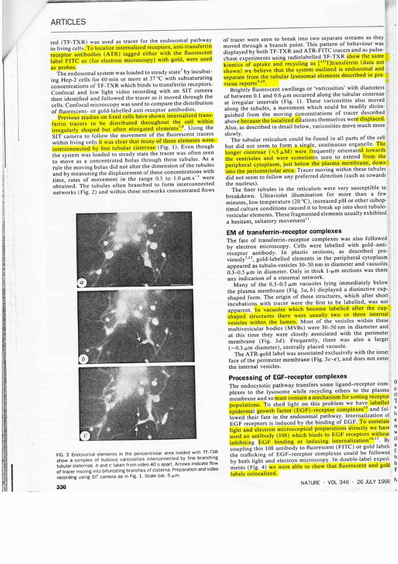
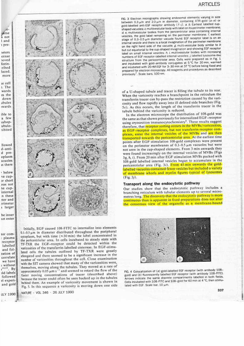
E ( I ) ) Y G, c o (U N P @ c o, d E rious fields lividing the rm 400 to s another een that, atr ons go coupled rl arity for tudies2-4. al, which 4 than is naly, and :esults on rakami el ,ow-angle certain ly re perfect r sensitive xista. The Although e second- difterent ystal con- exhibited magnet':z- ;le crystals tmpleso'2 t. rples gen- rt stage of unknown rre clearly' uch of the ly studied. has been varied the as to make rinate the 'ally found rr, decreas' J granular rnetlzatton alous mag- ibehaviour I I irulY 1990 t- I rel ated oxygen deficiency, even when 6 is of the order of o3fffilicit verification that these anomalies are absent is important for understanding the flux pinning, and the granular and reversible properties of the YBa2CusOz-6 compounds. In Received 12 April; accepted 19 June 1990 1. Dimos, D., Chaudhari,P., Mannhart, J. & LeGoues, F. K. Phys Rev. Lett 61, 2L9-222 (1988). 2 Daeumling, M , Seuntjens, J & Larbalestier, D C. Appl. Phys. Lett.52, 590-591 (1988). 3 Hibbs, A. D., Campbell, A. M & Male, S. IEEE Trans Magn. 25,2L42-21,45 (1989). 4. Kupfer, H. et at. Cryogenics 2.€t,268-280 (1989). 5 Shimizu, E. & lto, D Phys. Rev. B3 ), 292L-2923 (1989)' 6 Murakami. M el al Cryogenics 3O, 390-396 (1990). 7. Kaiser, D. L., l-{oltzberg, F., Scott, B. A. & McGuire, f . R. Appl. Phys. Lett.51' 1040-1042 (1e87). 8. Hottzberg, F, Strobel, P, & Worthington, T. K. J. Magn. magn, Mater,76177,626-630 (1988). 9. Salarna, K., Selvamanickam, V , Gao, L, & Sun, K. Appl. Phys Lett. Y,2352-2354 (1989). 10 Kes, P H., Pruymboom, A , van den Berg, J. & Mydosh, J A Cryogenics 29,228-231 (1989) 11 Swartzendruber, L. J, Roitburd, A , Kaiser, D 1., Gayle, F. W, & Bennett, L H Phys. Rev. Lett.64, 48s-486 (1990). 12. Welp, U.. Kwok, W. K., Crabtree, G. W., Vandervoort, K. G. & Liu, ).7. Appl. Phys. Lett. (submitted). 13. Van Dover, R B. et al. Nature 342, 55-59 (1989). 14 Daeumling. M. thesis, Univ Wisconsin (1990) 15. Tu, K. N, Yeh, N C., Park, S I & Tsuei, C C Phys Rev B39, 304-314 (1989). 16. Rothman, S J., Routbort, J L. & Baker, ) E Phys Rev. 840, 8852-8860 (1989). L7 Lindemer, T B et al. J. Am Ceram Soc.72, 1775-7788 (1989). 18. Cava, R J et al. Phys. Rev. B38, 5130-5133 (1987). 19. l-tylton, T. L & Beasley, M R Phys Rev. B(.L,11669-11672 (1990) 20 Livingston, ) D Appl. Phys. Lett.8, 319-320 (1966) 21. Kubo, Y & lgarashi, H Phys. Rev.839,725-728 (1989) 22. Xu, Y., Guan, W , Zeibig, K. & Heiden, C Cryogenics N,28L-285 (1989). 23. Beyers, R. et al. Nature 34O, 619-623 (1989) 24 Meingast, C,, Lee, P J. & Larbalestier, D C ). appl Phys. 66, 5962-5970 (1989). 25 Meingast, & Larbalestier, D. C. I appl. Phys 66, 5971-5983 (1989) ACKNOWLEDGEMENTS We thank F l-{oltzberg, A P. Malozemoff, T. W Worthington and D Kaiser for discussions and for the supply of flux-grown single crystals, A P Malozemoff and T, W. Worthington for sorReof the screening characterization on crystal FG1, and J McKinnell, J Vargas, S. E Babcock and K. Sawano for discussions. The work was supported by Electric Power Research lnstitute and the NSF. particular we'note that will require elimination tif,ed here, as well as granular weak links. ARTICLES practical applications of these materials of both the intra-grain weak links iden- the more generally recognized inter- tr Movement internalized ligand-receptor complexes along a continuous endosomal reticulum C. R, Hopkillsr A. Gibsorr M. Shipman & K. Miller Department of Biochemistry, lmperial College, London SW7 2AZ, UK Complexes of cell-surface receptors and their ligands are commonly internalized by endocytosis and enter a prelysosomal endosomal pathway for further processing. Fluorescence microscopy and video recording of living cells to trace the passage of ligand-receptor complexes has identified the endosomal ,compartment aS an extensive network of tubular cisternae. Endocytosed material entering this reticulum enters discrete swellings, identified as multivesicular bodies by electron microscopy, which move along the reticulum towards the peri- centriolar area. THp acidic endosomal compartment has a central role in the receptor-mediated uptake and processing of a wide range of metabolites (such as cholesterol ) and pathogens, and the recycling of receptor-ligand complexest-3. The membrane boun- dary of the endocytotic pathway is also probably involved in the transduction of mitogenic signals. Defining the limits of this system and identifying its regulatory mechanisms are therefore of considerable current interest in cell biology. The endosome is a difficult organelle to isolate by conventional methods of subcellular fractionation because it lacks a charac- teristic buoyant density and is highly pleiomorphic. has been studied most successfully by microscopical methods and some of its component parts have been described in detaila-6. However, the overall form of the organelle and the way in which its diverse functions are regulated remain unclear. We have apptied various microscopical methods to define the form of the endocytotic pathway in living cells. Here we describe NATURE VOL 346 , 26 JULY 1990 confocal microscopy and low light video recording of living epidermoid carcinoma cells (Hep-2) to follow the movement of fluorescent tracers through the endosomal compartment. Contrary to expectation we have found an extensive tubular reticulum with expanded swellings along its length. Electron microscopy shows that these structures are typical multivesicular bodies, endosomal elements which have been identified pre- viously in fixed cells and which hitherto had been thought to be free vacuoles. ldentiflcation of an endosomal reticulum The iron-carrying protein transferrin enters cells by receptor- mediated endocytosis. Transferrin labelled with the dye Texas FlG. 1 Cisternal elements peripheral cytoplasm of Hep-2 carcinoma cells. The fine branching cisternae have varicosities of varying sizes along them. Arrows indicate moving flows of tracer which on video show direction and rate of movement. Cells were grown in Dulbecco's MEM plus 5% fetal calf serum in sparse culture as described previously for A431 cellsT. They were incubated in subsaturating (tO-s M) concentrations of TF-TXR for 60 min or more at 37 'C. lmage obtained with a Hamamatsu C 24OO-08 SIT camera. Scale bar, 5 pm. 335