endothelial 5 and v integrins cooperate in remodeling of ...web.mit.edu/hyneslab/pdfs/endothelial...
TRANSCRIPT
2439RESEARCH ARTICLE
INTRODUCTIONVascular and congenital heart diseases are major causes of lethality(Bruneau, 2008) and much effort has been directed towardsdeveloping anti-angiogenic drugs (Ferrara and Kerbel, 2005).Therefore, there is considerable interest in understanding the rolesof molecules that drive and control vascular development.
One important participant in developmental and pathologicalangiogenesis is fibronectin (Astrof and Hynes, 2009), a secretedglycoprotein that assembles into a fibrillar extracellular matrix.Fibronectin-null mice show lethality at embryonic day (E) 9.5 andhave severe defects in the development of vasculature and somites(Francis et al., 2002; George et al., 1997; George et al., 1993).Integrins are heterodimeric cell adhesion receptors that mediatecell-matrix and cell-cell interactions (Hynes, 2002b; van der Flierand Sonnenberg, 2001). 51 integrin binds to the Arg-Gly-Asp(RGD) tri-peptide motif in fibronectin. This binding site is alsorecognized by v integrins (i.e. v1, v3, v5, v6 andv8) (Pankov and Yamada, 2002; Leiss et al., 2008). 5 integrin-null embryos show a similar phenotype to the fibronectin nulls, butdie 1 day later, at ~E10.5 (Francis et al., 2002; Goh et al., 1997;Yang et al., 1993). Similarly, targeted inactivation of the RGD sitein fibronectin (FnRGE) results in placental and cardiovascular
defects and lethality at E10.5 (Takahashi et al., 2007; Takahashi etal., 2009). All these data point to a key role for the 51 integrin-fibronectin interaction in angiogenesis.
Complete knockout of all 1 integrins is lethal at thepreimplantation stage (Fassler and Meyer, 1995; Stephens et al.,1995). Recently, several studies have shown that endothelial-specific knockout of 1 integrins results in severe vascular defectsand lethality at E10 (Carlson et al., 2008; Lei et al., 2008; Tanjoreet al., 2008; Zovein et al., 2010). This raises the question of which subunit is involved in developmental angiogenesis. Deletion of4 integrins is embryonic lethal (E10/11.5), with placental andcardiac defects (Yang et al., 1995). However, 4 endothelial-specific knockout mice are viable, with hematopoietic defects butno vascular phenotype (Priestley et al., 2007). 9 integrin-null micehave lymphatic defects and die of chylothorax soon after birth(Bazigou et al., 2009; Huang et al., 2000b), but lack other majorvascular defects. No major vascular developmental defects havebeen reported for knockouts of the other ten integrin subunits,including those of the laminin- and collagen-binding families.
The complete ablation of all five v integrins does not blockangiogenesis, but 80% of the embryos die at E11.5 due to placentaldefects and the remaining embryos succumb at birth with brainhemorrhage and cleft palate (Bader et al., 1998). The brainhemorrhage has been linked to defective activation of TGF by v8on glial cells supporting the brain vasculature (Cambier et al., 2005;McCarty et al., 2005; McCarty et al., 2002; Proctor et al., 2005).Although in vitro blocking experiments have suggested that v3 andv5 integrins promote angiogenesis, 3/5-null mice are viable andshow increased pathological angiogenesis (Hodivala-Dilke et al.,1999; Huang et al., 2000a; Reynolds et al., 2002). Furthermore, Tie2-Cre-mediated deletion of v integrins in endothelial cells does not
Development 137, 2439-2449 (2010) doi:10.1242/dev.049551© 2010. Published by The Company of Biologists Ltd
Howard Hughes Medical Institute, Koch Institute for Integrative Cancer Research,Massachusetts Institute of Technology, Cambridge, MA 02139, USA.
*Present address: Massachusetts General Hospital/Harvard Medical School, 55 Fruit Street, Jackson 14, Boston, MA 02114, USA†Author for correspondence ([email protected])
Accepted 6 May 2010
SUMMARYIntegrin cell adhesion receptors and fibronectin, one of their extracellular matrix ligands, have been demonstrated to beimportant for angiogenesis using functional perturbation studies and complete knockout mouse models. Here, we report on theroles of the 5 and v integrins, which are the major endothelial fibronectin receptors, in developmental angiogenesis. Wegenerated an integrin 5-floxed mouse line and ablated 5 integrin in endothelial cells. Unexpectedly, endothelial-specificknockout of integrin 5 has no obvious effect on developmental angiogenesis. We provide evidence for genetic interactionbetween mutations in integrin 5 and v and for overlapping functions and compensation between these integrins and perhapsothers. Nonetheless, in embryos lacking both 5 and v integrins in their endothelial cells, initial vasculogenesis and angiogenesisproceed normally, at least up to E11.5, including the formation of apparently normal embryonic vasculature and development ofthe branchial arches. However, in the absence of endothelial 5 and v integrins, but not of either alone, there are extensivedefects in remodeling of the great vessels and heart resulting in death at ~E14.5. We also found that fibronectin assembly issomewhat affected in integrin 5 knockout endothelial cells and markedly reduced in integrin 5/v double-knockoutendothelial cell lines. Therefore, neither 5 nor v integrins are required in endothelial cells for initial vasculogenesis andangiogenesis, although they are required for remodeling of the heart and great vessels. These integrins on other cells, and/orother integrins on endothelial cells, might contribute to fibronectin assembly and vascular development.
KEY WORDS: Aortic arch remodeling defect, Compensation, Fibronectin, Integrin, Mouse, Tie2-Cre
Endothelial 5 and v integrins cooperate in remodeling ofthe vasculature during developmentArjan van der Flier, Kwabena Badu-Nkansah, Charles A. Whittaker, Denise Crowley, Roderick T. Bronson,Adam Lacy-Hulbert* and Richard O. Hynes†
DEVELO
PMENT
2440
result in developmental vascular defects (McCarty et al., 2005), butinstead leads to colitis through loss of v integrins in hematopoieticcells causing defects in the activation of TGF (Lacy-Hulbert et al.,2007; Travis et al., 2007). These results suggest that v integrins,although not essential, have some involvement in vasculardevelopment, but their exact roles remain uncertain.
To test the roles of endothelial 51 integrin in developmentalangiogenesis we generated an integrin 5-floxed mouse line andablated 5 in endothelial cells using Tie2-driven expression of Crerecombinase. Surprisingly, 5 conditional knockout mice wereviable and lacked any immediately obvious phenotype.Characterization of 5 knockout endothelial cells suggestedcompensation by v integrins, which we tested by generatingendothelium-specific 5; v double-knockout mice. Interestingly,these mice form normal vasculature initially but most die at mid-gestation due to vascular remodeling defects. These results shedfurther light on the complex interplay among integrins incontrolling vascular development.
MATERIALS AND METHODSGeneration of 5 integrin-floxed miceA conditional 5 integrin targeting vector (see Fig. S1B in thesupplementary material) contained a thymidine kinase (TK) negative-selection cassette, an Frt-flanked PGK-neo cassette and the 255 bp exon 1of 5 integrin flanked by loxP sites. R1 embryonic stem (ES) cells wereelectroporated, selected and screened for correct recombination and singleintegration. The PGK-neo cassette was removed by transient expression ofFlip recombinase. Two karyotyped, correctly targeted ES cell clones (2H2and 3G3) gave germline transmission and identical results. Cre-mediatedexcision of exon 1 was confirmed by PCR genotyping and Southernblotting (see Fig. S1B,C in the supplementary material).
Mouse strainsAll mouse lines used were on a 129S4:C57BL/6 mixed background. Tie2-CreTg (Tek-Cre) (Kisanuki et al., 2001) (Jackson Laboratories) mice werecrossed with 5+/– mice (Yang et al., 1993). 5-cKO crosses were set upas follows: 5flox/flox � 5+/–; Tie2-Cre or 5flox/flox � 5flox/+; Tie2-Cre.5/v double-floxed mice, i.e. 5flox/flox; vflox/flox; R26RlacZ/lacZ and5flox/flox; vflox/flox; ImmortoTg mice, were generated by intercrossing5flox/flox mice withvflox mice (Lacy-Hulbert et al., 2007) and R26-lox-STOP-lox-lacZ reporter (R26R-lacZ) mice (Soriano, 1999) (JacksonLaboratories) or Immorto mice (Jat et al., 1991) (Charles RiverLaboratories). 5; v conditional double-knockout crosses (5/v-cdKO)were generally made by crossing 5flox/flox; vflox/flox; R26R-lacZ mice to5+/–; v+/–; Tie-CreTg/Tg mice. v+/– mice have been reported previously(Bader et al., 1998). Genotyping was performed in-house (see Fig. S1D inthe supplementary material) or by Transnetyx.
Histology, whole-mount staining and immunohistochemistryE14.5 embryos were fixed in Bouin’s (Sigma), paraffin embedded, seriallysectioned and analyzed after Hematoxylin and Eosin (H&E) staining.Whole-mount ears or trachea were fixed overnight in 4%glutaraldehyde/PBS; embryos were fixed overnight in methanol:DMSO(4:1). Tissue was blocked with wash buffer (PBS/0.5% Tween 20)containing 5% normal goat serum. Antibody incubations were in 1:1 PBS-diluted blocking buffer overnight at 4°C, followed by six 1-hour washes.Tissues were embedded in Fluoromount-G (Southern-Biotech).
Tissues for sectioning were either cryofixed in Tissue-Tek OCT (SakuraFinetek) or tissues/embryos were fixed in IHC Zinc Fixative (BD-Pharmingen) and paraffin embedded. Cryosections were postfixed for 10minutes in acetone at –20°C. Sections were blocked with PBS containing5% normal goat serum (depleted of fibronectin by gelatin-Sepharose forfibronectin staining). Primary antibody incubations were overnight at 4°C.Secondary antibody incubations were 1 hour at room temperature.Antibodies were: from Millipore, integrin 4 (9C10), 5 (5H10-27), v(AB1930), 1 (MB2.1) and PECAM1 (CD31) (390IHC); from Sigma,smooth-muscle actin-Cy3 (1A4), vinculin (hVin-1); from Abcam, LYVE1
(Ab14917) and pericentrin (Ab4448). Secondary goat anti-mouse, anti-ratand anti-rabbit antibodies conjugated with Alexa 488, Alexa 594 or Alexa647 were from Invitrogen and goat anti-rat-HRP from JacksonImmunoResearch. Staining of whole mouse embryos for -galactosidase(lacZ) activity followed a standard protocol (Nagy, 2003).
FACS analysisE10.5 yolk sacs or embryos were minced and incubated for 10 (yolk sac) or40 (embryos) minutes at 37°C in Dulbecco’s Modified Eagle’s Mediumcontaining 0.1% collagenase type I (Worthington) and 12 U/ml DNase.Subsequently, cells were incubated for 10 minutes in 1� Versene(Invitrogen) and strained through a 70-µm nylon mesh (Falcon). Endothelialcell lines (see below) were detached using sequential collagenase andVersene treatments. Antibody incubations were for 30 minutes at 4°C in PBScontaining 2 mM EDTA and 0.5% BSA, using antibodies conjugated toeither FITC, PE, APC or biotin, followed by conjugated streptavidin (BD-Pharmingen). Antibodies were obtained from BD-Pharmingen: integrin 4(R1-2), 5 (5H10-27), v (RMV-7), CD34 (RAM34); ICAM2 (CD102)(2C4), PECAM1 (CD31) (390), VE-cadherin (CD144) (11D4.1). Analysiswas on a FACSCalibur high-throughput sampler (BD Biosciences) and datawere analyzed using FlowJo software (Treestar).
Isolation of endothelial cell lines and cell cultureEndothelial cells were isolated from mice carrying the Immorto gene (Jat etal., 1991) with a 1-hour collagenase treatment. 5-KO and control lung(mLEC) or brain endothelial (mBEC) cells were isolated from adult 5flox/–;Tie2-Cre or 5flox/+; Tie2-Cre Immorto mice. Cells were grown tosubconfluency on coated plates (see below) and immune cells werenegatively selected with anti-CD18 (BD-Pharmingen, C71/16) followed bypositive selection for endothelial cells with conjugated anti-ICAM2antibodies using MACS beads (Miltenyi Biotec). After expansion, severalmLEC preparations were selected as PECAM1+. Eventually, all endothelialcell lines were subcloned by FACS sorting for ICAM2+ cells followed bylimited dilution cloning. 5/v double-floxed mLEC clones (5flox/flox;vflox/flox), derived from adult lungs, were incubated with AdCre (GeneTransfer Vector Core, University of Iowa, USA) to excise the 5 and vgenes, and the 5/v-dKO cells were isolated by FACS sorting for ICAM2+
and 5– v– cells followed by limited dilution cloning. Embryonicendothelial cells (eECs) were isolated from the heads and tails of E13.5embryos. 5-KO and control cell lines (mLEC and mBEC) were grown on0.1% gelatin-coated plates. The eECs, 5flox/flox; vflox/flox control and theirAdCre-derived 5/v-dKO mLECs were grown on plates coated with 20µg/ml Matrigel basement membrane matrix (BD Biosciences).
Cells were maintained at 33°C in low-glucose DME/Ham’s-F12 (1:1),20% normal bovine serum, 50 µg/ml endothelial mitogen (BiomedicalTechnologies, MA, USA) and 20 U/ml mouse interferon- (Millipore). Forexperiments, cells were transferred to a 37°C incubator and depleted ofinterferon-.Endothelial cells were reconstituted by retroviral expressionof human 5 integrin subcloned into LZRS-ms-IRES-zeo (Taverna et al.,1998; van der Flier et al., 2002).
Immunofluorescent staining of cellsCells were grown overnight on coated glass coverslips: mLECs andmBECs were plated on 10 µg/ml fibronectin (BD Biosciences), whereaseECs were plated on a mix of 20 µg/ml Matrigel and 10 µg/ml humanfibronectin. Cells were fixed for 10 minutes in 4% paraformaldehyde/PBS(or for 10 minutes in methanol at –20°C for v integrin), washed andpermeabilized for 10 minutes at room temperature with PBS containing0.2% Triton X-100. Cells were blocked and incubated overnight at 4°Cwith primary antibody in PBS/2% BSA. Sections were incubated for 1 hourat room temperature with secondary antibodies and embedded inVectashield mounting medium with DAPI (Vector Laboratories).
Fibronectin binding and assembly assaysNinety-six-well tissue culture plates were coated with the indicatedconcentrations of fibronectin, washed and blocked with 5% BSA, and20,000 endothelial cells/well were allowed to adhere for 2 hours inDMEM/0.2% BSA at 37°C. Plates were washed three times; adherent cellswere fixed with 4% formaldehyde and stained with 0.1% Crystal Violet.
RESEARCH ARTICLE Development 137 (14)
DEVELO
PMENT
After washes and permeabilization in 50 µl PBS/0.2% Triton X-100, theOD540 was measured in a plate reader. Two to three independentexperiments were performed in triplicate.
Endothelial cells were seeded on Matrigel-coated or gelatin-coated 6-well plates (400,000 cells/well) in fibronectin-depleted medium. Forincorporation of exogenous fibronectin, after culture overnight the mediumwas changed to fibronectin-depleted medium containing 10 µg/mlexogenous biotinylated human fibronectin. At the times indicated, mediumwas collected, cells were washed with PBS containing 1 mM Ca2+ andMg2+ and solubilized in 0.5 ml DOC buffer [2 mM EDTA, 1% sodiumdeoxycholate, 20 mM Tris pH 8.5, Complete Mini-protease Inhibitors(Roche)]. After passing eight times through a 22G needle, DOC-insolublematerial was spun down for 20 minutes at 20,000 g at 4°C and solubilizedin 120 µl 2� reducing SDS-PAGE loading buffer. Reduced (100 mMDDT) samples were loaded on 4-12% gels. For fibronectinimmunostaining, 15,000 cells/well were plated on coated 8-well Lab TekPermanox coverslips (Nunc). Treatments and immunostainings were asdescribed above.
ImmunoblottingNovex Tris-glycine precast gels (Invitrogen) were used and wet-transferredto nitrocellulose. Where indicated, samples were treated with PNGaseF(500 U/sample, New England Biolabs). Blots were blocked and incubatedwith antibodies in 5% non-fat dried milk, 0.2% NP40, Tris-buffered saline(pH 8). Primary antibodies were integrin 5 (AB1928), v (AB1930) andGAPDH (MAB374) (all from Millipore), vimentin (Sigma), and rabbitanti-fibronectin (297.1; generated in our laboratory). HRP-conjugatedsecondary antibodies were from Jackson ImmunoResearch: goat anti-rabbitand sheep anti-mouse IgM and HRP-streptavidin. Blots were developedusing Western-Lightning ECL (PerkinElmer)
RESULTSMice lacking endothelial 5 integrin exhibit noobvious vascular phenotypesSince complete integrin 5 knockout (5-KO) mice die at ~E11,preventing functional studies at later stages, we developed mouselines with a floxed 5 integrin gene (see Fig. S1B-D in thesupplementary material). To study the function of 5 integrin inangiogenesis we crossed 5flox/flox mice to those bearing a Tie2-Cretransgene, which is expressed in both endothelial andhematopoietic cells (Koni et al., 2001; Srinivasan et al., 2007).PCR genotyping and Southern blotting confirmed Cre-mediatedexcision of the floxed 5 allele (see Fig. S1C,D in thesupplementary material). We obtained several mice with a maternalgermline-excised 5-null allele, as Tie2-Cre is frequently expressedin the female germline and occasionally the male germline (deLange et al., 2008; Koni et al., 2001). Intercrosses of those 5+/–
mice confirmed the previously reported 5-KO phenotype (Yanget al., 1993). 5-null embryos at E10.5 (n8) were severely growthretarded, posterior somites were incompletely formed and embryosshowed reduced vascularization (data not shown). To excludepotential complications arising from germline-mediated excision ofthe 5flox allele in subsequent experiments, we intercrossed micecarrying the Tie2-Cre transgene and an 5-null allele with 5flox/flox
mice.Unexpectedly, we obtained normal Mendelian ratios of offspring
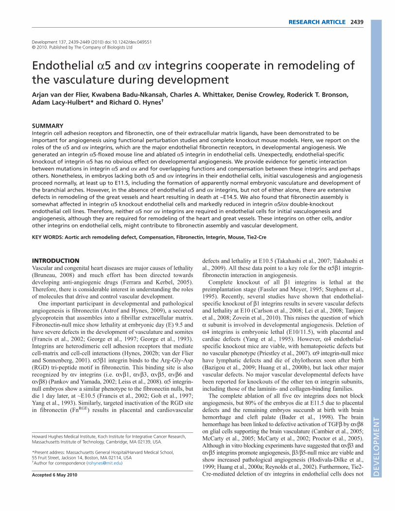
from such 5flox/flox � 5+/–; Tie2-CreTg/+ crosses (2 test, P>0.3; seeTable S1 in the supplementary material). FACS analysis for 5integrin expression in cell populations of freshly isolated samplesfrom E10.5 embryos (Fig. 1A,B) showed complete loss of 5integrin protein in both hematopoietic and endothelial cells derivedfrom the yolk sacs or from the integrin 5 conditional knockout (5-cKO) embryos themselves. This confirmed that 5 integrin wasindeed efficiently depleted early during embryonic development.
No obvious vascular defects were visualized by whole-mountstaining for platelet endothelial adhesion molecule [PECAM1(CD31)] and smooth muscle -actin (-SMA; ACTA2) ofvasculature of the inner-ear skin or trachea of adult 5-cKO mice(Fig. 1C). Similarly, immunohistochemical staining of sectionsfrom various other vascular beds showed no clear differences fromcontrols (data not shown).
The 5flox/–; Tie2-Cre (5-cKO) mice did not develop anyobvious spontaneous vascular or hematopoietic phenotypes for aperiod of 18 months. In addition, FACS analysis of the immunesystem showed no disparities in subsets of hematopoietic cells inadult 5-cKO mice (data not shown). These unexpected resultssuggested that deletion of 5 integrin from endothelial cells doesnot replicate the global 5-null phenotype. Together, these data
2441RESEARCH ARTICLEIntegrins in vascular development
Fig. 1. Tie2-Cre-mediated depletion of 5 integrin in endothelialcells results in no obvious vascular phenotypes. (A)FACS analysisof cells dissociated from E10.5 mouse yolk sacs, gated on immune cells(SSClow, PECAM1+) and endothelial cells (SSChigh, PECAM1+, VE-Cad+).Black line, isotype control antibody; green, 5 integrin on cells from5flox/+; Tie2-Cre embryos; red, 5 integrin on cells from 5flox/–; Tie2-Cre embryos. Note the complete loss of 5 integrin from endothelialand hematopoietic cells but not from control ‘other’ cells (PECAM1–).For details of FACS analysis, see Figs S2 and S4 in the supplementarymaterial. (B)Bar chart indicating loss of 5 integrin expression ongreater than 95% of endothelial cells (SSChigh, PECAM1+ VE-Cad+)isolated from E10.5 5flox/–; Tie2-Cre embryos. (C)Apparently normalvasculature in integrin 5 conditional knockout (5-cKO) mice (5f/–Tie versus 5f/+ Tie control) as seen by whole-mount staining forendothelial (PECAM1) and smooth muscle (-SMA) cells in (a-d) earskin and (e,f) trachea. Arteries (a), veins (v) and lymphatics (asterisk) areindicated. Scale bars: 180m in a,b,e,f; 45m in c,d.
DEVELO
PMENT
2442
show that early developmental deletion of 5 integrin inendothelial cells has no obvious effects on developmentalvasculogenesis or angiogenesis.
Integrin 5-deficient endothelial cells adhere tofibronectin in vitro but redistribute integrin v tofocal adhesionsWe isolated immortalized endothelial cell lines from adult lungs(mLEC) and brains (mBEC) using 5-cKO crosses into which aconditional SV40 large T antigen was introduced (Jat et al., 1991).FACS analysis and immunofluorescence staining showed that theclones were positive for ICAM2 and often PECAM1 and VE-cadherin (cadherin 5) (data not shown). FACS analysis andimmunoblots confirmed complete loss of 5 integrin in theseendothelial cell lines.5-KO endothelial cells showed reduced adhesion to fibronectin,
which was rescued by ectopic (over)expression of human 5integrin (see Fig. S2 in the supplementary material). Extendedadhesion times or increased fibronectin coating abolished thisdifference in adhesion between 5-KO and control cells (see Fig.S2C in the supplementary material). Immunofluorescence stainingfor vinculin of 5-KO endothelial cell cultures (mBECs, mLECs)plated on fibronectin consistently showed fewer, but larger andmore peripherally localized, focal adhesions (see Fig. S3 in thesupplementary material). We also observed relocalization of vintegrins from a diffuse surface expression in control cells to afocal adhesion localization in 5-KO cells (see Fig. S3 in thesupplementary material). FACS analysis revealed no, or minor,upregulation of v (see Fig. S2B in the supplementary material) or3 (not shown) integrin in 5-KO endothelial cells. Therelocalization and modest surface upregulation of v integrins in5-KO endothelial cells suggested that v integrin heterodimers(i.e. v1, 3, 5, 6 or 8) might be functionally compensatingfor loss of 5 integrins in endothelial cells. To explore thishypothesis, we knocked out both 5 and v integrins in endothelialcells using Tie2-Cre (5/v-dcKO).
Mice lacking both 5 and v integrins inendothelium die at ~E14.5 and haveorganizational defects in their great vesselsWe crossed doubly homozygous integrin 5;v floxed micecarrying the R26R-lacZ reporter gene (5flox/flox; vflox/flox;R26RlacZ/lacZ) to doubly heterozygous 5 and v integrin-null mice
carrying the Tie2-Cre allele (5+/–; v+/–; Tie2-CreTg/Tg). Wheneverpossible, we used mice homozygous for the Tie2-Cre transgene toincrease the numbers of offspring carrying a Tie2-Cre allele. Thesecrosses generate four potentially informative genotypes: 5/vconditional doubly hemizygous control mice (5flox/+; vflox/+;Tie2-Cre, hereafter designated 5/v-cdHemi), 5-cKO and vconditional hemizygous (5flox/–; vflox/+; Tie2-Cre, hereafter 5-cKO/v-cHemi), 5 conditional hemizygous and v-cKO (5flox/+;vflox/–; Tie2-Cre, hereafter 5-cHemi/v-cKO) and the 5/vconditional double knockout (5flox/–; vflox/–; Tie2-Cre, hereafter5/v-cdKO mice).
Table 1 shows that embryos lacking both 5 and v integrins inendothelial cells exhibit embryonic lethality and also that fewerthan half of the expected 5-cKO/v-cHemi mice survive (Table1). Strikingly, there were no obvious vascular defects in the 5-cHemi/v-cKO mice, suggesting that hemizygosity for 5 integrinis sufficient for survival in the absence of v integrins. Asexpected, 5-cHemi/v-cKO mice phenocopied the Tie2-Cre v-cKO mice and developed colitis at several months of age (Lacy-Hulbert et al., 2007). These 5/v Tie2-Cre crosses thus showed agenetic interaction between 5 and v mutations, which couldindicate overlap, compensation or convergence of the functions ofthese integrins. FACS analyses of E10.5 embryos confirmedefficient loss of both integrins (see Fig. S4 in the supplementarymaterial).
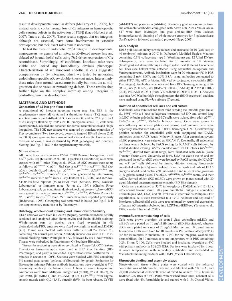
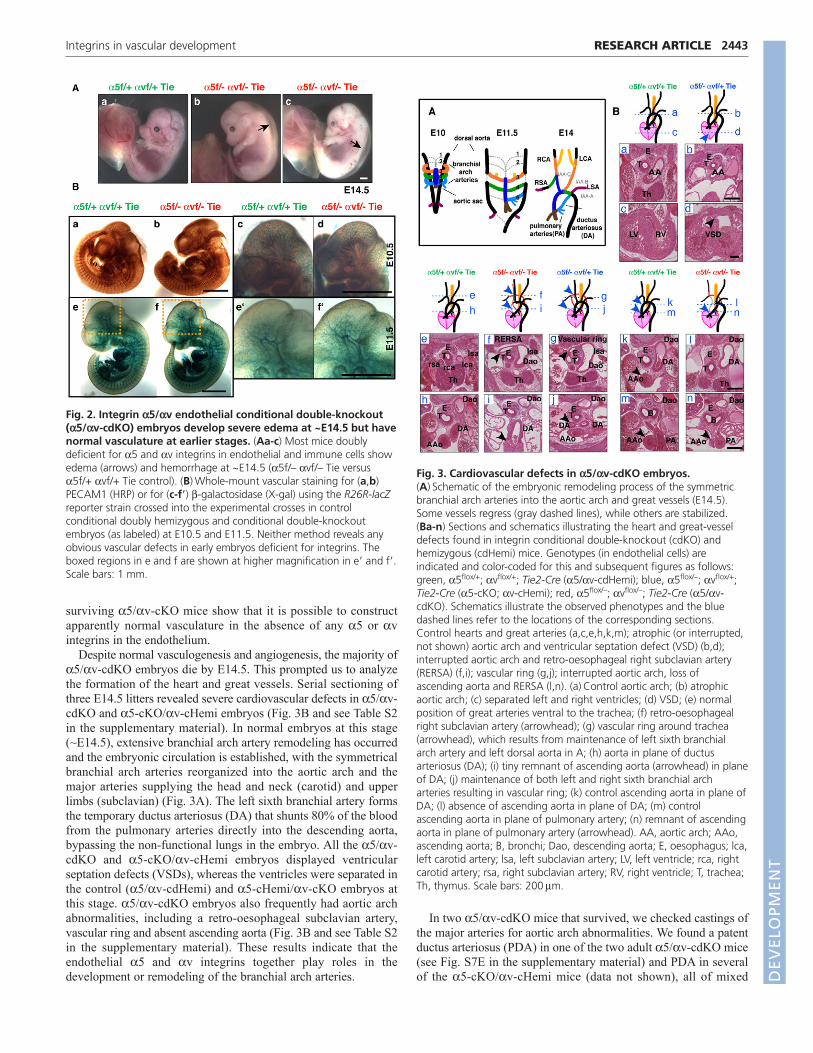
Analysis of timed matings confirmed that endothelial deletion ofboth the 5 and v integrin genes causes embryonic lethality. Mostembryos died at ~E14.5 and showed severe dorsal edema andsometimes hemorrhage (Fig. 2A). Similarly, a subgroup of 5-cKO/v-cHemi embryos was embryonic lethal (Table 1).Strikingly, all the embryos, as well as their vasculature, seemednormal until at least E11.5, as determined by whole-mount stainingfor PECAM1 (Fig. 2Ba-d; see Fig. S5 in the supplementarymaterial) and for -galactosidase expressed from the R26R-lacZreporter in 5/v-cdKO embryos (Fig. 2Be-f�).
A small number of 5/v-cdKO mice survived (<4% ofexpected) and were found to lack both 5 and v integrins on theirhematopoietic cells as determined by FACS analysis (data notshown). These adult 5/v-cdKO mice appeared to have normalvasculature and lymphatics, compared with control littermates,when examined by whole-mount vascular staining of ear skin andtrachea (see Fig. S6 in the supplementary material) or by sectioning(data not shown). The results on early embryos and on the rare
RESEARCH ARTICLE Development 137 (14)
Table 1. Genotypes of progeny from 5flox/flox; vflox/flox � 5+/–; v+/–; Tie2-Cre crosses Genotype
5flox/+; vflox/+; 5flox/–; vflox/+; 5flox/+; vflox/–; 5flox/–; vflox/–; Stage Tie2-Cre Tie2-Cre Tie2-Cre Tie2-Cre
P21 144 (46%) 48* (15%) 115 (37%) 5* (2%)
E9.5 4 5 6 2E10.5 39 31 31 48E11.5 10 16 20 (0, 1) 16E12.5 25 17 23 (0, 2) 31 (0, 1)E13.5 9 5 6 12 (1, 0)E14.5 21 25 (0, 5) 22 (0, 3) 31 (22, 4)E15.5 8 10 (0, 4) 10 11 (3, 4)E16.5 7 7 (0, 3) 2 5 (0, 5)E17.5 2 0 2 1 (0, 1)E18.5 8 (0, 1) 12 (0, 1) 6 8 (1, 6)
For postnatal progeny (P21), both numbers (total, 312) and percentages are given. For embryos, total numbers are listed and in parentheses are indicated first the number ofembryos with edema/death and second the number of absorbed embryos. Note the onset of edema and death in integrin-deficient embryos starting at E14.5.*Significantly different from expected; Poisson regression model, P<0.001. D
EVELO
PMENT
surviving 5/v-cKO mice show that it is possible to constructapparently normal vasculature in the absence of any 5 or vintegrins in the endothelium.
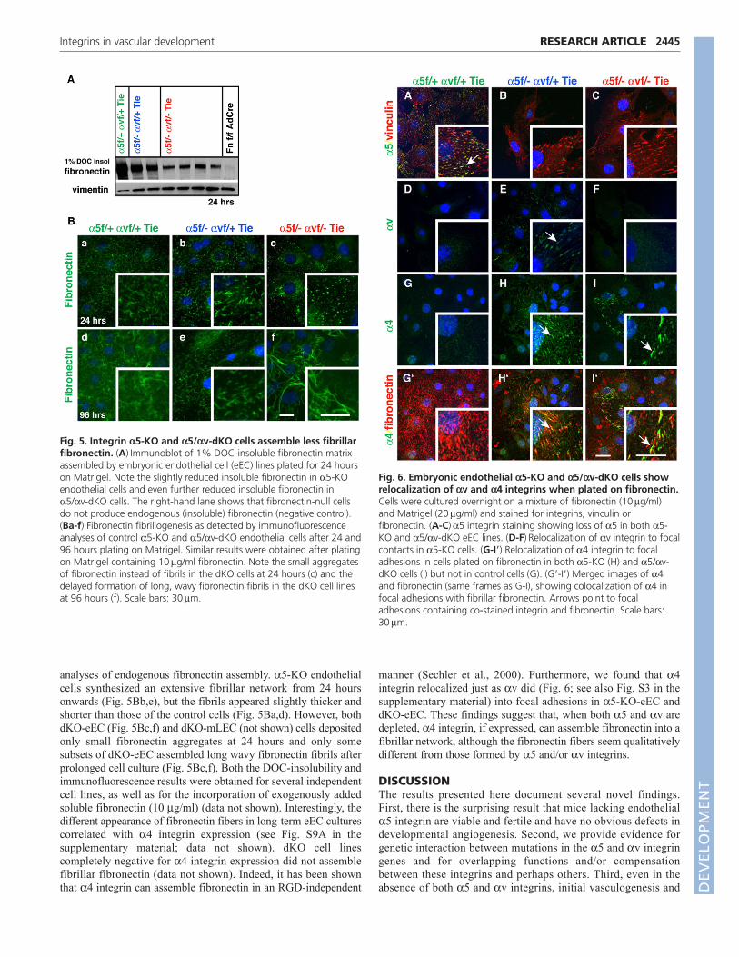
Despite normal vasculogenesis and angiogenesis, the majority of5/v-cdKO embryos die by E14.5. This prompted us to analyzethe formation of the heart and great vessels. Serial sectioning ofthree E14.5 litters revealed severe cardiovascular defects in 5/v-cdKO and 5-cKO/v-cHemi embryos (Fig. 3B and see Table S2in the supplementary material). In normal embryos at this stage(~E14.5), extensive branchial arch artery remodeling has occurredand the embryonic circulation is established, with the symmetricalbranchial arch arteries reorganized into the aortic arch and themajor arteries supplying the head and neck (carotid) and upperlimbs (subclavian) (Fig. 3A). The left sixth branchial artery formsthe temporary ductus arteriosus (DA) that shunts 80% of the bloodfrom the pulmonary arteries directly into the descending aorta,bypassing the non-functional lungs in the embryo. All the 5/v-cdKO and 5-cKO/v-cHemi embryos displayed ventricularseptation defects (VSDs), whereas the ventricles were separated inthe control (5/v-cdHemi) and 5-cHemi/v-cKO embryos atthis stage. 5/v-cdKO embryos also frequently had aortic archabnormalities, including a retro-oesophageal subclavian artery,vascular ring and absent ascending aorta (Fig. 3B and see Table S2in the supplementary material). These results indicate that theendothelial 5 and v integrins together play roles in thedevelopment or remodeling of the branchial arch arteries.
In two 5/v-cdKO mice that survived, we checked castings ofthe major arteries for aortic arch abnormalities. We found a patentductus arteriosus (PDA) in one of the two adult 5/v-cdKO mice(see Fig. S7E in the supplementary material) and PDA in severalof the 5-cKO/v-cHemi mice (data not shown), all of mixed
2443RESEARCH ARTICLEIntegrins in vascular development
Fig. 2. Integrin 5/v endothelial conditional double-knockout(5/v-cdKO) embryos develop severe edema at ~E14.5 but havenormal vasculature at earlier stages. (Aa-c) Most mice doublydeficient for 5 and v integrins in endothelial and immune cells showedema (arrows) and hemorrhage at ~E14.5 (5f/– vf/– Tie versus5f/+ vf/+ Tie control). (B)Whole-mount vascular staining for (a,b)PECAM1 (HRP) or for (c-f�) -galactosidase (X-gal) using the R26R-lacZreporter strain crossed into the experimental crosses in controlconditional doubly hemizygous and conditional double-knockoutembryos (as labeled) at E10.5 and E11.5. Neither method reveals anyobvious vascular defects in early embryos deficient for integrins. Theboxed regions in e and f are shown at higher magnification in e� and f�.Scale bars: 1 mm.
Fig. 3. Cardiovascular defects in 5/v-cdKO embryos.(A)Schematic of the embryonic remodeling process of the symmetricbranchial arch arteries into the aortic arch and great vessels (E14.5).Some vessels regress (gray dashed lines), while others are stabilized.(Ba-n) Sections and schematics illustrating the heart and great-vesseldefects found in integrin conditional double-knockout (cdKO) andhemizygous (cdHemi) mice. Genotypes (in endothelial cells) areindicated and color-coded for this and subsequent figures as follows:green, 5flox/+; vflox/+; Tie2-Cre (5/v-cdHemi); blue, 5flox/–; vflox/+;Tie2-Cre (5-cKO; v-cHemi); red, 5flox/–; vflox/–; Tie2-Cre (5/v-cdKO). Schematics illustrate the observed phenotypes and the bluedashed lines refer to the locations of the corresponding sections.Control hearts and great arteries (a,c,e,h,k,m); atrophic (or interrupted,not shown) aortic arch and ventricular septation defect (VSD) (b,d);interrupted aortic arch and retro-oesophageal right subclavian artery(RERSA) (f,i); vascular ring (g,j); interrupted aortic arch, loss ofascending aorta and RERSA (l,n). (a)Control aortic arch; (b) atrophicaortic arch; (c) separated left and right ventricles; (d) VSD; (e) normalposition of great arteries ventral to the trachea; (f) retro-oesophagealright subclavian artery (arrowhead); (g) vascular ring around trachea(arrowhead), which results from maintenance of left sixth branchialarch artery and left dorsal aorta in A; (h) aorta in plane of ductusarteriosus (DA); (i) tiny remnant of ascending aorta (arrowhead) in planeof DA; (j) maintenance of both left and right sixth branchial archarteries resulting in vascular ring; (k) control ascending aorta in plane ofDA; (l) absence of ascending aorta in plane of DA; (m) controlascending aorta in plane of pulmonary artery; (n) remnant of ascendingaorta in plane of pulmonary artery (arrowhead). AA, aortic arch; AAo,ascending aorta; B, bronchi; Dao, descending aorta; E, oesophagus; lca,left carotid artery; lsa, left subclavian artery; LV, left ventricle; rca, rightcarotid artery; rsa, right subclavian artery; RV, right ventricle; T, trachea;Th, thymus. Scale bars: 200m.
DEVELO
PMENT

2444
background. This led us to check 10- to 20-week-old 5-cKO mice(5flox/flox; Tie2-Cre) of which 9/10 had PDA (see Fig. S7A-D inthe supplementary material). These mice were from 5flox/flox �5flox/+; Tie2-Cre/+ crosses on a C57BL/6 N7 background andabout half of the 5flox/flox; Tie2-Cre (5-cKO) from this crossseemed to be lost before weaning (see Table S3 in thesupplementary material). However, in other crosses using a mixed129S4:C57BL/6 background and 5flox/–; Tie2-Cre mice the DAwas closed from postnatal day (P) 1 onwards, indicating a role ofpotential genetic background modifiers. These results indicate thatendothelial 5 or v integrins play roles in the development orremodeling of the vasculature.
Integrin 5/v-cdKO mice have normal branchialarch and cardiac cushion developmentWe next sought to define when and why defects arise in the greatarteries of 5/v-cdKO embryos. We first investigated whether thedevelopment of branchial arch arteries was normal (Fig. 4). Thebranchial arch arteries developed symmetrically and normallybetween E9.5 and E11.5 in 5/v-cdKO embryos, as shown bywhole-mount staining for PECAM1 and expression of the R26R-lacZ reporter (Fig. 4). Intracardial India ink injections alsoindicated normal symmetrical branchial arch formation (data notshown), as did histological analysis of branchial arch arterydevelopment in cdKO mice up to E11.5 (Fig. 4B and see Fig.S8G,H in the supplementary material). Furthermore, we detectedcomparable levels of staining for fibronectin around the vasculatureof control and cdKO embryos (Fig. 4B).
We also checked whether the cardiac cushions formed normallyin 5/v-cdKO mice. Endothelial cells migrate out of theendothelium into the cardiac jelly to form cardiac cushions, whichcontribute to the formation of the cardiac valves and closure of theseptum (Kisanuki et al., 2001; Savolainen et al., 2009; Webb et al.,1998) and affect the hemodynamics in the embryo, which couldplay a crucial role in the vascular remodeling process (Yashiro etal., 2007). However, whole-mount R26R-lacZ reporter staining ofE10.5 and E11.5 5/v-cdKO embryos did not show anydifferences in cardiac cushion formation (see Fig. S8 in thesupplementary material). In the outflow tract cushions, most cellsare derived from the neural crest and thus are not labeled by Tie2-Cre; R26R-lacZ (Kisanuki et al., 2001). However, the outflow tractcushions also appeared to have normal numbers of cells andexhibited comparable immunohistochemical staining forfibronectin (data not shown).
Great-artery remodeling phenotypes are found in many mutantsin which neural crest cell migration or differentiation of neural crestcells into smooth muscle cells covering the branchial arch arteriesis affected. However, we did not detect consistent differences insmooth muscle cell coverage of the branchial arch arteries betweenE10.5 and E11.5, indicating that the neural crest cells reach thebranchial arches and, at least at these stages, differentiate intosmooth muscle cells in a fashion comparable to that of their controllittermates (data not shown). Therefore, no obvious defects werefound in the formation of the branchial arches or of the cardiaccushions that could readily explain the subsequent arterialremodeling defects.
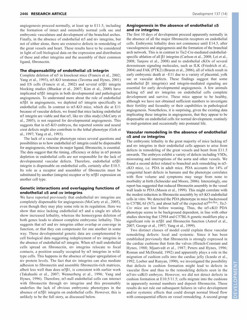
Integrin 5/v-dKO endothelial cells do notassemble normal fibronectin fibrilsTo analyze further the endothelial phenotype of 5/v-cdKO mice,we isolated dKO cell lines using two approaches. Adult lung-derived dKO endothelial (dKO-mLEC) cells were generated in
vitro from 5flox/flox; vflox/flox Immorto endothelial cells by AdCre-mediated excision of both 5 and v integrins. We also isolatedECs from E13.5 experimental cdKO crosses carrying the Immortogene. All 5/v-dKO cell lines failed to adhere effectively tofibronectin (see Fig. S9B in the supplementary material; data notshown) and were therefore maintained on Matrigel-coated tissueculture plates.
As mentioned above, integrin 5 plays a major role infibronectin matrix assembly (Wierzbicka-Patynowski andSchwarzbauer, 2003), so we analyzed fibronectin fibrillogenesis bydetermining the amount of fibronectin incorporated into 1% DOC-insoluble matrix fractions. 5-KO cells assembled less fibronectinthan did control cells, 5/v-dKO ECs assembled even less DOC-insoluble fibronectin, and dKO-mLEC cells assembled almost none(Fig. 5A; see Fig. S10 in the supplementary material; data notshown). These differences were also seen in immunofluorescence
RESEARCH ARTICLE Development 137 (14)
Fig. 4. Normal development of branchial arch arteries andfibronectin deposition until E11.5. (Aa-d) Whole-mount -galactosidase reporter expression at E9.5 (a,b) and whole-mountPECAM1 staining at E10.5 (c,d) showing normal symmetric formationof branchial arch arteries in 5/v-cdKO (b,d) as compared with controlcHemi (a,c) mouse embryos. (Ba-f)Frontal sections of E11.5 embryosshowing symmetric branchial arch formation and similar fibronectinstaining in (a) cHemi and (b) 5/v-cdKO embryos. Highermagnification images are shown of (c,d) carotid fibronectin stainingand (e,f) the left sixth arch artery. Note the similar fibronectin stainingpattern of several cell layers around the arteries. lca, left carotid artery;rca, right carotid artery; DoA, doral aorta. Scale bars: 100m.
DEVELO
PMENT
analyses of endogenous fibronectin assembly. 5-KO endothelialcells synthesized an extensive fibrillar network from 24 hoursonwards (Fig. 5Bb,e), but the fibrils appeared slightly thicker andshorter than those of the control cells (Fig. 5Ba,d). However, bothdKO-eEC (Fig. 5Bc,f) and dKO-mLEC (not shown) cells depositedonly small fibronectin aggregates at 24 hours and only somesubsets of dKO-eEC assembled long wavy fibronectin fibrils afterprolonged cell culture (Fig. 5Bc,f). Both the DOC-insolubility andimmunofluorescence results were obtained for several independentcell lines, as well as for the incorporation of exogenously addedsoluble fibronectin (10 g/ml) (data not shown). Interestingly, thedifferent appearance of fibronectin fibers in long-term eEC culturescorrelated with 4 integrin expression (see Fig. S9A in thesupplementary material; data not shown). dKO cell linescompletely negative for 4 integrin expression did not assemblefibrillar fibronectin (data not shown). Indeed, it has been shownthat 4 integrin can assemble fibronectin in an RGD-independent
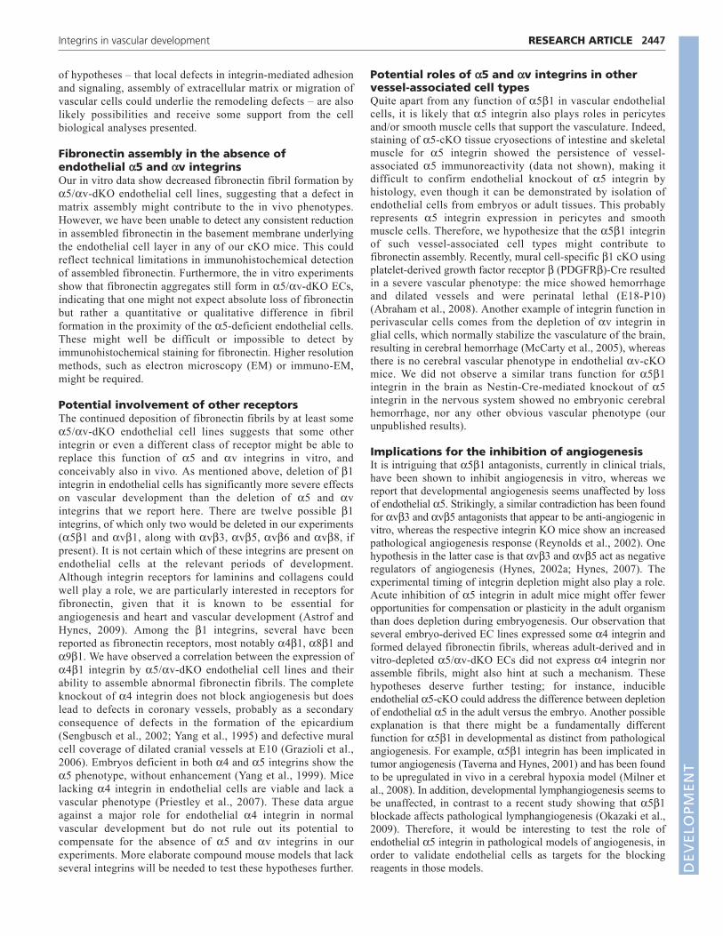
manner (Sechler et al., 2000). Furthermore, we found that 4integrin relocalized just as v did (Fig. 6; see also Fig. S3 in thesupplementary material) into focal adhesions in 5-KO-eEC anddKO-eEC. These findings suggest that, when both 5 and v aredepleted, 4 integrin, if expressed, can assemble fibronectin into afibrillar network, although the fibronectin fibers seem qualitativelydifferent from those formed by 5 and/or v integrins.
DISCUSSIONThe results presented here document several novel findings.First, there is the surprising result that mice lacking endothelial5 integrin are viable and fertile and have no obvious defects indevelopmental angiogenesis. Second, we provide evidence forgenetic interaction between mutations in the 5 and v integringenes and for overlapping functions and/or compensationbetween these integrins and perhaps others. Third, even in theabsence of both 5 and v integrins, initial vasculogenesis and
2445RESEARCH ARTICLEIntegrins in vascular development
Fig. 5. Integrin 5-KO and 5/v-dKO cells assemble less fibrillarfibronectin. (A)Immunoblot of 1% DOC-insoluble fibronectin matrixassembled by embryonic endothelial cell (eEC) lines plated for 24 hourson Matrigel. Note the slightly reduced insoluble fibronectin in 5-KOendothelial cells and even further reduced insoluble fibronectin in5/v-dKO cells. The right-hand lane shows that fibronectin-null cellsdo not produce endogenous (insoluble) fibronectin (negative control).(Ba-f) Fibronectin fibrillogenesis as detected by immunofluorescenceanalyses of control 5-KO and 5/v-dKO endothelial cells after 24 and96 hours plating on Matrigel. Similar results were obtained after platingon Matrigel containing 10g/ml fibronectin. Note the small aggregatesof fibronectin instead of fibrils in the dKO cells at 24 hours (c) and thedelayed formation of long, wavy fibronectin fibrils in the dKO cell linesat 96 hours (f). Scale bars: 30m.
Fig. 6. Embryonic endothelial 5-KO and 5/v-dKO cells showrelocalization of v and 4 integrins when plated on fibronectin.Cells were cultured overnight on a mixture of fibronectin (10g/ml)and Matrigel (20g/ml) and stained for integrins, vinculin orfibronectin. (A-C)5 integrin staining showing loss of 5 in both 5-KO and 5/v-dKO eEC lines. (D-F)Relocalization of v integrin to focalcontacts in 5-KO cells. (G-I�) Relocalization of 4 integrin to focaladhesions in cells plated on fibronectin in both 5-KO (H) and 5/v-dKO cells (l) but not in control cells (G). (G�-I�) Merged images of 4and fibronectin (same frames as G-I), showing colocalization of 4 infocal adhesions with fibrillar fibronectin. Arrows point to focaladhesions containing co-stained integrin and fibronectin. Scale bars:30m.
DEVELO
PMENT
2446
angiogenesis proceed normally, at least up to E11.5, includingthe formation of intact and ostensibly normal yolk sac andembryonic vasculature and development of the branchial arches.Finally, in the absence of endothelial 5 and v integrins, butnot of either alone, there are extensive defects in remodeling ofthe great vessels and heart. These results have to be consideredin light of cell biological data on the expression and distributionof these and other integrins and the assembly of their commonligand, fibronectin.
The dispensability of endothelial 5 integrinComplete deletion of 5 in knockout mice (Francis et al., 2002;Yang et al., 1993), 5-KO teratomas (Taverna and Hynes, 2001)and ES cells (Francis et al., 2002) and several 51 integrinblocking studies (Bhaskar et al., 2007; Kim et al., 2000) haveimplicated 51 integrin in both developmental and pathologicalangiogenesis. To understand more about the role of endothelial51 in angiogenesis, we depleted 5 integrin specifically inendothelial cells. In contrast to 5-KO mice, which die at E11because of vascular defects, we found that mice lacking endothelial5 integrin are viable and that 5, like v (this study) (McCarty etal., 2005), is not required for developmental angiogenesis. Thissuggests that in 5-KO embryos, the reported somite and neuralcrest defects might also contribute to the lethal phenotype (Goh etal., 1997; Yang et al., 1993).
The lack of a vascular phenotype raises several questions andpossibilities as to how endothelial 5 integrin could be dispensablefor angiogenesis, whereas its major ligand, fibronectin, is essential.The data suggest that the timing, efficiency or delay in 5 proteindepletion in endothelial cells are not responsible for the lack ofdevelopmental vascular defects. Therefore, endothelial 51integrin is dispensable on endothelial cells, certainly after E10.5.Its role as a receptor and assembler of fibronectin must besubstituted by another (integrin) receptor or by 51 expression onanother cell type.
Genetic interactions and overlapping functions ofendothelial 5 and v integrinsWe have reported previously that endothelial v integrins arecompletely dispensable for angiogenesis (McCarty et al., 2005),even though they may play some role in its regulation. Here weshow that mice lacking endothelial 5 and a single v alleleshow increased lethality, whereas the homozygous deletion ofboth genes leads to almost complete embryonic lethality. Thissuggests that 5 and v integrins either overlap or converge infunction, or that they can compensate for one another in someway. These developmental genetic data are complemented bycell biological data suggesting redeployment of v integrins inthe absence of endothelial 5 integrin. When 5-null endothelialcells spread on fibronectin, v integrins relocate to focalcontacts, a position usually occupied by 5 integrins in wild-type cells. This happens in the absence of major upregulation ofv protein levels. The fact that v integrins can also mediateadhesion to fibronectin and assemble fibronectin-rich matrices,albeit less well than does 51, is consistent with earlier work(Takahashi et al., 2007; Wennerberg et al., 1996; Yang andHynes, 1996). Therefore, 5-null endothelial cells can interactwith fibronectin through v integrins and this presumablyunderlies the lack of obvious embryonic phenotypes in theabsence of 51 integrin on endothelial cells. However, this isunlikely to be the full story, as discussed below.
Angiogenesis in the absence of endothelial 5and v integrinsThe first 10 days of development proceed apparently normally inthe absence of all the major fibronectin receptors on endothelialcells. Embryonic lethality does not commence until E14.5, aftervasculogenesis and angiogenesis and the formation of the branchialarch network. This is in contrast to Tie2-Cre-mediated endothelial-specific ablation of all 1 integrins (Carlson et al., 2008; Lei et al.,2008; Tanjore et al., 2008) and to endothelial cKOs of severaldownstream signaling molecules, such as ILK (Friedrich et al.,2004) and FAK (PTK2) (Braren et al., 2006), all of which result inearly embryonic death at ~E11 due to a variety of placental, yolksac or vascular defects. These findings suggest that someendothelial 1 integrin(s) and integrin-mediated signaling areessential for early developmental angiogenesis. A few animalslacking 5 and v integrins on endothelial cells completedevelopment and survive. These mice appear to be healthy,although we have not obtained sufficient numbers to investigatetheir fertility and fecundity or their capabilities in pathologicalangiogenesis. Nonetheless, it is surprising, given the existing dataimplicating these integrins in angiogenesis, that they appear to bedispensable on endothelial cells for normal development, routinelyto mid-gestation and occasionally into postnatal life.
Vascular remodeling in the absence of endothelial5 and v integrinsThe embryonic lethality in the great majority of mice lacking 5and v integrins in their endothelial cells appears to arise fromdefects in remodeling of the great vessels and heart from E11.5onwards. The embryos exhibit a variety of defects including VSDs,misrouting and interruptions of the aorta and other vessels. Wefound a second defect related to branchial arch remodeling in 5-cKO mice, i.e. PDA in adult mice. PDA accounts for 14% ofcongenital heart defects in humans and the phenotype correlateswith flow volume and symptoms may range from none tomorbidity at birth (Schneider and Moore, 2006). Interestingly, onereport has suggested that reduced fibronectin assembly in the vesselwall leads to PDA (Mason et al., 1999). This might correlate withthe modest reduction in fibronectin assembly by 5-KO endothelialcells in vitro. We detected the PDA phenotype in mice backcrossedto C57BL/6J (N7), and about half of the expected 5flox/flox; Tie2-Cre mice are lost before weaning. Thus, the 5-cKO PDAphenotype seems to be background dependent, in line with otherstudies showing that 129S4 and C57BL/6 genetic modifiers play asignificant role in 51 and fibronectin function (Astrof et al.,2007; George et al., 1997; Yang et al., 1999).
Two distinct classes of model could explain these vascularremodeling defects: local and systemic. Since it has beenestablished previously that fibronectin is strongly expressed inthe cardiac cushions that form the valves (ffrench-Constant andHynes, 1988; Mjaatvedt et al., 1987; Peters and Hynes, 1996;Roman and McDonald, 1992) and apparently plays a role in themigration of cushion cells into the cardiac jelly (Icardo et al.,1992; Loeber and Runyan, 1990), we investigated the possibilitythat defects in cushion formation might lead to defects invascular flow and thus to the remodeling defects seen in the5/v-cdKO embryos. However, we did not detect defects incushion formation at E10.5/11.5; cells migrate into the cushionsin apparently normal numbers and deposit fibronectin. Theseresults do not rule out subsequent failures in valve developmentand/or systemic effects that could disrupt normal blood flowwith consequential effects on vessel remodeling. A second group
RESEARCH ARTICLE Development 137 (14)
DEVELO
PMENT
of hypotheses – that local defects in integrin-mediated adhesionand signaling, assembly of extracellular matrix or migration ofvascular cells could underlie the remodeling defects – are alsolikely possibilities and receive some support from the cellbiological analyses presented.
Fibronectin assembly in the absence ofendothelial 5 and v integrinsOur in vitro data show decreased fibronectin fibril formation by5/v-dKO endothelial cell lines, suggesting that a defect inmatrix assembly might contribute to the in vivo phenotypes.However, we have been unable to detect any consistent reductionin assembled fibronectin in the basement membrane underlyingthe endothelial cell layer in any of our cKO mice. This couldreflect technical limitations in immunohistochemical detectionof assembled fibronectin. Furthermore, the in vitro experimentsshow that fibronectin aggregates still form in 5/v-dKO ECs,indicating that one might not expect absolute loss of fibronectinbut rather a quantitative or qualitative difference in fibrilformation in the proximity of the 5-deficient endothelial cells.These might well be difficult or impossible to detect byimmunohistochemical staining for fibronectin. Higher resolutionmethods, such as electron microscopy (EM) or immuno-EM,might be required.
Potential involvement of other receptorsThe continued deposition of fibronectin fibrils by at least some5/v-dKO endothelial cell lines suggests that some otherintegrin or even a different class of receptor might be able toreplace this function of 5 and v integrins in vitro, andconceivably also in vivo. As mentioned above, deletion of 1integrin in endothelial cells has significantly more severe effectson vascular development than the deletion of 5 and vintegrins that we report here. There are twelve possible 1integrins, of which only two would be deleted in our experiments(51 and v1, along with v3, v5, v6 and v8, ifpresent). It is not certain which of these integrins are present onendothelial cells at the relevant periods of development.Although integrin receptors for laminins and collagens couldwell play a role, we are particularly interested in receptors forfibronectin, given that it is known to be essential forangiogenesis and heart and vascular development (Astrof andHynes, 2009). Among the 1 integrins, several have beenreported as fibronectin receptors, most notably 41, 81 and91. We have observed a correlation between the expression of41 integrin by 5/v-dKO endothelial cell lines and theirability to assemble abnormal fibronectin fibrils. The completeknockout of 4 integrin does not block angiogenesis but doeslead to defects in coronary vessels, probably as a secondaryconsequence of defects in the formation of the epicardium(Sengbusch et al., 2002; Yang et al., 1995) and defective muralcell coverage of dilated cranial vessels at E10 (Grazioli et al.,2006). Embryos deficient in both 4 and 5 integrins show the5 phenotype, without enhancement (Yang et al., 1999). Micelacking 4 integrin in endothelial cells are viable and lack avascular phenotype (Priestley et al., 2007). These data argueagainst a major role for endothelial 4 integrin in normalvascular development but do not rule out its potential tocompensate for the absence of 5 and v integrins in ourexperiments. More elaborate compound mouse models that lackseveral integrins will be needed to test these hypotheses further.
Potential roles of 5 and v integrins in othervessel-associated cell typesQuite apart from any function of 51 in vascular endothelialcells, it is likely that 5 integrin also plays roles in pericytesand/or smooth muscle cells that support the vasculature. Indeed,staining of 5-cKO tissue cryosections of intestine and skeletalmuscle for 5 integrin showed the persistence of vessel-associated 5 immunoreactivity (data not shown), making itdifficult to confirm endothelial knockout of 5 integrin byhistology, even though it can be demonstrated by isolation ofendothelial cells from embryos or adult tissues. This probablyrepresents 5 integrin expression in pericytes and smoothmuscle cells. Therefore, we hypothesize that the 51 integrinof such vessel-associated cell types might contribute tofibronectin assembly. Recently, mural cell-specific 1 cKO usingplatelet-derived growth factor receptor (PDGFR)-Cre resultedin a severe vascular phenotype: the mice showed hemorrhageand dilated vessels and were perinatal lethal (E18-P10)(Abraham et al., 2008). Another example of integrin function inperivascular cells comes from the depletion of v integrin inglial cells, which normally stabilize the vasculature of the brain,resulting in cerebral hemorrhage (McCarty et al., 2005), whereasthere is no cerebral vascular phenotype in endothelial v-cKOmice. We did not observe a similar trans function for 51integrin in the brain as Nestin-Cre-mediated knockout of 5integrin in the nervous system showed no embryonic cerebralhemorrhage, nor any other obvious vascular phenotype (ourunpublished results).
Implications for the inhibition of angiogenesisIt is intriguing that 51 antagonists, currently in clinical trials,have been shown to inhibit angiogenesis in vitro, whereas wereport that developmental angiogenesis seems unaffected by lossof endothelial 5. Strikingly, a similar contradiction has been foundfor v3 and v5 antagonists that appear to be anti-angiogenic invitro, whereas the respective integrin KO mice show an increasedpathological angiogenesis response (Reynolds et al., 2002). Onehypothesis in the latter case is that v3 and v5 act as negativeregulators of angiogenesis (Hynes, 2002a; Hynes, 2007). Theexperimental timing of integrin depletion might also play a role.Acute inhibition of 5 integrin in adult mice might offer feweropportunities for compensation or plasticity in the adult organismthan does depletion during embryogenesis. Our observation thatseveral embryo-derived EC lines expressed some 4 integrin andformed delayed fibronectin fibrils, whereas adult-derived and invitro-depleted 5/v-dKO ECs did not express 4 integrin norassemble fibrils, might also hint at such a mechanism. Thesehypotheses deserve further testing; for instance, inducibleendothelial 5-cKO could address the difference between depletionof endothelial 5 in the adult versus the embryo. Another possibleexplanation is that there might be a fundamentally differentfunction for 51 in developmental as distinct from pathologicalangiogenesis. For example, 51 integrin has been implicated intumor angiogenesis (Taverna and Hynes, 2001) and has been foundto be upregulated in vivo in a cerebral hypoxia model (Milner etal., 2008). In addition, developmental lymphangiogenesis seems tobe unaffected, in contrast to a recent study showing that 51blockade affects pathological lymphangiogenesis (Okazaki et al.,2009). Therefore, it would be interesting to test the role ofendothelial 5 integrin in pathological models of angiogenesis, inorder to validate endothelial cells as targets for the blockingreagents in those models.
2447RESEARCH ARTICLEIntegrins in vascular development
DEVELO
PMENT

2448
ConclusionsThe results reported here reveal further complexity in the roles ofintegrins in vascular development. Although various lines ofevidence implicate 5 and v integrins in vascular developmentand angiogenesis, it is clear that these processes can proceedalmost, or completely, normally in the absence of either one ofthese integrin subunits and, to a significant extent, in the absenceof both. These results raise intriguing questions about overlappingfunctions or compensation among integrins. This possibilitypertains most strongly to the fibronectin receptor integrins becausethat ligand is the most essential extracellular matrix protein forvascular development. Since none of the clinical trials usingantagonists of these integrins has yet proven their efficacy as anti-angiogenic agents, it might be worth considering the possibility oftargeting both subunits at the same time. Basic developmentalquestions about which integrins play important roles in vasculardevelopment, and in which cells and cellular functions, requirefurther investigation. This will be challenging given the number ofpotential players, their embryonic lethality and possible overlap infunction.
AcknowledgementsWe acknowledge the Koch Institute Fannie E. Rippel Transgenics Facility andthe Flow Cytometry Core Facility for excellent technical assistance. We thankour colleagues from the R.O.H. laboratory for critically reading the manuscript.This work was supported by grants from the National Institutes of Health(PO1-HL66105 and the NIGMS Cell Migration Consortium, GC11451.126452,PI, A. F. Horwitz) and by the Howard Hughes Medical Institute of which R.O.H.is an Investigator. Deposited in PMC for release after 6 months.
Competing interests statementThe authors declare no competing financial interests.
Supplementary materialSupplementary material for this article is available athttp://dev.biologists.org/lookup/suppl/doi:10.1242/dev.049551/-/DC1
ReferencesAbraham, S., Kogata, N., Fassler, R. and Adams, R. H. (2008). Integrin beta1
subunit controls mural cell adhesion, spreading, and blood vessel wall stability.Circ. Res. 102, 562-570.
Astrof, S. and Hynes, R. O. (2009). Fibronectins in vascular morphogenesis.Angiogenesis 12, 165-175.
Astrof, S., Kirby, A., Lindblad-Toh, K., Daly, M. and Hynes, R. O. (2007). Heartdevelopment in fibronectin-null mice is governed by a genetic modifier onchromosome four. Mech. Dev. 124, 551-558.
Bader, B. L., Rayburn, H., Crowley, D. and Hynes, R. O. (1998). Extensivevasculogenesis, angiogenesis, and organogenesis precede lethality in micelacking all alpha v integrins. Cell 95, 507-519.
Bazigou, E., Xie, S., Chen, C., Weston, A., Miura, N., Sorokin, L., Adams, R.,Muro, A. F., Sheppard, D. and Makinen, T. (2009). Integrin-alpha9 is requiredfor fibronectin matrix assembly during lymphatic valve morphogenesis. Dev. Cell17, 175-186.
Bhaskar, V., Zhang, D., Fox, M., Seto, P., Wong, M. H., Wales, P. E., Powers,D., Chao, D. T., Dubridge, R. B. and Ramakrishnan, V. (2007). A functionblocking anti-mouse integrin alpha5beta1 antibody inhibits angiogenesis andimpedes tumor growth in vivo. J. Transl. Med. 5, 61.
Braren, R., Hu, H., Kim, Y. H., Beggs, H. E., Reichardt, L. F. and Wang, R.(2006). Endothelial FAK is essential for vascular network stability, cell survival,and lamellipodial formation. J. Cell Biol. 172, 151-162.
Bruneau, B. G. (2008). The developmental genetics of congenital heart disease.Nature 451, 943-948.
Cambier, S., Gline, S., Mu, D., Collins, R., Araya, J., Dolganov, G., Einheber,S., Boudreau, N. and Nishimura, S. L. (2005). Integrin alpha(v)beta8-mediatedactivation of transforming growth factor-beta by perivascular astrocytes: anangiogenic control switch. Am. J. Pathol. 166, 1883-1894.
Carlson, T. R., Hu, H., Braren, R., Kim, Y. H. and Wang, R. A. (2008). Cell-autonomous requirement for beta1 integrin in endothelial cell adhesion,migration and survival during angiogenesis in mice. Development 135, 2193-2202.
de Lange, W. J., Halabi, C. M., Beyer, A. M. and Sigmund, C. D. (2008). Germline activation of the Tie2 and SMMHC promoters causes noncell-specificdeletion of floxed alleles. Physiol. Genomics 35, 1-4.
Fassler, R. and Meyer, M. (1995). Consequences of lack of beta 1 integrin geneexpression in mice. Genes Dev. 9, 1896-1908.
Ferrara, N. and Kerbel, R. S. (2005). Angiogenesis as a therapeutic target. Nature438, 967-974.
ffrench-Constant, C. and Hynes, R. O. (1988). Patterns of fibronectin geneexpression and splicing during cell migration in chicken embryos. Development104, 369-382.
Francis, S. E., Goh, K. L., Hodivala-Dilke, K., Bader, B. L., Stark, M., Davidson,D. and Hynes, R. O. (2002). Central roles of alpha5beta1 integrin andfibronectin in vascular development in mouse embryos and embryoid bodies.Arterioscler. Thromb. Vasc. Biol. 22, 927-933.
Friedrich, E. B., Liu, E., Sinha, S., Cook, S., Milstone, D. S., MacRae, C. A.,Mariotti, M., Kuhlencordt, P. J., Force, T., Rosenzweig, A. et al. (2004).Integrin-linked kinase regulates endothelial cell survival and vasculardevelopment. Mol. Cell. Biol. 24, 8134-8144.
George, E. L., Georges-Labouesse, E. N., Patel-King, R. S., Rayburn, H. andHynes, R. O. (1993). Defects in mesoderm, neural tube and vasculardevelopment in mouse embryos lacking fibronectin. Development 119, 1079-1091.
George, E. L., Baldwin, H. S. and Hynes, R. O. (1997). Fibronectins are essentialfor heart and blood vessel morphogenesis but are dispensable for initialspecification of precursor cells. Blood 90, 3073-3081.
Goh, K. L., Yang, J. T. and Hynes, R. O. (1997). Mesodermal defects and cranialneural crest apoptosis in alpha5 integrin-null embryos. Development 124, 4309-4319.
Grazioli, A., Alves, C. S., Konstantopoulos, K. and Yang, J. T. (2006). Defectiveblood vessel development and pericyte/pvSMC distribution in alpha 4 integrin-deficient mouse embryos. Dev. Biol. 293, 165-177.
Hodivala-Dilke, K. M., McHugh, K. P., Tsakiris, D. A., Rayburn, H., Crowley,D., Ullman-Cullere, M., Ross, F. P., Coller, B. S., Teitelbaum, S. and Hynes,R. O. (1999). Beta3-integrin-deficient mice are a model for Glanzmannthrombasthenia showing placental defects and reduced survival. J. Clin. Invest.103, 229-238.
Huang, X., Griffiths, M., Wu, J., Farese, R. V., Jr and Sheppard, D. (2000a).Normal development, wound healing, and adenovirus susceptibility in beta5-deficient mice. Mol. Cell. Biol. 20, 755-759.
Huang, X. Z., Wu, J. F., Ferrando, R., Lee, J. H., Wang, Y. L., Farese, R. V., Jrand Sheppard, D. (2000b). Fatal bilateral chylothorax in mice lacking theintegrin alpha9beta1. Mol. Cell. Biol. 20, 5208-5215.
Hynes, R. O. (2002a). A reevaluation of integrins as regulators of angiogenesis.Nat. Med. 8, 918-921.
Hynes, R. O. (2002b). Integrins: bidirectional, allosteric signaling machines. Cell110, 673-687.
Hynes, R. O. (2007). Cell-matrix adhesion in vascular development. J. Thromb.Haemost. 5, 32-40.
Icardo, J. M., Nakamura, A., Fernandez-Teran, M. A. and Manasek, F. J.(1992). Effects of injecting fibronectin and antifibronectin antibodies on cushionmesenchyme formation in the chick. An in vivo study. Anat. Embryol. (Berl.) 185,239-247.
Jat, P. S., Noble, M. D., Ataliotis, P., Tanaka, Y., Yannoutsos, N., Larsen, L.and Kioussis, D. (1991). Direct derivation of conditionally immortal cell linesfrom an H-2Kb-tsA58 transgenic mouse. Proc. Natl. Acad. Sci. USA 88, 5096-5100.
Kim, S., Bell, K., Mousa, S. A. and Varner, J. A. (2000). Regulation ofangiogenesis in vivo by ligation of integrin alpha5beta1 with the central cell-binding domain of fibronectin. Am. J. Pathol. 156, 1345-1362.
Kisanuki, Y. Y., Hammer, R. E., Miyazaki, J., Williams, S. C., Richardson, J. A.and Yanagisawa, M. (2001). Tie2-Cre transgenic mice: a new model forendothelial cell-lineage analysis in vivo. Dev. Biol. 230, 230-242.
Koni, P. A., Joshi, S. K., Temann, U. A., Olson, D., Burkly, L. and Flavell, R. A.(2001). Conditional vascular cell adhesion molecule 1 deletion in mice: impairedlymphocyte migration to bone marrow. J. Exp. Med. 193, 741-754.
Lacy-Hulbert, A., Smith, A. M., Tissire, H., Barry, M., Crowley, D., Bronson, R.T., Roes, J. T., Savill, J. S. and Hynes, R. O. (2007). Ulcerative colitis andautoimmunity induced by loss of myeloid alphav integrins. Proc. Natl. Acad. Sci.USA 104, 15823-15828.
Lei, L., Liu, D., Huang, Y., Jovin, I., Shai, S. Y., Kyriakides, T., Ross, R. S. andGiordano, F. J. (2008). Endothelial expression of beta1 integrin is required forembryonic vascular patterning and postnatal vascular remodeling. Mol. Cell.Biol. 28, 794-802.
Leiss, M., Beckmann, K., Giros, A., Costell, M. and Fassler, R. (2008). The roleof integrin binding sites in fibronectin matrix assembly in vivo. Curr. Opin. CellBiol. 20, 502-507.
Loeber, C. P. and Runyan, R. B. (1990). A comparison of fibronectin, laminin, andgalactosyltransferase adhesion mechanisms during embryonic cardiacmesenchymal cell migration in vitro. Dev. Biol. 140, 401-412.
Mason, C. A., Bigras, J. L., O’Blenes, S. B., Zhou, B., McIntyre, B., Nakamura,N., Kaneda, Y. and Rabinovitch, M. (1999). Gene transfer in utero biologicallyengineers a patent ductus arteriosus in lambs by arresting fibronectin-dependentneointimal formation. Nat. Med. 5, 176-182.
RESEARCH ARTICLE Development 137 (14)
DEVELO
PMENT
McCarty, J. H., Monahan-Earley, R. A., Brown, L. F., Keller, M., Gerhardt, H.,Rubin, K., Shani, M., Dvorak, H. F., Wolburg, H., Bader, B. L. et al. (2002).Defective associations between blood vessels and brain parenchyma lead tocerebral hemorrhage in mice lacking alphav integrins. Mol. Cell. Biol. 22, 7667-7677.
McCarty, J. H., Lacy-Hulbert, A., Charest, A., Bronson, R. T., Crowley, D.,Housman, D., Savill, J., Roes, J. and Hynes, R. O. (2005). Selective ablation ofalphav integrins in the central nervous system leads to cerebral hemorrhage,seizures, axonal degeneration and premature death. Development 132, 165-176.
Milner, R., Hung, S., Erokwu, B., Dore-Duffy, P., LaManna, J. C. and delZoppo, G. J. (2008). Increased expression of fibronectin and the alpha 5 beta 1integrin in angiogenic cerebral blood vessels of mice subject to hypobarichypoxia. Mol. Cell. Neurosci. 38, 43-52.
Mjaatvedt, C. H., Lepera, R. C. and Markwald, R. R. (1987). Myocardialspecificity for initiating endothelial-mesenchymal cell transition in embryonicchick heart correlates with a particulate distribution of fibronectin. Dev. Biol.119, 59-67.
Nagy, A. (2003). Manipulating the Mouse Embryo: a Laboratory Manual. ColdSpring Harbor, NY: Cold Spring Harbor Laboratory Press.
Okazaki, T., Ni, A., Ayeni, O. A., Baluk, P., Yao, L. C., Vossmeyer, D.,Zischinsky, G., Zahn, G., Knolle, J., Christner, C. et al. (2009). alpha5beta1Integrin blockade inhibits lymphangiogenesis in airway inflammation. Am. J.Pathol. 174, 2378-2387.
Pankov, R. and Yamada, K. M. (2002). Fibronectin at a glance. J. Cell Sci. 115,3861-3863.
Peters, J. H. and Hynes, R. O. (1996). Fibronectin isoform distribution in themouse. I. The alternatively spliced EIIIB, EIIIA, and V segments show widespreadcodistribution in the developing mouse embryo. Cell Adhes. Commun. 4, 103-125.
Priestley, G. V., Ulyanova, T. and Papayannopoulou, T. (2007). Sustainedalterations in biodistribution of stem/progenitor cells in Tie2Cre+ alpha4(f/f) miceare hematopoietic cell autonomous. Blood 109, 109-111.
Proctor, J. M., Zang, K., Wang, D., Wang, R. and Reichardt, L. F. (2005).Vascular development of the brain requires beta8 integrin expression in theneuroepithelium. J. Neurosci. 25, 9940-9948.
Reynolds, L. E., Wyder, L., Lively, J. C., Taverna, D., Robinson, S. D., Huang,X., Sheppard, D., Hynes, R. O. and Hodivala-Dilke, K. M. (2002). Enhancedpathological angiogenesis in mice lacking beta3 integrin or beta3 and beta5integrins. Nat. Med. 8, 27-34.
Roman, J. and McDonald, J. A. (1992). Expression of fibronectin, the integrinalpha 5, and alpha-smooth muscle actin in heart and lung development. Am. J.Respir. Cell Mol. Biol. 6, 472-480.
Savolainen, S. M., Foley, J. F. and Elmore, S. A. (2009). Histology atlas of thedeveloping mouse heart with emphasis on E11.5 to E18.5. Toxicol. Pathol. 37,395-414.
Schneider, D. J. and Moore, J. W. (2006). Patent ductus arteriosus. Circulation114, 1873-1882.
Sechler, J. L., Cumiskey, A. M., Gazzola, D. M. and Schwarzbauer, J. E.(2000). A novel RGD-independent fibronectin assembly pathway initiated byalpha4beta1 integrin binding to the alternatively spliced V region. J. Cell Sci.113, 1491-1498.
Sengbusch, J. K., He, W., Pinco, K. A. and Yang, J. T. (2002). Dual functions of[alpha]4[beta]1 integrin in epicardial development: initial migration and long-term attachment. J. Cell Biol. 157, 873-882.
Soriano, P. (1999). Generalized lacZ expression with the ROSA26 Cre reporterstrain. Nat. Genet. 21, 70-71.
Srinivasan, R. S., Dillard, M. E., Lagutin, O. V., Lin, F. J., Tsai, S., Tsai, M. J.,Samokhvalov, I. M. and Oliver, G. (2007). Lineage tracing demonstrates the
venous origin of the mammalian lymphatic vasculature. Genes Dev. 21, 2422-2432.
Stephens, L. E., Sutherland, A. E., Klimanskaya, I. V., Andrieux, A., Meneses,J., Pedersen, R. A. and Damsky, C. H. (1995). Deletion of beta 1 integrins inmice results in inner cell mass failure and peri-implantation lethality. Genes Dev.9, 1883-1895.
Takahashi, S., Leiss, M., Moser, M., Ohashi, T., Kitao, T., Heckmann, D.,Pfeifer, A., Kessler, H., Takagi, J., Erickson, H. P. et al. (2007). The RGD motifin fibronectin is essential for development but dispensable for fibril assembly. J.Cell Biol. 178, 167-178.
Takahashi, S., Moser, M., Montanez, E., Nakano, T., Seo, M., Backert, S.,Inoue, I., Awata, T., Katayama, S., Komoda, T. et al. (2009). The fibronectinRGD motif is required for multiple angiogenic events during early embryonicdevelopment. Arterioscler. Thromb. Vasc. Biol. 30, e1.
Tanjore, H., Zeisberg, E. M., Gerami-Naini, B. and Kalluri, R. (2008). Beta1integrin expression on endothelial cells is required for angiogenesis but not forvasculogenesis. Dev. Dyn. 237, 75-82.
Taverna, D. and Hynes, R. O. (2001). Reduced blood vessel formation and tumorgrowth in alpha5-integrin-negative teratocarcinomas and embryoid bodies.Cancer Res. 61, 5255-5261.
Taverna, D., Disatnik, M. H., Rayburn, H., Bronson, R. T., Yang, J., Rando, T.A. and Hynes, R. O. (1998). Dystrophic muscle in mice chimeric for expressionof alpha5 integrin. J. Cell Biol. 143, 849-859.
Travis, M. A., Reizis, B., Melton, A. C., Masteller, E., Tang, Q., Proctor, J. M.,Wang, Y., Bernstein, X., Huang, X., Reichardt, L. F. et al. (2007). Loss ofintegrin alpha(v)beta8 on dendritic cells causes autoimmunity and colitis in mice.Nature 449, 361-365.
van der Flier, A. and Sonnenberg, A. (2001). Function and interactions ofintegrins. Cell Tissue Res. 305, 285-298.
van der Flier, A., Kuikman, I., Kramer, D., Geerts, D., Kreft, M., Takafuta, T.,Shapiro, S. S. and Sonnenberg, A. (2002). Different splice variants of filamin-B affect myogenesis, subcellular distribution, and determine binding to integrin[beta] subunits. J. Cell Biol. 156, 361-376.
Webb, S., Brown, N. A. and Anderson, R. H. (1998). Formation of theatrioventricular septal structures in the normal mouse. Circ. Res. 82, 645-656.
Wennerberg, K., Lohikangas, L., Gullberg, D., Pfaff, M., Johansson, S. andFassler, R. (1996). Beta 1 integrin-dependent and -independent polymerizationof fibronectin. J. Cell Biol. 132, 227-238.
Wierzbicka-Patynowski, I. and Schwarzbauer, J. E. (2003). The ins and outs offibronectin matrix assembly. J. Cell Sci. 116, 3269-3276.
Yang, J. T. and Hynes, R. O. (1996). Fibronectin receptor functions in embryoniccells deficient in alpha 5 beta 1 integrin can be replaced by alpha V integrins.Mol. Biol. Cell 7, 1737-1748.
Yang, J. T., Rayburn, H. and Hynes, R. O. (1993). Embryonic mesodermaldefects in alpha 5 integrin-deficient mice. Development 119, 1093-1105.
Yang, J. T., Rayburn, H. and Hynes, R. O. (1995). Cell adhesion events mediatedby alpha 4 integrins are essential in placental and cardiac development.Development 121, 549-560.
Yang, J. T., Bader, B. L., Kreidberg, J. A., Ullman-Cullere, M., Trevithick, J. E.and Hynes, R. O. (1999). Overlapping and independent functions of fibronectinreceptor integrins in early mesodermal development. Dev. Biol. 215, 264-277.
Yashiro, K., Shiratori, H. and Hamada, H. (2007). Haemodynamics determinedby a genetic programme govern asymmetric development of the aortic arch.Nature 450, 285-288.
Zovein, A. C., Luque, A., Turlo, K. A., Hofmann, J. J., Yee, K. M., Becker, M.S., Fassler, R., Mellman, I., Lane, T. F. and Iruela-Arispe, M. L. (2010) Beta1integrin establishes endothelial cell polarity and arteriolar lumen formation via aPar3-dependent mechanism. Dev. Cell 18, 39-51.
2449RESEARCH ARTICLEIntegrins in vascular development
DEVELO
PMENT