endovascular repair of traumatic thoracic aortic disruptions with “stacked” abdominal endograft...
TRANSCRIPT
From the Southern Association for Vascular Surgery
Endovascular repair of traumatic thoracic aorticdisruptions with “stacked” abdominal endograftextension cuffsDavid Rosenthal, MD, Eric D. Wellons, MD, Allison B. Burkett, MD, Paul V. Kochupura, MD, andSusan M. Hancock, MD, Atlanta, Ga
Objective: Endovascular stent graft repair of a traumatic thoracic aortic disruption (TTAD) is rapidly becoming anaccepted alternative to open surgical repair. The use of currently approved thoracic stent grafts especially in youngerpatients with small, “steep,” tapered aortas, remains a concern due to the acute thoracic endograft collapse and enfolding.The objective of this study, the largest report to date, was to evaluate the mid-term results of TTAD treated withabdominal aortic “stacked” extension cuffs, with follow-up extending to 41 months.Methods: Thirty-one patients with multi-system trauma (age range, 15 to 61; mean 31.4 years) were seen after motorvehicle accidents between January 1, 2003 and July 1, 2007. Chest x-ray findings warranted thoracic CT scans, whichrevealed disruptions of the thoracic aorta. Intra-operative arteriograms in all patients and intravascular ultrasound(IVUS) (n � 17) delineated the extent of the aortic injuries. The aortic length from the subclavian artery to the injuryaveraged 2.5 cm (range, 1.5 to 4.0 cm). The repairs were performed with Gore (W.L. Gore & Associates, Flagstaff, Ariz)(n � 15), Aneuryx (Medtronic, Santa Rosa, Calif) (n � 15), and Zenith (Cook, Inc., Bloomington, Ind) (n � 1) AorticExtension Cuffs. A femoral artery approach was used in 27 patients and a supra-inguinal retroperitoneal iliac approachin four. All patients underwent thoracic CT scans during follow-up.Results: In all patients, the stent-graft cuffs successfully excluded the TTAD: 21 patients had 2 cuffs, 9 had 3 cuffs, and1 had 4 cuffs. The aorta adjacent to the injury mean diameter was 18.5 mm (range, 17-24 mm). No subclavian arterieswere covered. Two patients required an additional cuff for exclusion of the Type I endoleaks at the distal attachment sitewithin 6 weeks of initial endograft repair. There were no procedure-related deaths; 2 patients died of other injuries. Atfollow-up, extending to 41 months (range, 3 to 41 months), two pseudo-aneurysms occurred which required openoperative repair: 1 due to infection (4 months) and a leaking pseudoaneurysm (14 months). A CT scan in all othersurvivors demonstrated no device-related complications, endoleaks, or cuff migrations.Conclusion: Stent-graft repair of TTAD is technically feasible. The technique of “stacked” aortic cuffs provides an acceptableoption when urgent therapy is needed, when patients are deemed high-risk for open operative repair, or until thoracic
endografts are designed which can safely treat focal, smaller aortic diameter injuries. (J Vasc Surg 2008;48:841-4.)Blunt trauma to the thoracic aorta is a life-threatening,often fatal condition, in patients who frequently havedevastating multiple system injuries. Indeed, operativerepair of thoracic aortic injuries are associated with mor-tality rates approaching 30%1,2 and paraplegia rates re-ported as high as 18%.3
The classic injury mechanism due to blunt thoracicaortic trauma is related to the combination of decelerationand traction at the immobile aortic isthmus, which repre-sents the junction between the mobile aortic arch and thefixed descending thoracic aorta. The isthmus is the mostcommon site of rupture (70%), followed by the ascendingaorta or aortic arch (18%) and the distal thoracic aorta(14%).3,4
The endovascular repair of traumatic thoracic aorticdisruption (TTAD) is rapidly emerging as a favorable alter-
From the Department of Vascular Surgery, Atlanta Medical Center.Competition of interest: none.Presented at the Annual Meeting of the Southern Association for Vascular
Surgery, Rio Grande, Puerto Rico, Jan 17-20, 2007.Reprint requests: David Rosenthal, MD, 315 Boulevard, NE, Suite 412,
Atlanta, GA 30312 (e-mail: [email protected]).0741-5214/$34.00Copyright © 2008 by The Society for Vascular Surgery.
doi:10.1016/j.jvs.2008.05.055native to open surgical repair, and several studies havereported lower peri-operative mortality, paraplegia, opera-tive time, operative blood loss, and hospital length ofstay.5-7 The use of currently approved thoracic stent-grafts,however, may not be possible, especially in younger pa-tients with small, “steep,” tapered aortas. The objective ofthis study, the largest reported to date, was to evaluate theresults of the use of commercially available “stacked” ab-dominal endograft extension cuffs to treat TTAD caused byblunt trauma.
METHODS
A retrospective review of 31 patients who underwentendoluminal repair of blunt TTAD between January 1,2003, and July 1, 2007, was performed. During the sametime interval, 2 other patients with TTADs underwentrepairs with the Gore TAG device (W.L. Gore & Associ-ates, Flagstaff, Ariz). These 2 patients’ thoracic aortas mea-sured 28 and 30 mm in diameter and they were notincluded in the study. No open operative procedures wereperformed during this interval. All patients had been inmotor vehicle accidents and sustained severe blunt trauma.Patients underwent chest x-rays and suspected findings for
TTAD mandated computed tomography angiography841

JOURNAL OF VASCULAR SURGERYOctober 2008842 Rosenthal et al
(CTA) scan (Fig 1). Measurements of the proximal anddistal thoracic aortic diameter and the length between theleft subclavian artery and the aortic disruption were ob-tained from the CTA scan.
Repairs were performed in the operating room with thepatient under general anesthesia with standby cardiovascu-lar surgery support available. Intravenous heparin was ad-ministered judiciously (ie, 50u IV/kg) as the majority ofthe patients had severe concomitant injuries (mean ISS of40). Cutaneous femoral artery access was used for aortog-raphy. A 5 French sheath was placed in the femoral arteryand a marker pigtail catheter was passed under fluoroscopicguidance to the aortic arch for aortography. The contralat-eral femoral artery was exposed in standard fashion forstent-graft access, and an 8 French sheath was placed. Afteraortography, intravascular ultrasound (IVUS) (n � 17)(Volcano Corporation, Rancho Cordova, Calif) was per-formed of the aortic arch and descending thoracic aorta tofurther delineate the anatomy and to visualize the locationof the disruption in relation to the left subclavian artery.After the IVUS catheter was removed, a wire exchange wasperformed and an Amplatz (Meditech/Boston Scientific,Natick, Mass) Super-Stiff Wire was placed in the ascendingarch for stent graft deployment. When the femoral arterieswere too small to accept the device, or the device was notlong enough to reach the thoracic aorta, a supra-inguinalincision was made for proximal access to the common iliacartery. In 4 patients, a supra-inguinal retroperitoneal iliacartery approach allowed access to the iliac. The endograftextension cuffs were oversized approximately 10-15%, anda distance of 1.5-2.0 cm between subclavian artery andinjury site was considered adequate for endograft cuffplacement. The endograft cuffs were passed “sheathless”(without a separate introducer sheath) as vessels were freeof occlusive disease and this allowed for the maximal lengthof device to reach the thoracic aorta. The endograft exten-sion cuffs were deployed, distal to proximal whenever pos-sible, (Fig 2). Ballooning was reserved for malposition or
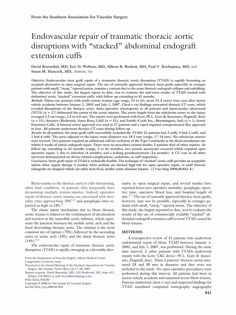
Fig 1. Computed tomography angiography demonstrates bluntaortic disruption with peri-aortic hematoma (arrow).
Type I endoleaks in order to minimize the potential for
further aortic injury and retrograde dissection. CT scanswere obtained within 72 hours after implantation, andfollow-up CT scans were obtained per endograft surveil-lance protocols.
RESULTS
The mechanism of the TTAD injury was motor vehiclecrash in 26 patients and motorcycle crash in 5. Twenty-fourpatients underwent endovascular repair less than 36 hoursafter injury. The delay from injury to repair in the other 7patients, all of whom were in an unstable condition frommultiple injuries, averaged 2.5 days (range, 2 to 5 days).The mean injury severity score (ISS) was 40 and concomi-tant injuries included coexisting head injuries (21), extrem-ity fractures (19), pelvic fractures (9), sternal/clavicle frac-tures (6), and solid organ injuries (5).
There were no procedure-related deaths, paraplegia,renal failure, or other cardiovascular complications. Twopatients died from causes unrelated to the endograft pro-cedures: 1 from multi-system organ failure and the otherwhen life support measures were withdrawn. The mean agewas 31.4 years (range, 15 to 61 years) and 19 patients weremen. Twenty-seven procedures were successfully donethrough a femoral artery approach. Iliac artery Dacron graftconduits for access were necessary in 2 patients due to smallfemoral arteries. In 2 other patients the device was too shortto read the site of injury and these patients had directpuncture of the iliac artery after the stent-graft device wastunneled under the inguinal ligament (similar to an aortofemoral bypass) for easy access to the common iliac artery;this obviated the need for a conduit and provided a lessangled approach to the iliac artery. No intra-operativeconversions to an open procedure were necessary.
In all patients, the endograft extension cuffs success-fully excluded the traumatic disruptions based on intra-operative aortography. Gore Excluder Aortic ExtensionCuffs (W.L. Gore & Associates, Flagstaff, Ariz) were used
Fig 2. Technique of “distal to proximal” deployment of endograftextension cuffs to improve seal and wall apposition (arrows indicateddistal to proximal deployment).
in 15 patients, AneuRx Extension Cuffs (Medtronic, Santa

JOURNAL OF VASCULAR SURGERYVolume 48, Number 4 Rosenthal et al 843
Rosa, Calif) were used in 15, and a modified Zenith (Cook,Inc., Bloomington, Ind) Cuff was used in 1 patient, persurgeon’s preference. Mean aortic diameter was 18.5 mm(range, 17 to 24 mm). No subclavian arteries were covered.Twenty-one patients had 2 cuffs, 9 had 3 cuffs, and 1 had 4cuffs. The aortic length from the subclavian artery to theaortic disruption, the “landing zone”, averaged 2.5 cm(range, l.5 to 4.0 cm). The average estimated blood losswas 150 mL (range, 75 to 300 mL).
Postoperative CT imaging was performed in all patientsprior to discharge. Two patients required an additional cufffor exclusion of Type I endoleaks at the distal attachmentsite within 6 weeks of initial endograft repair. These deviceswere deployed uneventfully. CT scan follow-up extendingto 41 months (range, 3 to 41 months) was performed in 27patients; 2 were lost to follow-up. Two pseudo-aneurysmsoccurred which required open operative repair: 1 was dueto a pulmonary abscess which infected the endograft 4months after endovascular repair, and the other a “leaking”pseudoaneurysm which occurred 14 months after endovas-cular repair. CT scans in the other 25 survivors demon-strated no device-related complications, endoleaks, or cuffmigrations compared to immediate postoperative images.
DISCUSSION
Since the initial report by Dake et al,8 of the successfulrepair of a traumatic aortic disruption with stent-grafts,many case reports and several institutional reviews havereproduced their results.9 However, traumatic aortic inju-ries tend to affect younger populations, in contrast tothoracic aneurysms, and commercially available devices arenot designed for use in young thoracic aortas. The youngaorta tends to be more narrow and the turn radius of theaortic arch much tighter and “steeper.”10
The small size of the aorta in young patients createsnew challenges that one rarely sees in patients with aneu-rysm disease and access vessels are often smaller as well. Inour series of 31 patients, however, the mean aortic diameterwas 18.5 mm (range, 17 to 24 mm), but only 4 patientsrequired iliac artery access, as the access vessels were free ofocclusive disease or tortuosity and readily dilated to accom-modate the endograft extension cuff delivery systems whichwere passed “sheathless” and were an approximate 18Fdiameter.
The smallest available thoracic endograft in the UnitedStates is the Gore TAG device (26 mm diameter) which wasdesigned for patients with thoracic aortic aneurysm whohave a larger diameter thoracic aorta, and “flatter” aorticarch curvature due to aortic elongation associated withaging. The 26 mm TAG device should be used in treatingaortas equal to or larger than 23 mm in diameter. If, forexample, a 26 mm TAG device is placed in 18.5 mm aorta,it represents an approximate 40% oversize. It appears thatthe adverse events reported to date with the use of the GoreTAG device to treat TTAD may be due to device oversizingbeyond the recommended Instructions for Use (IFU) ap-proved by the FDA, which may result in crimping, fracture,
endoleak, migration, and device collapse.11,12 Adverseevents, such as acute endoprosthesis collapse or enfolding,however, are not unique to the TAG endograft and havebeen reported with other endografts, when such grafts wereoversized as well.11 The unique packaging and releasemechanism of the TAG device may exacerbate the prob-lem of enfolding as the device is packaged in a furledconfiguration, and a smaller aorta may prevent it fromunfolding completely.13 Although small aortic diametermay promote collapse, other variables such as poor appo-sition of the endograft to the aortic wall, acute aortic archangulation, and other anatomic factors may also play acausative role in endoprosthesis collapse, although thisremains to be proven.13,14 Because of these concerns, manysurgeons are reluctant to use the TAG device in patientswith aortas measuring �23 mm in diameter.
Another concern with the use of thoracic aneurysmendografts for treating TTAD is the potential need to coverthe left subclavian artery. Coverage of the left subclavianartery is innocuous in most cases, but may be fraught withhazard when a left internal mammary-coronary bypass ordominant left vertebral artery is present, or a history ofprevious abdominal aortic surgery, or internal iliac arteryocclusions, which may increase the risk of postoperativespinal cord ischemia is noted.15 In this setting, carotid-subclavian bypass is indicated. Coverage of the left subcla-vian artery may also place the device in the transverseportion of the aortic arch which may lead to poor apposi-tion against the aortic wall, device collapse, and the creationof an inferior “lip” which may create a proximal endoleak.10
With stacked cuffs, both of these problems are readilyavoided and the left subclavian artery is not covered. Alimitation of abdominal endograft cuffs, however, is theirshort length, which necessitates the use of multiple cuffsand creates the potential for device separation and a poten-tial Type III endoleak. For this reason, the cuffs are de-ployed distal for proximal to improve seal and wall apposi-tion. In this series, 21 patients had 2 cuffs, 9 had 3 cuffs, 1had 4 cuffs, and only 2 endoleaks occurred. Another limi-tation with the abdominal endograft cuffs is the relativelyshort delivery system. However, in only 2 patients were weunable to reach the desired deployment location by femoralaccess; these patients underwent uneventful iliac arteryaccess.
Although the use of aortic extension cuffs to treatTTAD is “off label,” no enfolding or device collapse hasbeen reported with this technique. Two patients, however,had Type I endoleaks within 6 weeks of initial endograftrepair which were treated with an additional extension cuffearly in our experience (case 4 and 9). The wisest course ofaction appears to be to add an extra cuff if any doubt oftreatment length is present. Two late pseudo-aneurysmsalso occurred (one due to infection from a pulmonaryabscess at 4 months and one at the distal attachment site at14), for an overall complication rate of 13%.
Nevertheless, we believe that the use of abdominalextension cuffs are best to treat TTAD when the aorticdiameter is less than 23 mm or the radius of curvature is
small, which it inherently tends to be in the younger patient
JOURNAL OF VASCULAR SURGERYOctober 2008844 Rosenthal et al
population commonly affected by TTAD. The long-termperformance of abdominal aortic cuffs in the thoracic aortais unknown and will only be answered by other reports withlong-term follow-up. Patients with TTAD tend to beyoung and have decades of life expectancy within which theaorta may enlarge resulting in loss of fixation and materialfatigue (ie, stent fractures, fabric fatigue) may occur. Ifthese problems occur, it will likely be years later, butpatients must be made aware of the necessity for life longfollow-up. Regardless, until smaller diameter, shorterlength thoracic endografts are made with increased radialforce, better attachment systems, and increased flexibility tobetter oppose the inner wall of the aortic arch, the tech-nique of “stacked” abdominal aortic cuffs provides anacceptable option when urgent therapy to treat TTAD isneeded, especially in younger patients and when patientsare deemed high-risk for operative repair.
AUTHOR CONTRIBUTIONS
Conception and design: DR, EWAnalysis and interpretation: DR, EW, AB, PK, SHData collection: AB, PK, SHWriting the article: DR, AB, EWCritical revision of the article: DR, EW, AB, PK, SHFinal approval of the article: DR, EW, AB, PK, SHStatistical analysis: AB, PKObtained funding: Not applicableOverall responsibility: DR
REFERENCES
1. Cowley RA, Turney SZ, Hankins JR, Rodriquez A, Attar S, Shankar BS.Rupture of thoracic aorta caused by blunt trauma: a fifteen-year expe-rience. J Thorac Cardiovas Surg 1990;100:652-61.
2. von Oppell UO, Duanne TT, DeGroot MK, Zilla P. Traumatic aorticrupture: twenty-year meta analysis of mortality and risk for paraplegia.
Ann Thorac Surg 1994;58:585-93.3. Fabian TC, Richardson JD, Croce MA, Smith JS Jr, Rodman G Jr,Kearney PA, et al. Prospective study of blunt aortic injury: multicentertrial of the American Association for the Surgery of Trauma. J Trauma1997;42:374-80; discussion 380-3.
4. Jamieson WR, Janusz MT, Gudas VM, Burr LH, Fradet GJ, HendersonC. Traumatic rupture of the thoracic aorta third decade of experience.Am J Surg 2002;183:571-5.
5. Riesenman PJ, Farber MA, Rich PB, Sheridan BC, Mendes RR, Sheri-dan BC, Mendes RR, Marston WA, Keagy BA. Outcomes of surgicaland endovascular treatment of acute traumatic thoracic aortic injury. JVasc Surg 2007;46:934-40.
6. Ott MC, Stewart TC, Lawlor DK. Management of blunt thoracic aorticinjuries: endovascular stents versus open repair. J Trauma 2004;56:565-70.
7. Kasarajan K, Heffernan D, Langsfeld M. Acute thoracic aortic trauma: acomparison of endoluminal stent grafts with open repair and nonopera-tive management. Ann Vasc Surg 2003;17:589-95.
8. Dake MD, Miller DC, Semba CP, Mitchell RS, Walker PJ, Liddell RP.Transluminal placement of endovascular stent grafts for the treat-ment of descending thoracic aortic aneurysms. N Eng J Med 1994;331:1729-34.
9. Lin PH, Lumsden AB. Traumatic thoracic aortic injury. EndovascToday Feb 2006:58-66.
10. Neschis DG, Moaine S, Gutta R, Charles K, Scalea TM, Flynn WR,Griffith BP. Twenty consecutive cases of endograft repair of traumaticaortic disruptions: lessons learned. J Vasc Surg 2007;45:487-92.
11. Idu MM, Reckers JA, Balm R, Ponsen KJ, de Mol BA, Legemate DA.Collapse of a stent-graft following treatment of a traumatic thoracicaortic rupture. J Endovasc Ther 2005;12:503-7.
12. Steinbauer MG, Stehr A, Pfister K, Herold T, Zorger N, Topel I, et al.Endovascular repair of proximal endograft collapse after treatment forthoracic aortic disease. J Vasc Surg 2006;43:609-12.
13. Muhs BE, Balms R, White GH, Verhagen HJM. Anatomic factorsassociated with acute endograft collapse after Gore TAG treatment ofthoracic aortic dissection or traumatic rupture. J Vasc Surg 2007;45:655-61.
14. Riesenman PJ, Farber MA. Thoracic endovascular aortic repair oftraumatic injuries involving the descending thoracic aorta. J CardiovascSurg 2007;48:741-50.
15. Go MR, Barbato JE, Dillavou ED, Gupta N, Rhee RY, Makaroun MS,Cho JS. Thoracic endovascular aortic repair for traumatic aortic trans-action. J Vasc Surg 2007;46:928-33.
Submitted Jan 2, 2008; accepted May 14, 2008.