energetics of peptide recognition by the second pdz domain of human protein tyrosine phosphatase 1e ...
TRANSCRIPT
Energetics of Peptide Recognition by the Second PDZ Domain of Human ProteinTyrosine Phosphatase 1E†
Stoyan Milev,‡ Sasa Bjelic,‡ Oleg Georgiev,§ and Ilian Jelesarov*,‡
Biochemisches Institut der UniVersitat Zurich and Institut fur Molekularbiologie der UniVersitat Zurich, Winterthurerstrasse190, CH-8057 Zu¨rich, Switzerland
ReceiVed September 8, 2006; ReVised Manuscript ReceiVed October 31, 2006
ABSTRACT: Formation of protein-protein assemblies is essential in maintaining cell structure and function.Conservation of structural motifs and binding sites is the result of evolutionary pressure for solutionscompatible with both molecular economy and regulation. PDZ domains are a typical example: A conservedfold governs specificity toward recognition of C-terminal protein sequences by small sequential and/orstructural deviations within a canonical binding mode. The energetic principles underlying the strengthand specificity of PDZ-protein interactions are practically unknown. We use the second PDZ domain(PDZ2) of the human protein tyrosine phosphatase (hPTP1E) as a model to study the energetics of peptidebinding to a class I PDZ domain. Calorimetric experiments reveal the enthalpy, entropy, and heat capacitychanges accompanying PDZ2 binding to the C-terminal pentadecapeptide derived from the guaninenucleotide exchange factor RA-GEF2. Association is driven by favorable enthalpy and entropy changesbelow 18°C. Above that temperature the entropy change opposes complex formation. Structure-basedpredictions poorly reproduce the observed thermodynamic profile of the PDZ-peptide complex. On thebasis of MD simulations and experimental findings by others we suggest that changes in the dynamics ofthe PDZ domain upon peptide binding make a large contribution to the observed thermodynamic parameters.Possible impacts of subtle, ligand-induced structural “stiffening” of PDZ domains are discussed. In ourhands, the C-terminal segment of the tumor suppressor APC binds much less tightly to PDZ2 than whathas been proposed earlier from surface plasmon resonance experiments.
PDZ1 domains are a family of small, evolutionary well-represented protein binding modules. Usually they arearranged in tandems and facilitate formation of multiproteinnetworks involved in signaling, cytoskeletal organization, andsubcellular transport (1). Next to being “passive” scaffolds,PDZ domains can also modulate the function of partnerproteins such as ion channels and membrane receptors (refs2 and 3 and references cited therein). Mutations in genesencoding PDZ-containing proteins are linked to humandiseases (4-7).
Despite low overall sequential homology the known high-resolution structures of PDZ domains exhibit only littlestructural variation (CR RMSD on the order of 1.5 Å). Sixâ-strands form aâ-sandwich with nonparallel planes. Twoshort R-helices pack on the edges of theâ-sandwich to
complement the hydrophobic core. PDZ domains recognizeinternal sequences of partner proteins in some cases, yet thetypical binding mode is recognition of C-terminal segments.Binding takes place in a groove between strandâ2 and helixR2. The peptide is fixed in an extended conformation,essentially complementing theâ-sheet structure. The knownPDZ/peptide structures show a conserved, canonical bindingmotif. The main anchoring point is the very C-terminalresidue, which is invariantly hydrophobic. The apolar sidechain fills the hydrophobic pocket formed between strandsâ2 and â3 and R-helix R2. The terminal carboxylate iscoordinated by amide hydrogen bonds within a uniqueglycine-rich loop. Four upstream residues of the peptideligand are typically involved in interactions with the protein.(Conventionally, sequence position 0 is assigned to theC-terminal residue of the peptide ligand and the sequencenumbering is negative toward the N-terminus.) Dependingon the specificity of interactions made by the residue locatedin position-2 of the incoming peptide with the side chainin position 1 ofR-helix R2 of PDZ, three classes of PDZdomains have been defined so far (3). In class I PDZ, ahydrogen bond is formed between the hydroxyl group of Ser/Thr -2 and His 71. An apolar contact in this critical sitedefines class II PDZ. Aspartic acid or glutamic acid occupiesposition -2 in class III PDZ ligands. The preference isguided by the possibility of a hydrogen bond being formedbetween the side chain carboxylate and the hydroxyl groupof tyrosine from helixR2. Examples violating this paradigm
† This work was supported in part by the Swiss National ScienceFoundation (Grant 31-100197/1).
* To whom correspondence should be addressed: phone,++41 44635 5547; fax,++41 44 635 6805; e-mail, [email protected].
‡ Biochemisches Institut der Universita¨t Zurich.§ Institut fur Molekularbiologie der Universita¨t Zurich.1 Abbreviations: APC, tumor supressor adenomatous polyposis coli
protein; ASA, solvent-accessible surface (in Å);∆Cp, heat capacitychange; DSC, differential scanning calorimetry;∆G, free energy change;∆H, enthalpy change; ITC, isothermal titration calorimetry;Kd,equilibrium dissociation constant; PDZ, postsynaptic density protein-95; PDZ2, the second PDZ domain of the human protein tyrosinephosphatase 1E (hPTP1E); RA-GEF2, guanine nucleotide exchangefactor 2;∆S, entropy change; RG, synthetic pentadecapeptide compris-ing the C-terminus of RA-GEF2.
1064 Biochemistry2007,46, 1064-1078
10.1021/bi061869i CCC: $37.00 © 2007 American Chemical SocietyPublished on Web 01/06/2007
are documented, and some PDZ domains exhibit degeneratespecificity (8). Positions -1 and -3 are more solventexposed, exhibit no preference for specific side chains, andare thought to fine-tune the specificity and possibly theaffinity of interactions. In some cases the PDZ domainprovides anchoring sites for residues located as far as position-7 in the peptide ligand (9-13).
Although the structural determinants governing bindingof C-terminal peptides to PDZ domains are now wellestablished, much less is known about the energetics ofassociation. Solid-phase methods such as surface plasmonresonance and ELISA yield dissociation constants typicallyin the range 1-200 nM (14-16). Differently, solutionmethods (ITC, fluorescence, NMR) suggest that the typicalbinding affinity may be lower, in the micromolar range (17-20), although aKd of 270 nM was measured recently byITC for a RIM1R PDZ domain with unusual specificity (21).
We select the second PDZ domain (PDZ2) of the humantyrosine phosphatase (hPTP1E) as a model to study theenergetics of peptide binding to a class I PDZ domain.hPTP1E (alternatively known as PTPbas and PTPL1) isinvolved in maintaining the balance of tyrosine phosphory-lation and is thus implicated in the regulation of diversereceptor-mediated signal transduction pathways, among themthe regulation of cell growth and apoptosis in breast cancer(22, 23). The protein contains five PDZ domains. The seconddomain, PDZ2, is known to interact with the human Fas/CD95 receptor (24), the zyxin-related protein ZRP-1 (25),the tumor supressor adenomatous polyposis coli protein(APC), and the guanine nucleotide exchange factor RA-GEF2(26). C-Terminal peptides derived from the latter two proteinswere reported to bind to PDZ withKd ) 8 nM [APC (14)]andKd ) 10-30 µM [RA-GEF2 (10, 18)], respectively.
The energetics of binding of C-terminal sequences derivedfrom RA-GEF2 and APC proteins was characterized byisothermal titration calorimetry (ITC) and differential scan-ning calorimetry (DSC). The measurements confirm the lowmicromolar dissociation constant of the RA-GEF2-derivedpeptide (henceforth abbreviated as RG). Surprisingly, wefound that the affinity of PDZ2 for the APC peptide is muchlower than what has been suggested previously. To ourknowledge, we report here for the first time the completeenergetic profile of a PDZ-peptide complex. Analysis ofthe experimental thermodynamic parameters with respect tothe predictions of semiempirical methods identifies energeticcontributions from subtle structural changes in the PDZdomain induced by peptide binding.
EXPERIMENTAL PROCEDURES
Protein Cloning, Expression, and Purification.PDZ2 wasPCR amplified from a human brain cDNA library by use ofoligonucleotide primers designed from the DNA sequence(Genbank no. XM_172831). The primers PDZup (5′-GAAT-TcatatgCCTAAGCCTGGAGATATCTTTGAG-3′) and PDZ-do-1 (5′-GCCggatccTCATGTTGGAGATTGTCCCTTTTC-TAATAACAG-3 ′) containNdeI and BamHI sites, respec-tively. A stop codon was engineered into the PDZdo primerpreceding theBamHI site, so that a 96 amino acid fragmentcontaining PDZ2 would be expressed. The PCR product wascloned between theNdeI and BamHI sites of the pET21bbacterial expression vector and verified by sequencing.
Overexpression of PDZ2 was achieved by growingEscheri-chia colistrain BL21(DE3) transformed with the expressionvector in LB medium in the presence of ampicillin at 37°C. Expression was induced with 1 mM isopropylâ-D-thiogalactopyranoside (IPTG) ,and the cultures were grownfor an additional 4 h. Cells were harvested by centrifugation,resuspended in 50 mM sodium acetate and 10 mM EDTA,pH 5.0, and stored at-80 °C. The resuspended cells werelyzed by sonification. The supernatant was loaded on aHiTrap SP HP cation-exchange column in buffer A (50 mMsodium acetate, 10 mM EDTA, pH 5.0), and the PDZ2domain was eluted with buffer B (50 mM sodium acetate,10 mM EDTA, 1 M NaCl, pH 5.0). The protein was furtherpurified by reversed-phase HPLC in binary acetonitrile/watergradients containing 0.1% and 0.085% trifluoroacetic acidon a C8 column and lyophilized. The protein which wasrefolded from the lyophilized state was fully native as judgedby spectroscopy and scanning calorimetry. The bindingparameters obtained with material after cation exchange orHPLC as the final purification step were identical. The masswas verified by ESI mass spectrometry. Concentration wascalculated usingε280 ) 2125 M-1 cm-1 determined byquantitative amino acid analysis.
Peptide Synthesis and Purification.Peptide Ac-NH-YADSEADENEQVSAV-OH (RG) corresponding to the 15C-terminal amino acids of guanine nucleotide exchangefactor RA-GEF-2 and peptide Ac-NH-SSGTQSPKRHSG-SYLVTSV-OH (APC) corresponding to the 19 C-terminalamino acids of tumor suppressor APC were custom synthe-sized using the NR-Fmoc protection strategy. The naturallyoccurring phenylalanine in RG (N-terminal residue in thepentadecapeptide) was replaced by tyrosine to facilitateconcentration determination by UV. The N-terminal residuewas acetylated by reaction of the resin-bound and side chainprotected peptide with a 10-fold molar excess of aceticanhydride and a 5-fold molar excess of trimethylamine indimethylformamide. After deprotection and cleavage fromthe resin, crude peptide preparations were desalted on aSephadex G-25 column in 1 M acetic acid. Final purificationwas achieved by reversed-phase HPLC on a semipreparativeC8 column eluted with binary acetonitrile/water gradientscontaining 0.1% trifluoroacetic acid. The purity of peptideswas controlled by ESI mass spectrometry. The concentrationwas determined by UV absorption usingε280 ) 1280 M-1
cm-1.Buffer.All experiments were conducted in standard buffer
composed of 50 mM sodium phosphate and 150 mM NaCl,pH 6.8. The pH of samples containing urea was adjustedafter addition of the denaturant. Urea concentrations weredetermined by measuring the refraction index. All chemicalswere of analytical grade and were used without furtherpurification.
Differential Scanning Calorimetry (DSC). DSC experi-ments were performed on a VP-DSC calorimeter (MicroCalInc.) equipped with twin coin-shaped cells of 0.52 mLvolume. Details on the instrument’s performance are givenelsewhere (27). The heating rate was 1°C min-1. Samplescontaining protein and peptide (in isolation or as a mixture)were dialyzed for 18-24 h against the same batch of bufferused to establish the instrumental buffer-buffer baseline.Reversibility was checked by two to three cycles of heatingand cooling. The raw experimental data were corrected for
Peptide Binding to the hPTP1E PDZ2 Domain Biochemistry, Vol. 46, No. 4, 20071065
the instrumental baseline and transformed to partial molaror partial specific heat capacity using partial specific volumesof 0.724, 0.684, and 0.718 cm3 g-1 for PDZ2, RG, and thePDZ2/RG mixture, respectively, calculated from the aminoacid composition. The analysis of heat capacity traces of theprotein-peptide complex followed the formalism detailedelsewhere (28-30). Briefly, considering the unfolding of amonomeric protein, the temperature dependence of themeasured heat capacity is expressed as
whereKU is the equilibrium unfolding constant,fU ) KU/(1+ KU) is the fraction of unfolded protein,∆Hm is theunfolding enthalpy atTm, the temperature wherefU ) 0.5(i.e., KU ) 1), and∆Cp ) Cp,U - Cp,N is the unfolding heatcapacity change. The heat capacity of the folded protein,Cp,N,was modeled with a linear function. As shown before, theheat capacity of the unfolded state,Cp,U, can be calculatedwith good precision from the amino acid composition of theprotein and is well approximated by a second-order poly-nomial function of the general formCp,U ) a + bT + cT2,wherea, b, andc are coefficients (31). In eq 1, it is implicitlyassumed thatKU depends on the temperature according toeq 6 below. Regression analysis according to eq 1 returnsan optimized value for∆Hm, which is in fact the geometricmean of the model-independent calorimetric enthalpy,∆Hcal,and the model-dependent van’t Hoff enthalpy,∆HvH, i.e.,∆Hm ) (∆Hcal∆HvH)1/2. For a two-state transition,∆Hcal )∆HvH. The calorimetric enthalpy is obtained by integrationof theCp(T) function above the intrinsic heat capacity changefunction, which is defined by the first two terms on the right-hand side of eq 1. The van’t Hoff enthalpy can be calculatedas ∆HvH ) 2Tm[R(Cp,max - ∆Cp/2)]1/2, whereCp,max is theheat capacity atTm. Data handling and analysis were carriedout using the program CpCalc 2.1 (Applied Thermodynam-ics), subroutines for Origin provided by MicroCal, and in-house written scripts for NLREG (Phillip H. Sherrod).
Isothermal Titration Calorimetry (ITC).ITC experimentswere performed on a VP-ITC instrument (MicroCal Inc.).The calorimeter was calibrated according to the manufac-turer’s instruction. Samples of protein and peptide wereprepared in, and thoroughly dialyzed against, the same batchof buffer to minimize artifacts due to minor differences inbuffer composition. The concentration was determined afterdialysis. The sample cell (1.4 mL) was loaded with 30-80µM protein; peptide concentration in the syringe was 400-1000µM. A titration experiment typically consisted of 25-30 injections, each of 8 or 10µL volume and 10 or 12 sduration, with a 5 min interval between additions. The stirringrate was 300 rpm. Raw data were integrated, corrected fornonspecific heats, normalized for concentration, and analyzedaccording to a 1:1 binding model assuming a single set ofidentical binding sites.
Circular Dichroism Spectroscopy(CD). CD measurementswere carried out on a Jasco J-715 instrument equipped witha computer-controlled water bath using jacketed cuvettes of0.1 and 1.0 cm optical path. Isothermal urea unfolding
experiments were performed at 5, 15, 25, and 35°C. Thedata were analyzed following the linear extrapolation model(LEM) as detailed elsewhere (32). The∆GU values measuredat different temperatures define the stability curve of theprotein according to the Gibbs-Helmholtz equation:
Conveniently,TR is selected as the temperature where theprotein is half unfolded (fU ) 0.5 and∆HU ) ∆HR). Formonomeric proteins, ifTR is known from an independentexperiment (thermal unfolding),∆G(TR) ) -RT ln[fU/(1 -fU)] ) 0 can be included in the data set to reduce theuncertainty when three parameters are being optimized basedon (usually) few experimental points collected at lowtemperatures.
Binding Simulations.To simulate the temperature depen-dence of the excess heat capacity function of a mixture ofprotein and peptide, the theoretical framework developed byBrandts and Lin was used (33). We assume that a 1:1complex between protein and peptide is formed; the peptidebinds only to folded protein. The relevant equilibria to beconsidered are
The equilibrium association constant,KA, and the equilibriumunfolding constant are defined in the usual way as
The brackets indicate the equilibrium concentrations offolded unbound protein (PDZF), unfolded protein (PDZU),unbound peptide (RG), and the complex (PDZF/RG). Thetemperature dependencies ofKA andKU are given by
KA,R and ∆HA,R are the binding constant and the bindingenthalpy change at the reference temperatureTR, which wastaken as 298.15 K (25°C), and∆Cp,A ) d∆HA/dT is theheat capacity change of association. Parameters whichcharacterize unfolding of the protein [∆Hm, ∆Cp, andTm )322.6 K (49.4°C)] are defined as in eq 2. At any temperature,
Cp(T) ) Cp,N + fU∆Cp + ∆Hm
dfUdT
) Cp,N + fU∆Cp +
KU
(1 + KU)2(∆Hm2
RT2 ) (1)
∆GU(T) ) ∆HR(1 - TTR
) + ∆Cp[T - TR - T ln( TTR
)] (2)
PDZF + RG {\}KA
PDZF/RG
PDZF {\}KU
PDZU
KA )[PDZF/RG]
[PDZF][RG](3)
KU )[PDZU]
[PDZF](4)
KA(T) ) KA,R exp[- ∆HA,R
R (1T - 1TR
) +∆Cp,A
R (ln TTR
+
TR
T- 1)] (5)
KU(T) ) exp[- ∆Hm
RT (1 - TTm
) -∆Cp
RT(T - Tm -
T lnTTm
)] (6)
1066 Biochemistry, Vol. 46, No. 4, 2007 Milev et al.
the enthalpy and heat capacity changes can be calculatedfrom
The mass conservation equations link the two equilibria. Forthe total protein (PDZT) and peptide (RGT) concentration wecan write
Since PDZT and RGT are known, the combined eqs 3-10can be solved simultaneously to determine the concentrationsof the relevant species at any temperature:
Taking the protein-peptide complex as the reference state,the excess enthalpy function,⟨∆H⟩, is defined as
The minus sign accounts for complex dissociation. Numericaldifferentiation of eq 14 yields the temperature dependenceof the excess heat capacity function.
Electrostatic Modeling.The free protein was modeledeither with the structure extracted from the NMR ensembleof the complex or with the NMR ensembles of the freehuman PDZ2 (3PDZ) and the free mouse homologue (1GM1;94% sequence identity). Ten conformers of the correspondingNMR ensembles were used. The calculation was done withthe Poisson-Boltzmann model in the framework of thecontinuum approximation, as implemented in the programMEAD (34).
Molecular Dynamics Simulations.The MD simulationswere carried out with the GROMACS simulation suite(version 3.3.1) (35) using the OPLS all-atom force field.Models 1 and 20 of the NMR ensemble (1D5G) were usedas the starting point for MD simulation. The clashes in thestructure were removed with the WHATIF program (http://swift.cmbi.kun.nl/WIWWWI/). The structure (complex, pro-tein, or peptide) was solvated with TIP4 water (36), includingapproximately 150 mM NaCl (plus additional ions toneutralize the total system). A cubic periodic box was chosensuch that the minimum distance between the protein and theend of the box was more the 15 Å. After minimization usingthe steepest descent algorithm with a tolerance of 100 kJmol-1 Å-1, the system solvent was relaxed for 500 ps witha harmonic position restraint on all CR atoms (forceconstant: 10 kJ mol-1 Å-2). LINCS (37) and SETTLE (38)algorithms were applied for constraining the bond lengths.
The integration step was 2 fs. Short-range electrostatics werecalculated explicitly, and long-range electrostatic interactionswere calculated using the particle-mesh Ewald method (39).A 10 Å cutoff was used for van der Waals interactions. Along-range correction for the energy and the pressure wasapplied. The system was coupled to a Berendsen temperaturebath (τt ) 0.1 ps), separately for the protein and the solvent,and to a Berendsen pressure bath (τp ) 0.1 ps) (40). Thesimulations were performed at 300 K. The simulation timewas 15 ns. Trajectory visualization and analyzing was madewith the visualization and analyzing software VMD.
Structural Parametrization. In the following we brieflydescribe the methods used for calculating of energetic termsbased on the structure of the PDZ2/RG complex. Detaileddiscussion can be found elsewhere (41, 42). Solvent-accessible surface (ASA) calculations were performed withthe program NACCESS using a probe radius of 1.4 Å anda slice width of 0.25 Å, the default set of atomic radii (43).From each trajectory, the average ASA was calculated asthe arithmetic mean over ASAs of equally distant snapshots(step 20 ps) taken in the last 10 ns of simulation. Theensemble-averaged differences in ASA between the associ-ated and dissociated state (∆ASA) was partitioned into polar(∆ASApol) and apolar (∆ASAapol) components. The hydrationcontribution to the binding heat capacity (∆Cp) is calculatedby
where the terms∆ASA are in units of Å2 and the coefficientsCp,i are the elementary contributions (in units of kJ K-1 mol-1
Å-2) to the heat capacity of hydration of the correspondingtype of surface. The followingCp,i values were used:Cp,apol
) 1.82, Cp,pol ) -1.09, andCp,OH ) 0.71 (the last termaccounts for the heat capacity contribution of buried hydroxylgroups of serine and threonine). In the absence of protonrelease/uptake upon complex formation, the generic bindingenthalpy at 60°C can be calculated within an estimated errorrange of 10% as (in kJ mol-1):
At any other temperature the binding enthalpy is determinedusing∆H60° and the calculated∆Cp.
It has naturalized itself to partition the binding entropychange into three terms describing (i) the entropy of waterreorganization caused by the change in molecular surface(∆Shyd), (ii) the loss of conformational entropy due to sidechain and backbone immobilization (∆Sconf), and (iii) thechange of rotational/translational degrees of freedom whentwo free kinetic units form the bimolecular complex (∆Srt):
The change in hydration entropy is calculated by extrapola-tion, using∆Cp and the temperatures where the hydrationentropy of polar and apolar atoms is zero (335.15 and 385.15K, respectively):
∆Cp ) ∑Cp,i∆ASAi (15)
∆Hcalc60° ) 122∆ASApol,i - 30.4∆ASAapol (16)
∆S) ∆Shyd + ∆Sconf + ∆Srt (17)
∆Shyd ) Cp,apol∆ASAapol ln( T385.15) +
Cp,pol∆ASApol ln( T335.15) (18)
∆HA ) ∆HA,R + ∆Cp,A(T - TR) (7)
∆HU ) ∆Hm + ∆Cp(T - Tm) (8)
[PDZT] ) [PDZF] + [PDZU] + [PDZF/RG] ) [PDZF] +KU[PDZF] + KA[PDZF][RG] (9)
[RGT] ) [RG] + [PDZF/GR] ) [RG] + KA[PDZF][RG](10)
[PDZF] ) [PDZT]/(1 + KU + KA[RG]) (11)
[PDZU] ) KU[PDZT]/(1 + KU + KA[RG]) (12)
[PDZF/RG] ) KU[RG][PDZT]/(1 + KU + KA[RG])(13)
⟨∆H⟩ ) ∆HU
[PDZU]
[PDZT]- ∆HA
[PDZF] + [PDZU]
[PDZT](14)
Peptide Binding to the hPTP1E PDZ2 Domain Biochemistry, Vol. 46, No. 4, 20071067
The calculation of the∆Sconf term of eq 17 poses a majorproblem in estimating energetics from structural data. Ac-cording to the method used here, the side chain entropy losscan be estimated from the change in ASAi upon binding:
ASAAXA,i is ASA of the corresponding side chain in the fullyexposed state (modeled as Ala-X-Ala), and∆Sbu-ex,i is theentropy loss for a complete burial of the side chain of typei. Residues immobilized in the binding pocket experienceloss of conformational entropy from backbone immobiliza-tion (∆Sbb). An additional term (∆Sex-u) represents theunfavorable change in entropy for side chains which aresolvent exposed but structured in the protein-peptidecomplex. The values of ASAAXA,i , ∆Sbu-ex,i, ∆Sbb, and∆Sex-u
were taken from ref41.The magnitude of∆Srt is still uncertain (ref44 and cited
work therein). We use here∆Srt ) -35 J K-1 mol-1. Thisnumber is numerically close to the “cratic entropy” and hasbeen found to describe reasonably the loss of rotational/translational degrees of freedom in diverse experimentalsystems (44-46).
RESULTS AND DISCUSSION
PDZ2 was expressed in soluble form in the cytoplasm ofE. coli. The purified protein is monomeric at the concentra-tions and under the experimental conditions used in thisstudy, as evidenced by light scattering experiments. The far-UV CD spectrum has the spectral signature of proteinscontaining predominantlyâ-sheets. CD spectroscopy dem-onstrates that the two peptides used in this study adopt arandom coil conformation in solution (not shown). We firstdescribe thermal and isothermal urea-induced unfoldingexperiments aimed at determination of the thermodynamicstability and clarification of the unfolding mechanism ofPDZ2. This information is important in order to confinebinding experiments in the temperature range where PDZ2is fully native, as well as to interpret the results from thermalmelting of the protein-peptide complex. ITC and DSCexperiments revealing the energetics of peptide binding tothe protein are presented in the second part. In the third partwe search for correlations between the observed thermody-namic parameters and the structural features of the PDZ2/RG complex.
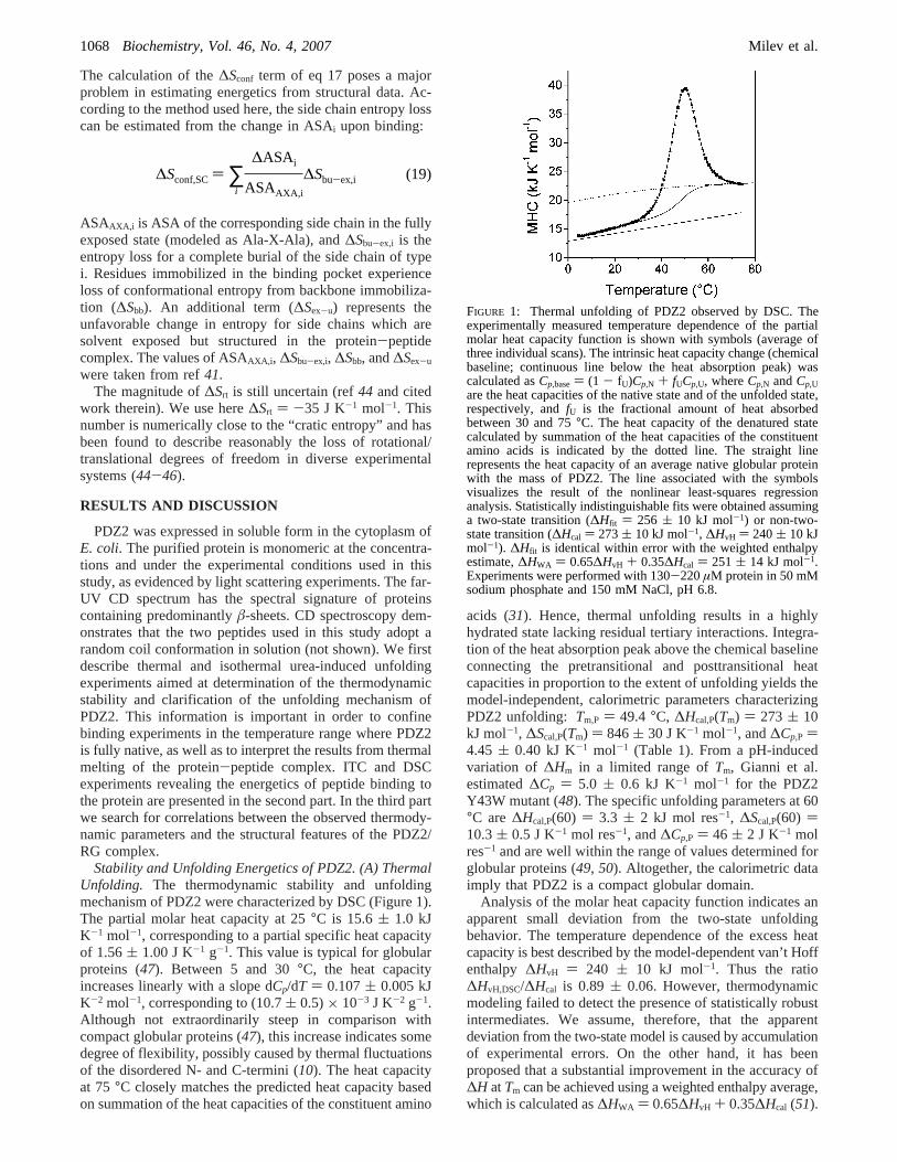
Stability and Unfolding Energetics of PDZ2. (A) ThermalUnfolding. The thermodynamic stability and unfoldingmechanism of PDZ2 were characterized by DSC (Figure 1).The partial molar heat capacity at 25°C is 15.6( 1.0 kJK-1 mol-1, corresponding to a partial specific heat capacityof 1.56( 1.00 J K-1 g-1. This value is typical for globularproteins (47). Between 5 and 30°C, the heat capacityincreases linearly with a slope dCp/dT ) 0.107( 0.005 kJK-2 mol-1, corresponding to (10.7( 0.5)× 10-3 J K-2 g-1.Although not extraordinarily steep in comparison withcompact globular proteins (47), this increase indicates somedegree of flexibility, possibly caused by thermal fluctuationsof the disordered N- and C-termini (10). The heat capacityat 75°C closely matches the predicted heat capacity basedon summation of the heat capacities of the constituent amino
acids (31). Hence, thermal unfolding results in a highlyhydrated state lacking residual tertiary interactions. Integra-tion of the heat absorption peak above the chemical baselineconnecting the pretransitional and posttransitional heatcapacities in proportion to the extent of unfolding yields themodel-independent, calorimetric parameters characterizingPDZ2 unfolding: Tm,P ) 49.4 °C, ∆Hcal,P(Tm) ) 273 ( 10kJ mol-1, ∆Scal,P(Tm) ) 846( 30 J K-1 mol-1, and∆Cp,P )4.45 ( 0.40 kJ K-1 mol-1 (Table 1). From a pH-inducedvariation of ∆Hm in a limited range ofTm, Gianni et al.estimated∆Cp ) 5.0 ( 0.6 kJ K-1 mol-1 for the PDZ2Y43W mutant (48). The specific unfolding parameters at 60°C are∆Hcal,P(60) ) 3.3 ( 2 kJ mol res-1, ∆Scal,P(60) )10.3( 0.5 J K-1 mol res-1, and∆Cp,P ) 46 ( 2 J K-1 molres-1 and are well within the range of values determined forglobular proteins (49, 50). Altogether, the calorimetric dataimply that PDZ2 is a compact globular domain.
Analysis of the molar heat capacity function indicates anapparent small deviation from the two-state unfoldingbehavior. The temperature dependence of the excess heatcapacity is best described by the model-dependent van’t Hoffenthalpy ∆HvH ) 240 ( 10 kJ mol-1. Thus the ratio∆HvH,DSC/∆Hcal is 0.89 ( 0.06. However, thermodynamicmodeling failed to detect the presence of statistically robustintermediates. We assume, therefore, that the apparentdeviation from the two-state model is caused by accumulationof experimental errors. On the other hand, it has beenproposed that a substantial improvement in the accuracy of∆H atTm can be achieved using a weighted enthalpy average,which is calculated as∆HWA ) 0.65∆HvH + 0.35∆Hcal (51).
FIGURE 1: Thermal unfolding of PDZ2 observed by DSC. Theexperimentally measured temperature dependence of the partialmolar heat capacity function is shown with symbols (average ofthree individual scans). The intrinsic heat capacity change (chemicalbaseline; continuous line below the heat absorption peak) wascalculated asCp,base) (1 - fU)Cp,N + fUCp,U, whereCp,N andCp,Uare the heat capacities of the native state and of the unfolded state,respectively, andfU is the fractional amount of heat absorbedbetween 30 and 75°C. The heat capacity of the denatured statecalculated by summation of the heat capacities of the constituentamino acids is indicated by the dotted line. The straight linerepresents the heat capacity of an average native globular proteinwith the mass of PDZ2. The line associated with the symbolsvisualizes the result of the nonlinear least-squares regressionanalysis. Statistically indistinguishable fits were obtained assuminga two-state transition (∆Hfit ) 256 ( 10 kJ mol-1) or non-two-state transition (∆Hcal ) 273( 10 kJ mol-1, ∆HvH ) 240( 10 kJmol-1). ∆Hfit is identical within error with the weighted enthalpyestimate,∆HWA ) 0.65∆HvH + 0.35∆Hcal ) 251 ( 14 kJ mol-1.Experiments were performed with 130-220µM protein in 50 mMsodium phosphate and 150 mM NaCl, pH 6.8.
∆Sconf,SC) ∑i
∆ASAi
ASAAXA,i
∆Sbu-ex,i (19)
1068 Biochemistry, Vol. 46, No. 4, 2007 Milev et al.
Indeed, as shown illustrated in Figure 1∆HWA simulatesperfectly the experimental trace.
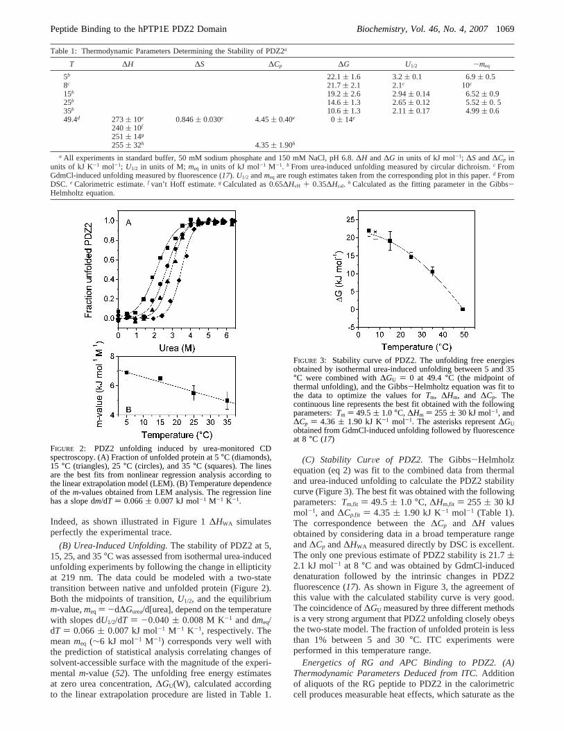
(B) Urea-Induced Unfolding.The stability of PDZ2 at 5,15, 25, and 35°C was assessed from isothermal urea-inducedunfolding experiments by following the change in ellipticityat 219 nm. The data could be modeled with a two-statetransition between native and unfolded protein (Figure 2).Both the midpoints of transition,U1/2, and the equilibriumm-value,meq ) -d∆Gurea/d[urea], depend on the temperaturewith slopes dU1/2/dT ) -0.040( 0.008 M K-1 and dmeq/dT ) 0.066( 0.007 kJ mol-1 M-1 K-1, respectively. Themeanmeq (∼6 kJ mol-1 M-1) corresponds very well withthe prediction of statistical analysis correlating changes ofsolvent-accessible surface with the magnitude of the experi-mentalm-value (52). The unfolding free energy estimatesat zero urea concentration,∆GU(W), calculated accordingto the linear extrapolation procedure are listed in Table 1.
(C) Stability CurVe of PDZ2. The Gibbs-Helmholzequation (eq 2) was fit to the combined data from thermaland urea-induced unfolding to calculate the PDZ2 stabilitycurve (Figure 3). The best fit was obtained with the followingparameters:Tm,fit ) 49.5( 1.0 °C, ∆Hm,fit ) 255 ( 30 kJmol-1, and∆Cp,fit ) 4.35 ( 1.90 kJ K-1 mol-1 (Table 1).The correspondence between the∆Cp and ∆H valuesobtained by considering data in a broad temperature rangeand∆Cp and∆HWA measured directly by DSC is excellent.The only one previous estimate of PDZ2 stability is 21.7(2.1 kJ mol-1 at 8 °C and was obtained by GdmCl-induceddenaturation followed by the intrinsic changes in PDZ2fluorescence (17). As shown in Figure 3, the agreement ofthis value with the calculated stability curve is very good.The coincidence of∆GU measured by three different methodsis a very strong argument that PDZ2 unfolding closely obeysthe two-state model. The fraction of unfolded protein is lessthan 1% between 5 and 30°C. ITC experiments wereperformed in this temperature range.
Energetics of RG and APC Binding to PDZ2. (A)Thermodynamic Parameters Deduced from ITC.Additionof aliquots of the RG peptide to PDZ2 in the calorimetriccell produces measurable heat effects, which saturate as the
Table 1: Thermodynamic Parameters Determining the Stability of PDZ2a
T ∆H ∆S ∆Cp ∆G U1/2 -meq
5b 22.1( 1.6 3.2( 0.1 6.9( 0.58c 21.7( 2.1 2.1c 10c
15b 19.2( 2.6 2.94( 0.14 6.52( 0.925b 14.6( 1.3 2.65( 0.12 5.52( 0. 535b 10.6( 1.3 2.11( 0.17 4.99( 0.649.4d 273( 10e 0.846( 0.030e 4.45( 0.40e 0 ( 14e
240( 10f
251( 14g
255( 32h 4.35( 1.90h
a All experiments in standard buffer, 50 mM sodium phosphate and 150 mM NaCl, pH 6.8.∆H and∆G in units of kJ mol-1; ∆S and∆Cp inunits of kJ K-1 mol-1; U1/2 in units of M; meq in units of kJ mol-1 M-1. b From urea-induced unfolding measured by circular dichroism.c FromGdmCl-induced unfolding measured by fluorescence (17). U1/2 andmeq are rough estimates taken from the corresponding plot in this paper.d FromDSC. e Calorimetric estimate.f van’t Hoff estimate.g Calculated as 0.65∆HvH + 0.35∆Hcal. h Calculated as the fitting parameter in the Gibbs-Helmholtz equation.
FIGURE 2: PDZ2 unfolding induced by urea-monitored CDspectroscopy. (A) Fraction of unfolded protein at 5°C (diamonds),15 °C (triangles), 25°C (circles), and 35°C (squares). The linesare the best fits from nonlinear regression analysis according tothe linear extrapolation model (LEM). (B) Temperature dependenceof the m-values obtained from LEM analysis. The regression linehas a slope dm/dT ) 0.066( 0.007 kJ mol-1 M-1 K-1.
FIGURE 3: Stability curve of PDZ2. The unfolding free energiesobtained by isothermal urea-induced unfolding between 5 and 35°C were combined with∆GU ) 0 at 49.4°C (the midpoint ofthermal unfolding), and the Gibbs-Helmholtz equation was fit tothe data to optimize the values forTm, ∆Hm, and ∆Cp. Thecontinuous line represents the best fit obtained with the followingparameters:Tm ) 49.5( 1.0 °C, ∆Hm ) 255( 30 kJ mol-1, and∆Cp ) 4.36 ( 1.90 kJ K-1 mol-1. The asterisks represent∆GUobtained from GdmCl-induced unfolding followed by fluorescenceat 8 °C (17)
Peptide Binding to the hPTP1E PDZ2 Domain Biochemistry, Vol. 46, No. 4, 20071069
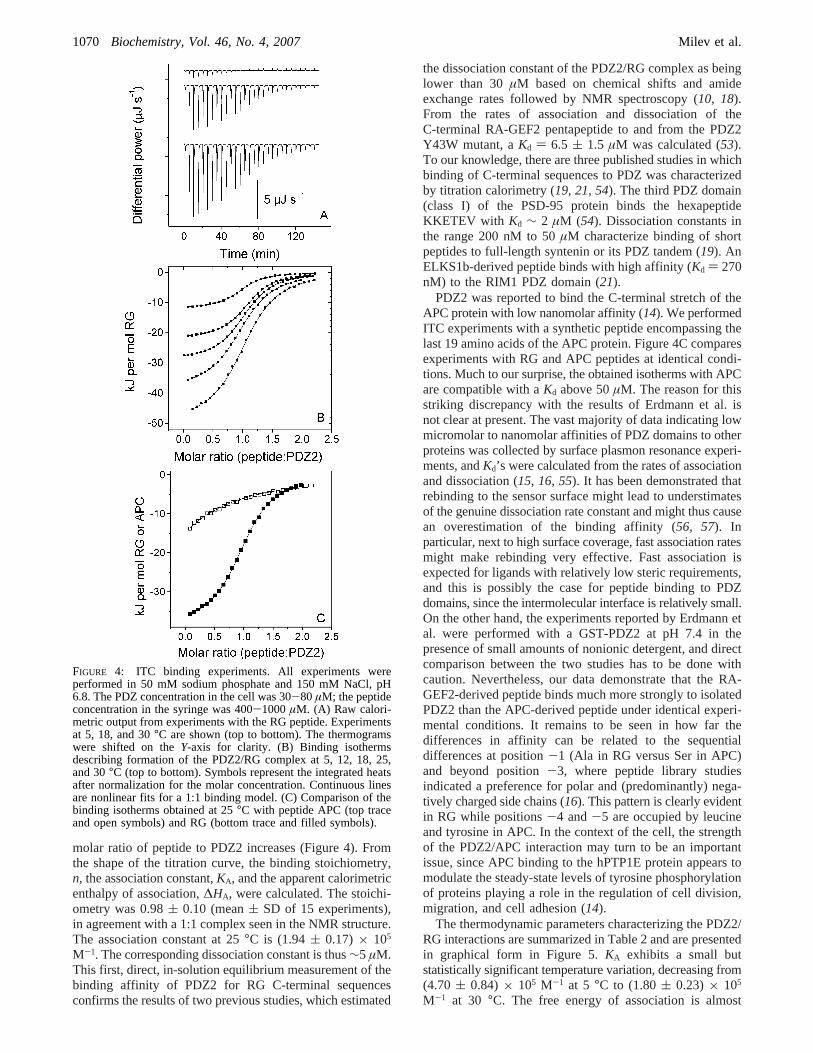
molar ratio of peptide to PDZ2 increases (Figure 4). Fromthe shape of the titration curve, the binding stoichiometry,n, the association constant,KA, and the apparent calorimetricenthalpy of association,∆HA, were calculated. The stoichi-ometry was 0.98( 0.10 (mean( SD of 15 experiments),in agreement with a 1:1 complex seen in the NMR structure.The association constant at 25°C is (1.94( 0.17) × 105
M-1. The corresponding dissociation constant is thus∼5 µM.This first, direct, in-solution equilibrium measurement of thebinding affinity of PDZ2 for RG C-terminal sequencesconfirms the results of two previous studies, which estimated
the dissociation constant of the PDZ2/RG complex as beinglower than 30µM based on chemical shifts and amideexchange rates followed by NMR spectroscopy (10, 18).From the rates of association and dissociation of theC-terminal RA-GEF2 pentapeptide to and from the PDZ2Y43W mutant, aKd ) 6.5 ( 1.5 µM was calculated (53).To our knowledge, there are three published studies in whichbinding of C-terminal sequences to PDZ was characterizedby titration calorimetry (19, 21, 54). The third PDZ domain(class I) of the PSD-95 protein binds the hexapeptideKKETEV with Kd ∼ 2 µM (54). Dissociation constants inthe range 200 nM to 50µM characterize binding of shortpeptides to full-length syntenin or its PDZ tandem (19). AnELKS1b-derived peptide binds with high affinity (Kd ) 270nM) to the RIM1 PDZ domain (21).
PDZ2 was reported to bind the C-terminal stretch of theAPC protein with low nanomolar affinity (14). We performedITC experiments with a synthetic peptide encompassing thelast 19 amino acids of the APC protein. Figure 4C comparesexperiments with RG and APC peptides at identical condi-tions. Much to our surprise, the obtained isotherms with APCare compatible with aKd above 50µM. The reason for thisstriking discrepancy with the results of Erdmann et al. isnot clear at present. The vast majority of data indicating lowmicromolar to nanomolar affinities of PDZ domains to otherproteins was collected by surface plasmon resonance experi-ments, andKd’s were calculated from the rates of associationand dissociation (15, 16, 55). It has been demonstrated thatrebinding to the sensor surface might lead to understimatesof the genuine dissociation rate constant and might thus causean overestimation of the binding affinity (56, 57). Inparticular, next to high surface coverage, fast association ratesmight make rebinding very effective. Fast association isexpected for ligands with relatively low steric requirements,and this is possibly the case for peptide binding to PDZdomains, since the intermolecular interface is relatively small.On the other hand, the experiments reported by Erdmann etal. were performed with a GST-PDZ2 at pH 7.4 in thepresence of small amounts of nonionic detergent, and directcomparison between the two studies has to be done withcaution. Nevertheless, our data demonstrate that the RA-GEF2-derived peptide binds much more strongly to isolatedPDZ2 than the APC-derived peptide under identical experi-mental conditions. It remains to be seen in how far thedifferences in affinity can be related to the sequentialdifferences at position-1 (Ala in RG versus Ser in APC)and beyond position-3, where peptide library studiesindicated a preference for polar and (predominantly) nega-tively charged side chains (16). This pattern is clearly evidentin RG while positions-4 and-5 are occupied by leucineand tyrosine in APC. In the context of the cell, the strengthof the PDZ2/APC interaction may turn to be an importantissue, since APC binding to the hPTP1E protein appears tomodulate the steady-state levels of tyrosine phosphorylationof proteins playing a role in the regulation of cell division,migration, and cell adhesion (14).
The thermodynamic parameters characterizing the PDZ2/RG interactions are summarized in Table 2 and are presentedin graphical form in Figure 5.KA exhibits a small butstatistically significant temperature variation, decreasing from(4.70 ( 0.84) × 105 M-1 at 5 °C to (1.80( 0.23) × 105
M-1 at 30 °C. The free energy of association is almost
FIGURE 4: ITC binding experiments. All experiments wereperformed in 50 mM sodium phosphate and 150 mM NaCl, pH6.8. The PDZ concentration in the cell was 30-80 µM; the peptideconcentration in the syringe was 400-1000µM. (A) Raw calori-metric output from experiments with the RG peptide. Experimentsat 5, 18, and 30°C are shown (top to bottom). The thermogramswere shifted on theY-axis for clarity. (B) Binding isothermsdescribing formation of the PDZ2/RG complex at 5, 12, 18, 25,and 30°C (top to bottom). Symbols represent the integrated heatsafter normalization for the molar concentration. Continuous linesare nonlinear fits for a 1:1 binding model. (C) Comparison of thebinding isotherms obtained at 25°C with peptide APC (top traceand open symbols) and RG (bottom trace and filled symbols).
1070 Biochemistry, Vol. 46, No. 4, 2007 Milev et al.
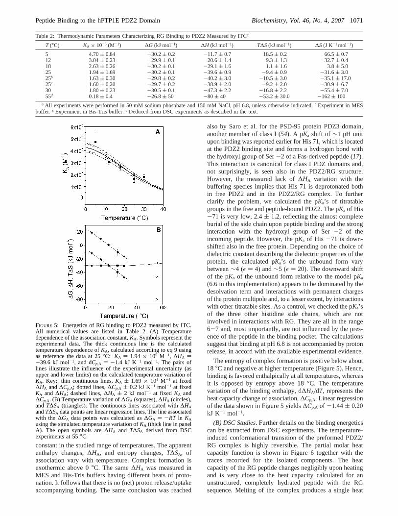
constant in the studied range of temperatures. The apparententhalpy changes,∆HA, and entropy changes,T∆SA, ofassociation vary with temperature. Complex formation isexothermic above 0°C. The same∆HA was measured inMES and Bis-Tris buffers having different heats of proto-nation. It follows that there is no (net) proton release/uptakeaccompanying binding. The same conclusion was reached
also by Saro et al. for the PSD-95 protein PDZ3 domain,another member of class I (54). A pKa shift of ∼1 pH unitupon binding was reported earlier for His 71, which is locatedat the PDZ2 binding site and forms a hydrogen bond withthe hydroxyl group of Ser-2 of a Fas-derived peptide (17).This interaction is canonical for class I PDZ domains and,not surprisingly, is seen also in the PDZ2/RG structure.However, the measured lack of∆HA variation with thebuffering species implies that His 71 is deprotonated bothin free PDZ2 and in the PDZ2/RG complex. To furtherclarify the problem, we calculated the pKa’s of titratablegroups in the free and peptide-bound PDZ2. The pKa of His-71 is very low, 2.4( 1.2, reflecting the almost completeburial of the side chain upon peptide binding and the stronginteraction with the hydroxyl group of Ser-2 of theincoming peptide. However, the pKa of His -71 is down-shifted also in the free protein. Depending on the choice ofdielectric constant describing the dielectric properties of theprotein, the calculated pKa’s of the unbound form varybetween∼4 (ε ) 4) and∼5 (ε ) 20). The downward shiftof the pKa of the unbound form relative to the model pKa
(6.6 in this implementation) appears to be dominated by thedesolvation term and interactions with permanent chargesof the protein multipole and, to a lesser extent, by interactionswith other titratable sites. As a control, we checked the pKa’sof the three other histidine side chains, which are notinvolved in interactions with RG. They are all in the range6-7 and, most importantly, are not influenced by the pres-ence of the peptide in the binding pocket. The calculationssuggest that binding at pH 6.8 is not accompanied by protonrelease, in accord with the available experimental evidence.
The entropy of complex formation is positive below about18 °C and negative at higher temperature (Figure 5). Hence,binding is favored enthalpically at all temperatures, whereasit is opposed by entropy above 18°C. The temperaturevariation of the binding enthalpy, d∆HA/dT, represents theheat capacity change of association,∆Cp,A. Linear regressionof the data shown in Figure 5 yields∆Cp,A of -1.44( 0.20kJ K-1 mol-1.
(B) DSC Studies. Further details on the binding energeticscan be extracted from DSC experiments. The temperature-induced conformational transition of the preformed PDZ2/RG complex is highly reversible. The partial molar heatcapacity function is shown in Figure 6 together with thetraces recorded for the isolated components. The heatcapacity of the RG peptide changes negligibly upon heatingand is very close to the heat capacity calculated for anunstructured, completely hydrated peptide with the RGsequence. Melting of the complex produces a single heat
Table 2: Thermodynamic Parameters Characterizing RG Binding to PDZ2 Measured by ITCa
T (°C) KA × 10-5 (M-1) ∆G (kJ mol-1) ∆H (kJ mol-1) T∆S(kJ mol-1) ∆S(J K-1 mol-1)
5 4.70( 0.84 -30.2( 0.2 -11.7( 0.7 18.5( 0.2 66.5( 0.712 3.04( 0.23 -29.9( 0.1 -20.6( 1.4 9.3( 1.3 32.7( 0.418 2.63( 0.26 -30.2( 0.1 -29.1( 1.6 1.1( 1.6 3.8( 5.025 1.94( 1.69 -30.2( 0.1 -39.6( 0.9 -9.4( 0.9 -31.6( 3.025b 1.63( 0.30 -29.8( 0.2 -40.2( 3.0 -10.5( 3.0 -35.1( 17.025c 1.60( 0.20 -29.7( 0.2 -38.9( 2.0 -9.2( 2.0 -30.9( 6.730 1.80( 0.23 -30.5( 0.1 -47.3( 2.2 -16.8( 2.2 -55.4( 7.055d 0.18( 0.4 -26.8( 50 -80 ( 40 -53.2( 30.0 -162( 100
a All experiments were performed in 50 mM sodium phosphate and 150 mM NaCl, pH 6.8, unless otherwise indicated.b Experiment in MESbuffer. c Experiment in Bis-Tris buffer.d Deduced from DSC experiments as described in the text.
FIGURE 5: Energetics of RG binding to PDZ2 measured by ITC.All numerical values are listed in Table 2. (A) Temperaturedependence of the association constant,KA. Symbols represent theexperimental data. The thick continuous line is the calculatedtemperature dependence ofKA, calculated according to eq 9 usingas reference the data at 25°C: KA ) 1.94 × 10⁵ M-1, ∆HA )-39.6 kJ mol-1, and dCp,A ) -1.4 kJ K-1 mol-1. The pairs oflines illustrate the influence of the experimental uncertainty (asupper and lower limits) on the calculated temperature variation ofKA. Key: thin continuous lines,KA ( 1.69 × 10⁴ M-1 at fixed∆HA and∆Cp,A; dotted lines,∆Cp,A ( 0.2 kJ K-1 mol-1 at fixedKA and ∆HA; dashed lines,∆HA ( 2 kJ mol-1 at fixed KA and∆Cp,A. (B) Temperature variation of∆GA (squares),∆HA (circles),and T∆SA (triangles). The continuous lines associated with∆HAandT∆SA data points are linear regression lines. The line associatedwith the ∆GA data points was calculated as∆GA ) -RT ln KAusing the simulated temperature variation ofKA (thick line in panelA). The open symbols are∆HA and T∆SA derived from DSCexperiments at 55°C.
Peptide Binding to the hPTP1E PDZ2 Domain Biochemistry, Vol. 46, No. 4, 20071071
absorption peak whose temperature of maximum heatabsorption (which can be regarded as the apparent transitiontemperature or melting temperature,Tm,C) is higher by∼6°C than the melting temperature of PDZ2 alone. Hence,PDZ2 is stabilized in the complex. The dissociation of thecomplex and the concurrent unfolding of the protein aretaking place within a relatively narrow temperature interval.
What can be learned from the thermogram? Figure 6 showsthe expected heat capacity of the system in a hypotheticalstate without intermolecular interactions. This function,Cp
SUM, was calculated by simple algebraic summation of the
heat capacities of free PDZ2 and free RG; i.e.,CpSUM )
CpPDZ2 + Cp
RG. Between 5 and 30°C CpSUM is higher than
the heat capacity of the (partly) associated state,CpPDZ2/RG.
This reflects the negative heat capacity change accompanyingbinding. The differenceCp
PDZ2/RG- CpSUM is ∼-1.6( 0.15
kJ K-1 mol-1 on average and is thus very close to∆Cp,A
obtained by ITC. However, the close correspondence of thetwo numbers is perhaps a fortituous coincidence in view ofthe uncertainties in determination of the absolute heatcapacities at relatively low concentrations and the use ofcalculated partial specific volumes. Much more importantfor the following discussion is the fact that the slopes ofCp
PDZ2/RG and CpSUM are very close to each other: 96( 5
and 112( 7 J K-2 mol-1, respectively (Table 3). Thesevalues imply, in principle, a temperature-dependent∆Cp,A.However, the integral∫T(Cp
PDZ2/RG - CpSUM) dT is a small
number, and the resulting deviation of the dHA/dT plot fromlinearity is certainly lower than the accuracy of the measure-ments. It follows that the formation of the PDZ2/RG complexis not linked to detectable temperature-dependent structuralchanges, or at least such changes contribute to the enthalpyof association but do not induce significant temperaturedependence of∆Cp.
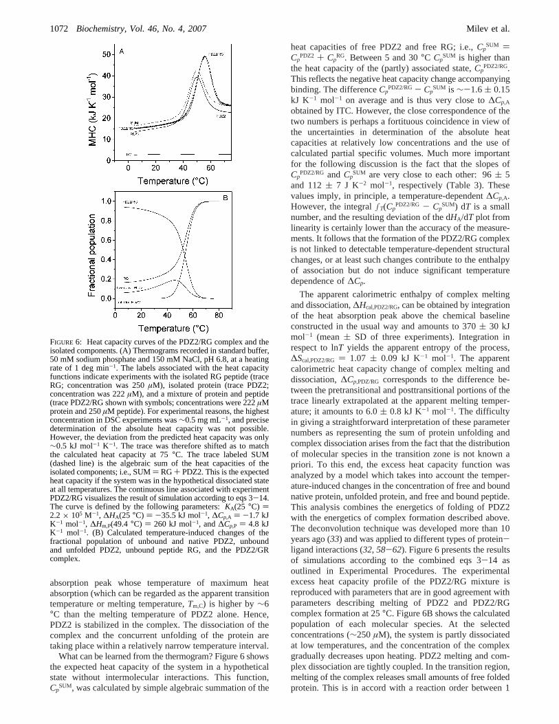
The apparent calorimetric enthalpy of complex meltingand dissociation,∆Hcal,PDZ2/RG, can be obtained by integrationof the heat absorption peak above the chemical baselineconstructed in the usual way and amounts to 370( 30 kJmol-1 (mean( SD of three experiments). Integration inrespect to lnT yields the apparent entropy of the process,∆Scal,PDZ2/RG ) 1.07 ( 0.09 kJ K-1 mol-1. The apparentcalorimetric heat capacity change of complex melting anddissociation,∆Cp,PDZ/RG corresponds to the difference be-tween the pretransitional and posttransitional portions of thetrace linearly extrapolated at the apparent melting temper-ature; it amounts to 6.0( 0.8 kJ K-1 mol-1. The difficultyin giving a straightforward interpretation of these parameternumbers as representing the sum of protein unfolding andcomplex dissociation arises from the fact that the distributionof molecular species in the transition zone is not known apriori. To this end, the excess heat capacity function wasanalyzed by a model which takes into account the temper-ature-induced changes in the concentration of free and boundnative protein, unfolded protein, and free and bound peptide.This analysis combines the energetics of folding of PDZ2with the energetics of complex formation described above.The deconvolution technique was developed more than 10years ago (33) and was applied to different types of protein-ligand interactions (32, 58-62). Figure 6 presents the resultsof simulations according to the combined eqs 3-14 asoutlined in Experimental Procedures. The experimentalexcess heat capacity profile of the PDZ2/RG mixture isreproduced with parameters that are in good agreement withparameters describing melting of PDZ2 and PDZ2/RGcomplex formation at 25°C. Figure 6B shows the calculatedpopulation of each molecular species. At the selectedconcentrations (∼250µM), the system is partly dissociatedat low temperatures, and the concentration of the complexgradually decreases upon heating. PDZ2 melting and com-plex dissociation are tightly coupled. In the transition region,melting of the complex releases small amounts of free foldedprotein. This is in accord with a reaction order between 1
FIGURE 6: Heat capacity curves of the PDZ2/RG complex and theisolated components. (A) Thermograms recorded in standard buffer,50 mM sodium phosphate and 150 mM NaCl, pH 6.8, at a heatingrate of 1 deg min-1. The labels associated with the heat capacityfunctions indicate experiments with the isolated RG peptide (traceRG; concentration was 250µM), isolated protein (trace PDZ2;concentration was 222µM), and a mixture of protein and peptide(trace PDZ2/RG shown with symbols; concentrations were 222µMprotein and 250µM peptide). For experimental reasons, the highestconcentration in DSC experiments was∼0.5 mg mL-1, and precisedetermination of the absolute heat capacity was not possible.However, the deviation from the predicted heat capacity was only∼0.5 kJ mol-1 K-1. The trace was therefore shifted as to matchthe calculated heat capacity at 75°C. The trace labeled SUM(dashed line) is the algebraic sum of the heat capacities of theisolated components; i.e., SUM) RG+ PDZ2. This is the expectedheat capacity if the system was in the hypothetical dissociated stateat all temperatures. The continuous line associated with experimentPDZ2/RG visualizes the result of simulation according to eqs 3-14.The curve is defined by the following parameters:KA(25 °C) )2.2× 10⁵ M-1, ∆HA(25 °C) ) -35.5 kJ mol-1, ∆Cp,A ) -1.7 kJK-1 mol-1, ∆Hm,P(49.4 °C) ) 260 kJ mol-1, and∆Cp,P ) 4.8 kJK-1 mol-1. (B) Calculated temperature-induced changes of thefractional population of unbound and native PDZ2, unboundand unfolded PDZ2, unbound peptide RG, and the PDZ2/GRcomplex.
1072 Biochemistry, Vol. 46, No. 4, 2007 Milev et al.
and 2. Indeed, the reaction order best describing the meltingtrace is 1.85 (not shown).
With help of the known molar fractions as a functionof the temperature (Figure 6B), we can now deconvolutethe total enthalpy change (∆Hcal,PDZ2/RG), entropy change(∆Scal,PDZ2/RG), and heat capacity change (∆Cp,PDZ2/RG) mea-sured upon melting of the PDZ2/RG mixture. The contribu-tion of the peptide to any parameter can be neglected sincetemperature-induced conformational changes are calori-metrically silent (see the essentially temperature-independentheat capacity function in Figure 6A). The following equationshold atTm,C:
The first terms on the right-hand side are the parametersdescribing PDZ2 unfolding atTm,C and are calculated as∆Hcal,P
TmC ) ∆Hcal,P + ∆Cp,P(Tm,C - Tm,P) and∆Scal,PTmC ) ∆Scal,P
+ ∆Cp,P ln(Tm,C/Tm,P); ∆Cp,P is a constant over the smalltemperature range of extrapolation. The second terms on theright-hand side are the energetic parameters of the PDZ/RGcomplex. The minus sign accounts for complex dissociation.The multiplierfC represents the molar fraction of the complexat 30°C, i.e., at the onset of the heat absorption peak, andequals 0.89. For the only unknowns in the above equations,one obtains∆Hcal,A
TmC ) -80 ( 40 kJ mol-1, ∆Scal,ATmC ) -165
( 100 kJ K-1 mol-1, and∆Cp,A ) -1.8( 0.8 kJ K-1 mol-1.The free energy change is thus-26 kJ mol-1. These are thethermodynamic parameters of association of the PDZ2/RGcomplex at 55°C (open symbols in Figure 5). Although therules of error propagation place a large uncertainty of thecalculated numbers, the correspondence between the meanvalues of the thermodynamic functions obtained by ITC atlower temperatures and by DSC at 55°C is surprisingly goodin view of the crude approximations underlying the calcula-tions. First, any temperature dependence of∆Cp is neglected.Second, both molecules are considered to be structurally andenergetically invariant in the entire temperature range. Third,there is no robust way to justify the validity of the two-statemodel, and any possible redistribution of binding modes inthe transition region is ignored. Fourth, the implicit assump-tion is that there are no interactions taking place betweenthe peptide and the protein in its denatured state. Neverthe-less, the success of the modeling (based on van’t Hoff
formalism) and the deduced calorimetric enthalpy, entropy,and heat capacity changes indicate that the thermodynamicparameters obtained by ITC and DSC describe the thermo-dynamics of RG binding to PDZ2 in a consistent way andthat the procedure is a valuable alternative to obtainthermodynamic information about protein-ligand bindingat temperatures beyond the usually limited temperature rangecovered by ITC experiments. More importantly, the proce-dure allows a reliable interpolation of the binding constantto the physiologically relevant temperature of 37°C.
Energetic Partitioning of the Binding Affinity.The drivingforce of RG binding to PDZ2 at 25°C (and in physiologicallyrelevant temperature range) is the favorable enthalpy change.The same is true for PDZ3 of PSD-95, which is also a classI PDZ domain (54). In contrast, favorable entropy changedominates the affinity of peptide binding to syntenin PDZ1and PDZ2 domains (19). The latter two domains exhibitdegenerate specificity and bind peptide ligands of classes I,II, and III. Neither of them has a histidine (typical for classI) or a hydrophobic residue (typical for class II) or a tyrosine(typical for class III) at the beginning of helixR2. With thelimited data available, it is impossible to interpret orgeneralize the different enthalpy-entropy signature of classI-specific complexes, as compared to PDZ-peptide com-plexes with degenerate specificity. From a structural perspec-tive, the large spread of binding enthalpies (-2 to -40 kJmol-1 at 25°C) and the apparent effective enthalpy/entropycompensation are surprising. First, the interactions anchoringthe very C-terminal hydrophobic residue are ubiquitous andconservative. Second, it appears that backbone-backbonehydrogen bonding contributes essentially to the bindingaffinity in a “nonspecific” manner. Although progress hasbeen made toward prediction of binding affinities and actualdesign of ligands capable of binding PDZ domains (63, 64),the emergent compatibility of the PDZ binding site withligands that bind, driven by enthalpy or entropy, or both,could turn out to be an obstacle in the rational optimizationof lead compounds for high affinityand specificity (65).
Structural Parametrization of Energetic Changes.How-ever good the precision of measured thermodynamic param-eters might be, they reflect the total energetic changes ofthe considered system. The challenging problem is indeedto relate energy terms to structural features in order tounderstand the molecular basis of the binding affinity.Starting from the early 1990s, different approaches have beenfollowed (41, 66-70). Among different parametrizationschemes, we use here the one developed by Murphy andFreire, which has been extensively tested against experi-mental data on unfolding and binding and enjoys com-
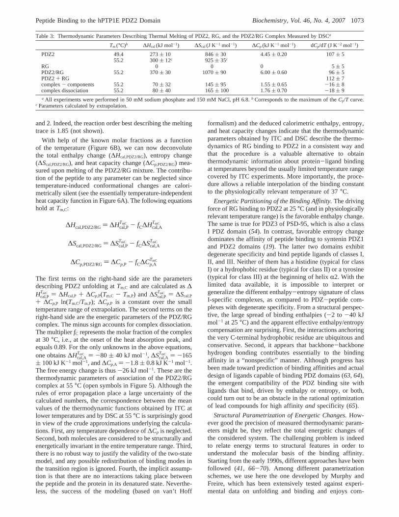
Table 3: Thermodynamic Parameters Describing Thermal Melting of PDZ2, RG, and the PDZ2/RG Complex Measured by DSCa
Tm (°C)b ∆Hcal (kJ mol-1) ∆Scal (J K-1 mol-1) ∆Cp (kJ K-1 mol-1) dCp/dT (J K-2 mol-1)
PDZ2 49.4 273( 10 846( 30 4.45( 0.20 107( 555.2 300( 12c 925( 35c
RG 0 0 0 5( 5PDZ2/RG 55.2 370( 30 1070( 90 6.00( 0.60 96( 5PDZ2+ RG 112( 7complex- components 55.2 70( 32 145( 95 1.55( 0.65 -16 ( 8complex dissociation 55.2 80( 40 165( 100 1.76( 0.70 -18 ( 9a All experiments were performed in 50 mM sodium phosphate and 150 mM NaCl, pH 6.8.b Corresponds to the maximum of theCp/T curve.
c Parameters calculated by extrapolation.
∆Hcal,PDZ2/RG) ∆Hcal,PTmC - fC∆Hcal,A
TmC
∆Scal,PDZ2/RG) ∆Scal,PTmC - fC∆Scal,A
TmC
∆Cp,PDZ2/RG) ∆Cp,P - fC∆Cp,ATmC
Peptide Binding to the hPTP1E PDZ2 Domain Biochemistry, Vol. 46, No. 4, 20071073
mensurate popularity (41, 42, 69). For brevity we henceforthrefer to the procedure as M&F (standing for Murphy andFreire; the contribution of numerous members of the Murphyand Freire laboratories to the refinement of the calculationis implicitly acknowledged).
Since the M&F calculations rely on solvent accessibilitydifferences, the structures of the binary complex and itscomponents in their free state should be known at highresolution or should be carefully modeled. The NMRstructure of the PDZ2/RG complex was solved [1D5G (10)].The NMR structure of PDZ2 in the unbound form is alsoavailable [3PDZ (11)]. The five C-terminal residues of RGare ordered in the binding site, yet free RG obtains a randomcoil conformation according to CD measurements, andtherefore, the RG structure extracted from the complex isinadequate to model the free RG conformation. To overcomethe problem and, more importantly, to create a consistentset of structures, we performed MD simulations of freePDZ2, free RG, and the PDZ2/RG complex in explicit water.According to the usual criteria (CR RMSD, radius of gyration,solute-solute and solute-solvent energy terms, intermo-lecular distances) the MD trajectories were well equilibratedto serve as a model of the dynamic behavior of the complexand its components (not shown). The average NMR and MDstructures of the PDZ2/RG complex and its components arevery close to each other, the CR RMSD (secondary structureelements) being between 0.87 and 1.36 Å in cross-comparisons. However, we noted a slight structure contrac-tion of the complex during MD. The change in ASA isrelatively small but significant in comparison with the spreadof ASA within the NMR ensemble. Therefore, we tested thecalculations against both ensembles according to the fol-lowing reaction schemes:
Reaction 1 represents the real process, taking into accountthe conformational changes of the protein domain ac-companying binding, while reaction 2 is a hypotheticalprocess, where binding is approximated as a rigid-bodyassociation. In model NMR, ensemble 3PDZ (human PDZ2)was used to represent the free protein, while the binding-competent conformation was extracted from the structure ofthe human PDZ2/RG complex (1D5G), which serves alsoas a model of the associated state of the system. In modelMD, the structures of the free and binding-competent PDZ2were taken from the trajectories of the free protein and ofthe complex, respectively. In both models, the free peptidewas modeled with the MD structure. The calculated surfacesrepresent averages over the corresponding ensembles.
The calculated parameters are listed in Table 4. Thecomparison with the experimental parameters is done at 25
and 60°C for ∆HA and at 18°C for ∆SA, where the totalentropy contribution is zero. Model NMRconf predicts thebinding enthalpy within the range of the other model’sestimates, yet all other terms are significantly different andcompletely contradict the experimental trend. As noted byWalma et al., the free PDZ2 structure suffers significantproblems according to several established structure qualitytests (71). However, no improvement was achieved by usingthe NMR structure of the mouse, highly homologous PDZ2domain (1GM1). The failure of the NMRconf model reiteratesthat the quality of structures is critical in ASA-basedparametrization and warns against the use of homologousstructures. Since model NMRrb predicts endothermic bindingat 25 °C, we focus the following discussion on the resultsobtained with the MD ensembles.
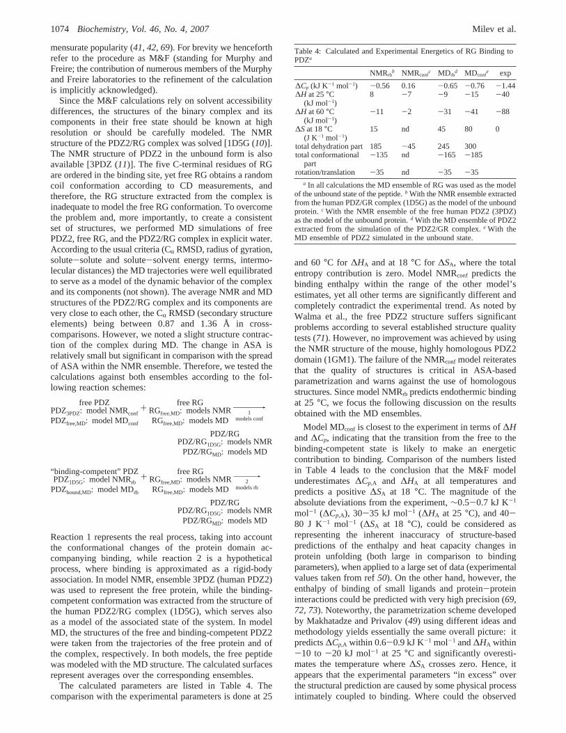
Model MDconf is closest to the experiment in terms of∆Hand∆Cp, indicating that the transition from the free to thebinding-competent state is likely to make an energeticcontribution to binding. Comparison of the numbers listedin Table 4 leads to the conclusion that the M&F modelunderestimates∆Cp,A and ∆HA at all temperatures andpredicts a positive∆SA at 18 °C. The magnitude of theabsolute deviations from the experiment,∼0.5-0.7 kJ K-1
mol-1 (∆Cp,A), 30-35 kJ mol-1 (∆HA at 25 °C), and 40-80 J K-1 mol-1 (∆SA at 18 °C), could be considered asrepresenting the inherent inaccuracy of structure-basedpredictions of the enthalpy and heat capacity changes inprotein unfolding (both large in comparison to bindingparameters), when applied to a large set of data (experimentalvalues taken from ref50). On the other hand, however, theenthalpy of binding of small ligands and protein-proteininteractions could be predicted with very high precision (69,72, 73). Noteworthy, the parametrization scheme developedby Makhatadze and Privalov (49) using different ideas andmethodology yields essentially the same overall picture: itpredicts∆Cp,A within 0.6-0.9 kJ K-1 mol-1 and∆HA within-10 to -20 kJ mol-1 at 25 °C and significantly overesti-mates the temperature where∆SA crosses zero. Hence, itappears that the experimental parameters “in excess” overthe structural prediction are caused by some physical processintimately coupled to binding. Where could the observed
free PDZPDZ3PDZ: model NMRconf
PDZfree,MD: model MDconf
+free RG
RGfree,MD: models NMRRGfree,MD: models MD
981
models conf
PDZ/RGPDZ/RG1D5G: models NMR
PDZ/RGMD: models MD
“binding-competent” PDZPDZ1D5G: model NMRrb
PDZbound,MD: model MDrb
+free RG
RGfree,MD: models NMRRGfree,MD: models MD
982
models rb
PDZ/RGPDZ/RG1D5G: models NMR
PDZ/RGMD: models MD
Table 4: Calculated and Experimental Energetics of RG Binding toPDZa
NMRrbb NMRconf
c MDrbd MDconf
e exp
∆Cp (kJ K-1 mol-1) -0.56 0.16 -0.65 -0.76 -1.44∆H at 25°C
(kJ mol-1)8 -7 -9 -15 -40
∆H at 60°C(kJ mol-1)
-11 -2 -31 -41 -88
∆Sat 18°C(J K-1 mol-1)
15 nd 45 80 0
total dehydration part 185 -45 245 300total conformational
part-135 nd -165 -185
rotation/translation -35 nd -35 -35a In all calculations the MD ensemble of RG was used as the model
of the unbound state of the peptide.b With the NMR ensemble extractedfrom the human PDZ/GR complex (1D5G) as the model of the unboundprotein.c With the NMR ensemble of the free human PDZ2 (3PDZ)as the model of the unbound protein.d With the MD ensemble of PDZ2extracted from the simulation of the PDZ2/GR complex.e With theMD ensemble of PDZ2 simulated in the unbound state.
1074 Biochemistry, Vol. 46, No. 4, 2007 Milev et al.
discrepancies stem from? A couple of possibilities arediscussed below.
(A) Heat Capacity Change. Experimental∆Cp values morenegative than those predicted from surface burial have beenreported for a variety of systems. Apart from the “trivial”case, where large refolding transitions accompanying bindingcontribute to the apparent∆Cp change, different explanationshave been forwarded with a common theme: The changesin hydration are not the only physical source of∆Cp decreaseconcomitant with binding. Additional contributions have beenascribed to (i) small structural perturbations leading toredistribution of easily excitable vibrational modes (74), (ii)ligand-induced narrowing of the distribution of enthalpicmicrostates (75), (iii) formation of highly cooperative arraysof noncovalent bonds (76), (iv) unusually large temperaturedependence of the intrinsic heat capacities of the associatedand dissociated states of the system (77), and (v) entrapmentof water at the binding interface (78, 79). Propositions i-iiirepresent general concepts that are difficult to verifyexperimentally. We find no experimental support for propo-sition iv because the heat capacity of the complex and thesum of the heat capacities of RG and PDZ2 change almostin parallel with temperature (Table 3 and Figure 6).Significant contribution from water entrapment is alsounlikely since the contribution of a trapped water moleculeto ∆Cp was estimated as being lower than-40 J K-1 mol-1
in an nonpolar environment (79, 80) and could probably notexceed-75 J K-1 mol-1 (the heat capacity of bulk water).One to three waters have been detected so far in PDZ-peptide complexes by crystallography, and the residence timeof each individual water molecule observed in our MDsimulations was never longer than 3.5 ns.2
(B) Enthalpy Change. We observe no measurable net heatsfrom protonation/deprotonation events. Also, no ions arepresent at the protein-peptide interface on the time scale ofthe MD simulations. The enthalpic effect of the watermolecules found at or close to the interface (one on averageover the 15 ns MD trajectory) cannot be sizable (81, 82).Hence, the structure-based prediction underestimates thegeneric binding enthalpy (41, 42). It should be noted that eq16 relates the enthalpy (of unfolding) to the average packingdensity of proteins and assumes no residual enthalpy afterbreakage of all relevant noncovalent bonds. We haveestimated the packing density by calculating the energy-weighted distance average between atom-atom pairs whichform the typical array of noncovalent bonds in proteins andprotein-protein complexes (81). The obtained values aremuch larger than those for the reference set analyzed in ref81 and, hence, indicate substantially looser packing of thePDZ2/RG interface in comparison with the interior of anaverage protein. Furthermore, the hydrogen bonds at theinterface are not particularly short or geometrically optimized,or networked, and visual inspection does not identify otherpolar interactions that might explain a strong enthalpic effect.We conclude that the experimental∆HA contains a contribu-
tion of an exothermic process that is not related to theformation of intermolecular contacts.
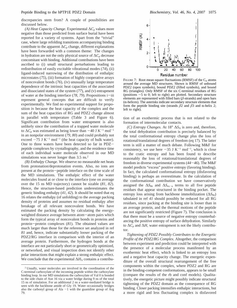
(C) Entropy Changes. At 18° ∆SA is zero and, therefore,the total dehydration contribution is precisely balanced bythe total conformational entropy change plus the loss ofrotational/translational degrees of freedom (eq 17). The latterterm is still a matter of much debate. Following M&F forconsistency, we use here-35 J K-1 mol-1, which is closeto the cratic entropy and has been found to describereasonably the loss of rotational/translational degrees offreedom in diverse experimental systems (44-46). The M&Fmodel predicts “excess” positive entropy (favoring binding).In fact, the calculated conformational entropy (disfavoringbinding) is perhaps an overestimate. In the calculation ofthe conformational contribution we have conservativelyassigned the∆Sbb and ∆Sex-u terms to all five peptideresidues that appear structured in the binding pocket. Theentropy loss arising from backbone immobilization (∆Sbb)tabulated in ref41 should possibly be reduced for all RGresidues, since packing at the binding site is looser than inan average protein, and the thermal motions of the peptideare not significantly restricted (Figure 7). The conclusion isthat there must be a source of negative entropy counterbal-ancing the positive entropy of surface dehydration. Similarlyto ∆Cp and∆H, water entrapment is not the likely contribu-tor.
Tightening of PDZ2 Possibly Contributes to the EnergeticProfile of the PDZ2/RG Complex.Altogether, the comparisonbetween experiment and prediction could be interpreted withthe presence of a molecular process manifested by anexothermic heat effect, which is linked to an entropy lossand a negative heat capacity change. The energetic expen-diture of the overall structural rearrangement of the freecomponents within the complex, where PDZ2 and RG arein the binding-competent conformations, appears to be small(compare the results of the rb and conf models). Qualita-tively, the energetic picture might possibly indicate structuraltightening of the PDZ2 domain as the consequence of RGbinding. Closer packing intensifies enthalpic interactions, buta more rigid and less fluctuating complex is disfavored
2 Usually, water molecules are involved in the coordination of theC-terminal carboxylate of the incoming peptide within the carboxylatebinding loop. In our MD simulations the carboxylate of Val 0 is bondedto the side chain of Asn 16 via a water bridge during 9 ns of the total15 ns of simulation. For an additional 2 ns a water-mediated bridge isseen with the backbone amide of Gly 19. Water occasionally bridgesalso the carbonyl group of Ala-1 with the guanidine group of Arg79.
FIGURE 7: Root mean square fluctuations (RMSF) of the CR atomsaround the average MD structures. Shown is RMSF of unboundPDZ2 (open symbols), bound PDZ2 (filled symbols), and boundRG (triangles). Only RMSF of the six C-terminal residues of RG(positions-5 to 0; left to right) are plotted. Secondary structureelements are represented with filled bars (â-strands) and open bars(R-helices). The asterisks indicate secondary structure elements thatform the peptide binding site (strandsâ2 andâ3 andR-helix 2;left to right).
Peptide Binding to the hPTP1E PDZ2 Domain Biochemistry, Vol. 46, No. 4, 20071075
entropically. The associated heat capacity decrement is notvery large since the proposed subtle increase in packingaffects mostly buried residues and does not include changesin hydration. A quantitative treatment of the data is notpossible to evaluate the associated free energy change, butwith the numbers taken at face value from Table 4, it appearsthat enthalpic stabilization dominates over entropic desta-bilization at 25°C. The deduced energetic signature mightrepresent thermodynamic manifestation of the more generalconcept that “stiffening” of a receptor and cooperativity inligand binding are two faces of the same phenomenon, asproposed by Cooper and Dryden (83) and more recentlydiscussed in detail by Williams et al. in the light of newexperimental findings (ref84 and references cited therein).The experimental evidence for ligand-induced structuraltightening of the PDZ2 domain is circumstantial at present.Visual inspection of Figure 7 reveals a uniform sequentialdistribution of regions with reduced dynamics. Peptidebinding to PDZ2 causes depression of the local unfolding/breathing rate around helixR1, far away from the peptidebinding site (17). Significant change in PDZ2 dynamics uponbinding was demonstrated on the basis of spin relaxationmeasurements (18). Coupling between binding and changesin the collective dynamics of the domain was also identifiedby normal mode analysis (85). Long-range residue-residuecoupling was detected very recently by pump-probe MDsimulations (86). Fuentes et al. found that the residues whichare dynamically affected are clustered in two regions, bothdistal from the binding site (18). The connectivity patternof residues undergoing changes in dynamics closely re-sembles the pattern of statistical couplings that has beenidentified in the PDZ domain family from multiple sequenceanalysis (87). Statistically significant, correlated, and evo-lutionarily conserved couplings between residues occupyingremote protein regions are thought to reflect a mechanismof allosteric regulation and/or cooperativity without large-scale conformational rearrangement. In physical terms,however, the relevant indicator of long-range interactions isthe existence of energetic, i.e., thermodynamic couplings.At least in one case of the PDZ family, many statisticallycoupled positions are in fact thermodynamically coupled (87).On the basis of the qualitative agreement between residuedynamics and thermodynamic residue-residue coupling,Fuentes et al. plausibly suggest modulation and diversifica-tion of the functional role of PDZ domains through long-range communication between the peptide binding site and“secondary, distal surfaces”. Alternatively, localized (orglobal) response of the PDZ domain may represent a generalmechanism to enhance or weaken binding (84). The en-thalpy-entropy balance of such long-range energetic cou-pling is not known. It should be noted that statisticalcouplings describe a protein fold as a whole and must nothold for individual family members; i.e., statistical couplings(a family property) must not translate equally in thermody-namic coupling (a family member property). Moreover, inview of the generally low sequence homology in the PDZfamily, the strength of thermodynamic coupling is likely tovary from one family member to another. It is tempting tospeculate that the different energetic signature of PDZ-peptide complexes, as discussed above, originates in partfrom differences in the energetic balance of subtle structuralresponse of PDZ domains to peptide binding.
ACKNOWLEDGMENT
We thank V. Sathya Devi for the preparation of peptidesamples and Serge Chesnov for mass spectrometry measure-ments and amino acid analysis.
REFERENCES
1. Sheng, M., and Sala, C. (2001) PDZ domains and the organizationof supramolecular complexes,Annu. ReV. Neurosci. 24, 1-29.
2. Bezprozvanny, I., and Maximov, A. (2001) PDZ domains: Morethan just a glue,Proc. Natl. Acad. Sci. U.S.A. 98, 787-789.
3. Hung, A. Y., and Sheng, M. (2002) PDZ domains: Structuralmodules for protein complex assembly,J. Biol. Chem. 277, 5699-5702.
4. Montell, C. (2000) A PDZ protein ushers in new links,Nat.e Genet.26, 6-7.
5. Nourry, C., Grant, S. G. N., and Borg, J.-P. (2003) PDZ domainproteins: Plug and play!,Sci. STKE 2003, re7.
6. Sherman, D. L., Fabrizi, C., Gillespie, C. S., and Brophy, P. J.(2001) Specific disruption of a Schwann cell dystrophin-relatedprotein complex in a demyelinating neuropathy,Neuron 30, 677-687.
7. Verpy, E., Leibovici, M., Zwaenepoel, I., Liu, X. Z., Gal, A.,Salem, N., Mansour, A., Blanchard, S., Kobayashi, I., Keats, B.J. B., Slim, R., and Petit, C. (2000) A defect in harmonin, a PDZdomain-containing protein expressed in the inner ear sensory haircells, underlies Usher syndrome type 1C,Nat. Genet. 26, 51-55.
8. Bezprozvanny, I., and Maximov, A. (2001) Classification of PDZdomains,FEBS Lett. 509, 457-462.
9. Birrane, G., Chung, J., and Ladias, J. A. A. (2003) Novel modeof ligand recognition by the erbin PDZ domain,J. Biol. Chem.278, 1399-1402.
10. Kozlov, G., Banville, D., Gehring, K., and Ekiel, I. (2002) Solutionstructure of the PDZ2 domain from cytosolic human phosphatasehPTP1E complexed with a peptide reveals contribution of the beta2-beta 3 loop to PDZ domain-ligand interactions,J. Mol. Biol.320, 813-820.
11. Kozlov, G., Gehring, K., and Ekiel, I. (2000) Solution structureof the PDZ2 domain from human phosphatase hPTP1E and itsinteractions with C-terminal peptides from the Fas receptors,Biochemistry 39, 2572-2580.
12. Skelton, N. J., Koehler, M. F. T., Zobel, K., Wong, W. L., Yeh,S., Pisabarro, M. T., Yin, J. P., Lasky, L. A., and Sidhu, S. S.(2003) Origins of PDZ domain ligand specificity: structuredeterminationand mutagenesis of the Erbin PDZ domain,J. Biol.Chem. 278, 7645-7654.
13. Tochio, H., Hung, F., Li, M., Bredt, D. S., and Zhang, M. J. (2000)Solution structure and backbone dynamics of the second PDZdomain of postsynaptic density-95,J. Mol. Biol. 297, 830-830.
14. Erdmann, K. S., Kuhlmann, J., Lessmann, V., Herrmann, L.,Eulenburg, V., Muller, O., and Heumann, R. (2000) The ad-enomatous polyposis coli-protein (APC) interacts with the proteintyrosine phosphatase PTP-BL via an alternatively spliced PDZdomain,Oncogene 19, 3894-3901.
15. Kim, E., DeMarco, S. J., Marfatia, S. M., Chishti, A. H., Sheng,M., and Strehler, E. E. (1998) Plasma membrane Ca2+ ATPaseisoform 4b binds to membrane-associated guanylate kinase(MAGUK) proteins via their PDZ (PSD-95/Dlg/ZO-1) domains,J. Biol. Chem. 273, 1591-1595.
16. Songyang, Z., Fanning, A. S., Fu, C., Xu, J., Marfatia, S. M.,Chishti, A. H., Crompton, A., Chan, A. C., Anderson, J. M., andCantley, L. C. (1997) Recognition of unique carboxyl-terminalmotifs by distinct PDZ domains,Science 275, 73-77.
17. Ekiel, I., Banville, D., Shen, S. H., and Gehring, K. (1998) Effectof peptide binding on amide proton exchange rates in the PDZ2domain from human phosphatase hPTP1E,Biochem. Cell. Biol.76, 334-340.
18. Fuentes, E. J., Der, C. J., and Lee, A. L. (2004) Ligand-dependentdynamics and intramolecular signaling in a PDZ domain,J. Mol.Biol. 335, 1105-1115.
19. Kang, B. S., Cooper, D. R., Jelen, F., Devedjiev, Y., Derewenda,U., Dauter, Z., Otlewski, J., and Derewenda, Z. S. (2003) PDZtandem of human syntenin: Crystal structure and functionalproperties,Structure 11, 459-468.
20. Niethammer, M., Valtschanoff, J. G., Kapoor, T. M., Allison, D.W., Weinberg, R. J., Craig, A. M., and Sheng, M. (1998) CRIPT,
1076 Biochemistry, Vol. 46, No. 4, 2007 Milev et al.
a novel postsynaptic protein that binds to the third PDZ domainof PSD-95/SAP90,Neuron 20, 693-707.
21. Lu, J., Li, H., Wang, Y., Sudhof, T. C., and Rizo, J. (2005) Solutionstructure of the RIM1[alpha] PDZ domain in complex with anELKS1b C-terminal peptide,J. Mol. Biol. 352, 455-466.
22. Freiss, G., and Vignon, F. (2004) Protein tyrosine phosphatasesand breast cancer,CRC Oncol. Hematol. 52, 9-17.
23. Palmer, A., Zimmer, M., Erdmann, K. S., Eulenburg, V., Porthin,A., Heumann, R., Deutsch, U., and Klein, R. (2002) EphrinBphosphorylation and reverse signaling: Regulation by Src kinasesand PTP-BL phosphatase,Mol. Cell 9, 725-737.
24. Sato, T., Irie, S., Kitada, S., and Reed, J. C. (1995) FAP-1: aprotein tyrosine phosphatase that associates with Fas,Science 268,411-415.
25. Murthy, K. K., Clark, K., Fortin, Y., Shen, S.-H., and Banville,D. (1999) ZRP-1, a zyxin-related protein, interacts with the secondPDZ domain of the cytosolic protein tyrosine phosphatasehPTP1E,J. Biol. Chem. 274, 20679-20687.
26. Gao, X., Satoh, T., Liao, Y., Song, C., Hu, C.-D., Kariya, K.-i.,and Kataoka, T. (2001) Identification and characterization of RA-GEF-2, a Rap guanine nucleotide exchange factor that serves asa downstream target of M-Ras,J. Biol. Chem. 276, 42219-42225.
27. Plotnikov, V. V., Brandts, J. M., Lin, L. N., and Brandts, J. F.(1997) A new ultrasensitive scanning calorimeter,Anal. Biochem.250, 237-244.
28. Freire, E. (1995) Thermal denaturation methods in the study ofprotein folding,Methods Enzymol. 259, 144-169.
29. Freire, E., and Biltonen, R. L. (1978) Statistical mechanicaldeconvolution of thermal transitions in macromolecules. I. Theoryand application to homogeneous systems,Biopolymers 17, 463-479.
30. Privalov, P. L., and Potekhin, S. A. (1986) Scanning microcalo-rimetry in studying temperature-induced changes in proteins,Methods Enzymol. 131, 4-51.
31. Privalov, P. L., and Makhatadze, G. I. (1990) Heat capacity ofproteins. II. Partial molar heat capacity of the unfolded polypeptidechain of proteins: Protein unfolding effects,J. Mol. Biol. 213,385-391.
32. Milev, S., Gorfe, A. A., Karshikoff, A., Clubb, R. T., Bosshard,H. R., and Jelesarov, I. (2003) Energetics of sequence-specificprotein-DNA association: Binding of integrase Tn916 to its targetDNA, Biochemistry 42, 34813491.
33. Brandts, J. F., and Lin, L. N. (1990) Study of strong to ultratightprotein interactions using differential scanning calorimetry,Bio-chemistry 29, 6927-6940.
34. Bashford, D. (2004) Macroscopic electrostatic models for proto-nation states in proteins,Front. Biosci. 9, 1082-1099.
35. Lindahl, E., Hess, B., and van der Spoel, D. (2001) GROMACS3.0: a package for molecular simulation and trajectory analysis,J. Mol. Model. 7, 306-317.
36. Jorgensen, W. L., Chandrasekhar, J., Madura, J. D., Impey, R.W., and Klein, M. L. (1983) Comparison of simple potentialfunctions for simulating liquid water,J. Chem. Phys. 79, 926-935.
37. Hess, B., Bekker, H., Berendsen, H. J. C., and Fraaije, J. G. E.M. (1997) LINCS: A linear constraint solver for molecularsimulations.J. Comp. Chem. 18, 1463-1472.
38. Miyamoto, S., and Kollman, P. A. (1992) Settle - an analyticalversion of the shake and rattle algorithm for rigid water models,J. Comput. Chem. 13, 952-962.
39. Darden, T., York, D., and Pedersen, L. (1993) Particle meshEwaldsan N.Log(N) method for Ewald sums in large systems,J. Chem. Phys. 98, 10089-10092.
40. Berendsen, H. J. C., Postma, J. P. M., Vangunsteren, W. F., Dinola,A., and Haak, J. R. (1984) Molecular-dynamics with coupling toan external bath,J. Chem. Phys. 81, 3684-3690.
41. Baker, B. M., and Murphy, K. P. (1998) Prediction of bindingenergetics from structure using empirical parameterization,Meth-ods Enzymol. 295, 294-315.
42. Luque, I., and Freire, E. (1998) Structure-based prediction ofbinding affinities and molecular design of peptide ligands,MethodsEnzymol. 295, 100-127.
43. Hubbard, S. J., and Thornton, J. M. (1993) NACCESS, ComputerProgram, Department of Biochemistry and Molecular Biology,University College, London.
44. Yu, Y. B., Privalov, P. L., and Hodges, R. S. (2001) Contributionof translational and rotational motions to molecular associationin aqueous solution,Biophys. J. 81, 1632-1642.
45. Antonsson, P., Kammerer, R. A., Schulthess, T., Hanisch, G., andEngel, J. (1995) Stabilization of the alpha-helical coiled-coildomain in laminin by C-terminal disulfide bonds,J. Mol. Biol.250, 74-79.
46. Bardi, J. S., Luque, I., and Freire, E. (1997) Structure-basedthermodynamic analysis of HIV-1 protease inhibitors,Biochem-istry 36, 6588-6596.
47. Gomez, J., Hilser, V. J., Xie, D., and Freire, E. (1995) The heatcapacity of proteins,Proteins: Struct., Funct., Genet. 22, 404-412.
48. Gianni, S., Calosci, N., Aelen, J. M. A., Vuister, G. W., Brunori,M., and Travaglini-Allocatelli, C. (2005) Kinetic folding mech-anism of PDZ2 from PTP-BL,Protein Eng., Des. Sci. 18, 389-395.
49. Makhatadze, G. I., and Privalov, P. L. (1995) Energetics of proteinstructure,AdV. Protein Chem. 47, 307-425.
50. Robertson, A. D., and Murphy, K. P. (1997) Protein structure andthe energetics of protein stability,Chem. ReV. 97, 1251-1267.
51. Haynie, D. T., and Freire, E. (1994) Estimation of the folding/unfolding energetics of marginally stable proteins using differentialscanning calorimetry,Anal. Biochem. 216, 33-41.
52. Myers, J. K., Pace, C. N., and Scholtz, J. M. (1995) Denaturantm-values and heat-capacity changessrelation to changes inaccessible surface-areas of protein unfolding,Protein Sci. 4,2138-2148.
53. Gianni, S., Engstrom, A., Larsson, M., Calosci, N., Malatesta, F.,Eklund, L., Ngang, C. C., Travaglini-Allocatelli, C., and Jemth,P. (2005) The kinetics of PDZ domain-ligand interactions andimplications for the binding mechanism,J. Biol. Chem. 280,34805-34812.
54. Saro, D., Klosi, E., Paredes, A., and Spaller, M. R. (2004)Thermodynamic analysis of a hydrophobic binding site: Probingthe PDZ domain with nonproteinogenic peptide ligands,Org. Lett.6, 3429-3432.
55. Marfatia, S. M., MoraisCabral, J. H., Kim, A. C., Byron, O., andChishti, A. H. (1997) The PDZ domain of human erythrocyte p55mediates its binding to the cytoplasmic carboxyl terminus ofglycophorin CsAnalysis of the binding interface by in vitromutagenesis,J. Biol. Chem. 272, 24191-24197.
56. Gopalakrishnan, M., Forsten-Williams, K., Cassino, T., Padro, L.,Ryan, T., and Ta˜uber, U. (2005) Ligand rebinding: self-consistentmean-field theory and numerical simulations applied to surfaceplasmon resonance studies,Eur. Biophys. J. 34, 943-958.
57. Nieba, L., Krebber, A., and Pluckthun, A. (1996) CompetitionBIAcore for measuring true affinities: Large differences fromvalues determined from binding kinetics,Anal. Biochem. 234,155-165.
58. Carra, J. H., and Privalov, P. L. (1997) Energetics of folding andDNA binding of the MAT alpha homeodomain,Biochemistry 36,526-535.
59. Celej, M. S., Dassie, S. A., Gonzalez, M., Bianconi, M. L., andFidelio, G. D. (2006) Differential scanning calorimetry as a toolto estimate binding parameters in multiligand binding proteins,Anal. Biochem. 350, 277-284.
60. Mitkevich, V. A., Schulga, A. A., Ermolyuk, Y. S., Lobachov, V.M., Chekhov, V. O., Yakovlev, G. I., Hartley, R. W., Nick, Pace,C., Kirpichnikov, M. P., and Makarov, A. A. (2003) Thermody-namics of denaturation of complexes of barnase and binase withbarstar,Biophys. Chem. 105, 383-390.
61. Plotnikov, V., Rochalski, A., Brandts, M., Brandts, J. F., Williston,S., Frasca, V., and Lin, L. N. (2002) An autosampling differentialscanning salorimeter instrument for studying molecular interac-tions,Assay Drug DeV. Technol. 1(Part 1), 83-90.
62. Waldron, T. T., and Murphy, K. P. (2003) Stabilization of proteinsby ligand binding: Application to drug screening and determi-nation of unfolding energetics,Biochemistry 42, 5058-5064.
63. Rupasinghe, C. N., and Spaller, M. R. (2006) The interplaybetween structure-based design and combinatorial chemistry,Curr.Opin. Struct. Biol. 10, 1-6.
64. Wiedemann, U., Boisguerin, P., Leben, R., Leitner, D., Krause,G., Moelling, K., Volkmer-Engert, R., and Oschkinat, H. (2004)Quantification of PDZ domain specificity, prediction of ligandaffinity and rational design of super-binding peptides,J. Mol. Biol.343, 703-718.
65. Ruben, A. J., Kiso, Y., and Freire, E. (2006) Overcomingroadblocks in lead optimization: A thermodynamic perspective,Chem. Biol. Drug Des. 67, 2-4.
Peptide Binding to the hPTP1E PDZ2 Domain Biochemistry, Vol. 46, No. 4, 20071077
66. Cummings, M. D., Hart, T. N., and Read, R. J. (1995) Atomicsolvation parameters in the analysis of protein-protein dockingresults,Protein Sci. 4, 2087-2099.
67. Horton, N., and Lewis, M. (1992) Calculation of the free-energyof association for protein complexes,Protein Sci. 1, 169-181.
68. Krystek, S., Stouch, T., and Novotny, J. (1993) Affinity andspecificity of serine endopeptidase protein inhibitor interactionssEmpirical free-energy calculations based on X-ray crystallographicstructures,J. Mol. Biol. 234, 661-679.
69. Luque, I., and Freire, E. (2002) Structural parameterization of thebinding enthalpy of small ligands,Proteins: Struct., Funct., Genet.49, 181-190.
70. Williams, D. H., Cox, J. P. L., Doig, A. J., Gardner, M., Gerhard,U., Kaye, P. T., Lal, A. R., Nicholls, I. A., Salter, C. J., andMitchell, R. C. (1991) Toward the semiquantitative estimation ofbinding constantssGuides for peptide peptide binding in aqueoussolution,J. Am. Chem. Soc. 113, 7020-7030.
71. Walma, T., Spronk, C. A. E. M., Tessari, M., Aelen, J., Schepens,J., Hendriks, W., and Vuister, G. W. (2002) Structure, dynamicsand binding characteristics of the second PDZ domain of PTP-BL, J. Mol. Biol. 316, 1101-1110.
72. Baker, B. M., and Murphy, K. P. (1997) Dissecting the energeticsof a protein-protein interaction: the binding of ovomucoid thirddomain to elastase,J. Mol. Biol. 268, 557-569.
73. Lavigne, P., Bagu, J. R., Boyko, R., Willard, L., Holmes, C. F.,and Sykes, B. D. (2000) Structure-based thermodynamic analysisof the dissociation of protein phosphatase-1 catalytic subunit andmicrocystin-LR docked complexes,Protein Sci. 9, 252-264.
74. Sturtevant, J. M. (1977) Heat-capacity and entropy changes inprocesses involving proteins,Proc. Natl. Acad. Sci. U.S.A. 74,2236-2240.
75. Eftink, M., and Biltonen, R. L. (1980) Thermodynamics ofinteraction biological systems., inBiological Microcalorimetry(Beezer, A. E., Ed.) pp 343-412, Academic Press, San Diego,CA.
76. Cooper, A., Johnson, C. M., Lakey, J. H., and Nollmann, M. (2001)Heat does not come in different colours: entropy-enthalpycompensation, free energy windows, quantum confinement, pres-sure perturbation calorimetry, solvation and the multiple causesof heat capacity effects in biomolecular interactions,Biophys.Chem. 93, 215-230.
77. Privalov, G. P., and Privalov, P. L. (2000) Problems and prospectsin microcalorimetry of biological macromolecules,MethodsEnzymol. 323, 31-62.
78. Cooper, A. (2005) Heat capacity effects in protein folding andligand binding: A re-evaluation of the role of water in biomo-lecular thermodynamics,Biophys. Chem. 115, 89-97.
79. Morton, C. J., and Ladbury, J. E. (1996) Water mediated protein-DNA interactions: The relationship of thermodynamics to struc-tural detail,Protein Sci. 5, 2115-2118.
80. Connelly, P. R. (1997)The Cost of Releasing Site-Specific, BoundWater Molecules from Protein: Towards a QuantitatiVe Guidefor Structure-Based Drug Design, Springer, Berlin.
81. Hilser, V. J., Gomez, J., and Freire, E. (1996) Enthalpy change inprotein folding and binding: Refinement of parameters forstructure-based calculations,Proteins: Struct., Funct., Genet. 26,123-133.
82. Holdgate, G. A., Tunnicliffe, A., Ward, W. H. J., Weston, S. A.,Rosenbrock, G., Barth, P. T., Taylor, I. W. F., Pauptit, R. A., andTimms, D. (1997) The entropic penalty of ordered water accountsfor weaker binding of the antibiotic novobiocin to a resistantmutant of DNA gyrase: A thermodynamic and crystallographicstudy,Biochemistry 36, 9663-9673.
83. Cooper, A., and Dryden, D. T. F. (1984) Allostery withoutconformational change. A plausible model,Eur. Biophys. J. 11,103-109.
84. Williams, D. H., Stephens, E., O’Brien, D. P., and Zhou, M. (2004)Understanding noncovalent interactions: Ligand binding energyand catalytic efficiency from ligand-induced reductions in motionwithin receptors and enzymes,Angew. Chem., Int. Ed. 43, 6596-6616.
85. De Los Rios, P., Cecconi, F., Pretre, A., Dietler, G., Michielin,O., Piazza, F., and Juanico, B. (2005) Functional dynamics ofPDZ binding domains: A normal-mode analysis,Biophys. J. 89,14-21.
86. Sharp, K., and Skinner, J. J. (2006) Pump-probe moleculardynamics as a tool for studying protein motion and long rangecoupling,Proteins: Struct., Funct., Bioinf. 65, 347-361.
87. Lockless, S. W., and Ranganathan, R. (1999) Evolutionarilyconserved pathways of energetic connectivity in protein families,Science 286, 295-299.
BI061869I
1078 Biochemistry, Vol. 46, No. 4, 2007 Milev et al.