enteric fever in children
TRANSCRIPT
Enteric Fever
Presented by Dr. Ankit Agarwal
Guided by: Dr. B.S. Natani
Dept. of Pediatrics
NIMS, Jaipur
Overview• History• Epidemiology• Etiology• Pathogenesis• Clinical Features• Complications• Diagnosis• Differential Diagnosis• Treatment• Prevention and Vaccination• References
History
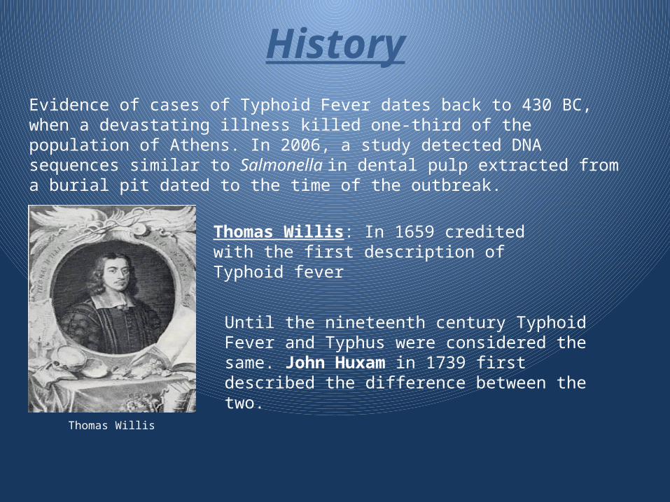
Thomas Willis: In 1659 credited with the first description of Typhoid fever
Evidence of cases of Typhoid Fever dates back to 430 BC, when a devastating illness killed one-third of the population of Athens. In 2006, a study detected DNA sequences similar to Salmonella in dental pulp extracted from a burial pit dated to the time of the outbreak.
Until the nineteenth century Typhoid Fever and Typhus were considered the same. John Huxam in 1739 first described the difference between the two.
Thomas Willis
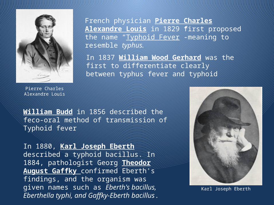
In 1880, Karl Joseph Eberth described a typhoid bacillus. In 1884, pathologist Georg Theodor August Gaffky confirmed Eberth's findings, and the organism was given names such as Eberth's bacillus, Eberthella typhi, and Gaffky-Eberth bacillus.
French physician Pierre Charles Alexandre Louis in 1829 first proposed the name “Typhoid Fever”-meaning to resemble typhus.
In 1837 William Wood Gerhard was the first to differentiate clearly between typhus fever and typhoid
William Budd in 1856 described the feco-oral method of transmission of Typhoid fever
Pierre Charles Alexandre Louis
Karl Joseph Eberth
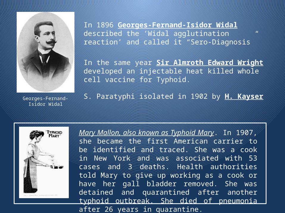
Georges-Fernand-Isidor Widal
In 1896 Georges-Fernand-Isidor Widal described the ‘Widal agglutination reaction’ and called it “Sero-Diagnosis”
In the same year Sir Almroth Edward Wright developed an injectable heat killed whole cell vaccine for Typhoid.
S. Paratyphi isolated in 1902 by H. Kayser
Mary Mallon, also known as Typhoid Mary. In 1907, she became the first American carrier to be identified and traced. She was a cook in New York and was associated with 53 cases and 3 deaths. Health authorities told Mary to give up working as a cook or have her gall bladder removed. She was detained and quarantined after another typhoid outbreak. She died of pneumonia after 26 years in quarantine.
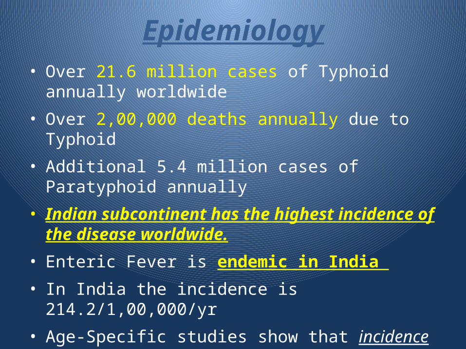
Epidemiology• Over 21.6 million cases of Typhoid annually
worldwide
• Over 2,00,000 deaths annually due to Typhoid
• Additional 5.4 million cases of Paratyphoid annually
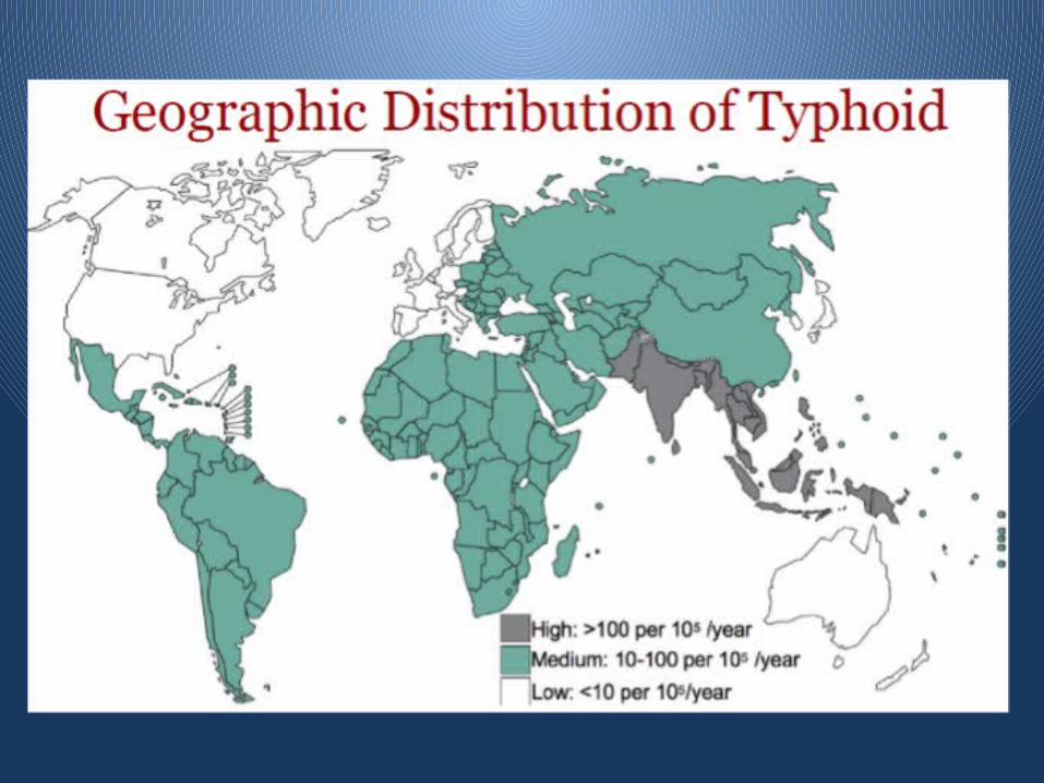
• Indian subcontinent has the highest incidence of the disease worldwide.
• Enteric Fever is endemic in India
• In India the incidence is 214.2/1,00,000/yr
• Age-Specific studies show that incidence in younger children are higher (0-4y:2,730; 5-19y:1,170; 20-40y:110 /1,00,000 per year)
Enteric Fever
is a collective term used for
Typhoid and Paratyphoid fevers.
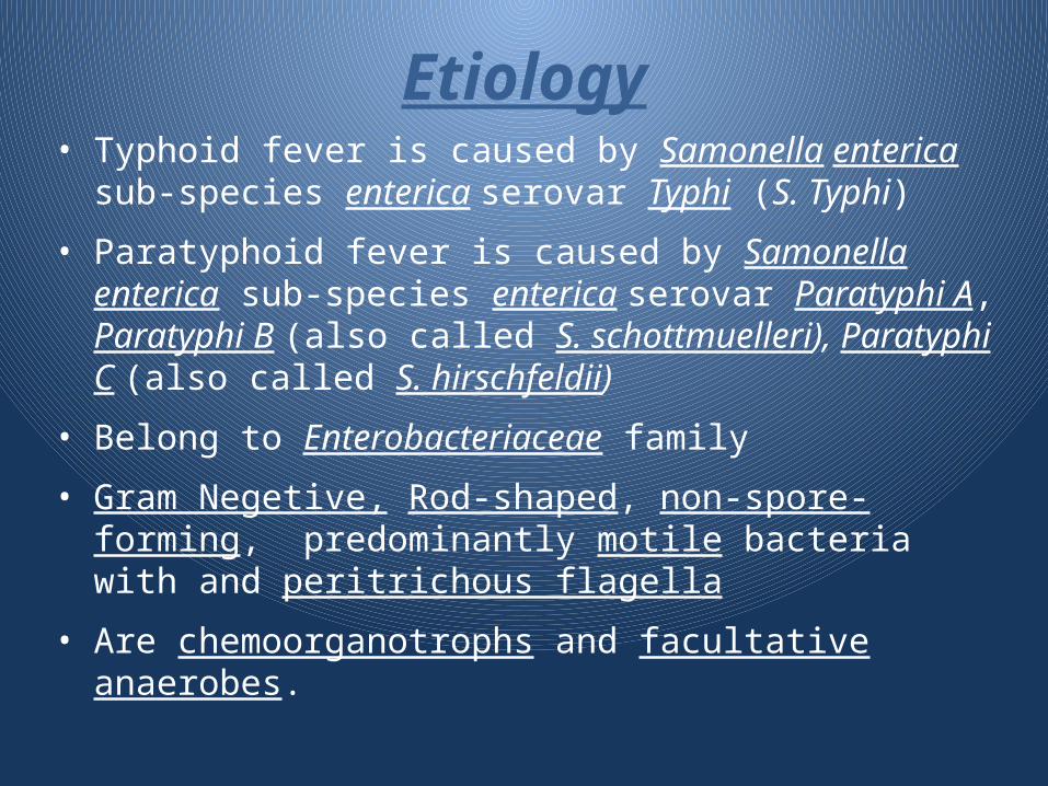
• Typhoid fever is caused by Samonella enterica sub-species enterica serovar Typhi (S. Typhi)
• Paratyphoid fever is caused by Samonella enterica sub-species enterica serovar Paratyphi A, Paratyphi B (also called S. schottmuelleri), Paratyphi C (also called S. hirschfeldii)
• Belong to Enterobacteriaceae family
• Gram Negetive, Rod-shaped, non-spore-forming, predominantly motile bacteria with and peritrichous flagella
• Are chemoorganotrophs and facultative anaerobes.
Etiology
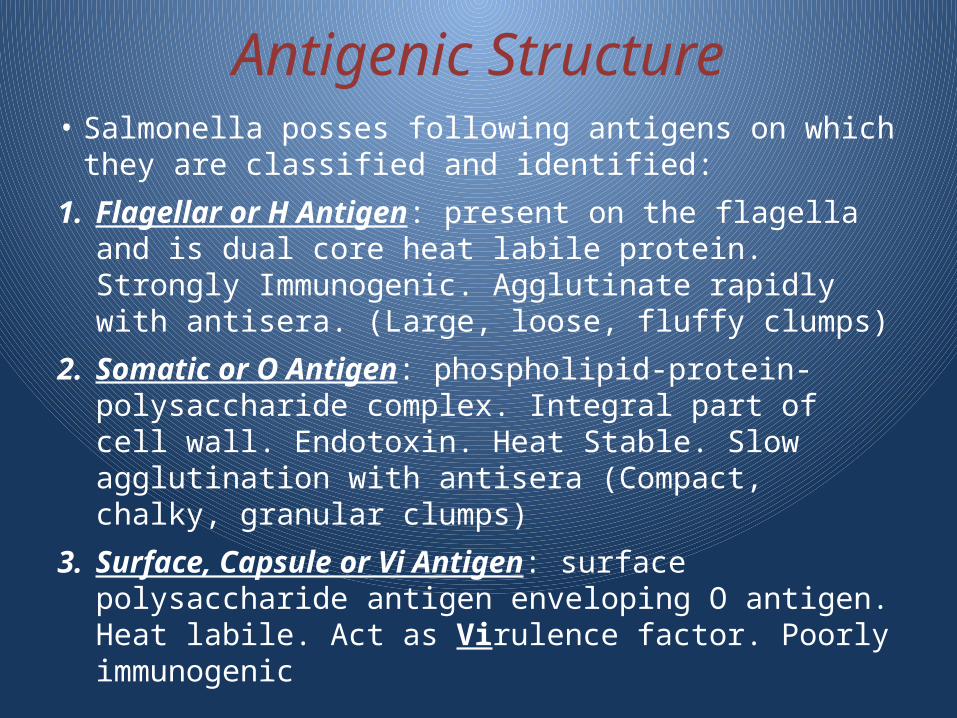
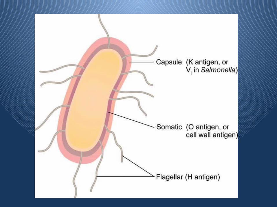
Antigenic Structure• Salmonella posses following antigens on which they
are classified and identified:
1. Flagellar or H Antigen: present on the flagella and is dual core heat labile protein. Strongly Immunogenic. Agglutinate rapidly with antisera. (Large, loose, fluffy clumps)
2. Somatic or O Antigen: phospholipid-protein-polysaccharide complex. Integral part of cell wall. Endotoxin. Heat Stable. Slow agglutination with antisera (Compact, chalky, granular clumps)
3. Surface, Capsule or Vi Antigen: surface polysaccharide antigen enveloping O antigen. Heat labile. Act as Virulence factor. Poorly immunogenic
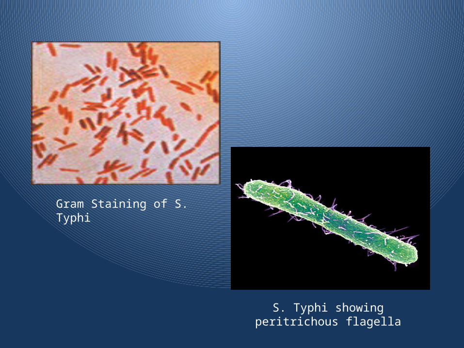
Gram Staining of S. Typhi
S. Typhi showing peritrichous flagella
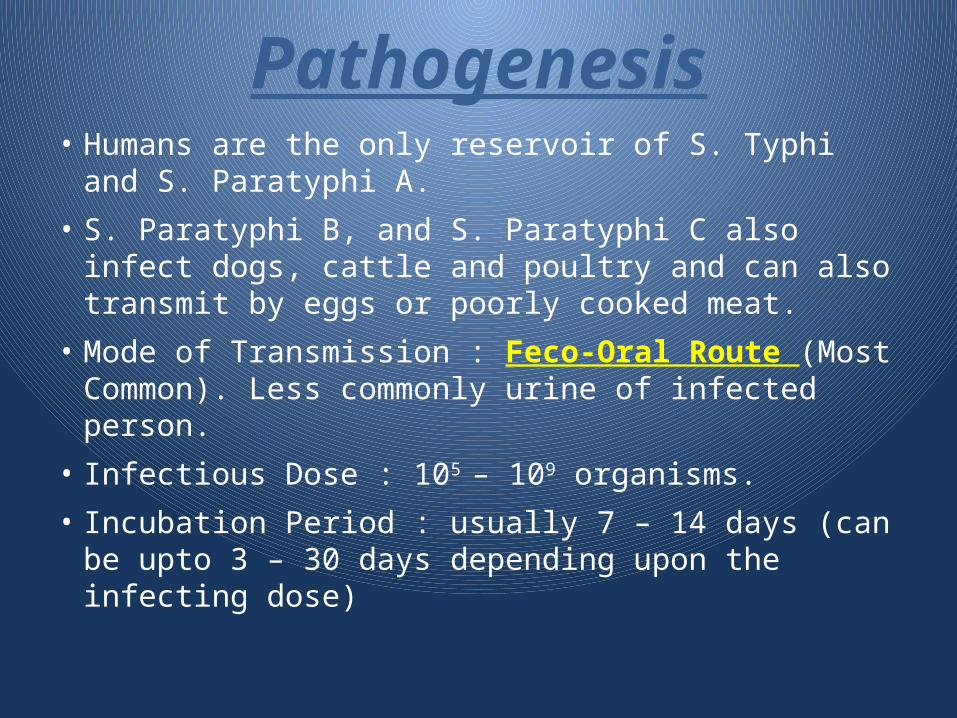
Pathogenesis• Humans are the only reservoir of S. Typhi and S.
Paratyphi A.
• S. Paratyphi B, and S. Paratyphi C also infect dogs, cattle and poultry and can also transmit by eggs or poorly cooked meat.
• Mode of Transmission : Feco-Oral Route (Most Common). Less commonly urine of infected person.
• Infectious Dose : 105 – 109 organisms.
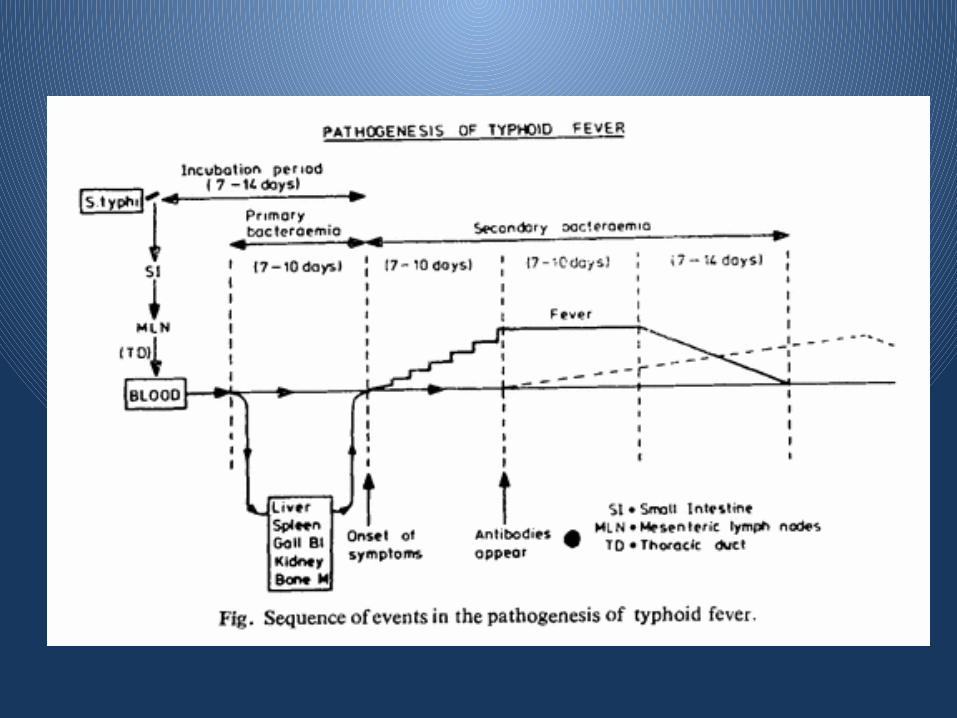
• Incubation Period : usually 7 – 14 days (can be upto 3 – 30 days depending upon the infecting dose)
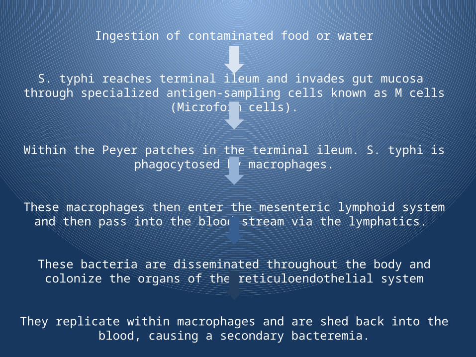
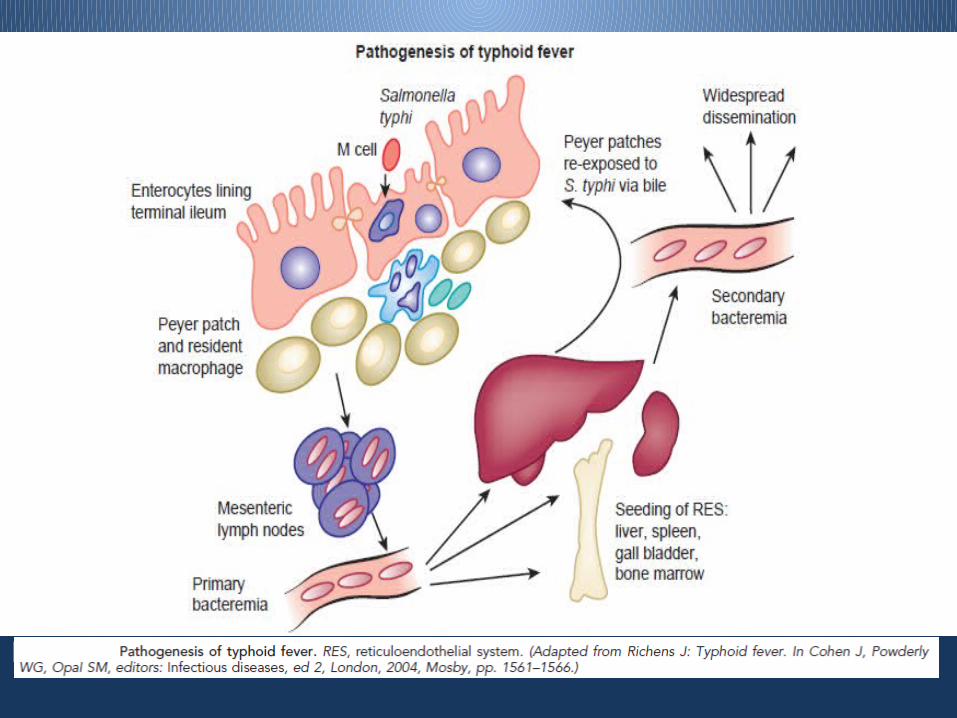
Ingestion of contaminated food or water
S. typhi reaches terminal ileum and invades gut mucosa through specialized antigen-sampling cells known as M cells (Microform cells).
Within the Peyer patches in the terminal ileum. S. typhi is phagocytosed by macrophages.
These macrophages then enter the mesenteric lymphoid system and then pass into the blood stream via the lymphatics.
These bacteria are disseminated throughout the body and colonize the organs of the reticuloendothelial system
They replicate within macrophages and are shed back into the blood, causing a secondary bacteremia.

• The surface Vi polysaccharide capsular antigen found in
S. Typhi interferes with phagocytosis and an virulence
trait encoded by the PhoP regulon gives ability to survive
within macrophages after phagocytosis
• S. typhi avoids triggering of an early inflammatory
response in the gut and instead colonize deeper tissues
and organ systems.
• Produces an inflammatory response in the deeper
mucosal layers and underlying lymphoid tissue causing
hyperplasia of Peyer patches and subsequent necrosis
and sloughing of overlying epithelium.
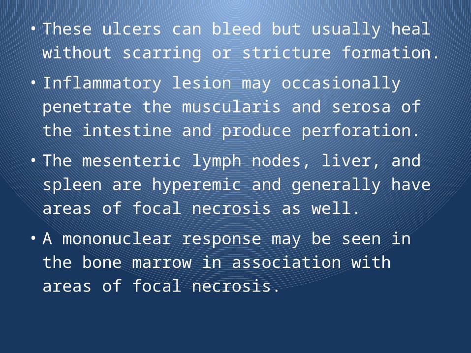
• These ulcers can bleed but usually heal without
scarring or stricture formation.
• Inflammatory lesion may occasionally penetrate
the muscularis and serosa of the intestine and
produce perforation.
• The mesenteric lymph nodes, liver, and spleen
are hyperemic and generally have areas of focal
necrosis as well.
• A mononuclear response may be seen in the
bone marrow in association with areas of focal
necrosis.
Carriers• The propensity to become a carrier follows the epidemiology of
gallbladder disease.
• Increasing with patient age and the antibiotic resistance of the
prevalent strains.
• Carriage are generally lower in children than adults.
• Types of Carriers:
– Convalescent carrier: passes bacilli in the excreta form 3weeks-
3months after clinical cure of typhoid.
– Temporary carrier: passes bacilli in the excreta form 3months-1year
after clinical cure of typhoid
– Chronic faecal carrier: continues to pass bacilli intermittently in the
excreta at least one year after infection.
– Chronic urinary carrier: the renal pelvis is infected & bacilli pass in
urine

Clinical Features• Mean age of onset in India is 10yrs.
• Onset is insidious and varies from a mild illness with low-grade fever, malaise, and slight, dry cough to a severe clinical picture with abdominal discomfort and multiple complications
• Presentation of typhoid may be more dramatic in children younger than 5 yrs of age, with comparatively higher rates of complications and hospitalization.
Features of 1st week of illness
• Rising Step ladder type of fever, with chills but rigors are rare
• Relative Bradycardia
• Dull, expressionless and toxic facies
• Dry skin with little sweating
• Coated tongue
• Musty, damp hay-like/baked bread like odour
• Vomiting
• Tender, doughy abdomen with slight guarding
• Constipation / Diarrhea (Pea Soup diarrhea)
• Occasionally minimal, non-productive cough
• Meningismus may occur early.
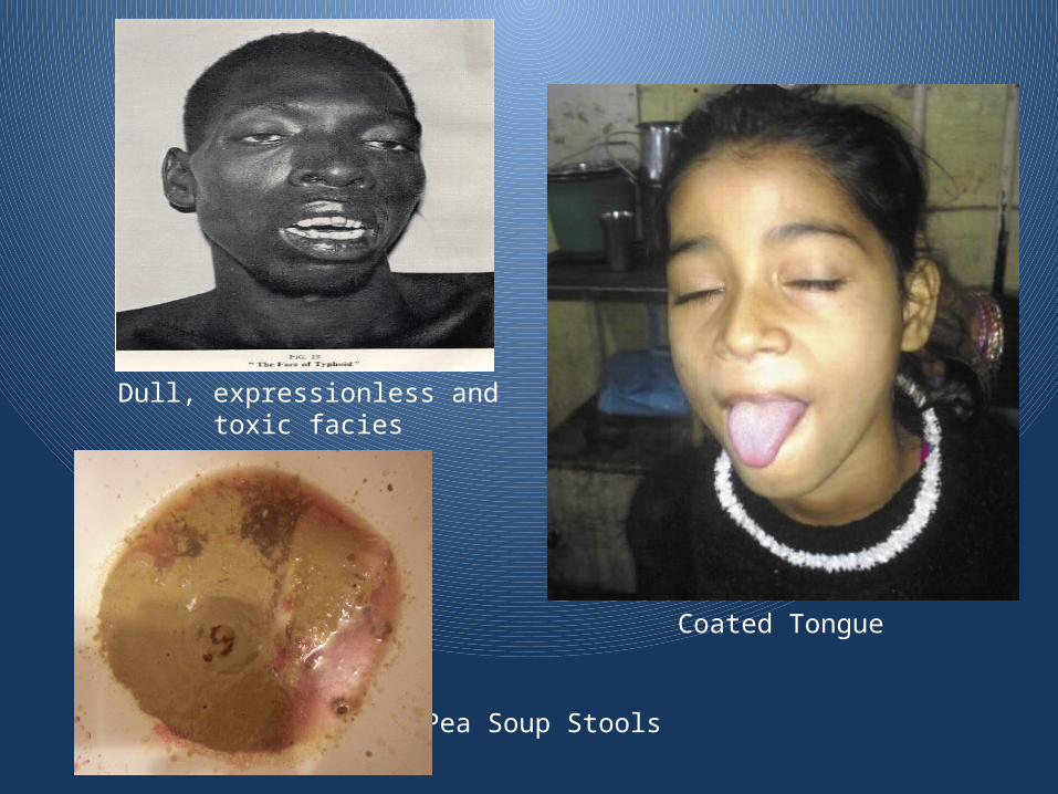
Dull, expressionless and toxic facies
Coated Tongue
Pea Soup Stools

Features of 2nd week of illness
• Continuous High grade fever (39.5-40.5 C)
• Worsening of cough may occur
• Rose Spots
• Abdominal Pain and tenderness
• Soft tender splenomegaly
• Soft tender hepatomegaly
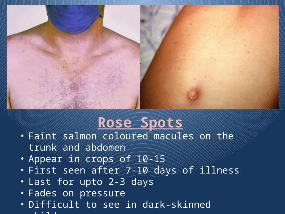
Rose Spots• Faint salmon coloured macules on the trunk and
abdomen • Appear in crops of 10-15• First seen after 7-10 days of illness • Last for upto 2-3 days • Fades on pressure• Difficult to see in dark-skinned children.
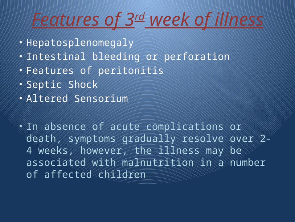
Features of 3rd week of illness
• Hepatosplenomegaly• Intestinal bleeding or perforation• Features of peritonitis• Septic Shock• Altered Sensorium
• In absence of acute complications or death, symptoms gradually resolve over 2-4 weeks, however, the illness may be associated with malnutrition in a number of affected children
• Enteric fever caused by S. Paratyphi organisms classically have a milder course.
• However, there have been several outbreaks of infection with drug-resistant S. paratyphi A, suggesting that paratyphoid fever may also be severe, with significant morbidity and complications
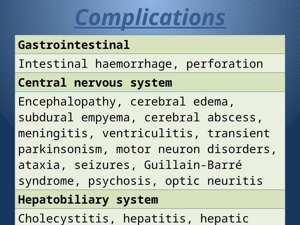
ComplicationsGastrointestinalIntestinal haemorrhage, perforationCentral nervous system Encephalopathy, cerebral edema, subdural empyema, cerebral abscess, meningitis, ventriculitis, transient parkinsonism, motor neuron disorders, ataxia, seizures, Guillain-Barré syndrome, psychosis, optic neuritisHepatobiliary systemCholecystitis, hepatitis, hepatic abscesses, splenic abscess, peritonitis, paralytic ileus, acute pancreatitis, splenic rupture
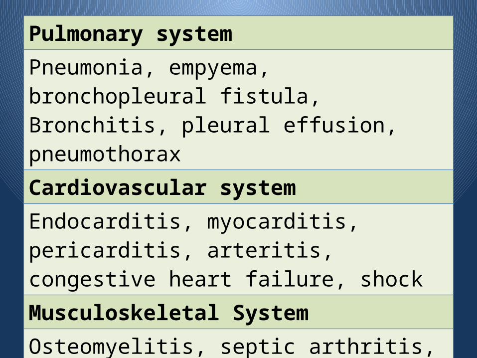
Pulmonary systemPneumonia, empyema, bronchopleural fistula, Bronchitis, pleural effusion, pneumothoraxCardiovascular system Endocarditis, myocarditis, pericarditis, arteritis, congestive heart failure, shockMusculoskeletal SystemOsteomyelitis, septic arthritis, Periostitis, typhoid spine, muscular rupture, Psoas abscess, gluteal abscess, cutaneous vasculitis
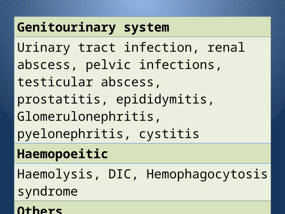
Genitourinary systemUrinary tract infection, renal abscess, pelvic infections, testicular abscess,prostatitis, epididymitis, Glomerulonephritis, pyelonephritis, cystitisHaemopoeiticHaemolysis, DIC, Hemophagocytosis syndromeOthersBed sores, hypercalcaemia, decubitus ulceration, abortion
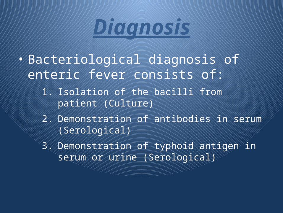
Diagnosis• Bacteriological diagnosis of enteric fever
consists of:1. Isolation of the bacilli from patient (Culture)
2. Demonstration of antibodies in serum (Serological)
3. Demonstration of typhoid antigen in serum or urine (Serological)
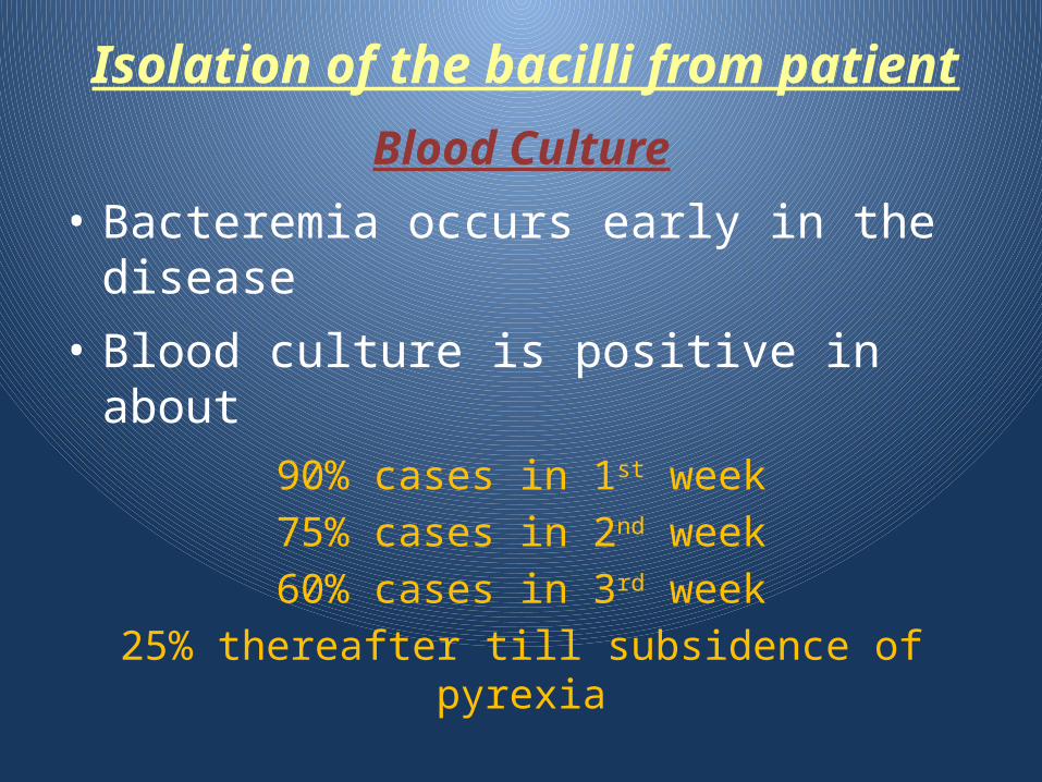
Blood Culture
• Bacteremia occurs early in the disease
• Blood culture is positive in about
90% cases in 1st week
75% cases in 2nd week
60% cases in 3rd week
25% thereafter till subsidence of pyrexia
• Blood cultures rapidly becomes negative on treatment with antibiotics.
Isolation of the bacilli from patient
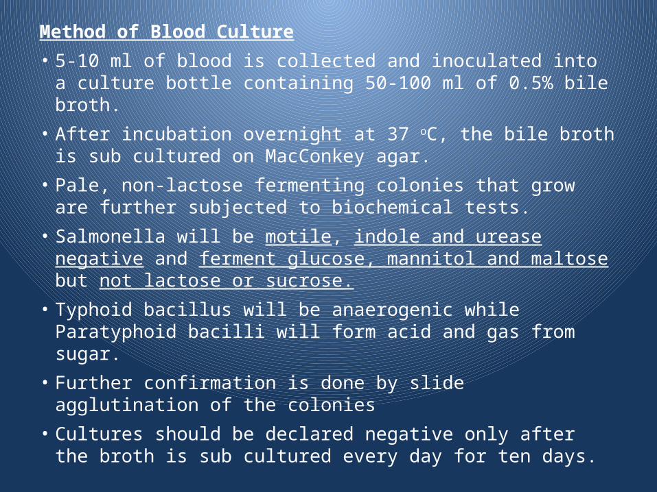
Method of Blood Culture
• 5-10 ml of blood is collected and inoculated into a culture bottle containing 50-100 ml of 0.5% bile broth.
• After incubation overnight at 37 oC, the bile broth is sub cultured on MacConkey agar.
• Pale, non-lactose fermenting colonies that grow are further subjected to biochemical tests.
• Salmonella will be motile, indole and urease negative and ferment glucose, mannitol and maltose but not lactose or sucrose.
• Typhoid bacillus will be anaerogenic while Paratyphoid bacilli will form acid and gas from sugar.
• Further confirmation is done by slide agglutination of the colonies
• Cultures should be declared negative only after the broth is sub cultured every day for ten days.
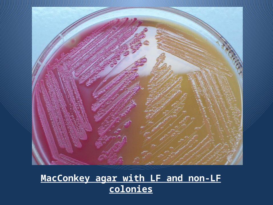
MacConkey agar with LF and non-LF colonies
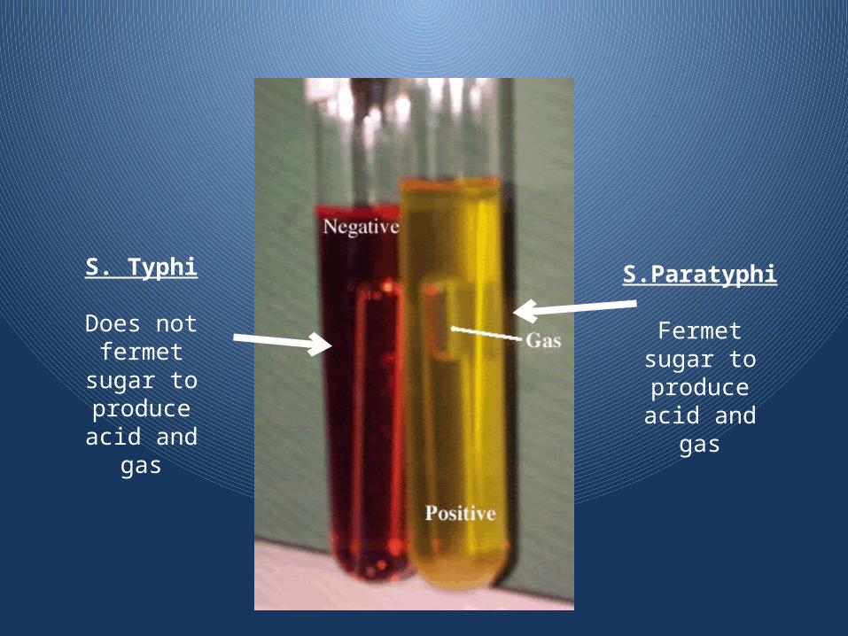
S. Typhi
Does not fermet sugar to produce acid
and gas
S.Paratyphi
Fermet sugar to produce acid
and gas
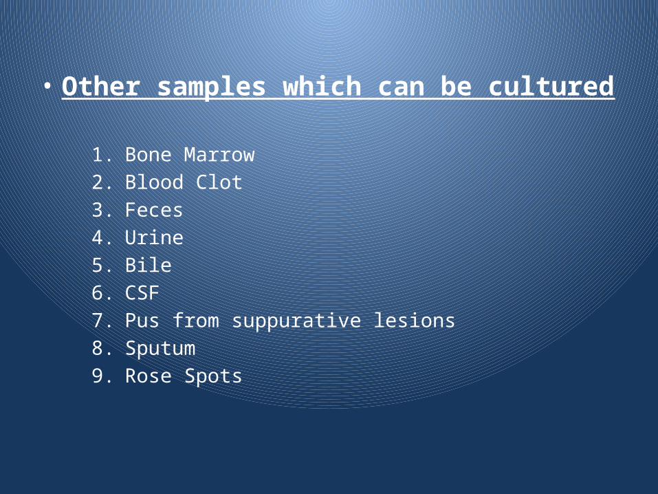
• Other samples which can be cultured
1. Bone Marrow
2. Blood Clot
3. Feces
4. Urine
5. Bile
6. CSF
7. Pus from suppurative lesions
8. Sputum
9. Rose Spots
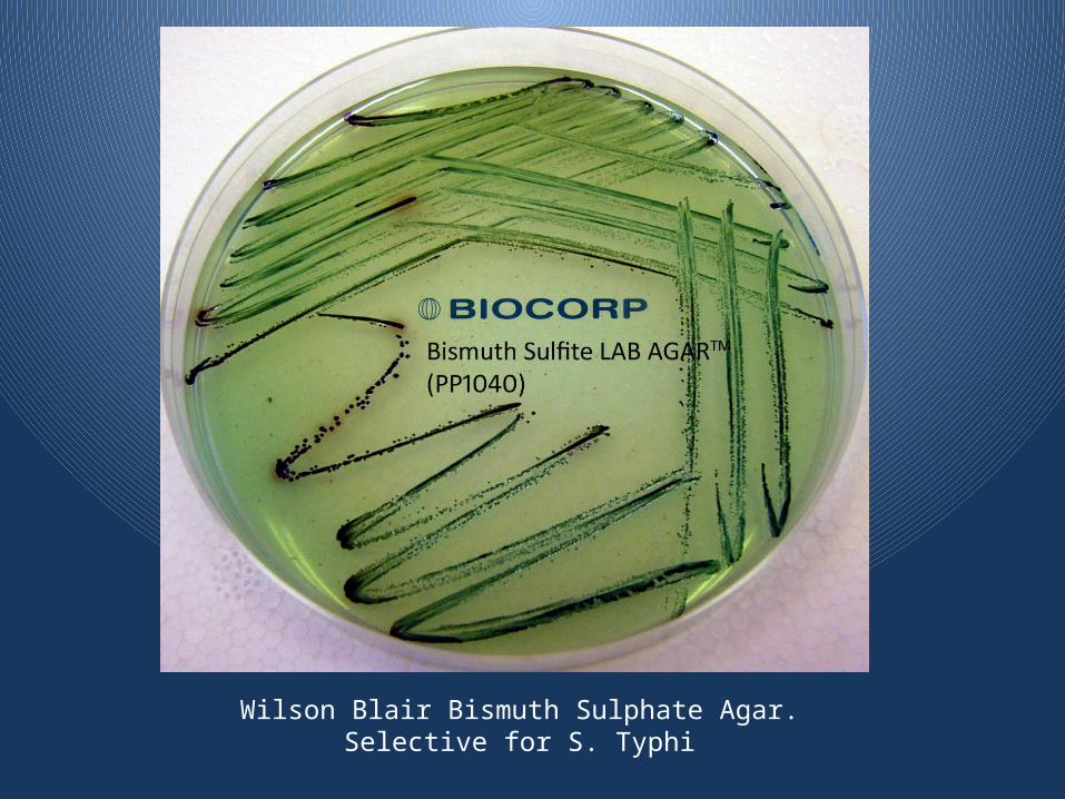
Wilson Blair Bismuth Sulphate Agar. Selective for S. Typhi
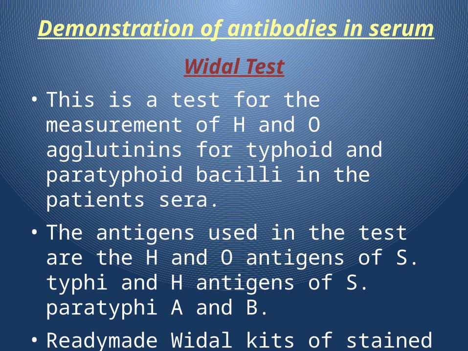
Demonstration of antibodies in serum
Widal Test
• This is a test for the measurement of H and O agglutinins for typhoid and paratyphoid bacilli in the patients sera.
• The antigens used in the test are the H and O antigens of S. typhi and H antigens of S. paratyphi A and B.
• Readymade Widal kits of stained antigen are now available commercially for use
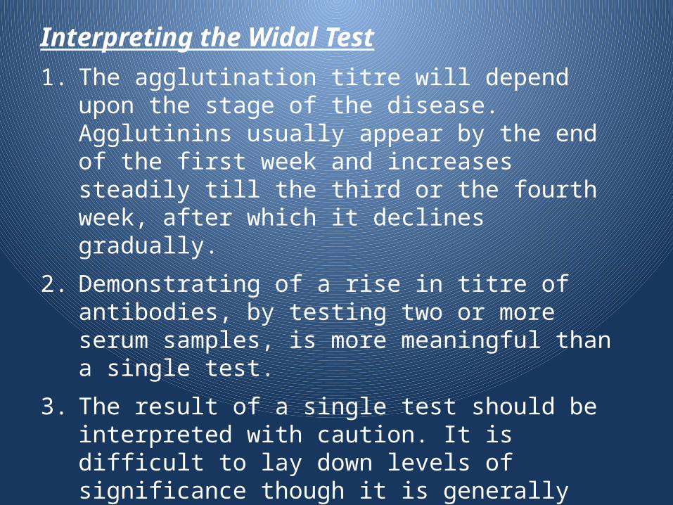
Interpreting the Widal Test
1. The agglutination titre will depend upon the stage of the disease. Agglutinins usually appear by the end of the first week and increases steadily till the third or the fourth week, after which it declines gradually.
2. Demonstrating of a rise in titre of antibodies, by testing two or more serum samples, is more meaningful than a single test.
3. The result of a single test should be interpreted with caution. It is difficult to lay down levels of significance though it is generally stated that titres of 1/100 or more of O agglutinins and 1/200 or more for H agglutinins are significant.
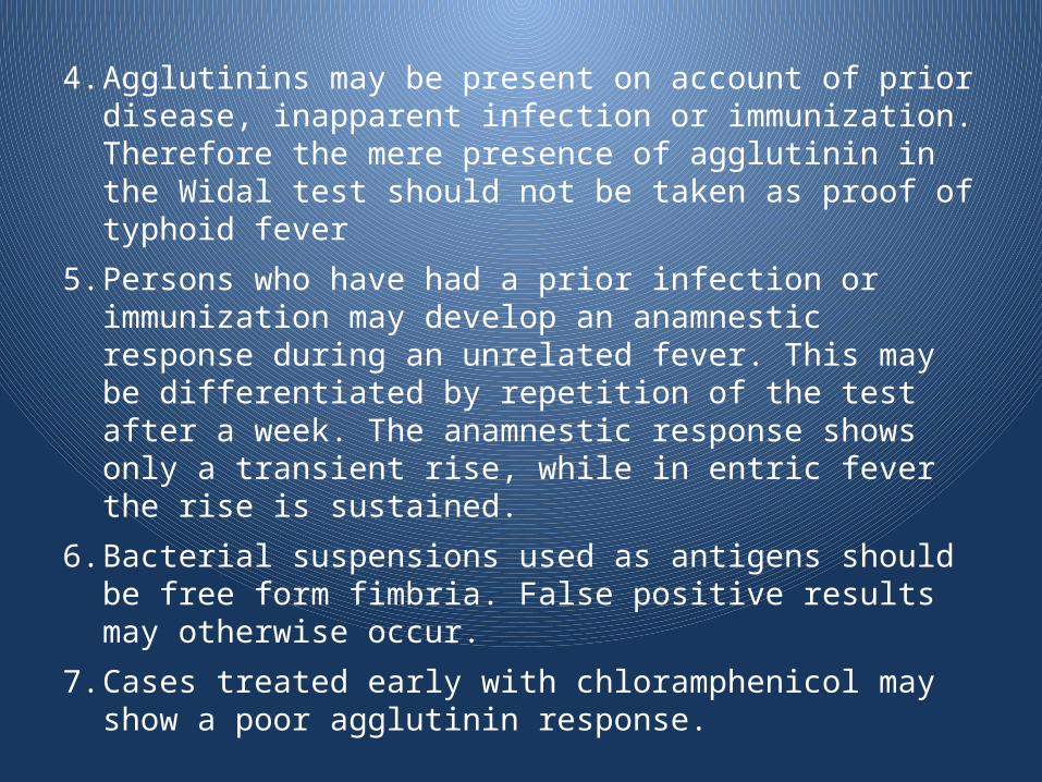
4. Agglutinins may be present on account of prior disease, inapparent infection or immunization. Therefore the mere presence of agglutinin in the Widal test should not be taken as proof of typhoid fever
5. Persons who have had a prior infection or immunization may develop an anamnestic response during an unrelated fever. This may be differentiated by repetition of the test after a week. The anamnestic response shows only a transient rise, while in entric fever the rise is sustained.
6. Bacterial suspensions used as antigens should be free form fimbria. False positive results may otherwise occur.
7. Cases treated early with chloramphenicol may show a poor agglutinin response.
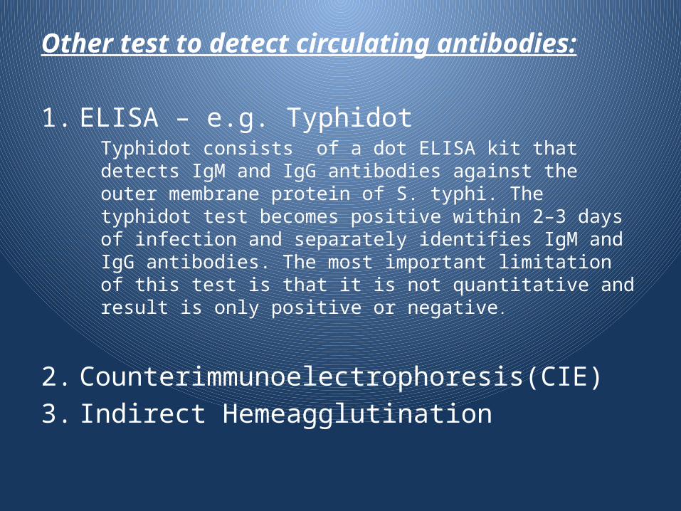
Other test to detect circulating antibodies:
1. ELISA – e.g. TyphidotTyphidot consists of a dot ELISA kit that detects IgM and IgG antibodies against the outer membrane protein of S. typhi. The typhidot test becomes positive within 2–3 days of infection and separately identifies IgM and IgG antibodies. The most important limitation of this test is that it is not quantitative and result is only positive or negative.
2. Counterimmunoelectrophoresis(CIE)
3. Indirect Hemeagglutination
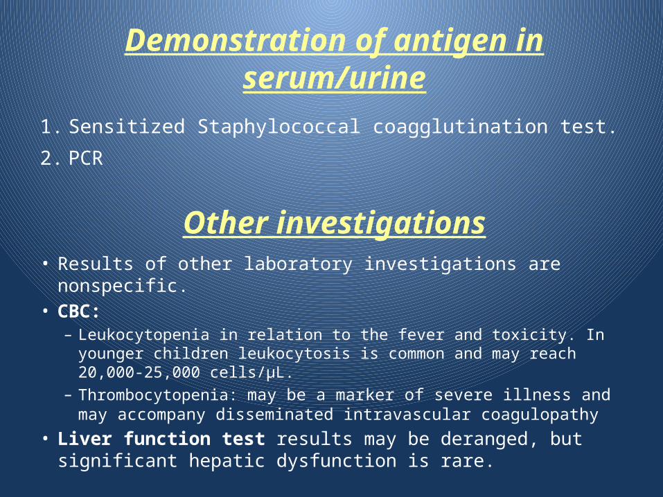
Demonstration of antigen in serum/urine
1. Sensitized Staphylococcal coagglutination test.
2. PCR
Other investigations• Results of other laboratory investigations are nonspecific. • CBC:
– Leukocytopenia in relation to the fever and toxicity. In younger children leukocytosis is common and may reach 20,000-25,000 cells/μL.
– Thrombocytopenia: may be a marker of severe illness and may accompany disseminated intravascular coagulopathy
• Liver function test results may be deranged, but significant hepatic dysfunction is rare.
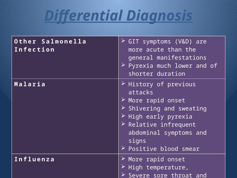
Differential Diagnosis
O t h e r S a l m o n e l l a I n f e c t i o n GIT symptoms (V&D) are more acute than the general manifestations
Pyrexia much lower and of shorter duration
M a l a r i a History of previous attacks More rapid onset Shivering and sweating High early pyrexia Relative infrequent abdominal
symptoms and signs Positive blood smear
I n f l u e n z a More rapid onset High temperature, Severe sore throat and cough Absence of a palpable spleen and
rose spots.
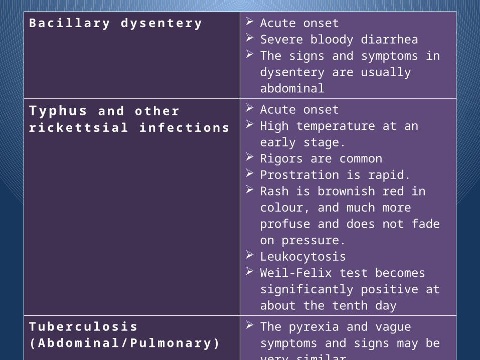
B a c i l l a r y d y s e n t e r y Acute onset Severe bloody diarrhea The signs and symptoms in
dysentery are usually abdominal
Ty p h u s a n d o t h e r r i c k e t t s i a l i n f e c t i o n s
Acute onset High temperature at an early stage. Rigors are common Prostration is rapid. Rash is brownish red in colour, and
much more profuse and does not fade on pressure.
Leukocytosis Weil-Felix test becomes
significantly positive at about the tenth day
Tu b e r c u l o s i s ( A b d o m i n a l / P u l m o n a r y )
The pyrexia and vague symptoms and signs may be very similar.
A chest X-ray, or laboratory confirmation of typhoid, may be the only sure method of diagnosis.
B r u c e l l o s i s Onset tends to be more insidious. Painful joint is frequently present.
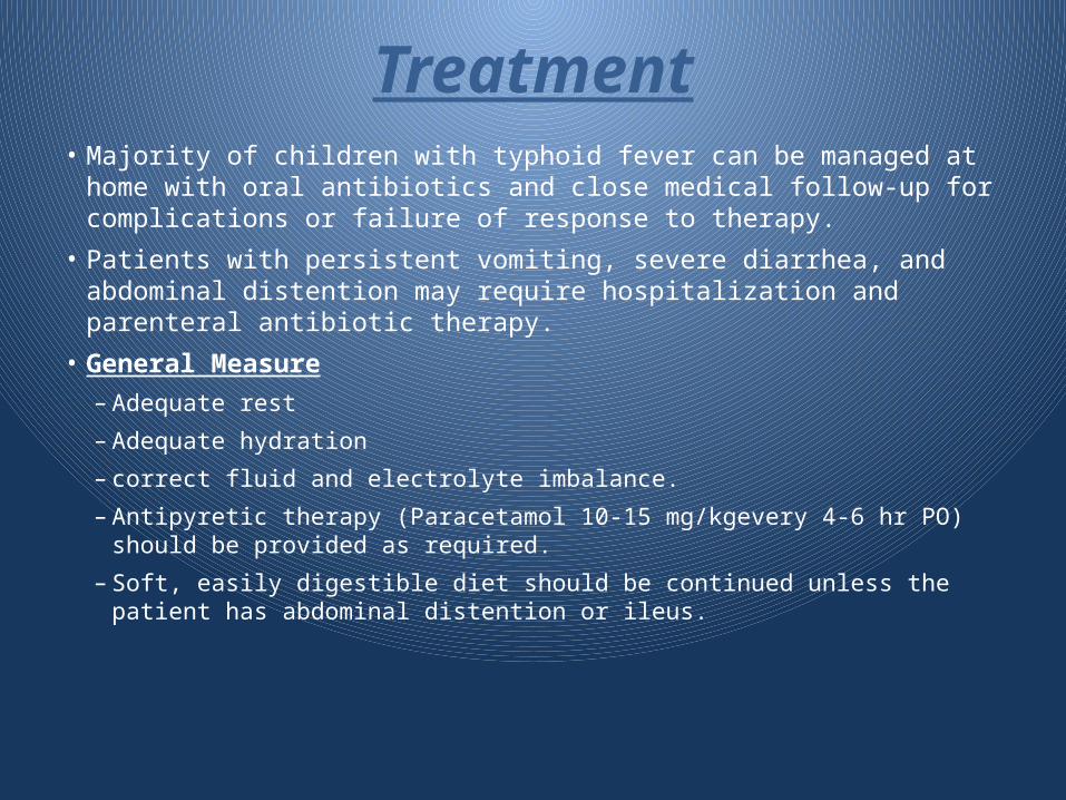
Treatment• Majority of children with typhoid fever can be managed at home with oral
antibiotics and close medical follow-up for complications or failure of response to therapy.
• Patients with persistent vomiting, severe diarrhea, and abdominal distention may require hospitalization and parenteral antibiotic therapy.
• General Measure
– Adequate rest
– Adequate hydration
– correct fluid and electrolyte imbalance.
– Antipyretic therapy (Paracetamol 10-15 mg/kgevery 4-6 hr PO) should be provided as required.
– Soft, easily digestible diet should be continued unless the patient has abdominal distention or ileus.
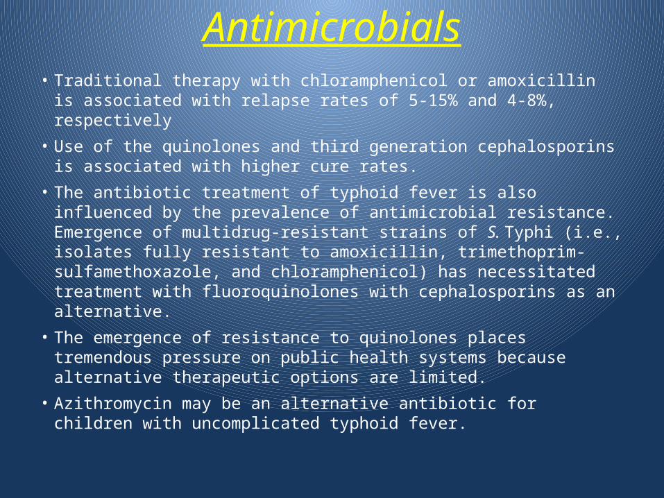
Antimicrobials• Traditional therapy with chloramphenicol or amoxicillin is
associated with relapse rates of 5-15% and 4-8%, respectively
• Use of the quinolones and third generation cephalosporins is associated with higher cure rates.
• The antibiotic treatment of typhoid fever is also influenced by the prevalence of antimicrobial resistance. Emergence of multidrug-resistant strains of S. Typhi (i.e., isolates fully resistant to amoxicillin, trimethoprim-sulfamethoxazole, and chloramphenicol) has necessitated treatment with fluoroquinolones with cephalosporins as an alternative.
• The emergence of resistance to quinolones places tremendous pressure on public health systems because alternative therapeutic options are limited.
• Azithromycin may be an alternative antibiotic for children with uncomplicated typhoid fever.
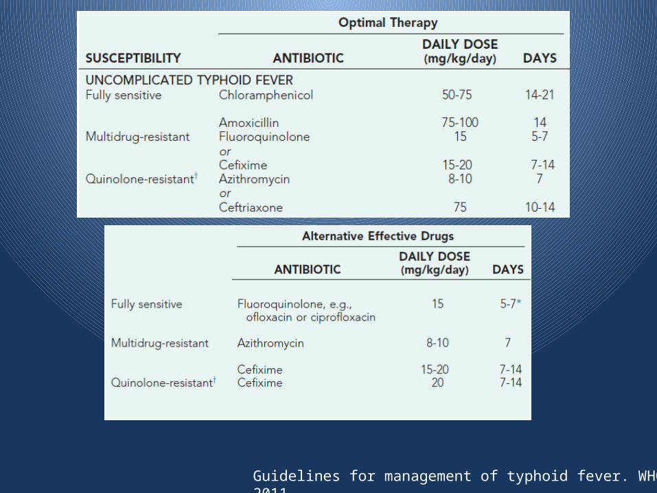
Guidelines for management of typhoid fever. WHO. July 2011
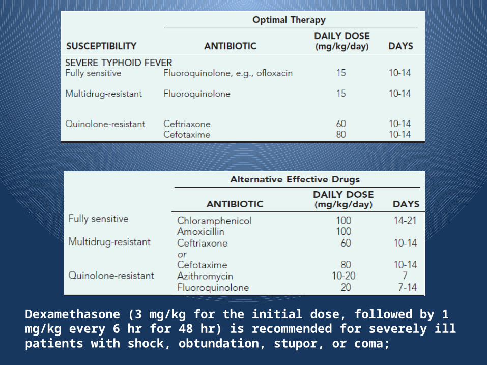
Dexamethasone (3 mg/kg for the initial dose, followed by 1 mg/kg every 6 hr for 48 hr) is recommended for severely ill patients with shock, obtundation, stupor, or coma;
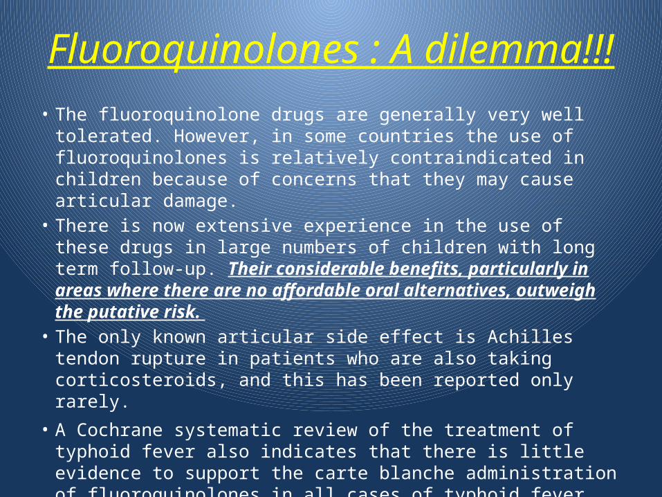
Fluoroquinolones : A dilemma!!!
• The fluoroquinolone drugs are generally very well tolerated. However, in some countries the use of fluoroquinolones is relatively contraindicated in children because of concerns that they may cause articular damage.
• There is now extensive experience in the use of these drugs in large numbers of children with long term follow-up. Their considerable benefits, particularly in areas where there are no affordable oral alternatives, outweigh the putative risk.
• The only known articular side effect is Achilles tendon rupture in patients who are also taking corticosteroids, and this has been reported only rarely.
• A Cochrane systematic review of the treatment of typhoid fever also indicates that there is little evidence to support the carte blanche administration of fluoroquinolones in all cases of typhoid fever
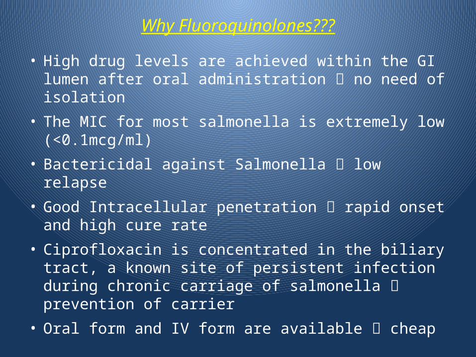
Why Fluoroquinolones???
• High drug levels are achieved within the GI lumen after oral administration no need of isolation
• The MIC for most salmonella is extremely low (<0.1mcg/ml)
• Bactericidal against Salmonella low relapse
• Good Intracellular penetration rapid onset and high cure rate
• Ciprofloxacin is concentrated in the biliary tract, a known site of persistent infection during chronic carriage of salmonella prevention of carrier
• Oral form and IV form are available cheap
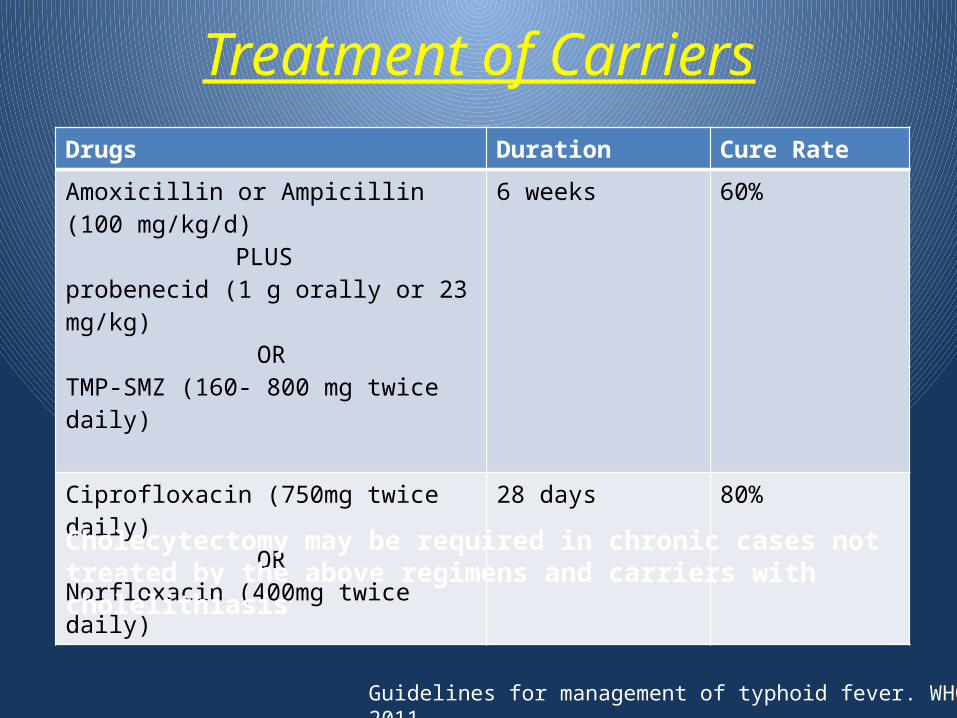
Treatment of CarriersDrugs Duration Cure Rate
Amoxicillin or Ampicillin (100 mg/kg/d)PLUS
probenecid (1 g orally or 23 mg/kg)OR
TMP-SMZ (160- 800 mg twice daily)
6 weeks 60%
Ciprofloxacin (750mg twice daily)OR
Norfloxacin (400mg twice daily)
28 days 80%
Cholecytectomy may be required in chronic cases not treated by the above regimens and carriers with cholelithiasis
Guidelines for management of typhoid fever. WHO. July 2011
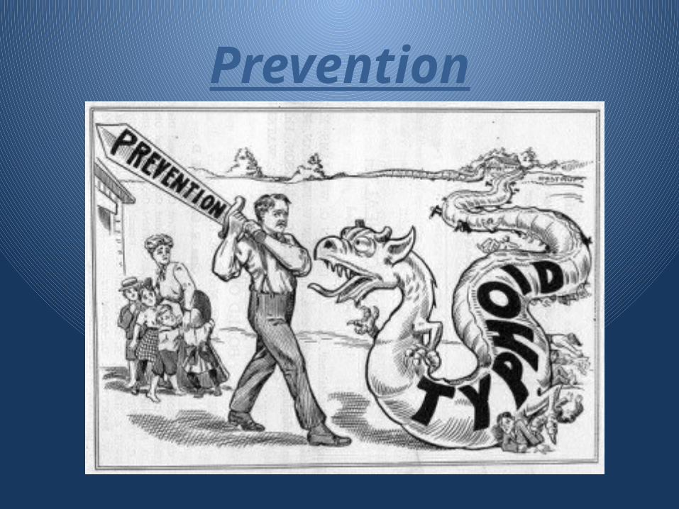
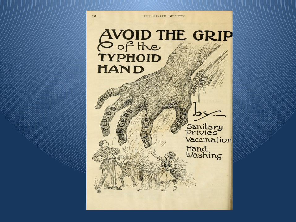
Prevention
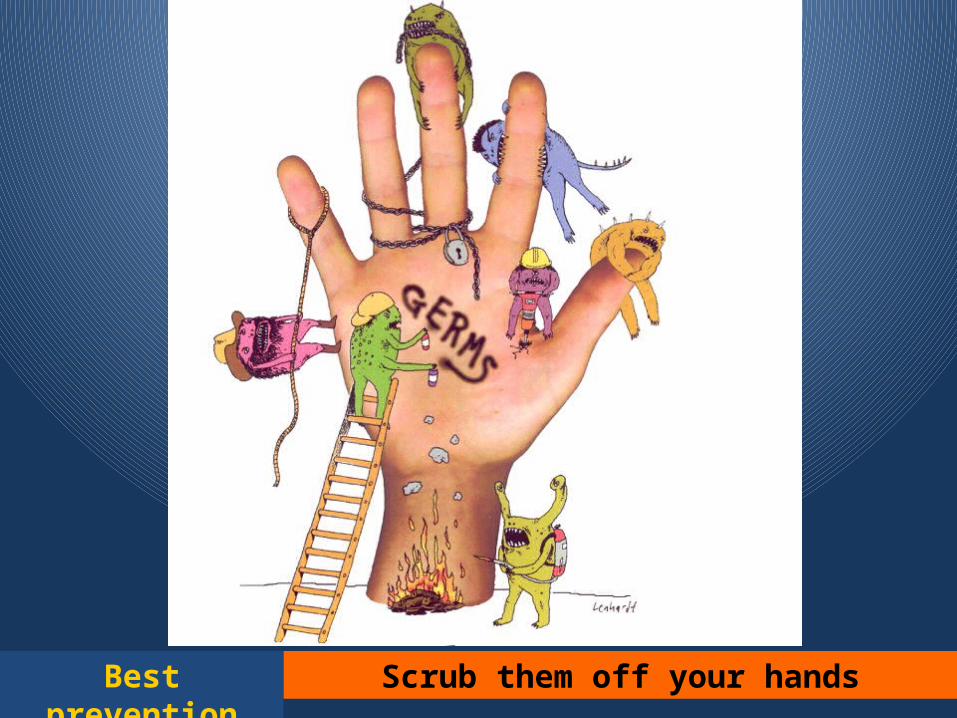
Best prevention Scrub them off your hands

Simple hand hygiene and washing can reduce several
cases of Typhoid
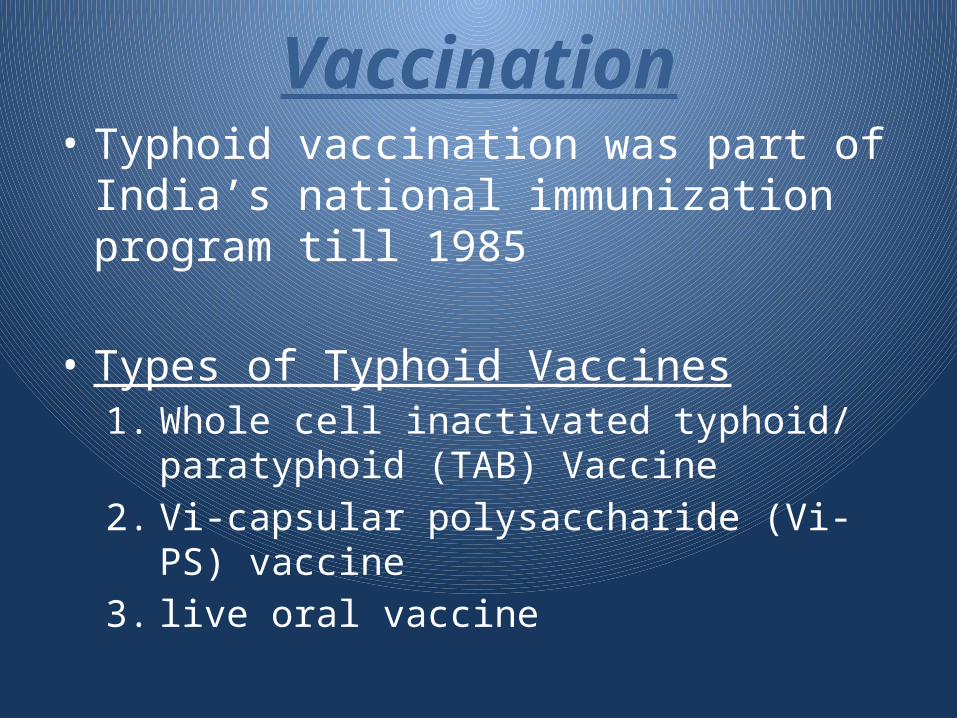
Vaccination• Typhoid vaccination was part of India’s
national immunization program till 1985
• Types of Typhoid Vaccines1. Whole cell inactivated typhoid/ paratyphoid
(TAB) Vaccine2. Vi-capsular polysaccharide (Vi-PS) vaccine3. live oral vaccine
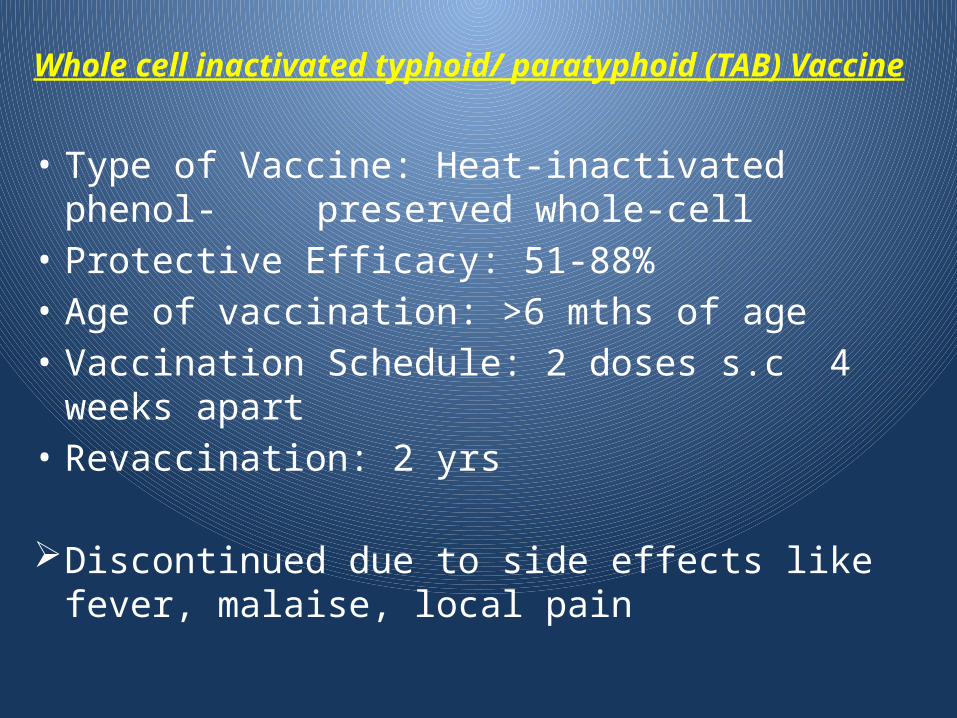
Whole cell inactivated typhoid/ paratyphoid (TAB) Vaccine
• Type of Vaccine: Heat-inactivated phenol- preserved whole-cell
• Protective Efficacy: 51-88%• Age of vaccination: >6 mths of age• Vaccination Schedule: 2 doses s.c 4 weeks apart• Revaccination: 2 yrs
Discontinued due to side effects like fever, malaise, local pain
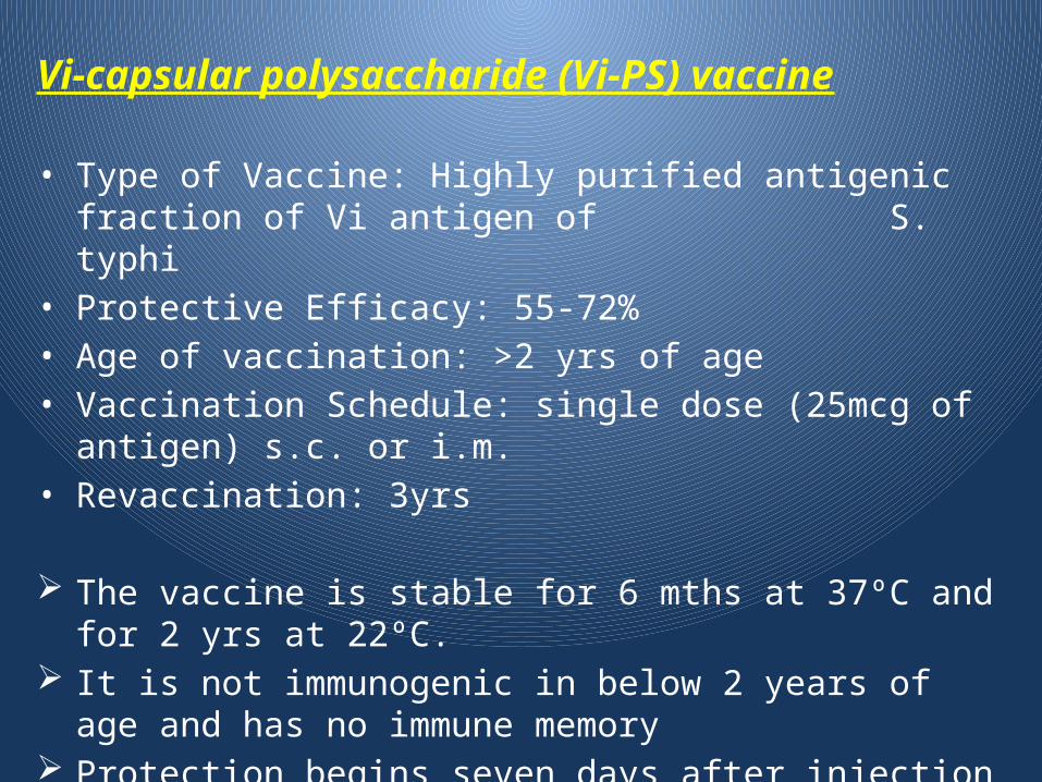
Vi-capsular polysaccharide (Vi-PS) vaccine
• Type of Vaccine: Highly purified antigenic fraction of Vi antigen of S. typhi
• Protective Efficacy: 55-72%• Age of vaccination: >2 yrs of age• Vaccination Schedule: single dose (25mcg of antigen) s.c. or i.m.• Revaccination: 3yrs
The vaccine is stable for 6 mths at 37ºC and for 2 yrs at 22ºC. It is not immunogenic in below 2 years of age and has no
immune memory Protection begins seven days after injection and maximum
reached at 28 days after injection
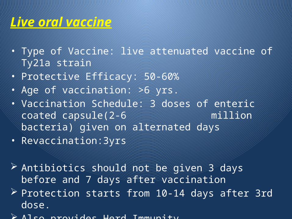
Live oral vaccine
• Type of Vaccine: live attenuated vaccine of Ty21a strain• Protective Efficacy: 50-60%• Age of vaccination: >6 yrs.• Vaccination Schedule: 3 doses of enteric coated capsule(2-6
million bacteria) given on alternated days
• Revaccination:3yrs
Antibiotics should not be given 3 days before and 7 days after vaccination
Protection starts from 10-14 days after 3rd dose. Also provides Herd Immunity Contraindicated in immunodeficiency
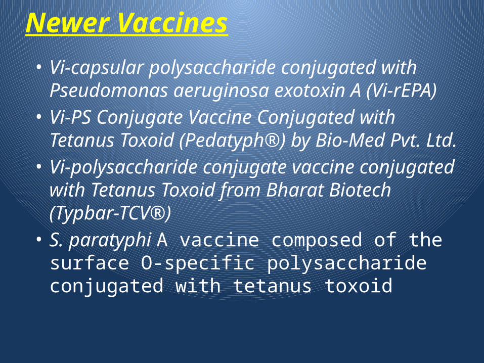
Newer Vaccines• Vi-capsular polysaccharide conjugated with
Pseudomonas aeruginosa exotoxin A (Vi-rEPA)• Vi-PS Conjugate Vaccine Conjugated with
Tetanus Toxoid (Pedatyph®) by Bio-Med Pvt. Ltd.• Vi-polysaccharide conjugate vaccine conjugated
with Tetanus Toxoid from Bharat Biotech (Typbar-TCV®)
• S. paratyphi A vaccine composed of the surface O-specific polysaccharide conjugated with tetanus toxoid
References1. Ananthnarayan and Panikarys textbook of microbiology 7th
edition
2. Atul Kothari, Amit Pruthi, Tulsi D. Chugh. The Burden of Enteric Fever. J Infect Developing Countries 2008; 2(4): 253-259.
3. Background document: The diagnosis, treatment and prevention of typhoid fever. World Health Organization. May-2003
4. Crump JA, Luby SP, Mintz ED (2004) The global burden of typhoid fever. Bull World Health Organ 82:346-353.
5. Ghai Essential Pediatrics 8th Edition
6. Guidelines for managemet of typhoid fever. WHO. July 2011
7. Huang DB, DuPont HL: Problem pathogens: extra-intestinal complications of Salmonella enterica serotype Typhi infection, Lancet Infect Dis 5:341–348, 2005
8. K. D. Moudgil M.D., B. S. Narang M.D., Pathogenesis of typhoid fever. The Indian Journal of Pediatrics July 1985, Volume 52, Issue 4, pp 371-378
9. IAP guidebook on immunization 2013-2014
10. IAP Task Force Report: Management of Enteric Fever in Children. Indian Pediatrics. Vol 43 Oct 2006
11. Manuela Raffatellu, R. Paul Wilson et al. Clinical pathogenesis of typhoid fever. J Infect Developing Countries 2008; 2(4): 260-266.
12. Nelson Text of Pediatrics 19th Edition
13. Ochiai RL, Acosta CJ, Danovaro-Holliday MC et al. (2008) A study of typhoid fever in five Asian countries: disease burden and implications for control. Bull World Health Organ 86(4):260-68.
14. PG textbook of Pediatrics
15. Sinha A, Sazawal S, Kumar R et al. (1999) Typhoid fever in children aged less than 5 years. Lancet 354:734-737
16. Victor Vaughan: A Biography of the pioneering Bacteriologist, 1851-1929 By Richard Adler
17. YK Joshi. SYMPOSIUM : TYPHOID FEVER. Journal Indian Academy of Clinical Medicine, Vol. 2, No. 1 and 2 , January-June 2001

Thank You…