entomopathogenic fungi: an effective biocontrol agent for …€¦ · entomopathogenic fungi: an...
TRANSCRIPT

Entomopathogenic Fungi: An Effective Biocontrol Agent for Management of Insect Populations
Naturally Digvijay Singha*, Tanveer Kour Rainab, Joginder Singha
aDepartment of Biotechnology, School of Biosciences, Lovely Professional University, Phagwara, Punjab, India. bDepartment of Biotechnology, School of Biosciences, Lovely Professional University, Phagwara, Punjab, India.
Abstract With the emerging need of switching over to sustainable agricultural practices, keeping in mind issues such as environmental protection, human safety and animal welfare, use of biocontrol agents like entomopathogenic fungi and other microorganisms, provides a better and safe substitute against chemical insecticdes and pesticides, associated with numerous environmental and health hazards. Entomopathogenic fungi used as a biocontrol agent can work as an effective biopesticides. These include class of fungi that can infect and kill insects. They help in regulating the insect and mite populations by causing lethal infections via epizootics. Some of the advantages associated with the use of entomopathogenic fungi in biocontainment strategies against insects and pests are; high host specificity, negligible effect on non-target organisms and easy mass production. Metarhizium anisopliae is one such entomopathogenic fungus that is testified to be effective against different insects and pests including termites, beetles and locusts. Understanding the biology and mechanism of action of these fungi is a prerequisite for using them as an effective biocontrol agent. Trichoderma is known for its parasitic activity against fungal plant pathogens, strains of Trichoderma have been reported to induce localised and systemic resistance in several plant pathogens as well as promote the growth and development of plant. Efficiency and cost are the two important parameters that need to be considered while comparing the entomopathogens (biopesticides) with the conventional chemical pesticides. This approach of using biocontrol agents instead of chemical pesticides seems to be very promising in the coming years as it heads towards sustainable agricultural practices and protecting environment, which is the need of the hour.
Keywords: Entomopathogenic fungi, biopesticides, biocontrol, Trichoderma, Metarhizium
INTRODUCTION Initial exploration in the field of biocontrol of the plant pathogens started in the mid-1920s; the techniques associated with the plant pathology have undergone vigorous investigation by the scientist, industry and research scholars in last few years [1-3]. Biological control can be defined as; "decreasing density of inoculums or disease fabricating actions of pathogen or parasite in its dynamic or static state, by one or more organisms, accomplished naturally or through alteration of surroundings, host or antagonist " [4, 5]. With the advances in the molecular technology, utilization of molecular techniques for natural control promptly growing, more powerful, widely applicable and easier to implement. Different molecular approaches and analysis are able to illustrate and annotate information regarding target pests that is crucial in improving control rates achieved and the success rate of the associated strategy of biocontrol. This information also includes taxonomic classification studies, probe generated through hybridization and ambiguous species, population structure and invasion origin. In addition to this, these can enhance knowledge about biocontrol agents, help in identification of new varieties of fungal pathogens and other related arthropods, provide taxonomic clarity, demonstrate genetic variability in agents, tranquil documentation of host association, and delivering an improved tool for tracing back evaluation of the biocontrol agent after their release into surroundings [6]. This review provides information
about the different fungal organisms used as biocontrol agents and the classical and predominantly molecular mechanism and approaches underlying the processes of biocontrol. Current molecular techniques accordant to classical biocontrol of pests and examples of molecular methods for biological control are described.
Entomopathogenic fungi – biocontrol agent Insects form the largest group of animals and cause the major damage in forest management, hence having a thorough understanding of the physiology of the natural parasites of these insects is very important. entomopathogenic fungi are one among these natural parasites. These form a heterogenous group belonging to diverse systematized groups and vary in their biology. All entomopathogenic fungi are mostly pathogenic in context to insects, and arthropods. These display a higher degree of effectiveness in infecting their host, thus can act as a regulator for regulating the abundance of harmful insects, including the forest insects (pests) [7]. Entomopathogenic fungi include numerous phylogenetically, morphologically and ecologically diverse fungal species, these organisms evolved to exploit insects. These are also present in omycota and water molds (kingdom-stramenopila) that are phylogenetically distinct and ecologically similar. Wide ranges of insect hosts from the aquatic larva to the adult insects are infected by these parasitic entomopathogenic fungi. Out of 31 orders of insects, 20 are infected by these
Digvijay Singh et al /J. Pharm. Sci. & Res. Vol. 9(6), 2017, 830-839
830

fungi in all the developmental stages: eggs, larvae, pupae, nymphs, and adults [8]. Entomopathogenic fungi differ substantially in the mechanism of action and virulence. Degree of attachment and penetrability of fungi inside host exoskeleton determines the success rate or the extent of infection. Insect pathogens can be controlled by using fungal entomopathogens, during the degradation of the insect integuments variety of extracellular enzymes is produced. In addition to efficiency, human safety, minimizing pesticide residues in food and biodiversity, safety of the non-target organisms; are few of the advantages associated with the utilization of microbial containment agents over conventional chemical pesticides [9]. Burges [10], Carruthers and Soper [11] and McCoy with co-workers [12] documented major breakthrough in their research that entomopathogenic fungi are capable of causing lethal infections and thus help in regulating the mite and insect population through epizootics. Because of having high host specificity, there is very minimal risk of confronting the non-target species. Entomopathogenic fungi are reported to confront and exploit a wide range of insect species including; lepidopteran larvae, thrips and aphids. These insects are of great concern in the agricultural sector globally [13]. Steps involved in virulence mechanism Different steps which are involved in adherence and degradation by this group of fungi infecting insects are summarized down under as;
a) Adhesion b) Germination c) Differentiation d) Penetration
Here in, wide array of intrinsic and extrinsic aspects collectively helps in determination of pathogenicity at each steps. For the infection, process to initiate attachment or adhesion of fungal spores onto the host cell is compulsory; this is generally brought about by the mucilage production. In addition to mucilage, other enzymes, lectins and different types of interactions viz, electrostatic forces and hydrophobic interactions also contribute majorly in this mechanism of infection [14]. The next step after adhesion that governs the virulence is the hydrolysis of the epidermis of the insect with the enzymes; among these secreted enzymes lipases, proteases and chitinases, produced sequentially, according to the substrate encountered are included [15]. Spore germination and behaviour is influenced by different factors including nutrients, fatty acids, water, cuticle surface ions, physiological state of host etc. [16]. Absorption of the consumable nutrients and the tolerance against the toxic compound existent on the surface, are the perquisites for successful germination [17]. Entry into insect lumen through cuticle layer is attained through the formation of a specialised structure called appressorium which attaches to the cuticle layer and forms a confined bridge or by the germ tube itself as described by Boucias and Pendland [18]. The bridge then provides
pathway for entry of fungal hyphae into insect’s body [13, 19, 20]. Penetration can be achieved through two processes including the mechanical and enzyme assisted pathways [12, 21, 22]. The exact ingression mechanism is idiosyncratic to the species. Various cuticle – degrading enzymes are produced during penetration process [23]. Entomopathogens are considered as the primary candidates for mycoinsecticides in agriculture, forestry and horticulture. Biology of the entomopathogenic fungi The life cycle of entomopathogenic fungi, synchronize with the life stages of the host insect and the prevailing external conditions. The infection levels, germination rate, insect host range and optimum temperature required may deviate between different fungal species [24, 25, 26]. Few species belonging to the Hyphomycetes family are categorized as opportunistic pathogens that infect many species in the range of insect orders; these are associated with production of a toxin which destroys the host defense response, leading to host death [27, 28]. Strongwellsea castrans, is the best example of a highly-evolved insect pathogenic fungi, that infects flies. Fungi display a unique mode of infestation. Bacteria and viruses are generally transmitted via contaminated food into the gut wall and finally enter the haemocoel either through the mouthparts or cuticle. Ingested fungal spores are voided through faeces and do not germinate in the gut. Factors responsible for the death of insect include; mechanical damage as a result of tissue invasion, toxicosis and starvation. Steps involved in infection process are summarized as under; 1. Adhesion and germination of conidia The primary event concerned with establishment of mycosis begins with the adhering of fungal spore onto the outer cuticle layer of the susceptible host. Locating the host is random event followed by passive attachment of spores with the aid of water or wind. For example, the dry spores of B. bassiana have an outer layer, which is composed of hydrophobic intermixed fascicle rodlets. The rodlet layer is unique to the conidial stage and has not been yet detected in vegetative cells. Hydrophobic forces of non-specific nature exerted by these rodlets were suggested as the possible reason for the attachment of dry spores to the cuticle [14]. In addition to this, a carbohydrate binding glycoprotein that is lectin, was detected on the conidia of B. bassiana, their presence on the conidia suggests their probable role in adhering of conidia to the insect cuticle as shown in figure1. However, the precise mechanism behind the interaction between fungal spores and the cuticle still is to be determined. The pathogen proceeds with rapid growth and germination, after adhering onto the surface of the host. The growth and germination of the pathogen on the host cell surface is influenced profoundly by the availability of nutrients, water, oxygen, pH, temperature and toxic compounds produced by the host on the surface.
Digvijay Singh et al /J. Pharm. Sci. & Res. Vol. 9(6), 2017, 830-839
831

Source: www.frontiersin.org/files/Articles/42518/fmicb-04-00024-HTML/image_m/fmicb-04-00024-g002.jpg
Figure 1: Adherence of fungal spore onto the outer cuticle layer of the susceptible host
Source: www.dovepress.com/entomopathogenic-organisms-conceptual-advances-and-real-world-applicat-peer-reviewed-fulltext-article-OAIP
Figure 2: Summary of the events involved in the infection cycle of an entomopathogenic fungus attacking an insect
Source:www.researchgate.net/publication/277017477_A_Review_of_Biopesticides_and_Their_Mode_of_Action_Against_Insect_Pests
Figure 3: Representation of Mode of action of entomopathogenic fungi against lepidopteran insects
Digvijay Singh et al /J. Pharm. Sci. & Res. Vol. 9(6), 2017, 830-839
832
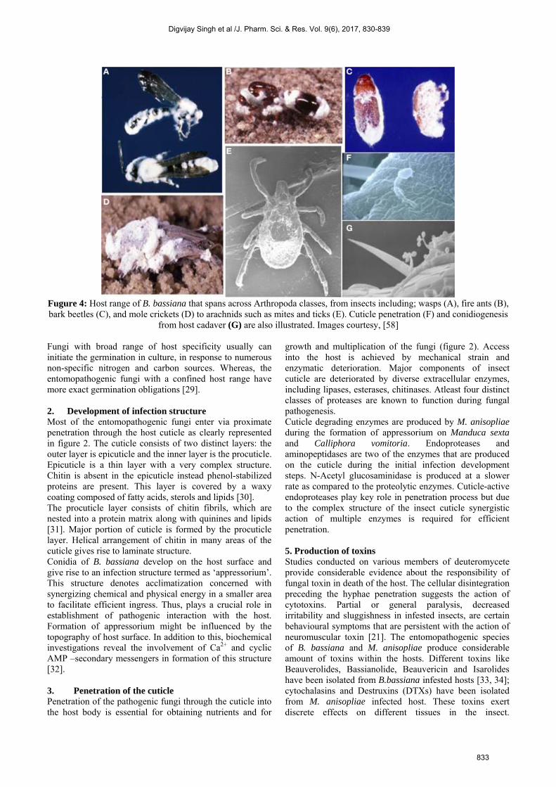
Fugure 4: Host range of B. bassiana that spans across Arthropoda classes, from insects including; wasps (A), fire ants (B), bark beetles (C), and mole crickets (D) to arachnids such as mites and ticks (E). Cuticle penetration (F) and conidiogenesis
from host cadaver (G) are also illustrated. Images courtesy, [58] Fungi with broad range of host specificity usually can initiate the germination in culture, in response to numerous non-specific nitrogen and carbon sources. Whereas, the entomopathogenic fungi with a confined host range have more exact germination obligations [29]. 2. Development of infection structure Most of the entomopathogenic fungi enter via proximate penetration through the host cuticle as clearly represented in figure 2. The cuticle consists of two distinct layers: the outer layer is epicuticle and the inner layer is the procuticle. Epicuticle is a thin layer with a very complex structure. Chitin is absent in the epicuticle instead phenol-stabilized proteins are present. This layer is covered by a waxy coating composed of fatty acids, sterols and lipids [30]. The procuticle layer consists of chitin fibrils, which are nested into a protein matrix along with quinines and lipids [31]. Major portion of cuticle is formed by the procuticle layer. Helical arrangement of chitin in many areas of the cuticle gives rise to laminate structure. Conidia of B. bassiana develop on the host surface and give rise to an infection structure termed as ‘appressorium’. This structure denotes acclimatization concerned with synergizing chemical and physical energy in a smaller area to facilitate efficient ingress. Thus, plays a crucial role in establishment of pathogenic interaction with the host. Formation of appressorium might be influenced by the topography of host surface. In addition to this, biochemical investigations reveal the involvement of Ca2+ and cyclic AMP –secondary messengers in formation of this structure [32]. 3. Penetration of the cuticle Penetration of the pathogenic fungi through the cuticle into the host body is essential for obtaining nutrients and for
growth and multiplication of the fungi (figure 2). Access into the host is achieved by mechanical strain and enzymatic deterioration. Major components of insect cuticle are deteriorated by diverse extracellular enzymes, including lipases, esterases, chitinases. Atleast four distinct classes of proteases are known to function during fungal pathogenesis. Cuticle degrading enzymes are produced by M. anisopliae during the formation of appressorium on Manduca sexta and Calliphora vomitoria. Endoproteases and aminopeptidases are two of the enzymes that are produced on the cuticle during the initial infection development steps. N-Acetyl glucosaminidase is produced at a slower rate as compared to the proteolytic enzymes. Cuticle-active endoproteases play key role in penetration process but due to the complex structure of the insect cuticle synergistic action of multiple enzymes is required for efficient penetration. 5. Production of toxins Studies conducted on various members of deuteromycete provide considerable evidence about the responsibility of fungal toxin in death of the host. The cellular disintegration preceding the hyphae penetration suggests the action of cytotoxins. Partial or general paralysis, decreased irritability and sluggishness in infested insects, are certain behavioural symptoms that are persistent with the action of neuromuscular toxin [21]. The entomopathogenic species of B. bassiana and M. anisopliae produce considerable amount of toxins within the hosts. Different toxins like Beauverolides, Bassianolide, Beauvericin and Isarolides have been isolated from B.bassiana infested hosts [33, 34]; cytochalasins and Destruxins (DTXs) have been isolated from M. anisopliae infected host. These toxins exert discrete effects on different tissues in the insect.
Digvijay Singh et al /J. Pharm. Sci. & Res. Vol. 9(6), 2017, 830-839
833
Lepidipteran muscle membrane is depolarized by the DTX, this depolarization achieved by activating calcium channels. DTX is also responsible for inhibiting the function of insect hemocytes [35]. There are presumably, many toxins that are yet not been isolated thus, their relevance to pathogenicity remains to be authenticated. Fungus plant interactions It is now evident that the entomopathogens are potentially engaged in the fungus-plant interactions. Investigation of the higher vascular plants have revealed that some of these are fungal endophytes [36, 37]. These include species of Clavicipitaceae, grouped under Hypocreales [38]. B. bassiana has entered in this group of fungi possessing endophytic activity by infecting the cereal plants like corn [39]. The Endophytic fungi are considered as the plant protective mutualists [40]. Recently, association of plants with M. anisopliae has been documented, and this association takes place in Rhizosphere layer that immediately surrounds the roots of the plant [41]. An experiment was conducted using a recombinant strain of M. anisopliae. The recombinant strain was released into a cabbage field in Maryland, USA. It was observed that the released isolate endured for longer duration in the soil that immediately surrounded the cabbage roots in comparison to the faraway bulk soil. It was believed that certain factors in the rhizosphere promoted the biological activity and persistence of M. anisopliae [41]. M. anisopliae expressed similar kind of genes when it was grown on the exudates from bean roots on a nutrient rich media, but when the fungus was growing on the insect cuticle and hemolymph, it expressed different genes, this indicated that the fungus acquired certain characteristics (adaptations) to grow saprophytically in the rhizosphere and function as a pathogen [42]. It is evident that the association of the fungi with the plants is important in the life cycle of both M. anisopliae and B. bassiana. Mechanism of host recognition by fungus The nutritional strategies of Trichoderma spp. Include both biotrophic and saprotrophic mode, they can also use dead fungal biomass as a food source. These can be classified as mycotrophic instead of mycoparasitic. The interaction between two fungi involves highly conserved signalling components [43]. Trichoderma generally proceeds with the penetration of fungal host by disintegrating its cell wall and then utilizing the intracellular contents of the host. This is aided by the action of certain hydrolytic enzymes like chitinases, proteases and glucanases. The enzymes are activated in Trichoderma prior to direct contact with the host and play crucial roles in the biocontrol [44]. A special diffusible factor of the host origin, known to act as an inducer for transcription of ech42 (endochitinase 42-encoding) gene in Trichoderma has been identified, during the interaction of Trichoderma with R. solani, before coming into physical contact [45]. In a similar way, the coiling of Trichoderma mycelia all over the host hyphae is triggered by lectins proteins existent in the host’s cell wall,
during the direct contact between the Trichoderma and its host [46]. The structural or extracellular chemical components of the host fungus trigger the formation of the infection structure and release of the lytic enzymes, as an induced response [45]. The understanding of these phenomena has been made much simpler by the advancements in gene sequencing and annotation of Trichoderma. The genomic and transcriptomic studies help in gaining the knowledge about the molecular physiology of mycrotrophic life cycle of Trichoderma. Expression of certain genes is triggered either on direct contact with the pathogenic fungi or when away from it. These genes are mostly protease encoding genes and oligopeptide transporter genes. T. harzianum strain CECT2413 growing in the similar conditions as provided during biological control, significantly express these proteases encoding genes belonging to subtilisin-like serine proteases [47]. The expression of these genes is abounding at the point of contact between T. atroviride and R. solani and S. sclerotiorum that are its fungal host species. Enhanced mycoparasitism activity is showcased in T. atroviride due to over expression of the protease gene prb1, attributing to its biocontrol activity. The released oligopeptides act as ligands and bind to the nitrogen starvation sensing receptors present on the surface of Trichoderma and trigger a host trapping mechanism which is similar to nematophagous fungi [48]. In Trichoderma, class IV G protein-coupled receptors (GPCRs) sense the released oligopeptides [49]. Two of class- IV GPCRs paralogs exist in each T. atroviride, T. virens and T. jecorina. GPCR gene Gpr1 of the cyclic AMP pathway is vital in mycoparasitic activity of T. atroviride [50]. A consensus G protein-signalling cascade comprising the three subunits, viz, Gα, Gβ; Gϒ subunits mediate downstream signal transduction from such receptors. Mutants with defective Gα subunit undergo loss of function in Tga1 and result in loss of mycoparasitic overgrowth on three hosts, in the strain. In addition to this, reduced chitinase activity and less accumulation of an antifungal compound 6-pentyl pyroneis observed [51, 52]. Consequently, deletion of tga1 homologue tgaA in T. virens reduced its mycoparasitic activity on phytopathogen S. rolfsii [53]. Mechanism of Signal transduction Mycoparasitism and mycotrophy are signal dependent mechanism wherein, the earlier recogonition of the lectins released by the fungal host is crucial and primary step. Generally, plant lectins promote coiling in Trichoderma but self-coiling is also observed in some species. Lectin induced coiling does not determine the attachment specificity of Trichoderma spp., to their host [51]. In Trichoderma, the hyphal growth is coupled with host and this is followed by formation of specialized papilla-like structures at the place where the cell wall disintegration and penetration of lumen take place prior to onset of mycoparasitism. At this stage, the Trichoderma spp. operates as a true fungal pathogen. Further expression of the genes in the host is regulated by downstream signals generated at receptors site. Three
Digvijay Singh et al /J. Pharm. Sci. & Res. Vol. 9(6), 2017, 830-839
834

important pathways associated with signal transduction have been observed in Trichoderma spp. These upregulate the expression of genes related to mycoparasitic activity and biocontrol. The genes which are over-expressed belong to the G-protein signalling, cAMP and MAPK (mitogen-activated protein kinases) pathways [43]. The MAP-kinase TVK1in T. virens I and its orthologs TmkA in T. asperellum and TMK1 in atroviride, are crucial in regulation of the signaling mechanism for an improved biocontrol. The genes transcription level increases respectively increase in T. virens and T. asperellum, during their interaction with the roots of the plants. Detailed analysis of the two genes belonging to G protein heterodimer signalling pathway class I (adenylate cyclase inhibiting) G-α subunit TGA1 of T. atroviridae and GNA3 of T. reesei, affirmed their role in biocontrol. TGA1 was identified important for regulation of coiling along the host hyphae. Alterations in the TGA1 results in reduction of the growth inhibiting effect on fungal host [51]. Host specific association linked to the MAP kinase activities was shown by TGA1 and TGA3 subunit was associated with the biological control as the deletion of the gene resulted in loss of virulence in the fungal strains [54]. A positive effect on mycoparasitism was observed on the activation of the GNA3 subunit in T. reesei, these results suggested a decisive role of cAMP and MAP-kinases in biological control by the Trichoderma species [55]. Entomopathogenic fungi in biological control of pests- approach of biological control: Undefined and unregulated use of conventional chemical insecticides has led to enhancement in developing resistance towards different chemicals present in the plant protection products, in the insects. More than 500 species of the arthropods have become resistant to more than one type of synthetic pesticides [56]. Invading and highly persistent species that are introduced accidently to a new continent or country and escape their natural pathogens and predators, pose another serious problem. Thus, there is a need to seek new, safer alternatives of reducing the outbreaks of pests [7]. Strategies for Biological Control The following strategies are employed for achieving biocontrol with the help of entomopathogenic fungi;
1) Classical biocontrol 2) Inundation biocontrol 3) Inoculation biocontrol 4) Conservation biocontrol
The classical and the inundation biocontrol strategies are widely used in forestry. Classical biocontrol encompass the deliberate entree of a foreign biocontrol agent that frequently co-evolves, for long-term pest control and permanent establishment [25]. For microorganisms, which are distributed throughout the planet, the term exotic refers to the distinct strain or biotype of the microbe used for pest control. These strains are not native to that particular area where they are being used.
The newly introduced exotic species has to acclimate to the new environment, multiply and spread to induce long-term effects, therefore understanding the biology of the exotic species and target and continuous monitoring of its presence in the area is of utmost importance. Inoculation biocontrol also includes the intended release of biocontrol agent in the similar manner, with an exception that the released variety will propagate for a short period of time and the control achieved by this strategy will be of longer duration and temporary. Permanent control as in case of classical biocontrol could not be achieved by this strategy [25]. In inundation biocontrol, the pest control is brought about entirely by the released variety, large number of mass-produced biocontrol agents or so-called pesticides are released into the desired area to reduce the pest population. The quantity of the biocontrol agents is maintained at such a level that it confers immediate results without achieving continuous establishment or impact. Works in the similar manner as chemical insecticides function. Conservation biological control works for the improvements of the existing practices or the environment, with the ultimate aim of protecting and exaggerating specific natural predators to reduce the effect of pests. The techniques used in the conservation biocontrol involve identification, handling and optimization of factors that enhance or suppress the effectiveness and abundance of the natural enemies [25]. Biocontrol Models: Group of fungi infecting major pests a) Metarhizium anisopliae First recognized as a biocontrol agent in 1880’s, found in soil; used as a biocontrol agent against different insects and pests including beetles, spittle bugs and locusts [57]. Different spores or conidial formulations of M. anisopliae are prepared and applied. After achieving the initiation of the fungal epizootic control, new spores and the vegetative cells are produced in the infected insect. These spores rapidly spread to the healthy insect population and promote persistent control. b) Beauveria bassiana – the entomopathogenic deuteromycete Beauveria bassiana belongs to the class deuteromycete (fungi imperfecta). These are filamentous fungi; different strains of beauveria are highly acquainted to a particular host insect. A broad range of medically or agriculturally significant strains of B. bassiana have been isolated from various insects worldwide. High host specificity is an interesting feature of many isolates of Beauveria. Tropical infectious diseases vectors such as the tsetse fly- Glossina morsitans, bugs of genera triatoma and rhodinus chagas' disease vector and the sand fly Phlebotomus that is potent transporter of leishmania are the hosts of medical importance. Consequently, Colorado potato beetle, few genera of termites and codling moths are categorized as the hosts of agricultural significance (figure 4). Entomopathogenic fungi are highly persisting in the environment thus, providing long-term suppression of the pest and better
Digvijay Singh et al /J. Pharm. Sci. & Res. Vol. 9(6), 2017, 830-839
835

control. B. bassiana is used as a biocontrol agent against leafhoppers Nephotettix spp., European borer Ostrinia nubilalis, and pine caterpillars Dendrolimus spp., in China. This group of fungi mostly exhibits dimorphic mode of growth. In asexual vegetative cycle germination of the spores is followed by filamentous growth of the fungi and formation of sympodial conidia. This mode is preferred when the specific insect host is not present. In contrast to this, Beauveria switches over to pathogenic life cycle in the presence of the host insect. Pathogenic life cycle follows, the germination of the conidiospores on the cuticle and subsequent penetration of the hyphal tubes directly into the integument of the insect. After penetrating the cuticle, alteration in the growth morphology of the fungus takes place resulting in switch over to yeast like phase and production of hyphal bodies, which are circulated into the haemolymph where they proliferate through budding. After causing the death of the host, the fungi reverts its growth back to saprotrophic stage. This ability to revert to yeast-like phase can be considered as a prerequisite for pathogenicity.
c) Trichoderma– as a Biocontrol agentDifferent species belonging to Trichoderma genus are wellknown for their ability to produce industrially applicableenzymes. Trichoderma species also have potential role inthe biological control of plant pathogens. Mycoparasitism(kill/parasitize fungal pathogens) against the fungalpathogens of crop plants is one of the major strategies ofbiocontrol, used by Trichoderma species. A number ofsignalling cascades are activated against fungal pathogenduring the mycoparisitic activity of Trichoderma.The Trichoderma species are found in almost every region,throughout the world and are isolated simply from differentsoil forms, sporocarps and decomposing woods.Trichoderma species have been demonstrated as potentbiocontrol agents against different pathogens, mostly soilborne which are causative agents of innumerable plantdiseases.Trichoderma has very high significance among otherfungal biocontrol agents due to its wide rangemycoparasititic potentiality against variety of fungalpathogens including Botrytis cinerea, Pythium spp.,Rhizoctonia solani, Sclerotium rolfsii and Sclerotiniasclerotiorum.Strains of Trichoderma are widely used as a substitute inplace of chemical pesticides to tackle many plantpathogens. This use is attributed to their antibiosis andmycolytic activity and to the responsiveness tophysiological changes mediated by host [59, 60]. Theyproduce various antimicrobial secondary metabolites likegliovirin, peptaibols and gliotoxin, which are known toinhibit numerous plant pathogens. T. virens and T.atroviride are the examples of two proven biocontrolspecies, these contain diverse reservoir of secondarymetabolite biosynthetic genes [55, 61]. Products of thesegenes aid for the secondary metabolite production and areassociated with mycoparasitism by Trichoderma againstother microbes. Steroids, terpenoids, pyrones andpolyketides are some of the highly characterized secondary
metabolites, these are non-polar in nature and have low molecular mass. Trichoderma spp. are known for the production of non- ribosomal peptides such as epipoly-thiodioxo-piperazines (ETPs) and siderophores that are mostly antimicrobial in nature, these enhance the cell wall lysis by acting in a synergistic manner with hydrolytic enzymes that are involved in cell wall disintegration [62]. Malmierca et al. [63] elucidated that the trichothecenes such as trichodermin and hazianum A (HA) are produced by Trichoderma species and disruption of the gene (tri gene) that impedes the synthesis of trichothecenes is responsible for lowering the biocontrol efficiency against Botrytis cinerea and Rhizoctonia solani pathogens. Silencing of the tri4 gene leads to down regulation of some defence genes of jasmonate (JA) and salicyclic acid (SA) pathways against B. cinerea in tomato plant whereas, the expression of these genes in the wild type strain is much higher. The results indicated that the pretreated plants were senisitized by the HA produced by Trichoderma, an increase in the expression of defence genes was also observed when they were challenged against B. cineria. Thus, Trichoderma species not only supress the propagation of fungal pathogen but also enhance the growth of treated plant and induce the expression of the defence genes. The widespread and special mechanisms prevailed in most of the Trichoderma spp. Include antagonism, parasitism, or even killing other fungi. The biocontrol efficient strains of Trichoderma spp. are found to successfully establish in the rhizosphere of the treated plants and promote growth of plants and stimulated defence responses when encountered by pathogens [64].
Genetic manipulations of entomopathogenic fungi Large-scale use of fungi as a biological control agent depends up to a large extent on the manipulations of wild–type strains and bringing together the desired attributes of different strains. Improvement in the effectiveness of the insecticide, reduction in the optimal dose necessary to kill the pest or to decrease crop damage by reducing the feeding time of the pest and expanding the host range are two approaches, among different types of improvements that may be considered. Complete understanding of the pathology of fungal infections is essential for the development of a hyper virulent and efficacious strain. Genetic transformation systems are an integral part of modern fungal research; these systems are the prerequisites for the experimental manipulation of the genes (virulence) in vivo and in vitro [65]. Availability of selectable transformation markers determines the success rate of these procedures. Techniques of genetic transformation have been employed in isolating specific virulent genes, investigating virulence determinants of M. anisophliae, and in the production of an enhanced virulence strain. Deciphering the molecular mechanism of fungal pathogenesis in insects will provide ground for the genetic engineering of the entomopathogenic fungi.
Digvijay Singh et al /J. Pharm. Sci. & Res. Vol. 9(6), 2017, 830-839
836

Field application of entomopathogenic fungi Laboratory tests always precede the venture for the practical application of the entomopathogenic fungi in the classical or inundation biological control strategies. These tests are conducted for the selection of highly virulent strains, determining inoculum dosage, to observe the impacts of both biotic and the abiotic factors on the fungus used as biocontrol agent and to test different mode in which the fungi can be introduced into the fields [66-73]. Although, sometimes these laboratory tests do not synchronize later with the practical use of the entomopathogenic fungi, but they provide beneficial and relevant information about the activity and the potential role of the fungus in biocontrol of many dangerous pests [74]. The practical use of microorganisms is not easy; it is associated with numerous problems. The biggest problem being the difficulty to predict the effects of these organisms used as biocontrol agents before actually releasing them into the environment. Different factors on which the success of the field trials depends need to be taken into consideration. The event of lesser efficiency of the biocontrol agent, applied in the field in contrast to that observed in the laboratory tests is observed quite often. Many characteristics of the entomopathogenic fungi like higher degree of virulence against the target species; no infestation in the non-target organisms including animals and humans; resistance towards abiotic and biotic factors of the environment are determinative in achieving satisfactory results in the field trials 75, 76]. The impact of entomopathogenic strains on the non-targets is always taken into account as side effects while the field application of the organism. It has been elucidated in various researches that entomopathogenic fungi show very less impact on the non-target insects [77-80]. Large-scale application of entomopathogenic fungi depends on economic and cheaper mass production of the synthetic media required. However, most of the fungal biopeticides are composed of the hypocrealen fungi, majority of them belonging to polyphagous species, demonstrating broad host spectrum. Among different species, the entomophthoralen fungi are highly specialized or monophagous and are not of great interest from the mycoinsecticides production point of view due to the complications in their propagation and development on artificial medium and mass scale propagation of the infective material [25]. Barley kernels colonized by Beauveria brongniarti based product was tried for field applicability under BIPESCO – EU-funded project, recently. The objective of the project was analysis and development of entomopathogenic fungi to control subterranean insect pests like weevils and scarabs [7]. Introduction of barley kernels colonized by fungus into the soil is the most commonly used method in case of soil-dwelling pests. This approach has been utilized in order to control the populations of larvae of Melolontha melolontha in different crop varieties [81-84]. Using fungal bands that are impregnated with entomopathogenic fungi, is another common method used for biocontrol. The bands are placed
near the trunk or around the branches of the tree and it provide protection against the invading pests. The method was first used to control Monochamus alternatus which is the major carrier of wilting in pines caused by Bursaphelenchus xylophylus [85]. Presently, the fiber band approach gives acceptable outcomes in biocontrol of Anoplophora glabripennis and Agrilusplani pennis invasive species [7]. Pros and Cons associated with use of entomopathogenic fungi as bio-control agent Fungi exhibit higher degree of host specificity. They can be used for controlling the virulent insect pests without causing any harm to beneficial insects. Advantages of using fungi as an insecticide as shown in Table 1. 1) less hazards encountered in contrast to chemical
insecticide application, such as environmental pollution and the absence of effects on mammals
2) Prolonged pest control and lack of insect resistance related problems.
3) Fungi show high degree of persistence and hence provide prolong pest control
4) Further development in this field by biotechnological research can help in producing better alternatives that can replace the chemical pesticides and insecticides.
Table 1: List of commercial and experimental products
derived from entomopathogenic fungi Product Name
Name of fungus used
Target insects
Mycotrol® Beauveria bassiana
Coding moth, pine caterpillar, European corn borer.
Boverin Beauveria bassiana
Colorado potato beetle
Mycar Culicinomyces clavisporus
Mosquito larvae
Metaquino Meta-sin®
Metarhizium anisopliae Nomurae arileyi
Spittle bug, sugarcane frog hopper ,Lepidopteran larvae
Vertalec Verticillium lecanii
Aphids, coffee green bug, greenhouse whitefly, thrips
Disadvantages associated with the use of fungi as insecticide 1) More time consuming as compared to chemical
insecticides, for instance, fungi requires 2-3 weeks’ time to kill the insect. In contrast to this chemical insecticide can do the same in may be 2-3 hours.
2) Additional control measures are required for other insects due to high specificity of fungal pesticide.
3) Production cost of bioinsecticides is relatively higher, shorter shelf life, necessity for cold storage of spores.
4) Application of the biocontrol agent needs to correspond with contributing factors like increased humidity level, low number of pests and a fungicidal free environment.
5) Poses a potential risk to immunodepressive or immunocompromised people.
6) Persistence and efficacy differs among insect species thus, the frequent optimization of the techniques is required to retain long-term impacts.
Digvijay Singh et al /J. Pharm. Sci. & Res. Vol. 9(6), 2017, 830-839
837

CONCLUSION Since thousands of years, the co-evolution of fungi and insects resulted in a wide range of intricate and complex interactions, including some of the beneficial interactions contributing to its utility as biological containment agent. Improvement of understanding about ecology of entomopathogenic fungi is vital for the further development of these organisms as biocontrol agent for wide range of serious insect pests. Biotechnology not only provides promising opportunities for the improvement of fungi for pest control but also, is more valuable in demonstrating the mechanism of pathogenicity. Due to the increased environmental awareness, failure of conventional chemical insecticides and pesticides, increased number of insecticide resistant species and food safety and concerns, the application of entomopathogenic fungi in biological control is amplifying abundantly. While determining the successful use of entomopathogenic fungi used, it is decisive to scrutinize each case individually and comparing the use directly with the chemical insecticides are usually disproportionate. For the successful use of any microbial agent, technical efficacy along with practical effectiveness, marketability, persistence and human welfare and safety are important. According to the recent existing research, the entomopathogenic fungi show minimal effects on the animals and other non-target organisms. In addition to this, they can also be used in integrated pest management replacing the conventional chemical insecticides [25]. Success of these programs is mostly based on significant, multi branched financial investments in R&D sector from industries, governments and other non-government organisations. One of the best utilization of entomopathogenic fungi is when complete wipe out of the pest is not needed instead, the pest populations are managed to a minimal level below which they could not be able to cause any effect on production or economy of crop production. Meanwhile, Trichoderma plays an important role in controlling plant pathogens, predominantly of fungal origin, inhabiting soils. Trichoderma-based products have proven to be safe for the farmers and consumers as well as they are favourable for the environment. However, more work needs to be done in this field to develop stable, easy to produce, cost effective and easy to apply formulations of the same.
REFERENCES [1] Cook, R.J., Ann. Rev PhytoPathol. 1993, 31, 53 – 80. [2] Utkhede, R.S., Can. J. Plant Pathol. 1996, 18, 455 – 462. [3] Weller, D.M., Ann. Rev. Phytopathol. 1988, 26, 379 – 407. [4] Boyetchko, S.M., in: Mukerji, K.G., Chamola, B.P., Upadhyay, R.K.
(Eds.), Biotechnological Approaches in Biocontrol of PlantPathogens, Springer Science+Business Media, New York 1999, pp.51 − 71
[5] Baker, K.F., Cook, J, Biological control of plant pathogens. Cambridge University Press. USA. 1974.
[6] Gaskin, J.F., Bon, M.C., Cock, M.J., Cristofaro, M., De Biase, A.,De Clerck-Floate, R., Ellison, C.A., Hinz, H.L., Hufbauer, R.A.,Julien, M.H., Sforza, R., Biol. Cont. 2011, 58, 1 – 21.
[7] Augustyniuk-Kram, A., Kram, K.J., in: Blanco, J.A., Lo, Y.H.(Eds.),. Forest Ecosystems–More than Just Trees, InTech, Croatia2012, p. 265.
[8] Araujo, J., Hughes, D.P., Adv. Genet. 2016, 94, 1 – 39. [9] Shahid, A.A., Rao, Q.A., Bakhsh, A., Husnain, T., Arch. Biol. Sci.
2012, 64, 21-42.
[10] Burges, H.D., in: Burges, H.D. (Ed.), Microbial control of pests andplant diseases 1970-1980. Academic Press, London, 1981, pp. 737 –767.
[11] Carruthers, R.I., Soper, R.S., in: Fuxa, J.R., Tanada, Y., (Eds.),Epizootiology of Insect Diseases, John Wiley and Sons, New York1987, pp. 357- 416
[12] McCoy, C.W., Samson, R.A., Boucias, D.G., in: Ignoffo, C.M.,Mandava, N.B., (Eds.), Handbook of Natural Pesticides: Volume 5a:Microorganisms, Volume 5, CRC-Press 1988
[13] Roberts, D.W., Humber, R.A., in: Cole, G.T., Kendrick, B. (Eds.)Biology of Conidial Fungi. Academic Press, New York 1981, pp.201 – 236.
[14] Boucias, D.G., Farmerie, W.G., Pendland, J.C., J. Invert. Path. 1998,72, 258 – 261.
[15] Smith, R.J., Pekrul, S., Grula, E.A., J. Invert. Path. 1981, 38, 335 –344.
[16] Hassa, M., Dillion, R.J., Charnley, A.K., J. Invert. Path. 1989, 54,227−279
[17] Latge, J.P., Sampeoro, L., Brey, P., Diaquin, M., J. Gen. Microbiol. 1987, 133, 1987 – 1997.
[18] Boucias, D.G., Pendland, J.C., J. Invert. Path. 1982, 39, 338. [19] Wraight, S.P., Carruthers, R., Bradley, C.A., Jaronski, S.T., Lacey,
L.A., Wood, P., Galaini-Wraight, S., J. Invert. Path. 1998, 71, 217 –226.
[20] Zacharuk, R.Y., 1973. Miscellaneous Publication of theEntomological Society of America, 9, p.112.
[21] Charnley, A.K., in: Andersonn, J.M., Rayner, D.M., Walton,D.W.H., Invertebrate-Microbial Interactions. British MycologicalSociety Symposium 6, Cambridge University Press, London 1984,pp 229 – 271.
[22] Leger, R.S., Durrands, P.K., Charnley, A.K., Cooper, R.M., J. Invert.Path. 1988, 52, 285 – 293.
[23] Gillespie, J. P., Bateman, R., Charnley, A.K., J. Invert. Path.1988,71, 128 – 137.
[24] Sierotzki, H., Camastral, F., Shah, P.A., Aebi, M., Tuor, U., Mycol.Res., 2000, 104, 213 – 219.
[25] Pell, J.K., Eilenberg, J., Hajek, A.E., Steinkraus, D.C., in: Butt,T.M., Jackson, C., Magan, N. (Eds.), Fungi as biocontrol agents:Progress, Problems and Potential. CAB International, Wallingford2001, pp. 71 – 153.
[26] Shaw, K.E., Davidson, G., Clark, S.J., Ball, B.V., Pell, J.K.,Chandler, D., Sunderland, K.D., Biol. Cont. 2002, 24, 266 – 276.
[27] Roberts, D.R., in: Burges, H.D. (Ed.), Microbial Control of Pestsand Diseases, 1970–1980. Academic Press, London 1981, pp. 441 –464.
[28] Samson, R.A., Evans, H.C., Latgé, J.P., Atlas of EntomopathogenicFungi. Springer Science & Business Media. 2013.
[29] Leger, R.S., Durrands, P.K., Charnley, A.K., Cooper, R.M., J. Invert.Path. 1988, 52, 285 – 293.
[30] Hackman, R.H., in: Bereiter-Hahn, J., Matolisy, A.G., Richards,K.S., (Eds.), Biology of the Integument, Vol. 1. Invertebrates,Springer-Verlag, Berlin, Heidelberg, New York, Tokyo 1984, pp.626 – 637.
[31] Neville, A.C., in: Bereiter-Hahn, J., Matoltsy, A.G., Richards, K.S.(Eds.), Biology of the Integument, Vol. 1. Invertebrates, Springer-Verlag, Berlin, Heidelberg, New York, Tokyo 1984, pp. 611 – 625.
[32] Leger, R.S., Roberts, D.W., Staples, R.C., J. Invert. Path. 1991, 57,299 – 310.
[33] Hamill, R.L., Sullivan, H.R., Appl. Microbiol. 1969, 18, 310 – 312. [34] Elsworth, J.F., Grove, J.F., J. Chem. Soc. 1977, 3, 270 – 273. [35] Bradfisch, G.A., Harmer, S.L., Toxicon, 1990, 28, 1249 – 1254. [36] Saikkonen, K., Faeth, S.H., Helander, M., Sullivan, T.J., Ann. Rev.
Ecol. Syst. 1998, 29, 319 – 343.[37] Arnold, A.E., Lewis, L.C., in: Vega, F.E., Blackwell, M., (Eds.),
Insect-Fungal Associations: Ecology and Evolution. OxfordUniversity Press, New York 2005, pp. 74 – 96.
[38] White, J.F., Belanger, F.A.I.T.H., Meyer, W.I.L.L.I.A.M., Sullivan,R.F., Bischoff, J.F., Lewis, E.A., Symbiosis, 2002, 33, 201 – 213.
[39] Bing, L.A., Lewis, L.C., Environ. Entomol. 1991, 20, 1207 – 1211. [40] Saikkonen, K., Wäli, P., Helander, M., Faeth, S.H., Trend Plt. Sci.
2004, 9, 275 – 280. [41] Hu, G., Leger, R.J.S., Appl. Environ. Microbiol. 2002, 68, 6383 –
6387. [42] Wang, C.S., Hu, G., Leger, R.J.S., Fungal Gen. Bio. 2005, 42, 704 –
718.
Digvijay Singh et al /J. Pharm. Sci. & Res. Vol. 9(6), 2017, 830-839
838
[43] Zeilinger, S., Omann, M., Gene Regul. Syst. Biol. 2007, 1, 227 −234.
[44] Hjeljord, L., Tronsmo, A., in: Harman, G.E., Kubicek, C.P., (Eds.),Trichoderma and Gliocladium. Taylor and Francis, London 1998,pp. 131 – 152.
[45] Zeilinger, S., Galhaup, C., Payer, K., Woo, S.L., Mach, R.L., Fekete,C., Lorito, M. and Kubicek, C.P., Fungal Genet. Biol. 1999, 26, 131– 140.
[46] Inbar, J., Chet, I., Microbiol. 1994, 140, 651 − 657. [47] Suárez, M.B., Vizcaíno, J.A., Llobell, A., Monte, E., Curr. Genet.
2007, 51, 331 – 342. [48] Dijksterhuis, J., Veenhuis, M., Harder, W., Nordbring-Hertz, B.,
Adv. Microbiol. Physiol. 1994, 36, 111 – 143.[49] Sarrocco, S., Matarese, F., Baroncelli, R., Vannacci, G., Seidl-
Seiboth, V., Kubicek, C.P., Vergara, M., Phytopathol. 2017, 107,537 – 544.
[50] Omann, M., Zeilinger, S., IOBC/WPRS Bull. 2009, 43, 105 – 108. [51] Rocha-Ramírez, V., Omero, C., Chet, I., Horwitz, B.A., Herrera-
Estrella, A., Eukaryot. Cell 2002, 1, 594-605.[52] Reithner, B., Brunner, K., Schuhmacher, R., Peissl, I., Seidl, V.,
Krska, R. and Zeilinger, S., Fungal Genet. Biol. 2005, 42, 749 – 760.[53] Mukherjee, M., Mukherjee, P.K., Kale, S.P., Microbiol. 2007, 153,
1734 – 1742. [54] Zeilinger, S., Reithner, B., Scala, V., Peissl, I., Lorito, M. and Mach,
R.L., Appl. Environ. Microbiol. 2005, 71, 1591 – 1597. [55] Druzhinina, I.S., Seidl-Seiboth, V., Herrera-Estrella, A., Horwitz,
B.A., Kenerley, C.M., Monte, E., Mukherjee, P.K., Zeilinger, S.,Grigoriev, I.V. and Kubicek, C.P., Nat. Rev. Microbiol. 2011, 9, 749– 759.
[56] Mota-Sanchez, D., Bills, P.S., Halon, M.E., in: Wheeler, W.B.,(Ed.), Pesticides in agriculture and the environment 2002, pp. 241 –272.
[57] Zimmermann, G., Pestic. Sci. 1993, 37, 375 – 379. [58] Cho, E.M., Boucias, D., Keyhani, N.O., Microbiol. 2006. 152, 2855
– 2864. [59] Howell, C.R., Plt. Dis. 2003, 87, 4 – 10. [60] Singh, A., Jain, A., Sarma, B.K., Upadhyay, R.S., Singh, H.B., Ann.
Appl. Biol. 2013, 163, 33 – 46.[61] Kubicek, C.P., Herrera-Estrella, A., Seidl-Seiboth, V., Martinez,
D.A., Druzhinina, I.S., Thon, M., Zeilinger, S., Casas-Flores, S.,Horwitz, B.A., Mukherjee, P.K. and Mukherjee, M., Genome Biol.2011, 12, 40.
[62] Lorito, M., Peterbauer, C., Hayes, C.K., Harman, G.E., Microbiol.1994, 140, 623 – 629.
[63] Malmierca, M.G., Cardoza, R.E., Alexander, N.J., McCormick, S.P.,Hermosa, R., Monte, E. and Gutiérrez, S., Appl. Environ. Microbiol. 2012, 78, 4856 – 4868.
[64] Benítez, T., Rincón, A.M., Limón, M.C., Codón, A.C., Int.Microbiol. 2004, 7, 249 – 260.
[65] Goettel, M.S., Leger, R.J., Bhairi, S., Jung, M.K., Oakley, B.R.,Roberts, D.W., Staples, R.C., Curr. Genet. 1990, 17, 129 – 132.
[66] Lingg, A.J., Donaldson, M.D., J. Invert. Path. 1981, 38, 191 – 200. [67] Markova, G., IOBC/WPRS Bullet. 2000, 23, 231 – 239. [68] Wegensteiner, R., IOBC/WPRS Bullet. 2000, 23, 161 – 166. [69] Kreutz, J., Vaupel, O., Zimmermann, G., J. Appl. Entomol. 2004,
128, 384 – 389. [70] Dubois, T., Lund, J., Bauer, L.S., Hajek, A.E., BioCont. 2008, 53,
517 – 528. [71] Shanley, R.P., Keena, M., Wheeler, M.M., Leland, J., Hajek, A.E.,
Biol. Cont. 2009, 50, 94 – 102.[72] Augustyniuk-Kram, A., J. Plt. Prot. Res. 2010, 50, 545 – 550. [73] Zhang, L.W., Liu, Y.J., Yao, J., Wang, B., Huang, B., Li, Z.Z., Fan,
M.Z., Sun, J.H., Insect Sci. 2011, 18, 209 – 216. [74] Nielsen, C., Keena, M., Hajek, A.E., J. Invert. Path. 2005, 89, 232 –
242. [75] Van Lenteren, J.C., Babendreier, D., Bigler, F., Burgio, G.,
Hokkanen, H.M.T., Kuske, S., Loomans, A.J.M., Menzler-Hokkanen, I., Van Rijn, P.C.J., Thomas, M.B., Tommasini, M.G.,BioCont. 2003, 48, 3 – 38.
[76] Jackson, M.A., Dunlop, C.A., Jaronski, S.T., BioCont. 2010, 55, 129– 145.
[77] James, R.R., Shaffer, B.T., Croft, B., Lighthart, B., Biocont. Sci.Technol. 1995, 5, 425 – 437.
[78] Parker, B.L., Skinner, M., Gouli, V., Brownbridge, M., Biol. Cont.1997, 8, 203 – 206.
[79] Traugott, M., Weissteiner, S., Strasser, H., Biol. Cont. 2005, 33, 107– 112.
[80] Nielsen, C., Vestergaard, S., Harding, S., Eilenberg, J., J. AnhuiAgri. Uni. 2007, 34, 184 – 194.
[81] Keller, S., Schweizer, C., Keller, E., Brenner, H., Biocont. Sci.Technol. 1997, 7, 105 – 116.
[82] Fröschle, M., Glas, M., IOBC/WPRS Bullet. 2000, 23, 27 – 34. [83] Bajan, C., Augustyniuk, A., Mierzejewska, E., Popowska-Nowak,
E., Biuletyn Naukowy 2001, 12, 159 – 167.[84] Vestergaard, S., Nielsen, C., Harding, S., Eilenberg, J., IOBC/WPRS
Bullet. 2002, 25, 51 – 58.[85] Shimazu, M., Appl. Entomol. Zoo. 2004, 39, 485 – 490.
Digvijay Singh et al /J. Pharm. Sci. & Res. Vol. 9(6), 2017, 830-839
839