environmental health criteria 219 - who · resumen, evaluacion y recomendaciones .....143. x note...
TRANSCRIPT

This report contains the collective views of an international group of experts and does notnecessarily represent the decisions or the stated policy of the United Nations EnvironmentProgramme, the International Labour Organisation, or the World Health Organization.
Environmental Health Criteria 219
Fumonisin B1
First draft prepared by Professor W.F.O. Marasas (MedicalResearchCouncil, Tygerberg, Douth Africa), Professor J.D. Miller(Carleton University, Ottawa, Canada), Dr.R.T. Riley (US Departmentof Agriculture, Athens, USA) and Dr A. Visconti (National ResearchCouncil, Bari, Italy)
Please note that the layout and pagination of this webversion are not identical with the printed document
Published under the joint sponsorship of the United NationsEnvironment Programme, the International LabourOrganisation, and the World Health Organization, andproduced within the framework of the Inter-OrganizationProgramme for the Sound Management of Chemicals.
World Health OrganizationGeneva, 2000
Corrigenda published by May 27, 2005 are included

The International Programme on Chemical Safety (IPCS), established in 1980, is a joint venture ofthe United Nations Environment Programme (UNEP), the International Labour Organization (ILO), and theWorld Health Organization (WHO). The overall objectives of the IPCS are to establish the scientific basis forassessment of the risk to human health and the environment from exposure to chemicals, through internationalpeer-review processes, as a prerequisite for the promotion of chemical safety, and to provide technical assistancein strengthening national capacities for the sound management of chemicals.
The Inter-Organization Programme for the Sound Management of Chemicals (IOMC) wasestablished in 1995 by UNEP, ILO, the Food and Agriculture Organization of the United Nations, WHO, theUnited Nations Industrial Development Organization, the United Nations Institute for Training and Research,and the Organisation for Economic Co-operation and Development (Participating Organizations), followingrecommendations made by the 1992 UN Conference on Environment and Development to strengthen cooperationand increase coordination in the field of chemical safety. The purpose of the IOMC is to promote coordinationof the policies and activities pursued by the Participating Organizations, jointly or separately, to achieve thesound management of chemicals in relation to human health and the environment.
WHO Library Cataloguing in Publication Data
Fumonisin B1.
(Environmental health criteria ; 219)
1.Carboxylic acids - toxicity 2.Food contamination 3.Environmental exposure4.Risk assessment I.Series
ISBN 92 4 157219 1 (NLM Classification: QD 341.P5)ISSN 0250-863X
The World Health Organization welcomes requests for permission to reproduce or translate its publications,in part or in full. Applications and enquiries should be addressed to the Office of Publications, World HealthOrganization, Geneva, Switzerland, which will be glad to provide the latest information on any changes made tothe text, plans for new editions, and reprints and translations already available.
©World Health Organization 2000
Publications of the World Health Organization enjoy copyright protection in accordance with the provisionsof Protocol 2 of the Universal Copyright Convention. All rights reserved.
The designations employed and the presentation of the material in this publication do not imply theexpression of any opinion whatsoever on the part of the Secretariat of the World Health Organization concerningthe legal status of any country, territory, city or area or of its authorities, or concerning the delimitation of itsfrontiers or boundaries.
The mention of specific companies or of certain manufacturers’ products does not imply that they areendorsed or recommended by the World Health Organization in preference to others of a similar nature that arenot mentioned. Errors and omissions excepted, the names of proprietary products are distinguished by initialcapital letters.
Computer typesetting by I. Xavier Lourduraj, Chennai, India

iii
CONTENTS
ENVIRONMENTAL HEALTH CRITERIA FORFUMONISIN B1
PREAMBLE . . . . . . . . . . . . . . . . . . . . . . . . . . . . . viii
ABBREVIATIONS . . . . . . . . . . . . . . . . . . . . . . . . . . . . . xvii
INTRODUCTION . . . . . . . . . . . . . . . . . . . . . . . . . . . . . . xix
1. SUMMARY, EVALUATION ANDRECOMMENDATIONS . . . . . . . . . . . . . . . . . . . . . . . . . . . . . . . . 1
1.1 Summary . . . . . . . . . . . . . . . . . . . . . . . . . . . . . . . 11.1.1 Identity, physical and chemical properties,
and analytical methods . . . . . . . . . . . . . . . . . . . . 11.1.2 Sources of human exposure . . . . . . . . . . . . . . . . 11.1.3 Environmental transport, distribution and
transformation . . . . . . . . . . . . . . . . . . . . . . . . . . . 11.1.4 Environmental levels and human exposure . . . 21.1.5 Kinetics and metabolism in animals . . . . . . . . . 21.1.6 Effects on animals and in vitro test systems . 21.1.7 Effects on humans . . . . . . . . . . . . . . . . . . . . . . . . 31.1.8 Effects on other organisms in the laboratory . 3
1.2 Evaluation of human health risks . . . . . . . . . . . . . . . . . . 41.2.1 Exposure . 41.2.2 Hazard identification . . . . . . . . . . . . . . . . . . . . . . 41.2.3 Dose-response assessment . . . . . . . . . . . . . . . . 61.2.4 Risk characterization . . . . . . . . . . . . . . . . . . . . . . 6
1.3 Recommendations for protection of human health . . . . 6
2. IDENTITY, PHYSICAL AND CHEMICALPROPERTIES, AND ANALYTICAL METHODS . . . . . . . . . . . 8
2.1 Identity . . . . . . . . . . . . . . . . . . . . . . . . . . . . . . . 82.2 Physical and chemical properties of the pure
substance . . . . . . . . . . . . . . . . . . . . . . . . . . . . . . . 92.3 Analytical methods . . . . . . . . . . . . . . . . . . . . . . . . . . . . . 10
2.3.1 Sampling and preparation procedures . . . . . . 10

EHC 219: Fumonisin B1____________________________________________________
iv
2.3.2 Extraction . . . . . . . . . . . . . . . . . . . . . . . . . . . . . . 102.3.3 Analysis . . . . . . . . . . . . . . . . . . . . . . . . . . . . . . 12
3. SOURCES OF HUMAN EXPOSURE . . . . . . . . . . . . . . . . . . . . 14
4. ENVIRONMENTAL TRANSPORT, DISTRIBUTIONAND TRANSFORMATION . . . . . . . . . . . . . . . . . . . . . . . . . . . 19
5. ENVIRONMENTAL LEVELS AND HUMANEXPOSURE . . . . . . . . . . . . . . . . . . . . . . . . . . . . . . 20
6. KINETICS AND METABOLISM IN ANIMALS . . . . . . . . . . 24
6.1 Absorption . . . . . . . . . . . . . . . . . . . . . . . . . . . . . . 246.2 Distribution . . . . . . . . . . . . . . . . . . . . . . . . . . . . . . 256.3 Elimination, excretion and metabolic
transformation . . . . . . . . . . . . . . . . . . . . . . . . . . . . . . 266.4 Retention and turnover . . . . . . . . . . . . . . . . . . . . . . . . . . 286.5 Reaction with body components . . . . . . . . . . . . . . . . . . 29
7. EFFECTS ON LABORATORY MAMMALSAND IN VITRO TEST SYSTEMS . . . . . . . . . . . . . . . . . . . . . . . 30
7.1 Laboratory animals and in vitro test systems . . . . . . . 307.1.1 Single exposure . . . . . . . . . . . . . . . . . . . . . . . . . 307.1.2 Repeated exposure . . . . . . . . . . . . . . . . . . . . . . 30
7.1.2.1 Body weight loss . . . . . . . . . . . . . . . 307.1.2.2 Hepatocarcinogenicity and
nephrotoxicity . . . . . . . . . . . . . . . . . 327.1.2.3 Immunotoxicity . . . . . . . . . . . . . . . . . 36
7.1.3 Skin and eye irritation . . . . . . . . . . . . . . . . . . . . 377.1.4 Reproductive toxicity, embryotoxicity
and teratogenicity . . . . . . . . . . . . . . . . . . . . . . . 377.1.5 Mutagenicity and related end-points . . . . . . . 417.1.6 Carcinogenicity . . . . . . . . . . . . . . . . . . . . . . . . . 42
7.1.6.1 Carcinogenicity bioassays . . . . . . . 427.1.6.2 Short-term assays for
carcinogenicity . . . . . . . . . . . . . . . . . 487.2 Other mammals . . . . . . . . . . . . . . . . . . . . . . . . . . . . . . 50
7.2.1 Equine leukoencephalomalacia . . . . . . . . . . . . 50

____________________________________________________
v
7.2.2 Porcine pulmonary oedema syndrome . . . . . . 537.2.3 Poultry toxicity . . . . . . . . . . . . . . . . . . . . . . . . . . 567.2.4 Non-human primate toxicity . . . . . . . . . . . . . . . 577.2.5 Other species . . . . . . . . . . . . . . . . . . . . . . . . . . . 57
7.3 Mechanisms of toxicity – mode of action . . . . . . . . . . 587.3.1 Disruption of sphingolipid metabolism . . . . . 58
7.3.1.1 Sphingolipids and theirmetabolism . . . . . . . . . . . . . . . . . . . . 58
7.3.1.2 Fumonisin-induced disruption ofsphingolipid metabolism in vitro . 59
7.3.1.3 Fumonisin disruption ofsphingolipid metabolism in vivo . . 64
7.3.1.4 Tissue and species specificity . . . . 687.3.1.5 Fumonisin-induced sphingolipid
alterations: effects on growth,differentiation and cell death . . . . . 69
7.3.1.6 Sphingolipid-mediated cellularderegulation and fumonisindiseases . . . . . . . . . . . . . . . . . . . . . . . 74
7.3.2 Altered fatty acid metabolism in liver . . . . . . . 767.3.3 Other biochemical changes . . . . . . . . . . . . . . . 77
7.4 Factors modifying toxicity; toxicity of metabolites . . 79
8. EFFECTS ON HUMANS . . . . . . . . . . . . . . . . . . . . . . . . . . . . . . 82
8.1 Transkei, South Africa . . . . . . . . . . . . . . . . . . . . . . . . . . 828.2 China . . . . . . . . . . . . . . . . . . . . . . . . . . . . . . 848.3 Northern Italy . . . . . . . . . . . . . . . . . . . . . . . . . . . . . . 85
9. EFFECTS ON OTHER ORGANISMS IN THELABORATORY . . . . . . . . . . . . . . . . . . . . . . . . . . . . . . 86
9.1 Microorganisms . . . . . . . . . . . . . . . . . . . . . . . . . . . . . . 869.2 Plants . . . . . . . . . . . . . . . . . . . . . . . . . . . . . . 86
9.2.1 Duckweed and jimsonweed . . . . . . . . . . . . . . . 869.2.2 Tomato . . . . . . . . . . . . . . . . . . . . . . . . . . . . . . 879.2.3 Maize . . . . . . . . . . . . . . . . . . . . . . . . . . . . . . 87
10. FURTHER RESEARCH . . . . . . . . . . . . . . . . . . . . . . . . . . . . . . 88

EHC 219: Fumonisin B1____________________________________________________
vi
11. PREVIOUS EVALUATIONS BYINTERNATIONAL ORGANIZATIONS . . . . . . . . . . . . . . . . . 89
REFERENCES . . . . . . . . . . . . . . . . . . . . . . . . . . . . . . 90
APPENDIX 1. NATIONAL GUIDELINES FORFUMONISINS . . . . . . . . . . . . . . . . . . . . . . . . . . . . . 126
APPENDIX 2. NATURAL OCCURRENCE OFFUMONISIN B1 (FB1) IN MAIZE-BASED PRODUCTS . . . . . . . . 127
RESUME, EVALUATION ET RECOMMANDATIONS . . . . . . . . 135
RESUMEN, EVALUACION Y RECOMENDACIONES . . . . . . . . . 143

x
NOTE TO READERS OF THE CRITERIAMONOGRAPHS
Every effort has been made to present information in the criteriamonographs as accurately as poss ible without unduly delaying theirpublication. In the interest of all users of the Environmental Health Criteriamonographs, readers are requested to communicate any errors that may haveoccurred to the Director of the International Programme on Chemical Safety,World Health Organization, Geneva, Switzerland, in order that they may beincluded in corrigenda.
* * *
A detailed data profile and a legal file can be obtained from theInternational Register of Potentially Toxic Chemicals, Case postale 356, 1219Châtelaine, Geneva, Switzerland (telephone no. + 41 22 - 9799111, fax no. +41 22 - 7973460, E-mail [email protected]).
* * *
This publication was made possible by grant number 5 U01 ES02617-15from the National Institute of Environmental Health Sciences, NationalInstitutes of Health, USA, and by financial support from the EuropeanCommission.
xi
Environmental Health Criteria
PREAMBLE
Objectives
In 1973 the WHO Environmental Health Criteria Programme was initiatedwith the following objectives:
(i) to assess information on the relationship between exposure toenvironmental pollutants and human health, and to provide guidelinesfor setting exposure limits;
(ii) to identify new or potential pollutants;
(iii) to identify gaps in knowledge concerning the health effects ofpollutants;
(iv) to promote the harmonization of toxicological and epidemiologicalmethods in order to have internationally comparable results.
The first Environmental Health Criteria (EHC) monograph, on mercury,was published in 1976 and since that time an ever-increasing number ofassessments of chemicals and of physical effects have been produced. Inaddition, many EHC monographs have been devoted to evaluating toxicologicalmethodology, e.g. for genetic, neurotoxic, teratogenic and nephrotoxic effects.Other publications have been concerned with epidemiological guidelines,evaluation of short-term tests for carcinogens, biomarkers, effects on theelderly and so forth.
Since its inauguration the EHC Programme has widened its scope, and theimportance of environmental effects, in addition to health effects, has beenincreasingly emphasized in the total evaluation of chemicals.
The original impetus for the Programme came from World HealthAssembly resolutions and the recommendations of the 1972 UN Conferenceon the Human Environment. Subsequently the work became an integral partof the International Programme on Chemical Safety (IPCS), a cooperativeprogramme of UNEP, ILO and WHO. In this manner, with the strong support

EHC 219: Fumonisin B1____________________________________________________
xii
of the new partners, the importance of occupational health and environmentaleffects was fully recognized. The EHC monographs have become widelyestablished, used and recognized throughout the world.
The recommendations of the 1992 UN Conference on Environment andDevelopment and the subsequent establishment of the IntergovernmentalForum on Chemical Safety with the priorities for action in the six programmeareas of Chapter 19, Agenda 21, all lend further weight to the need for EHCassessments of the risks of chemicals.
Scope
The criteria monographs are intended to provide critical reviews on theeffect on human health and the environment of chemicals and of combinationsof chemicals and physical and biological agents. As such, they include andreview studies that are of direct relevance for the evaluation. However, theydo not describe every study carried out. Worldwide data are used and arequoted from original studies, not from abstracts or reviews. Both publishedand unpublished reports are considered and it is incumbent on the authors toassess all the articles cited in the references. Preference is always given topublished data. Unpublished data are used only when relevant published dataare absent or when they are pivotal to the risk assessment. A detailed policystatement is available that describes the procedures used for unpublishedproprietary data so that this information can be used in the evaluation withoutcompromising its confidential nature (WHO (1990) Revised Guidelines for thePreparation of Environmental Health Criteria Monographs. PCS/90.69,Geneva, World Health Organization).
In the evaluation of human health risks, sound human data, wheneveravailable, are preferred to animal data. Animal and in vitro studies providesupport and are used mainly to supply evidence missing from human studies.It is mandatory that research on human subjects is conducted in full accordwith ethical principles, including the provisions of the Helsinki Declaration.
The EHC monographs are intended to assist national and internationalauthorities in making risk assessments and subsequent risk management
____________________________________________________
xiii
decisions. They represent a thorough evaluation of risks and are not, in anysense, recommendations for regulation or standard setting. These latter are theexclusive purview of national and regional governments.
Content
The layout of EHC monographs for chemicals is outlinedbelow.
• Summary – a review of the salient facts and the risk evaluation of thechemical
• Identity – physical and chemical properties, analytical methods• Sources of exposure• Environmental transport, distribution and transformation• Environmental levels and human exposure• Kinetics and metabolism in laboratory animals and humans• Effects on laboratory mammals and in vitro test systems• Effects on humans• Effects on other organisms in the laboratory and field• Evaluation of human health risks and effects on the environment• Conclusions and recommendations for protection of human health and the
environment• Further research• Previous evaluations by international bodies, e.g. IARC, JECFA, JMPR
Selection of chemicals
Since the inception of the EHC Programme, the IPCS has organizedmeetings of scientists to establish lists of priority chemicals for subsequentevaluation. Such meetings have been held in Ispra, Italy, 1980; Oxford, UnitedKingdom, 1984; Berlin, Germany, 1987; and North Carolina, USA, 1995. Theselection of chemicals has been based on the following criteria: the existenceof scientific evidence that the substance presents a hazard to human healthand/or the environment; the possible use, persistence, accumulation ordegradation of the substance shows that there may be significant human orenvironmental exposure; the size and nature of populations at risk (bothhuman and other species) and risks for environment; international concern, i.e.

EHC 219: Fumonisin B1____________________________________________________
xiv
the substance is of major interest to several countries; adequate data on thehazards are available.
If an EHC monograph is proposed for a chemical not on the priority list,the IPCS Secretariat consults with the Cooperating Organizations and all theParticipating Institutions before embarking on the preparation of themonograph.
Procedures
The order of procedures that result in the publication of an EHCmonograph is shown in the flow chart on p. xv. A designated staff member ofIPCS, responsible for the scientific quality of the document, serves asResponsible Officer (RO). The IPCS Editor is responsible for layout andlanguage. The first draft, prepared by consultants or, more usually, staff froman IPCS Participating Institution, is based initially on data provided from theInternational Register of Potentially Toxic Chemicals, and reference data basessuch as Medline and Toxline.
The draft document, when received by the RO, may require an initialreview by a small panel of experts to determine its scientific quality andobjectivity. Once the RO finds the document acceptable as a first draft, it isdistributed, in its unedited form, to well over 150 EHC contact pointsthroughout the world who are asked to comment on its completeness andaccuracy and, where necessary, provide additional material. The contactpoints, usually designated by governments, may be Participating Institutions,IPCS Focal Points, or individual scientists known for their particular expertise.Generally some four months are allowed before the comments are consideredby the RO and author(s). A second draft incorporating comments received andapproved by the Director, IPCS, is then distributed to Task Group members,who carry out the peer review, at least six weeks before their meeting.
The Task Group members serve as individual scientists, not asrepresentatives of any organization, government or industry. Their functionis to evaluate the accuracy, significance and relevance of the information in thedocument and to assess the health and environmental risks from exposure tothe chemical. A summary and recommendations for further research andimproved safety aspects are also required. The composition of the TaskGroup is dictated by the range of expertise required for the subject of themeeting and by the need for a balanced geographical distribution.

xv
Commitment to draf t EHCCommitment to draf t EHC
Document preparation initiated
Draft sent to IPCS Responsible Officer (RO)
EHC PREPARATION FLOW CHART
Revis ion asnecessary
Possible meeting of a few experts to resolve controversial issues
First DraftFirst Draft
Responsible Officer, Editor check for coherence of text and readability (not language editing)
Responsible Officer, Editor check for coherence of text and readability (not language editing)
International circulation to Contact Points (150+)
Comments to IPCS (RO)
Review of comments, reference cross-check;preparation of Task Group (TG) draft
Task Group meeting
Insert ion of TG changes
Post-TG draft; detailed reference cross-check
EditingEditing
Word-processing
Camera-ready copy
Final editing
Approval by Director, IPCS
WHO Publication Office
Printer Proofs PublicationPublication
Graphics
Library forCIP Data
French/Spanish translations of Summary
Working group,if requiredEditor
routine procedure
optional procedure

EHC 219: Fumonisin B1____________________________________________________
xvi
The three cooperating organizations of the IPCS recognize the importantrole played by nongovernmental organizations. Representatives from relevantnational and international associations may be invited to join the Task Groupas observers. Although observers may provide a valuable contribution to theprocess, they can only speak at the invitation of the Chairperson. Observersdo not participate in the final evaluation of the chemical; this is the soleresponsibility of the Task Group members. When the Task Group considersit to be appropriate, it may meet in camera .
All individuals who as authors, consultants or advisers participate in thepreparation of the EHC monograph must, in addition to serving in theirpersonal capacity as scientists, inform the RO if at any time a conflict ofinterest, whether actual or potential, could be perceived in their work. Theyare required to sign a conflict of interest statement. Such a procedure ensuresthe transparency and probity of the process.
When the Task Group has completed its review and the RO is satisfiedas to the scientific correctness and completeness of the document, it then goesfor language editing, reference checking and preparation of camera-ready copy.After approval by the Director, IPCS, the monograph is submitted to theWHO Office of Publications for printing. At this time a copy of the final draftis sent to the Chairperson and Rapporteur of the Task Group to check for anyerrors.
It is accepted that the following criteria should initiate the updating of anEHC monograph: new data are available that would substantially change theevaluation; there is public concern for health or environmental effects of theagent because of greater exposure; an appreciable time period has elapsed sincethe last evaluation.
All Participating Institutions are informed, through the EHC progressreport, of the authors and institutions proposed for the drafting of thedocuments. A comprehensive file of all comments received on drafts of eachEHC monograph is maintained and is available on request. The Chairpersonsof Task Groups are briefed before each meeting on their role and responsibilityin ensuring that these rules are followed.

xvii
WHO TASK GROUP ON ENVIRONMENTALHEALTH CRITERIA FOR FUMONISIN B 1
Members
Dr R.V. Bhat, Food and Drug Toxicology Research Centre, NationalInstitute of Nutrition, Indian Council of Medical Research, Hyderabad,India
Dr M. Hirose, Division of Pathology, Biological Research Centre,National Institute of Health Sciences, Tokyo, Japan
Dr P.C. Howard, Division of Biochemical Toxicology, National Center forToxicology Research, US Food and Drug Administration, Jefferson,Arkansas, USA
Dr S. Humphreys, Center for Food Safety and Applied Nutrition, USFood and Drug Administration, Washington DC, USA
Professor M. Kirsch-Volders, Laboratory for Cellular Genetics, Brussels, Belgium (Chairman)
Professor W.F.O. Marasas, Medical Research Council, Tygerberg, South Africa
Professor J.D. Miller, Department of Chemistry, Carleton University, Ottawa, Ontario, Canada
Dr J.H. Olsen, Institute of Cancer Epidemiology, Danish Cancer Society,Copenhagen, Denmark
Dr R. Plestina, Toxicology Unit, Institute for Medical Research andOccupational Health, Zagreb, Croatia
Dr R.T. Riley, Agricultural Research Service, US Department ofAgriculture, Athens, USA
Dr A. Visconti, Institute for Toxins and Mycotoxins of Plant Parasites,National Research Council, Bari, Italy (Vice-Chairman)

EHC 219: Fumonisin B1____________________________________________________
xviii
Secretariat
Dr A. Aitio, International Programme on Chemical Safety, World HealthOrganization, Geneva, Switzerland (Joint Secretary)
Mr Y. Hayashi, International Programme on Chemical Safety, WorldHealth Organization, Geneva, Switzerland (Joint Secretary)
Dr J.M. Rice, International Agency for Research on Cancer, Lyon, France

xix
WHO TASK GROUP ON ENVIRONMENTALHEALTH CRITERIA FOR FUMONISIN B 1
A WHO Task Group on Environmental Health Criteria for Fumonisin B1met at the World Health Organization, Geneva, Switzerland from 10 to 14May 1999. Dr M. Younes, Acting Coordinator, Programme for the Promotionof Chemical Safety, opened the meeting and welcomed the participants onbehalf of the IPCS and its three cooperating organizations (UNEP/ILO/WHO).The Task Group reviewed and revised the draft monograph and made anevaluation of the risks for human health and the environment from exposureto fumonisin B1.
Professor W.F.O. Marasas, Professor J.D. Miller, Dr R.T. Riley and DrA. Visconti prepared the first draft of this monograph. The second draftincorporated comments received following the circulation of the first draft tothe IPCS Contact Points for Environmental Health Criteria monographs.
Dr A. Aitio, Mr Y. Hayashi and Dr P. Jenkins of the IPCS Central Unitwere responsible for the overall scientific cont ent and technical editing,respectively.
The efforts of all who helped in the preparation and finalization of themonograph are gratefully acknowledged.
* * *
Financial support for this Task Group was provided by the US Food andDrug Administration as part of its contributions to the IPCS.

xx
ABBREVIATIONS
2-AAF 2-acetylaminofluorene
AAL-toxin Alternaria alternata lycopersici toxin
AMP adenosine monophosphate
AP aminopentol
CV coefficient of variation
CZE capillary zone electrophoresis
DEN diethylnitrosamine
DNA deoxyribonucleic acid
EDL effective dose level
EGF epidermal growth factor
ELEM equine leukoencephalomalacia
ELISA enzyme-linked immunosorbent assay
FA, FAK fumonisin A, fumonisin AK
FB fumonisin B
FC fumonisin C
FP fumonisin P
GC gas chromatography
GGT gamma-glutamyltranspeptidase
HPLC high-performance liquid chromatography
IC50 median inhibitory concentration
LC50 median lethal concentration
IFN-g interferon-gamma
LPS lipopolysaccharide
MAPK mitogen-activated protein kinase
MME monomethyl ester
MS mass spectrometry
NADH reduced nicotinamide adenine dinucleotide
NADPH reduced nicotinamide adenine dinucleotide phosphate
NCTR National Center for Toxicological Research (USA)

____________________________________________________
xxi
NOEL no-observed-effect level
NTD neural tube defect
NTP National Toxicology Program (USA)
NMBA N-methylbenzylnitrosamine
OPA o-phthaldialdehyde
PDI probable daily intake
PFC plaque-forming cell
PGST placental glutathione S-transferase
PIM pulmonary intravascular/interstitial macrophage
PKC protein kinase C
PPE porcine pulmonary oedema
PUFA polyunsaturated fatty acid
Sa/So sphinganine/sphingosine
TCA tricarbalyllic acid moiety
TLC thin-layer chromatography
TNF-" tumour necrosis factor-"

xxii
INTRODUCTION
In this document, the fungus previously referred to as Fusariummoniliforme Sheldon, is referred to as Fusarium verticillioides (Sacc.)Nirenberg in accordance with a decision taken at the 8th InternationalFusarium Workshop held at CABI BioScience, Egham, United Kingdom, 17-20 August 1998.
This monograph focuses on fumonisin B1, the most abundant naturallyoccurring fumonisin. Some information is also given on fumonisins B2 and B3,which frequently occur with FB1, both in culture material and in naturallycontaminated samples.


1
1. SUMMARY, EVALUATION ANDRECOMMENDATIONS
1.1 Summary
1.1.1 Identity, physical and chemical properties, and analytical methods
Fumonisin B1 (FB1) has the empirical formula C34H59NO15 and is thediester of propane-1,2,3-tricarboxylic acid and 2-amino-12,16-dimethyl-3,5,10,14,15-pentahydroxyeicosane (relative molecular mass: 721). It is themost prevalent of fumonisins, a family of toxins with at least 15 identifiedmembers. The pure substance is a white hygroscopic powder, which is solublein water, acetonitrile-water or methanol, is stable in acetonitrile-water (1:1), isunstable in methanol, and is stable at food processing temperature and to light.
Several analytical methods have been reported, including thin-layerchromatography (TLC) and liquid chromatographic (LC), mass spectroscopic(MS), post-hydrolysis gas chromatographic and immunochemical methods,although the majority of studies have been performed using LC analysis of afluorescent derivative.
1.1.2 Sources of human exposure
FB1 is produced by several Fusarium species, mainly by Fusariumverticillioides (Sacc.) Nirenberg (= Fusarium moniliforme Sheldon), whichis one of the most common fungi associated with maize worldwide. Significantaccumulation of FB1 in maize occurs when weather conditions favourFusarium kernel rot.
1.1.3 Environmental transport, distribution and transformation
There is evidence that fumonisins can be metabolized by some soilmicroorganisms. However, little is known about the environmental fate offumonisins after they are either excreted or processed.

EHC 219: Fumonisin B1____________________________________________________
2
1.1.4 Environmental levels and human exposure
FB1 has been detected in maize and maize-based products worldwide atmg/kg levels, sometimes in combination with other mycotoxins.Concentrations at mg/kg levels have also been reported in food for humanconsumption. Dry milling of maize results in the distribution of fumonisin intothe bran, germ and flour. In experimental wet milling, fumonisin was detectedin steep water, gluten, fibre and germ, but not in the starch. FB1 is stable inmaize and polenta, whereas it is hydrolysed in nixtamalized maize-basedfoods, i.e. foods processed with hot alkali solutions.
FB1 is not present in milk, meat or eggs from animals fed grain containingFB1 at levels that would not affect the health of the animals. Human exposureestimates for the USA, Canada, Switzerland, the Netherlands and the Transkei(South Africa) ranged from 0.017 to 440 :g/kg body weight per day. No dataon occupational inhalation exposure are available.
1.1.5 Kinetics and metabolism in animals
There have been no reports on the kinetics or metabolism of FB1 inhumans. In experimental animals it is poorly absorbed when dosed orally, israpidly eliminated from circulation and is recovered unmetabolized in faeces.Biliary excretion is important, and small amounts are excreted in urine. It canbe degraded to partially hydrolysed FB1 in the gut of non-human primates andsome ruminants. A small amount is retained in the liver and kidney.
1.1.6 Effects on animals and in vitro test systems
FB1 is hepatotoxic in all animal species tested including mice, rats, equids,rabbits, pigs and non-human primates. With the exception of Syrian hamsters,embryotoxicity or teratogenicity is only observed concurrent with orsubsequent to maternal toxicity. Fumonisins are nephrotoxic in pigs, rats,sheep, mice and rabbits. In rats and rabbits, renal toxicity occurs at lowerdoses than hepatotoxicity. Fumonisins are known to be the cause of equineleukoencephalomalacia and porcine pulmonary oedema syndrome, bothassociated with the consumption of maize-based feeds. Limited informationon immunological properties of FB1 is available. It was hepatocarcinogenic tomale rats in one strain and nephrocarcinogenic in another strain at the same

Summary, Evaluation and Recommendations____________________________________________________
3
dose levels (50 mg/kg diet), and was hepatocarcinogenic at 50 mg/kg diet infemale mice. There appears to be a correlation between organ toxicity andcancer development. FB1 was the first specific inhibitor of de novosphingolipid metabolism to be discovered and is currently widely used tostudy the role of sphingolipids in cellular regulation. FB1 inhibits cell growthand causes accumulation of free sphingoid bases and alterat ion of lipidmetabolism in animals, plants and some yeasts. It did not induce genemutations in bacteria or unscheduled DNA synthesis in primary rathepatocytes, but induced a dose-dependent increase in chromosomalaberrations at low concentration levels in one study on primary rathepatocytes.
1.1.7 Effects on humans
There are no confirmed records of acute fumonisin toxicity in humans.Available correlation studies from the Transkei, South Africa, suggest a linkbetween dietary fumonisin exposure and oesophageal cancer. This wasobserved where relatively high fumonisin exposure has been demonstrated andwhere environmental conditions promote fumonisin accumulation in maize,which is the staple diet. Correlation studies are also available from China.However, no clear picture on the relationship between either fumonisin or F.verticillioides contamination and oesophageal cancer emerged. Owing to theabsence of fumonisin exposure data, no conclusion can be drawn from a casecontrol study of males in Italy showing an association between maize intakeand upper gastrointestinal tract cancer among subjects with high alcoholconsumption.
There are no validated biomarkers for human exposure to FB1.
1.1.8 Effects on other organisms in the laboratory
FB1 inhibits cell growth and causes accumulation of free sphingoid basesand alteration of lipid metabolism in Saccharomyces cerevisiae.
FB1 is phytotoxic, damages cell membranes and reduces chlorophyllsynthesis. It also disrupts the biosynthesis of sphingolipids in plants and mayplay a role in the pathogenicity of maize by fumonisin-producing Fusariumspecies.

EHC 219: Fumonisin B1____________________________________________________
4
1.2 Evaluation of human health risks
1.2.1 Exposure
Human exposure as demonstrated by the occurrence of FB1 in maizeintended for human consumption is common worldwide. There areconsiderable differences in the extent of human exposure between differentmaize-growing regions. This is most evident when comparing fully developedand developing countries. For example, although FB1 can occur in maizeproducts in the USA, Canada and western Europe, human consumption ofthose products is modest. In parts of Africa, South-Central America and Asia,some populations consume a high percentage of their calories as maize mealwhere FB1 contamination may be high (see Appendix 2). Maize contaminatednaturally by FB1 can be simultaneously contaminated with other F.verticillioides or F. proliferatum toxins or with other agriculturallyimportant toxins including deoxynivalenol, zearalenone, aflatoxin andochratoxin.
FB1 is stable to food processing methods used in North America andwestern Europe. Treating maize with base and/or water washing effectivelylowers the FB1 concentrations. However, its hepatotoxicity and/ornephrotoxicity in experimental animals are still evident. Little is known abouthow food processing techniques used in the developing world affect FB1 inmaize products.
1.2.2 Hazard identification
The causal role of FB1 exposure in the disease equineleukoencephalomalacia has been established. Large-scale outbreaks of this fataldisease occurred in the USA during the 19th century and as recently as 1989-1990. The causal role of FB1 exposure in the fatal disease porcine pulmonaryoedema has been established. As observed in pregnant females, low exposuresto FB1 are fatal to rabbits. Exposure has been demonstrated to result in renaltoxicity and causes hepatotoxicity in all animal species studied, including non-human primates. FB1 exposure causes hypercholesterolaemia in several animalspecies, including non-human primates. There is good evidence for altered lipidmetabolism in the animal diseases associated with FB1 exposure. Disruptionof sphingolipid metabolism is evident either before or concurrent with in vitroand in vivo toxicity. The use of fumonisins as tools to study the function of

Summary, Evaluation and Recommendations____________________________________________________
5
sphingolipids has revealed that sphingolipids are required for cell growth andaffect signalling molecules in several pathways, leading to apoptotic andnecrotic cell death, cellular differentiation and altered immune responses.Altered lipid metabolism and changes in the activity and/or expression of keyenzymes responsible for normal cell cycle progress appear to be commonfactors following exposure to FB1. FB1 is not a developmental toxin to rat,mouse or rabbit. It induces fetotoxicity in Syrian hamster at high doseswithout maternal toxicity.
The carcinogenicity of FB1 in rodents varies between species, strains andsex. The only study with B6C3F1 mice indicated that FB1 washepatocarcinogenic to females at 50 mg/kg in the diet. Primary hepatocellularcarcinomas and cholangial carcinomas were induced in male BD IX rats feddiets at 50 mg FB1/kg for up to 26 months. Renal tubule adenomas andcarcinomas were detected in male F344/N Nctr rats fed 50 mg FB1/kg. Thereappears to be a correlation between organ toxicity and cancer development.
A limited number of genotoxicity studies are available. FB1 was notmutagenic in bacterial assays. In in vitro mammalian cells, unscheduled DNAsynthesis was not detected but FB1 caused chromosomal breaks in rathepatocytes in one study. Other studies have shown that FB1 causes increasedlipid peroxidation in v i v o and in vitro . It is possible that chromosome-breaking effects and lipid peroxidation are causally related.
FB1 levels above 100 mg/kg, which have been reported in maize consumedby humans in Africa and China, would probably cause leukoencephalomalacia,pulmonary oedema syndrome or cancer if fed to horses, pigs and rats or mice,respectively. Despite these cases of very high human exposure, there are noconfirmed records of acute fumonisin toxicity in humans. Available correlationstudies from the Transkei, South Africa, suggest a link between dietaryfumonisin exposure and oesophageal cancer. Elevated rates of oesophagealcancer have been observed where relatively high fumonisin exposure has beendemonstrated and where environmental conditions promote the accumulationof fumonisin in maize, which is the staple diet.
One case-control study in males from Italy found an association betweenmaize intake and cancers of the upper digestive tract, including oesophageal

EHC 219: Fumonisin B1____________________________________________________
6
cancer, among subjects with high alcohol consumption. There were no data onfumonisin exposure.
1.2.3 Dose-response assessment
The lowest dose of FB1 that induced hepatocarcinomas in experimentalanimals was 50 mg/kg diet in male BD IX rats and female B6C3F1/Nctr mice;no cancer induction was observed at 25 or 15 mg/kg diet, respectively. In eachcase, indications of hepatotoxicity or lipid alterations were noted at the sameor lower doses in studies with these same rat and mouse strains. The lowestdose of FB1 that induced renal carcinomas in the male F344/N Nctr rats was50 mg/kg diet; no cancer induction was observed at 15 mg/kg diet. Renaltubular apoptosis and cell proliferation, as well as tissue and urinarysphingolipid changes, occurred at lower doses than those required for theinduction of cancer in these studies.
No data are available to assess quantitatively the relationship betweenexposure to FB1 and possible effects in humans.
1.2.4 Risk characterization
FB1 is carcinogenic in mice and rats and induces fatal diseases in pigs andhorses at levels of exposure that humans encounter. The Task Group was notin a position to perform a quantitative estimation of the human health risks,but considered that such an estimation is urgently needed.
1.3 Recommendations for protection of human health
a) Limits for human dietary exposure should be established. Specialconsideration should be given to populations consuming a high percentageof their calories as maize meal.
b) Measures should be taken to limit fumonisin exposure and maizecontamination by:
• planting alternative crops in areas where maize is not well adapted;• developing maize resistant to Fusarium kernel rot;• practising better crop management;• segregating mouldy kernels.

Summary, Evaluation and Recommendations____________________________________________________
7
c) Early awareness of potential food contamination should be increased byimproving communication between veterinarians and public healthofficials on outbreaks of mycotoxicoses in domestic animals.
d) A robust, low-cost and simple screening method for the detection offumonisin contamination in maize should be developed.

8
12
34
56
7
8
910
1 112
131 4
1516
17
18
1 9
20
CH3
NH2
OHOH
CH3 O CH3 OH
OCCH2CHCH2COOH
CCH2CHCH2COOH
O COOH
OCOOH
2. IDENTITY, PHYSICAL AND CHEMICALPROPERTIES, AND ANALYTICAL METHODS
2.1 Identity
Common name: Fumonisin B1 (FB1)
Chemical formula: C34H59NO15
Chemical structure:
Relative molecular mass: 721
CAS Name: 1,2,3-Propanetricarboxylic acid, 1,1N-[1-(12-amino-4,9,11-trihydroxy-2-methyl-tridecyl)-2-(1-methylpentyl)-1,2-ethane-diyl] ester
IUPAC name: None
CAS registry number: 116355-83-0
RTECS No.: TZ 8350000

Identity, Physical and Chemical Properties, Analytical Methods____________________________________________________
9
Synonym: Macrofusine
At least 15 different fumonisins have so far been reported and other minormetabolites have been identified, although most of them have not been shownto occur naturally. They have been grouped into four main categories (Plattner,1995; Abbas & Shier, 1997; Musser & Plattner, 1997): FA1, FA2, FA3, FAK1;FB1, FB2, FB3, FB4; FC1, FC2, FC3, FC4; FP1, FP2 and FP3. FB2, FB3 and FB4differ from FB1 in that they lack hydroxyl groups present in FB1; FA1, FA2and FA3 are like FB1, FB2 and FB3, but are N-acetylated; FAK1 is like FA1 butis 15-keto functionalized; FCs are like FBs but lack the methyl group adjacentto the amino group; FPs have a 3-hydroxypyridium group instead of the aminegroup in the FBs. This monograph will focus mainly on FB1, the mos tabundant of the naturally occurring fumonisins.
2.2 Physical and chemical properties of the pure substance
Physical state: White hygroscopic powder
Melting point: Not known (has not been crystallized)
Optical rotation: Not known
Spectroscopy: Mass spectral and nuclear magneticresonance data are given in Bezuidenhout etal. (1988), Laurent et al. (1989a) and Savard& Blackwell (1994)
Solubility: Soluble in water to at least to 20 mg/ml(US NTP, 1999); soluble in methanol,acetonitrile-water.
n-Octanol/water par- 1.84 (Norred et al., 1997) tition coefficient (log P):
Stability: Stable in acetonitrile-water (1:1) for up to6 months at 25 °C; unstable in methanol(25% or 35% concentration decrease after

EHC 219: Fumonisin B1____________________________________________________
10
3 or 6 weeks at 25 °C, respectively), givingrise to monomethyl or dimethyl esters(Gelderblom et al., 1992a; Visconti et al.,1994); stable in methanol up to 6 weeks at–18 °C (Visconti et al., 1994); stable at78 °C for 16 h in buffer solutions at pHbetween 3.5 and 9 (Howard et al., 1998)
2.3 Analytical methods
Six general analytical methods have been reported: thin-layerchromatographic (TLC), liquid chromatographic (LC), mass spectrometric(MS), post-hydrolysis gas chromatographic, immunochemical andelectrophoretic methods (Sydenham & Shephard, 1996; Shephard, 1998). Themajority of studies have been performed using LC analysis of a fluorescentderivative.
2.3.1 Sampling and preparation procedures
In raw maize, FB1 is present in both visibly damaged and undamagedkernels (Bullerman & Tsai, 1994). This means that the problem that occurswith the mycotoxin aflatoxin, i.e., a few highly contaminated kernels inotherwise aflatoxin-free kernels, is probably less of an issue. However, it hasbeen shown that higher levels of fumonisins are concentrated in visiblydamaged kernels (Pascale et al., 1997). Studies to determine the minimumrepresentative sample in a lot of maize have not been reported. However,homogeneous material (CV < 10%) for fumonisin analysis was obtained bygrinding contaminated maize to a particle size less than 2 mm with test portionsizes of 25 and 10 g (Visconti & Boenke, 1995).
2.3.2 Extraction
Methanol-water (3:1) is the solvent of choice (e.g., Shephard et al., 1990;Stack & Eppley, 1992; Doko & Visconti, 1994; Scott & Lawrence, 1994) witha long shaking time or homogenization with a blender (Sydenham et al., 1992;Bennett & Richard, 1994; Visconti & Boenke, 1995; Visconti et al., 1995). Theuse of acetonitrile-water has also been reported, with conflicting data on itsperformance relative to methanol-water (Sydenham et al., 1992a; Bennett &Richard, 1994; Visconti & Boenke, 1995). Use of an acidic extraction

Identity, Physical and Chemical Properties, Analytical Methods____________________________________________________
11
procedure may lead to higher extraction efficiencies (Zoller et al., 1994;Meister, 1998). However, remarkable variability in extraction efficiency hasbeen reported by several authors, and more work needs to be done to establishthe best extraction solvents for various food products.
Clean-up involves the use of solid-phase extraction with strong anionexchange (Shephard et al., 1990) or C18 reversed-phase (Ross et al., 1990) ora combination of both (Miller et al., 1993). Improved recoveries can beachieved by using anion exchange instead of reversed-phase material for sampleclean-up (Stockenström et al., 1994; Dawlatana et al., 1995). Immunoaffinitycolumns (Scott & Trucksess, 1997) have also been shown to be useful forclean-up of crude extracts of maize (Ware et al., 1994; Duncan et al., 1998),sweet corn (Trucksess et al., 1995), beer (Scott & Lawrence, 1995) and milk(Scott et al., 1994).
Fumonisins are relatively stable compounds (Alberts et al., 1990; Dupuyet al., 1993a; Le Bars et al., 1994; Visconti et al., 1994; Pascale et al., 1995;Jackson et al., 1996a,b, 1997). A number of factors make them difficult toextract from processed food (Scott, 1993; Bullerman & Tsai, 1994). Bindingof FB1 to maize bran flour occurs at room temperature and above (Scott &Lawrence, 1994). Added iron may also affect recoveries of fumonisin (Scott& Lawrence, 1994). Unknown processing factors or ingredients can change therecovery of fumonisin from cereal products (Scott & Lawrence, 1994). Only45% of FB1 present in spiked corn meal was recovered following baking at175–200 °C for 20 min (Jackson et al., 1997). Fumonisins have been shownto react with reducing sugars at elevated temperatures (Murphy et al., 1996;Lu et al., 1997). The product of the reaction of FB1 with reducing sugars wasidentified as N-carboxymethyl-FB1 (Howard et al., 1998). This product wasfound in raw corn samples at 4% of the FB1 levels (Howard et al., 1998).Ammoniation and treatment with base reduces apparent fumonisinconcentrations while increasing the concentration of hydrolysed fumonisinswithout eliminating the toxicity of the treated product, again suggestinganalytical difficulties (Norred et al., 1991; Hendrich et al., 1993).
Methods have been reported for the extraction of FB1 and FB2 in plasmaand urine (Shephard et al., 1992c, 1995c; Shetty & Bhat, 1998), bile of rats andvervet monkeys (Shephard et al., 1994c, 1995a), faeces of vervet monkeys

EHC 219: Fumonisin B1____________________________________________________
12
(Shephard et al., 1994b), liver, kidney and muscle of beef cattle (Smith &Thakur, 1996), and milk (Maragos & Richard, 1994; Scott et al., 1994;Prelusky et al., 1996a).
2.3.3 Analysis
Normal phase silica TLC can be used for analysis, with fumonisins beingvisualized by spraying with p-anisaldehyde (Plattner et al., 1990; Sydenhamet al., 1990a; Dupuy et al., 1993b). For C18 HPLC or TLC, visualization hasbeen accomplished with fluorescamine (Rottinghaus et al., 1992; Miller et al.,1995) and vanillin (Pittet et al., 1992). The detection limit for fumonisins inmaize by these methods is 1 mg/kg (Miller et al., 1995). Improved TLCmethods with adequate sensitivity are needed, particularly to control maizecontamination in developing countries.
A number of fluorescent derivatives have been used for HPLC detectionincluding fluorescamine (Ross et al., 1991a,b), naphthalene-2,3-dicarboxaldehyde/potassium cyanide (Ware et al., 1993; Bennett & Richard,1994; Scott & Lawrence, 1994), 4-fluoro-7-nitrobenzo-2-oxa-1,3-diazole (Scott& Lawrence, 1992, 1994), 6-aminoquinolyl N-hydroxysuccinimidylcarbamate(Velázquez et al., 1995), 9-fluorenylmethyl chloroformat e (Holcomb et al.,1993) and o-phthaldialdehyde (OPA) (Shephard et al., 1990; Sydenham et al.,1992). In most laboratories, these methods have reported limits of detectionor limits of quantification ranging from 5 to 100 :g/kg. The OPA method iswidely used and methodology using this derivative has been the subject ofinternational collaborative trials (Thiel et al., 1993; Visconti et al., 1993;Sydenham et al., 1996). Particularly satisfactory results were achieved in thetrial by Sydenham et al. (1996) with FB1 concentrations ranging from 0.5 to8.0 mg/kg. Relative standard deviations for within-laboratory repeatabilityranged from 5.8% to 13.2% for FB1. Relative standard deviations for between-laboratory reproducibility were 13.9% to 22.2% for FB1. HORRAT ratios for7 samples in the test varied from 0.75 to 1.73 for FB1 (Sydenham et al., 1996).Ratios of less than 2 are considered acceptable. This method has been adoptedby the Association of Official Analytical Chemists International as an officialmethod for the analysis of maize.

Identity, Physical and Chemical Properties, Analytical Methods____________________________________________________
13
There are no standardized methodologies for fumonisin analysis indifferent food products. A method for the extraction and analysis of FB1 inbeer has been reported (Scott & Lawrence, 1994; Scott et al., 1997).
Hydrolysis of samples to the aminopentol chain followed by the GCanalysis of the trimethylsilyl or trifluoroacetate derivative by flame ionizationdetection or mass spectrometry has been reported (Plattner et al., 1990, 1992;Plattner & Branham, 1994). Determination of hydrolysed FB1 in alkali-processed corn foods by HPLC with fluorescent derivatives has also beenreported (Scott & Lawrence, 1996).
Analyses of maize extracts with antibodies reactive with FB1 (and FB2
plus FB3) by direct and indirect assays have been reported (Azcona-Olivera et al., 1992a,b; Usleber et al., 1994; Scott & Trucksess, 1997; Mullettet al., 1998). Detection limits using these methods have been reported to be0.1-100 :g/litre. In one study, an ELISA method gave higher estimates offumonisin concentrations compared to GC-MS and HPLC (Pestka et al.,1994).
To a very limited extent, fumonisins have also been determined bycapillary zone electrophoresis (CZE). In order to achieve resolution of theFB1 and FB2 analogues, samples were derivatized with either9-fluorenylmethyl chloroformate (Holcomb & Thompson, 1996) or fluoresceinisothiocyanate (Maragos, 1995) prior to separation.
As an analytical tool for the determination of fumonisins, MS wasinitially used as a detector after gas chromatographic separation of thehydrolysed fumonisins (Plattner et al., 1990). Although MS methods usingfast-atom bombardment (Plattner & Branham, 1994) and particle beaminterfaces (Young & Lafontaine, 1993) have been described, the application ofthe electrospray interface has led to the greatest advance in the use of MS forfumonisin determination. These methods rely on the LD separation of theunderivatized fumonisins and detection of the different analogues as theirprotonated molecular ions (Doerge et al., 1994; Plattner, 1995; Lukacs et al.,1996; Churchwell et al., 1997). A combined on-line immunoaffinity capture,HPLC/MS method has also been described, and this permits analysis of non-derivatized fumonisins at sub :g/kg levels (Newkirk et al., 1998).
14
3. SOURCES OF HUMAN EXPOSURE
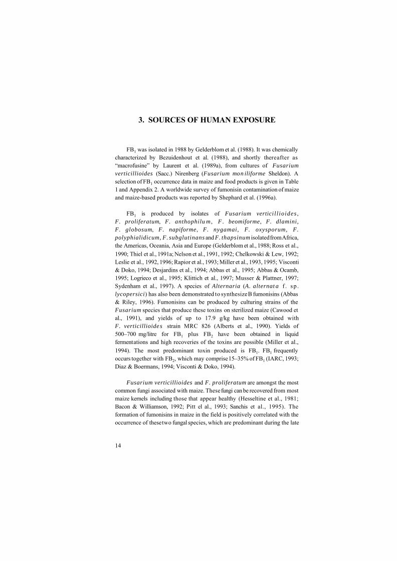
FB1 was isolated in 1988 by Gelderblom et al. (1988). It was chemicallycharacterized by Bezuidenhout et al. (1988), and shortly thereafter as“macrofusine” by Laurent et al. (1989a), from cultures of Fusariumverticillioides (Sacc.) Nirenberg (Fusarium mon iliforme Sheldon). Aselection of FB1 occurrence data in maize and food products is given in Table1 and Appendix 2. A worldwide survey of fumonisin contamination of maizeand maize-based products was reported by Shephard et al. (1996a).
FB1 is produced by isolates of Fusarium vertici l l io ides ,F. proliferatum, F. anthophilu m , F . beomiforme, F. dlamini,F. globosum, F. napiforme, F. nygamai, F. oxysporum, F.polyphialidicum, F. subglutinans and F. thapsinum isolated from Africa,the Americas, Oceania, Asia and Europe (Gelderblom et al., 1988; Ross et al.,1990; Thiel et al., 1991a; Nelson et al., 1991, 1992; Chelkowski & Lew, 1992;Leslie et al., 1992, 1996; Rapior et al., 1993; Miller et al., 1993, 1995; Visconti& Doko, 1994; Desjardins et al., 1994; Abbas et al., 1995; Abbas & Ocamb,1995; Logrieco et al., 1995; Klittich et al., 1997; Musser & Plattner, 1997;Sydenham et al., 1997). A species of Alternaria (A. alternat a f . sp .lycopersici) has also been demonstrated to synthesize B fumonisins (Abbas& Riley, 1996). Fumonisins can be produced by culturing strains of theFusarium species that produce these toxins on sterilized maize (Cawood etal., 1991), and yields of up to 17.9 g/kg have been obtained withF. verticillioide s strain MRC 826 (Alberts et al., 1990). Yields of500–700 mg/litre for FB1 plus FB2 have been obtained in liquidfermentations and high recoveries of the toxins are possible (Miller et al.,1994). The most predominant toxin produced is FB1. FB1 frequentlyoccurs together with FB2, which may comprise 15–35% of FB1 (IARC, 1993;Diaz & Boermans, 1994; Visconti & Doko, 1994).
Fusarium verticillioides and F. proliferatum are amongst the mostcommon fungi associated with maize. These fungi can be recovered from mostmaize kernels including those that appear healthy (Hesseltine et al., 1981;Bacon & Williamson, 1992; Pitt el al., 1993; Sanchis et al., 1995). Theformation of fumonisins in maize in the field is positively correlated with theoccurrence of these two fungal species, which are predominant during the late

Sources of Human Exposure____________________________________________________
15
maturity stage (Chulze et al., 1996). These species can cause Fusarium kernelrot of maize, which is one of the most important ear diseases in hot maize-growing areas (King & Scott, 1981; Ochor et al., 1987; De León & Pandey,1989) and is associated with warm, dry years and/or insect damage (Shurtleff,1980).
There is a strong relationship between insect damage and Fusarium kernelrot. A field survey demonstrated that the incidence of the European corn borerincreased F. verticillioides disease and fumonisin concentrations (Lew et al.,1991). Disease incidence was also shown to correlate to populations of thrips(Frankliniella occidentalis) (Farrar & Davis, 1991). Hybrids with a thinkernel pericarp were more susceptible to insect wounds, which allowed easieraccess to the fungus (Hoenisch & Davis, 1994). Hybrids with an increasedpropensity for kernel splitting had more disease (Odvody et al., 1990). Kernelsplitting is worse under drought conditions. Ears infected by F.graminearum may be predisposed to F. verticillioides infection andfumonisin accumulation (Schaafsma et al., 1993). In maize ears inoculated oneweek after silk emergence with F. verticillipodes fumonisins accumulated inthe visibly damaged (mouldy) kernels (Pascale et al., 1997; Desjardins et al.1998). Sydenham et al. (1995) showed that in lightly contaminated kernels FB1was concentrated in the pericarp of the maize kernel.
A study of fumonisin occurrence in hybrids grown across the USA maizebelt indicated that hybrids grown outside their range of adaptation had higherfumonisin concentrations (Shelby et al., 1994b), again suggesting the importantrole of temperature stress. Data from samples collected in Africa, Italy andCroatia also indicate fumonisin accumulation in lines grown outside their areaof adaptation (Doko et al., 1995; Visconti, 1996). The occurrence of fumonisinin Ontario, Canada (a cool maize-growing region) was limited to drought-stressed fields (Miller et al., 1995).
Significant fumonisin accumulation in maize occurs when weatherconditions favour Fusarium kernel rot, and the severity of ear infection hasbeen found to be a good indicator of fumonisin accumulation in maize earsartificially inoculated with F. verticillioides (Pascale et al., 1997). Sincemonitoring began in the USA, warm, dry years have greater concentrations

Table 1a Worldwide occurrence of fumonisin B1 (FB1) in maize-based products
Product Countries Detected / total FB1 (mg/kg)
North AmericaMaize Canada, USA 324/729 0.08–37.9Maize flour, grits Canada, USA 73/87 0.05–6.32Miscellaneous maize foodsb USA 66/162 0.004–1.21Maize feed USA 586/684 0.1–330
Latin AmericaMaize Argentina, Uruguay, Brazil 126/138 0.17–27.05Maize flour, alkali-treated kernels, polenta Peru, Venezuela, Uruguay 5/17 0.07–0.66Miscellaneous maize foodsb Uruguay, Texas-Mexico border 63/77 0.15–0.31Maize feed Brazil, Uruguay 33/34 0.2–38.5
EuropeMaize Austria, Croatia, Germany, Hungary, Italy, Poland, 248/714 0.007–250
Portugal, Romania, Spain, United KingdomMaize flour, maize grits, polenta, semolina Austria, Bulgaria, Czech Republic, France, Germany, 181/258 0.008–16
Italy, Netherlands, Spain, Switzerland, United Kingdom
Miscellaneous maize foodsb Czech Republic, France, Germany, Italy, Netherlands, 167/437 0.008–6.10Spain, Sweden, Switzerland, United Kingdom
Imported maize, grits and flour Germany, Netherlands, Switzerland 143/165 0.01–3.35Maize feed France, Italy, Spain, Switzerland, United Kingdom 271/344 0.02–70

Table 1 (contd).
AfricaMaize Benin, Kenya, Malawi, Mozambique, South Africa, 199/260 0.02–117.5
Tanzania, Uganda, Zambia, ZimbabweMaize flour, grits Botswana, Egypt, Kenya, South Africa, Zambia, 73/90 0.05–3.63
Zimbabwe
Miscellaneous maize foodsb Botswana, South Africa 8/17 0.03–0.35
Maize feed South Africa 16/16 0.47–8.85
AsiaMaize China, Indonesia, Nepal, Philippines, Thailand, 361/614 0.01–155
VietnamMaize flour, grits, gluten China, India, Japan, Thailand, Vietnam 44/53 0.06–2.60Miscellaneous maize foodsb Japan, Taiwan 52/199 0.07–2.39Maize feed Korea, Thailand 10/34 0.05–1.59
OceaniaMaize Australia 67/70 0.3–40.6Maize flour New Zealand 0/12 –
a This table is a summary of the information in Appendix 2b Includes maize snacks, canned maize, frozen maize, extruded maize, bread, maize-extruded bread, biscuits, cereals, chips, flakes,
pastes, starch, sweet maize, infant foods, gruel, purée, noodles, popcorn, porridge, tortillas, tortilla chips, masas, popped maize,soup, taco, tostada
EHC 219: Fumonisin B1____________________________________________________
18
than cooler years (Murphy et al., 1993). The direct influence of low moistureand dry weather on fumonisin accumulation could not be proven (Murphy etal., 1996; Pascale et al., 1997), although maize grown under normal conditionsin cooler maize-growing areas is not significantly contaminated by fumonisin(Doko et al., 1995; Miller et al., 1995).
Dry milling of maize results in the distribution of fumonisin into the bran,germ and flour (Bullerman & Tsai, 1994). Fumonisin may be present in beerwhere maize has been used as a wort additive (Scott et al., 1995). Littledegradation of fumonisin occurs during fermentation and the fumonisins arefound in the spent grain. No toxins can be detected in the distilled ethanol(Bothast et al., 1992; Scott et al., 1995; Bennett & Richard, 1996). Fumonisinis stable in polenta (Pascale et al., 1995), whereas it is hydrolysed, and thepericarp is removed, by nixtamalization, i.e. the treatment of maize-basedfoods with calcium hydroxide and heat (Hendrich et al., 1993). FB1 has beenshown to form N-(carboxymethyl)-FB1 when heated in the presence ofreducing sugars (Howard et al. 1998), and the latter substance has beendetected in raw corn (Howard et al., 1998).
FB1 is not significantly transferred into pork, chicken meat or eggs(Prelusky et al., 1994, 1996a; Vudathala et al., 1994), but a small amountaccumulates in the liver and kidney of pigs as a function of exposure (Preluskyet al., 1996b; see also section 6.2). Fumonisin is not significantly transferredinto milk from short-term dietary exposure (Scott et al., 1994; Prelusky et al.,1996a), and FB1 was found in only one of 165 samples of milk fromWisconsin, USA at a level close to 5 ng/ml (Maragos & Richard, 1994).

19
4. ENVIRONMENTAL TRANSPORT,DISTRIBUTION AND TRANSFORMATION
Maize is the only commodity that contains significant amounts offumonisins. It is consumed either directly or processed into products forhuman or animal consumption. Because fumonisins are known to be heatstable (Dupuy et al., 1993a; Howard et al., 1998), light stable (IARC, 1993),water soluble (US NTP, 1999), poorly absorbed, poorly metabolized andrapidly excreted by animals (see sections 6.1 to 6.5), most fumonisin willeventually end up being recycled into the environment in a manner that willconcentrate its spatial distribution. The amount that enters the environmentmay be quite large. For example, in the USA, maize production exceeds200 million tonnes per year. The concentration of FB1 and FB2 in field maizein the USA often exceeds 1 g/tonne of maize (Murphy et al., 1993 andAppendix 2). There is some evidence that fumonisins can be metabolized bysome microorganisms (Duvick et al., 1994, 1998). However, little is knownabout the environmental fate of fumonisin after it is either excreted orprocessed.

20
5. ENVIRONMENTAL LEVELS ANDHUMAN EXPOSURE
Table 1 summarizes the results of a number of surveys on the naturaloccurrence of FB1 in maize and maize-based foods and feeds (see Appendix 2for more detail). The list is not exhaustive of the surveys carried outworldwide as there is continual production of similar data from every cornerof the globe. Based on Table 1, 60% of the 5211 samples analysed have beenfound to be contaminated with FB1, the highest incidences of contaminationbeing in Oceania (82% of 82 samples) and Africa (77% of 383 samples),followed by Latin America (85% of 266 samples), North America (63% of1662 samples), Europe (53% of 1918 samples) and Asia (52% of 900samples).
The data show that levels and incidence of contamination varyconsiderably in relation to the commodities tested and the source. The highestincidence was recorded in maize feeds (82% of 1112 samples), followed byground maize products, such as flour, grits, polenta, semolina and gluten (73%of 517 samples), maize kernels (52% of 2525 samples) and miscellaneousmaize foods (40% of 892 samples).
FB1 levels in animal feedstuffs can be exceptionally high, and reachedmaximum values of 330, 70, 38, 9 and 2 mg/kg in North America (USA),Europe (Italy), Latin America (Brazil), Africa (South Africa) and Asia(Thailand), respectively. The majority of the highly contaminated feeds wereimplicated in cases of equine leukoencephalomalacia, porcine pulmonaryoedema and other mycotoxicoses.
In maize kernels available commercially or from experimental or breedingstations, FB1 has been detected in 96% (of 70 samples), 91% (of 138samples), 76% (of 260 samples), 59% (of 614 samples), 44% (of 729 samples)and 35% (of 714 samples) of samples from Oceania, Latin America, Africa,Asia, North America and Europe, respectively. Maximum FB1 levels were40.6 mg/kg (Australia), 27 mg/kg (Argentina), 117 mg/kg (South Africa),155 mg/kg (China), 38 mg/kg (USA) and 250 mg/kg (Italy).

Environmental Levels and Human Exposure____________________________________________________
21
The list of commercial retail foods subject to fumonisin contamination(Table 1) includes maize flour, grits, polenta, semolina, maize snacks,cornflakes, sweet maize, canned maize, frozen maize, extruded maize, bread,maize-extruded bread, biscuits, cereals, chips, pastes, starch, infant foods,gruel, purée, noodles, popcorn, porridge, tortillas, tortilla chips, masas,popped maize, soup, taco and tostada.
Of these samples, the global incidence of contamination in non-treated orminimally treated maize products (flour, grits, polenta, semolina) was 73% outof 517 samples analysed. The highest FB1 levels were recorded in Europe (16mg/kg), followed by North America (6.3 mg/kg), Africa (3.6 mg/kg), Asia (2.6mg/kg) and Latin America (0.7 mg/kg). In the remaining food products (892samples) the incidence of contamination was 40%, the highest level (6.1 mg/kgFB1) being found in a sample of extruded maize from Italy. Generallyprocessed maize foods have lower levels and incidence of contamination thannon-treated maize. These differences might be the results of dilution of maizein food commodities, or may depend on the differences in maize cultivar orquality requirements for various destinations.
Apart from maize and maize products, fumonisins have seldom beenfound in other food products, such as rice (Abbas et al., 1998), asparagus(Logrieco et al., 1998), beer (Torres et al., 1998) and sorghum (Shetty & Bhat,1997). Surveys on other cereals, such as wheat, rye, barley and oats, did notshow the occurrence of the toxin (Meister et al., 1996).
Human exposure estimates have been made for fumonisins in severalcountries, including Switzerland, Canada, South Africa, USA and theNetherlands (Zoller et al., 1994; Contaminants Standards Monitoring andPrograms Branch, 1996a,b; Gelderblom et al., 1996b; Kuiper-Goodman et al.,1996; Humphreys et al., 1997; Marasas, 1997; de Nijs, 1998). Humanexposure estimates of 0.017–0.089 :g/kg body weight per day have beenprepared for Canada for the period 1991 to early 1995 (Kuiper-Goodman etal., 1996). For the USA, a preliminary estimate of human exposure tofumonisins for maize eaters was 0.08 :g/kg body weight per day (Humphreyset al., 1997). The mean daily intake of fumonisins in Switzerland is estimatedto be 0.030 :g/kg body weight per day (Zoller et al., 1994).

EHC 219: Fumonisin B1____________________________________________________
22
Based on the daily average intakes of maize and maize products of 3 g(general population average), 42 g (regular maize product eaters) and 162 g(individuals with gluten intolerance) in the Netherlands, the respectivepopulation groups had an estimated daily intake of 4, 57 and 220 :g FB1 perperson, respectively, based on a mean FB1 content of 1.36 mg/kg maizeproduce. De Nijs et al. (1998a) estimated conservatively that 97% ofindividuals with gluten intolerance had a daily exposure of at least 1 :g FB1and 37% at least 100 :g, while the proportions of the general populationexposed to these levels of FB1 were 49% and 1%, respectively (de Nijs, 1998;de Nijs et al., 1998a).
Thiel et al. (1992) estimated that human exposures in the Transkei, SouthAfrica, are 14 and 440 :g FB1/kg body weight per day for healthy and mouldycorn, respectively. More recent estimates of the probable daily intake (PDI)of South Africans are summarized in Table 2. These vary from 1.2 to355 :g/kg body weight per day in rural blacks in Transkei consuming home-grown mouldy maize (Gelderblom et al., 1996b; Marasas, 1997).
These exposure estimates will vary considerably according to the sourceand extent of maize in the diet as well as the extent of Fusarium kernel rotprevalent in the harvested crop.
Occupational inhalation exposure could be a problem. In addition to thepresence of fumonisins in maize dust, FB1 is present in the spores and myceliaof F. verticillioides (Tejada-Simon et al., 1995). No data have been collectedon airborne levels of fumonisin during the harvesting, processing and handlingof fumonisin-contaminated maize.

Table 2. Probable daily intake of fumonisin in South Africaa
Product Country of No. of Mean FB1 + FB2 Probable daily intakeorigin samples concentration (:g/kg body weight per day)
(:g/kg)Rural population Urban population
Commercial maize South Africa 68 400 2.6 1.6
Commercial maize South Africa 209 300 2.0 1.2
Corn meal South Africa 52 200 1.3 0.8
Home-grown maize South Africab 18 7100 46.6 28.0Home-grown maize South Africac 18 54 000 354.9 212.9Imported maize USAd 1682 1100 7.2 4.3
Maize consumption (g/70 kg body weight per day) 460 276
a From: Marasas (1997)b Transkei, from individual farms in high oesophageal cancer area, healthy maizec Transkei, from individual farms in high oesophageal cancer area, mouldy maized Imported in 1993

24
6. KINETICS AND METABOLISMIN ANIMALS
There have been no reports on the kinetics and metabolism of fumonisinsin humans. Because fumonisins are known to be consumed by farm animalsand are the causative agent or a suspected contributing factor in farm animaldiseases, an effort has been made to understand the kinetics and metabolismin cows, pigs and poultry. Thus, this chapter will summarize results of studieson both laboratory and farm animals.
To date, published studies with radiolabelled FB1 or FB2 have beenconducted with either [21,22-14C]fumonisins, biosynthesized using L-[methyl-14C]methionine (Plattner & Shackelford, 1992; Alberts et al., 1993), or [U-14C]FB1 labelled using [1,2-14C]acetate (Blackwell et al., 1994). In thesestudies the final [14C]fumonisins had a specific activity of < 1 mCi/mmol andradiochemical purity of > 95%. Several studies have used unlabelledfumonisins with reported purities ranging from 70% (Hopmans et al., 1997)to 98% (Prelusky et al., 1996a).
Briefly, FB1 is: poorly absorbed when dosed orally; it is rapidlyeliminated from plasma or circulation and recovered in faeces; biliary excretionis important; enterohepatic cycling is clearly important in some animals; smallamounts are excreted in urine; a small but persistent (and biologically active)pool of [14C]label appears to be retained in liver and kidney; and some isdegraded to partially hydrolysed FB1 in the gut of vervet monkeys. In a studywith FB2 in rats, the results were similar to those of FB1 (Shephard et al.,1995b).
6.1 Absorption
There are no reports available of fumonisin absorption through inhalationor dermal exposure. However, because fumonisins are present in F.verticillioides cells (mycelia, spores and conidiophores) (Tejada-Simon et al.,1995), there is a potent ial for absorption through inhalation or buccalexposure. The risk from absorption due to dermal exposure would seem slight,since fumonisins are very water soluble and, typically, polar compounds donot easily penetrate the undamaged skin (Flynn, 1985).
Kinetics and Metabolism in Animals____________________________________________________
25
The quantity of FB1 detected in plasma after oral dosing in pigs, layinghens, vervet monkeys, dairy cows and rats is very low. In rats (BD IX,Sprague-Dawley or Wistar) administered [14C]FB1 orally, accumulation of 14C-labelled compounds in tissues is also very low, suggesting that absorption isvery poor (negligible to < 4% of dose) (Shephard et al., 1992a,b, 1994c;Norred et al., 1993). Similar results indicating that fumonisins are poorlyabsorbed (2 to < 6% of dose) have been reported in vervet monkeys, dairycows and pigs (Prelusky et al., 1994, 1995, 1996a,b; Shephard et al., 1994a,b).In orally dosed laying hens and dairy cows, systemic absorption based onplasma levels and accumulation of 14C-labelled compounds in tissues has beenestimated to be less than 1% of dose (Scott et al., 1994; Vudathala et al., 1994;Prelusky et al., 1996a). A study using beef cattle fed F. verticillioidesculture material (corn grits) containing FB1 plus FB2 (530 mg/kg) found thatthe majority of the fumonisin dose was recovered unmetabolized in faeces, andonly traces were det ected in blood and urine (Smith & Thakur, 1996).Following single gavage doses of 1 or 5 mg/kg body weight to cows, no FB1 orknown metabolites could be found in the plasma, indicating no or very limitedbioavailability in ruminants (Prelusky et al., 1995). Rumen metabolism mayreduce the bioavailability of FB1 as the hydrolysed form of FB1 comprised 60-90% of the total amount of FB1 found in faeces. In non-ruminants the parentcompound was the dominant species present (Rice & Ross, 1994).
6.2 Distribution
In rats and pigs orally dosed with [14C]FB1, the 14C label isdistributed to most tissues, with the liver and kidney containing the highestconcentration of radiolabel (Shephard et. al., 1992b; Norred et al., 1993;Prelusky et al., 1994, 1996a,b; Haschek et al., 1996). Typically, the livercontains more 14C label than the kidney, although in the study by Norred et al.(1993) the measured radioactivity in the kidney was greater than in the liver.In chickens and dairy cows the poor absorption of [14C]FB1 (< 1% of oraldose) was reflected in the fact that only trace amounts of radioactivity wererecovered in tissues (Prelusky et al., 1996a), no residues were recovered in eggsof laying hens (Vudathala et al., 1994) and no FB1 or aminopentol hydrolysisproducts were recovered in milk (Scott et al., 1994; Prelusky et al., 1996a). Inpregnant rats dosed intravenously with [14C]fumonisin, approximately 14.5%

EHC 219: Fumonisin B1____________________________________________________
26
and 4% of the dose were recovered in the liver and kidney, respectively, after1 h (Voss et al., 1996a). Based on the known pharmacokinetics (Norred et al.,1993) in the rat, 1-h exposure and intravenous injection were chosen so as tooptimize the presentation in blood of the [14C]FB1 to the placentae. Incontrast to liver and kidney, the uteri contained 0.24 to 0.44%, individualplacentae contained 0 to 0.04%, and total fetal recovery was # 0.015% ofdose/dam (Voss et al., 1996a). Recent studies have confirmed the lack ofplacental transfer of FB1 in rats (Collins et al., 1998a,b) and rabbits (LaBordeet al., 1997).
FB1 inhibition of the enzyme sphinganine N-acyltransferase results in alarge increase in intercellular free sphinganine (Wang et al., 1991; Yoo et al.,1992). In animal tissues the fumonisin-induced increase in free sphinganinetends to parallel the distribution of 14C label reported in the studies cited aboveusing [14C]FB1. For example, relative to other tissues examined, liver andkidney in rabbits, pigs and catfish showed the greatest increases in freesphinganine following exposure of animals to fumonisins or consumption ofdiets containing fumonisins (Goel et al., 1994; Gumprecht et al., 1995). Thefree sphinganine concentration in tissues has been shown to be an easilydetectable biomarker for exposure to fumonisins (Riley et al., 1994c), althoughit has not been validated as a biomarker in humans.
6.3 Elimination, excretion and metabolic transformation
When [14C]FB1 is dosed by intraperitoneal or intravenous injection in rats(BD IX, Sprague-Dawley or Wistar), initial elimination (subsequent to thedistribution phase) is rapid (half-life of approximately 10–20 min) with littleevidence of metabolism (Shephard et al., 1992a,b, 1994c; Norred et al., 1993).In rats the elimination kinetics based on intraperitoneal or intravenous dosingare consistent with a one- (Shephard et al., 1992b) or two-compartment model(Norred et al., 1993). Because FB1 is poorly absorbed from the ratgastrointestinal tract and extensively distributed in rat tissues (Norred et al.,1993), the tissue elimination kinetics following oral dosing is not as easilydescribed. In vervet monkeys, as in rats, the 14C label is widely distributed andrapidly eliminated (half-life of 40 min) after intravenous injection (Shephardet al., 1994a,b). The elimination kinetics following oral dosing in a non-humanprimate has not been determined. Following single intravenous injection of

Kinetics and Metabolism in Animals____________________________________________________
27
0.05 or 0.20 mg FB1/kg body weight to cows, the toxin is cleared rapidly fromthe blood. A two-compartment model (half-lives of < 2 and 15–18 min,respectively) satisfactorily described the plasma kinetics. No toxin could bedetected 120 min aft er dosing. No known metabolites were detected in theplasma (Prelusky et al., 1995).
In pigs, clearance of [14C]FB1 from blood following an intravenousinjection was best described by a 3-compartment model (half-lives of 2.5, 10.5and 183 min, respectively), and cannulation of the bile duct (bile removed)resulted in a much more rapid clearance (best described by a 2-compartmentmodel). The effect of bile removal was observed whether dosing wasintravenous or intragastric (Prelusky et al., 1994, 1996a). The half-life in pigsdosed intragastrically without bile removal was determined to be 96 min(Prelusky et al., 1996a). The studies with pigs strongly support theimportance of enterohepatic circulation of FB1 in pigs. As in the study withrats, the majority of 14C label dosed orally was recovered in faeces(approximately 90%) with less than 1% recovered in urine (Prelusky et al.,1994, 1996a). In the LLC-PK1 renal cell line, uptake of [14C]FB1 reached anequilibrium concentration with the extracellular [14C]FB1 concentration after4 to 16 h, and kinetics were indicative of a simple diffusion process (Riley &Yoo, 1995). Efflux was rapid with a half-life of less than 5 min.
Following intravenous injection into rats, FB1 is excreted unchanged inbile (Norred et al., 1993; Shephard et al., 1994c). In vervet monkeys there isevidence of metabolism to partially hydrolysed (one propane tricarboxylic acidresidue removed) FB1, and to a much lesser extent the fully hydrolysed (bothpropane tricarboxylic acid residues removed) aminopentol backbone, in faeceswhile in urine 96% of the 14C label was recovered as FB1 (Shephard et al.,1994a,b). Metabolism was most likely occurring in the gut since partiallyhydrolysed and fully hydrolysed FB1 were recovered in the faeces but not inthe bile of vervet monkeys (Shephard et al., 1995a). Because hydrolysed FB1and FB1-fructose adduct can be formed during processing, Hopmans et al.(1997) evaluated the excretion of these products and FB1 in Fischer-344 rats.Based on the amount of each FB1-related compound recovered in urine andfaeces, it was concluded that hydrolysed FB1 and the FB1-fructose adductwere better absorbed than FB1 (Hopmans et al., 1997).

EHC 219: Fumonisin B1____________________________________________________
28
Dairy cows dosed with pure FB1 either orally (1.0 and 5.0 mg FB1/kgbody weight) or by intravenous injection (0.05 and 0.20 mg FB1/kg bodyweight) showed no detectable residues of FB1, AP1 (the aminopentolhydrolysis product of FB1) or their conjugates in the milk (Scott et al. 1994).FB1 does not react with monoamine or diamine oxidase (Murphy et al., 1996).In vitro studies using rat primary hepatocytes and microsomal preparations(Cawood et al., 1994) or studies with the LLC-PK1 renal epithelial cell line(Riley & Yoo, 1995) indicated that there was no metabolism of FB1 in thesesystems.
Repeated intraperitoneal injection of FB1 resulted in induction ofcytochrome P-4501A1 and P-4504A1 activities (Martinez-Larrañaga et al.,1996). However there is no evidence that fumonisin is metabolized by P-450enzymes. Whether or not the induction was due to a direct interaction betweenfumonisins and the metabolizing systems could not be determined. However,it has been shown that some of the same sphingolipid metabolites that arealtered in fumonisin-treated animals also mediate the cytokine-inducedalterations in P-4502C11 in rat hepatocytes (Nikolova-Karakashian et al.,1997).
6.4 Retention and turnover
[14C]FB1 is widely distributed in tissues of the rat and pig. However, onlythe liver and kidney retain small but persistent amounts of 14C label based onmeasured radioactivity (Norred et al., 1993; Prelusky et al., 1994, 1996b). Inrats given three repeated oral doses, once accumulated, the measuredradioactivity in liver and kidney remained unchanged for at least 72 h after thelast intragastric dose (Norred et al., 1993). In pigs, it was estimated thatexposure to dietary FB1 at 2–3 mg/kg in feed would require a withdrawalperiod of at least 2 weeks for the 14C label to be eliminated from the liver andkidney (Prelusky et al., 1996b). The chemical nature of the 14C-labelledmaterial retained in liver and kidney was primarily FB1.
In vitro studies with rat primary hepatocytes and the cultured kidneycell line LLC-PK1 also indicate that a low but persistent pool of 14C-labelledmaterial is retained inside cells long after the rapidly diffusible pool of[14C]fumonisin has exited the cells (Cawood et al., 1994; Riley et al., 1998).This retained pool appears to be capable of maintaining the elevation of

Kinetics and Metabolism in Animals____________________________________________________
29
cellular (LLC-PK1 cells) and urinary (in rats) free sphingoid baseconcentration, a biomarker of fumonisin exposure (Solfrizzo et al. 1997b; Rileyet al., 1998; Wang et al., 1999).
6.5 Reaction with body components
Fumonisins are potent inhibitors of the enzyme sphinganine (sphingosine)N-acyltransferase in the de novo sphingolipid biosynthesis and sphingolipidturnover pathways (Wang et al., 1991). The consequences of this reaction willbe discussed in sections 7.8 and 7.9. FB1 may also interact directly withprotein kinase C (Yeung et al., 1996) and/or with mitogen-activated proteinkinases (Wattenberg et al., 1996). The only other information concerningreaction with body components is that FB1 does not bind strongly to chickenplasma proteins (Vudathala et al., 1994).
Cytotoxicity studies in primary rat hepatocytes and binding studies usingsubcellular fractions indicated that 14C-labelled FB1 binds tightly tohepatocytes and microsomal and plasma membrane fractions (Cawood et al.1994). FB1 has been shown to interact directly with liposomes (Yin et al.,1996). Since fumonisins are water soluble, are not accumulated and are rapidlyeliminated, the toxicological significance of this finding is unclear.
30
7. EFFECTS ON ANIMALS ANDIN VITRO TEST SYSTEMS
7.1 Laboratory animals and in vitro test systems
The st udies described below used either purified FB1, naturallycontaminated corn or cultures of Fusarium. It is generally accepted that thein vivo toxicity of Fusarium verticillioides MRC 826 culture material isthe result of its high FB1 content. Culture materials other than MRC 826 maycontain several other products such as other fumonisins, fusarins, moniliforminand beauvericin.
7.1.1 Single exposure
In the male Sprague-Dawley rat, intravenous injection of FB1 (95%purity) at 1.25 mg/kg body weight resulted in renal lesions localized to thetubules in the outer medulla and consisted of both proliferation and death ofcells. An increased number of mitotic figures, stained with 5-bromo-2N-deoxyuridine (not quantified), and apoptosis followed by severe nephrosiswere observed (Lim et al., 1996). Cell proliferation was also detected in theliver 24 h after dosing, but was not significantly different from control valuesat later times (day 2 to day 5). In the oesophagus, increased cell proliferationwas measured on day 3, but this returned to the control level on day 5. Whilekidney lesions were reported as severe, the increased mitotic activity in theliver and oesophagus occurred in the absence of morphological injury (Lim etal., 1996).
No information is available on the toxicological effects of single exposureto FB1 by the inhalation or dermal route.
7.1.2 Repeated exposure
7.1.2.1 Body weight loss
In male BD IX rats consuming a diet containing 1 g FB1/kg during a 4-week promotion treatment, the mean body weights were 50% lower than thoseof non-treated rats (P < 0.0001), both with and without initiation withdiethylnitrosamine (DEN) (Gelderblom et al., 1988). Similarly, the body

Effects on Animals and In Vitro Test Systems____________________________________________________
31
weight gains of male Fischer rats fed the same concentration of FB1 over a 26-day initiation period were 80% lower than those of the controls (P < 0.0025)(Gelderblom et al., 1992b).
Male Fischer rats fed diets containing 1 g FB1 (and FB2 plus FB3) per kgover a 21-day initiating period started to lose weight within the first week, andthe level of the FB compounds had to be reduced by half (Gelderblom et al.,1993). Body weight losses were first observed in rats fed FB2, where asignificant (P = 0.008) reduction compared to the controls was recorded after4–5 days. In the case of FB1 and FB3, significant (P = 0.01) reductions in bodyweight occurred after 7–8 days. Body weight loss induced by FB1 and FB2was significantly (P = 0.001) higher than that induced by FB3 (Gelderblom etal., 1993). In female Sprague-Dawley rats administered purified FB1 at gavagedoses of 0, 1, 5, 15, 35 or 75 mg FB1/kg body weight per day for 11consecutive days, significant depression of body weight and food consumptionwas observed at 35 and 75 mg FB1/kg body weight per day (Bondy et al.,1998).
The reduction in body weight gain of male Fischer rats induced by FB1 isapparently due to a feed refusal effect (Gelderblom et al., 1994). During afeeding study over 21 days, the body weight gains of rats receiving 750, 500,250 and 100 mg FB1/kg diet were significantly (0.01 < P < 0.05) lower thanthose of the controls as well as those of rats receiving 50 and 25 mg/kg. Basedon the weekly feed intake profiles, the reduction in body weight gain wasaccompanied by a concomitant reduction in feed intake. The reduction in feedintake was overcome after the second week, resulting in a feed intake similarto that of the controls at the end of the 21-day initiating treatment(Gelderblom et al., 1994).
In male Fischer-344/N Nctr BR rats, exposure to 234 and 484 mg FB1/kgdiet resulted in 10% and 17%, respectively, less gain in body weight after 28days of feeding in the range-finding study by the US National ToxicologyProgram (US NTP, 1999). Female rats had decreased body weight only at 484mg FB1/kg diet.
In the NTP 2-year carcinogenicity study (US NTP, 1999) (see section7.1.6.1), there was no difference in body weight or feed consumption in maleor female Fischer-344/N Nctr BR rats or B6C3F1/Nctr BR mice fed FB1when compared to rats or mice on control diets.

EHC 219: Fumonisin B1____________________________________________________
32
The characteristic reduction in the body weight of rats induced by FB1,was also induced by FB2, FB3 and the monomethyl esters of FB1 (MME, anartefact of the isolation procedure of FB1 and a minor contaminant of FB1
preparations), and to a much lesser extent by the N-acetylated analogue FA1,but not by the aminopolyol hydrolysis products AP 1 and AP2 or thetricarbalyllic acid moiety (TCA) (Gelderblom et al., 1993).
7.1.2.2 Hepatotoxicity and nephrotoxicity
The acute toxicity of FB1 was tested by dosing four male BD IX ratsorally with 240 mg FB1/kg body weight per day (Gelderblom et al., 1988).Three of the four rats died within 3 days and exhibited toxic hepatosischaracterized by scattered single-cell necrosis accompanied by mild fattychanges, hydropic (i.e., the abnormal accumulation of serous fluid in thecellular tissue or in a body cavity) degeneration and hyaline dropletdegeneration. Hepatocellular nuclei varied in size and some were markedlyenlarged. In addition to the hepatotoxic changes, fatty changes and scantnecrosis were present in the proximal convoluted tubules of the kidney,prominent lymphoid necrosis was observed in Peyer’s patches, and severedisseminated acute myocardial necrosis and severe pulmonary oedema wereobserved in two of the rats (Gelderblom et al., 1988).
In a separate experiment, male BD IX rats were dosed orally with 48 mgFB1/kg body weight per day for 12 days, followed by 70 mg FB1/kg bodyweight per day for the remaining 9 days of the experiment (Gelderblom et al.,1988). In the rats killed after 21 days, chronic toxic hepatosis was present andcharacterized by marked hydropic degeneration, single-cell necrosis and a fewhyaline droplets, early bile duct proliferation and fibrosis, and enlargement ofhepatocellular nuclei (Gelderblom et al., 1988).
In the livers of rats killed after 33 days on a diet containing 1 g FB1/kg, thehepatic changes were similar to those described above, but more advanced(Gelderblom et al., 1988). The proliferation of bile ducts and fibrosis causeddistortion of the lobular structure of the liver and, together with thedevelopment of hyperplastic nodules, gave the liver a distinctly nodularappearance. The authors reported that many nuclei were enlarged in hepatic

Effects on Animals and In Vitro Test Systems____________________________________________________
33
cells and numerous mitotic figures, some of which were abnormal, werepresent. The lesions in the kidneys were similar, but less severe, than thoseseen in the rats that died within 3 days (Gelderblom et al., 1988).
In male Fischer rats fed a diet containing 1 g FB1 (90-95% pure) per kgduring an initiating period of 26 days, followed by partial hepatectomy and apromoting regimen of 2-acetyl-aminofluorene (2-AAF) and carbontetrachloride, early pathological changes in the liver were very similar to thosedescribed above (Gelderblom et al., 1992b). Early hepatocyte nodules wereevident as discrete focal changes in hepatocytes characterized by somewhatbigger cells that displayed more mitotic figures than the cells in the surroundingliver and also showed vacuolization. Another prominent pathological featurewas the mild-to-moderate proliferation of bile ducts (Gelderblom et al.,1992b). Similar hepatic changes have been described in male Fischer rats feddiets containing, at a level of 0.5–1 g/kg, FB1, FB2, FB3 and MME during aninitiating period of 21 days followed by a promoting treatment of 2-AAF andpartial hepatectomy (Gelderblom et al., 1993). The short-term toxicologicaleffects in rats of FB2 and FB3 are similar to those of FB1 (Gelderblom et al.,1992a).
Changes including hydropic swelling, hyaline droplet accumulation,single-cell necrosis, increased mitotic figures, lipid accumulation, fibrosis, andbile duct proliferation were also observed in the liver of male Fischer rats thatdied after gavage treatment with 50 mg FB1/kg body weight in 6 dosages over11 days (Gelderblom et al., 1994).
A 4-week exposure of Sprague-Dawley rats to aqueous extracts ofFusarium verticillioides (MRC 826) cultures (containing fumonisins)resulted in decreased body weights, increased serum alanine and aspartateaminotransferase and alkaline phosphatase activities, decreased relative liverweights and microscopic liver lesions in rats (Voss et al., 1990).
Male and female Sprague-Dawley rats (3 of each sex per group) were feddiets containing 0, 15, 50 and 150 mg/kg of FB1 ($ 99% pure) for 4 weeks(Voss et al., 1993). No significant differences in weight gain or foodconsumption were found, but significant increases in serum triglycerides,cholesterol and alkaline phosphatase confirmed that a dietary level of 150

EHC 219: Fumonisin B1____________________________________________________
34
mg/kg was hepatotoxic to both sexes. Histopathological changes in the liver ofthese rats were characterized by scattered single-cell hepatocellular necrosis,variability in nuclear size and staining and hepatocellular cytoplasmicvacuolation. Nephrosis, consisting of focal cortical proximal tubular epithelialbasophilia, hyperplasia and single cell necrosis or pyknosis, was found inmales fed $ 15 mg/kg and in females fed $ 50 mg/kg (Voss et al., 1993). Theincidence and severity of ultrastructural alterations in kidney and liver wereclosely correlated with increased sphinganine concentration in tissues, serumand urine (Riley et al., 1994a).
The apparent no-observed-effect level (NOEL) for renal toxicity in FB1-fed rats was less than the NOEL for hepatic effects (4.1 < NOEL < 13.6 mg/kgdiet for 28 days), and renal toxicity was more severe in males (NOEL < 1.4mg/kg diet for 28 days) than females (1.4 < NOEL < 4.1 mg/kg diet for28 days). Furthermore, liver lesions found in females appeared (subjectively)more advanced than those found in males. The results of subacute toxicitystudies (7.5 and 10 mg/kg body weight per day for 4 days) (Bondy et al.,1995; Suzuki et al., 1995) and of an independent (Tolleson et al., 1996a) 4-week study in Fischer-344 rats fed 0, 99, 163, 234 or 484 mg FB1/kg dietcorroborated the findings of nephrotoxicity by Voss et al. (1993).Hepatopathy of the same type was found in males fed > 234 mg/kg diet andfemales fed $ 163 mg/kg diet. Nephropathy was found in males from all FB1-fed groups and in females fed $ 163 mg/kg diet (Tolleson et al., 1996a).Apoptotic hepatocytes and renal proximal tubule epithelial cells wereaccompanied by cell proliferation in Fischer-344 rats, suggesting thatfumonisin induces or accelerates programmed cell death in both liver andkidney (Tolleson et al. 1996a; US NTP, 1999).
In male and female B6C3F1 mice administered FB1 at gavage dosesranging from 1 to 75 mg FB1/kg body weight per day for 14 days, effects onliver, bone marrow, adrenals and kidneys were observed. In general, however,the degree of change observed indicates that mice are not as sensitive to FB1toxicity as rats (Bondy et al., 1995, 1997).
In B6C3F1 mice fed 99 to 484 mg FB1/kg diet for 4 weeks, the liver,not the kidney, was the target organ (US NTP, 1999). As for rats, the NOELwas lower in females as liver lesions were found in the females of all FB1-fed

Effects on Animals and In Vitro Test Systems____________________________________________________
35
groups, while in males hepatopathy was confined to the highest dose group.In male BALB/c mice dosed subcutaneously (0.25 to 6.25 mg FB1/kg bodyweight per day), a dose-dependent increase in apoptosis was observed in bothliver and kidney (Sharma et al., 1997).
To obtain dose–response data under longer-term exposure conditions,Fischer-344 rats and B6C3F1 mice were fed diets containing 0, 1, 3, 9, 27 or81 mg FB1/kg diet for 13 weeks (Voss et al., 1995). In rats, toxicity wasconfined to the kidneys. Lesions of the proximal tubule located in the outermedulla (sometimes referred to as the corticomedullary junction) were foundin males fed $ 9 mg/kg diet and in females fed 81 mg/kg diet. Qualitativelythese lesions were of the same type as those found in the 4-week study (Vosset al., 1993). No differences in the incidence or severity of nephropathybetween rats examined after 4 (n = 5 rats/group) or 13 (n = 10/group) weekswere found.
Renal lesions were accompanied by decreased relative kidney weight (asa percentage of body weight), which was found in males fed $ 27 mg/kg dietfor 4 weeks and in both sexes fed $ 9 mg/kg diet for 13 weeks. Serumcreatinine was increased after 13, but not 4, weeks in males fed $ 27 mg/kgdiet and in females fed 81 mg/kg diet (Voss et al. 1995).
In mice, hepatopathy and serum chemical evidence of liver dysfunctionwere found after 13 weeks in females fed 81 mg FB1/kg diet (Voss et al., 1995).Liver lesions in female mice were primarily centrilobular, although somemidzonal involvement and apparent “bridging” between adjacent central areaswere evident. Single cell hepatocyte necrosis, cytomegaly, increased numbersof mitotic figures, some mixed infiltration of neutrophils and macrophageswere present and, in more advanced lesions, the loss of hepatocytes caused anapparent collapse around the central vein. Hepatopathy was not found in malemice and FB1-related kidney lesions did not occur in either sex. A fewmacrophages containing minimal to mild amounts of cytoplasmic pigment,presumably ceroid, were also found in the adrenal cortex of high-dose (81mg/kg diet) females only.

EHC 219: Fumonisin B1____________________________________________________
36
Taken together, the findings from 4-week and 90-day toxicity studies inrats and mice (Voss et al., 1993, 1995; Tolleson et al., 1996a; US NTP, 1999)indicate that the liver is a target organ in both species, and the data seem toindicate that females exhibit hepatic effects at lower doses than males. In rats,however, the kidney is also an important target organ and, in contrast to liver,the males were affected at lower doses.
7.1.2.3 Immunotoxicity
There have been very few studies that address directly the potential forfumonisins to modify immune response in vivo. Nonetheless, there are manystudies with fumonisins or fumonisin-containing diets that show either alteredfunction of blood cells in vitro or changes in haematological parameters invivo. Fumonisins are inhibitors of ceramide synthase (see section 7.3) andceramide and glycosphingolipids are important signalling molecules andrecognition sites in the cellular immune response and attachment sites formany infectious agents and microbial toxins (Ballou et al., 1996; Merrill et al.,1997a).
In a study with pure FB1, changes in selected haematological parametersin pigs were reported at dietary levels as low as 1 mg/kg (Rotter et al., 1996).Consumption of culture-material diets (MRC 826) containing fumonisinsdecreased the ability to clear Pseudomonas aeruginosa and inhibitedpulmonary interstitial macrophage function (Smith et al., 1996c). It washypothesized that pulmonary intravascular macrophage (PIM) dysfunctioncould contribute to increase susceptibility to microbial diseases (Smith et al.,1996c).
Cytokine production has been shown to be modified by exposure tofumonisin. For example, serum tumour necrosis factor-" (TNF-")-likeactivity was increased in pigs fed culture material (M 1325 = MRC 826)containing 150 mg/kg fumonisins (Guzman et al., 1997). Fumonisin-inducedchanges in the TNF pathway have also been seen in lipopolysaccharide (LPS)-stimulated macrophages collected from BALB/c mice dosed with pure FB1(Dugyala et al., 1998).
Immunosuppression in chickens was produced in birds fed maize culturedwith F. verticillioides (MRC 826) (Marijanovic et al., 1991). Broiler chicks

Effects on Animals and In Vitro Test Systems____________________________________________________
37
fed diets containing 10 mg pure FB1/kg diet, or diets formulated fromFusarium verticillioides (MRC 826) culture material to contain 30 to 300mg FB1/kg diet, had reduced spleen and/or bursa weights and alteredhaematological parameters (Espada et al., 1994, 1997).
In male and female rats (10 rats/group) gavaged daily for 14 days withdoses of 0, 5, 15 or 25 mg FB1/kg body weight per day, a significant dose-related linear trend toward decreased plaque-forming cell number per 106
spleen mononuclear leukocytes (PFC per 106 splenocytes) (P = 0.003) andPFC per spleen cells (P = 0.001) was observed in the male rats. However, thePFC numbers in female rats were not affected significantly by treatment (P> 0.05) (Tryphonas et al., 1997).
7.1.3 Skin and eye irritation
No information is available on the effects of FB1 on skin and eye irritationand/or sensitization.
7.1.4 Reproductive toxicity, embryotoxicity and teratogenicity
Concern about the reproductive and developmental effects of fumonisinsoriginated with: (a) the observation of abortions in pregnant sows fedfumonisin-contaminated diets (Harrison et al., 1990); (b) the suggestion thata cluster of birth defects among residents in Brownsville, Texas, USA(Hendricks, 1999) might be associated with consumption of maize from the1989 maize crop; (c) the association of “mystery swine disease” withfumonisin-contaminated maize (Bane et al., 1992); and (d) the discovery thatfumonisins are inhibitors of sphingolipid biosynthesis (Wang et al., 1991).Currently there are no data to support the conclusion that consumption offumonisins is a developmental or reproductive toxicant in farm animals orhumans. There are also no data demonstrating that fumonisin consumptionresults in transfer to chicken eggs (Vudathala et al., 1994; Prelusky et al.,1996a) or that it crosses the placenta in rats (Voss et al., 1996a; Collins et al.,1998a,b), mice (Reddy et al., 1996) or rabbits (LaBorde et al., 1997).
Injection of purified FB1 into fertile chicken eggs resulted in time- anddose-dependent embryopathic and embryocidal effects (Javed et al., 1993b).

EHC 219: Fumonisin B1____________________________________________________
38
Embryonic changes included hydrocephalus, enlarged beaks and elongatednecks. Pathological changes were noted in most organ systems. At the low FB1dose (1 :M = 0.72 :g/ml), stimulation of chick embryo development wasobserved. Stimulated embryo development in vitro in pre-somite rat embryosexposed to 0.5–1 :g/ml of hydrolysed FB1 has been reported in an abstract(Flynn et al., 1994). Higher concentrations of fully hydrolysed FB1 (Flynn etal., 1997) and all concentrations of FB1 $ 0.2 :g/ml inhibited growth anddevelopment of pre-somite rat embryos in vitro (Flynn et al., 1994, 1996).Johnson et al. (1993) reported that FB1 was a weak developmental toxin toorganogenesis stage rat embryos (day 10.5; lowest-observed-effect level =0.5 mM). FB1 ($ 2.5 mM = 1.8 :g/ml) inhibited reaggregation and growth ofchicken embryo neural retina cells, a commonly used in vitro assay forscreening potential developmental toxins (Bradlaw et al., 1994). Bacon et al.(1995) found effects of FB1 in fertile chicken eggs similar to those reported byJaved et al. (1993b). In addition it was found that co-injection of fusaric acidand FB1 resulted in a synergistic toxic response (Bacon et al., 1995). Zachariaset al. (1996) found that morphological changes, due to direct administration ofFB1 to chick embryos, were correlated with inhibition of glycosphingolipidbiosynthesis.
Syrian hamsters orally gavaged with aqueous extracts of F.verticillioides (M 1325 = MRC 826) culture material containing fumonisins(0.25–18 mg FB1/kg body weight) or pure FB1 (12 mg/kg and 18 mg/kg) didnot exhibit maternal toxicity based on weight gain, serum aspartateaminotransferase activity or total bilirubin. Histological examination of liver,kidney and placenta did not reveal important changes, although mildkaryomegalic changes in liver were observed in the hamsters dosed with eitheraqueous extracts or pure FB1 at > 6 mg FB1/kg body weight (Floss et al.,1994a,b). When aqueous extracts were given by oral gavage from day 8 today 10 or 12 of gestation, there appeared to be an increase in the number offetal deaths, but statistical significance was not achieved (Floss et al., 1994a).Relative to controls, statistically significant increases in fetal deaths occurredonly in the hamsters given 18 mg FB1/kg body weight (aqueous culture extractsand pure material) (Floss et al., 1994b). Prenatal exposure to aqueous cultureextracts containing fumonisins or to pure FB1 were detrimental to fetal hamstersurvivability in the absence of maternal toxicity (Floss et al., 1994a,b; Penneret al. 1998).

Effects on Animals and In Vitro Test Systems____________________________________________________
39
In Fischer-344/N rats dosed orally from day 8 to 12 of gestation with 30or 60 mg purified FB1/kg body weight, the high dose significantly suppressedgrowth and fetal bone development while an extract of F. proliferatum (M5991) in corn culture did not (Lebepe-Mazur et al., 1995a). Voss et al. (1996a)formulated diets using F. verticillioides (MRC 826) culture material toprovide 0, 1, 10 or 55 mg FB1/kg diet. Based on consumption, the dietcontaining 55 mg/kg provided about 3 to 4 mg FB1/kg body weight per day tothe dams. The diets were fed to male and female Sprague-Dawley rats prior toand during the mating, gestational and lactational phases of the study.Nephropathy was observed in males and females fed diets containing$ 10 mg/kg and 55 mg/kg, respectively. No statistically significantreproductive effects were observed in any of the males or females, and nodevelopmental effects were found in fetuses during any phase of the study.Litter weight gains in the 10 and 55 mg/kg diet groups were slightly decreased.Increased levels of free sphinganine, a biomarker for fumonisin exposure, weredemonstrated in the livers of dams in the 55 mg/kg diet group on gestation day15. In contrast, no increase in the sphinganine/sphingosine (Sa/So) ratio wasobserved in fetuses at that time, suggesting that fetuses were not exposed inutero to FB1. This finding was supported by the study in which anintravenous injection of [14C]FB1 was given to dams on gestation day 15.Radiolabel was easily detected in tissues of pregnant females but was notdetected in their fetuses. Culture material containing fumonisins, and byinference FB1, did not have reproductive effects at doses that were minimallytoxic (Voss et al., 1996a). These findings have been recently confirmed (Sa/Soratios in fetuses were not affected and FB1 was not teratogenic at the dosestested) in a Charles River CD rats (Collins et al., 1998a,b).
Gross et al. (1994) gavaged pregnant CD1 mice daily between gestationdays 7 and 15 with a diet containing partially purified FB1 extracted from F.verticillioides (M 1325 = MRC 826) culture material. Maternal toxicity andfetal developmental abnormalities (e.g., hydrocephalus, digital and sternalossification) occurred at FB1 dosages greater than 12.5 mg/kg body weight perday. Similar results were obtained in a second study using purified FB1 (Reddyet al., 1996). As in the study by Voss et al. (1996b), the Sa/So ratio wassignificantly increased in maternal liver but not in fetal liver, suggesting thatdevelopmental effects were mediated through maternal toxicity (Reddy et al.,1996).

EHC 219: Fumonisin B1____________________________________________________
40
Unlike CD1 mice and Syrian hamsters, pregnant New Zealand whiterabbits are very sensitive to the toxic effects of FB1 (LaBorde et al., 1997).Maternal toxicity was observed at daily gavage dosages (in water) as low as0.25 mg/kg body weight from gestational day 3 to gestational day 19.Compared to controls there was no increase in fetal loss or in gross visceral orskeletal abnormalities, and no decrease in fetal weight or fetal organ weight atany dosage (0 to 1.75 mg/kg body weight) (LaBorde et al., 1997). The maternalkidney, serum and urine Sa/So ratios were increased, but there were noincreases in these ratios in fetal liver, brain or kidney (LaBorde et al., 1997).While FB1 is toxic in the pregnant dam, it is not a developmental toxin but ismaternally toxic in rabbits (LaBorde et al., 1997). However, the lowest-observed-effect level for maternal toxicity was 0.1 mg FB1/kg body weight,which is equivalent to a calculated dietary fumonisin level of 2.3 mg/kg diet(LaBorde et al., 1997). Thus, in sensitive species, maternal toxicity andconsequent fetal toxicity could occur at low dosages of FB1.
There is currently no evidence of neonatal toxicity. However, averagemean litter weights were reduced in litters from Sprague-Dawley dams fed F.verticillioides (MRC 826) culture material containing 10 or 55 mg FB1/kg(Voss et al., 1996a). The Sa/So ratio was increased in litters at lactation day 21.However, given the likelihood that offspring had consumed the contaminateddiets (Voss et al., 1996a), the authors could not ascertain the route of exposure(via milk or diet). Reduced weights and several alterations in haematologicalparameters were reported in mink kits lactationally exposed to fumonisins(Powell et al., 1996).
No FB1 was detected in the milk of lactating sows fed diets containingnon-lethal levels of FB1 and there was no evidence of toxicosis in their sucklingpigs (Becker et al., 1995). However, in a study with lactating cowsadministered FB1 intravenously, the carry-over rate of FB1 into the milkreached a maximum of 0.11% (Hammer et al., 1996), while in other studies nofumonisins were detected in cow’s milk (Scott et al., 1994; Richard et al.,1996). In a reproductive study with mink, fumonisins were detected in themilk at 0.7% of the dietary fumonisin concentrations (Powell et al., 1996).
The question of neonatal toxicity is of concern since neonates may bemore sensitive to fumonisins than adults. For example, a recent report by

Effects on Animals and In Vitro Test Systems____________________________________________________
41
Kwon et al. (1997b) indicated that subcutaneous injection of FB1 in neonatalrats caused elevation in the Sa/So ratio in brain tissue and reduced myelindeposition. The elevated sphinganine level was determined to be the result ofa direct effect on the neonate brain, indicating that FB1 can cross the blood-brain barrier (Kwon et al., 1997a). When maternal toxicity was minimal, therewas little or no evidence of neonatal toxicity in rats (Ferguson et al., 1997).
7.1.5 Mutagenicity and related end-points
The fumonisins FB1, FB2 and FB3 (98, 98, 90% pure, respectively) werenon-mutagenic in the Salmonella assay against the tester strains TA97a,TA98, TA100 and TA102, in both the presence and absence of the S-9microsomal preparation (Gelderblom & Snyman, 1991). The non-mutagenicityof FB1 (approximately 90% pure) to Salmonella tester strain TA100 atconcentrations up to 100 mg/plate was confirmed by Park et al. (1992).Similarly negative results were reported with FB1 in Salmonella TA98 andTA100, as well as in SOS chromotest in E. coli PQ37 and differential DNArepair assays with E. coli K12 strains (343/753, uvrB/recA and 343/765,uvr+rec+) (Knasmüller et al., 1997). In contrast, Sun & Stahr (1993), using acommercial bioluminescent bacterial (Vibrio fischeri) genotoxicity test,reported that FB1 showed in the concentration range 5–20 :g/ml genotoxicactivity without the metabolic activation of S-9 fraction.
FB1 (and FB2) were non-genotoxic in the in vitro rat hepatocyte DNArepair assay at concentrations ranging from 0.04 to 80 :M (and FB2 from 0.04to 40 :M) as well as in the in vivo assay at a dose of 100 mg/kg bodyweight administered by gavage (FB1 or FB2) (Gelderblom et al., 1989,1992b). The finding that FB1 does not induce unscheduled DNA synthesiswas confirmed in the in vitro assay in primary rat hepatocytes atconcentrations ranging from 0.5 to 250 :M (Norred et al., 1992a).
FB1 induced DNA strand breaks in isolated rat liver nuclei (Sahu et al.,1998).
FB1 induced a moderate increase in the micronucleus frequency in primaryrat hepatocytes at concentrations ranging from 0.01 to 1 :g/ml. Noconcentration-dependent increase of micronuclei occurred. A significant

EHC 219: Fumonisin B1____________________________________________________
42
concentration-dependent increase in chromosomal aberrations was observedin isolated hepatocytes exposed to FB1 at concentrations ranging from 1 to 100:g/ml (Knasmüller et al., 1997).
FB1 induced lipid peroxidation in isolated rat liver nuclei at concentrationsranging from 40 to 300 :M (Sahu et al., 1998). It also increased significantlythe level of thiobarbituric-acid-reactive substances in vitro in primary rathepatocytes at concentrations of 75 and 150 :M and in vivo in the liver ofrats fed a dietary FB1 level of 250 or 500 mg/kg for 21 days (Abel &Gelderblom, 1998).
Single gavage doses of 50, 100 and 200 mg FB1/kg body weightsignificantly (P = 0.05) inhibited hepatocyte proliferation as measured by theincorporation of radiolabelled thymidine into DNA in partiallyhepatectomized male Fischer rats (Gelderblom et al., 1994). Inhibition ofhepatocyte proliferation was also observed after dietary exposure to FB1($ 50 mg/kg diet) (Gelderblom et al., 1996c). FB1 also inhibited DNAsynthesis induced by epidermal growth factor in primary rat hepatocytes(Gelderblom et al., 1995).
In BALB/3T3 A31-1-1 mouse embryo cells, FB1 (90% pure) treatmentproduced transforming activity at 500 :g/ml but not at lower or higherconcentrations (Sheu et al., 1996).
7.1.6 Carcinogenicity
7.1.6.1 Carcinogenicity bioassays
When inbred BD IX rats were fed commercial diet containing freeze-driedor oven-dried culture material inoculated with F. verticillioides MRC 826for 2 years, the incidence of liver tumours (hepatocellular and cholangiocellularcarcinomas combined) was increased (control: 0/20, freeze-dried 13/20, oven-dried, 16/20) (Marasas et al, 1984b). When 30 rats of the same inbred strainwere given F. verticillioides MR 826 (containing fusarin C and later foundto produce FB1 and FB2) for 23-27 months, two hepatocellular and eightcholangiocellular cancers were observed. In addition, neoplastic hepaticnodules were observed in all surviving 21 animals, but none among thecontrols. However, in rats similarly administered F. verticillioides MRC1069 culture material containing 104 mg/kg fusarin C (but suspected of being

Effects on Animals and In Vitro Test Systems____________________________________________________
43
low in FB1), no increase in hepatic carcinomas was observed (Jaskiewicz et al.,1987b).
Maize from a field outbreak of equine leukoencephalomalacia in the USA,shown to be naturally contaminated with F. verticillioides, was fed to 12male Fischer-344 rats and commercial rodent feed to 12 controls by Wilson etal. (1985). All treated rats necropsied from 123 to 176 days had multiplehepatic neoplastic nodules, adenofibrosis and cholangiocarcinoma, whereas nosuch lesions were found in the controls. The authors considered these lesionsin the livers of male Fischer-344 rats to be similar to those described in maleBD IX rats by Marasas et al. (1984b). The fact that the lesions observed byWilson et al. (1985) developed more rapidly (as early as 123 days) than thosedescribed by Marasas et al. (1984b) (more than 450 days) was attributed byWilson et al. (1985) to the dietary deficiencies, which included choline andmethionine, in the maize-only diet used in their study.
Hendrich et al. (1993) reported that in Fischer-344/N rats fed dietscontaining Fusarium proliferatum maize culture material (with knownamounts of FB1) there was an increased incidence of hepatocellular adenomas,relative to rats fed the control diets. When rats were fed nixtamalizedFusarium proliferatum (M 5991) maize culture material diets (convertingFB1 to hydrolysed FB1) the incidence of hepatic adenomas andcholangiomas was reduced relative to rats fed the diets containing FB1. Thefrequency of hepatic/cholangiocellular adenomas in rats given the nixtamalizeddiet was higher in rats receiving nutrient-supplemented diet (equivalent toAIN-76) than in rats given a diet not supplemented with nutrients(nutritionally deficient relative to AIN-76). In a 4-week feeding study withmale Sprague-Dawley rats, Voss et al. (1996c) found that hydrolysed FB1 (58mg/kg diet) from nixtamalized F. verticillioides (MRC 826) was bothhepatotoxic and nephrotoxic. However, the extent and severity of thehepatotoxicity was significantly less than that caused by FB1 (71 mg/kg diet)from F. verticillioides (MRC 826), whereas the kidney toxicity was similar(Voss et al., 1996c).
In male BD IV rats treated with the known oesophageal carcinogen N-methylbenzylnitrosamine (NMBA) (2.5 mg/kg body weight) and FB1 (5 mg/kgbody weight), there was no synergistic interaction between NMBA and FB1

EHC 219: Fumonisin B1____________________________________________________
44
in the rat oesophagus when the two compounds were administered together(Wild et al., 1997).
A semi-purified maize-based diet containing FB1 (not less than 90% pure)at 50 mg/kg diet was fed to 25 inbred male BD IX rats over a period of 26months (Gelderblom et al., 1991). A control group received the same dietwithout FB1 (the FB1 content of the control diet was approximately 0.5 mg/kgand no aflatoxin B1 could be detected). Five rats from each group were killedat 6, 12, 20 and 26 months. All FB1-treated rats (50 mg/kg diet) that died orwere killed from 18 months onward suffered from a micro- and macronodularcirrhosis and had large expansile nodules of cholangiofibrosis at the hilus of theliver. The pathological changes terminating in cirrhosis and cholangiofibrosiswere already present in the liver of rats killed 6 months after the initiation ofthe experiment and included fibrosis, bile duct hyperplasia and lobulardistortion. The severity of the hepatic lesions increased with time and thehistological changes were consistent with those of a chronic toxic hepatosisprogressing to cirrhosis. Ten out of 15 FB1-treated (50 mg/kg diet) rats (66%)– but none in the controls – that were killed or died between 18 and 26 monthsdeveloped primary hepatocellular carcinoma. Metastases to the heart, lungs orkidneys were present in four of the rats with hepatocellular carcinoma. Apartfrom the hepatocellular carcinoma, FB1 also induced cholangiofibrosisconsistently from 6 months onward, and toward the end of the experiment,cholangiocarcinoma. However, the authors noted that the experiment wasperformed under nutritionally compromised conditions, using diets deficientin vitamins, methionine and choline, that may have had an enhancing effect onthe action of FB1 in the liver (Table 3).
The detailed results of a second long-term experiment in rats fed dietscontaining 0, 1, 10 and 25 mg FB1/kg diet over a period of 24 months have notyet been published (Gelderblom et al., 1996b). However, in preliminaryreports, Gelderblom et al. (1996b, 1997) noted that no cancers were observedin these rats, including those fed 25 mg FB1/kg diet.
Male and female Fischer-344/N Nctr BR rats and B6C3F1/Nctr BR micewere given diets containing FB1 for 2 years as part of the US NationalToxicology Program (NTP) tumorigenesis studies on FB1 (US NTP, 1999).

Table 3. Summary of the induction of neoplasia in long-term feeding studiesa
Neoplasia Species and strain Sex Fumonisin concentration (mg/kg feed) Reference
-----------------------------------------------------------------------0 5 15 50 80 100 150
Hepatocellular carcinoma BD IX rats male 0/15 10/15 GelderblomCholangiofibrosisb BD IX rats male 0/15 15/15 et al. (1991)
Renal tubule adenoma F-344/N Nctr rats male 0/48 0/40 0/48 2/48 5/48 US NTP (1999)Renal tubule carcinoma F-344/N Nctr rats male 0/48 0/40 0/48 7/48 10/48 US NTP (1999)Renal tubule adenoma F-344/N Nctr rats female 0/48 0/40 1/48 0/48 0/48 US NTP (1999)Renal tubule carcinoma F-344/N Nctr rats female 0/48 0/40 0/48 0/48 1/48 US NTP (1999)
Hepatocellular adenoma B6C3F1/Nctr mice female 5/47 3/48 1/48 16/47 31/45 US NTP (1999)
Hepatocellular carcinoma B6C3F1/Nctr mice female 0/47 0/48 0/48 10/47 9/45 US NTP (1999)
Hepatocellular adenoma B6C3F1/Nctr mice male 9/47 7/47 7/48 6/48 8/48 US NTP (1999)
Hepatocellular carcinoma B6C3F1/Nctr mice male 4/47 3/47 4/48 3/48 2/48 US NTP (1999)
a This summarizes the data for tissues where fumonisin dose-dependent induction of tumours was detected (US NTP, 1999)b In Gelderblom et al. (1991), cholangiofibrosis was considered to have progressed to cholangiocarcinoma

EHC 219: Fumonisin B1____________________________________________________
46
The FB1 that was used in these studies was > 96% pure. Dietary levels of FB1were 0, 5, 15, 50 and 150 mg/kg diet for the male rats, resulting in average dailyFB1 doses of 0, 0.25, 0.76, 2.5 and 7.5 mg/kg body weight. The dietary levels forfemale rats were 0, 5, 15, 50 and 100 mg/kg diet, resulting in average daily FB1doses of 0.31, 0.91, 3.0 and 6.1 mg/kg body weight.
There was no difference in body weight, survival or feed consumption inmale rats fed FB1 when compared to rats on control diets. The onlycompound-related change in tumour incidence was the induction of renaladenomas and carcinomas in male rats. The overall incidence of renal tubuletumours in male rats receiving 0, 5, 15, 50 and 150 mg FB1/kg diet were 0/48,0/40, 0/48, 2/48 and 5/48 for adenomas and 0/48, 0/40, 0/48, 7/48 and 10/48 forcarcinomas. Renal tubule adenomas were characterized as an expansiveproliferation of renal tubule epithelial cells that tended to be separated intolobules by a delicate fibrous stroma. The neoplastic cells had nuclei that wereslightly larger with increased cytoplasmic volume. The cytoplasmic changeswere uniform within individual lesions and varied from clear to basophilic.Renal tubule carcinomas were characterized by cellular atypia, necrosis withina lesion, invasion of the adjacent normal renal parenchyma, or metastasis todistant organs. Historically, renal tubule carcinomas have not occurred inFischer-344/N Nctr male rats on control diets. There was no apparentinvolvement of "-2 microglobulin in the tumorigenicity in male rat kidneys.The incidence in renal tumours was accompanied by an increased incidence inrenal tubule epithelial cell hyperplasia at 50 and 150 mg FB1/kg diet at 2 years(2/48, 1/40, 4/48, 14/48 and 8/48 of the male rats receiving 0, 5, 15, 50 and 150mg FB1/kg diet). Similarly, increased renal tubule epithelial cell apoptosis andproliferation were detected at 50 and 150 mg FB1/kg diet in male rats sacrificedfollowing 6, 10, 14 and 26 weeks on FB1-containing diets. As in the 28-dayrange-finding studies, the apoptosis was confined to tubules of the inner cortexand was characterized by cellular shrinkage from adjacent cells, cytoplasmiceosinophilia, and chromatin condensation and margination in the nucleus.Apoptotic cells were additionally detected using an in situ method fordetection of DNA fragmentation. In serum removed from male rats killed at 6,10, 14 or 26 weeks, no FB1-dependent changes were noted (e.g., in cholesterol,triglycerides or serum alanine aminotransferase levels). The urinary Sa/Soratio was increased in the urine of male rats fed 5, 15, 50 or 150 mg FB1/kg

Effects on Animals and In Vitro Test Systems____________________________________________________
47
diet, while kidney tissue Sa/So ratios were increased at 15, 50 or 150 mgFB1/kg diet. Increased tissue sphingoid base changes, and renal tubule tumourincidence and hyperplasia were detected at 50 and 150 mg FB1/kg diet, whileurinary sphingolipid changes were detected at 15, 50 or 150 mg FB1/kg diet.In the livers of the male rats, an increase in basophilic foci was detected at 150mg FB1/kg diet, but liver tissue Sa/So ratios were not affected.
FB1 at 5, 15, 50 and 100 mg/kg diet did not affect body weight, survival,feed consumption, serum analytes or tumour incidence in the female rats.Significant increases in urinary and kidney tissue Sa/So ratios were detected at50 and 100 mg FB1/kg diet, but the extent of induction was not as high as inthe male rats.
In the female mice, dietary levels of FB1 were 0, 5, 15, 50 and 80 mg/kgdiet, resulting in average daily FB1 consumption of 0.7, 2.1, 7.0 and 12.5 mg/kgbody weight, respectively. In the male mice, dose levels of 0, 5, 15, 80 and 150mg FB1/kg diet resulted in average daily FB1 doses of 0, 0.6, 1.7, 9.5 and 17mg/kg body weight, respectively.
In female B6C3F1/Nctr mice, there were essentially no differences in themean body weights and diet consumption. The body weights of the mice onthis study were less than those reported for the NTP and control studies at theNational Center for Toxicological Research (NCTR). This was attributed to anunintended restriction of the powdered feed in the individual feeders. Thefemale mice consuming diets containing 80 mg FB1/kg diet had a significantlyreduced survival compared to the female mice on control diets.
The only tissue that demonstrated FB1-dependent changes in tumourincidence was the liver in the female mice. Hepatocellular adenomas werepresent in 5/47, 3/48, 1/48, 16/47 and 31/45 female mice and hepatocellularcarcinomas were present in 0/47, 0/48, 0/48, 10/47 and 9/45 female miceconsuming 0, 5, 15, 50 and 80 mg FB1/kg diet, respectively. Hepatocellularadenomas were characterized as discrete lesions with compression of adjacentnormal tissues. The normal hepatic lobular structure was absent with unevengrowth patterns. The cells in the adenoma appeared to be well differentiatedand either eosinophilic, basophilic or vacuolated. Hepatocellular carcinomaswere characterized as foci of cells with distinct trabecular or adenoid structure.Histological evidence of local invasiveness or metastasis was usually evident.

EHC 219: Fumonisin B1____________________________________________________
48
The cells within the carcinoma were poorly differentiated or anaplastic. Someof the carcinomas appeared to arise within adenomas. The incidence ofhep atocellular adenomas and carcinomas in the female mice was within therange of historical occurrence in B6C3F1/Nctr mice. The increased tumourincidence was accompanied by increased hepatocellular hypertrophy (0/47,0/48, 0/48, 27/47, 31/45) and hepatocellular apoptosis (0/47, 0/48, 0/48, 7/47,14/45) in the female mice. The mean liver weights (relative to body weight) ofthe mice after 2 years were increased at 50 and 80 mg FB1/kg diet. There wereno consistent increases in serum analytes in mice killed at 3, 7, 9 and 24 weeks.Liver sphingoid bases were increased at 80 mg FB1/kg diet at weeks 3, 7 and9 but not week 24. Levels of urinary sphingoid bases were not determined.
No FB1-dependent changes in tumour incidences in the male micereceiving diets of 0, 5, 15, 80 and 150 mg FB1/kg diet were identified. Thebody weights, organ weights, survival, feed consumption and serum analyteswere not affected by FB1 dose. The lack of demonstration of statisticallysignificant sphingoid base increases in livers at intermediate sacrifices (3, 7 and9 weeks) were probably due to the low sample number (n = 4) (US NTP,1999).
7.1.6.2 Short-term assays for carcinogenicity
The short-term assays have mainly used rat liver nodules as the end-point, assessed either using traditional microscopy or histochemical analysisof different enzyme activities such as gamma-glutamyl transferase (GGT) orplacental glutathione S-transferase (PGST). In many of these assays, differentstages (initiation, promotion) of the multistage carcinogenesis model have beeninvestigated by combining the FB1 treatment with classical promotion assays.In these studies FB1 (or a Fusarium extract) was administered alone, or beforea promoting treatment, or after an initiating treatment.
a) Initiation studies
In male BD IX rats fed a diet containing 0.1% FB1 during 4 weeks, GGT-positive (GGT+) foci were induced in the liver (Gelderblom et al., 1988).

Effects on Animals and In Vitro Test Systems____________________________________________________
49
GGT+ foci were induced in the liver of male Fischer rats fed a dietcontaining 0.5–1 g/kg of FB1 (90–95% pure) for 21–26 days, followed bypartial hepatectomy and treatment with 2-AAF and carbon tetrachloride.However, foci were not induced when single or multiple doses (50–200 mg/kg)of FB1 (and FB2) were administered by gavage to hepatectomized rats(Gelderblom et al., 1992b, 1993).
In subsequent dose–response studies in male Fischer rats using the sameexperimental approach, Gelderblom et al. (1994) reported that the lowestdietary level to produce cancer initiation (GGT+-foci) over 21 days was250 mg FB1/kg diet. The lowest levels to cause cancer initiation over 14 and 7days were 500 mg and 750 mg FB1/kg diet, respectively. Based on the feedintake values, the effective dosage level (EDL) for cancer initiation over aperiod of 21 days was 142 < EDL < 308 mg FB1/kg body weight and over 14days the amount required for cancer initiation was 233 < EDL < 335 mgFB1/kg body weight. The dietary level of FB1 required for cancer initiation isdependent on the duration of exposure since a dose of 293 mg FB1/kg bodyweight over 7 days did not initiate cancer whereas a similar dose (308 mgFB1/kg body weight) over 21 days did.
Lebepe-Mazur et al. (1995b) fed female Fischer-344/N rats for one weekwith a semipurified diet with or without an aqueous extract of a Fusariumverticillioides (M 1325 = MRC 826) culture, providing 20 mg FB1/kg diet,and administered a single dose of 30 mg/kg body weight diethylnitrosamine(DEN) thereafter. Rats fed the Fusarium culture showed more PGST+
hepatocytes than those treated with DEN alone. Continued dietary treatmentwith the Fusarium moniliforme culture for 12 weeks after the DENadministration did not further increase the number of PGST foci. When theFB1 diet (one week) was followed by the DEN administration and a 7-day-non-treatment interval, no increase in PGST foci by FB1 was observed.
In another study (Lebepe-Mazur et al., 1995c), female Sprague-Dawleyrats were fed a diet supplemented with corn contaminated with Fusariumproliferatum (containing 20 or 50 mg/kg FB1) for six months; GGT-foci werenot observed but the number of PGST+ altered hepatic foci was increased intreated rats in comparison to rats fed a semipurified diet withoutsupplementation. In a similar study (Lebepe-Mazur et al., 1995b), feeding diet

EHC 219: Fumonisin B1____________________________________________________
50
containing 20 mg/kg FB1 for one or 13 weeks failed to induce a statisticallysignificant increase in PGST-altered foci.
In male Sprague-Dawley rats administered purified FB1 intraperitoneallyat 10 mg/kg body weight per day for 4 days, as well as in male and femaleSprague-Dawley rats given 35 and 75 mg/kg body weight per day orally for 11days, significant increases in PGST+ hepatocytes were observed (Mehta et al.,1998).
b) Promotion studies
In a study on the promotion activity of FB1, it was administered to maleFischer-344 rats (10, 50, 100, 250 or 500 mg/kg diet for 21 days) after a doseof DEN (200 mg/kg body weight) (Gelderblom et al., 1996c). Dietary levels of50 mg/kg or more markedly increased the number and size of the PGST+ fociin the liver. It was thus concluded (Gelderblom et al., 1996b,c) that the doseof FB1 required for cancer initiation was markedly higher than that required forcancer promotion.
Female Sprague-Dawley rats were fed a semipurified diet with or withoutan aqueous extract of a Fusarium verticillioides (M 1325 = MRC 826)culture, providing 20 or 50 mg FB1/kg for 6 months, after a single dose of 30mg DEN/kg. The number of PGST-altered foci was increased at 20 mg/kg butnot in the high-dose group, as compared to rats treated with DEN alone. GGT-altered foci were not observed (Lebepe-Mazur et al., 1995c).
Gelderblom et al. (1996b) suggested that FB1-induced hepato-carcinogenesis in male BD IX rats developed against a background of chronictoxic hepatosis culminating in cirrhosis. Chronic hepatotoxicity appears to bea prerequisite for the development of liver cancer in the BD IX rat(Gelderblom et al., 1996b).
7.2 Other mammals
7.2.1 Equine leukoencephalomalacia
Equine leukoencephalomalacia (ELEM) syndrome is characterized by thepresence of liquefactive necrotic lesions in the white matter of the cerebrum.The name is somewhat misleading since the gray matter may also be involved

Effects on Animals and In Vitro Test Systems____________________________________________________
51
(Marasas et al., 1988a). This fatal disease apparently occurs only in equids,although there has been one unconfirmed report of fumonisin-induced brainlesions and haemorrhage in rabbits gavaged with FB1 (Bucci et al., 1996) andthere is some evidence that FB1 can cross the blood-brain barrier and disruptbrain sphingolipid metabolism in neonatal rats (Kwon et al., 1997b). In equids,the ELEM syndrome has been recognized since the 19th century as asporadically occurring condition. ELEM was experimentally produced byfeeding mouldy maize obtained from a field case in Kansas by Butler (1902).The disease was known as “mouldy maize poisoning” but attempts to identifythe responsible fungus failed.
Wilson & Maronpot (1971) succeeded in establishing the causative agentwhen they isolated F. vertici l l ioides as the predominant contaminant ofmouldy maize that had caused cases of ELEM in Egypt and reproducedELEM by feeding culture material of the fungus on maize to two donkeys.Subsequently investigators in South Africa confirmed the ability of F.verticillioides (MRC 826) culture material to induce the characteristicclinical signs and pathological changes of ELEM as well as hepatosis in horsesand donkeys (Kellerman et al., 1972; Marasas et al., 1976, 1988a; Kriek et al.,1981).
The first symptoms of the syndrome are lethargy, head pressing andinappetence, followed by convulsions and death after several days. Elevatedserum enzyme levels indicative of liver damage (Wilson et al., 1992) arepreceded by elevation in the serum Sa/So ratio (Wang et al., 1992; Riley et al.,1997). Serum enzyme levels often return to near normal concentrations (Wanget al., 1992; Wilson et al., 1992; Ross et al., 1993; Riley et al., 1997) butusually increase markedly immediately prior to or at the onset of behavioralchanges (Kellerman et al., 1990; Wang et al., 1992; Ross et al., 1993; Riley etal., 1997).
In addition to the brain lesions, histopathological abnormalities in liverand kidney have been reported in horses orally dosed with pure fumonisins,maize screenings naturally contaminated with fumonisins, or culture materialcontaining known amounts of fumonisins (Kellerman et al., 1990; Wilson et al.,1992; Ross et al., 1993; Caramelli et al., 1993).

EHC 219: Fumonisin B1____________________________________________________
52
Shortly after the isolation and structure elucidation of fumonisins in 1988(Bezuidenhout et al., 1988; Gelderblom et al., 1988), Marasas et al. (1988a)successfully produced ELEM in a horse by the intravenous administration ofpure FB1. This was done by avoiding as much as possible hepatotoxicity usingserum enzymes indicative of it. ELEM has also been produced in horses givenpure FB1 by stomach tube, again monitoring for liver toxicity (Kellerman et al.,1990).
Fatal liver disease in the absence of any brain lesions has been induced byintravenous injection of FB1 (Laurent et al., 1989b). ELEM concurrent withsignificant liver disease has been observed in horses and ponies fed feedsnaturally contaminated with fumonisins at low concentrations (Wilson et al.,1992; Ross et al., 1993). The development of brain lesions in the absence ofmajor liver lesions does not preclude biochemical dysfunction in non-braintissue from contributing to the brain lesions. Ross et al. (1993) concluded thatlength of exposure, level of contamination, individual animal differences,previous exposure, or pre-existing liver impairment may all contribute to theappearance of the clinical disease.
To date, the lowest FB1 dose that has resulted in ELEM, in a controlledexperiment, is 22 mg/kg in diets formulated with naturally contaminated maizescreenings (Wilson et al., 1992). Analysis of feeds from confirmed cases ofELEM indicated that consumption of feed with a FB1 concentration greaterthan 10 mg/kg diet is associated with increased risk of development of ELEM,whereas, a concentration less than 6 mg/kg diet is not (Ross, 1994).
A study by the National Veterinary Services Laboratory of the USAnimal and Plant Health Inspection Agency (National Veterinary ServicesLaboratory, 1995) showed that horses fed 15 mg FB1/kg in diets formulatedfrom F. proliferatum (M 5991) culture material did not exhibit any clinicalsigns or altered serum biochemical parameters (including changes in the Sa/Soratio) after 150 days. A similar result was found with a pony fed a dietcontaining maize screenings naturally contaminated with 15 mg FB1/kg (Wanget al., 1992). Thus, the minimum toxic dose in equids appears to be < 22 mg/kg> 15 mg/kg based on studies with naturally contaminated maize screenings orculture material (F. proliferatum) containing fumonisins. The minimum toxicdose of pure fumonisins is unknown.

Effects on Animals and In Vitro Test Systems____________________________________________________
53
In a study using culture material containing primarily FB2 or FB3, Rosset al. (1994) found that a diet formulated from F. proliferatum (M 6290 andM 6104) culture material containing primarily FB2 at 75 mg/kg was capable ofinducing ELEM with hepatic involvement in ponies after 150 days. Incontrast, diets containing primarily FB3 (75 mg/kg) were without any effect(serum enzymes, clinical signs and histology were all normal relative to controlponies) after 57 to 65 days. It was concluded that FB3 was less toxic than FB2or FB1 (Ross et al., 1994). However, analysis of serum and tissues fromponies fed the FB3 diets revealed that the FB3 diets significantlyincreased concentrations of free sphingoid bases relative to controls and thatserum enzymes were elevated but within the normal range for ponies (Rileyet al., 1997).
7.2.2 Porcine pulmonary oedema syndrome
The first report of the disease now known as porcine pulmonary oedema(PPE) was by Kriek et al. (1981). In experimental trials, culture material of F.verticillioides (MRC 826) was fed to horses, pigs, sheep, rats and baboons(Kriek et al.,1981). Lung oedema occurred only in pigs. Clinical signs of PPEtypically occur soon (2–7 days) after pigs consume diets (culture material orcontaminated maize screenings) containing large amounts of fumonisins overa short period of time. Clinical signs usually include dyspnoea, weakness,cyanosis and death (Osweiler et al., 1992). At necropsy, the animals exhibitvarying degrees of interstitial and interlobular oedema, with pulmonary oedemaand hydrothorax (Colvin & Harrison, 1992; Colvin et al., 1993). Varyingamounts of clear yellow fluid accumulate in the pleural cavity.
Toxic hepatosis occurs concurrently with PPE (Osweiler et al., 1992;Colvin et al., 1993) and is also observed in animals that consume high levels offumonisins but do not develop PPE (Haschek et al., 1996). Typically, the livercontains multiple foci of coagulative necrosis that do not show zonaldistribution across the three zones of the liver (Osweiler et al., 1992; Colvinet al., 1993). Two studies have reported nodular hyperplasia in the pig liver(Casteel et al., 1993, 1994).

EHC 219: Fumonisin B1____________________________________________________
54
The physiological alteration that results in the inability of the lung tomaintain fluid equilibrium is unknown. However, several hypotheses havebeen proposed that are supported by experimentation. Casteel et al. (1994)found that feeding culture material diets (M 1325 = MRC 826) containing 150to 170 mg FB1/kg for 210 days resulted in right ventricular hypertrophy andmedial hypertrophy of the pulmonary arterioles. It was suggested that thiscardiotoxic effect was an indirect consequence of fumonisin-inducedhepatotoxicity. Cardiac failure is a well-known physiological mechanisminducing altered pulmonary haemodynamics which can result in pulmonaryoedema (Colvin et al., 1993). Significant changes in oxygen consumption andseveral haemodynamic parameters in pigs fed diets containing fumonisinssuggest that pulmonary hypertension caused by hypoxic vasoconstriction maycontribute to PPE (Smith et al., 1996a,b). It has been hypothesized that thecardiovascular alterations are a consequence of sphingoid-base-inducedinhibition of L-type calcium channels (Smith et al., 1996b).
Haschek et al. (1992) hypothesized that PPE might be induced bydysfunction of pulmonary interstitial macrophages (PIM) resulting in releaseof vasoactive mediators. The accumulation of membranous materials in PIM,secondary to hepatotoxicity, was postulated as the possible basis for PIMdysfunction (Haschek et al., 1992). A similar phenomena has been observedin alveolar endothelial cells (Gumprecht et al., 1998). It has been shown thatconsumption of culture material diets (MRC 826) containing fumonisins doesin fact alter PIM function (Smith et al., 1996c). How this might contribute topulmonary oedema is not clear. However, it has been hypothesized that PIMdysfunction could contribute to increased susceptibility to microbial diseases(Smith et al., 1996c). It has been shown that serum tumour necrosis factor-"(TNF-")-like activity was increased in pigs fed culture material (M 1325 =MRC 826) containing 150 mg FB1/kg (Guzman et al., 1997). Fumonisin-induced changes in the TNF pathway have also been seen inlipopolysaccharide-stimulated macrophages collected from BALB/c micedosed with pure FB1 (Dugyala et al., 1998).
In 1989-1990 outbreaks of this disease were reported in different partsof the USA (Harrison et al., 1990; Osweiler et al., 1992; Ross et al., 1992).Maize screenings obtained from farms (Harrison et al., 1990; Osweiler et al.,1992) where pigs died of PPE were predominantly contaminated with F.

Effects on Animals and In Vitro Test Systems____________________________________________________
55
verticillioides. Feeding (Kriek et al., 1981; Osweiler et al., 1992; Fazekas etal., 1998) or intubation (Colvin et al., 1993) of F. verticillioides culturematerial (MRC 826) produces PPE. Also, PPE and hepatotoxicity have beenproduced by feeding diets containing maize screenings naturally contaminatedwith fumonisins (Osweiler et al., 1992; Motelin et al., 1994). Purified FB1 hasbeen shown to produce the disease when administered intravenously (Harrisonet al., 1990; Haschek et al., 1992; Osweiler et al., 1992). However, PPE hasnot yet been produced by oral administration of pure fumonisins.
As with ELEM, there is a strong correlation between fumonisin contentof maize screenings obtained from different farms and outbreaks of PPE(Osweiler et al., 1992; Ross et al., 1992; Ross, 1994). The highestconcentration of FB1 ever reported was from maize screenings (330 mg/kg)associated with an outbreak of PPE (Ross et al., 1992). The minimum toxicdose has not been clearly established. Osweiler et al. (1992) induced PPE byfeeding F. verticillioides (MRC-3033) maize culture material reportedlycontaining 17 mg FB1/kg for 5 days. In the same study, maize screeningscontaining fumonisins at 92 mg/kg induced PPE in several pigs after 5–7 days;similar results were obtained in studies by Harrison et al. (1990), Haschek etal. (1992) and Motelin et al. (1994). Based on feeding studies with maizescreenings naturally contaminated with fumonisins, FB1 concentrations of 92to 166 mg/kg have induced PPE in 4–7 days.
Pigs fed diets containing fumonisins (formulated with culture material ornaturally contaminated maize screenings) often do not die of PPE, even whenfumonisins are reported to be present at very high concentrations in the diet.Concentrations of FB1 as low as 17 mg/kg in culture material diets (MRC-3033) induced PPE in 5 days (Osweiler et al.,1992). In contrast, culture-material-formulated (M 1325 = MRC 826) diets containing as much as 190mg/kg have been fed for 83 days with no reported evidence of respiratorydistress (Casteel et al., 1993) and a dose of 150 to 170 mg/kg diet for up to 210days caused liver effects early on but no evidence of pulmonary oedema(Casteel et al., 1994).
Colvin et al. (1993) concluded that the primary determinant of whetherpulmonary oedema or liver failure caused death was the quantity of fumonisinsfed or intubated per kg body weight per day. They proposed that > 16 mg/kg

EHC 219: Fumonisin B1____________________________________________________
56
body weight per day induced PPE and < 16 mg/kg body weight per dayinduced liver failure. However, daily oral intake levels of FB1 plus FB2(maize screenings) from 4.5 to 6.3 mg/kg body weight have induced PPE(Haschek et al., 1992; Motelin et al., 1994). The FB1 concentration in thesediets was 166 mg/kg and 129 mg/kg, respectively. Liver lesions have beeninduced with maize screenings at 1.1 mg/kg body weight per day (17 mgFB1/kg diet) (Motelin et al., 1994).
In pigs, tissues other than liver and lung have been reported to be targetsfor fumonisins, e.g., pancreas (Harrison et al., 1990), heart (Casteel et al.,1994), kidney (Colvin et al., 1993; Harvey et al., 1995, 1996), pulmonaryintravascular macrophages (Haschek et al., 1992), and oesophagus (Casteel etal., 1993). None of these studies were conducted with pure fumonisins. In arecent study with pure FB1 , altered growth and changes in selectedhaematological parameters in pigs were reported at dietary levels as low as 1mg/kg (Rotter et al., 1996).
7.2.3 Poultry toxicity
Several reports have been published implicating F. verticillioidescontamination of feed in diseases of poultry (Marasas et al., 1984a; Bryden etal., 1987; Jeschke et al., 1987; Prathapkumar et al., 1997). The clinical featuresof the disease often include diarrhoea, weight loss, increased liver weight andpoor performance. Immunosuppression in chickens was also produced in birdsfed maize cultured with several different isolates of the fungus (Marijanovicet al., 1991). Functional and morphological changes were observed in chickenexposed to FB1 (Qureshi & Hagler, 1992). Several studies have confirmed thatF. verticillioides, F. proliferatum, FB1 and moniliformin are toxic t opoultry (broiler chicks, turkeys, ducklings) (Ledoux et al., 1992, 1996; Brownet al., 1992; Dombrink-Kurtzman et al., 1993; Javed et al., 1993a, 1995;Weibking et al., 1993a,b, 1995; Kubena et al., 1995a,b; Hall et al., 1995;Bermudez et al., 1996; Vesonder & Wu, 1998) and chicken embryos (Javed etal., 1993b; Bacon et al., 1995). The levels of fumonisins used in these studieswere 75–644 mg/kg diet. Culture materials and naturally contaminated maizecontaining F. proliferatum may contain, in addition to fumonisins,moniliformin and beauvericin (Kriek et al., 1977; Logrieco et al., 1993; Plattner

Effects on Animals and In Vitro Test Systems____________________________________________________
57
& Nelson, 1994). Espada et al. (1994) reported toxicity and alteredhaematological parameters (Espada et al., 1997) in broiler chicks fed dietscontaining pure FB1 (10 mg/kg) and FB1 (30 mg/kg) from Fusariumverticillioides (MRC 826) culture material.
7.2.4 Non-human primate toxicity
Kriek et al. (1981) fed three baboons F. verticillioides culture material(MRC 826). Baboon 1 and baboon 2 died of acute congestive heart failure after248 and 143 days, respectively. The remaining baboon continued on feed for720 days, at which time it was killed. Autopsy of baboon 3 revealed that theprinciple lesion was cirrhosis of the liver. Vervet monkeys fed F.verticillioides culture material (MRC 826) for 180 days exhibited variousdegrees of toxic hepatosis (Jaskiewicz et al., 1987a). Subsequent long-termstudies (Fincham et al., 1992) with vervet monkeys fed MRC 826 culturematerial shown to contain fumonisins revealed an increase in serum cholesterol,plasma fibrinogen and blood coagulation factor VII (factors known to promoteatherosclerosis). These changes occurred secondary to chronic hepatotoxicityat a dose calculated to average 0.3 mg total fumonisins/kg body weight per day(low-dose diet) based on a retrospective analysis of the diets (Fincham et al.,1992) and a high-dose diet averaging approximately 0.8 mg total fumonisins/kgbody weight per day (Shephard et al., 1996b). Analysis of the free sphingoidbases in serum from some of the animals used in the study by Fincham et al.(1992) showed that in serum the free sphinganine concentration and Sa/So ratiowere significantly elevated in both the low-dose and high-dose animals(Shephard et al., 1996b). Free sphinganine and the Sa/So ratio were alsoelevated in urine at both dose levels, but not significantly (Shephard et al.,1996b).
7.2.5 Other species
Other species that have been studied using pure fumonisins, contaminatedmaize screenings or maize culture material of F. verticillioides include thefollowing: catfish (Brown et al., 1994; Goel et al., 1994); cattle (Osweiler etal., 1993); hamsters (Floss et al., 1994a,b); lambs (Edrington et al., 1995); mink(Restum et al., 1995); and rabbits (Gumprecht et al., 1995; Bucci et al., 1996;LaBorde et al., 1997). In all cases where toxicity was evident it involved liverand/or kidney or homologous organs.

EHC 219: Fumonisin B1____________________________________________________
58
7.3 Mechanisms of toxicity — mode of action
Several biochemical modes of action have been proposed to explain all orsome of the fumonisin-induced animal diseases. Two of these invokedisruption of lipid metabolism as initial site of action. There are also severalstudies that hypothesize fumonisin-induced changes in key enzymes involvedin cell cycle regulation, differentiation and/or apoptosis as initial or secondarysites of action.
7.3.1 Disruption of sphingolipid metabolism
The structural similarity between sphinganine and FB1 led Wang et al.(1991) to hypothesize that the mechanism of action of this mycotoxin mightbe via disruption of sphingolipid metabolism or a function of sphingolipids.At the moment, there are considerable data supporting the hypothesis thatfumonisin-induced disruption of sphingolipid metabolism is an importantevent in the cascade of events leading to altered cell growth, differentiation andcell injury observed both in vitro and in vivo.
7.3.1.1 Sphingolipids and their metabolism
The pathways of biosynthesis and turnover (Fig. 1) have not been as wellstudied in sphingolipids as in other lipid classes. In order to understand howdisruption of sphingolipid metabolism might contribute to the farm animal andlaboratory animal diseases associated with consumption of fumonisins, it isnecessary to understand how sphingolipids are biosynthesized. Eukaryoticcells synthesize a diverse array (over 400 distinct molecules) of sphingolipidswhich serve as important structural molecules in membranes and as regulatorsof many cell functions (Bell et al., 1993). While sphingolipids have also beenfound in procaryotes (Karlsson, 1970), their biosynthesis and role in cellularregulation is poorly understood.
Typically, de novo sphingolipid biosynthesis proceeds via the reactionsdescribed below (Merrill & Jones, 1990; Sweeley, 1991; Bell et al., 1993). Thefirst is the condensation of serine with palmitoyl-CoA by serinepalmitoyltransferase, a pyridoxal 5N-phosphate-dependent enzyme, and theresulting 3-ketosphinganine is reduced to sphinganine using NADPH.Sphinganine is acylated to dihydroceramides (also called N-acylsphinganines)

Effects on Animals and In Vitro Test Systems____________________________________________________
59
by ceramide synthase using various fatty acyl-CoAs. Headgroup s (e.g.,phosphorylcholine, glucose, etc.) are subsequently added to the 1-hydroxylgroup. The 4,5-trans-double bond of the sphingosine backbone is added afteracylation of the amino group of sphinganine by the enzyme dihydroceramidedesaturase (Michel et al., 1997). Both dihydroceramide anddihydrosphingomyelin are substrates for the enzyme. Thus, free sphingosineis not an intermediate of de novo sphingolipid biosynthesis (Merrill, 1991;Rother et al., 1992). Sphingolipid turnover is thought to involve the hydrolysisof complex sphingolipids to ceramides, then to sphingosine. Sphingosine iseither reacylated or phosphorylated and cleaved to a fatty aldehyde andethanolamine phosphate. The fatty aldehyde and ethanolamine phosphate canbe redirected into the biosynthesis of glycerophospholipids and other fats(Van Veldhoven & Mannaerts, 1993).
In animal cells the initial steps from the condensation of serine andpalmitoyl-CoA to the formation of ceramide take place in the endoplasmicreticulum. Subsequent processing of ceramide into glycosphingolipids andsphingomyelin takes place in the endoplasmic reticulum and Golgi apparatus.Degradation of complex sphingolipids occurs in the lysosomes, endosomes andthe plasma membrane with degradation of free sphingoid bases occurring in thecytosol. For reviews of sphingolipid metabolism, see Merrill & Jones (1990),Merrill (1991), Sweeley (1991) and the volumes edited by Bell et al. (1993).
7.3.1.2 Fumonisin-induced disruption of sphingolipid metabolism in vitro
The term “fumonisin disruption of sphingolipid metabolism” includesinhibition of sphingosine and ceramide biosynthesis, depletion of morecomplex sphingolipids, increase in free sphinganine, decrease in reacylation ofsphingosine derived from complex sphingolipid turnover and degradation ofdietary sphingolipids, increase in sphingoid base degradation products (i.e.sphingosine (sphinganine) 1-phosphate, ethanolamine phosphate and fattyaldehydes), and increase in lipid products derived from the increase in thesphingoid base degradation products. FB1 is now widely used to reveal thefunction of sphingolipids and sphingolipid metabolism in cells (Merrill et al.,1996a).

Fig 1A
Fig 1B
60

Effects on Animals and In Vitro Test Systems____________________________________________________
61
Fig. 1a. The pathway of de novo sphingolipid biosynthesis and turnover in amammalian cell. Large solid arrows indicate the enzymatic steps leading tobiosynthesis of ceramide, a known effector of cell death, and large brokenarrows show the enzymatic steps leading to the production of sphingosine 1-phosphate, an effector of cell survival. Also shown is the proposed role ofmitochondrial perturbations triggering a redirection of palmitate from beta-oxidation into the de novo pathway, resulting in increased biosynthesis ofceramide under conditions of oxidative stress.
Fig. 1b. The sites of action of serine palmitoyltransferase (SPTase) inhibitors(SPTI) such as ISP-I = myriocin = thermozymozydin, and ceramide synthase(CER synthase) inhibitors (FB) such as fumonisins. The block on the ceramidesynthase responsible for reacylation of sphingosine results in an increase infree sphingosine and possibly sphingosine 1-phosphate. However, it has beenreported that unlike sphingosine 1-phosphate, sphinganine 1-phosphate doesnot exert a marked cytoprotective effect, but does bind to and signal via theG protein-coupled receptor encoded by endothelial differentiation gene 1(EDG1) (Spiegel, 1999).
In both Fig 1a and Fig 1b, the biochemicals shown in all capital letters are thoseknown or suspected to be lipid secondary messengers. Also shown in Fi g 1 ais the generation of ceramide by ligand-induced sphingomyelin hydrolysis.
Abbreviations: DHC-desaturase (dihydroceramide desaturase), A-SMase(acidic sphingomyelinase), So-kinase and -lyase (sphingosine kinase andlyase).
Fumonisins potently inhibit the acylation of sphinganine and sphingosine(Wang et al., 1991; Yoo et al., 1992; Merrill et al., 1993c). In primary rathepatocytes, the IC50 for inhibition of serine incorporation into sphingosineis approximately 0.1 :M for FB1 and FB2 (Wang et al., 1991; Merrill et al.,1993c). In P388 murine macrophages, the IC50 is less than 0.5 :M (Balsindeet al., 1997). In cultured pig renal cells (LLC-PK1) the IC50 for inhibition of denovo sphingosine biosynthesis is approximately 20 :M FB1 (Yoo et al.,1992). The basis for this difference in sensitivity is unknown.
Inhibition of sphinganine (sphingosine) N-acyltransferase (ceramidesynthase) in cells leads to a concentration-dependent reduction in totalcomplex sphingolipids, including sphingomyelin and glycosphingolipids (Wanget al., 1991; Merrill et al., 1993b; Yoo et al., 1996). In LLC-PK1 cells, thedecrease in complex sphingolipids does not become apparent until 24 to 48 hafter cells have been exposed to FB1 but before inhibition of cell growth andincreased cell death (Yoo et al., 1996). In microsomal preparations fromcultured mouse cerebellar neurons, inhibition of ceramide synthase was

EHC 219: Fumonisin B1____________________________________________________
62
competitive with respect to both the long-chain (sphingoid) base and fattyacyl-CoA (Merrill et al., 1993b). This observation suggests that ceramidesynthase recognizes both the amino group (sphingoid binding domain) and thetricarballylic acid side-chains (fatty acyl-CoA domain) of FB1 (Merrill et al.,1996b). The reduced inhibition by the hydrolysed derivatives of the fumonisinB series (Norred et al., 1997; van der Westhuizen et al., 1998) supports thishypothesis. Ceramide synthase has recently been shown to acy la tehydrolysed FB1, AP1, to form N-palmitoyl-AP1. The product was also foundto be an inhibitor of ceramide synthase and to be 10 times more toxic than FB1in a human colonic cell line, HT29 (Humpf et al., 1998). Sphingomyelinbiosynthesis is approximately 10-fold more sensitive to inhibition by FB1 thanglycosphingolipids. This is true for other inhibitors of ceramide synthesis,such as $-fluoroalanine (Medlock & Merrill, 1988; Merrill et al.,1993b).
The complete inhibition of ceramide synthase by fumonisins causes theintracellular sphinganine concentration to increase rapidly (Wang et al., 1991;Yoo et al., 1992). The amount of free sphinganine that accumulates in cells isa function of several factors. These include the extent of inhibition of ceramidesynthase, the concentration of essential precursors (serine, palmitoyl-CoA),the growth rate of the cells, the rate of sphinganine degradation, and the rateof elimination from the cells. For example, in rat primary hepatocytes treatedwith a concentration of 1 :M FB1, which inhibits serine incorporation intosphingosine by > 90%, there was a significant increase in free sphinganine after1 h, which increased to 110-fold over controls after 4 days (Wang et al., 1991).In LLC-PK1 cells approximately 50-fold and 128-fold increases were measuredafter exposure to FB1 (35 :M, 50 to 60% inhibition) for 6 h and 24 h,respectively (Yoo et al., 1992). Proliferating LLC-PK1 cells accumulate muchhigher levels of free sphingoid bases than confluent monolayers andcytotoxicity is only observed in proliferating cells (Yoo et al., 1996).
While the level of free sphingosine does not increase in primary rathepatocytes (Wang et al., 1991; Gelderblom et al., 1995), it does in LLC-PK1cells, presumably due to inhibition of reacylation of sphingosine derived fromsphingolipid turnover or from the growth medium. When cells begin to die,sphingosine levels will increase due to the breakdown of membrane lipids.However, in LLC-PK1 cells the increase in free sphingosine occurs before anyevidence of increased cell death or inhibition of cell proliferation (Yoo et al.,

Effects on Animals and In Vitro Test Systems____________________________________________________
63
1992). Nonetheless, approximately 95% of the increase in the levels of freesphingoid bases in LLC-PK1 cells was found to be due to the increase in freesphinganine level (Yoo et al., 1992). The fate of the accumulated sphinganineis unclear. While sphingosine has little difficulty in crossing cell membranes(Hannun et al., 1991), the half-life of sphinganine inside LLC-PK1 cells ismuch longer than the half-life of FB1 in LLC-PK1 cells (Riley et al., 1998),which suggests either that the inhibition of sphinganine N-acyltransferase ispersistent, that sphinganine does not easily diffuse out of cells, or thatsphinganine degradation is slow relative to its biosynthesis. In urine from ratsfed FB1, > 95% of the free sphinganine is recovered in dead cells which haveapparently sloughed into the urine (Riley et al., 1994a). Thus, in urinesphinganine is tightly associated with the cells.
In rat hepatocytes, a portion of the accumulated sphinganine ismetabolized to sphinganine 1-phosphate and then cleaved into a fattyaldehyde and ethanolamine phosphate (Merrill et al., 1993c), both of whichcan be redirected into other biosynthetic pathways. The enzyme responsiblefor hydrolysis of sphingosine (sphinganine) 1-phosphate is sphingosine-phosphate lyase (Van Veldhoven & Mannaerts, 1993). In J774 cells exogenoussphinganine has been shown to be initially accumulated and then rapidlymetabolized (Smith & Merrill, 1995). About one-third of the ethanolamine inphosphatidylethanolamine is derived from long-chain base catabolism whenfumonisin is added (Smith & Merrill, 1995). Similar findings have beenreported using fumonisin-treated Chinese hamster ovary cells (Badiani et al.,1996), confirming that, in some cell types, accumulated free sphingoid basesare rapidly metabolized into ethanolamine and fatty aldehydes. In vitro andin vivo, rat liver lipid composition is markedly altered by FB1 (Wang et al.,1991; Gelderblom et al., 1997). In addition to changes in free sphingoid bases,more complex sphingolipids (ceramides, etc.) and phosphatidylethanolamine(Wang et al., 1991; Merrill et al., 1993c), there are many changes in the fattyacid composition of liver phospholipids (Gelderblom et al., 1997). The abilityof cells to rapidly metabolize bioactive sphingoid bases into other productsmay protect cells from the toxicity associated with accumulation of freesphingoid bases or ceramide (Spiegel, 1999). Chronically disruptedsphingolipid metabolism leads to imbalances in phosphoglycerolipid and fatty

EHC 219: Fumonisin B1____________________________________________________
64
acid metabolism. Because the accumulation of specific end-products andintermediates is dependent upon the balance between anabolic and catabolicprocesses, it is conceivable that changes in concentration of specific end-products could occur with no change in the steady-state concentration of freesphingoid bases.
7.3.1.3 Fumonisin disruption of sphingolipid metabolism in vivo
a) Equids
Typically free sphingosine and sphinganine are present in normal tissuesand cells in trace amounts (Merrill et al., 1988; Riley et al., 1994c). This is tobe expected since free sphinganine is a metabolic intermediate in thesphingolipid biosynthetic pathway, and free sphingosine is generatedprimarily as a consequence of sphingolipid turnover or degradation (Merrill,1991; Rother et al., 1992; Michel et al., 1997). The first in vivo study to testif dietary fumonisins could change free sphingoid base concentration usedserum obtained from ponies fed diets containing maize screenings naturallycontaminated with fumonisins (primarily FB1) (Wang et al., 1992) . Uponconsumption of diets containing fumonisin, all of the ponies exhibited largeincreases in sphinganine, although the magnitude of the changes varied amongthe animals. The elevation in serum sphinganine is reversible and the increasein free sphinganine and the Sa/So ratio occurred before increases in serumtransaminase activity and clinical signs of ELEM (Wang et al., 1992). Forexample, a pony was given feed (corn screenings) containing 15 to 22 mgFB1/kg. The Sa/So ratio increase by day 182 was followed by an increase inserum biochemical indices of cellular injury by day 223 (Wang et al., 1992).The pony died of ELEM on day 241.
In another study, horses were fed diets containing F. proliferatum (M6290 and M 6104) culture material, which contained primarily either FB2 (75mg/kg) or FB3 (75 mg/kg) (Ross et al., 1994). Analysis of serum and tissuesfrom these horses showed a qualitatively similar response to horses fed FB1(Wang et al. 1992), but the magnitude of the increase in free sphinganine inserum, liver and kidney was much less in the horses fed FB3 diets (Riley et al.,1997). While two of the three ponies fed FB2 diets developed ELEM, the threeponies fed FB3 diets showed no clinical signs of ELEM or evidence of liverdamage after 60 days (Ross et al., 1994).

Effects on Animals and In Vitro Test Systems____________________________________________________
65
There was also a reduction in the amount of complex sphingolipids in theserum, liver and kidney of ponies fed diets containing fumonisins (Wang et al.,1992; Riley et al., 1997), as would be expected if the elevation in sphinganinewas due to inhibition of de novo sphingolipid biosynthesis. In ponies feddiets containing FB2 or FB3 (75 mg/kg), the levels of complex sphingolipids inthe liver were reduced by 88% and 72%, respectively, and the kidney wassimilarly affected (Riley et al., 1997). In contrast to ponies fed FB2 diets,ponies fed FB3 diets exhibited no liver or kidney pathology (Ross et al.,1994). Thus, prolonged exposure to fumonisins can result in a markeddepletion of the complex sphingolipids in liver with no evidence of liverpathology.
b) Pigs
Similar results were obtained for pigs, confirming that there was adose–response relationship between the ratio of free sphinganine to freesphingosine in serum and tissues and the amount of fumonisin-contaminatedfeed consumed (Riley et al., 1993). Pigs were fed diets formulated fromnaturally contaminated maize screenings at 0 (< 1), 5, 23, 39, 101 and 175mg/kg total fumonisins (FB1 plus FB2). The results showed that the Sa/So ratiowas significantly elevated in the liver, lung and kidney from pigs consumingfeeds containing $ 23 mg/kg fumonisins. Liver injury was observed atfumonisin levels $ 23 mg/kg. However, injury to the kidney was not observedat any dose even though it contained equal or greater amounts of free sphingoidbases. In lung tissue, free sphingoid base content was elevated at doses$ 23 mg/kg, but lung lesions were only observed in pigs fed the diet containing175 mg/kg. Smith et al. (1996a) showed that in pigs fed fumonisins significanteffects on cardiovascular function were associated with significant increases infree sphingoid bases in heart tissue. Subsequent studies found that damage topig alveolar endothelial cells, in vivo, was preceded by accumulation of freesphingoid bases in lung tissue (Gumprecht et al., 1998).
Elevation of the Sa/So ratio in pig serum paralleled the increase in tissues(Riley et al., 1993). This finding supported the earlier hypothesis (Wang et al.,1992) that the elevated ratio in serum was due to the movement of freesphinganine (accumulating as a result of inhibition of sphinganine N-acyltransferase) from tissues into the blood. Statistically significant increasesin the serum ratio were observed at feed concentrations as low as 5 mg/kg total

EHC 219: Fumonisin B1____________________________________________________
66
fumonisins (after 14 days) and in pigs (at higher concentrations) in which otherserum biochemistry parameters were not changed and in which there were noobservable gross or microscopic lesions in liver, lung or kidney. Thus, theincrease in the Sa/So ratio was an earlier and more sensitive indicator offumonisin exposure than the development of lesions in liver or lung in pigsdetectable by light microscopy. Nonetheless, the increases in free sphinganinein tissues and serum closely paralleled the dose-dependent increases in otherbiochemical parameters measured at 14 days (Motelin et al., 1994).
It has been proposed that the ratio of free sphinganine to free sphingosineand the presence of elevated levels of free sphinganine in serum, urine andtissue be used as indicators for consumption of fumonisins by farm animals(Riley et al., 1994c). However, a subsequent study with pigs found alteredgrowth at doses of FB1 that did not cause an increase in free sphinganine(Rotter et al., 1996). Thus, in pigs, elevation of free sphinganine appears tooccur at dosages that are greater than those that cause subtle changes inperformance but lower than those that are toxic.
c) Poultry and other commercially important animals
Chickens fed diets supplemented with F. verticillioides culturematerials (Weibking et al., 1993a, 1995) or pure FB1 (Henry, 1993) exhibitedelevated sphinganine levels and elevated ratios in tissues and serum. Similarfindings have been made in the rabbit (Gumprecht, et al., 1995; LaBorde et al.,1997), catfish (Goel et al., 1994), mink (Restum et al., 1995; Morgan et al.,1997) and trout (Meredith et al., 1998).
d) Laboratory animals
In short-term studies with rats, rabbits and mice, disruption ofsphingolipid metabolism, as shown by statistically significant increases in freesphinganine concentration, occurs at or below the fumonisin dosages thancause liver or kidney lesions (Riley et al., 1994a; Martinova & Merrill, 1995;LaBorde et al., 1997; de Nijs, 1998; Tsunoda et al., 1998; Voss et al., 1998).In rats (Sprague-Dawley, RIVM:WU) and mice (BALB/c) dosed withfumonisins, the increase in free sphinganine concentration in the kidney and/or

Effects on Animals and In Vitro Test Systems____________________________________________________
67
liver is closely correlated with the extent and severity of lesions (Riley et al.,1994a; de Nijs, 1998; Tsunoda et al., 1998; Voss et al., 1998). In two separate21-day feeding studies (Fischer-344 rats), liver free sphinganine level wasincreased, although not significantly, at the lowest FB1 dose (50 mg/kg diet)that had liver cancer-promoting potential (Gelderblom et al., 1996c, 1997). Inrats and rabbits, the concentration of fumonisin that causes nephrotoxicity andan increase in kidney free sphinganine concentration is lower than thefumonisin dose that causes hepatotoxicity (Voss et al., 1993, 1996b, 1998;Gumprecht et al., 1995; LaBorde et al., 1997; de Nijs, 1998). For example, inSprague-Dawley rats significant elevation of free sphinganine levels andhepatosis were observed at $ 15 mg/kg # 50 mg/kg dietary FB1 (Riley et al.,1994a), whereas the NOEL for nephrosis in male Sprague-Dawley rats is9 mg/kg (Voss et al., 1995) and significant increases in kidney free sphinganinehave been detected in rats fed AIN-76 diets containing 1 mg FB1/kg (Wang etal., 1999). In male RIVM:WU rats, the liver free sphinganine level wassignificantly elevated at > 0.19 # 0.75 mg FB1/kg body weight (equivalent to1.9 and 7.5 mg/kg dietary FB1) in the absence of any evidence of hepatosis (deNijs, 1998) and the NOEL for tubular cell death and significant increases inkidney free sphinganine was # 0.19 mg FB1/kg body weight, which wasequivalent to 1.9 mg/kg in feed (de Nijs, 1998).
In the US National Toxicology Program, long-term feeding study withFischer-344/N Nctr BR rats, pure FB1 induced an increase in the Sa/So ratioin kidney tissue, which correlated with increased non-neoplastic and neoplasticlesions (US NTP, 1999). In B6C3F1/Nctr BR female mouse liver, freesphinganine and the Sa/So ratio were increased after 3 and 9 weeks at 50 and80 mg FB1/kg diet, which were the same doses that induced liver adenoma andcarcinoma (US NTP, 1999). Livers taken from rats (BD IX) in a long-termstudy (2 years) that were fed diets containing 10 and 25 mg/kg FB1 did notshow significant changes in liver free sphingoid bases, although the meanconcentration of free sphinganine and free sphingosine in the liver of rats fed25 mg FB1/kg diet was 8- and 3-fold, respectively, higher than control values(Gelderblom et al., 1997).
In rats, rabbits and vervet monkeys, increases in free sphinganineconcentration have been detected in the urine of animals fed fumonisin-

EHC 219: Fumonisin B1____________________________________________________
68
containing diets (Riley et al., 1994a; Castegnaro et al., 1996; Shephard et al.,1996b; LaBorde et al., 1997; Merrill et al., 1997b; Solfrizzo et al., 1997a,b;Wang et al., 1999). Accumulation of free sphinganine in rat urine (associatedwith accumulation of dead cells) closely reflected the changes which occurredin the kidney (Riley et al., 1994a). Analysis of urine from rats fedcommercially available chows showed a statistically significant correlationbetween the free sphinganine to free sphingosine ratio in urine and thefumonisin concentration in the chows. The FB1 concentration in the chowsranged from undetected to 3.3 mg/kg (Merrill et al., 1997b). Feeding studieswith pure FB1 in AIN-76 diets indicate that the no-observed-effect level(NOEL) for elevation of urinary free sphinganine level in Sprague-Dawley ratsis > 1 mg/kg diet # 5 mg/kg diet (Wang et al., 1999). In other studies, rats feda diet containing mixture of fumonisins (from culture material) for 13 daysshowed a NOEL of between 1 and 2 mg/kg diet (Solfrizzo et al., 1997b).
In rats fed an AIN-76 diet containing 10 mg FB1/kg for 10 days and thenput on a control diet, the urinary sphinganine concentration returned to controllevels in 10 days. However, if the diet contained 1 mg FB1/kg, the urinarysphinganine concentration remained markedly elevated for at least 10 daysafter changing the feed (Wang et al., 1999). Thus, in this study, once elevatedby feeding toxic levels of FB1, apparently non-toxic concentrations kept thefree sphinganine concentration significantly elevated to concentrations thatwere equivalent to those of the nephrotoxic fumonisin dosage. This result is,however, in contrast with the findings of Solfrizzo et al. (1997b), showing thatthe elevated levels of sphingoid bases after exposure to relatively high levelsof fumonisins (7–15 mg/kg for 13 days) return to their original values whenrats are exposed to a low-fumonisin diet (1 mg/kg or less) for a period of timethat is directly dependent on the previous level of exposure, in terms of doseand time (Solfrizzo et al., 1997b).
7.3.1.4 Tissue and species specificity
The tissue specificity and the severity of the pathology in rats (Sprague-Dawley, Fischer-344, Wistar, RIVM:WV) and mice (BALB/c) seem tocorrelate well with the disruption of sphingolipid biosynthesis (Riley et al.,1994a; Tsunoda et al., 1998; Voss et al., 1998). This is not the case in pigs and

Effects on Animals and In Vitro Test Systems____________________________________________________
69
horses, where the kidney appears to be equally or more sensitive than the liverwith regards to the fumonisin-induced increase in free sphinganine (Riley et al.,1993, 1997). There is significant liver pathology in horses and pigs (Ross etal., 1994; Haschek et al., 1996), with little evidence of kidney damage (Harveyet al., 1996).
It has been suggested that these differences in tissue and speciesspecificity may be due to differing susceptibility to the adverse cellular effectsof disrupted sphingolipid metabolism (Voss et al., 1996b). For example, liverand kidney may have different abilities to metabolize or eliminate freesphinganine or to compensate for depletion of complex sphingolipids. Inaddition, fumonisin, free sphingoid bases or their metabolites, in serum mayaffect the function of the vasculature and thus indirectly affect tissues that arenot directly affected by fumonisin inhibition of ceramide synthase (Ramasamyet al., 1995; Smith et al., 1996b). For example, the correlation between thefumonisin-induced increase in serum free-sphinganine (Wang et al., 1992) andthe onset of ELEM could be explained if the vascular function in horse brainwas altered due to elevated serum free sphinganine. In pigs, it has beenhypothesized that cardiovascular dysfunction, subsequent to increased freesphingoid base concentration in the heart, is the cause of PPE (Smith et al.,1996b).
Riley et al. (1996) have recommended that detection of highconcentrations of free sphinganine in urine, serum or tissues should be viewedas a clinical tool to be developed and used in conjunction with other clinicaltools in situations where animal toxicity resulting from exposure to fumonisinsis suspected. Changes in sphingolipid profiles in serum and urine in vervetmonkeys fed fumonisin-containing diets have been reported (Shephard et al.,1996b). Whether human exposure to fumonisins in maize and maize productswill result in increased free sphinganine concentration in tissues, urine or serumis not known. However, free sphingoid bases can be detected in human urine(Castegnaro et al., 1996; Solfrizzo et al., 1997a).
7.3.1.5 Fumonisin-induced sphingolipid alterations: effects on growth,differentiation and cell death
There are many ways that disruption of sphingolipid metabolism couldaccount for the cell damage caused by fumonisins. In order to fully understand

EHC 219: Fumonisin B1____________________________________________________
70
the possibilities, it is necessary to consider the multitude of functions ofcomplex sphingolipids (Bell et al., 1993), the potent bioactivity of sphinganineand its metabolites (Merrill et al., 1993a), and the parallel or branch metabolicpathways that can be affected by disruption of sphingolipid metabolism(Riley et al., 1996; Merrill et al., 1997a,b). Since the steady-state concentrationof many biologically active lipid intermediates and end-products could bealtered, there are also many potential molecular sites that could be affected byfumonisin-induced disruption of sphingolipid metabolism. Thus, it can beexpected that there will also be a diversity of alterations in cellular regulation.
The earliest effect of fumonisin on sphingolipid metabolism in vitro isthe decrease in serine incorporation into ceramide, followed by an increase infree sphinganine concentration (Yoo et al., 1992). There is also aconcentration-dependent decrease in more complex sphingolipids (Yoo et al.,1996). Because long-chain (sphingoid) bases are growth inhibitory, cytotoxicand induce apoptosis under some conditions (Merrill, 1983; Stevens et al.,1990; Nakamura et al., 1996; Sweeney et al., 1996; Yoo et al., 1996), and aregrowth stimulating under certain conditions (Zhang et al., 1990, 1991;Schroeder et al., 1994), the accumulation of sphinganine (and sometimessphingosine) might account for these same effects of fumonisins. Yoo et al.(1992) have shown that in the renal epithelial cell line (LLC-PKl cells) there isa concentration-dependent association between the inhibition of sphingolipidbiosynthesis by FBl and growth inhibition and cell death. After 24 h ofexposure to FB1 many cells began to develop a fibroblast-like appearance, withloss of cell-cell contact and an elongated, spindle shape. If fumonisin wasremoved, the cells that survived resumed growth and had a normal epithelialmorphology. Addition of exogenous sphinganine induces cell death atintracellular concentrations that are similar to those induced by FB1 (Yoo et al.,1996).
The two most likely explanations for the increased cell death afterinhibition of sphingolipid biosynthesis by fumonisins are: (1) that the freesphinganine (or a sphinganine degradation product) is growth inhibitory andcytotoxic for the cells, as has been seen in many other systems (Stevens et al.,1990; Hannun et al., 1991; Sweeney et al., 1996); and (2) that more complexsphingolipids are required for cell survival and growth, as has been proven

Effects on Animals and In Vitro Test Systems____________________________________________________
71
with mutants lacking serine palmitoyltransferase (Hanada et al., 1990, 1992)and in studies with specific inhibitors of glycosphingolipid biosynthesis(Radin, 1994; Nakamura et al., 1996). $-Chloroalanine, a non-specific serinepalmitoyltransferase inhibitor, in the presence of FB1 reduced the intracellularconcentration of free sphinganine and also reduced the inhibition of cell growth(50 to 60%) and the extent of cell death (50 to 60%) (Yoo et al., 1996). Morerecent studies with LLC-PK1 cells indicate that fumonisin inhibition of cellproliferation and increased cell death (apoptosis) are prevented by > 90%using the specific serine palmitoyltransferase inhibitor, myriocin (ISP-1)(Riley et al., 1999). Similar results have been obtained with HT29 cells, ahuman colonic cell line (Schmelz et al., 1998). However, in the LLC-PK1 cells,the morphological changes, such as decreased cell-cell contact and increasedfibroblast-like appearance, are not reversed. In primary human keratinocytes,both $-chloroalanine and N-acetylsphingosine partially protected against FB1-induced apoptosis (Tolleson et al., 1999). However, both exogenoussphinganine and N-acetylsphingosine alone induced apoptosis in these samecells (Tolleson et al., 1999). Thus, in cultured cells sphingolipid-dependentmechanisms for inducing apoptosis include accumulation of excess ceramideor sphingoid bases, or depletion of ceramide, or more complex sphingolipids.
In addition to the cell types described above, apoptosis in response toexposure to FB1 in vitro has been reported using turkey lymphocytes(Dombrink-Kurtzman et al., 1994a,b), human fibroblasts, oesophagealepithelial cells and hepatoma cells (Tolleson et al., 1996b), and CV-1 monkeykidney cells (Wang et al., 1996).
The adverse effects of fumonisin-induced depletion of more complexsphingolipids have been demonstrated in numerous other studies. For example,in hippocampal neurons, FB1 inhibition of complex sphingolipid biosynthesiswas correlated with decreased axonal growth (Harel & Futerman, 1993). TheFB1 inhibition of axonal growth could be reversed by addition of ceramide withFB1 (Harel & Futerman, 1993; Schwarz et al., 1995). The ability of growthfactors to stimulate axonal cell growth is dependent on sphingolipidbiosynthesis (Boldin & Futerman, 1997). In fibroblasts (Swiss 3T3 cells),fumonisin-induced morphological changes could be reversed by gangliosideGM 1. However, GM 1 did not prevent the inhibition of cell proliferation(Meivar-Levy et al., 1997). FB1 and/or myriocin (ISP-1) inhibition of

EHC 219: Fumonisin B1____________________________________________________
72
glycosphingolipid biosynthesis disrupts cell substratum adhesion in mousemelanoma cells (Hidari et al., 1996). FB1 has also been shown to alter themanner in which glycosyl phosphatidylinositol-anchored proteins, such as thefolate receptor, are organized and function in membranes (Hanada et al., 1993;Stevens & Tang, 1997). FB1 inhibition of galactosylceramide biosynthesis hasbeen shown to disrupt the assembly and disassembly of cytoskeletal proteinsresponsible for lipid transport and maintenance of the subcellular architecturein SW13 cells (derived from a human adrenal carcinoma) (Gillard et al., 1996).Thus, there is no doubt that the loss of complex sphingolipids also plays a rolein the abnormal behavior and altered morphology of fumonisin-treated cells.
Currently Swiss 3T3 cells are the only type of cell that respond tofumonisins with increased DNA synthesis (Schroeder et al., 1994). It wasproposed that this in vitro model would be useful for understanding if, withinthe complex in vivo milieu of cells in the liver, there might exist cells thatcould be inappropriately selected to enter the cell cycle. Defects in cell cyclecontrol have been shown to promote genomic instability and progression tomalignancy (Hartwell & Kastan, 1994). Incubation of Swiss 3T3 cells withDL-erythro-sphinganine caused an increase in [3H]thymidine incorporationinto DNA. Addition of FBl to the cells elevated sphinganine and induced acomparable increase in [3H]thymidine incorporation into DNA. These findingsassociated an accumulation of sphinganine with the induction of DNAsynthesis by FBl but did not prove that they were causally linked. However,this was proven using an inhibitor of serine palmitoyltransferase incombination with FB1. Reduction in cellular sphinganine when $-fluoro-L-alanine was added to Swiss 3T3 cells, demonstrated that this reduction insphinganine completely removed the insulin-dependent stimulation of[3H]thymidine incorporation into DNA by FBl. Therefore, the formation ofsphinganine is required for stimulation of DNA synthesis by fumonisins inSwiss 3T3 cells (Schroeder et al., 1994).
Fumonisin inhibition of ceramide synthesis can deregulate many normalcell functions including non-accidental programmed cell death. Some of theprocesses have been shown to be modulated by fumonisin inhibition ofceramide synthase in vitro (Table 4).

Effects on Animals and In Vitro Test Systems____________________________________________________
73
Table 4. Examples of cell functions modulated by de novo ceramidebiosynthesis as shown by inhibition of the process by fumonisin-treatment
• sphingosine-induced germinal vesicle breakdown and Xenopus oocyte maturation(Strum et al., 1995)
• daunorubicin-activated apoptosis in P388, U937 and chicken granulosa cells (Boseet al., 1995; Witty et al., 1996)
• chemotherapeutic agent (CPT-11)-induced interleukin 1-beta converting enzyme (ICE)cascade-dependent apoptosis in 4B1 (L929) mouse fibroblasts (Suzuki et al., 1997)
• carnitine palmitoyltransferase inhibition-induced apoptosis in LyD9 mousehaematopoietic precursor cells (Paumen et al., 1997)
• lipopolysaccharide (LPS)/platelet activating factor (PAF) induced arachidonic acidrelease in murine P388D 1 macrophages (Balsinde et al., 1997)
• chemical hypoxia-induced cell death in LLC-PK1 cells (Ueda et al., 1998)
• Fas-transduced-caspase-dependent T-cell proliferation (Sakata et al., 1998)
• fenretinide-induced poly-(ADP-ribose) polymerase (PARP) cleavage and apoptosisin HL-60 cells (DiPietrantonio et al., 1998)
• serum-stimulated retinoblastoma (Rb) protein dephosphorylation and cell cycleprogression (Lee et al., 1998)
• multidrug resistance modulator-dependent cytotoxicity (Cabot et al., 1998, 1999)
• TNF-"/cycloheximide-induced endothelial cell death (Xu et al., 1998)
• 12- O-tetradecanoylphorbol-13-acetate (TPA)-induced apoptosis in prostate cancercells (Garzotto et al., 1998)
• hexadecylphosphocholine-induced apoptosis in HaCaT cells (Wieder et al., 1998)
• fatty acid-induced nitric oxide synthase-dependent apoptosis in cultured ratprediabetic islets (Shimabukuro et al., 1998)
• ionizing radiation-induced DNA damaged and cell death in various cell types (Liao etal., 1999)
It is important to recognize that ceramide signaling is also mediated bysphingomyelin hydrolysis (Perry & Hannun, 1998) via enzymes that are not

EHC 219: Fumonisin B1____________________________________________________
74
inhibited by fumonisin. When fumonisins are added to cells for the purpose ofinhibiting de novo ceramide generation, there is also the potential foraccumulation of free sphingoid bases and their downstream sphingoid base 1-phosphates. Thus, there is the potential for misinterpreting the results ofexperiments using fumonisins as an inhibitor of ceramide biosynthesis as wasrecently pointed out by Lemmer et al. (1998).
The ability of fumonisin inhibition of ceramide biosynthesis to protectcells is of interest since primary rat hepatocyte necrotic cell death has beenshown to be mediated by ceramide-induced (but not dihydroceramide)mitochondrial dysfunction (Arora et al., 1997). The activity of the enzymeresponsible for desaturation of the inactive dihydroceramide (dihydroceramidedesaturase) is regulated by the intracellular redox state of the cell (Michel etal., 1997). Taken together, these findings suggest that ceramide metabolism issensitive to oxidative stress and that fumonisin-inhibition of ceramide willmodify apoptosis induced by mitochondrial damage or oxidative stress.
In primary rat hepatocytes and rat liver slices, large increases in freesphinganine occur at FB1 concentrations ranging from 0.1 to 1 :M (Wang etal., 1992; Gelderblom et al., 1996b; Norred et al., 1996) that are 300- to 3000-fold less than those that are cytotoxic and 10- to 100-fold less than those thatcause inhibition of epidermal growth factor-induced [3H]thymidineincorporation into DNA (Gelderblom et al., 1996b). There appears to be norelationship between fumonisin-induced increases in free sphinganine andfumonisin-induced inhibition of [3H]thymidine incorporation and cell death inprimary rat hepatocytes (Gelderblom et al., 1996a,b) or the cytotoxicity in ratliver slices (Norred et al., 1997). At the moment there is no adequateexplanation for the resistance of primary rat hepatocytes and rat liver slicesto fumonisin-induced cytotoxicity or inhibition of cell proliferation. This ispuzzling in light of the fact that other primary cell cultures, such as rabbitrenal epithelial cells, are very sensitive to fumonisin-induced inhibition of cellgrowth (Counts et al., 1996) and that in liver, in vivo, the intracellularconcentrations of FB1 that cause hepatotoxicity are relatively low based onpharmacokinetic considerations (see previous sections).
7.3.1.6 Sphingolipid-mediated cellular deregulation and fumonisin diseases
Sphingolipids have been associated with many facets of cellular regulation(Merrill et al., 1993a; Bell et al., 1993; Ballou et al., 1996; Merrill et al., 1997b;

Effects on Animals and In Vitro Test Systems____________________________________________________
75
Kolesnick & Krönke, 1998; Perry & Hannun, 1998) that could contribute toor modify the expression of fumonisin-associated diseases (Table 5).
Table 5. Examples of cellular regulatory processes that have been shown tobe modulated by sphingolipids and are known to be important in the control ofnormal cell growth, differentiation, apoptosis and immune response a
Sphingoid bases and their metabolites
• inhibition of protein kinase C
• activation of phospholipase D/inhibition of phosphatidic acid phosphatase
• activation of the epidermal growth factor (EGF) receptor kinase (probably viamitogen-activated protein kinase)
• control of intracellular calcium (seemingly via sphingosine 1-phosphate)
• control of plasma membrane potassium permeability in myocytes
• inhibition of DNA primase and increases in transcription factor AP-1, an early stepin the growth of some cell types
Ceramide
• second messenger in cytokine signal transduction
• activates protein kinases, phosphatases and MAP kinases
• inhibits phospholipase D
More complex sphingolipids
• binding of cytoskeletal proteins
• participation in cell-cell communication and cell-substratum interactions
• protein transport, sorting and targeting
a For additional processes regulated by ceramide generated de novo , see Table 4 andthe reviews cited in the text.

EHC 219: Fumonisin B1____________________________________________________
76
Chronic fumonisin disruption of sphingolipid metabolism has beenhypothesized to be a contributing factor leading to cellular deregulation andorgan toxicity (Merrill et al., 1993c; Riley et al., 1994b; Schroeder et al., 1994;Tolleson et al., 1996a,b, 1999). The consequences of disrupted sphingolipidmetabolism in vitro are specific to cell type.
The evidence for fumonisin-induced disruption of sphingolipidmetabolism in target tissues has been demonstrated repeatedly in manyindependent studies. Nonetheless, the precise mechanism by which disruptedsphingolipid metabolism contributes to the increased organ toxicity in rodentsis unclear. The current understanding of the sphingolipid signalling pathways(Merrill et al., 1997a,b; Kolesnick & Krönke, 1998; Perry & Hannun, 1998;Spiegel, 1999) indicates that the balance between the intracellular concentrationof sphingolipid effectors that protect cells from apoptosis (decreasedceramide, increased sphingosine 1-phosphate) and the effectors that induceapoptosis (increased ceramide, increased free sphingoid bases, increased fattyacids) will determine the observed cellular response (the critical set-points willbe cell-type specific). Since the balance between the rates of apoptosis andproliferation are critical determinants in the process of tumorigenesis, in cellsexposed to fumonisins, those cells sensitive to the proliferative effect ofdecreased ceramide and increased sphingosine 1-phosphate should be selectedto survive and proliferate when the conditions under which the cells areexposed to fumonisins are such that increased intracellular free sphingoid baseconcentration is not growth inhibitory. Conversely, when the rate of increasein free sphingoid bases exceeds a cell’s ability to convert sphing-anine/sphingosine to dihydroceramide/ceramide or their sphingoid base 1-phosphate, then free sphingoid bases will accumulate. In this case cells that aresensitive to sphingoid base-induced growth arrest will cease growing andinsensitive cells will survive. Another condition that would promote increasedapoptosis would be if the block on ceramide synthase was either reduced orceramide synthesis was increased while free sphinganine levels were still high.
7.3.2 Altered fatty acid metabolism in liver
Gelderblom et al. (1995) reported that there was no relationship betweenthe fumonisin-induced increase in free sphinganine and the mitoinhibitoryeffect or the cytotoxicity of FB1 in primary rat hepatocytes. In addition it was

Effects on Animals and In Vitro Test Systems____________________________________________________
77
found that free sphinganine concentration increased markedly even in primaryrat hepatocytes that had not been exposed to FB1. However, in cultured cellsthe simple act of changing the culture medium can result in a transient increasein free sphingoid bases (Smith & Merrill, 1995; Smith et al., 1997). Infumonisin-treated primary rat hepatocytes, the Sa/So ratio was maximallyelevated at 1 :M FB1, whereas cytotoxicity was observed at $ 250 :M FB1(Gelderblom et al., 1995, 1996b). Polyunsaturated fatty acids (PUFAs) wereshown to accumulate at the cytotoxic doses (Gelderblom et al., 1996b).
In other studies, fumonisins were shown to create a multitude of changesin liver cholesterol, phospholipids, sphingoid bases and free fatty acidcomposition (Gelderblom et al., 1996a, 1997). In both the short-term and thelong-term feeding studies, changes in fatty acid profiles indicated that FB1treatment altered the n-6 fatty acid metabolic pathway. In the long-term study(2 years), significant changes were observed in livers from rats fed 10 and 25mg FB1/kg diet (Gelderblom et al., 1997). These data suggested that theincrease in the n-3 fatty acid content of liver could, through altered eicosanoidbiosynthesis, modulate hepatocyte proliferation (Gelderblom et al., 1997).Recently, fumonisin treatment has been shown to increase the extent of lipidperoxidation in rat (Fischer-344) primary hepatocytes and liver in vivo in aconcentration- and dose-dependent manner (Abel & Gelderblom, 1998). Theincreased susceptibility to lipid peroxidation may be a consequence of theother lipid changes described above.
7.3.3 Other biochemical changes
Numerous studies using fumonisins have found changes in cellularregulation and cell function (Table 6). Many of these effects could be relevantto the organ toxicity of fumonisins.
In conclusion, fumonisin-induced disruption of sphingolipid metabolismis observed both in vitro and in vivo. With the exception of primary rathepatocytes, disruption of sphingolipid metabolism is closely correlated inboth a time- and concentration-dependent manner with alterations in cellproliferation and increased cell death. In vivo, evidence for disruption of

EHC 219: Fumonisin B1____________________________________________________
78
Table 6. Summary of studies using fumonisins that have foundchanges in cellular regulation and cell function
• repression of expression of protein kinase C (PKC), AP-1-dependent transcription,stimulation of a cyclic AMP response element in CV-I African green monkey kidneycells (1–10 :M FB1, 3 to 16 h) (Huang et al., 1995)
• decreased phorbol dibutyrate binding, increased cytosolic PKC activity, with bothexogenous sphinganine and FB1 to J774A.1 cells (Smith et al.,1997)
• inhibited phorbol dibutyrate binding in short-term incubations using crudecerebrocortical membrane preparation and both FB1 and exogenous sphingosine(Yeung et al., 1996)
• activation of the mitogen-activated protein kinase (MAPK) in Swiss 3T3 cells with FB1
(Wattenberg et al., 1996)
• inhibition of serine/threonine phosphatases (PP5, IC50 of 80 :M) in isolated enzymepreparations (Fukuda et al., 1996)
• over-expression of nuclear cyclin D1 and increased cyclin-dependent kinase 4(CDK4) activity in rat livers obtained from a long-term feeding study and a 21-dayfeeding study with FB1 (an abstract Ramljak et al., 1996)
• dephosphorylation of the retinoblastoma protein, repression of CDK2, and inductionof two CDK inhibitors in CV-1 cells with FB1 (Ciacci-Zanella et al., 1998)
• apoptosis inhibitor and protease inhibitor protection of CV-1 cells and primary humancells from FB1-induced apoptosis (Ciacci-Zanella & Jones, 1999)
• increased TNF secretion in LPS-activated intraperitoneal macrophages from FB1-treated mice (Dugyala et al., 1998)
• altered calcium homeostasis in frog ( Rana esculenta) atrial muscle in vitro (Sauviatet al., 1991)
• glutathione depletion and lipid peroxidation in cultured cells (Azuka et al., 1993; Kang& Alexander, 1996; Sahu et al., 1998; Abado-Becognee et al., 1998; Yin et al., 1998)and in vivo (Lim et al., 1996; Abel & Gelderblom, 1998)
• stimulation of nitric oxide production (Rotter & Oh, 1996)
sphingolipid metabolism is closely correlated with the onset and progressionof F. verticillioides-associated diseases in pigs, horses, rabbits, mice andrats. However, disrupted sphingolipid metabolism is also observed in tissues

Effects on Animals and In Vitro Test Systems____________________________________________________
79
that are not considered target organs (i.e., pig and horse kidney, pig heart,endothelial cells). Thus, fumonisin-induced disruption of sphingolipidmetabolism could contribute both directly and indirectly to the diseases knownto be caused by consumption of fumonisins. Fumonisins also affect other sitesof cellular regulation that are apparently independent of the disruption ofsphingolipid metabolism. However, disruption of various aspects of lipidmetabolism and signal transduction pathways mediated by lipid secondmessengers appears to be an important aspect of all the various proposedmechanisms of action.
7.4 Factors modifying toxicity; toxicity of metabolites
Voss et al. (1996c) found that nixtamalization of F. verticillioides(MRC 826) culture material effectively eliminated FB1, but the resultingmaterial (containing hydrolysed FB1) was less hepatotoxic but equallynephrotoxic when fed to rats. In a recent abstract, it was reported that pureFB1 at 50.5 and 101 mg/kg diet (70 and 140 :mol/kg diet) was toxic to femaleB6C3F1/Nctr mice when fed for 28 days, but FB2, FB3 and AP1 were nothepatotoxic (Howard et al., 1999). These considerations are important in theevaluation of the potential of calcium hydroxide treatment for thedetoxification of fumonisin-contaminated maize (Sydenham et al., 1995).
In naturally contaminated maize, the simultaneous occurrence of multiplefumonisins is likely. Therefore, the relative toxicity of the various fumonisinsis of concern for hazard assessment. Gelderblom et al. (1993) found that theaminopentols (AP1 and AP2) of FB1 and FB2 did not act as cancer initiators inorally dosed male Fischer rats, although they were more toxic than the parentcompounds in primary cultures of rat hepatocytes. AP1 is less toxic thanFB1 (Flynn et al., 1994, 1997). The aminopentols of FB1, FB2 and FB3
are also less effective inhibitors of sphinganine N-acyltransferase in ratprimary hepatocytes and liver slices (Merrill et al., 1993c; Norred et al., 1997).In gestation day 9.5 rat embryos exposed in vitro to 0, 3, 10, 30, 100 or 300:M AP1 throughout the entire 45-h cultured period, significant increase in theincidence of abnormal embryos including neural tube defects (NTD) wereobserved at concentrations of 100 :M and above (Flynn et al., 1997). Arecent study by Norred et al. (1997) found that the following mycotoxins hadno effect on sphinganine levels in rat liver slices: aflatoxin B1, cyclopiazonicacid, beauvericin, T-2 toxin, sterigmatocystin, luteoskyrin, verrucarin A,scirpentriol and zearalenone. Fumonisins FB1, FB2, FB3 , FB4 , FC4, and

EHC 219: Fumonisin B1____________________________________________________
80
hydrolysed FB1, FB2, FB3 and Aal-toxin all caused significant elevationin free sphinganine (Norred et al., 1997). Fumonisin B4, C4 and AAl-toxin arethe most effective inhibitors of sphinganine N-acyltransferase based onsphinganine accumulation in rat liver slices (Norred et al., 1997). However,their toxicity in vivo is unknown. Pure FB3 was less effective than FB1 or FB2in causing reduced weight gain in rats (Gelderblom et al., 1993) but FB2 andFB3 are equally effective inhibitors of sphinganine N-acyltranferase (Norredet al., 1997). A diet containing FB3 was less effective than a diet containingFB2 in inducing ELEM in ponies (Ross et al., 1994). The ability of dietscontaining FB3 to disrupt sphingolipid metabolism in vivo was less than thatof diets containing FB2 (Riley et al., 1997). However, in primary rathepatocytes, FB3 was found to be more cytotoxic (Gelderblom et al., 1993).Acetylated FB1 had no effect (relative to controls) on weight gain in rats nordid it have any cancer-initiating activity (Gelderblom et al., 1993). It does notcause sphinganine accumulation in liver slices (Norred et al., 1997) but didcause sphinganine accumulation in a study with primary rat hepatocytes (vander Westhuizen et al., 1998).
Because fumonisins occur naturally in combination with other fungaltoxins (Chu & Li, 1994; Bottalico et al., 1995; Logrieco et al., 1995; Yamashitaet al., 1995; Ueno et al., 1997), the possibility of toxic synergisms exists.There are numerous reports of additive effects but the dosages have been muchgreater than those known to occur naturally (Kubena et al., 1995a,b). Toxicsynergisms have been reported in growing pigs fed diets containing culturematerial with FB1 (50 mg/kg diet) and deoxynivalenol-contaminated wheat (4mg/kg diet) (Harvey et al., 1996) and in a similar study with aflatoxin (2.5mg/kg diet) and FB1 (100 mg/kg diet) culture material (Harvey et al., 1995).
Several reports have been published indicating that no toxic synergism isobserved in poultry. In turkeys fed a ration containing 200 mg FB1 and 100 mgmoniliformin/kg diet from 1 to 21 days of age, no additive or synergistic effectswere observed (Bermudez et al., 1997). Female turkey poults (Nicholas LargeWhites) from day of hatch to 3 week of age fed diets containing 300 mg FB1,as well as 4 mg diacetoxyscirpenol or 3 mg ochratoxin A, exhibited additive orless than additive toxicity, but not toxic synergy (Kubena et al., 1997a). In

Effects on Animals and In Vitro Test Systems____________________________________________________
81
male broiler chicks from day of hatch to 19 or 21 days of age fed dietscontaining 300 mg FB1, as well as 5 mg T-2 toxin/kg diet or 15 mgdeoxynivalenol/kg diet from naturally contaminated wheat, toxic synergy wasnot observed for either of these toxin combinations (Kubena et al., 1997b).
Fumonisins inhibit the in vitro biosynthesis of glycosphingolipidreceptors for cholera toxin and shiga-like toxins (Sandvig et al., 1996) andinhibit the accumulation of glycosphingolipids believed to be responsible formultidrug-resistance in certain cancer cells (Lavie et al. 1996).Glycosphingolipids are known to be receptors and adhesion sites for viruses,bacteria and fungi.

82
8. EFFECTS ON HUMANS
There has been one report of a disease outbreak characterized byabdominal pain, borborygmi and diarrhoea in India suspected to be associatedwith foodborne FB1 (Bhat et al., 1997).
8.1 Transkei, South Africa
The only studies available were correlation studies, most of whichindicated some relationship between oesophageal cancer rates and theoccurrence of F. verticillioides (IARC, 1993).
A very high incidence of oesophageal cancer among the black populationof the Transkei, South Africa, has been reported in several surveys (Jaskiewiczet al., 1987c; Makaula et al., 1996), some of which have been reviewed byIARC (1993). The incidence was higher in both sexes in the south(Butterworth and Kentani Districts) compared to the northern parts of theregion (Bizana and Lusikisiki Districts).
Based on the performance of hybrids in small experimental plots, maizegrows well in both areas (Rheeder et al., 1994). The sites are about 200 kmapart and the northern area is about 500 m higher. Although some soil fertilityfactors were different between the high and low oesophageal cancer areas, thereis no evidence that any nutrient was limiting at least for hybrid maizeproduction (Rheeder et al., 1994). Farmers grow open-pollinated maize ofvarying genotypes passed on from farm to farm and season to season. Kerneltypes include large flour-maize kernels as well as dent and flint-type white,yellow and blue kernels. Both areas depend on home-grown maize for around50–100% of the year’s supply, the remaining being purchased fromcommercial sources or imports.
Maize porridge is the staple diet (up to 100% of calories). Adults alsoconsume beer deliberately made from mouldy maize selected by the housewifefrom the harvest. This maize has been found to contain up to 118 mg/kgfumonisins (Rheeder et al., 1992). Based on experiments conducted on beermade from wort containing added FB1 (Scott et al., 1995), such beers couldcontain fumonisin concentrations of 30 mg/litre beer.

Effects on Humans____________________________________________________
83
Contamination of home-grown maize in Transkei by a number of toxigenicFusarium species, particularly F. verticillioides, has been observed sincethe early 1970s (Marasas et al., 1979a, 1981, 1988b, 1993; Marasas, 1993,1994, 1995, 1996, 1997). Another Fusarium species associated with maizein Transkei is F. graminearum, and the mycotoxins deoxynivalenol andzearalenone produced by this fungus occur in home-grown maize intended forhuman consumption (Marasas et al., 1979b). However, the occurrence of F.graminearum in maize kernels was found to be greater in low-risk than inhigh-risk areas for oesophageal cancer in later studies (Marasas et al., 1981,1988b), and Sydenham et al. (1990b) confirmed that deoxynivalenol andzearalenone levels were significantly higher in home-grown maize from areaswith low rates of oesophageal cancer than from those with high rates.
In contrast, the occurrence of F. verticillioides in maize kernels wassignificantly correlated to oesophageal cancer rates. However, the rules forselection of families was different between the two populations. Theprevalence of F. verticillioides was greater in home-grown maize collectedin 12 households in the high-incidence area compared to a similar collection ina low-incidence area. Households in the high-incidence area were selected onthe basis of cytological examination of cells collected from the oesophagus(Marasas et al., 1988b). Subsequent studies conducted after the chemicalcharacterization of the fumonisins in 1988 also found significantly higher levelsof F. verticillioides and fumonisins (20 times higher) in areas with high ratesof oesophageal cancer in Transkei than in areas with low rates (Sydenham etal., 1990a,b; Rheeder et al., 1992).
F. verticillioides and F. graminearum cause maize ear disease underquite different ecological conditions. The environmental conditions that prevailin the areas in the Transkei with high rates of oesophageal cancer clearly favourcolonization of maize ears by F. verticillioides. Significant fumonisinaccumulation in maize occurs periodically in all such environments examinedso far, primarily in relation to drought and other environmental stressors.Taking this into account, the South African studies have shown that the levelof fumonisin in home-grown maize has been consistently high in the areas inthe Transkei with high rates of oesophageal cancer. Cancer registry data have

EHC 219: Fumonisin B1____________________________________________________
84
shown these areas to have consistently high rates of oesophageal cancer since1955 (Jaskiewicz et al., 1987c; Makaula et al., 1996).
8.2 China
The only studies available were correlation studies where there was noclear picture on the association of either fumonisin or F. verticill ioidescontamination with oesophageal cancer.
Maize is consumed as a staple in a number of areas in China, includingLinxian and Cixian counties in Henan province (Zhen, 1984; Chu & Li, 1994;Yoshizawa et al., 1994). Mortality rates for males in the high-risk areas rangedfrom 26 to 36 per 100 000 in the low-risk counties and from 76 to 161 per100 000 in the high-risk counties. The incidence of F. verticillioides has beenreported to be higher in maize in high- than low-risk areas, but the mycologicaldata are fragmentary (Zhen, 1984) and difficult to evaluate.
Maize samples from Linxian and Cixian Counties, both high-incidenceareas of oesophageal cancer in China, were analysed for FB1 by Chu & Li(1994). All 31 samples contained FB1 at levels ranging from 18 to 155 mg/kg.These results established that home-grown maize in high incidence areas ofoesophageal cancer in China may be contaminated with very high levels ofFB1. Another investigation carried out on 246 maize samples showed thatpeople residing in an area with high incidence of human oesophageal cancer aremore exposed to fumonisins, although the exposure varied greatly (Zhang etal., 1997). However, no relationship between fumonisin and humanoesophageal cancer incidence was evident from this study. In a comparativestudy of FB1 levels in maize from high (Linxian) and low (Shangqiu)oesophageal cancer areas, Yoshizawa et al. (1994) found no significantdifferences between the areas, which was further confirmed in a subsequentstudy (Gao & Yoshizawa, 1997). Levels of FB1 in 13/27 samples from thehigh incidence area were 0.18–2.9 (mean 0.87) mg/kg, and in 5/20 samples fromthe low incidence areas, FB1 levels were 0.19–1.7 (mean 0.89) mg/kg(Yoshizawa et al., 1994).
In a comparative study of maize samples from high-risk (Haimen) andlow-risk (Penlai) areas for human primary liver cancer in China, Ueno et al.(1997) reported significantly higher levels (P < 0.01) of total fumonisins in the

Effects on Humans____________________________________________________
85
high- than the low-risk area. Fumonisin levels in 80/120 samples from the high-risk area for liver cancer were 0.14–34.9 mg/kg and from the low-risk area were0.08–15.1 mg/kg in 54/120 samples for 2 of the 3 years under investigation(Ueno et al., 1997).
8.3 Northern Italy
One analytical study was reported from Northern Italy.
Pordenone Province in the northeast of Italy has the highest mortality ratefor oral and pharyngeal cancers and oesophageal cancer in Italy and amongstthe highest in Europe (Franceschi et al., 1990). Risk factors identified includedalcohol and tobacco use, and significant associations with maize consumptionwere found for oral cancer (179 cases; odds ratios 3.3; confidence intervals2.0–5.3), pharyngeal cancer (170; 3.2; 2.0–5.3) and oesophageal cancer (68;2.8; 1.5–5.1). There were 505 hospital controls. The elevated risk of upperdigestive tract cancer was, however, limited to persons consuming more than42 weekly drinks of alcohol (Franceschi et al., 1990). The possibility ofreporting bias can not be excluded and no measures of fumonisin or F.verticillioides contamination were available. The analysis was restricted tomen.
In this region, most maize is locally produced and eaten as cooked maizemeal (polenta). Fumonisin-producing Fusarium species were found on maizeproduced in Northern Italy (Logrieco et al., 1995). One study showed that 20samples of polenta produced in Italy in 1993 and 1994 contained 0.15–3.76mg FB1/kg (Pascale et al., 1995).

86
9. EFFECTS ON OTHER ORGANISMSIN THE LABORATORY
9.1 Microorganisms
In the only available study on the effects of fumonisins on bacteria,Becker et al. (1997) reported that FB1 at concentrations from 50 to 1000 :M(36–721 mg/litre) did not inhibit the growth of various Gram-positive andGram-negative bacteria. There was also no indication that FB1 wasmetabolized by any of the bacteria tested.
Fumonisin was reported not to affect ethanol production (presumably bySaccharomyces) in distillers wash made from maize contaminated with 15and 36 mg FB1/kg (Bothast et al., 1992). Fumonisin concentrations of25–100 mg/litre resulted in altered sphingolipid precursors in Pichia ciferri(Kaneshiro et al., 1992). This species accumulated some trihydro fatty acidsin the presence of 50 mg FB1/litre. However, in Rhodotorula species, FB1depressed production of the same compounds (Kaneshiro et al., 1993). PureFB1 inhibits cell growth of Saccharomyces cerevisiae and causesaccumulation of free sphingoid bases and disruption of lipid metabolism (Wuet al., 1995).
9.2 Plants
9.2.1 Duckweed and jimsonweed
Because of their structural similarity to AAL toxins from Alternariaalternata f.sp. lycopersici (also called TA toxins; Bottini et al., 1981;Mirocha et al., 1992), fumonisins were suspected of being phytotoxic andvirulence factors by several investigators. Fumonisin reduced chlorophyllsynthesis by 59% in duckweed (Lemma minor) fronds at 10-6 M (Vesonderet al., 1992). Photobleaching occurred in excised jimsonweed (Daturastramonium) leaves, also in the :M range, and at approximately 10-4 Mdamage to mesophyll cells occurred after 6 h (Abbas et al., 1992). Fumonisinapparently causes membrane damage, as shown by electrolyte loss injimsonweed (Abbas et al., 1991, 1993). Additionally, fumonisin disrupts thesynthesis of sphingolipids in these plants (Abbas et al., 1994).

Effects on Other Organisms in the Laboratory____________________________________________________
87
9.2.2 Tomato
FB1 has similar toxicity to AAL-toxin-susceptible tomato cultivars andis not active against AAL-resistant lines. Leaf necrosis was reported at0.4 :M by Mirocha et al. (1992) and at > 0.1 :M by Lamprecht et al. (1994).Fumonisins were reported to cause a dose-dependent reduction in shoot androot length and dry mass in tomato seedlings (Lamprecht et al., 1994). As withduckweed, fumonisin has been shown to disrupt sphingolipid metabolism intomato (Abbas et al., 1994). In contrast, FB1 added directly to excised shootshas been reported to induce callus and roots at what appear to be high doses(Bacon et al., 1994).
9.2.3 Maize
Despite reports to the contrary (Abbas & Boyette, 1992), FB1 is toxic tomaize cells. FB1 exposure did not reduce maize seed germination but reducedradicle elongation when the solution concentration was above 10-4 M, and seedamylase production was inhibited (Doehlert et al., 1994). Fumonisin atconcentrations in the 10 :M range decreased shoot length, shoot dry mass androot length (Lamprecht et al., 1994). FB1 incorporated into plant tissue culturemedia reduced the growth of maize callus at 10-6 M (Van Asch et al., 1992).FB1 has been shown to disrupt sphingolipid metabolism in maize seedlings(Riley et al., 1996). In crosses of high- and low-fumonisin-producing strainsof F. verticillioides, only progeny that produced high concentrations offumonisin in vitro caused significant stem rot (Nelson et al., 1993). Thesedata provide indirect evidence that fumonisins play a role in the pathogenicityof F. verticillioides to maize (Miller, 1995).

88
10. FURTHER RESEARCH
• There is urgent need for an internationally available standard of purefumonisin B1.
• An understanding of the fate of fumonisins in maize food processing andcooking, particularly in developing countries, is urgently required.
• There is urgent need to develop a validated biomarker for human exposureto fumonisin.
• Epidemiological studies on the effects of fumonisins on human healthneed to be conducted, based on sound intake estimates and biomarkers.
• Valid methods for sampling for fumonisins in maize and for sampling,extracting and quantifying fumonisins in foods need to be developed.
• The influence of fumonisin on the carcinogenicity of other agriculturallyimportant mycotoxins (e.g., aflatoxin) and carcinogenic infectious agentsrequires further study.
• The importance of other routes of exposure to fumonisins, includingoccupational exposure through inhalation, needs to be determined.
• There is urgent need for increased research on non-carcinogenic end-pointsincluding hepatotoxicity, nephrotoxicity, neurotoxicity, immunotoxicity,gastrointestinal toxicity, cardiovascular toxicity, and the mechanistic basisfor the organ-selective toxicities.
• Research is needed to assess further the genotoxicity of fumonisin in bothgerm and somatic cells in vitro and in vivo.
• The basis for the sex differences in animals in the response to fumonisinrequires further investigation.
• The environmental fate of fumonisin in the ecosystem needs to beestablished.

89
11. PREVIOUS EVALUATIONS BYINTERNATIONAL ORGANIZATIONS
The International Agency for Research on Cancer evaluated FB1 in 1992(IARC, 1993) and reached the following conclusions.
There is inadequate evidence in humans for the carcinogenicity oftoxins derived from F. verticillioides.
There is sufficient evidence in experimental animals for thecarcinogenicity of cultures of F. verticillioides that contain significantamounts of fumonisins.
There is limited evidence in experimental animals for the carcinogenicityof FB1.
Overall Evaluation: Toxins derived from Fusarium verticillioides arepossibly carcinogenic to humans (Group 2B).

90
REFERENCES
Abado-Becognee K, Mobio TA, Ennamany R, Fleurat-Lessard F, Shier WT, Badria F, &Creppy EE (1998) Cytotoxicity of fumonisin B1: implication of lipid peroxidation andinhibition of protein and DNA syntheses. Arch Toxicol, 72: 233-236.
Abbas HK & Boyette CD (1992) Phytotoxicity of fumonisin B1 on weed and cropspecies. Weed Technol, 6: 548-552.
Abbas HK & Ocamb CM (1995) First report of production of fumonisin B1 by Fusariumpolyphialidicum collected from seeds of Pinus strobus. Plant Dis, 79: 642.
Abbas HK & Riley RT (1996) The presence and phytotoxicity of fumonisins and AAL-toxin in Alternaria alternata. Toxicon, 34: 133-136.
Abbas HK & Shier WT (1997) Phytotoxicity of australifungin and fumonisins to weeds.In: Proceedings of the 1997 Brighton Crop Protection Conference–Weeds. Croydon,United Kingdom, British Crop Protection Council, pp 795-800.
Abbas HK, Boyette CD, Hoagland RE, & Vesonder RF (1991) Bioherbicidal potential ofFusarium moniliforme and its phytotoxin, fumonisin. Weed Sci, 39: 673-677.
Abbas HK, Paul RN, Boyette CD, Duke SO, & Vesonder RF (1992) Physiological andultrastructural effects of fumonisin on jimsonweed leaves. Can J Bot, 70: 1824-1833.
Abbas HK, Gelderblom WCA, Cawood ME, & Shier WT (1993) Biological activities offumonisins, mycotoxins from Fusarium moniliforme in jimsonweed (Daturastramonium L.) and mammalian cell cultures. Toxicon, 31: 345-353.
Abbas HK, Tanaka T, Duke SO, Porter JK, Wray EM, Hodges L, Sessions AE, Wang E,Merrill AH, & Riley RT (1994) Fumonisin- and AAL-toxin-induced disruption ofsphingolipid metabolism with accumulation of free sphingoid bases. Plant Physiol, 106:1085-1093.
Abbas HK, Ocamb CM, Xie W, Mirocha CJ, & Shier WT (1995) First report of fumonisinB1, B2, and B3 production by Fusarium oxysporum var. redolens. Plant Dis, 79: 968.
Abbas HK, Cartwright RD, Shier WT, Abouzied MM, Bird CB, Rice LG, Ross PF,Sciumbato GL, & Meredith FI (1998) Natural occurrence of fumonisins in rice withFusarium sheat rot disease. Plant Disease, 82: 22-25.
Abel S & Gelderblom WCA (1998) Oxidative damage and fumonisin B1-induced toxicityin primary rat hepatocytes and rat liver in vivo. Toxicology, 131(2-3): 121-131.
Alberts JF, Gelderblom WCA, Thiel PG, Marasas WFO, Van Schalkwyk DJ, & BehrendY (1990) Effects of temperature and incubation period on production of fumonisin B1
by Fusarium moniliforme . Appl Environ Microbiol, 56: 1729-1733.
Alberts JF, Gelderblom WCA, Vleggaar R, Marasas WFO, & Rheeder JP (1993)Production of 14C-labelled fumonisin B1 by Fusarium moniliforme MRC 826 in cornculture. Appl Environ Microbiol, 59: 2673-2677.
Ali NS, Yamashita A, & Yoshizawa T (1998) Natural co-occurrence of aflatoxins andFusarium mycotoxins (fumonisins, deoxynivalenol, nivalenol and zearalenone) in cornfrom Indonesia. Food Addit Contam, 15: 377-384.

References
91
Arora A, Jones BJ, Patel TC, Bronk SF, & Gores GJ (1997) Ceramide induceshepatocyte cell death through disruption of mitochondrial function in the rat.Hepatology, 25: 958-963.
Azcona-Olivera JI, Abouzied MM, Plattner RD, Norred WP, & Pestka JJ (1992a)Generation of antibodies reactive with fumonisins B1, B2 and B3 by using cholera toxinas the carrier-adjuvant. Appl Environ Microbiol, 58: 169-173.
Azcona-Olivera JI, Abouzied MM, Plattner RD, & Pestka JJ (1992b) Production ofmonoclonal antibodies to the mycotoxins fumonisin B1, B2, and B3. J Agric Food Chem,40: 531-534.
Azuka CE, Osweiler GD, & Reynolds DL (1993) Effect of glutathione and vitamin E oncytotoxicity of fumonisins. Toxicologist, 13: 233.
Bacon CW & Williamson JW (1992) Interactions of Fusarium moniliforme , itsmetabolites and bacteria with corn. Mycopathologia, 117: 65-71.
Bacon CW, Hinton DM, Chamberlain WJ, & Norred WP (1994) De novo induction ofadventitious roots in excised shoots of tomatoes by fumonisin B1, a metabolite ofFusarium moniliforme . J Plant Growth Regul, 13: 53-57.
Bacon CW, Porter JK, & Norred WP (1995) Toxic interaction of fumonisin B1 andfusaric acid measured by injection into fertile chicken egg. Mycopathologia, 129: 29-35.
Badiani K, Byers DM, Cook HW, & Ridgway ND (1996) Effect of fumonisin B1 onphosphatidylethanolamine biosynthesis in Chinese hamster ovary cells. BiochimBiophys Acta, 1304: 190-196.
Ballou LR, Laulederkind SJF, Rosloniec EF, & Raghow R (1996) Ceramide signallingand the immune response. Biochim Biophys Acta, 1301: 273-287.
Balsinde J, Balboa MA, & Dennis EA (1997) Inflammatory activation of arachidonic acidsignaling in murine P388D1 macrophages via sphingomyelin synthesis. J Biol Chem,272: 20373-20377.
Bane DP, Neumann EJ, Hall WF, Harlin KS, & Slife LN (1992) Relationship betweenfumonisin contamination of feed and mystery swine disease. Mycopathologia, 117:121-124.
Becker BA, Pace L, Rottinghaus GE, Shelby R, Misfeldt M, & Ross MS (1995) Effectsof feeding fumonisin B1 in lactating sows and their suckling pigs. Am J Vet Res, 56:1253-1258.
Becker B, Bresch H, Schillinger U, & Thiel PG (1997) The effect of fumonisin B1 on thegrowth of bacteria. World J Microbiol Biotechnol, 13: 539-543.
Bell RM, Hannun YA, & Merrill AH Jr ed. (1993) Advances in lipid research:sphingolipids and their metabolites. Orlando, Florida, Academic Press, vol 25-26.
Bennett GA & Richard JL (1994) Liquid chromatographic method for the naphthalenedicarboxaldehyde derivate of fumonisins. J Assoc Off Anal Chem Int, 77: 501-505.
Bennett GA & Richard JL (1996) Influence of processing on Fusarium mycotoxins incontaminated grains. Food Technol, May: 235-238.

EHC 219: Fumonisin B1
92
Bermudez AJ, Ledoux DR, Turk JR, & Rottinghaus GE (1996) The chronic effects ofFusarium moniliforme culture material containing known levels of fumonisin B1, inturkeys. Avian Dis, 40: 231-235.
Bermudez AJ, Ledoux DR, Rottinghaus GE, & Bennett GA (1997) The individual andcombined effects of the Fusarium mycotoxins moniliformin and fumonisin B1 in turkeys.Avian Dis, 41: 304-311.
Bezuidenhout SC, Gelderblom WCA, Gorst-Allman CP, Horak RM, Marasas WFO,Spiteller G, & Vleggaar R (1988) Structure elucidation of the fumonisins, mycotoxinsfrom Fusarium moniliforme . J Chem Soc Chem Commun, 1988: 743-745.
Bhat RV, Shetty PH, Amruth RP, & Sudershan RV (1997) A foodborne diseaseoutbreak due to the consumption of moldy sorghum and maize containing fumonisinmycotoxins. Clin Toxicol, 35: 249-255.
Blackwell BA, Miller JD, & Savard ME (1994) Production of carbon 14-labeled fumonisinin liquid culture. J Assoc Off Anal Chem Int, 77: 506-511.
Boland MP, Foster SJ, & O’Neill LAJ (1997) Daunorubicin activates NFkB and induceskB-dependent gene expression in HL-60 promyelocytic and Jurkat T lymphoma cells. JBiol Chem, 272: 12952-12960.
Boldin S & Futerman AH (1997) Glucosylceramide synthesis is required for basicfibroblast growth factor and laminin to stimulate axonal growth. J Neurochem, 68: 882-885.
Bondy G, Suzuki C, Barker M, Armstrong C, Fernie S, Hierlihy L, Rowsell P, & Mueller R(1995) Toxicity of fumonisin B1 administered intraperitoneally to male Sprague-Dawleyrats. Food Chem Toxicol, 33: 653-665.
Bondy GS, Suzuki CAM, Fernie SM, Armstrong CL, Hierlihy SL, Savard ME, & BarkerMG (1997) Toxicity of fumonisin B1 to B6C3F1 mice: a 14-day gavage study. FoodChem Toxicol, 35: 981-989.
Bondy GS, Suzuki CAM, Mueller RW, Fernie SM, Armstrong CL, Hierlihy SL, Savard ME,& Barker MG (1998) Gavage administration of the fungal toxin fumonisin B1 to femaleSprague-Dawley rats. J Toxicol Environ Health, 53: 135-151.
Bose R, Verheij M, Haimovitz-Friedman A, Scotto K, Fuks Z, & Kolesnick R (1995)Ceramide synthase mediates daunorubicin-induced apoptosis: An alternativemechanism for generating death signals. Cell, 82: 404-414.
Bothast RJ, Bennett GA, Vancauwenberge JE, & Richard JL (1992) Fate of fumonisinB1 in naturally contaminated corn during ethanol fermentation. Appl Environ Microbiol,58: 233-236.
Bottalico A, Logrieco A, Ritieni A, Moretti A, Randazzo G, & Corda P (1995)Beauvericin and fumonisin B1 in preharvest Fusarium moniliforme maize ear rot inSardinia. Food Addit Contam, 12: 599-607.
Bottini AT, Bowen JR, & Gilchrist DG (1981) Phytotoxins: II. Characterization of aphytotoxic fraction from Alternaria alternata f. sp. lycopersici. Tetrahedron Lett, 22:2723-2726.

References
93
Bradlaw J, Pritchard D, Flynn T, Eppley R, & Stack M (1994) In vitro assessment offumonisin B1 toxicity using reaggregate cultures of chick embryo neural retina cells(CERC). In Vitro Cell Dev Biol, 30A: 92.
Brown DW, McCoy CP, & Rottinghaus GE (1994) Experimental feeding of Fusariummoniliforme culture material containing fumonisin B1 to channel catfish, Ictaluruspunctatus. J Vet Diagn Invest, 6: 123-124.
Brown TP, Rottinghaus GE, & Williams ME (1992) Fumonisin mycotoxicosis in broilers:Performance and pathology. Avian Dis, 36: 450-454.
Bryden WL, Love RJ, & Burgess LW (1987) Feeding grain contaminated withFusarium graminearum and Fusarium moniliforme to pigs and chickens. Aust Vet J,64: 225-226.
Bryden WL, Ravindran G, Amba MT, Gill RJ, & Burgess LW (1996) Mycotoxincontamination of maize grown in Australia, the Philippines and Vietnam. In: Miraglia M,Brera C, & Onori R ed. Ninth International IUPAC Symposium on Mycotoxins andPhycotoxins, Rome, 27-31 May 1996: Abstract book. Rome, Istituto Superiore di Sanità,p 41.
Bucci TJ, Hansen DK, & LaBorde JB (1996) Leukoencephalomalacia and hemorrhage inthe brain of rabbits gavaged with mycotoxin fumonisin B1. Nat Toxins, 4: 51-52.
Bullerman LB & Tsai W-Y (1994) Incidence and levels of Fusarium moniliforme,Fusarium proliferatum and fumonisins in corn and corn-based foods and feeds. JFood Prot, 57: 541-546.
Burdaspal PA & Legarda TM (1996) Occurrence of fumonisins in corn and processedcorn-based commodities for human consumption in Spain. In: Miraglia M, Brera C, &Onori R ed. IX International IUPAC Symposium on Mycotoxins and Phycotoxins, Rome,27-31 May 1996: Abstract book. Rome, Istituto Superiore di Sanità, p 167.
Butler T (1902) Notes on a feeding experiment to produce leucoencephalitis in a horse,with positive results. Am Vet Rev, 26: 748-751.
Cabot MC, Han T-Y, & Giuliano AE (1998) The multidrug resistance modulator SDZ PSC833 is a potent activator of cellular ceramide formation. FEBS Lett, 431: 185-188.
Cabot MC, Giuliano AE, Han T-Y, & Liu Y-Y (1999) SDZ PSC 833, the cyclosporine Aanalogue and multidrug resistance modulator, activates ceramide synthesis andincreases vinblastine sensitivity in drug-sensitive and drug-resistant cancer cells.Cancer Res, 59: 880-885.
Caramelli M, Dondo A, Cantini Cortellazzi G, Visconti A, Minervini F, Doko MB, & GuardaF (1993) [Equine leukoencephalomalacia from fumonisin: First case in Italy.] Ippologia,4: 49-56 (in Italian).
Casteel SW, Turk JR, Cowart RP, & Rottinghaus GE (1993) Chronic toxicity offumonisin in weanling pigs. J Vet Diagn Invest, 5: 413-417.
Casteel SW, Turk JR, & Rottinghaus GE (1994) Chronic effects of dietary fumonisin onthe heart and pulmonary vasculature of swine. Fundam Appl Toxicol, 23: 518-524.

EHC 219: Fumonisin B1
94
Castegnaro M, Garren L, Gaucher I, & Wild CP (1996) Development of a new methodfor the analysis of sphinganine and sphingosine in urine and tissues. Nat Toxins, 4:284-290.
Castella G, Bragulat MR, & Cabañes FJ (1997) Occurrence of Fusarium species andfumonisins in some animal feeds and raw materials. Cereal Res Commun, 25: 355-356.
Cawood ME, Gelderblom WCA, Vleggaar R, Behrend Y, Thiel PG, & Marasas WFO(1991) Isolation of the fumonisin mycotoxins: A quantitative approach. J Agric FoodChem, 39: 1958-1962.
Cawood ME, Gelderblom WCA, Alberts JF, & Snyman SD (1994) Interaction of 14C-labelled fumonisin B mycotoxins with primary rat hepatocyte cultures. Food ChemToxicol, 32: 627-632.
Chamberlain WJ, Voss KA, & Norred WP (1993) Analysis of commercial laboratory ratrations for fumonisin B1, a mycotoxin produced on corn by Fusarium moniliforme.Contemp Top, 32: 26-28.
Chelkowski J & Lew H (1992) Fusarium species of Liseola section - occurrence incereals and ability to produce fumonisins. Microbiol Alim Nutr, 10: 49-53.
Chu FS & Li GY (1994) Simultaneous occurrence of fumonisin B1 and other mycotoxinsin moldy corn collected from the People’s Republic of China in regions with highincidences of esophageal cancer. Appl Environ Microbiol, 60: 847-852.
Chulze SN, Ramirez ML, Farnochi MC, Pascale M, Visconti A, & March G (1996)Fusarium and fumonisin occurrence in Argentinian corn at different ear maturitystages. J Agric Food Chem, 44: 2797-2801.
Churchwell MI, Cooper WM, Howard PC, & Doerge DR (1997) Determination offumonisins in rodent feed using HPLC with electrospray mass spectrometric detection.J Agric Food Chem, 45: 2573-2578.
Ciacci-Zanella JR & Jones C (1999) Analysis of apoptosis induced by fumonisin B1, anovel mycotoxin frequently found in cereal grains. Food Chem Toxicol, 37: 703-712.
Ciacci-Zanella JR, Merrill AH Jr, Wang E, & Jones C (1998) Characterization of cellcycle arrest by fumonisin B1 in CV-1 cells. Food Chem Toxicol, 36: 791-804.
Collins TF, Shacklelford ME, Sprando RL, Black TN, LaBorde JB, Hansen DK, EppleyRM, Trucksess MW, Howard PC, Bryant MA, Ruggles DI, Olejnik N, & Rorie JI (1998a)Effects of fumonisin B1 in pregnant rats. Food Chem Toxicol, 36: 397-408.
Collins TF, Sprando RL, Black TN, Shacklelford ME, LaBorde JB, Hansen DK, EppleyRM, Trucksess MW, Howard PC, Bryant MA, Ruggles DI, Olejnik N, & Rorie JI (1998b)Effects of fumonisins in pregnant rats, part 2. Food Chem Toxicol, 36: 673-685.
Colvin BM & Harrison LR (1992) Fumonisin-induced pulmonary edema and hydrothoraxin swine. Mycopathologia, 117: 79-82.
Colvin BM, Cooley AJ, & Beaver RW (1993) Fumonisin toxicosis in swine: Clinical andpathologic findings. J Vet Diagn Invest, 5: 232-241.

References
95
Counts RS, Nowak G, Wyatt RD, & Schnellmann RG (1996) Nephrotoxicant inhibition ofrenal proximal tubule cell regeneration. Am J Physiol, 269: F274-F281.
Dawlatana M, Coker RD, Nagler MJ, & Blunden G (1995) A normal phase HPTLCmethod for the quantitative determination of fumonisin B1 in rice. Chromatographia, 41:187-190.
De León C & Pandey S (1989) Improvement of resistance to ear and stalk rots andagronomic traits in tropical maize gene pools. Crop Sci, 29: 12-17.
de Nijs M (1998) Public health aspects of Fusarium mycotoxins in food in TheNetherlands - a risk assessment. Wageningen, The Netherlands, AgriculturalUniversity, 140 pp (Thesis).
de Nijs M, van Egmond HP, Nauta M, Rombouts FM, & Notermans SHW (1998a)Assessment of human exposure to fumonisin B1. J Food Prot, 61: 879-884.
de Nijs M, Sizoo EA, Rombouts FM, Notermans SHW, & van Egmond HP (1998b)Fumonisin B1 in maize for food production imported in The Netherlands. Food AdditContam, 15: 389-392.
de Nijs M, Sizoo EA, Vermunt AEM, Notermans SHW, & van Egmond HP (1998c) Theoccurrence of fumonisin B1 in maize-containing foods in The Netherlands. Food AdditContam, 15: 385-388.
Desjardins AE, Plattner RD, & Nelson PE (1994) Fumonisin production and other traitsof Fusarium moniliforme strains from maize in northeast Mexico. Appl EnvironMicrobiol, 60: 1695-1697.
Desjardins AE, Plattner RD, Lu M, & Claflin LE (1998) Distribution of fumonisins in maizeears infected with strains of Fusarium moniliforme that differ in fumonisin production.Plant Dis, 82: 953-958.
Diaz GJ & Boermans HJ (1994) Fumonisin toxicosis in domestic animals: a review. VetHum Toxicol, 36: 548-555.
DiPietrantonio AM, Hsieh T, Olson SC, & Wu JM (1998) Regulation of G1/S transitionand induction of apoptosis in HL-60 leukemia cells by fenretinide (4HPR). Int J Cancer,78: 53-61.
Doehlert DC, Knutson CA, & Vesonder RF (1994) Phytotoxic effects of fumonisin B1 onmaize seedling growth. Mycopathologia, 127: 117-121.
Doerge DR, Howard PC, Bajic S, & Preece S (1994) Determination of fumonisins usingon-line liquid chromatography coupled to electrospray mass spectrometry. RapidCommun Mass Spectrom, 8: 603-606.
Doko MB & Visconti A (1994) Occurrence of fumonisins B1 and B2 in corn and corn-based human foodstuffs in Italy. Food Addit Contam, 11: 433-439.
Doko MB, Rapior S, & Visconti A (1994) Screening for fumonisins B1 and B2 in cornand corn-based foods and feeds from France. In: Abstracts of the 7th InternationalCongress of the IUMS Mycology Division, Prague, Czech Republic, 3-8 July 1994, p468.

EHC 219: Fumonisin B1
96
Doko MB, Rapior S, Visconti A, & Schjøth JE (1995) Incidence and levels of fumonisincontamination in maize genotypes grown in Europe and Africa. J Agric Food Chem, 43:429-434.
Doko MB, Canet C, Brown N, Sydenham EW, Mpuchane S, & Siame BA (1996) Naturalco-occurrence of fumonisins and zearalenone in cereals and cereal-based foods fromEastern and Southern Africa. J Agric Food Chem, 44: 3240-3243.
Dombrink-Kurtzman MA, Javid T, Bennett GA, Richard JL, Cote LM, & Buck WB (1993)Lymphocyte cytotoxicity and erythrocytic abnormalities induced in broiler chicks byfumonisins B1 and B2 and moniliformin from Fusarium proliferatum. Mycopathologia,124: 47-54.
Dombrink-Kurtzman MA, Bennett GA, & Richard JL (1994a) An optimized MTT bioassayfor determination of cytotoxicity of fumonisins in turkey lymphocytes. J Assoc Off AnalChem Int, 77: 512-516.
Dombrink-Kurtzman MA, Bennett GA, & Richard JL (1994b) Induction of apoptosis inturkey lymphocytes by fumonisin. FASEB J, 8: A488.
Dragoni I, Pascale M, Piantanida L, Tirilly Y, & Visconti A (1996) [Presence of fumonisinin foodstuff destined for feeding to pigs in Britanny (France).] Microbiol Alim Nutr, 14:97-103 (in Italian).
Dugyala RR, Sharma RP, Tsunoda M, & Riley RT (1998) Tumor necrosis factor-alphaas a contributor in fumonisin B1 toxicity. J Pharmacol Exp Ther, 285: 317-324.
Duncan K, Kruger S, Zabe N, Kohn B, & Prioli R (1998) Improved fluorometric andchromatographic methods for the quantification of fumonisins B1, B2 and B3. JChromatogr, A815: 41-47.
Dupuy J, Le Bars P, Boudra H, & Le Bars J (1993a) Thermostability of fumonisin B1, amycotoxin from Fusarium moniliforme , in corn. Appl Environ Microbiol, 59: 2864-2867.
Dupuy J, Le Bars P, Le Bars J, & Boudra H (1993b) Determination of fumonisin B1 incorn by instrumental thin layer chromatography. J Planar Chromatogr, 6: 476-480.
Dutton MF (1996) Fumonisins, mycotoxins of increasing importance: their nature andtheir effects. Pharmacol Ther, 70: 137-161.
Duvick J, Rood T, & Grant S (1994) Isolation of fumonisin-metabolizing fungi from maizeseed. In: Proceedings of the Fifth International Mycological Congress, Vancouver,Canada, 14-21 August 1994, 56 pp.
Duvick J, Rood T, Maddox J, & Gilliam J (1998) Detoxification of mycotoxins in plants asa strategy for improving grain quality and disease resistance: Identification offumonisin-degrading microbes from maize. In: Kohmoto K & Yoder OC ed. Moleculargenetics of host specific toxins in plant diseases. Dordrecht, Kluwer AcademicPublishers, pp 369-381.
Edrington TS, Kamps-Holtzapple CA, Harvey RB, Kubena LF, Elissalde MH, &Rottinghaus GE (1995) Acute hepatic and renal toxicity in lambs dosed with fumonisin-containing culture material. J Anim Sci, 73: 508-515.

References
97
Espada Y, Ruiz de Gopegui R, Cuadradas C, & Cabañes FJ (1994) Fumonisinmycotoxicosis in broilers: Weights and serum chemistry modifications. Avian Dis, 38:454-460.
Espada Y, Ruiz de Gopegui R, Cuadradas C, & Cabañes FJ (1997) Fumonisinmycotoxicosis in broilers: Plasma proteins and coagulation modifications. Avian Dis, 41:73-79.
Farrar JJ & Davis RM (1991) Relationships among ear morphology, western flowerthrips and Fusarium ear rot of corn. Phytopathology, 81: 661-666.
Fazekas B, Bajmócy E, Glávits R, Fenyvesi A, & Tanyi J (1998) Fumonisin B1
contamination of maize and experimental acute fumonisin toxicosis in pigs. J Vet Med,B45: 171-181.
Ferguson SA, Omer VE, Kwon OS, Holson RR, Houston RJ, Rottinghaus GE, & SlikkerW Jr (1997) Prenatal fumonisin (FB1) treatment in rats results in minimal maternal oroffspring toxicity. Neurotoxicology, 18: 561-569.
Fincham JE, Marasas WFO, Taljaard JJF, Kriek NPJ, Badenhorst CJ, Gelderblom WCA,Seier JV, Smuts CM, Faber M, Weight MJ, Slazus W, Woodroof CW, van Wyk MJ,Kruger M, & Thiel PG (1992) Atherogenic effects in a non-human primate of Fusariummoniliforme cultures added to a carbohydrate diet. Atherosclerosis, 94: 13-25.
Floss JL, Casteel SW, Johnson GC, & Rottinghaus GE (1994a) Developmental toxicityin hamsters of an aqueous extract of Fusarium moniliforme culture material containingknown quantities of fumonisin B1. Vet Hum Toxicol, 36: 5-10.
Floss JL, Casteel SW, Johnson GC, Rottinghaus GE, & Krause GF (1994b)Developmental toxicity of fumonisin in Syrian hamsters. Mycopathologia, 128: 33-38.
Flynn GL (1985) Mechanism of percutaneous absorption from physicochemicalevidence. In: Bronaugh RL & Maibach HI ed. Percutaneous absorption. New York,Marcel Dekker, pp 17-42.
Flynn TJ, Pritchard D, Bradlaw J, Eppley R, & Page S (1994) Effects of the mycotoxinfumonisin B1 and its alkaline hydrolysis product on pre-somite rat embryos in vitro .Teratology, 49: 404.
Flynn TJ, Pritchard D, Bradlaw JA, Eppley R, & Page S (1996) In vitro embryotoxicity offumonisin B1 evaluated with cultured postimplantation staged rat embryos. Toxicol InVitro, 9: 271-279.
Flynn TJ, Stack ME, Troy AL, & Chirtel SJ (1997) Assessment of the embryotoxicpotential of the total hydrolysis product of fumonisin B1 using cultured organogenesis-staged rat embryos. Food Chem Toxicol, 35: 1135-1141.
Franceschi S, Bidoli E, Barón AE, & La Vecchia C (1990) Maize and risk of cancer ofthe oral cavity, pharynx and esophagus in northeastern Italy. J Natl Cancer Inst, 82:1407-1411.
Fukuda H, Shima H, Vesonder RF, Tokuda H, Nishino H, Katoh S, Tamura S, SugimuraT, & Nagao M (1996) Inhibition of protein serine/threonine phosphatases by fumonisinB1, a mycotoxin. Biochem Biophys Res Commun, 220: 160-165.
EHC 219: Fumonisin B1
98
Furuya S, Ono K, & Hirabayashi Y (1995) Sphingolipid biosynthesis is necessary fordendrite growth and survival of cerebellar Purkinje cells in culture. J Neurochem, 65:1551-1561.
Gao H-P &Yoshizawa T (1997) Further study on Fusarium mycotoxins in corn andwheat from a high-risk area for human esophageal cancer in China. Mycotoxins, 45:51-55.
Garzotto M, White-Jones M, Jiang Y, Ehleiter D, Liao W-C, Haimovitz-Friedman A, FuksZ, & Kolesnick R (1998) 12-O-tetradecanoylphorbol-13-acetate-induced apoptosis inLNCaP cells is mediated through ceramide synthase. Cancer Res, 58: 2260-2264.
Gelderblom WCA & Snyman SD (1991) Mutagenicity of potentially carcinogenicmycotoxins produced by Fusarium moniliforme . Mycotoxin Res, 7: 46-52.
Gelderblom WCA, Jaskiewicz K, Marasas WFO, Thiel PG, Horak RM, Vleggaar R, &Kriek NPJ (1988) Fumonisins - Novel mycotoxins with cancer-promoting activityproduced by Fusarium moniliforme . Appl Environ Microbiol, 54: 1806-1811.
Gelderblom W, Marasas W, Thiel P, Semple E, & Farber E (1989) Possible non-genotoxic nature of active carcinogenic components produced by Fusariummoniliforme . Proc Am Assoc Cancer Res, 30: 144.
Gelderblom WCA, Kriek NPJ, Marasas WFO, & Thiel PG (1991) Toxicity andcarcinogenicity of the Fusarium moniliforme metabolite, fumonisin B1, in rats.Carcinogenesis, 12: 1247-1251.
Gelderblom WCA, Marasas WFO, Vleggaar R, Thiel PG, & Cawood ME (1992a)Fumonisins: Isolation, chemical characterization and biological effects. Mycopathologia,117: 11-16.
Gelderblom WCA, Semple E, Marasas WFO, & Farber E (1992b) The cancer-initiatingpotential of the fumonisin B mycotoxins. Carcinogenesis, 13: 433-437.
Gelderblom WCA, Cawood ME, Snyman SD, Vleggaar R, & Marasas WFO (1993)Structure-activity relationships of fumonisins in short-term carcinogenesis andcytotoxicity assays. Food Chem Toxicol, 31: 407-414.
Gelderblom WCA, Cawood ME, Snyman SD, & Marasas WFO (1994) Fumonisin B1
dosimetry in relation to cancer initiation in rat liver. Carcinogenesis, 15: 209-214.
Gelderblom WCA, Snyman SD, van der Westhuizen L, & Marasas WFO (1995)Mitoinhibitory effect of fumonisin B1 on rat hepatocytes in primary culture.Carcinogenesis, 16: 625-631.
Gelderblom WCA, Smuts CM, Abel S, Snyman SD, Cawood MA, van der Westhuizen L,& Swanevelder S (1996a) Effect of fumonisin B1 on protein and lipid synthesis inprimary rat hepatocytes. Food Chem Toxicol, 34: 361-369.
Gelderblom WCA, Snyman SD, Abel S, Lebepe-Mazur S, Smuts CM, van derWesthuizen L, Marasas WFO, Victor TC, Knasmüller S, & Huber W (1996b)Hepatotoxicity and carcinogenicity of the fumonisins in rats: A review regardingmechanistic implications for establishing risk in humans. Adv Exp Med Biol, 392: 279-296.

References
99
Gelderblom WCA, Snyman SD, Lebepe-Mazur S, van der Westhuizen L, Kriek NPJ, &Marasas WFO (1996c) The cancer-promoting potential of fumonisin B1 in rat liver usingdiethylnitrosamine as a cancer initiator. Cancer Lett, 109: 101-108.
Gelderblom WCA, Smuts CM, Abel S, Snyman SD, van der Westhuizen L, Huber WW,& Swanevelder S (1997) Effect of fumonisin B1 on the levels and fatty acidcomposition of selected lipids in rat liver, in vivo. Food Chem Toxicol, 35: 647-656.
Gillard BK, Harrell RG, & Marcus DM (1996) Pathways of glycosphingolipidbiosynthesis in SW13 cells in the presence and absence of vimentin intermediatefilaments. Glycobiology, 6: 33-42.
Goel S, Lenz SD, Lumlertdacha S, Lovell RT, Shelby RA, Li M, Riley RT, & KemppainenBW (1994) Sphingolipid levels in catfish consuming Fusarium moniliforme corn culturematerial containing fumonisins. Aquat Toxicol, 30: 285-294.
Gross SM, Reddy RV, Rottinghaus GE, Johnson G, & Reddy CS (1994) Developmentaleffects of fumonisin B1-containing Fusarium moniliforme culture extract in CD1 mice.Mycopathologia, 128: 111-118.
Gumprecht LA, Marcucci A, Weigel RM, Vesonder RF, Riley RT, Showker JL, BeasleyVR, & Haschek WM (1995) Effects of intravenous fumonisin B1 in rabbits:nephrotoxicity and sphingolipid alterations. Nat Toxins, 3: 395-403.
Gumprecht LA, Beasley VR, Weigel RM, Parker HM, Tumbleson ME, Bacon CW,Meredith FI, & Haschek WM (1998) Development of fumonisin-induced hepatotoxicityand pulmonary edema in orally dosed swine: morphological and biochemicalalterations. Toxicol Pathol, 26: 777-788.
Guzman RE, Bailey K, Casteel SW, Turk J, & Rottinghaus GE (1997) Dietary Fusariummoniliforme culture material induces in vitro tumor necrosis factor-alpha like activity insera of swine. Immunopharmacol Immuntoxicol, 19: 279-289.
Hall JO, Javed T, Bennett GA, Richard JL, Dombrink-Kurtzman MA, Côté LM, & BuckWB (1995) Serum vitamin A (retinol) reduction in broiler chicks on feed amended withFusarium proliferatum culture material or fumonisin B1 and moniliformin. J Vet DiagnInvest, 7: 416-418.
Hammer P, Blüthgen A, & Walte HG (1996) Carry-over of fumonisin B1 into the milk oflactating cows. Milchwissenschaft, 51: 691-695.
Hanada K, Nishijima M, & Akamatsu Y (1990) A temperature-sensitive mammalian cellmutant with thermolabile serine palmitoyltransferase for the sphingolipid biosynthesis. JBiol Chem, 265: 22137-22142.
Hanada K, Nishijima M, Kiso H Jr, Hasegawa A, Fujita S, Ogawa T, & Akamatsu Y(1992) Sphingolipids are essential for the growth of Chinese hamster ovary cells:Restoration of the growth of a mutant defective in sphingoid base biosynthesis byexogenous sphingolipids. J Biol Chem, 267: 23527-23533.
Hanada K, Izawa K, Nishijima M, & Akamatsu Y (1993) Sphingolipid deficiency induceshypersensitivity of CD14, a glycosyl phosphatidylinositol-anchored protein, tophosphatidylinositol-specific phospholipase C. J Biol Chem, 268: 13820-13823.

EHC 219: Fumonisin B1
100
Hannun YA, Merrill AH Jr, & Bell RM (1991) Use of sphingosine as an inhibitor ofprotein kinase C. Meth Enzymol, 201: 316-328.
Harel R & Futerman AH (1993) Inhibition of sphingolipid synthesis affects axonaloutgrowth in cultured hippocampal neurons. J Biol Chem, 268: 14476-14481.
Harrison LR, Colvin BM, Greene JT, Newman LE, & Cole JR Jr (1990) Pulmonary edemaand hydrothorax in swine produced by fumonisin B1, a toxic metabolite of Fusariummoniliforme . J Vet Diagn Invest, 2: 217-221.
Hartwell LH & Kastan MB (1994) Cell cycle control and cancer. Science, 266: 1821-1828.
Harvey RB, Edrington TS, Kubena LF, Elissalde MH, & Rottinghaus GE (1995) Influenceof aflatoxin and fumonisin B1-containing culture material on growing barrows. Am JVet Res, 56: 1668-1672.
Harvey RB, Edrington TS, Kubena LF, Elissalde MH, Casper HH, Rottinghaus GE, & TurkJR (1996) Effects of dietary fumonisin B1-containing culture material, deoxynivalenol-contaminated wheat, or their combination on growing barrows. Am J Vet Res, 57:1790-1794.
Haschek WM, Motelin G, Ness DK, Harlin KS, Hall WF, Vesonder R, Peterson RE, &Beasley VR (1992) Characterization of fumonisin toxicity in orally and intravenouslydosed swine. Mycopathologia, 117: 83-96.
Haschek WM, Gumprecht LA, Smith GW, Parker HM, Beasley VR, & Tumbleson ME(1996) Effects of fumonisins in swine. In: Advances in swine biomedical research.New York, Plenum Press, pp 99-112.
Hendrich S, Miller KA, Wilson TM, & Murphy PA (1993) Toxicity of Fusariumproliferatum-fermented nixtamalized corn-based diets fed to rats: Effect of nutritionalstatus. J Agric Food Chem, 41: 1649-1654.
Hendricks K (1999) Fumonisins and neural tube defects in South Texas. Epidemiology,10: 198-200.
Henry MH (1993) Bioproduction of fumonisin B1 in chicken embryos and broiler chicks.University of Georgia, 93 pp (Masters of Science Thesis).
Hesseltine CW, Rogers RF, & Shotwell OL (1981) Aflatoxin and mold flora in NorthCarolina in 1977 corn crop. Mycologia, 73: 216-228.
Hidari KI-PJ, Ichikawa S, Fujita T, Sakiyama H, & Hirabayashi Y (1996) Completeremoval of sphingolipids from the plasma membrane disrupts cell to substratumadhesion of mouse melanoma cells. J Biol Chem, 271: 14636-14641.
Hirooka EY, Yamaguchi MM, Aoyama S, Sugiura Y, & Ueno Y (1996) The naturaloccurrence of fumonisins in Brazilian corn kernels. Food Addit Contam, 13: 173-183.
Hoenisch RW & Davis RM (1994) Relationship between kernel pericarp thickness andsusceptibility to Fusarium ear rot in field corn. Plant Dis, 78: 517-519.

References
101
Holcomb M & Thompson HC Jr (1996) Analysis of fumonisin B1 in rodent feed by CEwith fluorescence detection of the FMOC derivative. J Cap Electrophor, 3: 205-208.
Holcomb M, Thompson HC Jr, & Hankins LJ (1993) Analysis of fumonisin B1 in rodentfeed by gradient elution HPLC using precolumn derivatization with FMOC andfluorescence detection. J Agric Food Chem, 41: 764-767.
Hopmans EC & Murphy PA (1993) Detection of fumonisins B1, B2, and B3 andhydrolyzed fumonisin B1 in corn-containing foods. J Agric Food Chem, 41: 1655-1658.
Hopmans EC, Hauck CC, Hendrich S, & Murphy PA (1997) Excretion of fumonisin B1,hydrolyzed fumonisin B1, and the fumonisin FB1-fructose adduct in rats. J Agric FoodChem, 45: 2618-2625.
Horvath A, Sutterlin C, Manning-Krieg U, Movva NR, & Riezman H (1994) Ceramidesynthesis enhances transport of GPI-anchored proteins to the Golgi apparatus inyeast. EMBO J, 13:3687-3695.
Howard PC, Churchwell MI, Couch LH, Marques MM, & Doerge DR (1998) Formation ofN-(carboxymethyl)fumonisin B1, following the reaction of fumonisin B1 with reducingsugars. J Agric Food Chem, 46: 3546-3557.
Howard PC, Couch LH, Muskhelishvili M, Eppley RM, Doerge DR, & Okerberg C (1999)Comparative toxicity of fumonisin derivatives in 28-day feeding study using femaleB6C3F1 mice. Toxicologist, 48: 34.
Huang C, Dickman M, Henderson G, & Jones C (1995) Repression of protein kinase Cand stimulation of cyclic AMP response elements by fumonisin, a fungal encoded toxinwhich is a carcinogen. Cancer Res, 55: 1655-1659.
Humpf H-U, Schmelz E-M, Meredith FI, Vesper H, Vales TR, Wang E, Menaldino DS,Liotta DC, & Merrill AH Jr (1998) Acylation of naturally occurring and synthetic1-deoxysphinganines by ceramide synthase: Formation of N-palmitoyl-aminopentolproduces a toxic metabolite of hydrolyzed fumonisin, AP1, and a new category ofceramide synthase inhibitor. J Biol Chem, 273: 19060-19064.
Humphreys SH, Carrington C, & Bolger PM (1997) Fumonisin risk scenarios.Toxicologist, 36: 170.
IARC (1993) Toxins derived from Fusarium moniliforme : Fumonisins B1 and B2 andfusarin C. In: Some naturally occurring substances: Food items and constituents,heterocyclic aromatic amines and mycotoxins. Lyon, International Agency forResearch on Cancer, pp 445-466 (IARC Monographs on the Evaluation of CarcinogenicRisk to Humans, Volume 56).
Inooka S & Toyokuni T (1996) Sphingosine transfer in cell-to-cell interaction. BiochemBiophys Res Commun, 218: 872-876.
Jackson LS, Hlywka JJ, Senthil KR, Bullerman LB, & Musser SM (1996a) Effects oftime, temperature, and pH on the stability of fumonisin B1 in an aqueous model system.J Agric Food Chem, 44: 906-912.
Jackson LS, Hlywka JJ, Senthil KR, & Bullerman LB (1996b) Effects of thermalprocessing on the stability of fumonisin B2 in an aqueous system. J Agric Food Chem,44: 1984-1987.

EHC 219: Fumonisin B1
102
Jackson LS, Katta SK, Fingerhut DD, DeVries JW, & Bullerman LB (1997) Effects ofbaking and frying on the fumonisin B1 content of corn-based foods. J Agric FoodChem, 45: 4800-4805.
Jaffrezou J-P, Levade T, Bettaieb A, Andrieu N, Bezombes C, Maestre N, VermeerschS, Rousse A, & Laurent G (1996) Daunorubicin-induced apoptosis: Triggering ofceramide generation through sphingomyelin hydrolysis. EMBO J, 15: 2417-2424.
Jaskiewicz K, Marasas WFO, & Taljaard JJF (1987a) Hepatitis in vervet monkeyscaused by Fusarium moniliforme . J Comp Pathol, 97: 281-291.
Jaskiewicz K, Van Rensburg SJ, Marasas WF, & Gelderblom WC (1987b)Carcinogenicity of Fusarium moniliforme culture material in rats. J Natl Cancer Inst, 78:321-325.
Jaskiewicz K, Marasas WFO, & Van der Walt FE (1987c) Oesophageal and other maincancer patterns in four districts of Transkei, 1981-1984. S Afr Med J, 72: 27-30.
Javed T, Bennett GA, Richard JL, Dombrink-Kurtzman MA, Côté LM, & Buck WB(1993a) Mortality in broiler chicks on feed amended with a Fusarium proliferatumculture material or with purified fumonisin B1 and moniliformin. Mycopathologia, 123:171-184.
Javed T, Richard JL, Bennett GA, Dombrink-Kurtzman MA, Bunte RM, Koelkebeck KW,Côté LM, Leeper RW, & Buck WB (1993b) Embryopathic and embryocidal effects ofpurified fumonisin B1 or Fusarium proliferatum culture material extract on chickenembryos. Mycopathologia, 123: 185-193.
Javed T, Dombrink-Kurtzman MA, Richard JL, Bennett GA, Côté LM, & Buck WB (1995)Sero-hematologic alterations in broiler chicks on feed amended with Fusariumproliferatum culture material or fumonisin B1 and moniliformin. J Vet Diagn Invest, 7:520-526.
Jayadev S, Liu B, Bielawska AE, Lee JY, Nazaire F, Pushkareva MY, Obeid LM, &Hannun YA (1995) Role for ceramide in cell cycle arrest. J Biol Chem, 270: 2047-2052.
Jeschke N, Nelson PE, & Marasas WFO (1987) Toxicity to ducklings of Fusariummoniliforme isolated from corn intended for use in poultry feed. Poult Sci, 66: 1619-1623.
Johnson P, Smith EE, Phillips TD, Gentles AB, Small M, & Duffus E (1993) The effectsof fumonisin B1 on rat embryos in culture. Toxicologist, 13: 257.
Kaneshiro T, Vesonder RF, & Peterson RE (1992) Fumonisin-stimulated N-acetyldihydro-sphingosine, N-acetylphytosphingosine, and phytosphingosine productsof Pichia (Hansenula) ciferri , NRRL Y-1031. Curr Microbiol, 24: 319-324.
Kaneshiro T, Vesonder RF, Peterson RE, & Bagby MO (1993) 2-hydroyhexadecanoicand 8,9,13-trihydroxydocosanoic acid accumulation by yeasts treated by fumonisinsB1. Lipids, 28: 397-401.
Kang YK & Alexander JM (1996) Alterations of the glutathione redox cycle status infumonisin B1-treated pig kidney cells. J Biochem Toxicol, 11: 121-126.
References
103
Kang HJ, Kim JC, Seo JA, Lee YW, & Son DH (1994) Contamination of Fusariummycotoxins in corn samples imported from China. Agric Chem Biotechnol, 37: 385-391.
Karlsson K-A (1970) Sphingolipid long chain bases. Lipids, 5: 878-891.
Kellerman TS, Marasas WFO, Pienaar JG, & Naudé TW (1972) A mycotoxicosis ofEquidae caused by Fusarium moniliforme Sheldon. Onderstepoort J Vet Res, 39: 205-208.
Kellerman TS, Marasas WFO, Thiel PG, Gelderblom WCA, Cawood M, & Coetzer JAW(1990) Leukoencephalomalacia in two horses induced by oral dosing of fumonisin B1.Onderstepoort J Vet Res, 57: 269-275.
King SB & Scott GE (1981) Genotypic differences in maize to kernel infection byFusarium moniliforme . Phytopathology, 71: 1245-1247.
Klittich CJR, Leslie JF, Nelson PE, & Marasas WFO (1997) Fusarium thapsinum(Gibberella thapsina): A new species in section Liseola from sorghum. Mycologia,89: 643-652.
Knasmüller S, Bresgen N, Kassie F, Mersch-Sundermann V, Gelderblom W, Zöhrer E,& Eckl PM (1997) Genotoxic effects of three Fusarium mycotoxins, fumonisin B1,moniliformin and vomitoxin in bacteria and in primary cultures of rat hepatocytes. MutatRes, 391: 39-48.
Kolesnick RN & Krönke M (1998) Regulation of ceramide production and apoptosis.Annu Rev Physiol, 60: 643-665.
Kriek NPJ, Marasas WFO, Steyn PS, van Rensburg SL, & Steyn M (1977) Toxicity of amoniliformin-producing strain of Fusarium moniliforme var. subglutinans isolated frommaize. Food Cosmet Toxicol, 15: 579-587.
Kriek NPJ, Kellerman TS, & Marasas WFO (1981) A comparative study of the toxicity ofFusarium verticillioides (= F. moniliforme ) to horses, primates, pigs, sheep and rats.Onderstepoort J Vet Res, 48: 129-131.
Kubena LF, Edrington TS, Kamps-Holtzapple C, Harvey RB, Elissalde MH, &Rottinghaus GE (1995a) Influence of fumonisin B1, present in Fusarium moniliformeculture material, and T-2 toxin on turkey poults. Poult Sci, 74: 306-313.
Kubena LF, Edrington TS, Kamps-Holtzapple C, Harvey RB, Elissalde MH, &Rottinghaus GE (1995b) Effects of feeding fumonisin B1 present in Fusariummoniliforme culture material and aflatoxin singly and in combination to turkey poults.Poult Sci, 74: 1295-1303.
Kubena LF, Edrington TS, Harvey RB, Phillips TD, Sarr AB, & Rottinghaus GE (1997a)Individual and combined effects of fumonisin B1 present in Fusarium moniliformeculture material and diacetoxyscirpenol or ochratoxin A in turkey poults. Poult Sci, 76:256-264.
Kubena LF, Edrington TS, Harvey RB, Buckley SA, Phillips TD, Rottinghaus GE, &Casper HH (1997b) Individual and combined effects of fumonisin B1 present inFusarium moniliforme culture material and T-2 toxin or deoxynivalenol in broilerchicks. Poult Sci, 76: 1239-1247.
Kuiper-Goodman T, Scott PM, McEwen NP, Lombaert GA, & Ng W (1996) Approachesto the risk assessment of fumonisins in corn-based foods in Canada. Adv Exp MedBiol, 392: 369-393.

EHC 219: Fumonisin B1
104
Kwon OS, Sandberg JA, & Slikker W Jr (1997a) Effects of fumonisin B1 treatment onblood-brain barrier transfer in developing rats. Neurotoxicol Teratol, 19: 151-155.
Kwon OS, Schmued LC, & Slikker W Jr (1997b) Fumonisin B1 in developing rats altersbrain sphinganine levels and myelination. Neurotoxicology, 18: 571-579.
LaBorde JB, Terry KK, Howard PC, Chen JJ, Collins FX, Shackelford ME, & Hansen DK(1997) Lack of embryotoxicity of fumonisin B1 in New Zealand white rabbits. FundamAppl Toxicol, 40: 120-128.
Lamprecht SC, Marasas WFO, Alberts JF, Cawood ME, Gelderblom WCA, ShephardGS, Thiel PG, & Calitz FJ (1994) Phytotoxicity of fumonisins and TA-toxin to corn andtomato. Phytopathology, 84: 383-391.
Laurent D, Platzer N, Kohler F, Sauviat MP, & Pellegrin F (1989a) Macrofusine etmicromoniline: Deux nouvelles mycotoxines isolées de maïs infesté par Fusariummoniliforme Sheld. Microbiol Alim Nutr, 7: 9-16.
Laurent D, Pellegrin F, Kohler F, Costa R, Thevenon J, Lambert C, & Huerre M (1989b)La fumonisine B1 dans la pathogénie de la leucoencéphalomalacie équine. MicrobiolAlim Nutr, 7: 285-291.
Lavie Y, Cao H-t, Bursten SL, Guiliano AE, & Cabot MC (1996) Accumulation ofglucosylceramides in multidrug-resistant cancer cells. J Biol Chem, 271: 19530-19536.
Le Bars J, Le Bars P, Dupuy J, Boudra H, & Cassini R (1994) Biotic and abiotic factorsin fumonisin B1 production and stability. J Assoc Off Anal Chem Int, 77: 517-521.
Lebepe-Mazur S, Bal H, Hopmans E, Murphy P, & Hendrich S (1995a) Fumonisin B1 isfetotoxic in rats. Vet Hum Toxicol, 37: 126-130.
Lebepe-Mazur S, Hopmans E, Murphy PA, & Hendrich S (1995b) Fed before diethyl-nitrosamine, Fusarium moniliforme and F. proliferatum mycotoxins alter thepersistence of placental glutathione S-transferase-positive hepatocytes in rats. VetHum Toxicol, 37: 209-214.
Lebepe-Mazur S, Wilson T, & Hendrich S (1995c) Fusarium proliferatum-fermentedcorn stimulates development of placental glutathione S-transferase-positive alteredhepatic foci in female rats. Vet Hum Toxicol, 37: 55-59.
Ledoux DR, Brown TP, Weibking TS, & Rottinghaus GE (1992) Fumonisin toxicity inbroiler chicks. J Vet Diagn Invest, 4: 330-333.
Ledoux DR, Bermudez AJ, & Rottinghaus GE (1996) Effects of feeding Fusariummoniliforme culture material, containing known levels of fumonisin B1, in the youngturkey poult. Poult Sci, 75: 1472-1478.
Lee U-S, Lee M-Y, Shin K-S, Min Y-S, Cho C-M, & Ueno Y (1994) Production offumonisin B1 and B2 by Fusarium moniliforme isolated from Korean corn kernels forfeed. Mycotoxin Res, 10: 67-72.

References
105
Lee JY, Leonhardt LG, & Obeid LM (1998) Cell-cycle-dependent changes in ceramidelevels preceding retinoblastoma protein dephosphorylation in G2/M. Biochem J, 334:457-461.
Lemmer ER, Hall P De La M, Gelderblom WCA, & Marasas WFO (1998) Poor reportingof oocyte apoptosis. Nat Med, 4: 373.
Leslie JF, Plattner RD, Desjardins AE, & Klittich CJR (1992) Fumonisin B1 production bystrains from different mating populations of Gibberella fujikuroi (Fusarium sectionLiseola). Phytopathology, 82: 341-345.
Leslie JF, Marasas WFO, Shephard GS, Sydenham EW, Stockenström S, & Thiel PG(1996) Duckling toxicity and the production of fumonisin and moniliformin by isolates inthe A and F mating populations of Gibberella fujikuroi (Fusarium moniliforme ). ApplEnviron Microbiol, 62: 1182-1187.
Lew H, Adler A, & Edinger W (1991) Moniliformin and the European corn borer.Mycotoxin Res, 7: 71-76.
Liao W-C, Haimovitz-Friedman A, Persaud RS, McLoughlin M, Ehleiter D, Zhang N, GateiM, Lavin M, Kolesnick R, & Fuks Z (1999) Ataxia telangiectasia-mutated gene productinhibits DNA damage-induced apoptosis via ceramide synthase. J Biol Chem, 274:17908-17917.
Lim CW, Parker HM, Vesonder RF, & Haschek WM (1996) Intravenous fumonisin B1
induces cell proliferation and apoptosis in the rat. Nat Toxins, 4: 34-41.
Logrieco A, Moretti A, Ritieni A, Chelkowski J, Altomare C, Bottalico A, & Randazzo G(1993) Natural occurrence of beauvericin in preharvest Fusarium subglutinansinfected corn ears in Poland. J Agric Food Chem, 41: 2149-2152.
Logrieco A, Moretti A, Ritieni A, Bottalico A, & Corda P (1995) Occurrence andtoxigenicity of Fusarium proliferatum from preharvest maize ear rot, and associatedmycotoxins, in Italy. Plant Dis, 79: 727-731.
Logrieco A, Doko MB, Moretti A, Frisullo S, & Visconti A (1998) Occurrence offumonisin B1 and B2 in Fusarium proliferatum infected asparagus plants. J Agric FoodChem, 46: 5201-5204.
Lu Z, Dantzer WR, Hopmans EC, Prisk V, Cunnick JE, Murphy PA, & Hendrich S (1997)Reaction with fructose detoxifies fumonisin B1 while stimulating liver-associatednatural killer cell activity in rats. J Agric Food Chem, 45: 803-809.
Lukacs Z, Schaper S, Herderich M, Schreier P, & Humpf H-U (1996) Identification anddetermination of fumonisin B1 and FB2 in corn products by high-performance liquid chromatographic-electrospray-ionization tandem mass spectrometry (HPLC-ESI-MS-MS). Chromatographia, 43: 124-128.
Makaula NA, Marasas WFO, Venter FS, Badenhorst CJ, Bradshaw D, & SwanevelderS (1996) Oesophageal and other cancer patterns in four selected districts of Transkei,Southern Africa: 1985-1990. Afr J Health Sci, 3:11-15.

EHC 219: Fumonisin B1
106
Maragos CM (1995) Capillary zone electrophoresis and HPLC for the analysis offluorescein isothiocyanate-labelled fumonisin B1. J Agric Food Chem, 43: 390-394.
Maragos CM & Richard JL (1994) Quantitation and stability of fumonisins B1 and B2 inmilk. J Assoc Off Anal Chem Int, 77: 1162-1167.
Marasas WFO (1993) Occurrence of Fusarium moniliforme and fumonisins in maize inrelation to human health (Editorial). S Afr Med J, 83: 382-383.
Marasas WFO (1994) Fusarium. In: Hui YM, Gorham JR, Murrell KD, & Cliver DO ed.Foodborne disease handbook. New York, Marcel Dekker, vol 2, pp 521-573.
Marasas WFO (1995) Fumonisins: Their implications for human and animal health. NatToxins, 3: 193-198.
Marasas WFO (1996) Fumonisins: History, world-wide occurrence and impact. AdvExp Med Biol, 392: 1-17.
Marasas WFO (1997) Risk assessment of fumonisins produced by Fusariummoniliforme in corn. Cereal Res Commun 25: 399-406.
Marasas WFO, Kellerman TS, Pienaar JG, & Naudé TW (1976) Leukoencephalomalacia:A mycotoxicosis of Equidae caused by Fusarium moniliforme Sheldon. OnderstepoortJ Vet Res, 43: 113-122.
Marasas WFO, Kriek NPJ, Wiggins VM, Steyn PS, Towers DK, & Hastie TJ (1979a)Incidence, geographic distribution and toxigenicity of Fusarium species in SouthAfrican corn. Phytopathology, 69: 1181-1185.
Marasas WFO, Van Rensburg SJ, & Mirocha CJ (1979b) Incidence of Fusariumspecies and the mycotoxins, deoxynivalenol and zearalenone, in corn produced inesophageal cancer areas in Transkei. J Agric Food Chem, 27: 1108-1112.
Marasas WFO, Wehner FC, Van Rensburg SJ, & Van Schalkwyk DJ (1981) Mycofloraof corn produced in human esophageal cancer areas in Transkei, Southern Africa.Phytopathology, 71: 792-796.
Marasas WFO, Nelson PE, & Toussoun TA (1984a) Toxigenic Fusarium species:Identity and mycotoxicology. University Park, Pennsylvania, The Pennsylvania StateUniversity Press, 328 pp.
Marasas WFO, Kriek NPJ, Fincham JE, & Van Rensburg SJ (1984b) Primary livercancer and oesophageal basal cell hyperplasia in rats caused by Fusariummoniliforme . Int J Cancer, 34: 383-387.
Marasas WFO, Kellerman TS, Gelderblom WCA, Coetzer JAW, Thiel PG, & Van derLugt JJ (1988a) Leukoencephalomalacia in a horse induced by fumonisin B1 isolatedfrom Fusarium moniliforme . Onderstepoort J Vet Res, 55: 197-203.
Marasas WFO, Jaskiewicz K, Venter FS, & Van Schalkwyk DJ (1988b) Fusariummoniliforme contamination of maize in oesophageal cancer areas in Transkei. S AfrMed J, 74: 110-114.

References
107
Marasas WFO, Thiel PG, Gelderblom WCA, Shephard GS, Sydenham EW, & RheederJP (1993) Fumonisins produced by Fusarium moniliforme in maize: Foodbornecarcinogens of Pan African importance. Afr Newslett Occup Health Saf, 2/93(suppl):11-18.
Marijanovic DR, Holt P, Norred WP, Bacon CW, Voss KA, Stancel PC, & Ragland WL(1991) Immunosuppressive effects of Fusarium moniliforme corn cultures inchickens. Poult Sci, 70: 1895-1901.
Martinez-Larrañaga MR, Anadón A, Diaz MJ, Fernandez R, Sevil B, Fernandez-CruzML, Fernandez MC, Martinez MA, & Anton R (1996) Induction of cytochrome P4501A1and P4504A1 activities and peroxisomal proliferation by fumonisin B1. Toxicol ApplPharmacol, 141: 185-194.
Martinova EA & Merrill AH Jr (1995) Fumonisin B1 alters sphingolipid metabolism andimmune function in BALB/c mice: Immunological responses to fumonisin B1.Mycopathologia, 130: 163-170.
Matteri RL, Becker BA, Pace L, & Rottinghaus GE (1994) Effects of thermalenvironment and maternal exposure to fumonisin B1 on luteinizing hormone secretion inpreweaning gilts. Biol Reprod, 50 (suppl 1): 68.
Medlock KA & Merrill AH Jr (1988) Inhibition of serine palmitoyltransferase in vitro andlong-chain base biosynthesis in intact Chinese hamster ovary cells by beta-chloroalanine. Biochemistry, 27: 7079-7084.
Mehta R, Lok E, Rowsell PR, Miller JD, Suzuki CA, & Bondy GS (1998) GlutathioneS-transferase-placental form expression and proliferation of hepatocytes in fumonisinB1-treated male and female Sprague-Dawley rats. Cancer Lett, 128: 31-39.
Meister U, Symmank H, & Dahlke H (1996) [Investigation and evaluation of thecontamination of native and imported cereals with fumonisins.] Z Lebensm UntersForsch, 203: 528-533 (in German).
Meister U (1998) [Effect of extraction and extract clean-up on the determinable levelsof fumonisins in maize and maize products: Investigations on the acid extraction andthe use of immunoaffinity columns.] In: Wolff J & Betsche T ed. [Proceedings 20.Workshop on Mycotoxins, Detmold, 8-10 June 1998.] Institute of Biochemistry of Grainand Potatoes, Federal Agency for Grain, Potatoes and Fat Research, pp 241-245 (inGerman).
Meivar-Levy I, Sabanay H, Bershadsky AD, & Futerman AH (1997) The role ofsphingolipids in the maintenance of fibroblast morphology. J Biol Chem, 272: 1558-1564.
Meredith FI, Riley RT, Bacon CW, Williams DE, & Carlson DB (1998) Extraction,quantification, and biological availability of fumonisin B1 incorporated into Oregon testdiet and fed to rainbow trout. J Food Prot, 61: 1034-1038.
Merrill AH Jr (1983) Characterization of serine palmitoyltransferase activity in Chinesehamster ovary cells. Biochim Biophys Acta, 754: 284-291.
Merrill AH Jr (1991) Cell regulation by sphingosine and more complex sphingolipids. JBioenerget Biomembr, 23: 83-104.

EHC 219: Fumonisin B1
108
Merrill AH Jr & Jones DD (1990) An update of the enzymology and regulation ofsphingomyelin metabolism. Biochim Biophys Acta, 1044: 1-12.
Merrill AH Jr, Wang E, Mullins RE, Jamison WCL, Nimkar S, & Liotta DC (1988)Quantitation of free sphingosine in liver by high-performance liquid chromatography.Anal Biochem, 171: 373-381.
Merrill AH Jr, Hannun YA, & Bell RM (1993a) Sphingolipids and their metabolites in cellregulation. In: Bell RM, Hannun YA, & Merrill AH Jr ed. Advances in lipid research:Sphingolipids and their metabolites. Orlando, Florida, Academic Press, vol 25, pp 1-24.
Merrill AH Jr, van Echten G, Wang E, & Sandhoff K (1993b) Fumonisin Bl inhibitssphingosine (sphinganine) N-acetyltransferase and de novo sphingolipid biosynthesisin cultured neurons in situ. J Biol Chem, 268: 27299-27306.
Merrill AH Jr, Wang E, Gilchrist DG, & Riley RT (1993c) Fumonisins and other inhibitorsof de novo sphingolipid biosynthesis. In: Bell RM, Hannun YA, & Merrill AH Jr ed.Advances in lipid research: Sphingolipids and their metabolites. Orlando, Florida,Academic Press, vol 26, pp 215-234.
Merrill AH Jr, Wang E, Schroeder JJ, Smith ER, Yoo H-S, & Riley RT (1995) Disruptionof sphingolipid metabolism in the toxicity and carcinogenicity of fumonisins. In: EklundM, Richard J, & Mise K ed. Molecular approaches to food safety issues involving toxicmicroorganisms. Fort Collins, Colorado, Alaken Press, pp 429-443.
Merrill AH Jr, Liotta DC, & Riley RT (1996a) Fumonisins: fungal toxins that shed light onsphingolipid function. Trends Cell Biol, 6: 218-224.
Merrill AH Jr, Wang E, Vales TR, Smith ER, Schroeder JJ, Menaldino DS, Alexander C,Crane HM, Xia J, Liotta DC, Meredith FI, & Riley RT (1996b) Fumonisin toxicity andsphingolipid biosynthesis. Adv Exp Med Biol, 392: 297-306.
Merrill AH Jr, Schmelz E-M, Dillehay DL, Spiegel S, Shayman JA, Schroeder JJ, RileyRT, Voss KA, & Wang E (1997a) Sphingolipids – the enigmatic lipid class:Biochemistry, physiology, and pathophysiology. Toxicol Appl Pharmacol, 142: 208-225.
Merrill AH Jr, Schmelz E-M, Wang E, Dillehay DL, Rice LG, Meredith FI, & Riley RT(1997b) Importance of sphingolipids and inhibitors of sphingolipid metabolism ascomponents of animal diets. J Nutr, 127: 830S-833S.
Michel C, Van Echten-Deckert G, Rother J, Sandoff K, Wang E, & Merrill AH Jr (1997)Characterization of ceramide synthesis. J Biol Chem, 272: 22432-22437.
Miller JD (1995) Fungi and mycotoxins in grain: Implications for stored productresearch. J Stored Prod Res, 31: 1-16.
Miller JD, Savard ME, Sibilia A, Rapior S, Hocking AD, & Pitt JI (1993) Production offumonisins and fusarins by Fusarium moniliforme from southeast Asia. Mycologia, 85:385-391.
Miller JD, Savard ME, & Rapior S (1994) Production and purification of fumonisins froma stirred jar fermenter. Nat Toxins, 2: 354-359.

References
109
Miller JD, Savard ME, Schaafsma AW, Seifert KA, & Reid LM (1995) Mycotoxinproduction by Fusarium moniliforme and Fusarium proliferatum from Ontario andoccurrence of fumonisin in the 1993 corn crop. Can J Plant Pathol, 17: 233-239.
Miller MA, Honstead JP, & Lovell RA (1996) Regulatory aspects of fumonisins withrespect to animal feed. Adv Exp Med Biol, 392: 363-368.
Minervini F, Bottalico C, Pestka J, & Visconti A (1992) [On the occurrence offumonisins in feeds in Italy.] In [Proceedings of the 46th National Congress of the ItalianSociety of Veterinary Science, Venice, Italy, 30 September-3 October], pp 1365-1368(in Italian).
Mirocha CJ, Gilchrist DG, Shier WT, Abbas HK, Wen Y, & Vesonder RF (1992) AALtoxins, fumonisins (biology and chemistry) and host-specificity concepts.Mycopathologia, 17: 47-56.
Morgan MK, Schroeder JJ, Rottinghaus GE, Powell DC, Bursian SJ, & Aulerich RJ(1997) Dietary fumonisins disrupt sphingolipid metabolism in mink and increase the freesphinganine to sphingosine ratio in urine but not in hair. Vet Hum Toxicol, 39: 334-336.
Motelin GK, Haschek WM, Ness DK, Hall WF, Harlin KS, Schaeffer DJ, & Beasley VR(1994) Temporal and dose-response features in swine fed corn screeningscontaminated with fumonisin mycotoxins. Mycopathologia, 126: 27-40.
Mullett W, Lai EP, & Yeung JM (1998) Immunoassay of fumonisins by a surfaceplasmon resonance biosensor. Anal Biochem, 258: 161-167.
Murphy PA, Rice LG, & Ross PF (1993) Fumonisin B1, B2, and B3 content of Iowa,Wisconsin and Illinois corn and corn screenings. J Agric Food Chem, 41: 263-266.
Murphy PA, Hopmans EC, Miller K, & Hendrich S (1995) Can fumonisins in foods bedetoxified? In: Toxicants in food. Lancaster, Pennsylvania, Technomic Publishing, vol 1,pp 105-117.
Murphy PA, Hendrich S, Hopmans EC, Hauck CC, Lu Z, Buseman G, & Munkvold G(1996) Effect of processing on fumonisin content of corn. Adv Exp Med Biol, 392: 323-334.
Musser SM & Plattner RD (1997) Fumonisin composition in cultures of Fusariummoniliforme , Fusarium proliferatum, and Fusarium nygami. J Agric Food Chem, 45:1169-1173.
Nakamura S, Kozutsumi Y, Sun Y, Miyake Y, Fujita T, & Kawasaki T (1996) Dual role ofsphingolipids in signaling of the escape from and onset of apoptosis in a mousecytotoxic T-cell line, CTLL-2. J Biol Chem, 271: 1255-1257.
National Veterinary Services Laboratory (1995) Toxicity of fumonisins in horses(equine leukoencephalomalacia [ELEM]) - Developmental project final report. US Animaland Plant Health Inspection Agency, National Veterinary Services Laboratory (ReportNo. PL/TC/94-2).
Nelson PE, Plattner RD, Shackelford DD, & Desjardins AE (1991) Production offumonisins by Fusarium moniliforme strains from various substrates and geographicareas. Appl Environ Microbiol, 57: 2410-2412.

EHC 219: Fumonisin B1
110
Nelson PE, Plattner RD, Shackelford DD, & Desjardins AE (1992) Fumonisin B1
production by Fusarium species other than F. moniliforme in section Liseola and bysome related species. Appl Environ Microbiol, 58: 984-989.
Nelson PE, Desjardins AE, & Plattner RD (1993) Fumonisins, mycotoxins produced byFusarium species: Biology, chemistry, and significance. Ann Rev Phytopathol, 31:233-252.
Newkirk DK, Benson RW, Howard PC, Churchwell MI, Doerge DR, & Roberts DW(1998) On-line immunoaffinity capture, coupled with HPLC and electrospray ionizationmass spectrometry, for automated determination of fumonisins. J Agric Food Chem, 46:1677-1688.
Nikolova-Karakashian M, Morgan ET, Alexander C, Liotta DC, & Merrill AH Jr (1997)Bimodal regulation of ceramidase by interleukin-1$. J Biol Chem, 272: 18718-18724.
Norred WP, Voss KA, Bacon CW, & Riley RT (1991) Effectiveness of ammoniatreatment in detoxification of fumonisin-contaminated corn. Food Chem Toxicol, 29:815-819.
Norred WP, Plattner RD, Vesonder RF, Bacon CW, & Voss KA (1992a) Effects ofselected secondary metabolites of Fusarium moniliforme on unscheduled synthesisof DNA by rat primary hepatocytes. Food Chem Toxicol, 30: 233-237.
Norred WP, Wang E, Yoo H, Riley RT, & Merrill AH Jr (1992b) In vitro toxicology offumonisins and the mechanistic implications. Mycopathologia, 117: 73-78.
Norred WP, Plattner RD, & Chamberlain WJ (1993) Distribution and excretion of[14C]fumonisin B1 in male Sprague-Dawley rats. Nat Toxins, 1: 341-346.
Norred WP, Riley RT, Meredith FI, Bacon CW, & Voss KA (1996) Time- and dose-response effects of the mycotoxin, fumonisin B1 on sphingoid base elevation inprecision-cut rat liver and kidney slices. Toxicol In Vitro, 10: 349-358.
Norred WP, Plattner RD, Dombrink-Kurtzman MA, Meredith FI, & Riley RT (1997)Mycotoxin-induced elevation of free sphingoid bases in precision-cut rat liver slices:specificity of the response and structure-activity relationships. Toxicol ApplPharmacol, 147: 63-70.
Ochor TE, Trevathan LE, & King SB (1987) Relationship of harvest date and hostgenotype to infection of maize kernels by Fusarium moniliforme . Plant Dis, 71: 311-313.
Odvody GN, Remmers JC, & Spencer NM (1990) Association of kernel splitting withkernel and ear rots of corn in a commercial hybrid grown in the coastal bend of Texas.Phytopathology, 80: 1045.
Ostrý V & Ruprich J (1998) Determination of the mycotoxin fumonisins in gluten-freediet (corn-based commodities) in the Czech Republic. Cent Eur J Public Health, 6: 57-60.
Osweiler GD, Ross PF, Wilson TM, Nelson PE, Witte ST, Carson TL, Rice LG, & NelsonHA (1992) Characterization of an epizootic of pulmonary edema in swine associatedwith fumonisin in corn screenings. J Vet Diagn Invest, 4: 53-59.
Osweiler GD, Kehrli ME, Stabel JR, Thruston JR, Ross PF, & Wilson TM (1993) Effectsof fumonisin-contaminated corn screenings on growth and health of feeder calves. JAnim Sci, 71: 459-466.

References
111
Park DL, Rua SM Jr, Mirocha CJ, Abd-Alla EAM, & Weng CY (1992) Mutagenicpotentials of fumonisin contaminated corn following ammonia decontaminationprocedure. Mycopathologia, 117: 105-108.
Pascale M, Doko MB, & Visconti A (1995) [Determination of fumonisins in polenta byhigh performance liquid chromatography.] In: [Proceedings of the 2nd NationalCongress on Food Chemistry, Giardini-Naxos, 24-27 May 1995.] Messina, Italy, LaGrafica Editoriale, pp 1067-1071 (in Italian).
Pascale M, Visconti A, Proñczuk M, WiÑniewska H, & Chelkowki J (1997)Accumulation of fumonisins in maize hybrids inoculated under field conditions withFusarium moniliforme Sheldon. J Sci Food Agric, 74: 1-6.
Patel S, Hazel CM, Winterton AGM, & Mortby E (1996) Survey of ethnic foods formycotoxins. Food Addit Contam, 13: 833-841.
Patel S, Hazel CM, Winterton AGM, & Gleadle AE (1997) Surveillance of fumonisins inUK maize-based foods and other cereals. Food Addit Contam, 14: 187-191.
Paumen MB, Ishida Y, Muramatsu M, Yamamoto M, & Honjo T (1997) Inhibition ofcarnitine palmitoyltransferase I augments sphingolipid synthesis and palmitate-inducedapoptosis. J Biol Chem, 272: 3324-3329.
Penner JD, Casteel SW, Pittman L Jr, Rottinghaus GE, & Wyatt RD (1998)Developmental toxicity of purified fumonisin B1 in pregnant Syrian hamsters. J ApplToxicol, 18 :197-203.
Perez GI, Knudson CM, Leykin L, Korseyer SJ, & Tilley JL (1997) Apoptosis-associatedsignaling pathways are required for chemotherapy-mediated female germ celldestruction. Nat Med, 3: 1228-1232.
Perry DK & Hannun YA (1998) The role of ceramide in cell signaling. Biochi BiophysActa, 1436: 233-243.
Pestka JJ, Azcona-Olivera JI, Plattner RD, Minervini F, Doko MB, & Visconti A (1994)Comparative assessment of fumonisin in grain-based foods by ELISA, GC-MS andHPLC. J Food Prot, 57: 169-172.
Piñeiro MS, Silva GE, Scott PM, Lawrence GA, & Stack ME (1997) Fumonisin levels inUruguayan corn products. J Assoc Off Anal Chem Int, 80: 825-828.
Pitt JI, Hocking AD, Bhudhasamai K, Miscambe BF, Wheeler KA, & Tanboon-Ek P (1993)The normal mycoflora of commodities from Thailand: 1. Nuts and oilseeds. Int J FoodMicrobiol, 20: 211-226.
Pittet A, Parisod V, & Schellenberg M (1992) Occurrence of fumonisins B1 and B2 incorn-based products from the Swiss market. J Agric Food Chem, 40: 1352-1354.
Plattner RD (1995) Detection of fumonisins produced in Fusarium moniliforme culturesby HPLC with electrospray MS and evaporative light scattering detectors. Nat Toxins,3: 294-298.

EHC 219: Fumonisin B1
112
Plattner RD & Branham BE (1994) Labeled fumonisins: Production and use of fumonisinB1 containing stable isotopes. J Assoc Off Anal Chem Int, 77: 525-532.
Plattner RD & Nelson PE (1994) Production of beauvericin by a strain of Fusariumproliferatum isolated from corn fodder for swine. Appl Environ Microbiol, 60: 3894-3896.
Plattner RD & Shackelford DD (1992) Biosynthesis of labeled fumonisins in liquidcultures of Fusarium moniliforme . Mycopathologia, 117: 17-22.
Plattner RD, Norred WP, Bacon CW, Voss KW, Peterspn R, Shackelford DD, &Weisleder D (1990) A method of detection of fumonisins in corn samples associatedwith field cases of equine leukoencephalomalacia. Mycologia, 82: 698-702.
Plattner RD, Weisleder D, Shackelford DD, Peterson R, & Powell RG (1992) A newfumonisin from solid cultures of Fusarium moniliforme . Mycopathologia, 117: 23-28.
Powell DC, Bursian SJ, Bush CR, Render JA, Rottinghaus GE, & Aulerich RJ (1996)Effects of dietary exposure to fumonisins from Fusarium moniliforme culture material(M1325) on the reproductive performance of female mink. Arch Environ ContamToxicol, 31: 286-292.
Prathapkumar SH, Rao VS, Paramkishan RJ, & Bhat RV (1997) Disease outbreak inlaying hens arising from the consumption of fumonisin-contaminated food. Br Poult Sci,38: 475-479.
Prelusky DB, Trenholm, HL, & Savard ME (1994) Pharmacokinetic fate of 14C-labelledfumonisin B1 in swine. Nat Toxins, 2: 73-80.
Prelusky DB, Savard ME, & Trenholm HL (1995) Pilot study on the plasmapharmacokinetics of fumonisin B1 in cows following a single dose by oral gavage orintravenous administration. Nat Toxins, 3: 389-394.
Prelusky DB, Trenholm HL, Rotter BA, Miller JD, Savard ME, Yeung JM, & Scott PM(1996a) Biological fate of fumonisin B1 in food-producing animals. Adv Exp Med Biol,392: 265-278.
Prelusky DB, Miller JD, & Trenholm HL (1996b) Disposition of 14C-derived residues intissues of pigs fed radiolabelled fumonisin B1. Food Addit Contam, 13: 155-162.
Price WD, Lovell RA, & McChesney DG (1993) Naturally occurring toxins in feedstuffs:Center for Veterinary Medicine perspective. J Anim Sci, 71: 2556-2562.
Qureshi MA & Hagler WM Jr (1992) Effect of fumonisin-B 1 exposure on chickenmacrophage function in vitro . Poult Sci, 71: 104-112.
Radin NS (1994) Glucosylceramide in the nervous system: a mini review. NeurochemRes, 19: 533-540.
Ramasamy S, Wang E, Hennig B, & Merrill AH Jr (1995) Fumonisin Bl alters sphingolipidmetabolism and disrupts the barrier function of endothelial cells in culture. Toxicol ApplPharmacol, 133: 343-348.
Ramirez ML, Pascale M, Chulze S, Reynoso MM, March G, & Visconti A (1996) Naturaloccurrence of fumonisins and their correlation to Fusarium contamination incommercial corn hybrids growth in Argentina. Mycopathologia, 135: 29-34.

References
113
Ramljak D, Diwan BA, Ramakrishna G, Victor TC, Marasas WFO, & Gelderblom WCA(1996) Overexpression of cyclin D1 is an early event in, and possible mechanismresponsible for, fumonisin B1 liver tumorigenesis in rats. Proc Am Assoc Cancer Res,38: 495.
Rapior S, Miller JD, Savard ME, & Apsimon JW (1993) Production in vitro defumonisines et de fusarines par des souches européennes de Fusarium moniliforme .Microbiol Alim Nutr, 11: 327-333.
Reddy RV, Johnson G, Rottinghaus GE, Casteel SW, & Reddy CS (1996)Developmental effects of fumonisin B1 in mice. Mycopathologia, 134: 161-166.
Restum JC, Bursian SJ, Millerick M, Render JA, Merrill AH Jr, Wang E, Rottinghaus GE,& Aulerich RJ (1995) Chronic toxicity of fumonisins from Fusarium moniliforme culturematerial (M-1325) to mink. Arch Environ Contam Toxicol, 29: 545-550.
Rheeder JP, Marasas WFO, Thiel PG, Sydenham EW, Shephard GS, & van SchalkwykDJ (1992) Fusarium moniliforme and fumonisins in corn in relation to humanesophageal cancer in Transkei. Phytopathology, 82: 353-357.
Rheeder JP, Marasas WFO, Farina MPW, Thompson GR, & Nelson PE (1994) Soilfertility factors in relation to oesophageal cancer risk areas in Transkei, SouthernAfrica. Eur J Cancer Prev, 3: 49-56.
Rice LG & Ross PF (1994) Methods for detection and quantitation of fumonisins incorn, cereal products and animal excreta. J Food Prot, 57: 536-540.
Richard JL, Meerdink G, Maragos CM, Tumbleson M, Bordson G, Rice LG, & Ross PF(1996) Absence of detectable fumonisins in the milk of cows fed Fusariumproliferatum (Matsushima) Nirenberg culture material. Mycopathologia, 133: 123-126.
Riley RT & Yoo H-S (1995) Time and dose relationship between the cellular effects offumonisin B1 (FB1) and the uptake and accumulation of [14C]FB1 in LLC-PK1 cells.Toxicologist, 15(1): 290.
Riley RT, An N-H, Showker JL, Yoo H-S, Norred WP, Chamberlain WJ, Wang E, MerrillAH Jr, Motelin G, Beasley VR, & Haschek WM (1993) Alteration of tissue and serumsphinganine to sphingosine ratio: An early biomarker of exposure to fumonisin-containing feeds in pigs. Toxicol Appl Pharmacol, 118: 105-112.
Riley RT, Hinton DM, Chamberlain WJ, Bacon CW, Wang E, Merrill AH Jr, & Voss KA(1994a) Dietary fumonisin B1 induces disruption of sphingolipid metabolism in Sprague-Dawley rats: A new mechanism of nephrotoxicity. J Nutr, 124: 594-603.
Riley RT, Voss KA, Yoo H-S, Gelderblom WCA, & Merrill AH Jr (1994b) Mechanism offumonisin toxicity and carcinogenesis. J Food Prot, 57: 638-645.
Riley RT, Wang E, & Merrill AH Jr (1994c) Liquid chromatographic determination ofsphinganine and sphingosine: Use of the free sphinganine-to-sphingosine ratio as abiomarker for consumption of fumonisins. J Assoc Off Anal Chem Int, 77: 533-540.
Riley RT, Wang E, Schroeder JJ, Smith ER, Plattner RD, Abbas H, Yoo H-S, & Merrill AHJr (1996) Evidence for disruption of sphingolipid metabolism as a contributing factor inthe toxicity and carcinogenicity of fumonisins. Nat Toxins, 4: 3-15.

EHC 219: Fumonisin B1
114
Riley RT, Showker JL, Owens DL, & Ross PF (1997) Disruption of sphingolipidmetabolism and induction of equine leukoencephalomalacia by Fusarium proliferatumculture material containing fumonisin B1 or B3. Environ Toxicol Pharmacol, 3: 221-228.
Riley RT, Voss KA, Norred WP, Wang E, & Merrill AH Jr (1998) Fumonisins: mechanismof mycotoxicity. Rev Méd Vét, 149: 617-626.
Riley RT, Voss KA, Norred WP, Bacon CW, Meredith FI, & Sharma RP (1999) Serinepalmitoyltransferase inhibition reverses anti-proliferative effects of ceramide synthaseinhibition in cultured renal cells and suppresses free sphingoid base accumulation inkidney of BALBc mice. Environ Toxicol Pharmacol, 7: 109-118.
Ross PF (1994) What are we going to do with this dead horse? J Assoc Off AnalChem Int, 77: 491-494.
Ross PF, Nelson PE, Richard JL, Osweiler GD, Rice LG, Plattner RD, & Wilson TM(1990) Production of fumonisins by Fusarium moniliforme and Fusarium proliferatumisolates associated with equine leukoencephalomalacia and a pulmonary edemasyndrome in swine. Appl Environ Microbiol, 56: 3225-3226.
Ross PF, Rice LG, Plattner RD, Osweiler GD, Wilson TM, Owens DL, Nelson HA, &Richard JL (1991a) Concentrations of fumonisin B1 in feeds associated with animalhealth problems. Mycopathologia, 114: 129-135.
Ross PF, Rice LG, Reagor JC, Osweiler GD, Wilson TM, Nelson HA, Owens DL,Plattner RD, Harlin KA, Richard JL, Colvin BM, & Banton MI (1991b) Fumonisin B1
concentrations in feeds from 45 confirmed equine leukoencephalomalacia cases. J VetDiagn Invest, 3: 238-241.
Ross PF, Rice LG, Osweiler GD, Nelson PE, Richard JL, & Wilson TM (1992) A reviewand update of animal toxicoses associated with fumonisin-contaminated feeds andproduction of fumonisins by Fusarium isolates. Mycopathologia, 117: 109-114.
Ross PF, Ledet AE, Owens DL, Rice LG, Nelson HA, Osweiler GD, & Wilson TM (1993)Experimental equine leukoencephalomalacia, toxic hepatosis, and encephalopathycaused by corn naturally contaminated with fumonisins. J Vet Diagn Invest, 5: 69-74.
Ross PF, Nelson PE, Owens DL, Rice LG, Nelson HA, & Wilson TM (1994) Fumonisin B2
in cultured Fusarium proliferatum, M-6104, causes equine leukoencephalomalacia. JVet Diagn Invest, 6: 263-265.
Rother J, Van Echten G, Schwarzmann G, & Sandhoff K (1992) Biosynthesis ofsphingolipids: Dihydroceramide and not sphinganine is desaturated by cultured cells.Biochem Biophys Res Commun, 189: 14-20.
Rotter BA, & Oh Y-N (1996) Mycotoxin fumonisin B1 stimulates nitric oxide productionin a murine macrophage cell line. Nat Toxins, 4: 291-294.
Rotter BA, Thompson BK, Prelusky DB, Trenholm HL, Stewart B, Miller JD, & Savard ME(1996) Response of growing swine to dietary exposure pure fumonisin B1 during aneight-week period: Growth and clinical parameters. Nat Toxins, 4: 42-50.

References
115
Rottinghaus GE, Coatney CE, & Minor HC (1992) A rapid, sensitive thin layerchromatography procedure for the detection of fumonisin B1 and B2. J Vet DiagnInvest, 4: 326-329.
Rumbeiha WK & Oehme FW (1997) Fumonisin exposure to Kansans throughconsumption of corn-based market foods. Vet Hum Toxicol, 39: 220-225.
Sahu SC, Eppley RM, Page SW, Gray GC, Barton CN, & O’Donnell MW (1998)Peroxidation of membrane lipids and oxidative DNA damage by fumonisin B1 in isolatedrat liver nuclei. Cancer Lett, 125: 117-121.
Sakata K, Sakata A, Vela-Roch N, Espinosa R, Escalante A, Kong L, Nakabayashi T,Cheng J, Talal N, & Dang H (1998) Fas (CD95)-transduced signal preferentiallystimulates lupus peripheral T lymphocytes. Eur J Immunol, 28: 2648-2660.
Sanchis V, Abadias M, Oncins L, Sala N, Viñas I, & Canela R (1994) Occurrence offumonisins B1 and B 2 in corn-based products from the Spanish market. Appl EnvironMicrobiol, 60: 2147-2148.
Sanchis V, Abadias M, Oncins L, Sala N, Viñas I, & Canela R (1995) Fumonisins B1 andB2 and toxigenic Fusarium strains in feeds from the Spanish market. Intl J FoodMicrobiol, 27: 37-44.
Sandvig K, Garred O, Van Helvoort A, Van Meer G, & Van Deurs B (1996) Importanceof glycolipid synthesis for butryic acid-induced sensitization to Shiga toxin andintracellular sorting of toxin in A431 cells. Mol Biol Cell, 7: 1391-1404.
Sauviat MP, Laurent D, Kohler F, & Pellegrin F (1991) Fumonisin, a toxin from thefungus Fusarium moniliforme Sheld, blocks both the calcium current and themechanical activity in frog atrial muscle. Toxicon, 29: 1025-1031.
Savard ME & Blackwell BA (1994) Spectral characteristics of secondary metabolitesfrom Fusarium fungi. In: Miller JD & Trenholm HL ed. Mycotoxins in grain: Compoundsother than aflatoxin. St. Paul, Minnesota, Eagan Press, pp 59-260.
Schaafsma AW, Miller JD, Savard ME, & Ewing RJ (1993) Ear rot development andmycotoxin production in corn in relation to inoculation method, corn hybrid, and speciesof Fusarium. Can J Plant Pathol, 15: 185-192.
Schmelz EM, Dombrink-Kurtzman MA, Roberts PC, Kozutsumi Y, Kawasaki T, & MerrillAH Jr (1998) Induction of apoptosis by fumonisin B1 in HT29 cells is mediated by theaccumulation of endogenous free sphingoid bases. Toxicol Appl Pharmacol, 148: 252-260.
Schroeder JJ, Crane HM, Xia J, Liotta DC, & Merrill AH Jr (1994) Disruption ofsphingolipid metabolism and stimulation of DNA synthesis by fumonisin B1: A molecularmechanism for carcinogenesis associated with Fusarium moniliforme . J Biol Chem,269: 3475-3481.
Schwarz A, Rapaport E, Hirschberg K, & Futerman H (1995). A regulatory role forsphingolipids in neuronal growth: Inhibition of sphingolipid synthesis and degradationhave opposite effects on axonal branching. J Biol Chem, 270: 10990-10998.
Scott PM (1993) Fumonisins. Int J Food Microbiol, 18: 257-270.

EHC 219: Fumonisin B1
116
Scott PM & Lawrence GA (1992) Liquid chromatographic determination of fumonisinswith 4-fluoro-7-nitrobenzofurazan. J Assoc Off Anal Chem Int, 75: 829-834.
Scott PM & Lawrence GA (1994) Stability and problems in recovery of fumonisinsadded to corn-based foods. J Assoc Off Anal Chem Int, 77: 541-545.
Scott PM & Lawrence GA (1995) Analysis of beer for fumonisins. J Food Prot, 58:1379-1382.
Scott PM & Lawrence GA (1996) Determination of hydrolysed fumonisin B1 in alkali-processed corn foods. Food Addit Contam, 13: 823-832.
Scott PM & Trucksess MW (1997) Application of immunoaffinity columns to mycotoxinanalysis. J Assoc Off Anal Chem Int, 80: 941-949.
Scott PM, Delgado T, Prelusky DB, Trenholm HL, & Miller JD (1994) Determination offumonisins in milk. J Environ Sci Health, B29: 989-998.
Scott PM, Kanhere SR, Lawrence GA, Daley EF, & Farber JM (1995) Fermentation ofwort containing added ochratoxin A and fumonisins B1 and B2. Food Addit Contam, 12:31-40.
Scott PM, Yeung JM, Lawrence GA, & Prelusky DB (1997) Evaluation of enzyme-linkedimmunosorbent assay for analysis of beer for fumonisins. Food Addit Contam, 14: 445-450.
Scudamore KA & Chan HK (1993) Occurrence of fumonisin mycotoxins in maize andmillet imported into the United Kingdom. In: Scudamore K ed. Occurrence andsignificance of mycotoxins. Slough, United Kingdom, Ministry of Agriculture, Fisheriesand Food, pp 186-189.
Scudamore KA, Nawaz S, & Hetmanski MT (1998) Mycotoxins in ingredients of animalfeeding stuffs: II. Determination of mycotoxins in maize and maize products. Food AdditContam, 15: 30-55.
Sharma RP, Dugyala RR, & Voss KA (1997) Demonstration of in-situ apoptosis inmouse liver and kidney after short-term repeated exposure to fumonisin B1. J CompPathol, 117: 371-381.
Shelby RA, Rottinghaus GE, & Minor HC (1994a) Comparison of thin-layerchromatography and competitive immunoassay methods for detecting fumonisin onmaize. J Agric Food Chem, 42: 2064-2067.
Shelby RA, White DG, & Bauske EM (1994b) Differential fumonisin production in maizehybrids. Plant Dis, 78: 582-584.
Shephard GS (1998) Chromatographic determination of the fumonisin mycotoxins. JChromatogr, A815: 31-39.
Shephard GS, Sydenham EW, Thiel PG, & Gelderblom WCA (1990) Quantitativedetermination of fumonisins B1 and B 2 by high-performance liquid chromatographywith fluorescence detection. J Liq Chromatogr, 13: 2077-2087.

References
117
Shephard GS, Thiel PG, & Sydenham EW (1992a) Initial studies on the toxicokinetics offumonisin B1 in rats. Food Chem Toxicol, 30: 277-279.
Shephard GS, Thiel PG, Sydenham EW, Alberts JF, & Gelderblom WCA (1992b) Fate ofa single dose of the 14C-labelled mycotoxin fumonisin B1, in rats. Toxicon, 30: 768-770.
Shephard GS, Thiel PG, & Sydenham EW (1992c) Determination of fumonisin B1 inplasma and urine by high-performance liquid chromatography. J Chromatogr, 574: 299-304.
Shephard GS, Thiel PG, Sydenham EW, Alberts JF, & Cawood ME (1994a) Distributionand excretion of a single dose of the mycotoxin fumonisin B1 in a non-human primate.Toxicon, 32: 735-741.
Shephard GS, Thiel PG, Sydenham EW, Vleggaar R, & Alberts JF (1994b)Determination of the mycotoxin fumonisin B1 and identification of its partially hydrolysedmetabolites in the faeces of non-human primates. Food Chem Toxicol, 32: 23-29.
Shephard GS, Thiel PG, Sydenham EW, & Alberts JF (1994c) Biliary excretion of themycotoxin fumonisin B1 in rats. Food Chem Toxicol, 32: 489-491.
Shephard GS, Thiel PG, Sydenham EW, & Savard ME (1995a) Fate of a single dose of14C-labelled fumonisin B1 in vervet monkeys. Nat Toxins, 3: 145-150.
Shephard GS, Thiel PG, Sydenham EW, & Snijman PW (1995b) Toxicokinetics of themycotoxin fumonisin B2 in rats. Food Chem Toxicol, 33: 591-595.
Shephard GS, Thiel PG, & Sydenham EW (1995c) Liquid chromatographic determinationof the mycotoxin fumonisin B2 in physiological samples. J Chromatogr, A692: 39-43.
Shephard GS, Thiel PG, Stockenström S, & Sydenham EW (1996a) Worldwide surveyof fumonisin contamination of corn and corn-based products. J Assoc Off Anal ChemInt, 79: 671-687.
Shephard GS, van der Westhuizen L, Thiel PG, Gelderblom WCA, Marasas WFO, &Van Schalkwyk DJ (1996b) Disruption of sphingolipid metabolism in non-humanprimates consuming diets of fumonisin-containing Fusarium moniliforme culturematerial. Toxicon, 34: 527-534.
Shetty PH & Bhat RV (1997) Natural occurrence of fumonisin B1 and its co-occurrencewith aflatoxin B1 in Indian sorghum, maize and poultry feeds. J Agric Food Chem, 45:2170-2173.
Shetty PH & Bhat RV (1998) Sensitive method for the detection of fumonisin B1 inhuman urine. J Chromatogr, B705: 171-173.
Sheu CW, Rodriguez I, Eppley RM, & Lee JK (1996) Lack of transforming activity offumonisin B1 in BALB/3T3 A31-1-1 mouse embryo cells. Food Chem Toxicol, 34: 751-753.
Shimabukuro M, Zhou Y-T, Levi M, & Unger RH (1998) Fatty acid-induced $ cellapoptosis: a link between obesity and diabetes. Proc Natl Acad Sci (USA), 95: 2498-2502.
Shurtleff MC (1980) Compendium of corn diseases. St. Paul, Minnesota, AmericanPhytopathological Society, p 105.

EHC 219: Fumonisin B1
118
Smith ER & Merrill AH Jr (1995) Differential roles of de novo sphingolipid biosynthesisand turnover in the “burst” of free sphingosine and sphinganine, and their 1-phosphates and N-acyl-derivatives, that occurs upon changing the medium of cells inculture. J Biol Chem, 270: 18749-18758.
Smith JS & Thakur RA (1996) Occurrence and fate of fumonisins in beef. Adv Exp MedBiol, 392: 39-55.
Smith GW, Constable PD, Bacon CW, Meredith FI, & Haschek WM (1996a)Cardiovascular effects of fumonisins in swine. Fundam Appl Toxicol, 31: 169-172.
Smith GW, Constable PD, & Haschek WM (1996b) Cardiovascular responses to short-term fumonisin exposure in swine. Fundam Appl Toxicol, 33: 140-148.
Smith GW, Constable PD, Smith AR, Bacon CW, Meredith FI, Wollenberg GK, & HaschekWM (1996c) Effects of fumonisin-containing culture material on pulmonary clearance inswine. Am J Vet Res, 57: 1233-1238.
Smith ER, Jones PL, Boss JM, & Merrill AH Jr (1997) Changing J774A.1 cells to newmedium perturbs multiple signaling pathways, including the modulation of protein kinaseC by endogenous sphingoid bases. J Biol Chem, 272: 5640-5646.
Solfrizzo M, Avantaggiato G, & Visconti A (1997a) Rapid method to determinesphinganine/sphingosine in human and animal urine as a biomarker for fumonisinexposure. J Chromatogr Biomed Appl, 692: 87-93.
Solfrizzo M, Avantaggiato G, & Visconti A (1997b) In vivo validation of thesphinganine/ sphingosine ratio as a biomarker to display fumonisin ingestion. CerealRes Commun, 25: 437-441.
Spiegel S (1999) Sphingosin 1-phosphate: a prototype of a new class of secondmessengers. J Leukoc Biol, 65: 341-344.
Stack ME (1998) Analysis of fumonisin B1 and its hydrolysis product in tortillas. JAssoc Off Anal Chem Int, 81: 737-740.
Stack ME & Eppley RM (1992) Liquid chromatographic determination of fumonisins B1
and B2 in corn and corn products. J Assoc Off Anal Chem Int, 75: 834-837.
Stevens VL & Tang J (1997) Fumonisin B1-induced sphingolipid depletion inhibitsvitamin uptake via the glycosylphosphatidylinositol-anchored folate receptor. J BiolChem, 272: 18020-18025.
Stevens VL, Nimkar S, Jamison WC, Liotta DC, & Merrill AH Jr (1990) Characteristics ofthe growth inhibition and cytotoxicity of long-chain (sphingoid) bases for Chinesehamster ovary cells: Evidence for an involvement of protein kinase C. Biochim BiophysActa, 1051: 37-45.
Stockenström S, Sydenham EW, & Thiel PG (1994) Determination of fumonisins in corn:Evaluation of two purification procedures. Mycotoxin Res, 10: 9-14.
Strum JC, Swenson KI, Turner JE, & Bell RM (1995) Ceramide triggers meiotic cell cycleprogression in Xenopus oocytes. A potential mediator of progesterone-inducedmaturation. J Biol Chem, 270: 13541-13547.

References
119
Sun TSC & Stahr HM (1993) Evaluation and application of a bioluminescent bacterialgenotoxicity test. J Assoc Off Anal Chem Int, 76: 893-898.
Suzuki CAM, Hierlihy L, Barker M, Curran I, Mueller R, & Bondy GS (1995) The effectsof fumonisins on several markers of nephrotoxicity in rats. Toxicol Appl Pharmacol,133: 207-214.
Suzuki A, Iwasaki M, Kato M, & Wagai N (1997) Sequential operation of ceramidesynthesis and ICE cascade in CPT-11-initiated apoptotic death signaling. Exp Cell Res,233: 41-47.
Sweeley CC (1991) Sphingolipids. In: Vance DE & Vance JE ed. Biochemistry of lipids,lipoproteins and membranes. Amsterdam, Elsevier Science Publishers, pp 327-361.
Sweeney EA, Sakakura C, Shirahama T, Masamune A, Ohta H, Hakomori S-I, &Igarashi Y (1996) Sphingosine and its methylated derivative N,N-methylsphingosine(DMS) induce apoptosis in a variety of human cancer cell lines. Int J Cancer, 66: 358-366.
Swiss Federal Office of Public Health (1997a) Révision d’ordonnance sur lessubstances étrangères et les composants OSEC (RS 817.021.23). Bern, SwissFederal Office of Public Health, 2 pp (Circular No. 11).
Swiss Federal Office of Public Health (1997b) Liquid chromatographic determination offumonisins B1, B2, and B3 in foods and feeds. J Assoc Off Anal Chem Int, 75: 313-318.
Sydenham EW (1994) Fumonisins: Chromatographic methodology and their role inhuman and animal health. Cape Town, South Africa, University of Cape Town (Ph.D.Thesis).
Sydenham EW & Shephard GS (1996) Chromatographic and allied methods of analysisfor selected mycotoxins. In: Gilbert J ed. Progress in food contaminant analysis.London, Blackie, pp 65-146.
Sydenham EW, Gelderblom WCA, Thiel PG, & Marasas WFO (1990a) Evidence for thenatural occurrence of fumonisin B1, a mycotoxin produced by Fusarium moniliforme ,in corn. J Agric Food Chem, 38: 285-290.
Sydenham EW, Thiel PG, Marasas WFO, Shephard GS, Van Schalkwyk DJ, & Koch KR(1990b) Natural occurrence of some Fusarium mycotoxins in corn from low and highesophageal cancer prevalence areas of the Transkei, Southern Africa. J Agric FoodChem, 38: 1900-1903.
Sydenham EW, Shephard GS, Thiel PG, Marasas WFO, & Stockenström S (1991)Fumonisin contamination of commercial corn-based human foodstuffs. J Agric FoodChem, 39: 2014-2018.
Sydenham EW, Marasas WFO, Shephard GS, Thiel PG, & Hirooka EY (1992) Fumonisinconcentrations in Brazilian feeds associated with field outbreaks of confirmed andsuspected animal mycotoxicoses. J Agric Food Chem, 40: 994-997.
Sydenham EW, Shepard GS, Gelderblom WCA, Thiel PG, & Marasas WFO (1993a)Fumonisins: their implications for human and animal health. In: Scudamore K ed.Proceedings of the UK Workshop on Occurrence and Significance of Mycotoxins.Slough, United Kingdom, Ministry of Agriculture, Fisheries and Food, Central ScienceLaboratory, pp 42-48.
EHC 219: Fumonisin B1
120
Sydenham EW, Shephard GS, Thiel PG, Marasas WFO, Rheeder JP, Sanhueza CEP,González HHL, & Resnick SL (1993b) Fumonisins in Argentinian field-trial corn. J AgricFood Chem, 41: 891-895.
Sydenham EW, Stockenström S, Thiel PG, Shephard GS, Koch KR, & Marasas WFO(1995) Potential of alkaline hydrolysis for the removal of fumonisins from contaminatedcorn. J Agric Food Chem, 43: 1198-1201.
Sydenham EW, Shephard GS, Thiel PG, Stockenström S, Snijman PW, & VanSchalkwyk DJ (1996) Liquid chromatographic determination of fumonisins B1, B2, andB3 in corn: AOAC-IUPAC collaborative study. J Assoc Off Anal Chem Int, 79: 688-696.
Sydenham EW, Shephard GS, Stockenström S, Rheeder JP, Marasas WFO, & van derMerwe MJ (1997) Production of fumonisin B analogues and related compounds byFusarium globosum, a newly described species from corn. J Agric Food Chem, 45:4004-4010.
Tejada-Simon MV, Marovatsanga LT, & Pestka JJ (1995) Comparative detection offumonisin by HPLC, ELISA, and immunocytochemical localization in Fusarium cultures.J Food Prot, 58: 666-672.
Thiel PG, Marasas WFO, Sydenham EW, Shephard GS, Gelderblom WCA, &Nieuwenhuis JJ (1991a) Survey of fumonisin production by Fusarium species. ApplEnviron Microbiol, 57: 1089-1093.
Thiel PG, Shephard GS, Sydenham EW, Marasas WFO, Nelson PE, & Wilson TM(1991b) Levels of fumonisins B1 and B2 in feeds associated with confirmed cases of equine leukoencephalomalacia. J Agric Food Chem, 39: 109-111.
Thiel PG, Marasas WFO, Sydenham EW, Shephard GS, & Gelderblom WCA (1992) Theimplications of naturally occurring levels of fumonisins in corn for human and animalhealth. Mycopathologia, 117: 3-9.
Thiel PG, Sydenham EW, Shephard GS, & Van Schalkwyk DJ (1993) Study of thereproducibility characteristics of a liquid chromatographic method for the determinationof fumonisins B1 and B2 in corn: IUPAC collaborative study. J Assoc Off Anal Chem Int,76: 361-366.
Tolleson WH, Dooley KL, Sheldon WG, Thurman JD, Bucci TJ, & Howard PC (1996a)The mycotoxin fumonisin induces apoptosis in cultured human cells and in livers andkidneys of rats. Adv Exp Med Biol, 392: 237-250.
Tolleson WH, Melchior WB Jr, Morris SM, McGarrity LJ, Domon OE, Muskhelishvili L,James SJ, & Howard PC (1996b) Apoptotic and antiproliferative effects of fumonisinB1 in human keratinocytes, fibroblasts, esophageal epithelial cells and hepatoma cells.Carcinogenesis, 17: 239-249.
Tolleson WH, Couch LH, Melchior WB Jr, Jenkins GR, Muskhelishvili M, Muskhelishvili L,McGarrity LJ, Domon O, Morris SM, & Howard PC (1999) Fumonisin B1 inducesapoptosis in cultured human keratinocytes through sphinganine accumulation andceramide depletion. Int J Oncol, 14: 833-843.

References
121
Torres MR, Sanchis V, & Ramos AJ (1998) Occurrence of fumonisins in Spanish beersanalyzed by an enzyme-linked immunosorbent assay method. Int J Food Microbiol, 39:139-143.
Trucksess MW, Stack ME, Allen S, & Barrion N (1995) Immunoaffinity column coupledwith liquid chromatography for determination of fumonisin B1 in canned and frozensweet corn. J Assoc Off Anal Chem Int, 78: 705-710.
Tryphonas H, Bondy G, Miller JD, Lacroix F, Hodgen M, McGuire P, Fernie S, Miller D, &Hayward S (1997) Effects of fumonisin B1 on the immune system of Sprague-Dawleyrats following a 14-day oral (gavage) exposure. Fundam Appl Toxicol, 39: 53-59.
Tseng T-C & Liu C-Y (1997) Natural occurrence of fumonisins in corn-basedfoodstuffs in Taiwan. Cereal Res Commun, 25: 393-394.
Tsunoda M, Sharma RP, & Riley RT (1998) Early fumonisin B1 toxicity in relation todisrupted sphingolipid metabolism in male BALB/c mice. J Biochem Mol Toxicol, 12: 281-289.
Ueda N, Kaushal GP, Hong X, & Shah SV (1998) Role of enhanced ceramidegeneration in DNA damage and cell death in chemical hypoxic injury to LLC-PK1 cells.Kidney Int, 54: 399-406.
Ueno Y, Aoyama S, Sugiura Y, Wang D-S, Lee U-S, Hirooka EY, Hara S, Karki T, ChenG, & Yu S-Z (1993) A limited survey of fumonisins in corn and corn-based products inAsian countries. Mycotoxin Res, 9: 27-34.
Ueno Y, Umemori K, Niimi E, Tanuma S, Nagata S, Sugamata M, Ihara T, Sekijima M,Kawai K, Ueno I, & Tashiro F (1995) Induction of apoptosis by T-2 toxin and othernatural toxins in HL-60 human promyelotic leukemia cells. Nat Toxins, 3: 129-137.
Ueno Y, Iijima K, Wang S-D, Sugiura Y, Sekijima M, Tanaka T, Chen C, & Yu S-Z (1997)Fumonisins as a possible contributory risk factor for primary liver cancer: A 3-yearstudy of corn harvested in Haimen, China by HPLC and ELISA. Food Chem Toxicol, 35:1143-1150.
Usleber E, Straka M, & Terplan G (1994a) Enzyme immunoassay for fumonisin B1
applied to corn-based food. J Agric Food Chem, 42: 1392-1396.
Usleber E, Schlichtherle C, Märtlbauer E (1994b) DMZ. Mag Food Dairy Ind, 115:1220-1227.
US NTP (1999) NTP technical report on the toxicology and carcinogenesis studies offumonisin B1 (CAS No. 116355-83-0) in F344/N rats and B6C3F1 mice (feed studies). Research Triangle Park, North Carolina, US Department of Health and HumanServices, National Toxicology Program (NTP TR 496; NIH Publication No. 99-3955).
Van Asch MAJ, Rijkenberg FHJ, & Coutinho TA (1992) Phytotoxicity of fumonisin B1,moniliformin, and T-2 toxin to corn callus cultures. Phytopathology, 82: 1330-1332.
Van der Westhuizen L, Shephard GS, Snyman SD, Abel S, Swanevelder S, &Gelderblom WCA (1998) Inhibition of sphingolipid biosynthesis in rat primaryhepatocyte cultures by fumonisin B1 and other structurally related compounds. FoodChem Toxicol, 36: 497-503.

EHC 219: Fumonisin B1
122
Van Veldhoven PP & Mannaerts GP (1993) Sphingosine-phosphate lyase. In: Bell RM,Hannun YA, & Merrill AH Jr ed. Advances in lipid research: Sphingolipids and theirmetabolites. Orlando, Florida, Academic Press, vol 26, pp 69-98.
Velázquez C, van Bloemendal C, Sanchis V, & Canela R (1995) Derivation offumonisins B1 and B2 with 6-aminoquinolyl N-hydroxysuccinimidylcarbamate. J AgricFood Chem, 43: 1535-1537.
Vesonder RF & Wu W (1998) Correlation of moniliformin, but not fumonisin B1 levels, inculture materials of Fusarium isolates to acute death in ducklings. Poult Sci, 77: 67-72.
Vesonder RF, Labeda DP, & Peterson RE (1992) Phytotoxic activity of selected water-soluble metabolites of Fusarium against Lemma minor L. (Duckweed).Mycopathologia, 118: 185-189.
Viljoen JH, Marasas WFO, & Thiel PG (1994) Fungal infection and mycotoxiccontamination of commercial maize. In: Du Plessis JG, Van Rensberg JBJ, McLarenNW, & Flett BC ed. Proceedings of the Tenth South African Maize BreedingSymposium. Potchefstroom, South Africa, Department of Agriculture, pp 26-37.
Visconti A (1996) Fumonisins in maize genotypes grown in various geographic areas.Adv Exp Med Biol, 392: 193-204.
Visconti A & Boenke A (1995) Improvement of the determination of fumonisins (FB1
and FB2) in maize and maize based feeds. Brussels, European Commission, BCRInformation, 101 pp (Report EUR 16617 EN).
Visconti A & Doko MB (1994) Survey of fumonisin production by Fusarium isolatedfrom cereals in Europe. J Assoc Off Anal Chem Int, 77: 546-550.
Visconti A, Doko MB, Schurer B, & Boenke A (1993) Intercomparison study for theanalysis of fumonisins B1 and B2 in an unknown solution. In. Scudamore K ed.Proceedings of the UK Workshop on Occurrence and Significance of Mycotoxins.Slough, United Kingdom, Ministry of Agriculture, Fisheries and Food, Central ScienceLaboratory, pp 200-202.
Visconti A, Doko MB, Bottalico C, Schurer B, & Boenke A (1994) Stability of fumonisins(FB1 and FB2) in solution. Food Addit Contam, 11: 427-431.
Visconti A, Boenke A, Doko MB, Solfrizzo M, & Pascale M (1995) Occurrence offumonisins in Europe and the BCR-measurements and testing projects. Nat Toxins, 3:269-274.
Voss KA, Plattner RD, Bacon CW, & Norred WP (1990) Comparative studies ofhepatotoxicity and fumonisin B1 and B2 content of water and chloroform/methanolextracts of Fusarium moniliforme strain MRC 826 culture material. Mycopathologia,112: 81-92.
Voss KA, Chamberlain WJ, Bacon CW, & Norred WP (1993) A preliminary investigationon renal and hepatic toxicity in rats fed purified fumonisin B1. Nat Toxins, 1: 222-228.
Voss KA, Chamberlain WJ, Bacon CW, Herbert RA, Walters DB, & Norred WP (1995)Subchronic feeding study of the mycotoxin fumonisin B1 in B6C3F1 mice and Fischer344 rats. Fundam Appl Toxicol, 24: 102-110.

References
123
Voss KA, Bacon CW, Norred WP, Chapin RE, Chamberlain WJ, Plattner RD, & MeredithFI (1996a) Studies on the reproductive effects of Fusarium moniliforme culturematerial in rats and the biodistribution of [14C]fumonisin B1 in pregnant rats. Nat Toxins,4: 24-33.
Voss KA, Riley RT, Bacon CW, Chamberlain WJ, & Norred WP (1996b) Subchronictoxic effects of Fusarium moniliforme and fumonisin B1 in rats and mice. Nat Toxins,4: 16-23.
Voss KA, Bacon CW, Meredith FI, & Norred WP (1996c) Comparative subchronictoxicity studies of nixtamalized and water-extracted Fusarium moniliforme culturematerial. Food Chem Toxicol, 34: 623-632.
Voss KA, Riley RT, Bacon CW, Meredith FI, & Norred WP (1998) Toxicity andsphinganine levels are correlated in rats fed fumonisin B1 (FB1) or hydrolyzed FB1.Environ Toxicol Pharmacol, 5: 101-104.
Vudathala DK, Prelusky DB, Ayroud M, Trenholm HL, & Miller JD (1994)Pharmacokinetic fate and pathological effects of 14C-fumonisin B1 in laying hens. NatToxins, 2: 81-88.
Wang D-S, Sugiura Y, Ueno Y, Buddhanont P, & Suttajit M (1993) A limited survey forthe natural occurrence of fumonisins in Thailand. Thai J Toxicol, 9: 42-46.
Wang E, Norred WP, Bacon CW, Riley RT, & Merrill AH Jr (1991) Inhibition ofsphingolipid biosynthesis by fumonisins: implications for diseases associated withFusarium moniliforme . J Biol Chem, 266: 14486-14490.
Wang E, Ross PF, Wilson TM, Riley RT, & Merrill AH Jr (1992) Increases in serumsphingosine and sphinganine and decreases in complex sphingolipids in ponies givenfeed containing fumonisins, mycotoxins produced by Fusarium moniliforme . J Nutr,122: 1706-1716.
Wang D-S, Liang Y-X, Chau NT, Dien LD, Tanaka T, & Ueno Y (1995) Natural co-occurrence of Fusarium toxins and aflatoxin B1 in corn for feed in North Vietnam. NatToxins, 3: 445-449.
Wang H, Jones C, Ciacci-Zanella J, Holt T, Gilchrist DG, & Dickman MB (1996)Fumonisins and Alternaria alternata lycopersici toxins: Sphinganine analog mycotoxinsinduce apoptosis in monkey kidney cells. Proc Natl Acad Sci (USA), 93: 3461-3465.
Wang E, Riley RT, Meredith FI, & Merrill AH Jr (1999) Fumonisin B1 consumption by ratscauses reversible, dose-dependent increases in urinary sphinganine and sphingosine.J Nutr, 129: 214-220.
Ware GM, Francis O, Kuan SS, Umrigar P, Carman A, Carter L, & Bennett GA (1993)Determination of fumonisin B1 in corn by high performance liquid chromatography withfluorescence detection. Anal Lett, 26: 1751-1770.
Ware GM, Umrigar PP, Carman AS Jr, & Kuan SS (1994) Evaluation of fumonitestimmunoaffinity columns. Anal Lett, 27: 693-715.
Wattenberg EV, Badria FA, & Shier WT (1996) Activation of mitogen-activated proteinkinase by the carcinogenic mycotoxin fumonisin B1. Biochem Biophys Res Commun,227: 622-627.

EHC 219: Fumonisin B1
124
Weibking TS, Ledoux DR, Brown TP, & Rottinghaus GE (1993a) Fumonisin toxicity inturkey poults. J Vet Diagn Invest, 5: 75-83.
Weibking TS, Ledoux DR, Bermudez AJ, Turk JR, Rottinghaus GE, Wang E, & Merrill AHJr (1993b) Effects of feeding Fusarium moniliforme culture material, containing knownlevels of fumonisin B1, on the young broiler chick. Poult Sci, 72: 456-466.
Weibking T, Ledoux DR, Bermudez AJ, Turk JR, & Rottinghaus GE (1995) Effects onturkey poults of feeding Fusarium moniliforme M-1325 culture material grown underdifferent environmental conditions. Avian Dis, 39: 32-38.
Wieder T, Orfanos CE, & Geilen CC (1998) Induction of ceramide-mediated apoptosisby the anticancer phospholipid analog, hexadecylphosphocholine. J Biol Chem, 273:11025-11031.
Wild CP, Castegnaro M, Ohgaki H, Garren L, Galendo D, & Miller JD (1997) Absence ofa synergistic effect between fumonisin B1 and N-nitrosomethylbenzylamine in theinduction of oesophageal papillomas in the rat. Nat Toxins, 5: 126-131.
Wilson BJ & Maronpot RR (1971) Causative fungus agent of leukoencephalomalacia inequine animals. Vet Rec, 88: 484-486.
Wilson TM, Nelson PE, & Knepp CR (1985) Hepatic neoplastic nodules, adenofibrosis,and cholangiocarcinomas in male Fisher 344 rats fed corn naturally contaminated withFusarium moniliforme . Carcinogenesis, 6: 1155-1160.
Wilson TM, Ross PF, Rice LG, Osweiler GD, Nelson HA, Owens DL, Plattner RD,Reggiardo C, Noon TH, & Pickrell JW (1990) Fumonisin B1 levels associated with anepizootic of equine leukoencephalomalacia. J Vet Diagn Invest, 2: 213-216.
Wilson TM, Ross PF, Owens DL, Rice LG, Green SA, Jenkins SJ, & Nelson HA (1992)Experimental reproduction of ELEM–A study to determine the minimum toxic dose inponies. Mycopathologia, 117: 115-120.
Witty JP, Bridgham JT, & Johnson AL (1996) Induction of apoptotic cell death in hengranulosa cells by ceramide. Endocrinology, 137: 5269-5277.
Wu W-I, McDonough VM, Nickels JT, Ko SJ, Fischl AS, Vales TR, Merrill AH Jr, &Carman GM (1995) Regulation of lipid biosynthesis in Saccharomyces cerevisiae byfumonisin B1. J Biol Chem, 270: 13171-13178.
Xu J, Yeh C-H, Chen S, He L, Sensi SL, Canzoniero LMT, Choi DW, & Hsu CY (1998)Involvement of de novo ceramide biosynthesis in tumor necrosis factor-"/cycloheximide-induced cerebral endothelial cell death. J Biol Chem, 273: 16521-16526.
Yamashita A, Yoshizawa T, Aiura Y, Sanchez P, Dizon EI, Arim RH, & Sardjono (1995)Fusarium mycotoxins (fumonisins, nivalenol and zearalenone) and aflatoxins in cornfrom southeast Asia. Biosci Biotechnol Biochem, 59: 1804-1807.
Yeung JM, Wang H-Y, & Prelusky DB (1996) Fumonisin B1 induces protein kinase Ctranslocation via direct interaction with diacylglycerol binding site. Toxicol ApplPharmacol, 141: 178-184.

References
125
Yin J-J, Smith MJ, Eppley RM, Troy AL, Page SW, & Sphon JA (1996) Effects offumonisin B1 and (hydrolyzed) fumonisin backbone AP1 on membranes: A spin-label study. Arch Biochem Biophys, 335: 13-22.
Yin J-J, Smith MJ, Eppley RM, Page SW, & Sphon JA (1998) Effects of fumonisin B1 onlipid peroxidation in membranes. Biochim Biophys Acta, 1371: 134-142.
Yoo H-S, Norred WP, Wang E, Merrill AH Jr, & Riley RT (1992) Fumonisin inhibition ofde novo sphingolipid biosynthesis and cytotoxicity are correlated in LLC-PKl cells.Toxicol Appl Pharmacol, 114: 9-15.
Yoo H-S, Norred WP, Showker JL, & Riley RT (1996) Elevated sphingoid bases andcomplex sphingolipid depletion as contributing factors in fumonisin-inducedcytotoxicity. Toxicol Appl Pharmacol, 138: 211-218.
Yoshizawa T, Yamashita A, & Luo Y (1994) Fumonisin occurrence in corn from high-and low-risk areas for human esophageal cancer in China. Appl Environ Microbiol, 60:1626-1629.
Young JC & Lafontaine P (1993) Detection and characterization of fumonisinmycotoxins as their methyl esters by liquid chromatography/particle-beam massspectrometry. Rapid Commun Mass Spectrom, 7: 352-359.
Zacharias C, Van Echten-Deckert G, Wang E, Merrill AH Jr, & Sandoff K (1996) Theeffect of fumonisin B1 on developing chick embryos: Correlation between de novosphingolipid biosynthesis and gross morphological changes. Glycoconj J, 13: 167-175.
Zhang H, Buckley NE, Gibson K, & Spiegel S (1990) Sphingosine stimulates cellularproliferation via a protein kinase C-independent pathway. J Biol Chem, 265: 76-81.
Zhang H, Desai NN, Olivera A, Seki T, Booker G, & Spiegel S (1991) Sphingosine-1-phosphate, a novel lipid, involved in cellular proliferation. J Cell Biol, 114: 155-167.
Zhang H, Nagashima H, & Goto T (1997) Natural occurrence of mycotoxins in corn,samples from high and low risk areas for human esophageal cancer in China.Mycotoxins, 44: 29-35.
Zhen YZ (1984) [The culture and isolation of fungi from the cereals in five high andthree low incidence counties of esophageal cancer in Henan Province.] J Chin Tumor,6: 27-29 (in Chinese).
Zoller O, Sager F, & Zimmerli B (1994) [Occurrence of fumonisins in food.] Mitt GebieteLebensm Hyg, 85: 81-99 (in German).

126
APPENDIX 1. NATIONAL GUIDELINESFOR FUMONISINS
The US Food and Drug Administration, Center for Veterinary Medicineunofficial guidelines (Miller et al., 1996) recommend that the non-roughageportion of feeds for equine species should be less than 5 ppm FB1 (< 5 mg/kg),for porcine species the total diet should contain less than 10 ppm FB1(< 10 mg/kg), for beef cattle the non-roughage portion should be less than50 ppm FB1 (< 50 mg/kg), and for poultry the complete feed should containless than 50 ppm FB1 (< 50 mg/kg).
An official tolerance value for dry maize products (1 mg/kg FB1 plus FB2)has been issued in Switzerland (Swiss Federal Office of Public Health, 1997).

127
APPENDIX 2. NATURAL OCCURRENCEOF FUMONISIN B1 (FB1) INMAIZE-BASED PRODUCTS
Product Country Positive/ FB1 Referencetotal (mg/kg)
North America
maize Canada 1/1 0.08 Stack & Eppley(1992)
maize Canada 9/98 <1–2.5 Miller et al. (1995)
maize flour Canada 1/2 0.05 Sydenham et al.(1991)
maize feed USA 3/3 37–122 Wilson et al. (1990)
maize feed USA 2/2 12–130 Plattner et al. (1990)
maize feed USA 81/93 <1–126 Ross et al. (1991a)
maize feed USA 158/213 <1–330 Ross et al. (1991b)
maize feed USA 15/15 1.3–5.2 Thiel et al. (1991b)
maize feed USA 2/2 105–155 Colvin & Harrison(1992)
maize feed USA 29/29 3–330 Osweiler et al. (1992)
maize feed USA 20/21 <1–73 a Bane et al. (1992)
maize USA 6/6 1.7–196.5 Stack & Eppley screenings (1992) (feed)
maize feed USA 1/1 86.0 Park et al. (1992)
maize feed USA 14/14 1.3–27.0 Sydenham et al.(1992)
maize feed USA 160/160 0.1–239 Murphy et al. (1993)
maize feed USA 85/85 2.6–32 a Price et al. (1993)
maize feed USA 5/5 0.22–1.41 Hopmans & Murphy(1993)
maize feed USA 0/29 – Chamberlain et al.(1993)
maize feed USA 0/1 – Holcomb et al. (1993)
maize feed USA 5/5 0.77–6.2 Rumbeiha & Oehme(1997)
maize USA 6/6 0.14–16.31 Stack & Eppley (1992)
maize USA 13/99 1.2–3.2 Price et al. (1993)
maize USA 155/175 < 1–37.9 Murphy et al. (1993)
maize USA 24/28 av. 0.87 Chamberlain et al.(1993)

EHC 219: Fumonisin B1____________________________________________________
128
Appendix 2 (contd),
Product Country Positive/ FB1 Reference
total (mg/kg)
maize USA 116/322 1–> 10 Shelby et al. (1994a)
maize flour USA 13/25 0.05–0.35 Rumbeiha & Oehme
(1997)
maize flour USA 7/7 0.40–6.32 Pestka et al. (1994)
maize flour USA 15/16 0.05–2.79 Sydenham et al.(1991)
maize flour USA 16/16 0.28–2.05 Stack & Eppley(1992)
maize flour USA 6/6 0.21–0.84 Hopmans & Murphy(1993)
maize grits USA 10/10 0.11–2.55 Sydenham et al.(1991)
maize grits USA 5/5 0.14–0.27 Stack & Eppley(1992)
maize flakes USA 0/2 – Sydenham et al.(1991)
maize flakes USA 2/5 0.01 Stack & Eppley(1992)
maize cereals USA 7/12 0.06–0.33 Stack & Eppley (bran, fibre, (1992) pops)
popcorn USA 1/18 0.07 Rumbeiha & Oehme(1997)
sweet maize USA 1/1 0.026 Hopmans & Murphy(1993)
sweet maize USA 37/97 0.004–0.35 Trucksess et al.(1995)
tortillas USA 1/3 0.05–0.06 Sydenham et al.(1991)
miscellaneous USA 4/4 0.09–0.70 Sydenham et al. maize foods b (1991)
miscellaneous USA 6/11 0.01–0.12 Stack & Eppley maize foods b (1992)
miscellaneous USA 4/4 0.02–0.32 Hopmans & Murphy maize foods b (1993)
miscellaneous USA 3/5 0.05–1.21 Pestka et al. (1994) maize foods b

Appendix____________________________________________________
129
Appendix 2 (contd),
Product Country Positive/ FB1 Reference
total (mg/kg)
Latin America
maize Argentina 17/17 1.11–6.70 Sydenham et al.(1993b)
maize Argentina 51/51 0.18–27.05 Visconti et al.(1995); Ramirez etal. (1996)
maize feed Brazil 20/21 0.2–38.5 Sydenham et al.(1992)
maize Brazil 47/48 0.6–18.5 Hirooka et al. (1996)
tortillas Texas-Mexico 50/52 av. 0.19 Stack (1998)border
masas Texas-Mexico 8/8 av. 0.26 Stack (1998)border
maize flour Peru 1/2 0.66 Sydenham et al.(1991)
alkali-treated Peru 0/2 – Sydenham et al. kernels (1991)
maize feed Uruguay 13/13 0.26–6.3 Piñeiro et al. (1997)
maize Uruguay 11/22 0.17–3.7 Piñeiro et al. (1997)
maize snacks Uruguay 4/10 0.15–0.31 Piñeiro et al. (1997)
frozen maize Uruguay 1/7 0.16 Piñeiro et al. (1997)
polenta Uruguay 3/12 0.1–0.43 Piñeiro et al. (1997)
maize flour Venezuela 1/1 0.07 Stack & Eppley(1992)
Europe
maize Austria 6/9 1–15 Lew et al. (1991)
maize flour Austria –/3 f 0.05–1.15 Sydenham et al.(1993a)
maize flour Bulgaria –/15 f 0.05–0.21 Sydenham et al.(1993a)
maize Croatia 11/19 0.01–0.06 Doko et al. (1995)
maize flour Czech 22/22 0.01–0.49 a Ostrý & Ruprich(1998)
maize pastes Czech 6/11 0.01–0.51 a Ostrý & Ruprich(1998)
maize-extruded Czech 30/35 0.01–1.8 a Ostrý & Ruprich bread (1998)

EHC 219: Fumonisin B1____________________________________________________
130
Appendix 2 (contd),
Product Country Positive/ FB1 Reference
total (mg/kg)
polenta Czech 6/7 0.01–1.2 a Ostrý & Ruprich(1998)
porridge Czech 18/19 0.01–0.79 a Ostrý & Ruprich
(1998)maize feed France 43/58 0.02–8.82 Doko et al. (1994)
maize feed France 35/35 0.02–2.17 Dragoni et al. (1996)
maize flour France 1/1 1.24 Sydenham et al.(1993a)
miscellaneous France 10/22 0.02–1.50 Visconti et al. (1995) maize foods b
maize Germany 49/458 0.007–4.83 Meister et al. (1996)
imported maize Germany 21/21 0.014–1.11 a Meister et al. (1996)
maize grits Germany 1/2 0.01 Usleber et al. (1994a)
grits flour, Germany 60/71 0.01–16.00 Usleber et al. semolina (1994b) c
semolina Germany 10/11 0.01–1.23 Usleber et al. (1994a)
popcorn Germany 13/29 0.01–0.16 Usleber et al.(1994b) c
popcorn Germany 4/6 0.01–0.11 Usleber et al. (1994a)
infant foods Germany 0/91 – Usleber et al.(1994b) c
maize Hungary 56/92 0.05–75.10 Fazekas et al. (1998)
maize feed Italy 23/25 0.02–8.40 Minervini et al. (1992)
maize screen Italy 3/3 55.2–70.0 Caramelli et al. (1993) (feed)
maize feed Italy 1/1 60 Doko & Visconti(1994)
maize Italy 7/7 0.1–5.3 Doko & Visconti(1994)
maize Italy 6/6 125–250 Bottalico et al. (1995)
maize Italy 26/26 0.01–2.33 Doko & Visconti genotypes (1994)
commercial Italy 7/7 0.10–5.31 Doko & Visconti maize kernels (1994)
maize flour Italy 7/7 0.42–3.73 Doko & Visconti(1994)
maize grits Italy 1/1 3.76 Doko & Visconti(1994)
polenta Italy 20/20 0.15–3.76 Pascale et al. (1995)

Appendix____________________________________________________
131
Appendix 2 (contd),
Product Country Positive/ FB1 Reference
total (mg/kg)
popcorn, Italy 6/10 0.01–0.06 Doko & Visconti maize flakes, (1994) tortilla chips
extruded maize Italy 6/6 0.79–6.10 Doko & Visconti(1994)
sweet maize Italy 5/5 0.06–0.79 Doko & Visconti(1994)
maize flour Netherlands 5/7 0.008–0.09 de Nijs et al. (1998c)
maize for bread Netherlands 8/19 0.008–0.38 de Nijs et al. (1998c)
maize for Netherlands 2/10 0.008–0.11 de Nijs et al. (1998c) popcorn
maize foods Netherlands 12/42 0.008–1.43 de Nijs et al. (1998c)
imported maize Netherlands 61/62 0.03–3.35 de Nijs et al. (1998b)
maize Poland 2/7 0.01–0.02 Doko et al. (1995)
maize Portugal 9/9 0.09–2.30 Doko et al. (1995)
maize Romania 3/6 0.01–0.02 Doko et al. (1995)
maize feed Spain 136/171 av. 3.3 Castella et al. (1997)
maize kernels Spain 1/1 0.72 Visconti et al. (1995)
maize flour Spain 1/3 0.05–0.07 Sanchis et al. (1994)
maize flour Spain 16/17 # 0.50 a Burdaspal & Legarda(1996)
maize grits Spain 3/15 0.05–0.09 Sanchis et al. (1994)
maize flakes Spain 2/12 0.05–0.10 Sanchis et al. (1994)
maize starch Spain 1/13 0.03 Burdaspal & Legarda(1996)
sweet maize Spain 3/3 0.72 Visconti et al. (1995)
miscellaneous Spain 2/20 0.05–0.20 Sanchis et al. (1994) maize foods b
maize flour Sweden 1/1 0.13 Visconti et al. (1995)
popcorn Sweden 1/1 0.13 Visconti et al. (1995)
maize feed Switzerland 6/22 av. 0.24 Pittet et al. (1992)
maize flour Switzerland 2/7 av. 0.09 Pittet et al. (1992)
maize grits d Switzerland 34/55 av. 0.26 Pittet et al. (1992)
maize grits, Switzerland 27/27 0.01–2.20 Zoller et al. (1994) flour d
maize flakes Switzerland 1/12 0.06 Pittet et al. (1992)
popcorn Switzerland 8/13 0.005–0.25 Zoller et al. (1994)

EHC 219: Fumonisin B1____________________________________________________
132
Appendix 2 (contd),
Product Country Positive/ FB1 Referencetotal (mg/kg)
sweet maize Switzerland 1/7 0.07 Pittet et al. (1992)
miscellaneous Switzerland 0/17 – Pittet et al. (1992) maize foods b
maize feed UK 24/29 0.05–4.55 Scudamore & Chan(1993)
maize UK 65/67 0.03–24 Scudamore et al.(1998)
polenta UK 16/20 0.02–2.12 a Patel et al. (1997)
maize snacks UK 31/40 0.01–0.22 a Patel et al. (1997)
popcorn UK 6/22 0.01–0.78 a Patel et al. (1997)
Africa
maize Benin 9/11 0.02–2.63 Doko et al. (1995)
maize flour Botswana 5/5 0.18–0.45 Sydenham et al.(1993a)
miscellaneous Botswana 6/6 0.03–0.35 Doko et al. (1996) maize foods b
maize flour Egypt 2/2 1.78–2.98 Sydenham et al.(1991)
maize kernels Kenya 1/1 0.78 Doko et al. (1996)
maize flour Kenya –/3 f 0.05–0.11 Sydenham et al.(1993a)
maize kernels Malawi 7/8 0.02–0.11 Doko et al. (1996)
maize kernels Mozambique 3/3 0.24–0.29 Doko et al. (1996)
maize feed South Africa 15/15 0.47–4.34 Viljoen et al. (1994)
mixed feed South Africa 1/1 8.85 Shephard et al. (1990)
maize South Africa 3/3 10–83 Sydenham et al.(1990a)
maize South Africa 60/60 0.2–46.9 Sydenham et al.(1990b)
maize South Africa e 62/74 0.05–117.5 Rheeder et al. (1992)
maize South Africa 24/68 0.05–0.87 Sydenham (1994) c
maize flour South Africa 46/52 0.05–0.48 Sydenham et al.(1991)
maize flour South Africa 2/2 0.06–0.07 Doko et al. (1996)
maize grits South Africa 10/18 0.05–0.19 Sydenham et al.(1991)
maize flakes South Africa 0/3 – Sydenham et al.(1991)

Appendix____________________________________________________
133
Appendix 2 (contd),
Product Country Positive/ FB1 Referencetotal (mg/kg)
miscellaneous South Africa 2/8 0.05–0.09 Sydenham et al. maize foods b (1991)
maize kernels Tanzania 8/9 0.02–0.16 Doko et al. (1996)
maize kernels Uganda 1/1 0.60 Doko et al. (1996)
maize Zambia 20/20 0.02–1.42 Doko et al. (1995)
maize flour Zambia 1/1 0.74 Doko et al. (1996)
maize kernels Zimbabwe 1/2 0.12 Doko et al. (1996)
maize flour Zimbabwe 3/3 1.06–3.63 Sydenham et al.(1993a)
maize flour Zimbabwe 4/4 0.05–1.91 Doko et al. (1996)
Asia
maize China 2/5 5.3–8.4 Ueno et al. (1993)
maize China 18/47 0.18–2.9 Yoshizawa et al.(1994)
maize China 27/68 0.01–1.4 Kang et al. (1994)
maize China e 34/34 18–155 Chu & Li (1994)
maize China e 134/240 0.08–34.87 Ueno et al. (1997)
maize China e 37/54 0.08–21 Gao & Yoshizawa(1997)
maize flour China 0/3 – Sydenham et al.(1993a)
maize flour China 3/4 0.06–0.2 Ueno et al. (1993)
gluten India 1/1 0.7 Scudamore & Chan(1993)
maize Indonesia 7/12 0.05–1.8 Yamashita et al.(1995)
maize Indonesia 16/16 0.05–2.44 Ali et al. (1998)
maize grits Japan 14/17 0.20–2.60 Ueno et al. (1993)
sweet maize Japan 0/8 – Ueno et al. (1993)
maize snack Japan 0/31 – Ueno et al. (1993)
maize soup Japan 0/7 – Ueno et al. (1993)
maize feed Korea 5/12 0.05–1.33 Lee et al. (1994)
maize Nepal 12/24 0.05–4.6 Ueno et al. (1993)
maize Philippines 26/50 0.05–1.8 Yamashita et al.(1995)
maize Philippines 9/10 0.3–10.0 Bryden et al. (1996)
miscellaneous Taiwan 52/153 0.07–2.39 Tseng & Liu (1997) maize foods b

EHC 219: Fumonisin B1____________________________________________________
134
Appendix 2 (contd),
Product Country Positive/ FB1 Referencetotal (mg/kg)
maize Thailand 19/27 0.06–18.8 Yamashita et al.(1995)
maize feed Thailand 5/22 0.05–1.59 Wang et al. (1993)
maize flour Thailand 6/6 0.48–0.88 Wang et al. (1993)
maize grits Thailand 5/5 0.25–1.82 Wang et al. (1993)
maize Vietnam 12/12 0.3–9.1 Bryden et al. (1996)
maize Vietnam 8/15 0.27–3.45 Wang et al. (1995)
maize powder Vietnam 15/17 0.27–1.52 Wang et al. (1995)
Oceania
maize Australia 67/70 0.3–40.6 Bryden et al. (1996)
maize flour New Zealand 0/12 – Sydenham et al.(1993a)
a Fumonisins B1 + B2 + B3
b Includes maize snacks, canned maize, frozen maize, extruded maize, bread, maize-extruded bread, biscuit, cereals, chips, flakes, pastes, starch, sweet maize, infantfoods, gruel, purée, noodles popcorn, porridge, tortillas, tortilla chips, masas, poppedmaize, soup, taco, tostada
c From: Shephard et al. (1996a)d Maize grits and flour samples analysed were imported cereals (mainly from Argent ina)e Fumonisin levels in low- and high-risk area for human oesophageal cancerf The number of positive samples was not indicated in the original report

135
RESUME, EVALUATION ETRECOMMANDATIONS
1. Résumé
1.1 Identité, propriétés physique et chimiques et méthodesd’analyse
La fumonisine B1 (FB1), de formule brute C34H59NO15, est le diesterde l’acide propane-1,2,3-tricarboxylique et du 2-amino-12,16-diméthyl-3,5,10,14,15-pentahydroxyeicosane (masse moléculaire relative: 721). C’estla plus abondante des fumonisines, qui constituent une famille de toxines donton a identifié au moins 15 membres. A l’état pur, ce composé se présente sousla forme d’une poudre hygroscopique de couleur blanche, soluble dans l’eau,le mélange eau-acétonitrile et le méthanol. Elle est stable dans le mélange eau-acétonitrile à 1:1 et instable dans le méthanol. Elle est stable aux températuresutilisées pour la transformation des denrées alimentaires ainsi qu’à la lumière.
Plusieurs méthodes d’analyse ont été proposées, notamment lachromatographie sur couche mince ou en phase liquide, la spectrométrie demasse, la chromatographie en phase gazeuse après hydrolyse et un certainnombre de méthodes immunochimiques. En fait, la plupart des dosages se fontpar chromatographie en phase liquide d’un dérivé fluorescent.
1.2 Sources d’exposition humaine
La FB1 est produite par plusieurs espèces de Fusarium, maisessentiellement par Fusarium verticill i o i d e s (Sacc.) Nirenberg(= Fusarium moniloforme Sheldon) qui est l’un des parasitescryptogamiques les plus fréquents du maïs. La FB1 peut s’accumuler enquantités importantes dans le maïs lorsque les conditions météorologiques sontfavorables à l’apparition de la fusariose.
1.3 Transport, distribution et transformation dansl’environnement
On est fondé à penser que les fumonisines peuvent être métabolisées parcertains microorganismes terricoles. On sait toutefois peu de choses de leur
EHC 219: Fumonisin B1____________________________________________________
136
devenir dans l’environnement une fois qu’elles ont été excrétées ou que lesproduits qui en contiennent ont été transformés.
1.4 Concentrations dans l’environnement et expositionhumaine
On a mis en évidence de la FB1 dans le maïs et les produits qui en dériventpartout dans le monde à des concentrations de l’ordre du mg/kg, parfois enassociation avec d’autres mycotoxines. Des concentrations de cet ordre ontégalement été observées dans des denrées alimentaires destinées à laconsommation humaine. Lors de la mouture à sec du maïs, la fumonisine serépartit dans le son, le germe et la farine. Lors d’essais de mouture par voiehumide, on a mis en évidence la toxine dans l’eau de macération, dans le gluten,dans les fibres et les germes, à l’exclusion de l’amidon. La FB1 reste stable dansle maïs et la polenta, mais elle s’hydrolyse dans les aliments à base de maïstraités par des solutions alcalines à chaud.
La FB1 est absente du lait, de la viande et des oeufs provenant d’animauxnourris avec du maïs dont la teneur en toxine ne représente aucun danger poureux. On estime que l’exposition humaine journalière aux Etats-Unis, au Canada,en Suisse, aux Pays-Bas et au Transkei (Afrique du Sud) varie entre 0,017 et440 :g/kg de poids corporel. On ne possède aucune donnée sur l’expositionprofessionnelle par inhalation.
1.5 Cinétique et métabolisme chez l’animal
On ne dispose d’aucune donnée sur la cinétique ou le métabolisme de laFB1 chez l’Homme. Chez les animaux de laboratoire, le composé est peurésorbé après ingestion; il disparaît rapidement du courant sanguin et seretrouve inchangé dans les matières fécales. Il est excrété en quantitéimportante par la voie biliaire et en faible proportion dans les urines. Chez lesprimates non humains et certains ruminants, la fumonisine peut subir unedégradation hydrolytique partielle dans l’intestin. Elle subsiste en petitequantité dans le foie et les reins.

Résumé, evaluation et recommandations
137
1.6 Effets sur l’animal et les systèmes d’épreuve in vitro
La FB1 s’est révélée hépatotoxique pour les espèces animales surlesquelles elle a été testée, notamment le rat, la souris, les équidés, le lapin, leporc et les primates non humains. Sauf dans le cas du hamster doré, onn’observe d’effets embryotoxiques ou tératogènes que concurremment oupostérieurement aux manifestations toxiques qui se produisent chez la mère.Les fumonisines sont néphrotoxiques chez le porc, le rat, le mouton, la souriset le lapin. Dans le cas du rat et du lapin, la néphrotoxicité se manifeste à desdoses plus faibles que l’hépatotoxicité. On sait que la leucoencéphalomalacieéquine et l’oedème pulmonaire porcin observés après consommation deprovendes à base de maïs sont dus à la présence de fumonisines. Les donnéesdont on dispose sur les propriétés immunologiques de la FB1 sont limitées. Ala dose de 50 mg/kg de nourriture, le composé a provoqué des cancers du foiechez une souche de rats et des cancers du rein chez une autre souche; dans lesmêmes conditions de dosage et d’administration, il a également provoqué descancers du foie chez des souris femelles. Il semble qu’il existe une corrélationentre les effets toxiques sur les organes et l’apparition de cancers. La FB1 a étéle premier inhibiteur du métabolisme des sphingolipides qui ait été découvertet on l’utilise beaucoup depuis lors pour étudier le rôle des sphingolipides dansla régulation cellulaire. La FB1 inhibe la croissance cellulaire; elle entraînel’accumulation de bases sphingoïdes libres et modifie le métabolisme deslipides chez les animaux, les plantes et certaines levures. Elle ne provoque pasde mutation génique chez les bactéries et mise en présence d’une cultureprimaire d’hépatocytes de rat, elle n’entraîne pas une synthèse nonprogrammée de l’ADN. On a constaté par contre qu’elle pouvait provoquerdes aberrations chromosomiques à faible dose dans ces mêmes culturescellulaires.
1.7 Effets sur l’Homme
On ne dispose d’aucune donnée confirmée relative à la toxicité aiguë desfumosinines pour l’Homme. Des études effectuées au Transkei (Afrique duSud) sur la corrélation entre divers effets toxiques et la présence defumonisines dans la ration alimentaire suggèrent l’existence d’un lien entre cescomposés et le cancer de l’oesophage. Cette corrélation a été observée dans descirconstances où il y avait une forte exposition aux fumonisines et où lesconditions environnementales étaient favorables à une importante accumulation
EHC 219: Fumonisin B1____________________________________________________
138
de toxines dans le maïs, qui constitue une denrée alimentaire de base dans larégion. Des études du même genre ont également été effectuées en Chine. Cesdernières n’ont toutefois pas permis de dégager une relation claire entre lacontamination par les fumonisines ou F. verticillioides et le cancer del’oesophage. Fautes de données concernant l’exposition aux fumonisines, iln’est pas possible non plus de tirer de conclusions d’une étude cas-témoinseffectuée en Italie sur des sujets de sexe masculin et qui révèle l’existence d’uneassociation entre la consommation de maïs et les cancers des voies digestivessupérieures chez les gros buveurs.
Il n’existe pas de marqueurs biologiques valables de l’exposition à la FB1.
1.8 Effets sur les autres organismes en laboratoire
La FB1 inhibe la croissance cellulaire et provoque l’accumulation de basessphingoïdes libres ainsi que la modification du métabolisme des lipides chezSaccharomyces cerevisiae.
La FB1 est phytotoxique, elle provoque des lésions de la membranecellulaire et réduit la synthèse chlorophyllienne. Elle perturbe également labiosynthèse des sphingolipides chez les végétaux et pourrait jouer un rôle dansles maladies du maïs dues aux espèces de Fusarium qui produisent desfumonisines
2. Evaluation des risques pour la santé humaine
2.1 Exposition
L’Homme est exposé partout dans le monde puisque la FB1 est présentedans le maïs destiné à la consommation humaine. Toutefois, des différencesnotables existent entre les régions du culture de cette céréale. Cetteconstatation s’impose lorsque l’on compare pays développés et pays endéveloppement. Par exemple, même si aux Etats-Unis, au Canada et en Europeoccidentale la FB1 est présente dans les produits tirés du maïs, laconsommation de ces produits y reste à un niveau modeste. Dans certainesrégions d’Afrique, d’Amérique centrale et d’Asie, il y a des populations dontl’apport calorique alimentaire est constitué pour une large part de farine de

Résumé, evaluation et recommandations
139
maïs et la contamination par la FB1 peut y être importante (voir Appendice 2).Le maïs naturellement contaminé par la FB1 peut également l’être par d’autrestoxines de F. verticillioides ou F. proliferatum ou encore par des toxinesd’importance agricole telles que le désoxynivalénol, la zéaralénone, l’aflatoxineou l’ochratoxine.
Les procédés de transformation des denrées alimentaires utilisés enAmérique du Nord ou en Europe occidentale n’ont aucun effet sur la stabilitéde la FB1. Le traitement du maïs par une base ou son lavage à l’eau réduisentsensiblement la teneur en toxine. Cependant on constate toujours des effetshépatotoxiques et néphrotoxiques chez les animaux de laboratoire. On sait peude choses sur la manière dont les techniques de préparation des alimentsutilisées dans le monde en développement peuvent agir sur la FB1 présentedans les produits tirés du maïs.
2.2 Nature des dangers
Le rôle étiologique de la FB1 dans la leucoencéphalomalacie équine estétabli. D’importantes flambées de cette zoonose mortelle se sont produites auxEtats-Unis au cours du 19ème siècle et plus récemment en 1989-1990. Demême, on a également montré que cette toxine était à l’origine d’une zoonosetout aussi mortelle, l’oedème pulmonaire porcin. Une faible dose de FB1 peutégalement être mortelle pour le lapin, comme on a pu l’observer sur des lapinesgravides. Chez toutes les espèces animales étudiées, y compris les primatesnon humains, on a constaté que cette toxine provoquait des lésions rénales ethépatiques. La FB1 provoque une hypercholestérolémie chez plusieurs espècesanimales, dont les primates non humains. On de bonnes raisons de penser quedans les maladies animales dues à une exposition à la FB1, il y a modificationdu métabolisme des lipides. Les effets toxiques observés in vivo ou in vitrosont précédés ou accompagnés d’une perturbation du métabolisme dessphingolipides. L’utilisation des fumonisines dans l’étude de la fonction dessphingolipides a montré que ces composés sont nécessaires à la croissancecellulaire et qu’il peuvent affecter les molécules signal de différentes manières,avec pour conséquence la mort cellulaire par apoptose ou nécrose, ladifférenciation cellulaire ou encore la modification de la réponse immunitaire.

EHC 219: Fumonisin B1____________________________________________________
140
Après exposition à la FB1, on observe communément une modification dumétabolisme des lipides et de l’expression ou de l’activité des enzymes quijouent un rôle clé dans la progression du cycle cellulaire. La FB1 n’exerce pasd’effets toxiques sur le développement chez le rat, la souris ou le lapin. Enrevanche, elle est foetotoxique chez le hamster doré à forte dose même quandelle n’a pas d’effet sur la mère.
Chez les rongeurs, la cancérogénicité de la FB1 varie selon l’espèce, lasouche et le sexe. La seule étude qui ait été effectuée sur des souris B6C3F1 amontré que la toxine provoquait des cancers du foie chez les femelles à la dosede 50 mg/kg de nourriture. Des cancers primitifs du foie et des cholangiomesont été observés chez des rats mâles BD IX qui avaient reçu, pendant unedurée allant jusqu’à 26 mois, une alimentation contenant 50 mg/kg de FB1.Chez des rats mâles F344/N alimentés dans les mêmes conditions de dosage,on a mis en évidence des adénomes et des carcinomes au niveau des tubulesrénaux. Il semble qu’il y ait corrélation entre la toxicité pour tel ou tel organeet l’apparition de cancers à ce niveau.
Les études de génotoxicité sont en nombre limité. Celles qui portent surdes bactéries n’ont pas révélé d’effets mutagènes. Dans des cultures de cellulesmammaliennes on n’a pas non plus décelé de synthèse non programmée del’ADN mais selon une étude, la FB1 a provoqué des cassures chromosomiquesdans des hépatocytes de rat. Selon d’autres travaux, la FB1 accroît laperoxydation des lipides in vivo et in vitro . Il est possible qu’il existe unerelation de cause à effet entre la peroxydation des lipides et les cassureschromosomiques.
Les teneurs en FB1 supérieures à 100 mg/kg constatées dans le maïsconsommé par l’Homme en Afrique et en Chine pourraient probablementprovoquer selon le cas, des leucoencéphalomalacies, des oedèmes pulmonaireset des cancers si on donnait à manger ce maïs à des chevaux, des porcs, des ratsou des souris. On connaît des cas où l’exposition humaine à cette toxine esttrès importante, mais aucun cas d’intoxication aiguë par une fumonisine n’a étédécrit. Les études de corrélation effectuées au Transkei (Afrique du Sud)incitent à penser qu’il pourrait y avoir un lien entre une exposition à lafumonisine par voie alimentaire et le cancer de l’oesophage. On a effectivementconstaté un taux élevé de cancers de l’oesophage là où l’exposition à cettetoxine était importante et où les conditions environnementales étaient

Résumé, evaluation et recommandations
141
favorables à l’accumulation de fumonisines dans le maïs, qui constitue unaliment de base dans ces régions.
Une étude cas-témoins réalisée en Italie a mis en évidence une associationentre la consommation de maïs et les cancers des voies digestives supérieures,et notamment le cancer de l’oesophage chez les gros buveurs. Aucune donnéesur l’exposition à la toxine n’a été fournie.
2.3 Relation dose-réponse
La dose la plus faible capable de provoquer l’apparition de cancers du foiechez l’animal de laboratoire est égale à 50 mg par kg de nourriture dans le casde rats mâles BD IX et de souris femelles B6C3F1/Nctr; aucun cancer n’a étéobservé aux doses respectives de 25 ou 15 mg/kg de nourriture. Dans chaquecas et avec les mêmes souches de rats et de souris, on a observé des signesd’hépatotoxicité ou de modification du métabolisme des lipides à ces mêmesdoses ou à des doses inférieures. La dose la plus faible ayant provoqué descancers du rein chez des rats mâles de souche F344/Nctr était égale à 50 mg/kg de nourriture; aucun effet cancérogène n’a été observé à la dose de 15 mg/kgde nourriture. Ces études ont également montré qu’à des doses inférieures àcelles qui provoquaient l’apparition de cancers, il y avait apoptose etprolifération des cellules des tubules rénaux, avec des modifications au niveaudes sphingolipides tissulaires et urinaires.
On ne possède aucune donnée qui puisse permettre d’établir une relationquantitative entre l’exposition à la FB1 et d’éventuels effets sur l’organismehumain.
2.4 Caractérisation du risque
La FB1 est cancérogène pour la souris et le rat et elle provoque desmaladies mortelles chez le porc et le cheval à des concentrations auxquellesl’Homme pourrait être exposé. Le Groupe de travail n’a pas été en mesure deformuler une estimation quantitative du risque pour la santé humaine mais ila été d’avis qu’il y avait urgence à cet égard.

EHC 219: Fumonisin B1____________________________________________________
142
3. Recommandations pour la protection de la santéhumaine
a) Il faudrait établir les limites d’exposition par voie alimentaire. On devras’attacher en particulier aux populations dont l’apport calorique provientpour une grande part de la farine de maïs.
b) Des mesures devraient être prises pour limiter l’exposition auxfumonisines et la contamination du maïs par ces toxines; elles pourraientconsister
C à changer de culture là où le maïs n’est pas vraiment adapté
C à mettre au point des variétés de maïs qui résistent à la fusarioseC à mieux gérer les cultures
C à éliminer les grains parasités
c) On veillera à faire prendre conscience suffisamment tôt du risque decontamination alimentaire en faisant en sorte qu’il y ait une meilleurecommunication entre les vétérinaires et les responsables de la santépublique en cas de flambées de mycotoxicoses parmi les animauxdomestiques.
d) Il faudrait mettre au point une méthode de recherche de la contaminationdu maïs par les fumonisines qui soit bon marché, simple et peu sensibleaux conditions d’application.

143
RESUMEN, EVALUACION YRECOMENDACIONES
1. Resumen
1.1 Identidad, propiedades físicas y químicas y métodosanalíticos
La fumonisina B1 (FB1) tiene la fórmula empírica C34H59NO15 y es eldiéster del ácido propano-1,2,3-tricarboxílico y el 2-amino-12, 16-dimetil-3, 5,10, 14, 15-pentahidroxieicosano (masa molecular relativa: 721). Es la másfrecuente de las fumonisinas, familia de toxinas con 15 miembros identificadospor lo menos. La sustancia pura es un polvo higroscópico blanco, soluble enagua, acetonitrilo-agua o metanol, estable en acetonitrilo-agua (1:1), inestableen metanol y estable a la temperatura de elaboración de los alimentos y la luz.
Se han notificado varios métodos analíticos, en particular la cromatografíaen capa fina y la cromatografía líquida, la espectrometría de masas, lacromatografía de gases poshidrólisis y métodos inmunoquímicos, aunque lamayoría de los estudios se han realizado utilizando análisis por cromatografíalíquida de un derivado fluorescente.
1.2 Fuentes de exposición humana
La FB1 se produce en varias especies de Fusarium, principalmenteFusarium verticillioides (Sacc.) Niremberg (= Fusarium moniliformeSheldon), que es uno de los hongos más comunes asociados con el maíz en todoel mundo. En el maíz hay una acumulación importante de FB1 cuando lascondiciones climatológicas favorecen la podredumbre del grano debida aFusarium.
1.3 Transporte, distribución y transformación en el medioambiente
Hay pruebas de que algunos microorganismos del suelo puedenmetabolizar las fumonisinas. Sin embargo, se sabe poco acerca del destino delas fumonisinas en el medio ambiente tras la excreción o la transformación.
EHC 219: Fumonisin B1____________________________________________________
144
1.4 Niveles en el medio ambiente y exposición humana
Se ha detectado FB1 en el maíz y sus productos en todo el mundo enconcentraciones de varios mg/kg, a veces combinada con otras micotoxinas. Sehan notificado también concentraciones de mg/kg en alimentos para consumohumano. Como consecuencia de la elaboración en seco del maíz, la fumonisinase distribuye en el salvado, los gérmenes y la harina. En la elaboración enhúmedo experimental, se detectó fumonisina en el agua de remojo, el glúten, lafibra y los gérmenes, pero no en el almidón. La FB1 es estable en el maíz y lapolenta, mientras que se hidroliza en los alimentos nixtamalizados a base demaíz, es decir, alimentos elaborados con soluciones alcalinas calientes.
La FB1 no está presente en la leche, la carne o los huevos de animalesalimentados con grano que contiene FB1 en concentraciones que no afectaríana la salud de los animales. Las estimaciones de la exposición humana para losEstados Unidos, el Canadá, Suiza, los Países Bajos y el Transkei (Sudáfrica)oscilaron entre 0,017 y 440 :g/kg de peso corporal al día. No se dispone dedatos sobre la exposición por inhalación en el puesto de trabajo.
1.5 Cinética y metabolismo en los animales
No hay informes sobre la cinética o el metabolismo de la FB1 en el serhumano. En animales experimentales se absorbe muy poco cuando seadministra por vía oral, se elimina rápidamente de la circulación y se recuperasin metabolizar en las heces. La excreción biliar es importante y se excretanpequeñas cantidades en la orina. Se puede degradar a FB1 parcialmentehidrolizada en el intestino de primates no humanos y en algunos rumiantes. Seretiene una pequeña cantidad en el hígado y el riñón.
1.6 Efectos en los animales y en los sistemas de prueba in vitro
La FB1 es hepatotóxica en todas las especies animales sometidas a prueba,en particular ratones, ratas, équidos, conejos, cerdos y primates no humanos.Con la excepción de los hámsteres sirios, sólo se observa embriotoxicidad oteratogenicidad cuando se produce toxicidad materna o después de ella. Lasfumonisinas son nefrotóxicas en cerdos, ratas, ovejas, ratones y conejos. En

Resumen, evaluación y recomendaciones
145
ratas y conejos se produce toxicidad renal a dosis inferiores a las de lahepatotoxicidad. Se sabe que las fumonisinas producen leucoencefalomalaciaequina y síndrome de edema pulmonar porcino, asociados ambos con elconsumo de piensos a base de maíz. Es limitada la información sobre laspropiedades inmunológicas de la FB1. Fue hepatocarcinogénica para las ratasmachos de una raza y nefrocarcinogénica en otra raza, utilizando la mismadosificación (50 mg/kg de alimentos) y fue hepatocarcinogénica con 50 mg/kgde alimentos en ratones hembra. Parece haber una correlación entre la toxicidaden los órganos y la aparición de cáncer. La FB1 fue el primer inhibidorespecífico del metabolismo de los esfingolípidos de novo que se descubrió yse está utilizando ampliamente en la actualidad para estudiar su función en laregulación celular. La FB1 inhibe el crecimiento celular y produce laacumulación de bases esfingoideas libres y la alteración del metabolismolipídico en animales, plantas y algunas levaduras. No indujo mutaciones génicasen bacterias o síntesis no programada de ADN en hepatocitos primarios derata, pero sí un aumento de las aberraciones cromosómicas dependiente de ladosis con concentraciones bajas en un estudio sobre los hepatocitos primariosde rata.
1.7 Efectos en el ser humano
No hay registros confirmados de toxicidad aguda de la fumonisina en el serhumano. Los estudios de correlación disponibles procedentes de Transkei(Sudáfrica) parecen indicar una vinculación entre la exposición a la fumonisinaen los alimentos y el cáncer de esófago. Esto se ha observado en lugares dondese ha demostrado una exposición relativamente alta a la fumonisina y donde lascondiciones ambientales favorecen la acumulación de fumonisina en el maíz,que es el alimento básico. También hay estudios de correlación de China. Sinembargo, no se obtuvo una imagen clara de la relación entre la contaminaciónbien por fumonisina o bien por F. verticillioides y el cáncer de esófago.Debido a la ausencia de datos sobre la exposición a la fumonisina, no se puedellegar a ninguna conclusión a partir de un estudio de casos y testigos de varonesen Italia que mostraba una asociación entre el consumo de maíz y el cáncer enla parte superior del aparato gastrointestinal en personas con un elevadoconsumo de alcohol.

EHC 219: Fumonisin B1____________________________________________________
146
No hay biomarcadores validados para la exposición humana a la FB1
1.8 Efectos en otros organismos en el laboratorio
La FB1 inhibe el crecimiento celular y produce acumulación de basesesfingoideas libres y la alteración del metabolismo lipídico en Saccharomycescerevisiae.
La FB1 es fitotóxica, provoca lesiones en las membranas celulares y reducela síntesis de clorofila. También altera la biosíntesis de esfingolípidos en lasplantas y puede desempeñar una función en la patogenicidad del maíz por lasespecies de Fusarium que producen fumonisina.
2. Evaluación de los riesgos para la salud humana
2.1 Exposición
La exposición humana, demostrada por la presencia de FB1 en el maízpara consumo humano, es común en todo el mundo. Hay diferenciasconsiderables en el grado de exposición humana entre las diferentes regiones decultivo de maíz. Esto se pone de manifiesto sobre todo cuando se establece unacomparación entre países plenamente desarrollados y en desarrollo. Porejemplo, aunque puede haber FB1 en productos de maíz en los EstadosUnidos, el Canadá y Europa occidental, el consumo humano de estosproductos es pequeño. En algunas partes de Africa, América del Sur y Centraly Asia, algunas poblaciones consumen un elevado porcentaje de sus caloríascomo harina de maíz, cuya contaminación por FB1 puede ser alta (véase elapéndice 2). El maíz contaminado de forma natural por FB1 puede estarcontaminado simultáneamente por otras toxinas de F. verticillioides o F.proliferatum o por otras toxinas importantes desde el punto de vista agrícola,en particular el deoxinivalenol, la zearalenona, la aflatoxina y la ocratoxina.
La FB1 es estable en los métodos de elaboración de alimentos que seutilizan en América del Norte y Europa occidental. El tratamiento del maíz conbases y/o el lavado con agua reduce de manera eficaz las concentraciones deFB1. Sin embargo, en animales experimentales siguen siendo evidentes su

Resumen, evaluación y recomendaciones
147
hepatotoxicidad y/o nefrotoxicidad. Se sabe poco acerca de la influencia de lastécnicas de elaboración de alimentos utilizadas en el mundo en desarrollo en laFB1 en los productos de maíz.
2.2 Identificación de peligros
Se ha demostrado la función causal de la exposición a la FB1 en laleucoencefalomalacia equina. Durante el siglo XIX se produjeron en losEstados Unidos brotes en gran escala de esta enfermedad letal, y también enépocas tan recientes como 1989-1990. Se ha establecido asimismo la funcióncausal de la exposición a la FB1 en la enfermedad mortal del edema pulmonarporcino. Tal como se observó en hembras preñadas, una exposición baja a laFB1 es letal para los conejos. Se ha demostrado que la exposición provocatoxicidad renal y hepatotoxicidad en todas las especies animales estudiadas,incluidos los primates no humanos. La exposición a la FB1 produce hiper-colesterolemia en varias especies animales, en particular en primates nohumanos. Hay pruebas convincentes de que en las enfermedades animalesasociadas con la exposición a la FB1 se altera el metabolismo lipídico. Esmanifiesta la perturbación del metabolismo de los esfingolípidos antes de latoxicidad in vitro e in vivo o coincidiendo con ella. El uso de fumonisinascomo instrumento para estudiar la función de los esfingolípidos ha puesto demanifiesto que éstos se requieren para el crecimiento celular y afectan de variasformas a la señalización de las moléculas, provocando muerte celularapoptótica y necrótica, diferenciación celular y respuestas inmunitariasalteradas. Parece que son factores comunes tras la exposición a la FB1 laalteración del metabolismo lipídico y los cambios en la actividad y/o laexpresión de enzimas fundamentales encargados del funcionamiento normal delciclo celular. La FB1 no es tóxica para el desarrollo en ratas, ratones o conejos.A dosis elevadas sin toxicidad materna induce fetotoxicidad en el hámster sirio.
La carcinogenicidad de la FB1 en roedores varía en función de las especies,las razas y el sexo. El único estudio con ratones B6C3F1 puso de manifiestoque la FB1 era hepatocarcinogénica para las hembras a 50 mg/kg en losalimentos. En ratas BD IX macho que recibieron alimentos con 50 mg deFB1/kg durante un período de hasta 26 meses se observó la inducción decarcinomas hepatocelulares primarios y carcinomas colangiales. Se detectaron

EHC 219: Fumonisin B1____________________________________________________
148
adenomas y carcinomas de los túbulos renales en ratas F344/N Nctr macho alas que se suministraron 50 mg de FB1/kg. Parece existir una correlación entrela toxicidad en los órganos y la aparición de cáncer.
El número de estudios de genotoxicidad disponible es limitado. La FB1 nofue mutagénica en valoraciones bacterianas. En un estudio realizado con célulasde mamífero in vitro no se detectó síntesis de ADN no programado, pero laFB1 provocó roturas cromosómicas en hepatocitos de rata. En otros estudiosse ha puesto de manifiesto que la FB1 provoca un aumento de la peroxidaciónde los lípidos in vivo e in vitro . Es posible que los efectos de la rotura de loscromosomas y la peroxidación de los lípidos tengan una relación causal.
Las concentraciones de FB1 superiores a 100 mg/kg, notificadas en el maízde consumo humano en Africa y China, probablemente provoquenleucoencefalomalacia, síndrome de edema pulmonar o cáncer si se administrana caballos, cerdos y ratas o ratones, respectivamente. A pesar de estos casosde exposición humana muy alta, no hay datos confirmados de intoxicaciónaguda por fumonisina en personas. Los estudios de correlación disponibles delTranskei, Sudáfrica, parecen indicar una relación entre la exposición a lafumonisina a través de los alimentos y el cáncer de esófago. Se han observadoíndices elevados de cáncer de esófago donde se ha demostrado una exposiciónrelativamente elevada a la fumonisina y donde las condiciones ambientalesfavorecen la acumulación de fumonisina en el maíz, que es el alimento básico.
En un estudio de casos y testigos realizado con varones en Italia seobservó una asociación entre el consumo de maíz y la aparición de cáncer enla parte superior del aparato digestivo, incluido el cáncer de esófago, entrepersonas habituadas a un consumo de alcohol alto. No había datos sobre laexposición a la fumonisina.
2.3 Evaluación de la respuesta en función de la dosis
La dosis más baja de FB1 que indujo hepatocarcinomas en animalesexperimentales fue de 50 mg/kg de alimentos en ratas BD IX macho y enratones B6C3F1/Nctr hembra; no se observó inducción de cáncer con 25 ó 15mg/kg de alimentos, respectivamente. En cada caso, se detectaron indicios de

Resumen, evaluación y recomendaciones
149
hepatotoxicidad o alteraciones lipídicas con dosis iguales o inferiores enestudios con esas mismas razas de ratas y ratones. La dosis más baja de FB1que indujo carcinomas renales en ratas F344/N Nctr macho fue de 50 mg/kg dealimentos; no se observó inducción de cáncer con 15 mg/kg de alimentos. Seprodujo apoptosis tubular renal y proliferación celular, así como cambios enlos esfingolípidos tisulares y urinarios, con dosis inferiores a las normalmentenecesarias para la inducción de cáncer en esos estudios.
No se dispone de datos para evaluar cuantitativamente la relación entre laexposición a la FB1 y los posibles efectos en el ser humano.
2.4 Caracterización del riesgo
La FB1 es carcinogénica en ratones y ratas e induce enfermedades letalesen cerdos y caballos en concentraciones de exposición a las que estánsometidas los seres humanos. El Grupo Especial no estuvo en condiciones derealizar una estimación cuantitativa de los riesgos para la salud humana, peroconsideró que se necesitaba con urgencia dicha estimación.
3. Recomendaciones para la protección de la saludhumana
a) Se deben establecer límites para la exposición de las personas a través delos alimentos. Se debe prestar particular atención a las poblaciones queconsumen un porcentaje elevado de sus calorías como harina de maíz.
b) Se deben adoptar medidas para limitar la exposición a la fumonisina y lacontaminación del maíz mediante:
C plantación de cultivos alternativos en zonas donde el maíz no estébien adaptado;
C obtención de maíz resistente a la podredumbre del grano porFusarium;
C aplicación de mejores prácticas de cultivo;C separación de los granos mohosos.
c) Se debe aumentar la sensibilización temprana acerca de la posibilidad decontaminación de los alimentos, mejorando la comunicación entre los

EHC 219: Fumonisin B1____________________________________________________
150
veterinarios y los funcionarios de salud pública sobre los brotes demicotoxicosis en animales domésticos.
d) Se debe elaborar un método sólido, de bajo costo y sencillo para ladetección de la contaminación por fumonisina en el maíz.

continued at end of book
THE ENVIRONMENTAL HEALTH CRITERIA SERIES
Acetaldehyde (No. 167, 1995)Acetone (No. 207, 1998)Acetonitrile (No. 154, 1993)Acrolein (No. 127, 1991)Acrylamide (No. 49, 1985)Acrylic acid (No. 191, 1997)Acrylonitrile (No. 28, 1983)Aged population, principles for evaluating the effects of chemicals (No. 144, 1992)Aldicarb (No. 121, 1991)Aldrin and dieldrin (No. 91, 1989)Allergic hypersensitization associated with exposure to chemicals, principles and methods for assessing (No. 212, 1999)Allethrins (No. 87, 1989)Aluminium (No. 194, 1997)Amitrole (No. 158, 1994)Ammonia (No. 54, 1986)Anticoagulant rodenticides (No. 175, 1995)Arsenic (No. 18, 1981)Asbestos and other natural mineral fibres (No. 53, 1986)Assessment of risks to human health from exposure to chemicals, principles for the (No. 210, 1999)Bacillus thuringiensis (No. 217, 1999)Barium (No. 107, 1990)Benomyl (No. 148, 1993)Benzene (No. 150, 1993)Beryllium (No. 106, 1990)Biomarkers and risk assessment: concepts and principles (No. 155, 1993)Biotoxins, aquatic (marine and freshwater) (No. 37, 1984)Boron (No. 204, 1998)Brominated diphenylethers (No. 162, 1994)Butanols – four isomers (No. 65, 1987)Cadmium (No. 134, 1992)Cadmium – environmental aspects (No. 135, 1992)Camphechlor (No. 45, 1984)Carbamate pesticides: a general introduction (No. 64, 1986)Carbaryl (No. 153, 1994)Carbendazim (No. 149, 1993)Carbon disulfide (No. 10, 1979)Carbon monoxide (No. 13, 1979, 1st edition) (No. 213, 1999, 2nd edition)Carbon tetrachloride (No. 208, 1999)Carcinogens, summary report on the evaluation of short-term in vitro tests (No. 47, 1985)Carcinogens, summary report on the evaluation of short-term in vivo tests (No. 109, 1990)Chlordane (No. 34, 1984)Chlordimeform (No. 199, 1997)Chlordecone (No. 43, 1984)Chlorendic acid and anhydride (No. 185, 1996)Chlorinated paraffins (No. 181, 1996)Chlorine and hydrogen chloride (No. 21, 1982)Chloroalkyl ethers, selected (No. 201, 1998)
Chlorobenzenes other than hexachlorobenzene (No. 128, 1991)Chlorofluorocarbons, fully halogenated (No. 113, 1990)Chlorofluorocarbons, partially halogenated (ethane derivatives) (No. 139, 1992) (methane derivatives) (No. 126, 1991)Chloroform (No. 163, 1994)Chlorophenols (No. 93, 1989)Chlorothalonil (No. 183, 1996)Chromium (No. 61, 1988)Chrysotile asbestos (No. 203, 1998)Copper (No. 200, 1998)Cresols (No. 168, 1995)Cyhalothrin (No. 99, 1990)Cypermethrin (No. 82, 1989)Cypermethrin, alpha- (No. 142, 1992)DDT and its derivatives (No. 9, 1979)DDT and its derivatives – environmental aspects (No. 83, 1989)Deltamethrin (No. 97, 1990)Demeton-S-methyl (No. 197, 1997)Diaminotoluenes (No. 74, 1987)Diazinon (No. 198, 1997)1,2-Dibromoethane (No. 177, 1996)Di-n-butyl phthalate (No. 189, 1997)1,2-Dichloroethane (No. 62, 1987, 1st edition) (No. 176, 1995, 2nd edition)2,4-Dichlorophenoxyacetic acid (2,4-D) (No. 29, 1984)2,4-Dichlorophenoxyacetic acid – environmental aspects (No. 84, 1989)1,3-Dichloropropene, 1,2-dichloropropane and mixtures (No. 146, 1993)Dichlorvos (No. 79, 1988)Diesel fuel and exhaust emissions (No. 171, 1996)Diethylhexyl phthalate (No. 131, 1992)Diflubenzuron (No. 184, 1996)Dimethoate (No. 90, 1989)Dimethylformamide (No. 114, 1991)Dimethyl sulfate (No. 48, 1985)Diseases of suspected chemical etiology and their prevention, principles of studies on (No. 72, 1987)Disinfectants and disinfectant by-products (No. 216, 1999)Dithiocarbamate pesticides, ethylenethiourea, and propylenethiourea: a general introduction (No. 78, 1988)Electromagnetic fields (No. 137, 1992)Endosulfan (No. 40, 1984)Endrin (No. 130, 1992)Environmental epidemiology, guidelines on studies in (No. 27, 1983)Epichlorohydrin (No. 33, 1984)Ethylbenzene (No. 186, 1996)Ethylene oxide (No. 55, 1985)Extremely low frequency (ELF) fields (No. 36, 1984)Fenitrothion (No. 133, 1992)Fenvalerate (No. 95, 1990)Flame retardants: a general introduction (No. 192, 1997)

THE ENVIRONMENTAL HEALTH CRITERIA SERIES (continued)
_________________
a Out of print
Flame retardants: tris(chloropropyl) phosphate and tris(2-chloroethyl) phosphate (No. 209, 1998)Flame retardants: tris(2-butoxyethyl) phosphate, tris(2-ethylhexyl) phosphateand tetrakis(hydroxymethyl) phosphonium salts (No. 218, 2000)Fluorine and fluorides (No. 36, 1984)Food additives and contaminants in food, principles for the safety assessment of (No. 70, 1987)Formaldehyde (No. 89, 1989)Genetic effects in human populations, guidelines for the study of (No. 46, 1985)Glyphosate (No. 159, 1994)Guidance values for human exposure limits (No. 170, 1994)Heptachlor (No. 38, 1984)Hexachlorobenzene (No. 195, 1997)Hexachlorobutadiene (No. 156, 1994)Alpha- and beta-hexachlorocyclohexanes (No. 123, 1992)Hexachlorocyclopentadiene (No. 120, 1991)n-Hexane (No. 122, 1991)Human exposure assessment (No. 214, 1999)Hydrazine (No. 68, 1987)Hydrogen sulfide (No. 19, 1981)Hydroquinone (No. 157, 1994)Immunotoxicity associated with exposure to chemicals, principles and methods for assessment (No. 180, 1996)Infancy and early childhood, principles for evaluating health risks from chemicals during (No. 59, 1986)Isobenzan (No. 129, 1991)Isophorone (No. 174, 1995)Kelevan (No. 66, 1986)Lasers and optical radiation (No. 23, 1982)Lead (No. 3, 1977)a
Lead, inorganic (No. 165, 1995)Lead – environmental aspects (No. 85, 1989)Lindane (No. 124, 1991)Linear alkylbenzene sulfonates and related compounds (No. 169, 1996)Magnetic fields (No. 69, 1987)Man-made mineral fibres (No. 77, 1988)Manganese (No. 17, 1981)Mercury (No. 1, 1976) a
Mercury – environmental aspects (No. 86, 1989)Mercury, inorganic (No. 118, 1991)Methanol (No. 196, 1997)Methomyl (No. 178, 1996)2-Methoxyethanol, 2-ethoxyethanol, and their acetates (No. 115, 1990)Methyl bromide (No. 166, 1995)Methylene chloride (No. 32, 1984, 1st edition) (No. 164, 1996, 2nd edition)Methyl ethyl ketone (No. 143, 1992)Methyl isobutyl ketone (No. 117, 1990)
Methylmercury (No. 101, 1990)Methyl parathion (No. 145, 1992)Methyl tertiary-butyl ether (No. 206, 1998)Mirex (No. 44, 1984)Morpholine (No. 179, 1996)Mutagenic and carcinogenic chemicals, guide to short-term tests for detecting (No. 51, 1985)Mycotoxins (No. 11, 1979)Mycotoxins, selected: ochratoxins, trichothecenes, ergot (No. 105, 1990)Nephrotoxicity associated with exposure to chemicals, principles and methods for the assessment of (No. 119, 1991)Neurotoxicity associated with exposure to chemicals, principles and methods for the assessment of (No. 60, 1986)Nickel (No. 108, 1991)Nitrates, nitrites, and N-nitroso compounds (No. 5, 1978) a
Nitrogen oxides (No. 4, 1977, 1st edition) a
(No. 188, 1997, 2nd edition)2-Nitropropane (No. 138, 1992)Noise (No. 12, 1980) a
Organophosphorus insecticides: a general introduction (No. 63, 1986)Paraquat and diquat (No. 39, 1984)Pentachlorophenol (No. 71, 1987)Permethrin (No. 94, 1990)Pesticide residues in food, principles for the toxicological assessment of (No. 104,1990)Petroleum products, selected (No. 20, 1982)Phenol (No. 161, 1994)d-Phenothrin (No. 96, 1990)Phosgene (No. 193, 1997)Phosphine and selected metal phosphides (No. 73, 1988)Photochemical oxidants (No. 7, 1978)Platinum (No. 125, 1991)Polybrominated biphenyls (No. 152, 1994)Polybrominated dibenzo-p-dioxins and dibenzofurans (No. 205, 1998)Polychlorinated biphenyls and terphenyls (No. 2, 1976, 1st edition) a
(No. 140, 1992, 2nd edition)Polychlorinated dibenzo-p-dioxins and dibenzofurans (No. 88, 1989)Polycyclic aromatic hydrocarbons, selected non-heterocyclic (No. 202, 1998)Progeny, principles for evaluating health risks associated with exposureto chemicals during pregnancy (No. 30, 1984)1-Propanol (No. 102, 1990)2-Propanol (No. 103, 1990)Propachlor (No. 147, 1993)Propylene oxide (No. 56, 1985)Pyrrolizidine alkaloids (No. 80, 1988)Quintozene (No. 41, 1984)Quality management for chemical safety testing (No. 141, 1992)
THE ENVIRONMENTAL HEALTH CRITERIA SERIES (continued)
Radiofrequency and microwaves (No. 16, 1981)Radionuclides, selected (No. 25, 1983)Resmethrins (No. 92, 1989)Synthetic organic fibres, selected (No. 151, 1993)Selenium (No. 58, 1986)Styrene (No. 26, 1983)Sulfur oxides and suspended particulate matter (No. 8, 1979)Tecnazene (No. 42, 1984)Tetrabromobisphenol A and derivatives (No. 172, 1995)Tetrachloroethylene (No. 31, 1984)Tetradifon (No. 67, 1986)Tetramethrin (No. 98, 1990)Thallium (No. 182, 1996)Thiocarbamate pesticides: a general introduction (No. 76, 1988)Tin and organotin compounds (No. 15, 1980)Titanium (No. 24, 1982)Tobacco use and exposure to other agents, health effects of interactionsbetween (No. 211, 1999)Toluene (No. 52, 1986)Toluene diisocyanates (No. 75, 1987)Toxicity of chemicals (Part 1), principlesand methods for evaluating the (No. 6, 1978)Toxicokinetic studies, principles of (No. 57, 1986)Tributyl phosphate (No. 112, 1991)Tributyltin compounds (No. 116, 1990)Trichlorfon (No. 132, 1992)1,1,1-Trichloroethane (No. 136, 1992)Trichloroethylene (No. 50, 1985)Tricresyl phosphate (No. 110, 1990)Triphenyl phosphate (No. 111, 1991)Tris- and bis(2,3-dibromopropyl) phosphate (No. 173, 1995)
Ultrasound (No. 22, 1982)Ultraviolet radiation (No. 14, 1979, 1st edition) (No. 160, 1994, 2nd edition)Vanadium (No. 81, 1988)Vinyl chloride (No. 215, 1999)Vinylidene chloride (No. 100, 1990)White spirit (No. 187, 1996)Xylenes (No. 190, 1997)
THE CONCISE INTERNATIONAL CHEMICAL ASSESSMENT DOCUMENT SERIES
CICADs are IPCS risk assessment documents that provide concise but critical summaries ofthe relevant scientific information concerning the potential effects of chemicals upon humanhealth and/or the environment.
Azodicarbonamide (No. 16, 1999)Benzyl butyl phthalate (No. 17, 1999)Biphenyl (No. 6, 1999)2-Butoxyethanol (No. 10, 1998)1,2-Diaminoethane (No. 15, 1999)3,3'-Dichlorobenzidine (No. 2, 1998)1,2-Dichloroethane (No. 1, 1998)Ethylenediamine (No. 15, 1999)Limonene (No. 5, 1998)Manganese and its compounds (No. 12,1999)
Methyl methacrylate (No. 4, 1998)N-Phenyl-1-naphthylamine (No. 9, 1998)1,1,2,2-Tetrachloroethane (No. 3, 1998)1,1,2,2-Tetrafluoroethane (No. 11, 1999)o-Toluidine (No. 7, 1998)Tributylin oxide (No. 14, 1999)Triglycidyl isocyanurate (No. 8, 1998)Triphenyltin compounds (No. 13, 1999)
To obtain further copies of monographs in this series, please contact Marketing and Dissemination,World Health Organization, 1211 Geneva 27, Switzerland (Fax: +41-22-7914857; email [email protected]

Price: Sw.fr. 36.- ISBN 92 4 157219 1Price in developing countries: Sw.fr. 25.20