environmental monitoring using a rapid non- destructive
TRANSCRIPT

1
Environmental monitoring using a rapid non-destructive automated compendial method
Andrew Sage, Principle ScientistRapid Micro Biosystems
New England PDA16 May, 2008

2
Overview
• overview of the automated compendial rapid microbial enumeration technology- the Growth Direct system
• application to environmental testing in manufacturing facilities:– water– air– surface

3
The business problem: high cost of culture-based QC microbiological testing in pharmaceutical manufacturing
↓cost of materials↓regulatory risk: “gold standard”↓skills required↑sensitivity (for culturable bugs)
↑ time to results↑ cost of labor↑ cost of held inventory↑ cost of product scrap↑ cost of plant downtime↑ cost of cleanup
$$$$

4
Goals in automating the compendial method
• Improve accuracy & decrease time-to-results– replace human eye with digital imaging
• facilitate system validation– use same procedures and method
principles as traditional culture
• save labor & improve compliance– automate analysis and documentation

5
Automating the compendial method by replacing the human eye with sensitive digital imaging- a better set of eyes
~100 cells ~5x10⁶ cells
Growth Direct system visual plate counting
6
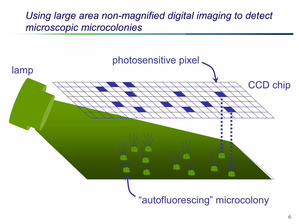
Using large area non-magnified digital imaging to detect microscopic microcolonies
CCD chip
photosensitive pixellamp
“autofluorescing” microcolony

7
How the image analysis software enumerates growing microbes

8
Image analysis using Growth Direct software
make a stack of images from the various time points

9
Image analysis using Growth Direct™ software
find objects on each image using image analysis software

10
Image analysis using Growth Direct™ software
align images

11
Image analysis using Growth Direct™ software
trace all objects backwards through time

12
identify growing objects (intensity increases over time)
Image analysis using Growth Direct™ software

13
ignore debris (objects that do not grow over time)
Image analysis using Growth Direct™ software

14
report number of growing objects
Image analysis using Growth Direct™ software
5 growing microcolonies
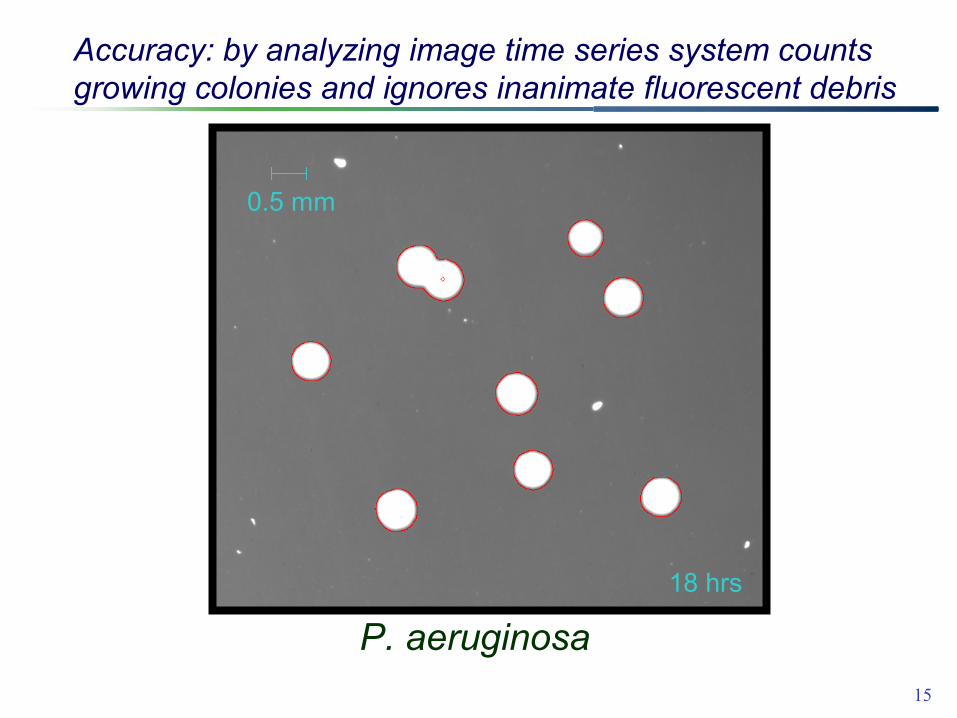
15
2 hrs3 hrs4 hrs5 hrs6 hrs7 hrs8 hrs9 hrs10 hrs11 hrs12 hrs13 hrs14 hrs15 hrs16 hrs17 hrs18 hrs
0.5 mm
Accuracy: by analyzing image time series system counts growing colonies and ignores inanimate fluorescent debris
P. aeruginosa

16
The work flow of the automated compendial test
�
���
����
������
�������
� ����
�������� �
�� �����

17
Labor savings and improved compliance from an automated compendial test
• labor savings – data acquisition is automated – documentation is electronic, and easily
transferred to data management systems
• increased compliance – fewer data management errors – greater reproducibility

18
Automating the compendial test preserves its advantages while addressing its weaknesses
• captures the positive features of the compendial tests– non-destructive– ultra-sensitive (1 CFU )– breadth of testing applications– enumerates replicating cells– high throughput – no added reagents– industry standard media, membranes
• addresses the limitations of the compendial tests– automation: ↓labor, ↑compliance, ↑reproducibility–speed: saves days, generally ~50% faster

19
Bacteria detected by cellular autofluorescenceAcidovorax delafieldii Curtobacterium sp. Proteus vulgaris Acidovorax sp. Deinococcus proteolyticus Pseudomonas aeruginosa Acidovorax temperans Dermacoccus nishinomiyaensis Pseudomonas fluorescens Acinetobacter junii Enterococcus faecalis Pseudomonas putida Afipia broomeae Escherichia coli Pseudomonas stutzeri Arthrobacter sp. Geobacillus stearothermophilus Ralstonia pickettii Bacillus cereus Hydrogenophagea sp. Rhodococcus erythropolis Bacillus clausii Hyphomicrobium sp. Roseomonas gilardii Bacillus fusiformis Kocuria kristinae Roseomonas sp. Bacillus gibsonii Kocuria rhizophila Salmonella enterica Bacillus licheniformis Kytococcus sedentarius Serratia marcesens Bacillus megaterium Macrococcus caseolyticus Sphingomonas paucimobilis Bacillus pumilus Methylobacterium extorquens Sphingomonas spp. Bacillus sp. Methylobacterium radiotolerans Sphingomonas terrae Bacillus subtilis Microbacterium luteolum Staphylococcus aureus Bacillus vortex Microbacterium maritypicum Staphylococcus capitis Bacteriodes fragilis Microbacterium sp. Staphylococcus epidermidis Brachybacterium sp. Micrococcus luteus Staphylococcus equorum Bradyrhizobium spp. Moraxella osloensis Staphylococcus haemolyticus Brevibacterium sp. Myxococcus xanthus Staphylococcus hominis Brevundimonas diminuta Neisseria sp. Staphylococcus saccharolyticus Burkholderia cepacia Paenibacillus lautus Staphylococcus sp. Caulobacter leidyii Paenibacillus sp. Staphylococcus warneri Cellulomas sp. Pantoea agglomerans Streptococcus sp. Chromobacterium violaceum Paracoccus sp. Streptomyces chrysolmalus complex Clostridium sporogenes Porphyromonas gingivalis Streptomyces coelicolor Corynebacterium sp. Prevotella melaninogenica Streptomyces sp. Corynebacterium xerosis Propionibacterium acnes Vibrio natriegens Corynebacterium pseudodiptheriticum

20
Fungi detected by cellular autofluorescence
Penicillium roquefortii Candida albicans Zygosaccharomyces rouxiiPenicillium notatum Aureobasidium pullulans Trichoderma asperellumPenicillium corylophylumAspergillus versicolor Sporotrichum pruinosum Penicillium chrysogeneumAspergillus sp.Sporidiobolus johnsoniiPenicillium camemberti Aspergillus niger Schizosaccharomyces pombeFusarium solaniAspergillus fumigatus Schizophyllum fasciatumEpicoccum nigrumAspergillus flavusSchizophyllum communeCladosporium herbarumArthrinium sacchariSaccharomyces cerevisiae Chaetomium globosumAlternaria geophilaRhizopus oligosporusCandida parapsilosis Alternaria alternata
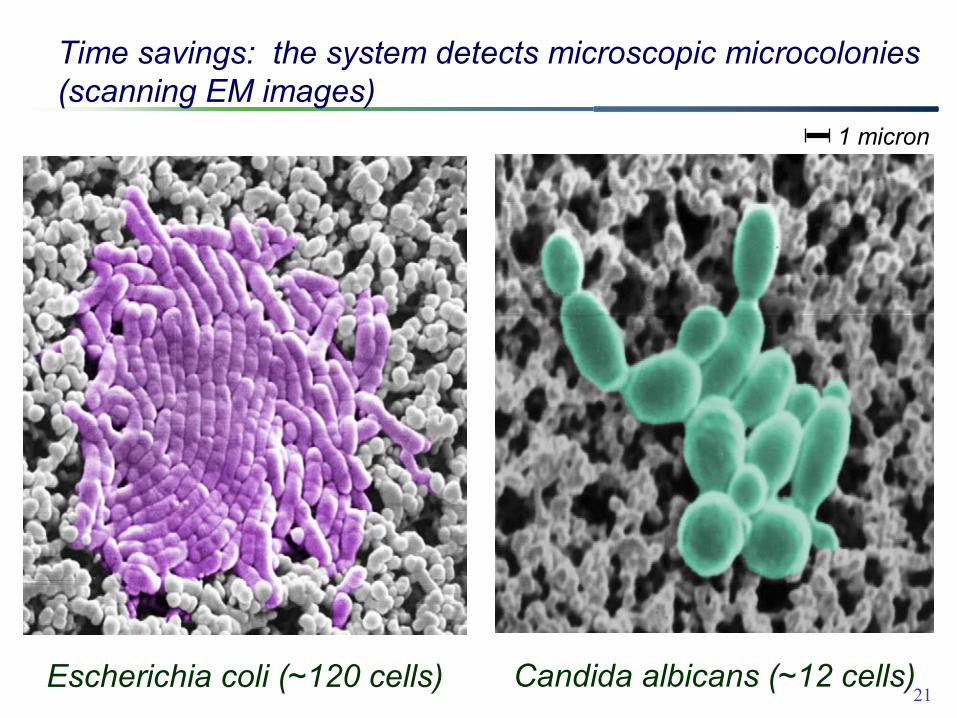
21Candida albicans (~12 cells)Escherichia coli (~120 cells)
Time savings: the system detects microscopic microcolonies(scanning EM images)
1 micron

22
The automated compendial method saves days for slow growing strains
2.73.60.9Proionibacterium acnes
1.21.80.6Clostridium sporogenes1.62.40.8Aspergillus niger1.931.1Ralstonia picketii2.13.61.5Aspergillus versicolor2.43.71.3Mycoplasma bovis2.441.6Deinococcus proteolyticus
4.86.71.9Mycobacterium chelonae6.170.9Bacteroides vulgatis
14.617.22.6Methylobacterium extorquens
Visual(days)
Growth Direct(days)
Days saved

23
Time savings is greatest for slow growing microbes

24
Water testing

25
Rapid detection of water microbes: autofluorescent detection detects the same colonies that later become visible by eye
2.5 days
Growth Direct microcolonies
5 days
visual plate counting
sample: purified water from a pharmaceutical facility

26
Correlation of Growth Direct and visible counts in pharma water samples
Growth Direct (68 hr) vs visible counts
visible colonies at 120 hr0 50 100 150 200 250 300 350G
row
th D
irect
col
onie
s at
68
hr
0
50
100
150
200
250
300
350
y = 1.02x
R2 = 0.90
R2A, 32.5ºC

27
visual plate counting (5 days)
Growth Direct (1.5 days)
Accuracy: resolving at the microcolony stage colonies that are uncountable by traditional visible plate

28
Air monitoring

29
20 hr
Growth Direct microcolonies
72 hr
visual plate counting
Rapid detection of airborne microbes at a pharma plant

3072 hrs (TSA, 32.5ºC)
21 hrs39 hrs
12 hrs
18 hrs
12 hrs
Rapid detection of diverse airborne microbes at a pharma facility

31
Air monitoring: co-trending of rapid (1.5 day) and traditional (3 day) tests at a pharma facility
y = 1.105x + 0.41R2 = 0.7899
0
20
40
60
0 20 40 60visible colonies (72 hr)
Gro
wth
Dire
ct c
olon
ies
(36
hr)
4-Sep-0719-Sep-072-Oct-0715-Nov-07
32.5 ºC, TSA
32
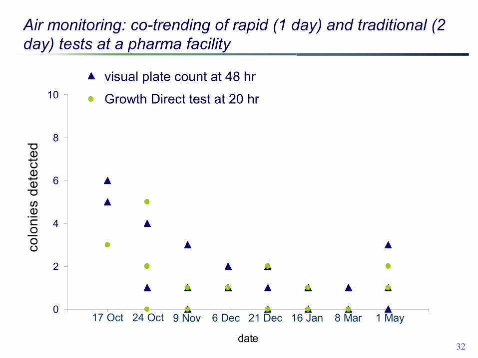
Air monitoring: co-trending of rapid (1 day) and traditional (2 day) tests at a pharma facility
0
2
4
6
8
10
2 3 4 5 6 7 8 9 10 11
date
colo
nies
det
ecte
d
Naked agar (48 hours)
Growth Direct (18 hours)
6 Dec24 Oct 9 Nov 16 Jan 21 Dec 17 Oct 8 Mar 1 May
visual plate count at 48 hr
Growth Direct test at 20 hr
17 Oct 24 Oct 9 Nov 6 Dec 21 Dec 16 Jan 8 Mar 1 May

33
Comparing recovery on membranes and agar in air testing using a “half membrane” strategy

34
y = 1.04x - 5.1, R2 = 0.96
020406080
100120140160180
0 50 100 150agar (CFU)
mem
bran
e (C
FU)
y = 0.93x - 1.7, R2 = 0.87
0
50
100
150
200
0 50 100 150 200agar (CFU)
mem
bran
e (C
FU)
y = 0.94x - 0.93, R2 = 0.97
0
50
100
150
200
250
0 50 100 150 200 250agar (CFU)
mem
bran
e (C
FU)
y = 1.00x + 2.1, R2 = 0.77
010203040506070
0 20 40 60
agar (CFU)
mem
bran
e (C
FU)
Various air samplers, “half-membrane” experiments show equivalent recovery on membrane and agar
Air Ideal (90 mm) Air Ideal (55 mm)
SAS 180 SMA P201
TSA medium

35
Surface monitoring

36
Rapid detection of microbes on surfaces at a pharma site
15 hr
Growth Direct microcolonies
72 hr
visual plate counting

37
0
5
10
15
20
2 3 4 5 6 7 8 9 10 11date
colo
nies
det
ecte
d
Naked agar (48 hours)
Growth Direct (24 hours)
Oct 17 Nov 9Oct 24 Dec 6 Dec 21 Jan 16 Mar 8 May 1
Surface testing: co-trending of rapid (1 day) and traditional (2 day) tests at a pharma facility
visual plate count at 48 hr
Growth Direct test at 24 hr
TSA, 32.5 °C

38
Comparing recovery on membranes and agar in surface testing using capture efficiency (Whyte et al, 1989)
Efficiency (E) = fraction recovered of total microbes/ replicate
• Sample multiple times on same location (e.g. 5 replicates)
• Incubate• Count each plate

39
Comparing efficiency of recovery for surface contact plates: membrane Vs agar
Whyte et al, 1989. J. Hosp. Inf., 13: 33-41
Efficiency (E) = fraction recovered of total microbes/ replicateE = [1 − log (slope)]
1
10
100
1000
0 2 4 6 8 10replicate contact plate number
colo
nies
reco
vere
d
membrane
agar
Emembrane = 0.34Eagar = 0.28

40
0.32 ± 0.100.26 ± 0.09latex gloves9 “thumbs”
0.40 ± 0.08
0.32 ± 0.13
0.38 ± 0.12
0.40 ± 0.13
membrane
average capture efficiency
0.40 ± 0.09
0.31 ± 0.10
0.49 ± 0.07
0.38 ± 0.11
agar
plexiglass10 sites
tyvek8 sites
glass10 sites
stainless steel12 sites
Surface
Surface contact testing: equivalent capture efficiencies on membranes vs. agar

41
Validation Question -Growth Direct System, New Technology?
• Growth Direct is not an alternative technology– it is based on standard USP growth based membrane filtration methods– the results are given as CFU’s.
• The “novel” Growth Direct is an automated compendial method:– the system is an “Automated” colony counter and can be linked to
the USP Chapter <16> Automated Methods of Analysis. – validation requires proof that the camera sees as many micro-colonies
as the eye would see colonies on the membrane surface.– Performance Qualification would follow standard requirements in
chapter <1227>, <61> etc.– other validation components are standard incubator and software
validation protocols.

42
Summing up• Advantages of the an automated compendial
enumeration method:– addresses same broad spectrum of QC applications
as the compendial method– sensitive digital imaging detects microcolonies– non-destructive, compatible with microbial ID– equivalent counts to current method
• Autofluorescence-based detection offers equivalent results with substantial time savings for environmental applications:– water– air– surfaces

43
Environmental monitoring using a rapid non-destructive automated
compendial method