enzymatic colorimetric method for the determination of
TRANSCRIPT

: Berti et al.: Determination of inorganic phosphorus in serum and urine 399
J. Clin. Chem. Clin. Biochem.Vol. 26, 1988, pp. 399-404
'. © 1988 Walter de Gruyter & Co.Berlin · New York
Enzymatic Colorimetric Method for the Determination ofInorganic Phosphorus in Serum and Urine
By G. Berti, P. Fossati, G. Tarenghi, C. Musitelli
i Laboratorio Ricerca & Sviluppo e Direzione Scientifica Miles Italiana SpA, Divisione Ames, Cavenago Brianza(Milano), Italia and
G. K Mehi d'Eril11
Laboratorio di Chimica-Clinica, IRCCS Policlinico S. Matteot Pavia, Italia
(Received December 12, 1987/February 29, 1988)
Summary: The performance of an enzymatic colorimetric method for the determination of inorganic phos-phorus in serum and urine is described. Phosphate ions react with inosine in the presence of purine nucleosidephosphorylase to form hypoxanthine; this is oxidized by xanthine oxidase to uric acid with production ofhydrogen peroxide. The latter is determined with the aid of the chromogen system peroxidase/4-aminophen-azone/N-ethyl-N-iS^methylpheny^-N'-acetylethylenediamine, the coloured product being measured at 555 nm.This series of reactions is completed in 5 min at 37 °C. The test is linear up to 240 mg/1. Analytical recoveryin serum averaged 101.2 ± 1.2% and in urine 101.9 + 3.2%. Within-run and between-run precision studiesin serum and urine samples gave CVs < 4.54% (at 22.0 mg/1). Results obtained by this method agree(r = > 0.983) with the molybdate UV and molybdenum blue methods. Interference by endogenous substances,including organic phosphate, was negligible.
η ο uc on these methods are used nevertheless in the clinicalMost techniques for the determination of inorganic laboratory, especially those based on direct proce-phosphorus in serum or other biological fluids are dures without deproteinization. Many attempts havebased on phosphomolybdate complex formation by been made to develop enzymatic methods: phospho-reaction of inorganic phosphate with molybdate in rylase, phosphoglucomutase and glucose-6-phosphateacid solution. dehydrogenase have been used to generate NADPH-ι- ι ι ,. ι n * * Λ fr°m inorganic phosphate and glycogen (2, 3). Gly-This colourless to pale yellow compound can be de- i J i . J n u u * ^ u j Λ. · . , . ' , t τΑτ , - t_ , - ceraldehyde-3-phosphate dehydrogenase was used astermmed directly by UV absorption, or by colon- t · i* *· *· * ι Λ* ' . , ~ a key enzyme in an alternative enzymatic route lead-metric measurement after reduction to molybdenum . J ΧΤΑΤ^ΤΤ /A\ rr /c\ «. · -+u« , t . . t ' . . ;;. ing to NADH (4). Hwang (5) reacts inosine withblue or complexmg with dyes such as malachite green * u * - * u % · i -A uphosphate in the presence of punne nucleoside phos-
^" phorylase:1) the resulting hypoxanthine, in the pres-These methods have several drawbacks: the direct UV ence of milk xanthine oxidase,1) produces uric acid,method is affected by serum turbidity or pigmenta- which is determined from its absorbance at 293 nm.tion; the molybdenum blue method is influenced inits specificity, sensitivity and colour stability by the — -type of reducing agent used; and the strongly acidic !) Enzymes:
.... "* *t; ι i ·* i_ j i^ Purme nucleoside phosphorylase (EC 2.4.2.1)conditions of the malachite green method may result Xanthine oxidase (EC 1.1.3.22)in hydrolysis of organic phosphate. Peroxidase (EC 1.11.1.7)
J. Clin. Chem. Clin. Biochem. / Vol. 26, 1988 / No. 6
400 Berti et al.: Determination of inorganic phosphorus in serum and urine
All these enzymatic methods, however, have variouslimitations such as the need for reading in the UVrange; poor stability of substrates or enzymes; unfa-vourable reaction equilibrium; and laborious proce-dures. Significant developments have been reportedby Fossati (6) and Machida (7), both using the Hwangenzymatic sequence. More precisely, Fossati assayedthe Superoxide ion (formed as hydrogen peroxide pre-cursor during hypoxanthine oxidation) by reducing atetrazolium salt to formazane; Machida assayed phos-phate, likewise in the visible region, using the perox-idase/phenol/4-aminophenazone chromogen systemwith an appropriate microbial xanthine oxidase.
A fully enzymatic colorimetric method based uponthe Machida enzymatic sequence has been recentlydescribed (8, 9).
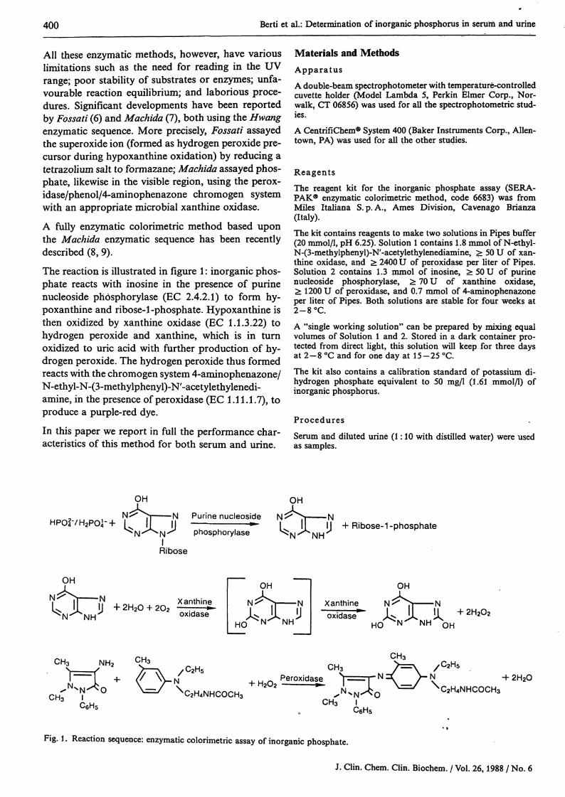
The reaction is illustrated in figure 1: inorganic phos-phate reacts with inosine in the presence of purinenucleoside phosphorylase (EC 2.4.2.1) to form hy-poxanthine and ribose-1-phosphate. Hypoxanthine isthen oxidized by xanthine oxidase (EC 1.1.3.22) tohydrogen peroxide and xanthine, which is in turnoxidized to uric acid with further production of hy-drogen peroxide. The hydrogen peroxide thus formedreacts with the chromogen system 4-aminophenazone/N-ethyl-N-(3-methylphenyl)-N'-acetylethylenedi-amine, in the presence of peroxidase (EC 1.11.1.7), toproduce a purple-red dye.
In this paper we report in full the performance char-acteristics of this method for both serum and urine.
Materials and MethodsApparatusA double-beam spectrophotometer with temperature-controlledcuvette holder (Model Lambda 5, Perkin Elmer Corp., Nor-walk, CT 06856) was used for all the spectrophotometric stud-ies.A CentrifiChem® System 400 (Baker Instruments Corp., Allen-town, PA) was used for all the other studies.
ReagentsThe reagent kit for the inorganic phosphate assay (SERA-PAK® enzymatic colorimetric method, code 6683) was fromMiles Italiana S. p. A., Ames Division, Cavenag Brianza(Italy).
The kit contains reagents to make two solutions in Pipes buffer(20 mmol/1, pH 6.25). Solution 1 contains 1.8 mmol of N-ethyl-N-(3-methylphenyl)-N'-acetylethylenediamine, ^ 50 U of xan-thine oxidase, and > 2400 U of peroxidase per liter of Pipes.Solution 2 contains 1.3 mmol of inosine, >50U of purinenucleoside phosphorylase, > 70 U of xanthine oxidase,> 1200 U of peroxidase, and 0.7 mmol of 4-aminophenazoneper liter of Pipes. Both solutions are stable for four weeks at2-8 °C.A "single working solution" can be prepared by mixing equalvolumes of Solution 1 and 2. Stored in a dark container pro-tected from direct light, this solution will keep for three daysat 2-8 °C and for one day at 15-25 °C.The kit also contains a calibration standard of potassium di-hydrogen phosphate equivalent to 50 mg/l (1.61 mmol/1) ofinorganic phosphorus.
Procedures
Serum and diluted urine (1:10 with distilled water) were usedas samples.
L^^P ,n ι ,HP042-/H2P04--l·
OH
^ Π Ν Purine nucleosideI I I I ^
Ν -""̂ Ν ̂ phosphorylaseIRibose
OH
NIf II + Ribose-1 -phosphate
OH
N
rsr NHXanthineoxidase
OH
Xanthineoxidase"
OH
Ν
HC>
Ν2H2°>
CH3 NH2
N^^OCH3 I
C6H5
C2H5
CH3
C2H4NHCOCH3
, u Λτ Η2Ο2•2H2O
C2H4NHCOCH3CH3
C6H5
Fig. 1. Reaction sequence: enzymatic colorimetric assay of inorganic phosphate.
J. Clin. Chem. Clin. Biochem. / Vol. 26,1988 / No. 6
Berti et al.: Determination of inorganic phosphorus in serum and urine 401
Manual two-step procedureAdd 20 μΐ of sample (or standard) to 1.5 ml of Solution 1, mixand incubate at 37 °C for 2-5 minutes. Add 1.5 ml of Solution2; mix and incubate 5 minutes at 37 °C, then read at 555 nmagainst the reagent blank. The results reported for the manualprocedure were obtained by this method.
Manual one-step procedureThe assay can also be carried out as follows: add 20 μΐ ofsample (or standard) to 3.0 ml of "single working solution",mix and incubate 5 minutes at 37 °C. Read at 555 nm vs thereagent blank.
Automated procedureThe following instrument setting was used for CentrifiChem:temperature 37 °C; wavelength 550 nm; sample volume 5 μΐ,wash volume 55 μΐ; single working solution 350 μΐ; 1st readingtime 3 s; time interval 300 s; control standard 50 mg/1.This procedure was validated by assaying 60 human sera bythe automated procedure (y) and by the manual one-step pro-cedure (x). Calculated linear regression was: y = 0.990x + 0.3mg/1, r = 0.993, Syx = 1.4, χ = 43.7, y = 43.6, range 20-80mg/1.
4 6 8Incubation time [min]
10
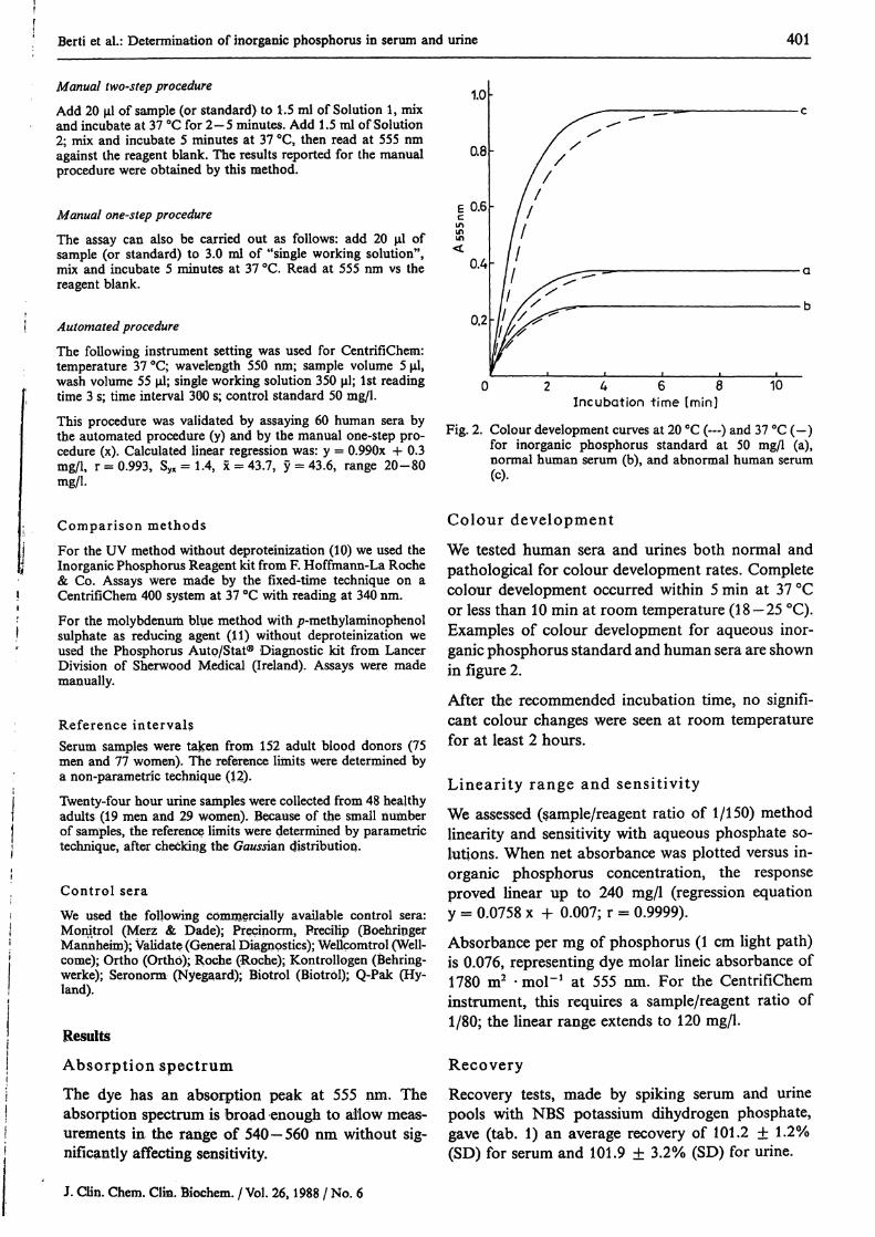
Fig. 2. Colour development curves at 20 °C (—) and 37 °C (-)for inorganic phosphorus standard at 50 mg/1 (a),normal human serum (b), and abnormal human serum(c).
Comparison methodsFor the UV method without deproteinization (10) we used theInorganic Phosphorus Reagent kit from F. Hoffmann-La Roche& Co. Assays were made by the fixed-time technique on aCentrifiChem 400 system at 37 °C with reading at 340 nm.For the molybdenum blue method with /?-methylaminophenolsulphate as reducing agent (11) without deproteinization weused the Phosphorus Autq/Stat® Diagnostic kit from LancerDivision of Sherwood Medical (Ireland). Assays were mademanually.
Reference intervalsSerum samples were taken from 152 adult blood donors (75men and 77 women). The reference limits were determined bya non-parametric technique (12).Twenty-four hour urine samples were collected from 48 healthyadults (19 men and 29 women). Because of the small numberof samples, the reference limits were determined by parametrictechnique, after checking the Gaussian distribution.
Control seraWe used the following commercially available control sera:Monitrol (Merz & Dade); Precinorm, Precilip (BoehringerMannheim); Validate (General Diagnostics); Wellcomtrol (Well-come); Ortho (Ortho); Roche (Roche); Kontrollogen (Behring-werke); Seronorm (Nyegaard); Biotrol (Biotrol); Q-Pak (Hy-land).
Results
Absorption spectrum
The dye has an absorption peak at 555 nm. Theabsorption spectrum is broad enough to allow meas-urements in the range of 540—560 nm without sig-nificantly affecting sensitivity.
Colour development
We tested human sera and urines both normal andpathological for colour development rates. Completecolour development occurred within 5 min at 37 °Cor less than 10 min at room temperature (18 — 25 °C).Examples of colour development for aqueous inor-ganic phosphorus standard and human sera are shownin figure 2.
After the recommended incubation time, no signifi-cant colour changes were seen at room temperaturefor at least 2 hours.
Linearity range and sensitivity
We assessed (sample/reagent ratio of 1/150) methodlinearity and sensitivity with aqueous phosphate so-lutions. When net absorbance was plotted versus in-organic phosphorus concentration, the responseproved linear up to 240 mg/1 (regression equationy = 0.0758 χ + 0.007; r = 0.9999).
Absorbance per mg of phosphorus (1 cm light path)is 0.076, representing dye molar lineic absorbance of1780 m2 -mol"1 at 555 nm. For the CentrifiCheminstrument, this requires a sample/reagent ratio of1/80; the linear range extends to 120 mg/1.
Recovery
Recovery tests, made by spiking serum and urinepools with NBS potassium dihydrogen phosphate,gave (tab. 1) an average recovery of 101.2 ± 1.2%(SD) for serum and 101.9 ± 3.2% (SD) for urine.
J. CUn. Chem. Clia. fciochem. / Vol. 26,1988 / No. 6

402 Berti et al.: Determination of inorganic phosphorus in serum and urine
Tab. 1. Analytical recovery Tab. 2. Precision data
Sample Added Found RecoveryInorganic phosphorus (mg/1)
Serum pool
Urine pool
—25.050.0
100.0150.0170.0200.0—240275315504
37.662.488.2
139.8191.4209.3240.0
300544586607826
—99.2101.2102.2102.5101.0101.2
—101.7104.097.5
104.4
SerumMean(mg/1)
SD(mg/1)
Within-run (n =22.038.890.0
1.000.601.50
Between-run (n =40.751.580.0
1.001.702.20
CV(%)
10)4.541.551.67
= 12)a
2.453.302.75
UrineMean(mg/ψ
Within-run903.6
1424.6
SD(mg/1)
(n = 12)19.121.9
CV(%)
2.111.54
Between-run (n = 12)a
216.0508.1630.0
5.515.110.5
2.552.971.67
a over a 15-day period
Precision
We assessed assay precision by replicate analysis ofvarious human serum and urine pools at differentphosphate concentrations. Table 2 shows the resultsobtained.
Method comparison
We explored the correlation between the enzymaticcolorimetric method and two chemical methods, thephosphomolybdate UV method and the molybdenumblue method, in routine serum and urine assays. Re-sults were processed by least-square regression anal-ysis (13); correlation plots and statistical parametersare shown in figures 3 and 4.
Interferences
We explored the effects of known potential interfer-ents in Trinder-type reactions, either endogenous orexogenous (bilirubin, haemoglobin, ascorbic acid,drugs such as L-dopa and its metabolite 3,4-dihy-droxyphenylacetate, and α-rnethyldopa), by spikingpooled human sera at normal inorganic phosphorusconcentration with known amounts of these sub-stances (for haemoglobin we used washed and lysederythrocytes). We also tested the effect of hypoxan-thine and xanthine, since these occur in the reaction*.Allopurinol was tested as a known inhibitor of xan-thine oxidase. The effect on test results of some rep-resentative organic phosphates, as well as of the morecommonly used anticoagulants, was evaluated simi-
Ο)
120.-.110|M ooX 90Ό
80
70
1 60
1 5°ο 40
is 3°Ι 20*~ 10
Ο 10 20 30 40 50 60 70 80 90 100 110 120PJ (ρ hosphom ly bdate U V rnelhod) f mg /1 ]
10 20 30 40 50 60 70 80 90 100 110 120PJ (molybdenum blue method) [mg/l]
Fig. 3. Correlation plots for serum samples. Regression parameters are fora) enzymatic colorimetric method (y) vs phosphomolybdate UV method (xV
y = 0.949x + 2.6; r = 0.985; Syx = 2.8; χ = 44.2; y = 44.5; n = 98;b) enzymatic colorimetric method (y) vs molybdenum blue method (xY
y = 0.969x + 3.8; r = 0.983; S^ = 3.1; χ = 43.0; y = 45.5; n = 75.
J. Clin. Chem. Clin. Biochem. / Vol. 26,1988 / No. 6
Berti et al,: Determination of inorganic phosphorus in serum and urine 403
1600
200 400 600 800 1000 1200 1400 1600Pj (phosphomolybdate UV method) [ mg/ l ]
1500
500 1000 1500 2000P· (molybdenum blue method} [mg/l]
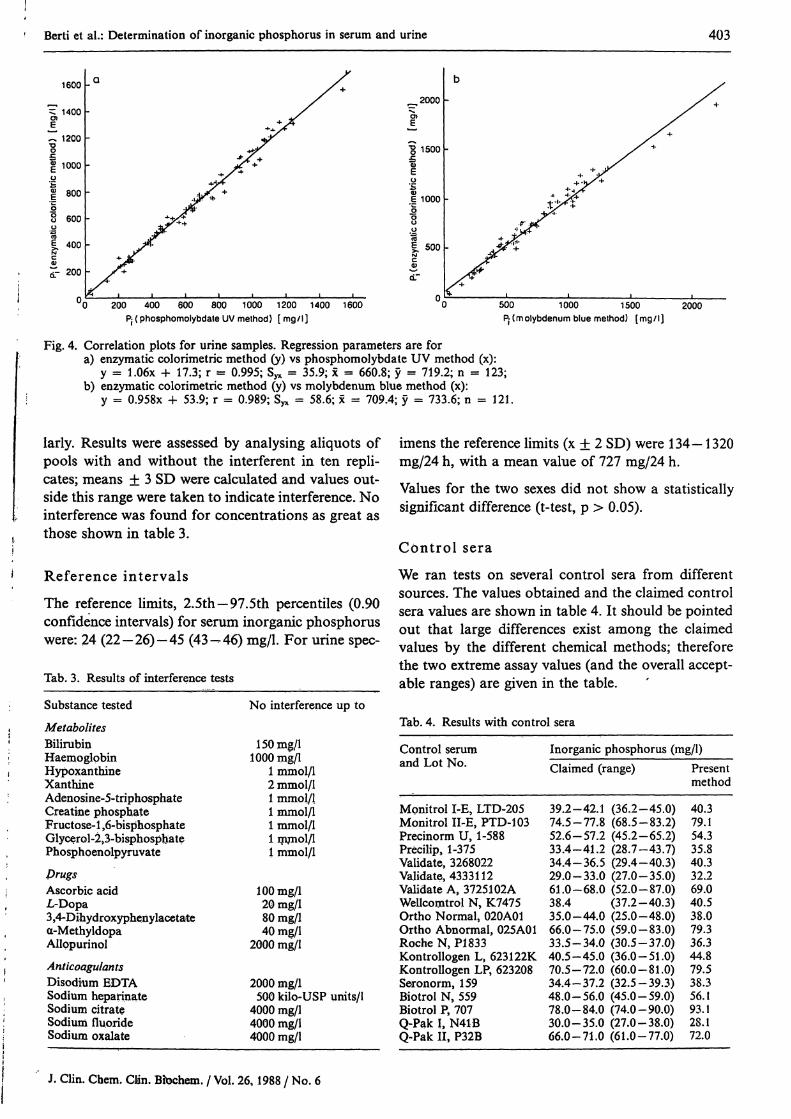
Fig. 4. Correlation plots for urine samples. Regression parameters are fora) enzymatic colorimetric method (y) vs phosphomolybdate UV method (x):
y = 1.06x + 17.3; r = 0.995; Syx = 35.9; x = 660.8; y = 719.2; n = 123;b) enzymatic colorimetric method (y) vs molybdenum blue method (x):
y = 0.958x + 53.9; r = 0.989; S^ = 58.6; x = 709.4; y = 733.6; n = 121.
larly. Results were assessed by analysing aliquots ofpools with and without the interferent in ten repli-cates; means ± 3 SD were calculated and values out-side this range were taken to indicate interference. Nointerference was found for concentrations as great asthose shown in table 3.
Reference intervals
The reference limits, 2.5th-97.5th percentiles (0.90confidence intervals) for serum inorganic phosphoruswere: 24 (22—26)-45 (43—46) mg/l. For urine spec-
Tab. 3. Results of interference tests
Substance tested
MetabolitesBilirubinHaemoglobinHypoxanthineXanthineAdenosine-5-triphosphateCreatine phosphateFructose-1,6-bisphosphateGlycerol-l^-bisphosphatePhosphoenolpyruvate
DrugsAscorbic acidL-Dopa3,4-Dihydroxypheny lacetatea-MethyldopaAllppurinol
AnticoagulantsDisodhim EDTASodium hepannateSodium citrateSodium fluorideSodium oxalate
No interference up to
150 mg/l1000 mg/l
1 mmol/12 mmol/11 mmol/11 mmol/11 mmol/11 mmol/11 mmol/1
100 mg/l20 mg/l80 mg/l40 mg/l
2000 mg/l
2000 mg/l500 kilo-USP units/1
4000 mg/l4000 mg/l4000 mg/l
imens the reference limits (χ ± 2 SD) were 134-1320mg/24 h, with a mean value of 727 mg/24 h.
Values for the two sexes did not show a statisticallysignificant difference (t-test, p > 0.05).
Control sera
We ran tests on several control sera from differentsources. The values obtained and the claimed controlsera values are shown in table 4. It should be pointedout that large differences exist among the claimedvalues by the different chemical methods; thereforethe two extreme assay values (and the overall accept-able ranges) are given in the table.
Tab. 4. Results with control sera
Control serumand Lot No.
Monitrol I-E, LTD-205Monitrol II-E, PTD-103Precinorm U, 1-588Precilip, 1-375Validate, 3268022Validate, 4333112Validate A, 3725102AWeilcomtrol N, K7475Ortho Normal, 020A01Ortho Abnormal, 025A01Roche N, PI 833Kontrollogen L, 623122KKontrollogen LP, 623208Seronorm, 159Biotrol N, 559Biotrol P, 707Q-Pak I, N41BQ-Pak 11, P32B
Inorganic phosphorus (mg/l)Claimed (range)
39.2-42.1 (36.2-45.0)74.5 -77. * (68.5-83.2)52.6-57.2 (45.2-65.2)33.4-41.2 (28.7-43.7)34.4-36.5 (29.4-40.3)29.0-33.0 (27.0-35.0)61.0-68.0 (52.0-87.0)38.4 (37.2-40.3)35.0-44.0 (25.0-48.0)66.0-75.0 (59.0-83.0)33.5-34.0 (30.5-37.0)40.5-45.0 (36.0-51.0)70.5-72.0 (60.0-81.0)34 .4 -37. 2 (32.5-39.3)48,0-56.0 (45.0-59.0)78.0-84.0 (74.0-90.0)30.0-35.0 (27.0-38.0)66.0-71.0 (61.0-77.0)
Presentmethod
40.379.154.335.840.332.269.040.538.079.336.344.879.538.356.193.128.172.0
J. Clin. Chem. Clin. Bibchem. / Vol. 26,1988 / No. 6

404 Berti et al.: Determination of inorganic phosphorus in serum and urine
Discussion
Existing chemical methods for assaying inorganicphosphorus in biological fluids generally lack suffi-cient specificity and ease of execution — two mainconsiderations for routine use. The method at issueis highly specific and easy to perform, being based onan appropriate enzymatic reaction coupled with asensitive colorimetric measurement.
Method linearity (up to at least 240 mg/1) is broadenough to make reassays unnecessary. Owing to thehigh sensitivity, precision is very good at the variousphosphate levels tested, including the low ones. Com-parative studies versus the phosphomolybdate UVmethod and the molybdenum blue method showedgood agreement for both serum and urine.
High bilirubin and ascorbic acid concentrations, aswell as moderate haemolysis do not affect results;haemoglobin above 1 g/1 causes increased absorptionand leads to phosphorus overassay.
Endogenous hypoxanthine and xanthine might inter-fere by generating stoichiometric amounts of hydro-gen peroxide; but they are neutralized in the two-stepprocedure by reaction with N-ethyl-N-(3-methyl-phenyl)-N'-acetylethylenediamine during preincuba-tion, forming a colourless product in the absence ofinosine and 4-aminophenazone.
No interference from these oxypurines was seen withconcentrations in excess of values ever found in serum(14). It should, however, be noted that even in theone-step procedure the results are affected only atvery high values, such as those of patients receivingallopurinol (15). Allopurinol showed no inhibitoryeffect of its own. For L-dopa and α-methyldopa, aborderline negative interference may occur; no inter-ference from 3,4-dihydroxyphenylacetate was noted.Organic phosphates with relatively high free energyof hydrolysis (> 21 kJ/mol ^ ^ 5.0 kcal/mol) areeasily degraded in an acidic medium and are reportedto interfere more or less significantly in chemical assaymethods for inorganic phosphate (1). We tested avariety of phosphate links (ester, acetal, phosphoricacid anhydride, and amidophosph te) and found thatnone would interfere in the present method, even atconcentrations much higher than those found inserum (16) or likely to be released by moderate hae-molysis. Anticoagulants did not interfere.
In conclusion the method discussed in this papershows good sensitivity, linearity and precision. Withrespect to accuracy, the method offers the advantageover chemical procedures of being free of interferencefrom organic phosphate. Added advantages are theuse of noncorrosive reagents, ease of handling, aridadaptability to clinical laboratory instruments.
References1. Weissman, N. & Pileggi, V. J. (1974) Inorganic Ions: Phos-
phate in Clinical Chemistry: Principles and Technics, 2ndedn. (Henry, R. J., Cannon, D. C. & Winkelmann, J. W.,eds.) pp. 720-728, Harper & Row, Hagerstown.
2. Schulz, D. W., Passonneau, J. V. & Lowry, Ο. Η. (1967)Anal. Biochem. 19, 300-314.
3. Pesce, A. M., Bodourian, S. H. & Nicholson, J. F. (1974)Clin. Chem. 20, 332-336.
4. Scopes, R. K. (1972) Anal. Biochem. 49, 88-94.5. Hwang, W. I. & Cha, S. (1973) Anal. Biochem. 55, 379-
387.6. Fossati, P. (1985) Anal. Biochem. 149, 62-65.7. Machida, Y. & Nakanishi, T. (1982) Agric. Biol. Chem. 46,
807-808.8. Colombi, M., Bosoni, A., Fossati, P. & Musitelli, C. (1985)
Isr. J. Clin. Biochem. 4, 31 (Abstract).9. Berti, G., Fossati, P., Melzi d'Eril, G. V., Tarenghi, G. &
Musitelli, C. (1987) Clin. Chem. 33, 312 (Technical Brief).
10. Daly, J. A. & Ertingshausen, G. (1972) Clin. Chem. 18,263-265.
11. Power, M. H. (1953) in Standard Methods of ClinicalChemistry I (Reiner, M., Ed.) pp. 84—87, Academic Press,New York.
12. Solberg, H. E. (1983) J. Clin. Chem. Clin. Biochem. 21,749-760.
13. Westgard, J. O. (1973) Clin. Chem. 19, 49-57.14. H nde, K. E., Perini, F., Putterman, G. & Elin, R. (1979)
Clin. Chem. 25, 1492-1494 (Case Report).15. Kito, M., Tawa, R., Takeshima, S. & Hirose, R. (1982) J.
Chromatog. 257, 183-188.16. Natelson, S. & Natelson, E. A. (1975) Mechanisms Con-
trolling Serum Phosphate Levels in Principles of AppliedClinical Chemistry, 1, pp. 169-174, Plenum Press, NewYork.
Dr. Giovanni BertiLaboratorio Ricerca & SviluppoMiles Italiana SpA, Div. AmesVia F. L. Miles 101-20040 Cavenago Brianza (Milano)
J. Clin. Chem. Clin. Biochem. / Vol. 26,1988 / No. 6