epidemiology of peste des petits ruminants virus in ... · domestiques ou sauvages. ... les moutons...
TRANSCRIPT

i
N° d’ordre : 2274
Thèse
présentée
pour obtenir
LE TITRE DE DOCTEUR DE L’INSTITUT NATIONAL POLYTECHNIQUE DE TOULOUSE
PhD
École doctorale : S.E.V.A.B
Spécialité : Sciences Vétérinaires Par Mr GOPILO Abraham
Titre de la thèse EPIDEMIOLOGY OF PESTE DES PETITS RUMINANTS VIRUS IN ETHIOPIA AND
MOLECULAR STUDIES ON VIRULENCE
Soutenue le 3 Novembre 2005 devant le jury composé de :
Prof P. DORCHIES Président
Prof P.D. PICAVET Directeur de thèse
Dr P.C. LEFEVRE Rapporteur
Dr S. ZIENTARA Rapporteur
Dr G. LIBEAU Membre
Dr S. BERTAGNOLI Membre

ii
EPIDEMIOLOGY OF PESTE DES PETITS RUMINANTS VIRUS IN
ETHIOPIA AND MOLECULAR STUDIES ON VIRULENCE
Abraham Gopilo
2005
© Copyright

i
Acknowledgements
I praise and thank God Almighty for providing me health and strength to undertake the training
project.
The National Animal Health Research Center (Ethiopia) allowed the study leave of four years
and the French Ministry of Foreign Affairs granted financial support for the scholarship. I am
highly indebted to them.
I am also highly indebted to CIRAD-EMVT for welcoming me and helping me through out all
these years. Many people were kind and very patient with me. My special thanks go to the Co-
Director of the Thesis Dr Emmanuel Albina. I really appreciate his interest, guidance and
leadership through out the work. My thanks also to Dr Genevieve Libeau for availability and all
the co-ordination work when I needed it most.
Many thanks to Professor P.D. Picavet for being very kind for accepting to be Director of the
Thesis. My whole hearted appreciations to Professor P. Dorchies and to Dr P.C. Lefèvre, Dr S.
Zientara and Dr S. Bertagnoli for accepting the burden of being President and Rapporters in the
Jury, respectively.
My special love goes to my wife Fantaye Abamegal and my daughters Debora Abraham and
Abigel Abraham for allowing me to undertake this training, while being in constant distress in
my absence. They have been extremely patient with me.

ii
Curriculum Vitae
Abraham Gopilo is born in June 26, 1956 in Kemba in southern Ethiopia. He graduated in 1982
as Doctor of Veterinary Medicine (DVM) from Ukraine Academy of Agriculture, Kiev in the
former USSR. He also graduated in Master of Philosophy in Veterinary Science (Veterinary
Virology) from Massey University, New Zealand in 1989. He has undertaken short courses (upto
three months) in diagnostic virology in 1994 (Maison Alfort, France), 1995 (Pirbright, UK), 1996
(Vienna, Austria). He was in a training visit to Australian National Animal Health Laboratory in
Geelong in 1990.
He has a working experience in veterinary clinic of four years (1983-87) and of diagnostic
laboratory two years (1990-91) and in the Central Disease Investigation Laboratory (CDIL)
which later became National Animal Health Research Center (NAHRC) for ten years (1992-
2001). In the laboratory, he worked as National Coordinator for the Pan African Rinderpest
Campaign (PARC) serological monitoring and disease surveillance programmes and as National
project counter part for the International Atomic Energy Agency (IAEA) and the Ethiopian
Science and Technology Commission programmes.
In addition to his assignment in the laboratory he has been a visiting lecturer in Veterinary
Faculty in Addis Ababa University (1991-92, 2000) and one time visiting lecturer in tropical
veterinary virology in Free University of Berlin in Germany in 2000. He had been successful
field supervisor for MSc students (from Zambia and the Sudan) in Free University of Berlin. He
had letters of appreciation of remarkable achievement from African Union (AU), Free University
of Berlin and International Atomic Energy Agency for expert missions for the Agency in
Tanzania (1995), Egypt (1997) and the Sudan (2000).

iii
List of Publications
1. Abraham, G., Sintayehu, A., Libeau G., Albina, E., Roger, F., Laekemariam, Y., Abayneh
D., Awoke, K.M (2005) Antibody seroprevalences against peste des petits ruminants
(PPR) virus in camels, cattle, goats and sheep in Ethiopia. Preventive Veterinary
Medicine 70: 51-57.
2. Abraham, G., Berhan, A. (2001) The use of antigen-capture enzyme-linked
immunosorbent assay (ELISA) for the diagnosis of rinderpest and peste des petits
ruminants in Ethiopia. Tropical Animal Health and Production 33: 423-30.
3. Abraham, G., Roman, Z., Berhan, A., Huluagerish, A. (1998) Eradication of rinderpest
from Ethiopia. Tropical Animal Health and Production 30: 269-72.
4. Abraham, G., Roeder, P., Zewdu, R. (1992) Agents associated with neonatal diarrhea in
Ethiopian dairy calves. Tropical Animal Health and Production 24: 74-80.
5. Roeder, P.L., Abraham, G., Kenfe, G., Barrett, T (1994) Peste des petits ruminants in
Ethiopian goats. Tropical Animal Health and Production 26: 69-73.
6. Roeder, P.L., Abraham, G., Mebratu, G.Y., Kitching R.P. (1994) Foot-and-mouth disease
in Ethiopia from 1988-1991. Tropical Animal Health and Production 26: 163-167.
7. Shiferaw, F., Abditcho, S., Gopilo, A., Laurenson, M.K. (2002) Anthrax outbreak in
Mago National Park, southern Ethiopia. Vet Rec. 150: 318-320.
8. Tibbo, M., Woldemeskel, M., Gopilo, A. (2001) An outbreak of respiratory disease
complex in sheep in Central Ethiopia. Tropical Animal Health and Production 33: 355-65.

iv
Résumé
La peste des petits ruminants (PPR) est une maladie infectieuse, contagieuse des petits ruminants
domestiques ou sauvages. Elle se caractérise par une hyperthermie élevée (autour de 41°C), du
jetage, des écoulements oculaires, une stomatite nécrosante, de la diarrhée profuse et
généralement une forte mortalité. En Afrique, elle peut avoir différentes incidences cliniques sur
les moutons ou les chèvres, depuis l’infection subclinique jusqu’à une infection aiguë létale. En
Ethiopie, la PPR clinique est rarement décrite et l’étude de la circulation virale était jusqu’à
présent peu développée. Dans ce travail, nous montrons la présence d’anticorps contre le virus de
la PPR sur un grand nombre de moutons, chèvres, vaches et chameaux éthiopiens et nous
confirmons la transmission naturelle du virus PPR chez ces animaux sans manifestation clinique
détectable. Cette absence apparente de pathogénicité peut être liée à une résistance génétique
particulière des races de petits ruminants présentes en Ethiopie ou à une variation de la virulence
des souches de virus PPR. Afin d’étudier ce dernier point, nous avons entrepris des études in
vitro sur des souches isolées en Ethiopie et dans différents pays en comparaison avec une souche
vaccinale obtenue par atténuation par passages en série sur culture cellulaire et d’autres souches
de morbillivirus.
Dans un premier temps, nous avons testé la capacité du virus PPR à infecter différents systèmes
cellulaires. Nous établissons que les cellules VERO (fibroblastes de rein de singe) et 293T
(cellules épithéliales de rein humain) permettent la réplication du virus PPR comme celle du virus
de la peste bovine. En revanche, les cellules B95a (cellules lymphoblastoïdes B de singe) ne
multiplient que le virus de la peste bovine. La capacité d’une cellule à supporter la réplication du
virus est de nature à influer son pouvoir pathogène et l’épidémiologie de la maladie. La
différence de sensibilité des cellules au virus PPR peut être lié à l’affinité de la glycoprotéine
d’enveloppe virale H pour son ou ses récepteurs cellulaires utilisés notamment par le virus de la
peste bovine. Pour aborder cette question, nous avons entrepris des comparaisons de séquences

v
au niveau de la protéine H du virus PPR, en lien avec ce qui a été déjà décrit sur d'autres
morbillivirus.
Pour compléter cette étude sur la virulence, nous avons séquencé les promoteurs de plusieurs
souches de virus PPR et conduit une analyse des mutations pouvant jouer un rôle dans
l'atténuation. En effet, les promoteurs viraux des morbillivirus déterminent la transcription des
ARNm viraux et la réplication du génome viral : la modification de leur séquence peut donc
affecter leur efficacité et influer sur la virulence de la souche concernée. Nous observons 7
mutations sur les promoteurs de la souche vaccinale du virus PPR en comparaison avec les autres
souches virulentes. Certaines mutations sont retrouvées sur les autres morbillivirus, d'autres sont
spécifiques du virus PPR. De cette approche moléculaire, nous déduisons également l’intérêt
d’utiliser les séquences des promoteurs du virus, relativement très variables par rapport au reste
du génome, pour mener des études de phylogéographie et de comparaison entre paramyxovirus.
Le document de thèse a été organisé en 6 chapitres. Le premier concerne l’histoire naturelle de la
PPR avec la description du virus, du génome, de l’épidémiologie, de la transmission, des
symptômes, de la pathologie, de l’immunologie, du diagnostic, de la lutte contre la maladie et des
aspects économiques en Afrique sub-saharienne. Le deuxième chapitre traite de la biologie
comparative du virus PPR avec les autres morbillivirus. Le troisième chapitre concerne les
travaux d’épidémiologie de la PPR effectués en Ethiopie. Le quatrième volet de ce travail reprend
les études sur la spécificité cellulaire du virus PPR et la comparaison des séquences sur la
protéine H. Le cinquième chapitre expose les analyses de séquence des promoteurs génomique et
antigénomique du virus PPR. Enfin, la dernière partie comprend une discussion générale et des
perspectives.

vi
Summary Peste des petits ruminants (PPR) is an acute and highly contagious viral disease of small
ruminants, which is characterised by high fever, ocular and nasal discharge, pneumonia, necrosis
and ulceration of the mucuous membrane and inflammation of the gastro-intestinal tract leading
to severe diarrhoea and high mortality. In Africa, goats are severely affected while sheep undergo
a mild form or rarely suffer clinical disease. PPR is one of the most important economical
diseases in Ethiopia. Clinical PPR is confirmed in Ethiopian goats, however, its circulation in
other animals has never been described. In the present work, we showed that the antibody
seroprevalence in camel, cattle, goat and sheep confirmed natural transmission in these animals
without clinical disease. The apparent absence of pathogenicity in these animals may have been
due to host resistance or loss of virulence of the virus strain. We have further investigated the
latter point by in vitro studies on PPRV comparing strains from Ethiopia and other countries with
the vaccine strain which has been attenuated after several cell culture passages.
In a first approach, virulence of PPRV was monitored in cell culture system and the use of virus
specific monoclonal antibodies enabled to detect differences in virulence between PPRV and
RPV. Vero (primate origin) and 293T (human) cell lines supported virus replication permitting
the in vitro growth of both PPRV and RPV. In contrast to RPV, B95a (marmoset B) cells infected
with PPRV were non permissive. The capability of cells to support active virus replication, which
may result in intercellular spread and induce damages in infected cells, has implications on the
pathogenesis and epidemiology. Cellular receptors are major determinants of host range and
tissue tropism of a virus. The difference in infectivity of PPRV and RPV may have depended on
the H protein epitopes and their cellular receptors. Therefore, we decided to compare the amino
acid epitope of H protein of PPRV with that of other morbilliviruses.

vii
As part of our investigation of virulence factors, we have sequenced and compared genome and
antigenome promoters of a vaccine strain with field strains of PPRV. The promoters contain the
polymerase binding sites to initiate and generate the positive-strand replication and transcription
of mRNAs. Nucleotide base change differences between vaccine strain and field strains would
provide molecular basis for attenuation. Alignment of the genome promoter sequences revealed
seven nucleotide mutations at certain positions. Our finding on nucleotide mutation on PPRV are
in agreement with the nucleotide changes in rinderpest virus and other morbillivirus promoter
regions between vaccine strain and wild type virus. Certain mutations were specific to PPRV.
The promoter sequences were clustered around the geographic origin of the viruses and were
lineage specific. Phylogenetic analysis of PPRV promoters was used for PPR phylogeograhy, and
for comparison with other paramyxoviruses.
The thesis is divided in 6 chapters. The first chapter deals with the natural history of PPR
including the virus, the genome, epidemiology, transmission, clinical signs, immunology,
diagnosis, control and its economic cost in the low income subsistence farming systems in sub-
Saharan Africa. The second chapter is about comparative biology of PPRV with regard to other
groups of morbillivirus genus in the Paramyxoviridae family.
The third chapter deals with field study and observations on epidemiology of PPR in Ethiopia. In
chapter four, PPRV virulence was monitored in cell culture system and comparison of H protein
epitopes. In chapter five, sequence analysis of genome and antigenome promoters of PPRV was
described In chapter six, general discussion and recommendations were forwarded.

viii
Table of Contents
Title Page
Acknowledgement
Curriculum Vitae
List of publications
List of Abbreviation
Résumé
Summary
Chapter I Review of Literature 1
1.1 Introduction 1
1.2. History 2
1.3. Causative agent 2
1.3. Geographic distribution 10
1.5. Epidemiology 13
1.5.1. Transmission 13
1.5.2. Host range and pathogenicity 13
1.5.3. Pattern of the disease 17
1.6. Clinical signs 18
1.7. Pathology 21
1.8 Immunity 23
1.9. Diagnosis 24
1.9.1. Virus Isolation 24
1.9.2. Antigen detection 26
1.9.2.1. Agar gel immunodiffusion test 26

ix
1.9.2.2. Hyper immune serum 26
1.9.2.3. Counter immunoelectrophoresis 26
1.9.2.4. ELISA for antigen detection 26
1.9.2.5. cDNA probe 27
1.9.2.6. Reverse transcription polymerase chain raction (RT/PCR) 28
1.9.3. Serology 29
1.9.3.1 Virus neutralisation test 29
1.9.3.2. Competitive enzyme-linked immunosorbent assay (c-ELISA) 30
1.10. Control and prophylaxis 35
1.11. Disease economy 36
Chapter 2 Comparative biology of PPRV and other morbilliviruses 37
2.1. Introduction 37
2.2. Pathogenicity and host ranges 37
2.3. Serologic relationships 40
2.4. Cross protection studies 40
2.5. Antigenic relationships 41
2.6. The comparison of proteins of morbilliviruses 42
2.7. Genetic relationships 47
2.7.1. Phylogenetic analysis 49
2.8. Conclusions 49
Scope of the study 51
Objectives 51
Chapter 3 PPR occurrence in Ethiopia 54
3.1 Introduction 54
3.1.1. Population 54

x
3.1.2. The Agriculture 54
3.1.3. The Livestock sub-sector 55
3.1.4. Animal Health 56
3.1.5. PPR in Ethiopia 57
3.3 Antibody seroprevalence against PPRV (Article 1) 61
Chapter 4 Differences in sensitivity of cells to PPRV (Article 2) 70
Chapter 5 Sequence analysis of genome and antigenome promoters of PPRV, comparison with other Morbillivirus (Article 3) 92
Chapter 6 General discussion 119
6.1. Epidemiology of PPR in Ethiopia 119
6.2. Monitoring of PPRV virulence in vitro cell culture systems 121
6.3. Sequence analysis of promoters of PPRV and other Morbillivirus123
Recommendations 128
1. Epidemiology and control 128
2. Diagnostic tools 129
3. Molecular tools 130
4. Further research 131
Annexe I Virus neutralisation test protocol 132
Annexe II PCR protocol 133
Bibliography 134

xi
List of Figures
Description Page
Fig. 1-1 Genome of morbillivirus 8
Fig. 1-2 Promoters, genes, transcription units and non-coding regions 9
Fig. 1-3 RNA replication pathway 9
Fig. 1-4 Geographic distribution of PPRV lineages 11
Fig. 1-5 Phylogenetic relationship of PPR isolates and lineages 12
Fig. 1-6 PPR resistant breeds of goats in sahelian region in Africa 15
Fig. 1-7 PPR resistant breeds of sheep in West Africa 15
Fig. 1-8 PPR clinical sign, discharge 20
Fig. 1-9 PPR clinical sign, mouth lesion 20
Fig. 1-10 Body temperature and clinical phases of PPR 20
Fig. 1-11 Immunocapture ELISA 32
Fig. 1-12 PCR reaction steps 33
Fig. 1-13 Indirect and cELISA steps 34
Fig. 3-1 Map of Ethiopia (Regional States) 59
Fig. 3-2 PPR point epidemic in one gathering site 59
Fig 3-3 Seasonal pattern of PPR in endemic areas 59
Fig.1. PPR seroprevalence study sites in Ethiopia in 2001 (Article 1) 63
Fig. 1. Flow cytometry analysis of cells infected with PPRV (Article 2) 81
Fig. 2. Flow cytometry analysis of cells infected with RPV (Article 2) 82
Fig. 3. Percentage of cells indicating positive fluorescence (Article 2) 83
Fig. 4. Titration of PPRV and RPV in different cell lines (Article 2) 83

xii
Fig. 5. Analysis of H amino-acid sequences of PPRV and morbilliviruses (Article 2)
84-87
Fig. 6. a,b 3D surface models of globular head of H protein of PPRV (Article 2) 88
Fig. 6. c,d 3D surface models of globular head of H protein of MV (Article 2 ) 89
Fig.1a. Morbillivirus replication pathway (Article 3) 112
Fig.1b. Genes and promoters of morbillivirus (Article 3) 112
Fig 2a. Alignment of genome promoter (Article 3) 113
Fig. 2b. Alignment of antigenome promoter (Article 3) 113
Fig. 3a. PCR amplification using universal (morbillivirus) primers (Article 3) 114
Fig. 3b. PCR amplification using PPRV specific primers (Article 3) 114
Fig. 4a. Analysis of genome promoters of wild type and vaccine strains of PPRV,
RPV, MV and CDV (Article 3) 115
Fig. 4b. Analysis of antigenome promoters wild type and vaccine strains of PPRV,
RPV, MV and CDV (Article 3) 115
Fig. 5a. Phylogenetic analysis of promoter sequences of PPRV strains
(Article 3) 117
Fig. 5b. Phylogenetic analysis of promoter sequences of Paramyxoviridaes
(Article 3) 117

xiii
List of Tables
Description Page
Table 2-1 Comparative electrophoretic mobilities of morbillivirus proteins 48
Table 2-2 Homology at amino acid level in percentage 48
Table 2-3 Protein homology of morbilliviruses 48
Table 1 Questionnaire survey on respiratory diseases in Ethiopia
(Article 1) 65
Table 2 Serological results of peste des petits ruminants in Ethiopia
(Article 1) 65
Table 1 Samples processed for RT-PCR and genome and antigenome promoter sequence
analysis (Article 3) 109
Table 2 Selected strains of Paramyxoviridae from gene bank (Article 3) 110
Table 3 Results of RT - PCR using target gene specific primers 111

xiv
List of Abbreviation
AGID Agar gel immunodiffusion
AGP Antigenome promoter
ATCC American type cell culture
cDNA Complementary deoxiribonucleic acid
CDV Canine distemper virus
C-ELISA Competitive enzyme –linked immunosorbent assay
CIEP Counter immunoelectrophoresis
CMV Cetacean morbillivirus
CPE Cytopathic effects
DEPC Diethyl pyrocarbonate
DMEM Dulbeco’s minimum essential medium
DMV Dolphin morbillivirus
EDI ELISA data information
EDTA Ethylenediaminetetraacetic acid
ELISA Enzyme-linked immunosorbent assay
FACS Fluorescence activated cells sorter
FAO/GREP Food and Agriculture organization/Global Rinderpest Eradication Project
FAO/IAEA FAO/ International Atomic Energy Agency
FBS Fetal bovine serum
FITC Fluorescein isothiocyanate
GDP Gross domestic product
GP Genome promoter
HRPO Horseradish peroxidase

xv
IC-ELISA Immuno capture enzyme-linked immunosorbent assay
IFAT Indirect fluorescent antibody test
Ig Immunoglobulin
IgA Immunoglobulin A
IgG Immunoglobulin G
IgM Immunoglobulin M
ISCOM Immune stimulating complex
MAbs Monoclonal antibodies
MEM Minimum essential medium with Earle’s salts
MIBE Measles inclusion body encephalitis
MOCL monocyte derived cells lines (cloned sheep origin)
MOI Multiplicity of infection (a proportion of cell/ml to virus, TCID50)
mRNA Messenger ribonucleic acid
MV Measles virus
NPV net present value
O.I.E. Office International des Epizooties
OD optical density
ODE Old dog encephalitis
ORF open reading frame
PANVAC Pan African Veterinary Vaccine Centre
PARC Pan African Rinderpest Campaign
PBL Peripheral blood lymphocytes
PBMC Peripheral blood monocyte cells
PBS Phosphate-buffered saline
PBST PBS plus 0,05% Tween 20

xvi
PCR Polymerase chain reaction
PCV Packed cell volume
PDV Phocine distemper virus
PMV Porpoise morbillivirus
PPR Peste des petits ruminants
PPRV Peste des petits ruminants virus
RACE Rapid amplification of cDNA ends
RBC Red blood cells
RBOK Rinderpest bovine old Kabete
RBT/1 Reedbuck/1 strain of rinderpest isolated in Kenya in 1960s
RNA ribonucleic acid
RP Rinderpest
RPV Rinderpest virus
RT/PCR Reverse transcriptase polymerase chain reaction
SDS-PAGE Sodium dodecyl sulfate polyacrylamide gel electrophoresis
SLAM Signaling Lymphocyte Activation Molecules (CD150)
SSPE Subacute sclerosing panencephalitis
TBE Tris-borate-EDTA
TCID 50 Tissue culture infectious dose fifty
TCRV Tissue culture rinderpest vaccine
Vero cells African green monkey kidney cells
VNT Virus neutralization test
VV Vaccinia virus

1
Chapter 1.
Review of Literature
1.1. Introduction
For centuries, morbillivirus infections have had a huge impact on both human beings and
animals. Morbilliviruses are highly contagious pathogens that cause some of the most devastating
viral diseases of humans and animals worldwide (Murphy et al., 1999). They include measles
virus (MV), canine distemper virus (CDV), rinderpest virus (RPV), and peste des petits
ruminants virus (PPRV). Furthermore, new emerging infectious diseases of morbilliviruses with
significant ecological consequences for marine mammals have been discovered in the past
decade. Phocid distemper virus (PDV) in seals and the cetacean morbillivirus (CMV) have been
found in dolphins, whales and porpoises (Barrett et al., 1993, Domingo et al., 1990, McCullough
et al., 1991).
The great cattle plagues of the 18th and 19th centuries in Europe were introduced by traders from
the East (Wilkinson, 1984). Subsequently, rinderpest was introduced into Africa from India
during colonial wars in Abyssinia in the 1890s, with devastating effects on the susceptible
domestic and wildlife species (Mack, 1970). International campaigns are under way to eradicate
globally both MV and RPV. Peste des petits ruminants virus (PPRV), originally endemic in west
Africa has spread across East Africa, the Middle East and southern Asia as far as Bangladesh
(Shaila et al., 1996) and Turkey (Ozkul et al., 2002).
Morbilliviruses are enveloped, nonsegmented negative strand RNA viruses and constitute a genus
within the family Paramyxoviridae. They cause fever, coryza, conjunctivitis, gastroenteritis, and
pneumonia in their respective host species. The major sites of viral propagation are lymphoid
tissues, and acute diseases are usually accompanied by profound lymphopenia and

2
immunosuppression, leading to secondary and opportunistic infections (Appel and Summers,
1995; Murphy et al., 1999).
1.2. History
Peste des petits ruminants (PPR) is a highly contagious and infectious viral disease of domestic
and wild small ruminants (Furley et al., 1987). It is an economically significant disease of small
ruminants such as sheep and goats (Dhar et al., 2002). It was first described in Côte d'Ivoire in
West Africa (Gargadennec and Lalanne, 1942) where it used to be named as Kata, psuedo-
rinderpest, pneumoenteritis complex and stomatitis-pneumenteritis syndrome (Braide, 1981).
Investigators soon confirmed the existence of the disease in Nigeria, Senegal and Ghana. For
many years it was thought that it was restricted to that part of the African continent until a disease
of goats in the Sudan, which was originally diagnosed as rinderpest in 1972, was confirmed to be
PPR (Diallo, 1988). The realization that many of the cases diagnosed as rinderpest among small
ruminants in India may, instead, have involved the PPR virus, together with the emergence of the
disease in other parts of western and South Asia (Shaila et al., 1996), signified its ever-increasing
importance. It has received a growing attention because of its wide spread, economic impacts
(Lefèvre and Diallo, 1990) and the role it plays in complication of the ongoing global eradication
of rinderpest and epidemiosurveillance programmes (Couacy-Hymann et al., 2002).
1.3. Causative Agent:
PPR is caused by a virus that was assumed for a long time to be a variant of rinderpest adapted to
small ruminants. However, studies based on virus cross neutralization and electron microscopy
showed that it was a morbillivirus that had the physicochemical characteristic of a distinct virus
though biologically and antigenically related to RPV. It was also shown to be an
immunologically distinct virus with a separate epizootiology in areas where both viruses were
enzootic (Taylor, 1979a). The development of specific nucleic acid probes for hybridisation
3
studies and nucleic acid sequencing have proved that PPR virus is quite distinct from rinderpest
virus (Diallo et al., 1989a). PPRV is in the Morbillivirus genus of the Paramyxoviridae family
(Gibbs et al., 1979). The Morbillivirus genus also includes other six viruses: measles virus (MV),
rinderpest virus (RPV), canine distemper virus (CDV), phocine morbillivirus (PMV), porpoise
distemper virus (PDV) and dolphin morbillivirus (DMV) (Barrett et al., 1993a, Barrett, 2001).
1.3.1. Virus structure and genome organization
When viewed through electronmicroscope, morbilliviruses display the typical structure of
Paramyxoviridae: a pleomorphic particle with a lipid envelope which encloses a helical
nucleocapsid (Gibbs et al., 1979). The nucleocapsids have a characteristic herring-bone
appearance. Morbilliviruses are linear, non-segmented, single stranded, negative sense RNA
viruses with genomes approximately 15–16 kb in size and 200 nm diameter (Norrby and Oxman,
1990). Full length genome sequences are available for MV (Cattaneo et al., 1989), RPV (Baron
and Barrett, 1995), CDV (Barrett et al., 1987), PPRV (Bailey et al., 2005) and the dolphin
morbillivirus (DMV) (Rima et al., 2003). These data have been used to establish reverse genetics,
a technology critical for negative sense RNA virus research (Nagai, 1999; Neumann et al., 2002).
The sequence data show striking similarities and it is believed that the morbilliviruses have an
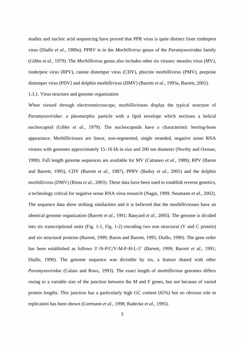
identical genome organization (Barrett et al., 1991; Banyard et al., 2005). The genome is divided
into six transcriptional units (Fig. 1-1, Fig. 1-2) encoding two non structural (V and C protein)
and six structural proteins (Barrett, 1999; Baron and Barrett, 1995; Diallo, 1990). The gene order
has been established as follows 3’-N-P/C/V-M-F-H-L-5’ (Barrett, 1999; Barrett et al., 1991;
Diallo, 1990). The genome sequence was divisible by six, a feature shared with other
Paramyxoviridae (Calain and Roux, 1993). The exact length of morbillivirus genomes differs
owing to a variable size of the junction between the M and F genes, but not because of varied
protein lengths. This junction has a particularly high GC content (65%) but no obvious role in
replication has been shown (Liermann et al., 1998; Radecke et al., 1995).

4
The nucleocapsid (N) protein: The N protein is the most abundant viral protein both in the virion
and in infected cells (Diallo et al., 1987). It directly associates with the RNA genome to form the
typical herring bone structure of morbillivirus nucleocapsids. There is a single transcription
promoter at the 3’ end, upstream to the first codon of the N gene, including the non coding part of
the N gene and a 52-56 bases leader sequence (Billeter et al., 1984, Crowley et al., 1988, Ray et
al., 1991). Various roles have been proposed for the leader RNA, including RNA binding site for
the polymerase to initiate and generate positive strand RNA replication (Fig. 1-3) (Norrby and
Oxman, 1990; Walpita, 2004), and down regulation of host cell transcription (Ray et al., 1991).
Transcription and replication of morbilliviruses are controlled by untranslated regions (UTRs) at
the 3’ and 5’ ends of the genome, known as the genome (GP) and antigenome (AGP) promoters
(Lamb and Kolakofsky, 2001). In PPRV, these are represented by nucleotides 1–107 and 15840–
15948, respectively. A conserved stretch of 23–31 nucleotides at the 3’ terminus of both the GP
and the AGP has been shown to be an essential domain required for promoter activity. The
sequence of the promoters was highly conserved in PPRV (Mioulet et al., 2001). Conserved
sequences at the junction between the GP and the N gene start, which includes the intergenic
triplet, are also required for transcription (Mioulet et al., 2001). The intergenic regions are made
up of four elements: a semi conseved polyadenylation signal, a highly conserved GAA sequence,
a semi conserved start signal for the next gene and variable length of 5’ and 3’ untranslated
regions (UTRs) (Barrett et al., 1991).
A poly U tract which, is responsible for polyadenylation of the positive sense transcripts
produced by the viral RNA-dependent RNA polymerase, was located 52 bases downstream of the
N ORF stop. This sequence is highly conserved in the morbilliviruses and acts as part of a gene
stop and polyadenylation signal. Reduction in size of the poly U tract of the paramyxovirus
simian virus 5 (SV5) from six residues to four was shown to diminish downstream initiation to
20–30% of wild-type levels indicating a possible role as a critical spacer region between gene

5
stops and starts (Rassa et al., 2000). This sequence was maintained throughout the genome of
PPRV except at the M/F, and F/H junctions where the U tract is interrupted by a G residue. It is
unknown if this G insertion has any modulating effect on the polymerase stuttering mechanism
employed to polyadenylate nascent mRNAs, or on the transcription of the downstream mRNA(s).
Immediately following the poly U tract was a conserved triplet (GAA) that has been shown to be
an intergenic region which is not transcribed during mRNA synthesis but which is an essential
signal for the activity of the viral polymerase since mutations or deletions in this region can
reduce or abrogate viral replication (Kolakofsky et al., 1998). Deletion mutagenesis studies
indicate that the 5’ UTRs for CDV and RPV F genes may serve to direct translation initiation
from a particular AUG, thus ensuring efficient translation of F protein. (Evans et al., 1990). The
rate of transcription of mRNAs from each gene is proportional to its distance from the genome
promoter, since there is a chance that at each intergenic junction the polymerase may detach from
the template and reinitiate transcription from the 3’ end (Barrett et al., 1991).
The second transcription unit encodes the P, C and V proteins. The P protein of morbilliviruses
interacts with both the N and L proteins to form the viral polymerase. The N terminus of V is
identical to P but polymerase slipping at the editing site can result in a frame shift whereby an
inserted G residue in the mRNA directs the production of an alternative C terminus (Cattaneo et
al., 1989; Wardrop and Briedis, 1991, Mahapatra et al., 2003). The hexamer phasing of the P
gene editing site is also thought to play a critical role in this process (Kolakofsky et al., 1998).
The alternative C terminus of PPRV has seven conserved cysteine residues that are thought to
interact to form a motif for binding metal ions (especially zinc). This was shown experimentally
for both Sendai virus-5 (SV5) and MV V proteins (Paterson et al., 1995). The C and V proteins
of paramyxoviruses, although essentially non-structural, have been shown to have critical roles in
infection. In RPV they were shown to be important for replication (Baron and Barrett, 2000). C-
minus mutants showed growth defects in vitro, this being related to a reduced level of mRNA

6
transcription. In contrast V-minus mutants were not defective in vitro, but had an altered
cytopathic effect and increased genome/antigenome RNA production. The C and V proteins of
paramyxoviruses also act as interferon antagonists, modifying the cellular immune response to
infection (Gotoh et al., 2001; Horvath, 2004).
The Matrix (M) proteins are basic membrane associated molecules that interact with surface
glycoproteins in the lipid envelope as well as the virion RNP. They are the most highly conserved
proteins in the Paramyxovirus family and this was reflected in the conservation of the PPRV M
protein when compared to that of other morbilliviruses. This high conservation is probably due to
the pivotal role the M protein plays in virion morphogenesis. A small protein with such a critical
role is likely to be intolerant to variation, especially within a genus whose members are
antigenically so similar. It is a non-glycosylated envelope protein thought to be involved in
nucleocapsid-envelope recognition and envelope formation during the budding process of virions
from the host cell membrane (Kingsbury, 1990). M interacts with both the nucleocapsid and the
cytoplasmic tails of the F and H glycoproteins.
The F protein is also highly conserved. Paramyxoviruses generate an inactive precursor (F0)
which is cleaved by host cell enzymes to yield an active di-sulphide linked protein F1–F2 and the
cleavage site was also conserved (Lamb and Kolakofsky, 2001). The F protein is one of two
glycosylated envelope proteins that constitute the peplomers or surface projections. Synthesized
as a precursor, F0 is subsequently cleaved by cellular proteases into two disulfide-linked
polypeptides, F1 and F2 (Sato et al., 1988). Proteolytic cleavage is believed to be essential for F
protein biologic activity.
The H protein is responsible for attachment of the virus to the host cell (Choppin and Scheid,
1980, Lamb and Kolakofsky, 2001). The biological activity of the H protein is one of the criteria
for classification of Paramyxoviridae. H proteins are highly variable (Blixenkrone- Moller et al.,
1996). Indeed, along with the P, the H is the least conserved of the morbillivirus proteins. The H

7
protein is the least conserved among CDV, RPV and MV (37% identity between CDV and MV)
(Blixenkrone-Moller et al., 1996) and 37% amino acid homology between MV and CDV (Wild
et al., 1995). Members of the Paramyxovirus genus (e.g. Newcastle disease virus) have H protein
with both hemagglutinating and neuraminidase activities (Scheid and Choppin, 1974); the
Morbillivirus H protein exhibits only hemagglutinating activity, and the H protein of members of
Pneumovirus genus (Respiratory syncytial viruses) has neither hemagglutinating nor
neuraminidase activities (Kingsbury et al., 1978).
The large (L) protein is the enzymatic component of the viral transcriptase and replicase. The L
proteins are multi-functional and, in addition to their polymerase activity, have methylation,
capping and polyadenylation activities (Lamb and Kolakofsky, 2001). Morbillivirus L proteins
have three highly conserved domains (designated A, B and C), separated by two hinge regions
which vary greatly between morbilliviruses (McIlhatton et al., 1997). The conservation of the D
and N residue in this motif is known to be an absolute requirement for polymerase activity
(Chattopadhyay et al., 2004). Specifically, the GD residues of this motif constitute part of a turn
structure that is predicted to be the core polymerase motif (Poch et al., 1990). This protein region
is involved in nucleic acid binding and is formed when leucine residues, from adjacent alpha-
helices, interdigitate to stabilise the L protein (Ramji and Foka, 2002; Vinson et al., 1989).

8
Fig. 1-1 Genome of Morbilliviruses. ( image, anonym )
9
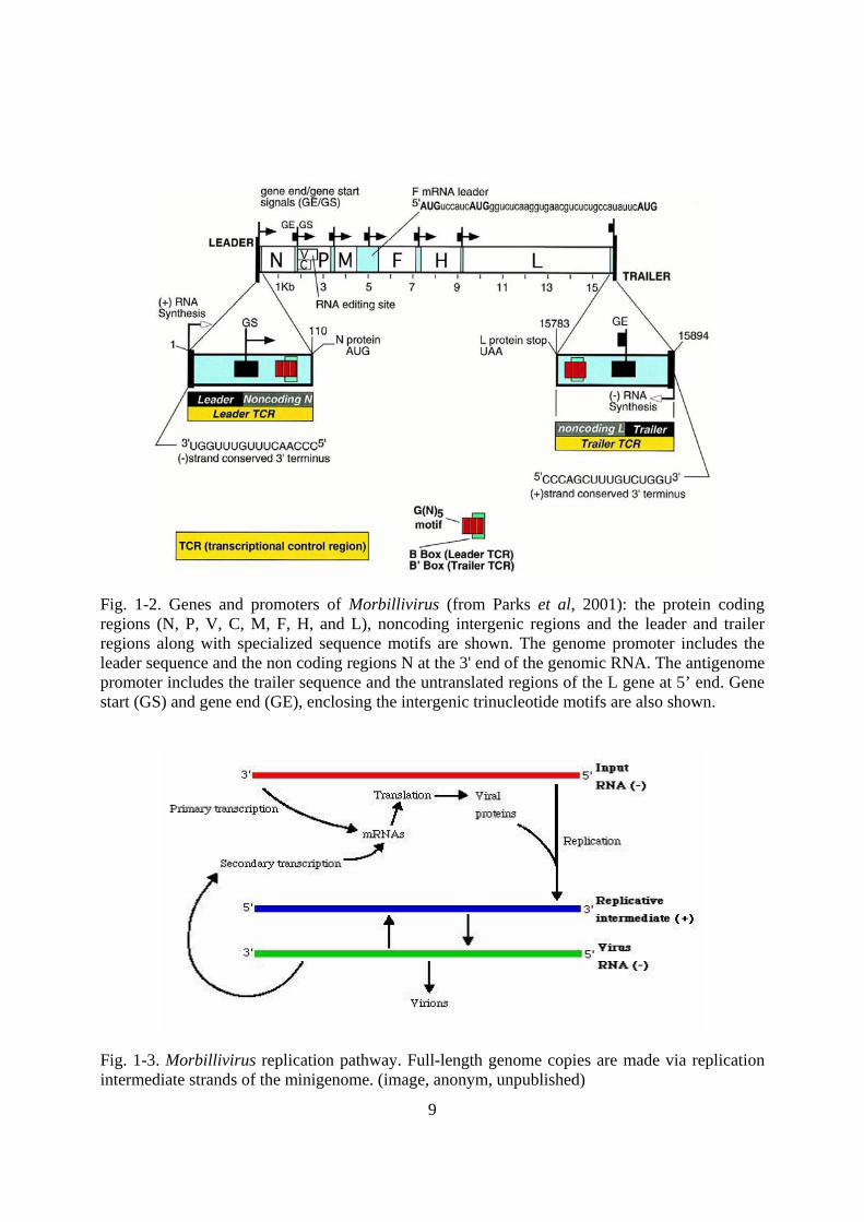
Fig. 1-2. Genes and promoters of Morbillivirus (from Parks et al, 2001): the protein coding regions (N, P, V, C, M, F, H, and L), noncoding intergenic regions and the leader and trailer regions along with specialized sequence motifs are shown. The genome promoter includes the leader sequence and the non coding regions N at the 3' end of the genomic RNA. The antigenome promoter includes the trailer sequence and the untranslated regions of the L gene at 5’ end. Gene start (GS) and gene end (GE), enclosing the intergenic trinucleotide motifs are also shown.
Fig. 1-3. Morbillivirus replication pathway. Full-length genome copies are made via replication intermediate strands of the minigenome. (image, anonym, unpublished)

10
1.4. Geographical Distribution:
PPR is known to be present in a broad belt of sub-Saharan Africa, Arabia, the Middle East and
Southern Asia. Major outbreaks in Turkey and India in recent years have indicated a marked rise
in the global incidence of PPR (Nanda et al., 1996; Ozkul et al., 2002; Shaila et al., 1996).
The virus was isolated in Nigeria (Taylor and Abegunde, 1979), Sudan (ElHag and Taylor,
1984), Saudia Arabia (Abu Elzein et al., 1990), India (Shaila et al., 1989, Nanda et al., 1996) and
Turkey (Ozkul et al., 2002). Serological evidences were detected in Syria, Niger and Jordan,
while the virus presence was confirmed with cDNA probe in Ethiopia (Roeder et al., 1994) and
Eritrea (Sumption et al., 1998), respectively. Genetic relationship between PPR viruses isolated
from different geographical regions was studied by sequence comparison of the F-protein gene.
Four lineages were revealed (Shaila et al., 1996, Dhar et al., 2002) (Fig. 1-4, Fig. 1-5). Lineage 1
is represented by viruses isolated in Africa in 1970s (Nigeria/1975/1, Nigeria/1975/2,
Nigeria1975/3, Nigeria/1976/1 and Senegalese strain). Lineage 2 which includes viruses isolated
in the late 1980s in West Africa (Ivory Coast and Guinea) is the only African lineage that did not
cross the Red Sea to the Asian countries. Lineage 3 is a combination of isolates from Sudan
(Meilig /1972) (Diallo, 1988), Ethiopia (Roeder et al., 1994). Lineage 4 of PPR virus isolates
which includes the Asian isolates from Israel/1994, Iran/1994, Nepal/1995, Bangeldesch/1993
and India (Shaila et al., 1996), is confined to Asia. Recently, it was reported in Turkey (Ozkul et
al., 2002). The presence of the two African lineages in Asia beside a distinct Asian lineage may
be taken as indication of the trade route of spread of the disease.

11
Fig. 1-4 Geographic distribution of PPRV lineages (Dhar et al., 2002)

12
Fig. (1-5) Phylogenetic relationships of the Peste des petits ruminants virus isolates based on (F)
protein gene (Ozkul et al., 2002)

13
1.5. Epidemiology
1.5.1. Transmission:
Transmission requires close contact between infected animals in the febrile stage and susceptible
animals (Braide, 1981) because of the lability of the virus outside the living host. The discharges
from eyes, nose and mouth, as well as the loose faeces, contain large amounts of the virus. Fine
infective droplets are released into the air from these secretions and excretions, particularly when
affected animals cough and sneeze (Bundza et al., 1988; Taylor, 1984). Animals in close contact
inhale the droplets and are likely to become infected. Although close contact is the most
important way of transmitting the disease, it is suspected that infectious materials can also
contaminate water and feed troughs and bedding, turning them into additional sources of
infection. These particular hazards are, however, probably fairly short-term since the PPRV, like
rinderpest, would not be expected to survive for long outside the host. Indirect transmission
seems to be unlikely in view of the low resistance of the virus in the environment and its
sensitivity to lipid solvent (Lefèvre and Diallo, 1990). There is no known carrier state for PPRV.
Trade in small ruminants, at markets where animals from different sources are brought into close
contact with one another, affords increased opportunities for PPR transmission, as does the
development of intensive fattening units.
1.5.2. Host Range and pathogenicity:
PPR is mainly a disease of small ruminants. It affects goats and sheep. PPR virus exhibits
different levels of virulence between sheep and goats. Goats are severely affected while sheep
generally undergo a mild form (Lefèvre and Diallo, 1990). Although infected, sheep rarely suffer
clinical disease (El Hag Ali and Taylor, 1988; Roeder et al., 1994). An outbreak with a high
mortality in sheep was reported by Taylor (1984) who hypothesised that sheep possessed an
innate resistance to the clinical effects of disease, but occasional field strains could overcome this
resistance and produce high mortality (Taylor, 1984). Breed may affect the outcome of PPR virus

14
infection and its epidemiology, the Guinean breeds (West African dwarf, Iogoon, kindi and
Djallonke) are known to be highly susceptible (Lefèvre and Diallo, 1990). This is in agreement
with the finding that British breed exhibited severe clinical reaction when infected experimentally
while the Sudanese breeds failed to develop a characteristic clinical response (El Hag and Taylor,
1984). A more recent observation detected variations in breed susceptibility within goats in West
Africa. The acute form of the disease was observed in WAD goats while WALL breed developed
only mild form (Diop et al., 2005). (Fig. 1-6 and Fig. 1-7).

15
Fig. 1-6 PPR resistance goat breeds in sahelian region (Photo by Dr V. Martin)
Fig. 1-7 PPR resistance sheep in West Africa (Photo, anonym, Dakar, Senegal)

16
In India and the Middle East both goats and sheep are affected with equally devastating
consequences. In India, morbidity and case fatality reach 10 and 25% respectively in flocks of
indigenous sheep (Shaila et al., 1989). The outbreak will not involve cattle, whether rinderpest
vaccinated or not, even if they are in contact with affected goats and sheep. Cattle and pigs are
known to be a dead end host and all attempt to induce clinical disease in adult cattle
experimentally failed; they undergo a silent or subclinical infection that protect them against
subsequent challenge with virulent strain of RP (Gibbs et al., 1979; Taylor, 1984). Sero-
neutralization test for the presence of PPR antibodies detected 4.2% in 142 camels (Ismail et al.,
1995). PPR affect wildlife animals both under field condition and experimentally. The disease
was induced experimentally in American white deer (Odocoileus virginianus) which was found
to be susceptible (Hamdy et al., 1976) and a field outbreak was reported from a zoological
collection in Alain (Furley et al., 1987). It caused a high mortality and severe disease in Dorcas
Gazelles (Gazella dorcas), Nubian Ibex (Capra ibex nubiana), Laristan sheep (Ovis orientalis
laristani) and gemsbok (Oryx gazellaa). Subclinical involvement of Nigale (Tragelaphinae) was
suspected. In another report from Saudi Arabia, PPR was suspected on clinical and serological
base in Gazaelle and deer (Abu Elzein et al., 1990). Antelope and other small wild ruminant
species can also be severely affected (Abu Elzein et al., 2004).

17
1.5.3. Pattern of the disease:
In general, a morbidity is common, particularly in fully susceptible goat populations. Milder
forms of the disease may occur in sheep and partially immune goat populations. There are
considerable differences in the epidemiological pattern of the disease in the different ecological
systems and geographical areas. In the humid Guinean zone where PPR occurs in an epizootic
form, it may have dramatic consequences with morbidity of 80%-90% accompanied with a
mortality between 50 and 80% (Lefèvre and Diallo, 1990). While in arid and semi-arid regions,
PPR is seldomly fatal but usually occurs as a subclinical or inapparent infection opening the door
for other infections such as Pasteurellosis (Lefèvre and Diallo, 1990). Though outbreaks in West
Africa coincide with the wet rainy season, Opasina and Putt (1985) observed outbreaks during
the dry season in two different ecological zones. A high morbidity of 90% accompanied with
70% case fatality was reported from Saudi Arabia (Abu Elzein et al., 1990).
Serological data from Nigeria revealed that antibodies occur in all age groups from 4-24 months
indicating a constant circulation of the virus (Taylor, 1979b). In Oman the disease persisted on a
year round basis maintaining itself in the susceptible yearling population (Taylor et al., 1990).
Therefore, an increase in incidence reflects an increase in number of susceptible young goats
recruited into the flocks rather than seasonal upsurge in the virus activity, since its upsurge pend
on the peak of kidding seasons (Taylor et al., 1990). Moreover, the susceptibility of young
animals aged 3 to 18 months was proved to be very high, being more severely affected than
adults or unweaned animals (Taylor et al., 1990).

18
1.6. Clinical Signs
Clinical signs of PPR have been well documented (Hamdy et al., 1976; Obi, 1984; Lefèvre,
1987; Taylor, 1984; Bundza et al., 1988; Roeder et al., 1994; Roeder and Obi, 1999).
Introduction of PPR into a flock may be associated with any of the following:
• history of recent movement or gathering together of sheep and/or goats of different ages
with or without associated changes in housing and feeding;
• introduction of recently purchased animals; contact in a closed/village flock with sheep
and/or goats that had been sent to market but returned unsold;
• change in weather such as the onset of the rainy season (hot and humid) or dry, cold
periods, contact with trade or nomadic animals through shared grazing, water and/or
housing;
• a change in husbandry (e.g. towards increased intensification) and trading practices.
Following infection there is a 3–4 day incubation period during which the virus replicates in the
draining lymph nodes of the oro-pharynx before spreading via the blood and lymph to other
tissues and organs including the lungs causing a primary viral pneumonia. The predominant
form of the disease is the acute form. The salient clinical signs start with sudden rise in body
temperature to 39.5 - 41°C. Affected animals breathe fast, sometimes so fast that they exhibit
rocking movements with both the chest and abdominal walls moving as the animal breathes.
Severely affected cases show difficult and noisy breathing marked by extension of the head and
neck, dilation of the nostrils, protrusion of the tongue and soft painful coughs. They have
obvious signs of pneumonia. A clear watery discharge starts to issue from the eyes, nose and
mouth, later becoming thick and yellow as a result of secondary bacterial infection. Appearance
of a serous to mucopurulent nasal discharge which may crust over and occlude the nostril,
sneezing, ocular discharge resulting in matting of the eyelids. The discharges wet the chin and

19
the hair below the eye; they tend to dry, causing matting together of the eyelids, obstruction of
the nose and difficulty in breathing (Fig. 1-8). Unlike RP, there is a definite but inconstant,
respiratory system component (Brown et al., 1991; Bundza et al., 1988). One to two days after
fever has set in, the mucous membranes of the mouth and eyes become very reddened. Then,
epithelial necrosis causes small pin-point greyish areas on the gums, dental pad, palate, lips,
inner aspects of the cheeks and upper surface of the tongue. These areas increase in number and
size and join together. The lining of the mouth is changed in appearance. It becomes pale and
coated with dying cells and, in some cases, the normal membrane may be completely obscured
by a thick cheesy material. Underneath the dead surface cells, there are shallow erosions. Gentle
rubbing across the gum and palate with a finger may yield a foul-smelling material containing
shreds of epithelial tissue (Braide, 1981) (Fig. 1-9). Body temperature usually remains high for
about 5-8 days, and then slowly returns to normal prior to recovery or drops below normal
before death (Fig. 1-10).

20
Fig. 1-8 Clinical signs, discharges (Photo, Abraham and Roeder, Ethiopia)
Fig. 1-9 Mouth lesions (Photo, Abraham and Roeder, Ethiopia)
Fig. 1-10 Body temperature fluctuation and phases of clinical disease (Scott et al., 1986)

21
Diarrhoea commonly appears about two to three days after the onset of fever although, in early
or mild cases, it may not be obvious. The faeces are initially soft and then watery, foul-smelling
and may contain blood streaks and pieces of dead gut tissue. Where diarrhoea is not an obvious
presenting sign, the insertion of a cotton wool swab into the rectum may reveal evidence of soft
faeces which may be stained with blood. Such victims may eventually become dehydrated with
sunken eyeballs, and death often follows within seven to ten days from onset of the clinical
reaction. Other animals will recover after a protracted convalescence.
The affected animals had lymphocytopenia, elevated PCV (above 60% while normal 35-45%),
very high RBCs count while the level of hemoglobin and the white blood cells was normal
(Furley et al., 1987). A common feature in later stages of the sub-acute disease is the formation
of small nodular lesions in the skin on the outside of the lips around the muzzle. The exact cause
of this is not known.
1.7. Pathology
Pathogenesis: PPR virus, like other morbilliviruses, is lymphotropic and epitheliotropic (Scott,
1981). Consequently, it induces the most severe lesions in organ systems rich in lymphoid and
epithelial tissues. The respiratory route is the likely portal to entry. After the entry of the virus
through the respiratory tract system, it localizes first replicating in the pharyngeal and
mandibular lymph nodes as well as tonsil. Viremia may develop 2-3 days after infection and 1-2
days before the first clinical sign appears. Subsequently viremia results in dissemination of the
virus to spleen, bone marrow and mucosa of the gastro-intestinal tract and the respiratory system
(Scott, 1981).
Post mortem findings: The carcass of an affected animal is usually emaciated, the hindquarters
soiled with soft/watery faeces and the eyeballs sunken. The eyes and nose contain dried-up
discharges. Lips may be swollen; erosions and possibly scabs or nodules in late cases. The nasal
cavity is congested (reddened) lining with clear or creamy yellow exudates and erosions. They

22
may be dry with erosions on the gums, soft and hard palates, tongue and cheeks and into the
oesophagus. The lung is dark red or purple with areas firm to the touch, mainly in the anterior
and cardiac lobes (evidence of pneumonia). Lymph nodes (associated with the lungs and the
intestines) are soft and swollen. Abomasum congested with lining haemorrhages.
The pathology caused by PPR is dominated by necrotizing and ulcerative lesions in the mouth
and the gastro-intestinal tract (Roeder et al., 1994). Erosion in the oral cavity is a constant
feature. The rumen reticulum and omasum rarely exhibit lesions. Occasionally, there may be
erosions on the pillars of the rumen. The omasum is a common site of regularly outlined
erosions often with oozing blood. Lesions in the small intestine are generally moderate, being
limited to small streaks of hemorrhages and, occasionally, erosions in the first portions of the
duodenum and the terminal ileum. The large intestine is usually more severely affected, with
congestion around the ileo-cecal valve, at the ceco-colic junction and in the rectum. In the
posterior part of the colon and the rectum, discontinuous streaks of congestion “zebra stripes”
form on the crests of the mucosal folds.
In the respiratory system, small erosion and petechiae may be visible on the nasal mucosa,
turbinates, larynx and trachea. Bronchopneumonia may be present, usually confined to the
anteroventral areas, and is characterized by consolidation and atelectasis.
Histopathology: PPR virus causes epithelial necrosis of the mucosa of the alimentary and
respiratory tracts marked by the presence of eosinophilic intracytoplasmic and intranuclear
inclusion bodies. Multinucleated giant cells (syncytia) can be observed in all affected epithelia
as well as in the lymph nodes (Brown et al., 1991). In the spleen, tonsil and lymph nodes, the
virus causes necrosis of lymphocytes evidenced by pyknotic nuclei and karyorrhexis (Rowland
et al., 1971). Brown and others (1991) using immunohistochemical methods detected viral
antigen in both cytoplasm and nuclei of tracheal, bronchial and bronchio-epithelial cell, type II
pneumocytes, syncytial cells and alveolar macrophages.

23
Small intestines are congested with lining haemorrhages and some erosions. Large intestines
(caecum, colon and rectum) have small red haemorrhages along the folds of the lining, joining
together as time passes and becoming darker, even green/black in stale carcasses.
1.8. Immunity:
The surface glycoproteins hemagglutinin (H) and fusion protein (F) of morbilliviruses are highly
immunogenic and confer protective immunity. PPRV is antigenically closely related to rinderpest
virus (RPV) and antibodies against PPRV are both cross-neutralizing and cross protective
(Taylor, 1979a). A vaccinia virus double recombinant expressing H and F glycoproteins of RPV
has been shown to protect goats against PPR disease (Jones et al., 1993) though the animals
developed virus-neutralizing antibodies only against RPV and not against PPRV. Capripox
recombinants expressing the H protein or the F protein of RPV or the F protein of PPRV
conferred protection against PPR disease in goats, but without production of PPRV-neutralizing
antibodies (Romero et al., 1995) or PPRV antibodies detectable by ELISA (Berhe et al, 2003).
These results suggested that cell-mediated immune responses could play a crucial role in
protection. Goats immunized with a recombinant baculovirus expressing the H glycoprotein
generated both humoral and cell-mediated immune responses (Sinnathamby et al., 2001). The
responses generated against PPRV-H protein in the experimental goats are also RPV cross-
reactive suggesting that the H protein presented by the baculovirus recombinant ‘resembles’ the
native protein present on PPRV (Sinnathamby et al., 2001). Lymphoproliferative responses were
demonstrated in these animals against PPRV-H and RPV-H antigens (Sinnathamby et al., 2001).
N-terminal T cell determinant and a C-terminal domain harboring potential T cell determinant(s)
in goats was mapped (Sinnathamby et al., 2001). Though the sub-set of T cells (CD4+ and CD8+
T cells) in PBMC that responded to the recombinant protein fragments and the synthetic peptide
could not be determined, this could potentially be a CD4+ helper T cell epitope, which has been
shown to harbor an immunodominant H restricted epitope in mice (Sinnathamby et al., 2001).

24
Identification of B- and T-cell epitopes on the protective antigens of PPRV would open up
avenues to design novel epitope based vaccines against PPR.
Sheep and goats are unlikely to be infected more than once in their economic life (Taylor, 1984).
Lambs or kids receiving colostrum from previously exposed or vaccinated with RP tissue culture
vaccine were found to acquire a high level of maternal antibodies that persist for 3-4 months. The
maternal antibodies were detectable up to 4 months using virus neutralization test compared to 3
month with competitive ELISA (Libeau et al., 1992). Measles vaccine did not protect against
PPR, but a degree of cross protection existed between PPR and canine distemper (Gibbs et al.,
1979).
Though PPR disease can be effectively controlled by RPV vaccine, rinderpest eradication
programmes have been launched in many countries and if these campaigns are successful, Office
International des Epizooties (OIE) recommends the cessation of vaccination of all the animals
with RPV vaccine so that any residual foci of RPV could be identified. Under these
circumstances, small ruminants could only be protected against PPR by using homologous
attenuated vaccine. In addition, the successful use of an attenuated PPRV vaccine against RPV
has been reported in goats, opening the possibility to use it as a differentiable vaccine for cattle
(Couacy-Hymann et al., 1995).
1.9. Diagnosis
Goats and sheep can be infected with RP and PPR as well. Clinical differential diagnosis is not
possible as similar disease is produced by both viruses in small ruminants. Therefore, tentative
clinical diagnosis may have to be confirmed by laboratory analysis. Diagnosis of PPR may be
performed by virus isolation, detection of viral antigens, and nucleic acid sequencing and
detection of specific antibody in serum.
1.9.1. Virus isolation

25
Samples for virus isolation include heparinized blood, eye and nasal swabs (from live animals),
tonsil, mesenteric lymph nodes, spleen, section of colon and lung. For successful isolation,
samples must be collected during the hyperthermic phase (Lefevre, 1987) and submitted to the
testing laboratory in cold ice. The most widely used cell culture systems are primary lamb kidney
and ovine skin (Gilbert and Monnier, 1962; Laurent, 1968, Taylor and Abegune, 1979) and Vero
cells (Hamdy et al., 1976).
The sensitivity of virus isolation technique could be increased when the virus is grown in lamb
and goats kidney cells (Taylor, 1984). Vero cells are however widely used for their continuity
and low liability of contamination. PPRV has also been adapted to grow in other continuous cell
lines including MDBK and BHK-21 (Lefèvre, 1987). Vero cells, derived from African green
monkey kidney are currently the most widely used cell line for PPRV and RPV. A culture of
Vero cells from American type cell culture (ATCC # CCL81) was found to yield very high titres
and is currently used in many laboratories working on PPRV and RPV. Appearance of cytopathic
effects (CPE) may require at least 8-10 days or several blind passages. In Vero cells, the
cytopathic effects (CPE) produced by PPRV consist of cell rounding, clumping into typical
grape-like clusters, formation of small syncytia and appearance of long fine often anastomosing
“spindle cells” (Hamdy et al., 1976). Like other morbilliviruses, PPRV produces eosinophilic
intracytoplasmic and intranuclear inclusion bodies both in primary cells (Laurent, 1968) and
continuous cell lines (Hamdy et al., 1976).
T-lymphoblast cell line transformed by Theileria parva proved to be more sensitive when
compared to other cell culture and gave a result within 24 hours (at least 6 days for other cell
culture) for both PPRV and RPV (Rossiter, 1994).
Once isolated in cell culture, a candidate PPRV may be identified by one of the three procedures:
• animal inoculation: PPR causes clinical disease in goats and sheep but not in cattle (Gibbs
et al., 1979);

26
• reciprocal cross neutralization (differential neutralization): PPRV is neutralized by both
PPR and RPV reference sera, but is neutralized at greater titre with the homologous serum
(Taylor and Abegunde and, 1979, Taylor, 1979a);
• molecular techniques: cDNA probe, (Diallo et al., 1989a, Pandey et al., 1992),
electrophoretic profile in polyacrylamide gel (PAGE) (Diallo et al., 1987) and PCR,
(Barret et al., 1993, Forsyth et al., 1995, Couacy-Hymann et al., 2002).
1.9.2. Antigen detecting methods:
1.9.2.1. Agar Gel Immunodiffusion Test
Agar gel immunodiffusion test (AGID) is widely used and can detect 42.6% of antemortem
specimens and necropsy specimens (Obi, 1984; Abraham and Berhan, 2001). It can be used to
test the presence of both antigen and antibodies and can give results within 2-4 hours when RP
hyperimmune serum is used while it needs 4-6 hours with PPR hyperimmune serum (Obi, 1984).
One of the important advantages of this test that it is highly specific (92%), though it can not
differentiate between PPR and RP.
1.9.2.2. Hyperimmune serum:
Standard antiserum is made by immunising sheep with 5 ml of PPR virus with a titre of 104
TCID50 (50% tissue culture infective dose) per ml given at weekly intervals for 4 weeks. The
animals are bled 5-7 days after the last injection. Standard RP hyperimmune antiserum is also
effective in detecting PPR antigen.
1.9.2.3. Counter immunoelectrophoresis
Counter immunoelectrophoresis (CIEP) is in the same principle as the AGID except that the gel
is electrically charged to improve the sensitivity of the test.
1.9.2.4. ELISA for antigen detection:
A monoclonal antibody-based sandwich ELISA was found to be highly sensitive in detection of
antigen in tissues and secretions of infected goats (Saliki et al., 1994). Another format of antigen
27
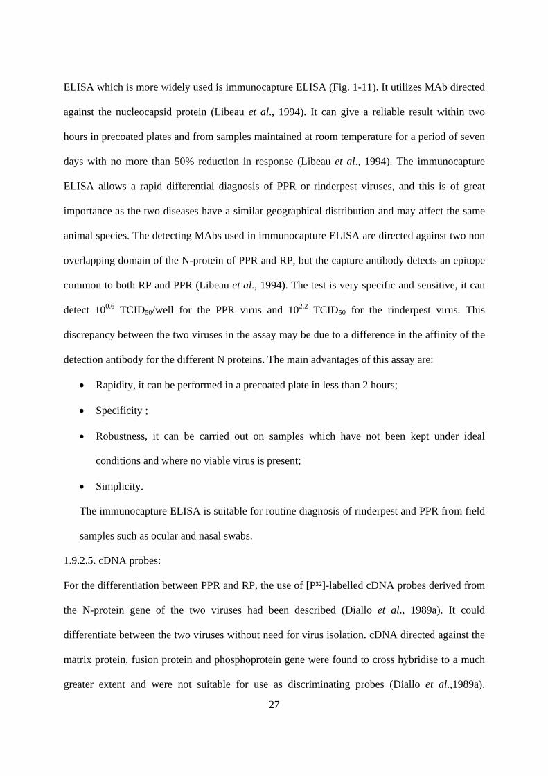
ELISA which is more widely used is immunocapture ELISA (Fig. 1-11). It utilizes MAb directed
against the nucleocapsid protein (Libeau et al., 1994). It can give a reliable result within two
hours in precoated plates and from samples maintained at room temperature for a period of seven
days with no more than 50% reduction in response (Libeau et al., 1994). The immunocapture
ELISA allows a rapid differential diagnosis of PPR or rinderpest viruses, and this is of great
importance as the two diseases have a similar geographical distribution and may affect the same
animal species. The detecting MAbs used in immunocapture ELISA are directed against two non
overlapping domain of the N-protein of PPR and RP, but the capture antibody detects an epitope
common to both RP and PPR (Libeau et al., 1994). The test is very specific and sensitive, it can
detect 100.6 TCID50/well for the PPR virus and 102.2 TCID50 for the rinderpest virus. This
discrepancy between the two viruses in the assay may be due to a difference in the affinity of the
detection antibody for the different N proteins. The main advantages of this assay are:
• Rapidity, it can be performed in a precoated plate in less than 2 hours;
• Specificity ;
• Robustness, it can be carried out on samples which have not been kept under ideal
conditions and where no viable virus is present;
• Simplicity.
The immunocapture ELISA is suitable for routine diagnosis of rinderpest and PPR from field
samples such as ocular and nasal swabs.
1.9.2.5. cDNA probes:
For the differentiation between PPR and RP, the use of [P³²]-labelled cDNA probes derived from
the N-protein gene of the two viruses had been described (Diallo et al., 1989a). It could
differentiate between the two viruses without need for virus isolation. cDNA directed against the
matrix protein, fusion protein and phosphoprotein gene were found to cross hybridise to a much
greater extent and were not suitable for use as discriminating probes (Diallo et al.,1989a).

28
Unfortunately, this hybridization cannot be used widely because it requires fresh specimens and
in addition to the short half life of [P³²], there is constraints with the handling of isotopes.
Therefore, probes using non radioactive labels such as biotin (Pandey et al., 1992) or dioxin
(Diallo et al., 1995) were developed. The biotin labeled cDNA was found to be as specific as the
one using the radioactive label and more rapid in differentiation between PPR and RP (Pandey et
al., 1992). However, it was reported elsewhere, that the expected sensitivity had never been
obtained using non-radioactive labels (Diallo et al., 1995).
1.9.2.6. Reverse transcription polymerase chain reaction (RT-PCR)
Conventional serological techniques and virus isolation are normally used to diagnose
morbillivirus infection in samples submitted for laboratory diagnosis. However, such techniques
are not suitable for use on decomposed tissue samples, the polymerase chain reaction (PCR), has
proved invaluable for analysis of such poorly preserved field samples.
Saiki and others (1988) first demonstrated the efficiency of amplifying in vitro a selected
sequence flanked by two oligonucleotide primers of opposite orientation. The method consists of
repetitive cycles of DNA denaturation, primer annealing and extension by a DNA polymerase
effectively doubling the target with each cycle leading, theoretically, to an exponential rise in
DNA product. The replacement of the polymerase Klenow fragment by thermostable polymerase
derived from Thermus aquaticus (Taq) has greatly improved the usefulness of PCR. Using this
system, a rate of amplification up to 107 to 109 times has been reported. The efficiency achieved
actually can vary enormously, however, since it is dependent on factors such as the number of
cycles, the quantity of the starting material, the length of the target DNA, the temperature
conditions of annealing and priming, and the polymerase used. When the starting material is
DNA, high purification of the nucleic acid is not necessary so the procedure is greatly simplified.
These qualities have made the PCR one of the essential techniques in molecular biology today
and it is starting to have a wide use in laboratory disease diagnosis.

29
Since the genome of all morbilliviruses consists of a single strand of RNA, it must be first copied
into DNA, using reverse transcriptase, in a two-step reaction known as reverse
transcription/polymerase chain reaction (RT-PCR). RT-PCR has been shown to be useful for the
rapid detection of morbillivirus-specific RNA in samples submitted for laboratory diagnosis
(Shaila et al., 1996). It has proved especially useful in identifying the new morbilliviruses found
in marine mammals (Barrett et al., 1993b). Both genus-specific and universal morbillivirus
primer sets have been produced that can be used to distinguish all known morbilliviruses (Forsyth
and Barrett, 1995).
Two sets of primers have been made, based on sequences in the 3’ end of N genes (messenger
sense), which are least conserved regions between the two viruses. They enable specific
amplification of 300 base pair (bp) fragments for RPV and PPRV (Couacy-Hymann et al., 2002).
Reverse transcription-polymerase chain reaction tests (RT-PCR) using phosphoprotein (P)
universal primer and fusion (F) protein gene specific primer sets to detect and differentiate
between PPR and RP were described (Barrett et al., 1993b; Forsyth and Barret, 1995; Couacy-
Hymann et al., 2002).
1.9.3. Serology:
Many tests have been used for the demonstration of PPR antibodies in serum: virus neutralization
test, agar gel diffusion test, immunoelectrophoresis and recently blocking and competitive
ELISA.
1.9.3.1. Virus neutralisation:
The virus neutralisation test (VNT) is sensitive and specific, but time-consuming and expensive.
The standard neutralisation test is carried out in roller-tube cultures of primary lamb kidney cells
or Vero cells when primary cells are not available. VNT is the most reliable test for detection of
morbillivirus antibodies (Rossitter, 1994). Serum against either PPR or RP may neutralise both

30
viruses, but would neutralize the homologous virus at a higher titre than the heterologous virus.
Therefore for differentiation purpose reciprocal cross neutralization is used (Taylor and
Abegunde, 1979).
1.9.3.2. cELISA
Competitive and blocking ELISA based on monoclonal antibodies specific for N-protein (Libeau
et al., 1995) and H-protein (Anderson and Mckay, 1994; Saliki et al., 1993; Singh et al., 2004)
were developed for detection of antibodies in animal sera. These tests either used gradient
purified virus or expressed antigens. In the N-protein cELISA, the serum antibodies and the MAb
compete on specific epitope on nucleoprotein obtained from recombinant baculovirus. Though no
cross reaction in N-protein cELISA was reported, a high level of competition up to 45% was
observed among the negative (Libeau et al., 1995). Despite the fact that neutralizing antibodies
are not directed against the N-protein, but the H-protein (Diallo et al., 1995), a correlation of 0.94
between VNT and cELISA was observed suggesting that the former was more sensitive (Libeau
et al., 1995). The relative sensitivity of this cELISA to VNT was 94.5, while the specificity was
99.4%. Both blocking ELISA and cELISA detecting anti-H antibodies are based on competition
between an anti-H monoclonal antibody (MAb) and serum antibodies, but in case of blocking
ELISA the test sera are preincubated with antigen and then incubated with the MAb (Saliki et al.,
1993). The sensitivity and specificity of the H-blocking ELISA were found to be 90.4% and
98.9% respectively (Saliki et al., 1993). PPR cELISA using MAb directed against the H-protein
cross reacted to some extent with rinderpest, while RP cELISA is specific, therefore an animal
was assumed to have experienced RP if it is positive in both PPR and RP ELISA (Anderson and
McKay, 1994). The protocol of cELISA is illustrated in Fig. 1-13. The absorbance in PPR ELISA
is converted to percentage of inhibition (PI) using the formula:
PI=100-(absorbance of the test wells/ absorbance of the mAb control wells) x 100. Sera showing
PI greater than 50% are scored positive. The overall specificity of c-ELISA test was 98.4% with a

31
sensitivity of 92.2% when compared with VNT. The diagnostic efficacy of the assay in terms of
sensitivity and specificity was calculated using two-sided contingency table (Singh et al., 2004).
Sensitivity of the assay was taken as proportion of positive samples out of actual positive
samples. Specificity was calculated as proportion of negative samples out of total negative
samples. The anti-H RP cELISA has been successfully used for serological monitoring of post
vaccination herd immunity in the Pan African Rinderpest Campaign (PARC) project to control
and eradicate rinderpest from the African continent and which later became part of the FAO
Global Rinderpest Eradication Project (GREP). Its wide spread use in (OIE) rinderpest
serological surveillance run into controversial difficulties as apparent missing of antibody
detection in rinderpest lineage II.

32
Fig. 1-11 Immunocapture ELISA (Image, Dr G. Libeau)

33
Fig. 1-12 PCR reaction steps (Image, anonym, unpublished)

34
Fig. 1-13. Indirect and Competitive ELISA for antibody detection (Dr G. Libeau)

35
1.10.Control and prophylaxis:
There is no specific treatment against the disease. Control of PPR in non infected countries may
be achieved using classical measures such as restriction of importation of sheep and goats from
affected areas, quarantine, slaughter and proper disposal of carcasses and contact fomites and
decontamination of affected premises in case of introduction. Control of PPR outbreaks can also
rely on movement control (quarantine) combined with the use of focused ("ring") vaccination and
prophylactic immunization in high-risk populations. Immunization of small ruminants with
lymph node and spleen materials containing virulent virus inactivated with 1.5-5% chloroform
was tried and the animals were immune to subsequent challenge 18 months latter (Braide, 1981).
Until recently, the most practical vaccination against PPR was based on the use of tissue culture
adapted rinderpest vaccine. Vaccination of animals with RP attenuated virus has been practiced
for a long time. The tissue culture rinderpest vaccine (TCRV) at a dose of 102.5 TCID50 protected
goats against PPR for 12 months and the animals were not able to transmit the infection
following challenge with PPR virus (Taylor, 1979a), although the antigen was detected in
lachrymal swabs from vaccinated animals after challenge with virulent virus (Gibbs et al., 1979).
However, it was reported previously that considerable residues of virulence were detected after
32, 42, even 65 serial passages in embryonic lamb kidney cells (Taylor, 1979a). This vaccine was
successfully used to control PPR in some countries in west Africa (Bourdin, 1973) and is widely
used in many African countries (Lefèvre and Diallo, 1990). It has been withheld from being used
because of its interference with the Pan-African Rinderpest Campaign (PARC), since it is
impossible to determine if seropositive small ruminants have been vaccinated or naturally
infected with RPV. Sera from animals vaccinated with RP vaccine contain substantial level of RP
antibodies with little or no cross neutralising antibodies to PPR but after challenge with PPR,
neutralizing antibodies to PPR increase sharply. RP thermostable vaccine was developed for

36
protection of goats against PPR (Stem, 1993). Homologous PPR vaccine attenuated after 63
passages in vero cell (Diallo et al., 1989b) was used and produced a solid immunity for 3 years
(Diallo et al., 1995). The PPRV homologous vaccine was found to be safe under field conditions
even for pregnant animals and it induced immunity in 98% of the vaccinated animals (Diallo et
al., 1995). The PPRV vaccine has been tried for protection of cattle against RP and it was found
very effective (Couacy-Hymann et al., 1995). PPR vaccine seed is available through the Pan
African Veterinary Vaccine Centre (PANVAC) at Debre Zeit, Ethiopia, for Africa, or CIRAD-
EMVT at Montpellier, France, for other areas.
1.11. Disease Economy:
The PPR epidemics can cause mortality rates of 50–80% in naive sheep and goats populations
(Kitching, 1988). Due to the confusion with other diseases, the economic impacts of PPR are
probably underestimated, but it is believed that PPR is one of the major constraints of small
ruminant farming in the tropic (Taylor, 1984). Based on assumption that goats experience an
outbreak every 5 years, Opasina and Putt (1985) estimated an annual sum ranging from 2.47£ per
goat at high loss and 0.36 £ per goat at lowest. The loss due to PPR in Nigeria was estimated to
be 1.5 million dollars annually (Hamdy et al., 1976). The economic losses due to PPR alone in
India have been estimated annually to 1,800 millions Indian Rupees (39 millions US$)
(Bandyopadhyay, 2002). An economic analysis for assessing benefits of vaccination against PPR
in Niger revealed that such a programme was highly beneficial with an anticipated net present
value (NPV) return in five years of 24 millions USD following an investment of two millions
USD.

37
Chapter 2
Comparative biology of PPRV among other morbilliviruses
2.1 Introduction
Peste des petits ruminants virus (PPRV) is a very recent addition to the Morbillivirus genus in
comparison to measles virus (MV), canine distemper virus (CDV) and rinderpest virus (RPV).
The disease PPR was described as a clinical entity only in 1942 (Gargadennec and Lalanne,
1942). Measles, canine distemper and rinderpest have been known to exist for several centuries
(Appel et al., 1981). PPRV was isolated in cell culture (Gilbert and Monnier, 1962) at least 10
years after the three other morbilliviruses were cultured in the early 1950s. For a long time after
the description of PPR and even after the agent was isolated, it was thought to be a variant of RP
that was adapted to goats and sheep and had lost its virulence for cattle (Laurent, 1968).
PPR is an important disease in its own right, but it has also created problems because of its
apparent similarity to rinderpest. The clinical signs of rinderpest in small ruminants closely
resemble those of PPR, making differential diagnosis difficult. It should, however, be kept in
mind that clinical disease caused by rinderpest in small ruminants is a relatively rare event, even
in Asia.
2.2 Pathogenicity and host range
Since morbilliviruses do not persist in an infectious form following an acute infection, and
infection results in life-long immunity in recovered hosts, the virus relies on a constant supply of
new susceptible hosts for its maintenance. It has been estimated that a population of at least
300,000 individuals is required to maintain MV in circulation (Black, 1991). Each of the
morbilliviruses has its different natural host ranges (Appel et al., 1981). MV causes disease only
in primates. Non-human primates are highly susceptible to MV, but their numbers are too small
to maintain the virus in circulation and infection occurs through contact with humans. Canine

38
distemper virus is naturally pathogenic in the Canidae (dog, wolf, fox), Mustilidae (ferret, mink,
weasel), Tayassuidae (javelins) (Appel et al., 1991) and Procyonidae (raccoon) families. The
recent outbreaks of distemper in seals in Lake Baikal (Visser et al., 1993), in lions in the
Serengeti National Park (Roelke-Parker et al., 1996), and in leopards and other large cats in zoos
(Appel et al., 1994) have underscored the ability of CDV to invade new host species. Phocine
distemper virus, the most recently described member of the Morbillivirus genus can infect many
species of seal (Duignan et al., 1995; Osterhaus, 1992). Little is known of the host range of the
cetacean morbillivirus, but serological evidence for its presence in many species of cetacean has
been obtained (Duignan et al., 1995). Rinderpest virus is primarily a pathogen of cattle and water
buffaloes but also could cause disease in goats, sheep, and rarely in pigs (Murphy et al., 1999)
and most species belonging to other Artiodactyla. Dogs fed infected meat can develop antibodies
to RPV, indicating a subclinical infection (Rossiter, 1994). Peste des petits ruminants virus
occurs in goats and sheep but it has been described in captive wild small ruminants belonging to
three families: Gazellinae (dorcas gazelle), Caprinae (Nubian ibex) and Hippotraginae (gemsbok)
(Furley et al., 1987).
Another difference among morbilliviruses is the ability of some members to establish persistent
infections in their natural hosts. Acute MV infection occurs mainly in childhood and is
characterized by fever, cough, coryza, conjunctivitis and skin rash. In about 1/ 106 cases, a fatal
degenerative neurological condition known as subacute sclerosing panencephalitis (SSPE) may
occur 4-10 years after the acute clinical disease of measles (Appel et al., 1981). A related
condition designated measles inclusion body encephalitis (MIBE) has also been described
(Norrby and Oxman, 1990). Similarly, a very rare but fatal progressive motor and mental
deteriorating condition known as old dog encephalitis (ODE) has been described in dogs many
years after acute CDV infection (Appel et al., 1981). No carrier state or persistent infections have
been described for the RPV, PPR or CDV.

39
Cellular receptors and tropism: Cellular receptors are one of the major determinants of the host
range and tissue tropism of a virus. Human signaling lymphocyte activation molecule (SLAM;
also known as CD150), a membrane glycoprotein expressed on some lymphocytes and dendritic
cells (Cocks et al., 1995), is a cellular receptor for MV (Tatsuo et al., 2000). The tissue
distribution of human SLAM can explain the pathology of measles. Selective infection and
destruction of SLAM positive cells may be the principal mechanism underlining the
immunosuppressive effect of morbilliviruses in general (Tatsuo et al., 2000). The target cells for
RPV are epithelial cells, activated lymphocytes, and macrophages (Rey Nores et al., 1995). Field
isolates of CDV also replicate in dog or ferret macrophages (Brugger et al. 1992) as well as in
primary dog brain cell cultures (Zurbriggen et al., 1987). Cell lines such as Vero (African green
monkey kidney) which use mainly CD46 receptors do not allow the propagation of field isolates,
whereas cell culture adapted CDV strains such as the Onderstepoort vaccine strain are able to
replicate in many cell lines (Appel and Gillespie, 1972). It is known that virulence for the natural
host may be lost when CDV is adapted to cell culture (Harrison et al., 1968). Furthermore, the
marmoset B cell line B95a, which is commonly used to isolate MV from clinical specimens
(Kobune et al., 1990) expresses a high level of SLAM on the cell surface (Tatsuo et al., 2000).
B95a cells have been shown to be very sensitive to CDV and RPV (Kai et al., 1993, Kobune et
al., 1991). Vero, 293T (human kidney), and L (mouse fibroblast) cells developed syncytia after
transfection with the H and F genes of the RPV Kabete O strain. Thus, SLAMs appeared to act
most efficiently as receptors for MV, CDV, and RPV, respectively (Castro et al., 1999).
Morbilliviruses use SLAMs of their respective host species as cellular receptors (Tatsuo et al.,
2001). However, MV, CDV, and RPV strains could use SLAMs of non host species as receptors,
albeit at reduced efficiencies (Tatsuo et al., 2001). Thus, the finding that these three
morbilliviruses use SLAMs as cellular receptors suggests that the usage of SLAM as a receptor
has been maintained from the ancestral virus, accounting for an essential part of the pathogenesis

40
of morbillivirus infections. Recently, B95a was commonly used to isolate morbilliviruses from
clinical specimens (Kai et al., 1993). A high level of SLAM expression on B95a cells (Tatsuo et
al., 2000) appears to be a reason for its usefulness. B95a has been shown to be very sensitive to
both virulent field virus and vaccine strains of RPV (Lund and Barrett, 2000). Despite sequence
differences, the structure required for the interaction with morbillivirus H proteins may be well
conserved among SLAMs of many different species. Therefore, the use of SLAM as a cellular
receptor may be included in their characteristic properties (Tatsuo et al., 2001).
2.3. Serologic relationships
A possible relationship between CDV and RPV was first suspected from the observation that
dogs fed on rinderpest infected meat became immune to CDV (Polding and Simpson, 1957). For
a long time after PPR was first described, and even the virus was isolated (Gilbert and Monnier,
1962), the causal virus was believed to be a variant of RPV partly because sera against the two
agents cross-neutralized and vaccination with RPV vaccine protected against PPR (Bourdin,
1973). Initial studies on the relationship among the morbilliviruses were done using classical
serological tests (agar gel precipitation, complement fixation, hemagglutination and virus
neutralization) and cross protection studies (Orvell and Norrby, 1974).
Cross neutralization has been adopted as a means for differentiating between PPRV and RPV
whose host ranges overlap in small ruminants, serum raised against one virus neutralizing the
homologous virus at a higher titre than the heterologous one (Gibbs et al., 1979). A practical
consequence of serologic cross reactivity between morbilliviruses is that diagnostic tests based on
polyclonal antibody with notable exception of VNT, are incapable of distinguishing between
PPRV and RPV.
2.4 Cross protection studies
Several investigators have examined one sided and or reciprocal cross protection between various
morbillivirus pairs: MV/CDV (Appel et al., 1984), MV/RPV (Provost et al., 1971), RPV/PPRV

41
(Gibbs et al., 1979), and CDV/PDV (Osterhaus et al., 1990). Although the results from various
studies may differ because of differences in virus strains, type of vaccine, size of inoculum and
experimental methods, it can be stated that any two morbilliviruses show some degree of cross
protection. The tissue culture rinderpest vaccine TCRV (Plowright and Ferris, 1962) has been
shown to provide complete protection against PPR in goats for at least one year (Taylor, 1979a).
TCRV had been used in many countries in control of PPR (Lefèvre and Diallo, 1990). Its use,
however, complicated global efforts to eradicate RP.
Indeed, after RP is eradicated in cattle, small ruminants may serve as a reservoir from which RPV
could re-emerge. Since there is presently no test for distinguishing between vaccine and wild-
type RPV, one cannot determine the origin (vaccinal or natural infection) of RPV antibodies in
goats and sheep. A homologous attenuated PPR vaccine has been developed (Diallo et al.,
1989b), but the problem of differentiating between vaccine and wild-type PPRV may still be
posed. This may be the stimulating force for developing marked vaccines for both rinderpest and
PPR.
2.5 Antigenic relationships
Panels of monoclonal antibodies (MAbs) have been generated to study antigenic relationships
among the morbilliviruses (Harder et al., 1991, Libeau and Lefèvre, 1990). Two studies (Norrby
et al., 1985, Sheshberadaran et al, 1986) used anti–CDV and anti-MV MAbs to study the
antigenic relationships among CDV, MV and RP by virus neutralization and or direct
immunofluorescence. It could be concluded:
• Most MAbs specific for the nucleoprotein (N) and fusion (F) proteins reacted with all
three viruses, CDV, RPV or MV;
• MAbs to the phosphoprotein (P) and matrix (M) proteins showed only partial cross
reactivity with the greatest variation occurring between CDV and MV;
• The H protein MAbs cross reacted at a low level and only between RPV and MV.

42
Based on the systematic observation of epitopes shared between RPV and either CDV or MV,
never between MV and CDV alone, it has been proposed that RPV may be the archevirus of
the Morbillivirus genus from which the other viruses evolved, CDV having branched off
earlier than MV (Norrby et al., 1985). Genetic evidence tends to support this proposal.
Further more studies with RPV MAbs though more limited in scope, also indicate that, more
MAbs cross react between the MV/RPV pair than the CDV/RPV pair (Libeau and Lefèvre,
1990; Sugiyama et al., 1991). The two studies that have included PPRV or anti-PPRV MAbs
concluded that PPRV was more closely related to RPV than to MV (McCullough et al.,
1986). However, the results of Libeau and Lefèvre (1990) using anti-RPV MAbs indicate that
PPRV may be also as distant from RPV as it is from MV.
Monoclonal antibodies have also been instrumental in the characterization of morbilliviruses
recently isolated from wild aquatic mammals: phocine distemper, dolphin morbillivirus
(DMV) and porpoise morbillivirus. The available data suggest that:
• Phocine distemper virus (PDV) isolated from seals in Lake Baikal in Siberia is
closely related to CDV to be considered a strain of that virus (Visser et al., 1990).
• Phocine distemper virus isolated from seals in the North Sea, while being a distinct
morbillivirus, is more closely related antigenically to CDV (Harder et al., 1991).
• DMV and PMV isolated respectively from dolphin and porpoises in the
Mediterranean Sea, are similar but distinct morbilliviruses, more closely related to
PPRV and RPV than to CDV or MV (Visser et al., 1993).
2.6. The Comparison of proteins of Morbilliviruses
Eight distinct proteins have been described in morbilliviruses (Tables 2-1, 2-2, 2-3).
Properties and biological functions of these proteins include three viral structural proteins
(N,P and L) which are internal polypeptides complexed with the viral genome to form the
nucleocapsid, while the other three (M,F,H) form the virus envelope (Norrby and

43
Oxmann, 1990). Comparison using sodium dodecyl sulphate polyacrylamide gel
electrophoresis (SDS-PAGE) techniques of the structural proteins for MV (Cattaneo et
al., 1989), CDV (Orvell, 1980), RPV and PPRV (Diallo et al., 1987) detected variability
in electrophoretic mobility. Variability in mobility has been detected in the N (Campbell
et al., 1980), P, M, and H proteins (Rima, 1983; Saito et al., 1992). The N, M, F and L
proteins appeared to be the most conserved proteins (Table 2-1). The M, F and P proteins
of the vaccine strain of PPRV are most closely related to those of DMV (Diallo et al.,
1994; Haffar et al., 1999; Meyer and Diallo, 1995).
Nucleocapsid (N) Protein: The N protein is phosphorylated in at least some strains of
MV (Campbell et al., 1980), CDV (Campbell et al., 1980) and RPV (Grubman et al.,
1988). It is very susceptible to non specific intracellular proteolysis resulting in the
appearance of extra bands on SDS-PAGE gels (Rima, 1983). Its breakdown may be
prevented by addition of protease inhibitors. Apart from its physical role of protecting the
viral RNA genome, the N protein has been shown to play a role in the immune response
to MV. Vaccinia virus (VV) recombinants expressing the N protein (VV-N) of MV have
been shown to induce partial protection in mice (Wild et al., 1992) and full protection in
rats (Brinckmann et al., 1991) against MV challenge. Since N protein immunized
individuals do not produce any neutralizing antibody, the mechanism of protection is
most likely a cell mediated response. CD4+ T-cell response has recently been obtained
using lymphocytes from MV N proteins immunized mice (Giraudon et al., 1991). The N
protein of MV may play a critical role in viral assembly since it has been shown that
vaccinia virus (VV) expressing the N gene of MV could assemble into nucleocapsid like
structures (Spehner et al., 1991).
Mobility differences have been detected in the N and M proteins of RPV (Anderson et
al., 1990; Diallo et al., 1987) and the N protein of PPRV (Taylor et al., 1990). In RPV

44
the N protein from strains of low virulence migrate faster than the N protein from more
virulent strains (Diallo et al., 1987). This difference, however, is not useable as a
virulence marker since the highly attenuated RBOK vaccine strain has an N protein of
intermediate mobility similar to its virulent ancestor, the RBOK wild-type. In PPRV, all
isolates from the African continent have N protein that migrates at a slightly faster rate
than the N proteins of isolates from the Arabian Peninsula (Taylor et al., 1990). This
difference has been proposed as a biochemical marker for differentiation of the two
groups of PPRV isolates (Lefèvre and Diallo, 1990).
Phosphoprotein: The polymerase associated (P) protein is a minor component of virions.
It is phosphorylated (hence the acronym phosphoprotein) in at least some strains of all
morbilliviruses (Diallo et al., 1987). Phosphorylation may be partly responsible for
observed variations in the electrophoretic mobility of the P proteins of various CDV and
MV strains (Orvell, 1980). Because of its association with the nucleocapsid, the P protein
is thought to be required for formation of an active transcription complex (Norrby and
Oxman, 1990). A similar role has been shown for the P protein of Newcastle disease
virus, another Paramyxoviridae (Hamaguchi et al., 1983). The P protein of MV may also
play a role in the immune response, since VV expressed protein partially protected rats
against MV challenge (Brinckmann et al., 1991). Finally, the P protein of MV is required
for retention of N protein in the cytoplasm, where the latter is needed for encapsidation of
replicating genome (Huber et al., 1991).
Non structural proteins: The C protein or its putative mRNA has been identified in cells
infected by MV (Bellini et al., 1985), CDV (Hall et al., 1980), RPV (Grubman et al.,
1988) and PDV (Blixenkrone-Moller et al., 1992; Curran and Rima, 1992). It is
transcribed independently of the full length P from a second reading frame on the
functionally bicistronic P gene. The V protein or its putative mRNA have so far been

45
described in cells infected by MV (Cattaneo et al., 1989; Wardrop and Briedis, 1991) and
PDV (Curran and Rima, 1992). The roles of C and V proteins remain unknown (C
increased mRNA transcription in vitro and is an interferon antagonist; V has a putative
regulatory role on transcription and replication and is also an inhibitor of interferon
response).
The Matrix (M) protein is the smallest in size but it is one of the most abundant of the six
structural proteins (Rima, 1983). The M protein may play a role in the immune response
as indicated by partial protection of rats against MV using a VV expressed protein
(Brinckmann et al., 1991). In recent years, research on the M proteins of MV has
intensified following the findings that SSPE viruses had a non-expressed M proteins
(Hall and Choppin, 1981) and that sera from SSPE patients had high levels of antibodies
against all MV structural proteins except M protein (Hall and Choppin, 1981).
Subsequently, it was shown that differences between wild-type MV and SSPE viruses
involved abnormalities in the expression, structure or stability of not only the M protein
(Cattaneo et al., 1988) but also the F and H proteins (Baczko et al., 1986). Such changes
are thought to be responsible for the lack of budding viral particles, infectious virus or
cell fusion in the presence of detectable nucleocapsids in SSPE infected brain tissue (Ter
Meulen and Carter, 1984). Abnormalities in protein structure and function particularly
the M protein, may thus be responsible for establishment or maintenance of MV
persistence. Indeed it has recently been shown that normal MV M protein is associated
with intracellular viral nucleocapsids whereas M protein from an SSPE strain of the same
lineage is localized mainly in the cytosol of infected cells (Hirano et al., 1992).
The fusion (F) protein mediates fusion between the virus envelope and cell membrane or
between the infected cell and adjacent cells, thereby playing a major role in viral
penetration and spread within the host (Norrby and Oxman, 1990). The comparison of

46
the nucleic acid sequences of different morbillivirus fusion (F) protein genes revealed
that the 5'-end sequence of the mRNA is specific to each virus. The F protein plays an
important role in the immune response. Purified VV- expressed or ISCOM (immune
stimulating complexes) incorporated F proteins have been successfully used to protect
rabbits and cattle against RPV (Barrett et al., 1989). Dogs against CDV (De vries et al.,
1988) and rats and mice against MV (Wild et al., 1992). Although animals immunized
with F protein may generate neutralizing antibodies which are protective, other
mechanisms such as inhibition of cell fusion may be important in protection, since non-
neutralizing MAbs induced protection of mice against lethal CDV challenge (Hirayama
et al., 1991). Recent sequencing data on SSPE viruses indicate that alterations in the F
protein cytoplasmic domain may also play a role in establishment of MV persistence
(Schmid et al., 1992).
The hemagglutinin (H) protein is responsible for the virus-cell membrane interaction for
binding the cellular receptors. Among morbilliviruses, only MV has an H protein with
hemagglutinating properties, although sera against CDV and RPV inhibit
hemagglutination by MV (Orvell and Norrby, 1974). The H protein may also play a role
in cell to cell fusion as indicated by the capacity of anti-H MAbs to inhibit cell to cell
fusion in vitro (Wild et al., 1991). The role of the H protein in protection has been
extensively investigated. Vaccinia virus-expressed or ISCOM incorporated H proteins
induce complete protection against RPV in cattle (Giavedoni et al., 1991) and CDV in
mice (Hirayama et al., 1991). Virus neutralization is an important protection mechanism,
since passively administered anti-H neutralizing MAbs induce full protection in mice
(Varsanyi et al., 1987). Inhibition of cell to cell fusion may also be a protection
mechanism. Furthermore, MAbs against the H proteins of MV have been shown to
decrease the expression of the P, F, and M proteins a phenomenon called (antibody-

47
induced antigenic modulation) which may also play a role in protection (Fujinami et al.,
1984).
The polymerase (L) protein is the largest polypeptide of morbilliviruses (Rima,1983).
The L protein is a very minor viral component, its gene being the last to be transcribed.
In association with the N and P proteins, it forms the nucleocapsid. The L protein is also
necessary to the transcription or ribonucleoprotein complex (Hamaguchi et al., 1983).
Because of its large size the L protein is believed to exhibit the major RNA dependent
RNA polymerase activities (nucleotide polymerization, capping and polyadenylation) of
viral mRNA (Kingsbury, 1990).
2.7 Genetic relationships
2.7.1. Nucleotide and amino acid sequence homologies
Full length genome sequences are available for MV (Cattaneo et al., 1989), RPV (Baron
and Barrett, 1995), CDV (Barrett et al., 1987), PPRV (Bailey et al., 2005) and the
dolphin morbillivirus (DMV) (Rima et al., 2003). The MV-RPV and CDV-PDV pairs
exhibit the closest relationship. The nucleotides or amino acid sequence may be a good
indicator of the degree of relatedness. A graphical representation of the similarity
between PPRV and other members of the Morbillivirus genus showed regions of high
and low nucleotide conservation. Regions of high conservation include the L and M
genes. Non-coding regions show very little similarity (Bailey et al., 2005). The PPRV
genome encoded the same eight proteins as the type virus (MV), also its length was
divisible by six, a feature shared with other Paramyxoviridae (Calain and Roux, 1993).
The genome was most similar at the nucleotide (nt) level to that of RPV. The full length
genome sequence shows that the N, V and H proteins of PPRV had close similarity with
DMV, indicating a close antigenic relationship between the two viruses (Bailey et al.,
2005).

48
Table 2-1 Comparative electrophoretic mobilities of Morbillivirus proteins
Molecular weights are expressed in kilodaltons (Kd). (Diallo et 1987) Table 2-2 Homology at amino acid sequence level in percentage Virus pair N P M F H MV-CDV 67 45 76 66 37 MV-RPV 74 - 78 77 60 MV-PDV 69 45 77 57 38 CDV-RPV - - 77 66 38 CDV-PDV 76 76 90 83 74 RPV-PDV - - 73 56 37
Table 2-3 Protein homology of morbilliviruses
Protein compared CDV MV RPV PPRV
N 77 64 66 69
P 75 43 47 na
M 91 78 76 77
F 83 57 56 68
H 75 33 32 36
L 90 72 73 na
Barrett, 1999 *na = data not available
Estimated molecular weight
Postanslational modification
CDV MV RP PPRV Polymerase (L) 180-200 180-200 200-212 200 Glycosylated Hemagglutinin (H) 76-85 79-80 74-81 78 Phosphorylated Phosphoprotein (P) 66-73 70-72 71-92 82-86 Phosphorylated Nucleocapsid (N) 58-60 59-60 65-68 60-62 Glycosylated Fusion (F0) 59-62 59-62 62 - F1 subunit 40-41 40-41 49.5 - F2 subunit 16-23 16-23 - - Glycosylated Matrix (M) 34-35 34-35 38 39 Non structural (C) 15 18-20 19 19.5 Non structural (V) - 40 - -

49
2.8. Phylogenetic relationships
On the basis of phylogenetic analysis of morbilliviruses, it is thought that when cattle
were domesticated, they passed a morbillivirus, a progenitor of modern RPV, to humans,
which eventually evolved into MV. Similarly, carnivores could have contracted a
morbillivirus infection from their ruminant prey, which then evolved into CDV (Barrett
and Rossiter, 1999). MV and RPV are closely related, and CDV and phocine distemper
virus are the most distantly related to MV and RPV among morbilliviruses (Barret and
Rossiter, 1999). Furthermore, among all viral proteins, the H protein is the least
conserved among CDV, RPV, and MV (37% identity between CDV and MV)
(Blixenkrone-Moller, 1993). PDV antibodies, were found in sera obtained from Arctic
seals (Barrett, 1999). Arctic seals may have been infected with CDV by contact with
terrestrial carnivores that can carry the virus (wolves, foxes, dogs, polar bears) and have
evolved into the phocid virus (Barrett, 1999).
2.9. Conclusions
PPRV exhibits the typical characteristics of the Morbillivirus genus in the
Paramyxoviridae family. PPRV is not only a distinct virus but may be less closely related
to RPV than MV to RPV. Data from the three members of the Morbillivirus genus (MV,
CDV, and RPV) indicate that strains of varying pathogenicity may occur naturally. In
RPV for example strains of low virulence have been identified (e.g. RBT/1 and
Reedbuck). Such strains distinguish themselves from virulent strains by having a faster
migrating N protein (Diallo et al., 1987) or by their MAb reactivity spectrum (Libeau and
Lefèvre, 1990). The available PPR isolates have been the subject of only a few studies
and there is no indication that they differ in pathogenicity. Variations in clinical disease
of PPR in Africa may have resulted from breed resistance or a default in appropriate
cellular receptors rather than of variations in virulence of the PPRV. Therefore, the

50
epidemiology, pathogenicity and host resistance of PPR is more complex than thought
earlier. It is hoped that marked RP and PPR vaccines and diagnostic tests capable of
differentiating infected and vaccinated animals will improve the diagnostic and
epidemiosurveillance capability. Control of PPR using tissue culture PPR homologous
vaccine is currently underway in endemic countries. However, the predominant
husbandry system in Africa where most households keep a few free ranging goats or
sheep, means that vaccination coverage of the population will be difficult to attain in
small ruminants.

51
Scope of the thesis
The ability of PPRV to infect and produce disease (pathogenicity) in a range of animals
appears to be variable based on breed and species of animals, endemic situation and eco-
climatic (environmental) or field conditions. The hypothesis was that infection with
PPRV does not always result in clinical disease. Field serosurveillance was conducted to
determine the probability of infection in several species of animals in Ethiopia. From the
observation that the virus could circulate without inducing clinical signs in some
susceptible species, it was decided to carry out in vitro studies on the virulence of PPRV.
Virulence of PPRV was first monitored by measuring the severity of infectivity in
different cell culture systems. Three cell lines, which are frequently used for morbillivirus
replication in vitro, were infected with a defined MOI of virus and monitored for a period
of time sequence. Infectivity of PPRV was compared with RPV using virus specific
monoclonal antibodies.
The infectivity of the virus varied and indicated differences in cell permissivenes and
cellular tropism. Cellular receptors are one of the major determinants of the host range
and tissue tropism of a virus. However, virus and cellular interaction is a complex process
involving attachment and fusion genes, cellular environment and immune status of
animals. This approach was completed by a molecular analysis of the H protein, the virus
ligand of the cell receptor and of the virus promoters, comparing strains of different
virulence. The objective was to identify molecular determinants for virulence.
OBJECTIVES
The objectives of the study were to:
1. Undertake studies on epidemiology of PPR in Ethiopia: In Africa, goats are severely
affected while sheep undergo a mild form or rarely suffer clinical disease. PPR is one of

52
the most important economical diseases in Ethiopia. Clinical PPR is confirmed in
Ethiopian goats, however, its circulation in other animals has never been described. The
apparent absence of pathogenicity in these animals may have been due to host resistance
or loss of virulence of the virus strain. The objective of the present work is to detect the
antibody seroprevalence in camel, cattle, goat and sheep, which may confirm natural
transmission in these animals without clinical disease.
2. Monitor PPR virulence in cell culture and analyse the H protein: PPRV exhibited different
levels of virulence between sheep and goats. Goats were severely affected while sheep
generally underwent a mild form. However, the virus circulation in both species was the
same level, suggesting a difference in host susceptibility. In addition, other species, the
camel, was infected by the virus but without showing clear disease. This difference may
result from a difference in cell susceptibility to the virus. The cell susceptibility can be
affected by the rate of infection, the affinity of the virus for its cell membrane receptor,
the attachement (H) protein, the efficiency of intercellular spreading of the virus and
capacity to induce damages in infected cells. However, the factors which determine the
virulence remain largely unknown and have not been related to a single event. Low
virulence outcome may result from a lesser infection of the cells or a lower replication of
the virus in the cells, both resulting in a lower viral antigen distribution through different
organs and tissues. Such characteristics may account for the milder clinical disease and
lower mortality. The capability of cells to be infected and support active virus replication
has important implications on the pathogenesis of the disease. The second objective was
designed to monitor sensitivity of different cell lines to PPRV.
3. Sequence analysis of genome and antigenome promoters of wild-type and vaccine strains
of PPRV: molecular basis for attenuation of the virus vaccine strain was sought with
comparative sequence analysis of the genome and antigenome promoters (GP and AGP).

53
As demonstrated by Banyard et al. (2005) in the case of RPV, the virus genome
promoters play a role in the pathogenicity of morbilliviruses. Thus, GP and AGP of the
peste des petits ruminants (PPR) virus, amplified from different pathological samples of
sheep and goat origins, were sequenced and compared with corresponding sequences of
PPR vaccine strain and other morbilliviruses.

54
Chapter 3
PPR occurrence in Ethiopia
3.1. Introduction
Ethiopia, is 471,776 sq miles (1,221,900 sq km) which is located in the Horn of Africa (Fig. 3-
1). It borders with Eritrea in the north, Djibouti in the northeast, Somalia in the east and
southeast, Kenya in the south, and the Sudan in the west. The capital city is Addis Ababa, which
is located in the center of the country.
3.1.1 Population
The population of Ethiopia in 2004 is estimated at 71.066 million comprising 59.867 million
rural (84 percent) and 11.199 million urban (16 percent), respectively. The male female ratio is
approximately 50%. The overall annual population growth rate is estimated at 2.8 percent.
3.1.2. The Agricultural Sector
Ethiopia is comprised of several major ecological zones leading to extreme variations in agro-
climatic conditions and diverse genetic resource bases. Agriculture is the cornerstone of the
economy, where small-holder farmers are responsible for 96% of the cropped area. The crops
grown varied according with soil types ranging from vertisol to sand, and cropping altitudes
ranging from less than 600 m to more than 3,000 m above sea level. The main cereal staples are
wheat, barley, teff (Eragrostis abyssinica), finger millet, maize and sorghum. Cash crops include
coffee, oilseeds and spices.
Rainfall has two distinct seasons: the belg, a minor season that usually begins in January –
February and ends in April–May and the meher or kiremt, the main rainy season, which starts in
June–July and ends in September–October.

55
3.1. 3. The Livestock Sub-Sector
Livestock production is an integral part of the country's agricultural system and is determined by
ecology, climate, and it’s economic importance for the farmer. Common grasslands provide
extensive pasture and browse in Afar, Somali, the southern zones of Bale, Borena and South
Omo, and in the western lowlands that reach from Gambella to Tigray.
Livestock are significant components of small scale and pastoralist farming systems and are
reserves for family emergency needs. Manure is the cheapest and easily available fertilizer to
increase soil fertility. Draft oxen are used for ploughing the land for crop production. Sheep and
goats supply more than 30% of the domestic meat consumption. Animals and their products
including hides and skins are major export commodity, which is estimated to be 50 million USD
per annum. Pastoralists and semi-pastoralists sustain their culture, life style and pride on their
livestock.
The livestock population is estimated at 35 million tropical livestock units (TLU) which includes
30 million cattle, 21 million sheep, 24 million goats, 7 million equids, one million camels and
over 53 million chicken. The main cattle breeds include the Arsi (highland zebu), Boran, Fogera,
Horo, Sheko (Gimira), Abigar (Nuer), and the Adal. The Fogera and Horo, well known for their
milk, are reared around lake Tana and Eastern Welega regions, respectively. The Boran, a beef
breed, is found in the southern and eastern parts of the country, while the Gimira and Abigar
breeds in the south-west are considered to have tolerance to high tse-tse challenge. European
breeds, especially Friesian and Jersey, have been imported and used for cross breeding with the
indigenous animals.
Sheep and goat breeds include the Horro, Menz, Adal (Afar) Arsi and Black-Head Ogaden
Sheep, and the Adal (Afar) goat. Few exotic breeds of sheep and goats have been introduced for
cross breeding. Awassi and Corriedale sheep have been used in the highlands while the Anglo-

56
Nubians are kept for milk and meat production in the lower altitude of the mixed farming
systems.
3.1. 4. Animal Health
Animal diseases have a significant impact on household food security. Some of these diseases
wipe out the entire herds and threaten the livelihoods of the farmers. They contribute to a general
decline in the productivity and have been determinant factors for poverty in rural communities.
Animal diseases affect productivity by 50 to 60% a year by reducing production potentials of the
indigenous stock and restricting the introduction of more productive exotic breeds.
The major causes of economic loss and poor productivity in livestock is the prevalences of a wide
ranges of diseases (CBPP, FMD, CCPP, PPR, AHS and HS among others) and parasites
(Trypanosomiasis, Anaplasmosis, other external and internal parasites). The direct losses due to
mortality is estimated 8-10% of cattle, 14-16% of sheep flock and 11-13% of goat per annum.
Indirect economic losses occur through slow growth, low fertility and decreased work output.
Three major impacts of diseases were: socio-economic 85% (primarily production losses and
control costs incurred by the poor), zoonotic (for those diseases transmissible from animals to
humans) and national average of 15% a combination of marketing impacts on the poor with
public-sector expenditures on disease control. Therefore, impact of animal diseases on the
economy is that, they impede investment in the sector, and are sanitary barriers of export
markets. Therefore, improving health and productivity would provide an important opportunity
for increasing food security and could be a promising and cost effective way of stimulating the
national economy.
While recognising the extent of the animal health challenges, it is important to note the success of
recent efforts to eradicate rinderpest. Ethiopia has formally declared itself provisionally free of
rinderpest to the Office International des Epizooties.

57
3.2. PPR in Ethiopia
PPR was suspected on clinical grounds to be present in goat herds in Afar region of Eastern
Ethiopia in 1977 (Pegram et al., 1981). Moreover, serological and clinical evidences were
reported by Taylor (1984). However, the presence of the virus was only confirmed in 1991 with
cDNA probe in lymph nodes and spleen specimens collected from an outbreak in a holding land
near Addis Ababa. PPR was characterized by ocular and nasal discharges, mouth lesions,
pneumonia, gastro enteritis and diarrhoea (Figs. 1-8, 1-9). The disease in this outbreak caused
more than 60% mortality (Fig. 3-2). ). The disease probably was introduced into Ethiopia in 1989
in the Southern Omo river valley from where it moved eastward to Borena region and then
northwards along the Rift valley to Awash (Gopilo et al., 1991, Roeder et al., 1994). The disease
became endemic in goats (Abraham and Berhan, 2001).
Small ruminants in this country mainly thrive on free-range pasture land, shrubs and forest
cover. Due to the shrinkage in pasture land and forest area, these animals move to long distance
in search of fodder and water during dry season. This phenomenon is common due to different
summer and winter grazing grounds depending upon the altitude. PPR is transmitted through
direct contact between infected animal and susceptible population. During nomadism, animals
come in contact with local sheep and goat population from where they pick up the infection or
spread disease if nomadic flock is pre-exposed. Therefore, migratory flocks play an important
role in transmission epidemiology of PPR. Movement of animals and introduction of newly
purchased animals from the market also play an important role in transmission and maintenance
of the virus. This could be one of the possible reasons for higher frequency of PPR outbreaks
between March to June (Fig. 3-3), which also correspond to lean period of kidding. Although
seasonal occurrence of PPR virus outbreaks is disputed, disease transmission is certainly
affected by animal movement for which socioeconomic factors and variations in agro climatic
conditions are responsible. Large group of animals move to large areas and even between

58
different districts. With the start of rains, the movement of animals is restricted due to the easy
availability of local fodder. Nutritional status of the animals also gets improved during the rains.
This may reduce disease transmission after the start of rains and during the period of easy
availability of fodder. Similar observations were also recorded in tropical humid zone of
Southern Nigeria during a period of 5 years of observations (Taylor, 1984).

59
Fig 3-1 Map of Ethiopia
Fig. 3-2 PPR point epidemic in goats in one gathering site for marketing. (Roeder et al., 1994)
Fig. 3-3 PPR has seasonal disease pattern in endemic areas. (Animal health reports, 2001)
Months of the year
No. outbreaks

60
In Ethiopia, PPR in goats was described and confirmed by the laboratory. However, there was
always a question if other species of animals (camel, cattle and sheep) could be infected in the
absence of detectable clinical signs. Our first work was to address this question. The following
published article describes the antibody seroprevalence which was carried out in these animals.

61
Chapter 3
Article 1

62

63

64

65

66

67

68
From this study, we confirmed that PPRV exhibited different levels of virulence between sheep
and goats. Goats were severely affected while sheep generally underwent a mild form. However,
the virus circulation in both species was the same level, suggesting a difference in host
susceptibility. In addition, another species, the camel, was infected by the virus but without
showing clear disease. This difference may result from a difference in cell susceptibility to the
virus. The cell susceptibility can be affected by the rate of infection, the affinity of the virus for
its cell membrane receptor, the efficiency of intercellular spreading of the virus and capacity to
induce damages in infected cells. However, the factors which determine the virulence remain
largely unknown and have not been related to a single event. Low virulence outcome may result
from a lesser infection of the cells or a lower replication of the virus in the cells, both resulting in
a lower viral antigen distribution through different organs and tissues. Such characteristics may
account for the milder clinical disease and lower mortality. The capability of cells to be infected
and support active virus replication has important implications on the pathogenesis of the disease.
Rinderpest virus is primarily a pathogen of cattle and water buffaloes but also could cause disease
in goats, sheep, and rarely in pigs (Murphy et al., 1999) and most species belonging to other
Artiodactyla. Dogs fed infected meat can develop antibodies to RPV, indicating a subclinical
infection (Rossiter, 1994). Morbilliviruses use SLAMs of their respective host species as cellular
receptors (Tatsuo et al., 2001). However, MV, CDV, and RPV strains could use SLAMs of
nonhost species as receptors, albeit at reduced efficiencies (Tatsuo et al., 2001). Thus, the finding
that these three morbilliviruses use SLAMs as cellular receptors suggests that the usage of SLAM
as a receptor has been maintained from the ancestral virus, accounting for an essential part of the
pathogenesis of morbillivirus infections. Genetically, it was assumed that when cattle were
domesticated, they passed a morbillivirus, a progenitor of modern RPV, to humans, which
eventually evolved into MV. Similarly, carnivores could have contracted a morbillivirus infection
from their ruminant prey, which then evolved into CDV (Barrett and Rossiter, 1999). PPRV has

69
also probably arisen from a RPV ancestor through adaptation to small ruminants. Interestingly,
adaptation of RPV to another species seems to have resulted in a reduction of the host range
(basically primates for MV, carnivores for CDV and small ruminants for PPRV). In this context,
we were interested to check if PPRV may have different cell affinity than the one already
described for other morbilliviruses. The second article was about comparing PPRV and
rinderpest (RPV) vis-à-vis three different cell lines.

70
Chapter 4
Article 2. Submitted to Virus Research Differences in sensitivity of B95a, MOCL-5, 293T and Vero cells to peste des petits ruminants virus (PPRV) G. Abraham, G. Libeau, C. Minet, C. Grillet, Catherine Cetre-Sossah, and E. Albina*
CIRAD- EMVT, TA30/G, Campus International de Baillarguet, 34398 Montpellier, Cedex 5,
France.
Abstract
A panel of cells infected with peste des petits ruminants virus (PPRV) and rinderpest virus
(RPV) and harvested at different times post infection, were stained with virus specific
monoclonal antibodies and with specific mouse antisera (IgG) conjugated with fluorescein
isothiocyanate. After fixation they were analysed using flow cytometry and the results were later
compared with the cell culture titration. Vero and 293T cell lines supported virus replication of
PPRV and RPV. However, antigen was not detected in B95a cells infected with PPRV. Further
efforts to adapt PPRV in B95a cells by six blind passages failed to result in productive infection.
Differences in sensitivity of B95a cells between PPRV and RPV may reflect differences in the
interaction of viral protein with the SLAM (CD150) receptor. Comparative amino-acid sequence
analysis of the H proteins of PPRV, RPV, measles virus (MV) and canine distemper virus (CDV)
showed conserved critical residues for the interaction with the SLAM. Three point mutations
were detected specific for PPRV and hypothetical 3D structural model of globular head of H
protein confirmed these modifications at positions 508, 525 and 526. Whether these
modifications may account for a defective interaction of the PPRV with B95a cells will need
further analysis.
Key words: B95a, H protein, peste des petits ruminants, rinderpest, SLAM, receptor
* Corresponding author: Dr Emmanuel Albina Tel:(33)467593705 or (33)467593724
Fax : (33) 4 67 59 37 98. Email : [email protected]

71
1. Introduction
Peste des petits ruminants (PPR) is an acute and highly contagious viral disease of domestic and
wild small ruminants, which is characterised by high fever, ocular and nasal discharge,
pneumonia, necrosis and ulceration of the mucuous membrane and inflammation of the gastro-
intestinal tract leading to severe diarrhoea and death (Gibbs et al., 1979). PPRV exhibits different
levels of virulence between sheep and goats (Lefèvre and Diallo, 1990, Roeder et al., 1994 ).
However, the factors which determine the virulence remain unknown and have not been related
to a single event (Brown and Torres, 1994; Rey Nores and McCullough, 1996). Low virulence
outcome may result from a lower replication of the virus, resulting in a lower viral antigen
distribution through different organs and tissues. Such characteristics may account for the milder
clinical disease and lower mortality (Wohlsein et al., 1995). The capability of cells to be infected
and support active virus replication has important implications on the pathogenesis and
epidemiology of the disease.
PPR and RP viruses are classified in the genus Morbillivirus of the family Paramyxoviridae.
They are genetically and antigenically very closely related to other viruses in the genus.
Morbilliviruses are enveloped viruses with non segmented negative-strand RNA genomes. They
have two envelope glycoproteins, the hemagglutinin (H) and fusion (F) proteins, mediating
receptor binding and membrane fusion, respectively. The H (attachment) glycoproteins of
Morbilliviruses are more divergent (Blixenkrone-Müller et al., 1996) and may play a role in host
cell specificity. Cellular receptors are one of the major determinants of the host range and tissue
tropism of a virus (Baron et al., 1996). B95a lymphoblastoid cells are a good host for rinderpest
virus, but they did not support replication of PPRV unless the virus was adapted through five or
six blind passages (Das et al., 2000). This appeared to be in contradiction with results obtained
with other morbilliviruses in the same cells. RPV (Kobune et al., 1991), MV (Kobune et al.,
1990) and canine distemper virus (Seki et al., 2003) replicated and were isolated successfully in

72
this cell line. SLAM receptors have been used by CDV, MV and RPV and H gene sequences
interacted with this receptor in B95a cell infections.
The present work was designed to monitor sensitivity of different cell lines with PPRV and RPV
was used as positive control of the assay.
2. Materials and methods
2.1. Cells and virus: Vero (African green monkey), 293T (transformed human embryo kidney)
and B95a (marmoset B cell lines) from American Type Culture Collection and MOCL-5 (sheep
monocyte derived cell line) (Olivier et al., 2001), were grown in Eagles minimum essential
medium (Gibco, UK), Dulbecco’s modified medium with L glutamine (Gibco, UK) and RPMI
1640 (Gibco, UK), respectively and supplemented with 10% fetal bovine serum and 1%
Glutamine. All cultures were maintained in 5% CO2 incubator at 37°C.
2.2. In vitro infection: The above cultured cells were infected with vaccine strain of PPR virus
(Diallo et al., 1989b) and Rinderpest virus vaccine strain (Pirbright, UK). For negative controls,
an inoculum of uninfected cell lysate prepared in the same manner that for the virus was used.
After an adsorption period of 30 minutes at 37°C, the cell cultures were incubated and harvested
at different times post infection (0 – 144 hours).
2.3. Monoclonal antibody (MAb): PPRV anti-N monoclonal antibody (Libeau et al., 1992) and
the PPRV anti-H, RPV (anti-H and N) monoclonal antibodies (Pirbright, UK) were used for
immuno labelling. The fluorescein conjugated isotype was anti mouse IgG (H+L) (Sigma, USA).
2.4. Flow cytometry: Non-adherent cells were removed by shaking of culture flasks followed by
pipetting to dislodge loosely adherent cells. Adherent cells were removed from the tissue culture
flasks by incubation with EDTA/trypsin for 5 minutes at 37°C. After being harvested, cells were
washed with washing buffer (0.1% PBS-azide, 5% horse serum and 0.1% saponin (w/v)
solution). Immunolabelling was achieved in a two step procedure: (I) incubation with

73
predetermined optimal concentrations of anti-N or anti-H monoclonals at 4°C for 30 minutes and
(II) staining with isotype specific mouse antisera (IgG) conjugated with fluorescein
isothiocyanate. Each step was followed by two washings with washing buffer, after which the
cells were fixed with 1% PFA.
Fluorescence staining was determined by flow cytometry using a FACScan flow cytometer
(Becton Dickinson, USA) and the Lysis II software program (CellQuest, UK). Forward scatter
and side-scatter profiles were used to place gates on live cells and excluding cell debris. At least
5,000 live cells were acquired.
2.5. Virus infectivity assay: Cell and supernatant fractions from PPR infected and non-infected
cultures were harvested at different times post infection (0 – 144 hours) and stored at –70°C until
titration. The virus titre was determined by titration of serial 10 fold dilutions on semi-confluent
monolayers of cells in tissue culture micro-plates. The virus titre was calculated by the method of
Reed and Muench (1938).
2.6. Adaptation of PPRV to B95a cells : Two strains, the Vero-adapted Nigeria 75/1 vaccine
strain and the wild type virulent Nigeria 75/1 strain, were serially passaged to six times. The
cytopathic effect was regularly checked over the period of 7-10 days for each passage.
2.7. Comparative analysis of H gene amino-acid sequences: The amino-acid sequences of the
hemagglutinin protein of PPRV and other vaccine cell-adapted and wild type strains of
Morbillivirus were aligned using CLUSTAL W included in the Vector NTI-9 package (Informax
Inc., USA). Critical residues for H-MV/SLAM (CD150) interaction, identified so far, were
placed on the multiple alignments. Specific amino-acid mutations on the H-PPRV in highly
conserved positions for other morbillivirus were also identified.
2.8. Modelling : The sequence of the H globular head of the vaccine strain PPRV (Nig75/1, was
compared other morbilliviruses : Accession numbers used are PPRV (vac: X74443, wt:
AJ512718 and NC_006383), RPV (vac: Z30697, wt: X98291), MV (vac: AF266286 and

74
AF266289, wt: AF266288 and AB012948), CDV (vac: AF305419 and AF378705, wt:
AF164967 and AY466011).
The sequences were then introduced in a 3D model building and visualization using SwissModel
(Peitsch, 2005, Guex et Peitsch, 1997, Schwede et al, 2003) and DeepView version 3.7 (Swiss-
PdbViewer) and the H-MV model from Massé et al (2004) as molecule template.
3. Results
At times indicated, cells were harvested and stained with anti-N and anti-H monoclonal
antibodies specific for PPRV or RPV. After staining and acquisition, data were plotted on gated
live cells using a 3D histogram (fluorescence intensity (x-axis), number of cells (y-axis) and
incubation time (z-axis). The maximum antigen expression was measured at 120 hours post
infection (Fig. 1 and 2). The flow cytometry detected viral N antigen in 293T cells earlier, while
in Vero cells the N antigen was detected only after 72 hours post infection. Peak fluorescence on
both cell populations was achieved between 120 and 144 hours (Fig. 3). The B95a, 293T and
Vero cell lines supported rinderpest virus replication and expression. However, B95a cells did
not support replication of PPR virus and so PPR virus antigen was not detected in these cells.
Further efforts to adapt PPRV in B95a cells by six blind passages failed to result in productive
infection. Antigen was not detected in the sheep MOCL 5 cells infected with both PPRV and
RPV (data not shown).
In cell culture titration assay CPE was detected in 293T earlier than in Vero cells. Peak titres
were detected at 120 hours post infection (Fig. 4). The titres varied with cell type, but clearly
both cells were able to support the growth of PPRV and RPV. Results of cell culture titration and
flow cytometry analysis were comparable and fairly confirmed the sensitivity of the test.
H gene sequences of PPRV, RPV, CDV and MV retrieved from gene bank database were aligned
and sequences were compared.
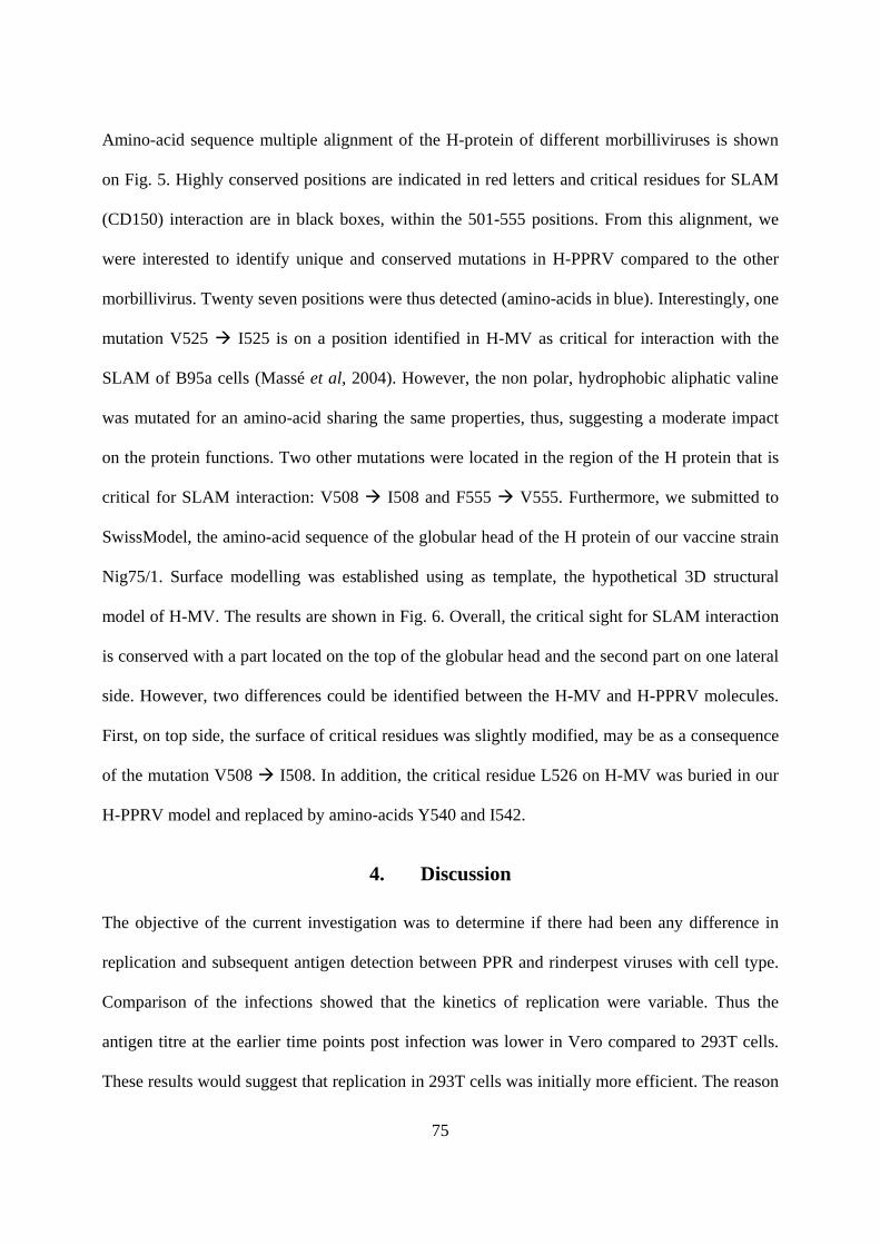
75
Amino-acid sequence multiple alignment of the H-protein of different morbilliviruses is shown
on Fig. 5. Highly conserved positions are indicated in red letters and critical residues for SLAM
(CD150) interaction are in black boxes, within the 501-555 positions. From this alignment, we
were interested to identify unique and conserved mutations in H-PPRV compared to the other
morbillivirus. Twenty seven positions were thus detected (amino-acids in blue). Interestingly, one
mutation V525 I525 is on a position identified in H-MV as critical for interaction with the
SLAM of B95a cells (Massé et al, 2004). However, the non polar, hydrophobic aliphatic valine
was mutated for an amino-acid sharing the same properties, thus, suggesting a moderate impact
on the protein functions. Two other mutations were located in the region of the H protein that is
critical for SLAM interaction: V508 I508 and F555 V555. Furthermore, we submitted to
SwissModel, the amino-acid sequence of the globular head of the H protein of our vaccine strain
Nig75/1. Surface modelling was established using as template, the hypothetical 3D structural
model of H-MV. The results are shown in Fig. 6. Overall, the critical sight for SLAM interaction
is conserved with a part located on the top of the globular head and the second part on one lateral
side. However, two differences could be identified between the H-MV and H-PPRV molecules.
First, on top side, the surface of critical residues was slightly modified, may be as a consequence
of the mutation V508 I508. In addition, the critical residue L526 on H-MV was buried in our
H-PPRV model and replaced by amino-acids Y540 and I542.
4. Discussion
The objective of the current investigation was to determine if there had been any difference in
replication and subsequent antigen detection between PPR and rinderpest viruses with cell type.
Comparison of the infections showed that the kinetics of replication were variable. Thus the
antigen titre at the earlier time points post infection was lower in Vero compared to 293T cells.
These results would suggest that replication in 293T cells was initially more efficient. The reason

76
may relate to the difference levels of cytoplasmic, particularly lysosomal, activity in the cell
types. Nevertheless, these differences did not prevent the virus from eventually spreading to all
cells in the culture. Moreover, vaccine strains of both PPR and rinderpest viruses are well
adapted to Vero cells.
B95a and MOCL5 cells did not support active PPR virus replication. It may be that these cells
lack or are deficient in specific attachment protein sequence or an intercellular host protein
necessary for efficient virus replication. This may also reflect the use of a different cell receptor
(Wild et al., 1991). Cellular receptors are one of the major determinants of the host range and
tissue tropism of a virus. The H (attachment) glycoproteins of morbilliviruses are variable
(Blixenkrone-Müller et al., 1996) and this may play a role in host cell specificity. B95a
lymphoblastoid cells are a good host for rinderpest virus, but they did not support replication of
PPRV. Further efforts to adapt PPRV in B95a cells by six blind passages failed to result in
productive infection, contrary to the reports of Das and others (2000). Differences in sensitivity
of B95a cells between PPRV and RPV may reflect the use of a different cell receptor, which is
believed to be determinant in the host range and tissue tropism. This appeared to be in
contradiction with results obtained with other morbilliviruses in the same cells. RPV (Kobune et
al., 1991), MV (Kobune et al., 1990) and canine distemper virus (Seki et al., 2003) replicate and
can be isolated successfully in B95a cell line. MV uses CD46 receptor for Vero but SLAM for
B95a (Takeuchui et al., 2002 ).
Amino-acid sequence analysis of the H protein of several vaccine and wild type strains of PPRV,
RPV, measles virus (MV) and canine distemper virus (CDV) showed that the critical residues
identified on the H-MV protein for the interaction with the SLAM (CD150 receptor) are
conserved in all morbillivirus H sequences we have analysed and surface modelling using
hypothetical 3D structural model of H-MV identified one mutation as critical for interaction with
the SLAM of B95a cells (Massé et al, 2004). Thus, the hydrophobic non polar aliphatic H-MV

77
residue V525 was replaced by the hydrophobic non polar aliphatic I525. The effect of this
conservative mutation on the H-SLAM interaction is probably limited but requires further
investigation. In addition, the SwissModel (Schwede et al., 2003), using the amino-acid sequence
of the globular head of the H protein detected two differences between the H-MV and H-PPRV
molecules. First, on top side, the surface of critical residues was slightly modified, may be as a
consequence of the mutation V508 I508. Second, the critical residue L526 on H-MV was
replaced by amino-acids Y540 and I542. Whether these modifications may account for a
defective interaction of the H-PPRV with the B95a SLAM receptor will need further analysis, as
the non-susceptibility of B95a cells to PPRV cannot be explained simply by sequence
modification of the H protein.
Acknowledgements
This work was supported by a grant from the French Ministry of Foreign Affairs. We also thank
Dr. Michel Olivier (INRA, France) for the kind supply of the MOCL-5 cell lines.

78
Reference
1. Baron, M.D., Kamata, Y., Barras, V., Goatley, L., Barrett, T. 1996. The genome sequence of
the virulent Kabete ‘O’ strain of rinderpest virus: comparison with the derived vaccine.
Journal of General Virology, 77, 3041-3046.
2. Blixenkrone-Müller, M, Bolt, G. Jensen, T.D. Harder, T. and Svansson, V. 1996.
Comparative analysis of the attachment protein (H) of dolphin morbillivirus. Virus Research,
40, 47-55.
3. Brown, C.C. and Torres, A. 1994. Distribution of antigen in cattle infected with rinderpest
virus. Veterinary Pathology, 3, 1 194 – 200.
4. Das, S.C., Baron, M.D. and Barrett, T. 2000. Recovery and characterization of a chimeric
rinderpest virus with the glycoproteins of peste des petits ruminants virus: Homologous F and
H proteins are required for virus viability. Journal of Virology, 74, 9039-9047.
5. Diallo, A. Taylor, W.P. Lefèvre and Provost, A. 1989. Atténuation d’une souche de virus de
la peste des petits ruminants: candidat pour un vaccin homologue vivant. Rev. Elev. Méd.
Vét. Pays Trop., 42, 311-319.
6. Gibbs, P.J.E., Taylor, W.P. Lawman, M.P. and Bryant, J. 1979. Classification of the peste des
petits ruminants virus as the fourth member of the genus Morbillivirus. Intervirology, 11,
268 – 274.
7. Kobune, F. Sakata, H. Sugiura, A. 1990. Marmoset lymphoblastoid cells as a sensitive host
for isolation of measles virus. Journal of Virology, 64, 700-705.
8. Kobune, F. Sakata, H. Sugiyama, M. Sugiura, A. 1991. B95a, a marmoset lymphoblastoid
cell line as a sensitive host for rinderpest virus. Journal of General Virology, 72, 687-692.
9. Lefèvre, P.C. and Diallo, A. 1990. Peste des petites ruminants. Revue Scientifique Office
International.des Epizooties, 9, 951- 965.

79
10. Libeau, G. Diallo, A., Calvez, D. Lefevre, P.C. 1992. A competitive ELISA using anti-N
monoclonal antibodies for specific detection of RP antibodies in cattle and small ruminantss.
Veterinary Microbiology, 31, 147-160.
11. Masse, N., T. Barrett, C.P. Muller, T.F. Wild, and Buckland, 2004. Identification of a second
major site for CD46 binding in the hemagglutinin protein from a laboratory strain of measles
virus (MV): potential consequences for wild-type MV infection. J.Virol. 76, 13034-13038.
12. Olivier, M., Berthon, P., Chastang, J., Cordier, G., Lantier, F. 2001. Establishment and
characterisation of ovine blood monocyte-derived cell lines. Veterinary Immunology and
Immunopathology, 82, 139-151.
13. Reed, L.J, and Muench, H. 1938. A simple method for estimating 50% endpoints. American
Journal of Hygiene 27, 493 – 497.
14. Rey Nores, J.E. and McCullough, K.C. 1996. Relative ability of different bovine leukocyte
populations to support active replication of rinderpest virus. Journal of Virology 70, 4419-
4436.
15. Roeder, P.L., Abraham, G., Kenfe, G., Barrett, T. 1994. Peste des petits ruminants in
Ethiopian goats. Tropical Animal Health and Production 26, 69-73.
16. Schwede, T., Kopp, J., Guex, N., and Peitsch, M.C. 2003. SWISS-MODEL: an automated
protein homology-modelling server. Nucleic Acids Research 31, 3381-3385.
17. Seki, F. Ono, N. Yamaguchi, R. and Yanagi, Y. 2003. Efficient isolation of wild strains of
canine distemper virus in vero cells expressing canine SLAM (CD150) and their adaptability
to marmoset B95a cells. Journal of Virology 77, 9943-9950.
18. Taylor, W.P. 1986. Epidemiology and control of rinderpest. Rev. Sci. Tech. Off. Int. Epizoot.
5, 407-410.
19. Wild, T.F. Malvoisin, E. and Buckland, R. 1991. Measles virus: both the haemagglutin and
fusion glycoproteins are required for fusion. J. General Virology 72, 439 – 442.

80
20. Wohlsein, P.G. Wamwayi, H.M. Trautwein, G. Pohlenz, J., Liess, B. and Barrett, T. 1995.
Pathomorphological and immunohistological findings in cattle experimentally infected with
rinderpest virus isolates of different pathogenicity. Veterinary Microbiology 44, 141 – 147.

81
Vero-PPR-H Vero-PPR-N
293T-PPR-H 293T-PPR-N
B95a-PPR-H B95a-PPR-N
Fig. 1 Flow cytometry analysis of cells infected with peste des petits ruminants virus. The
relative cell number (in %) is plotted on the Y-axis(0, 20, 40, 60), fluorescence intensity is
plotted on the X-axis (101 , 102 , 103, 104 of each square) and time is plotted on the Z – axis
(starting from 0, 24, 48, 72, 96, 120, 144 hours post infection for each square).

82
RP-vero-H RP-vero-N
RP-293T-H RP-293T-N
RP-B95a-H RP-B95a-N
Fig. 2 Flow cytometry analysis of cells infected with rinderpest virus. The relative cell number
(in %) is plotted on the Y-axis(0, 20, 40, 60), fluorescence intensity is plotted on the X-axis (101 ,
102 , 103, 104 of each square) and time is plotted on the Z – axis (starting from 0, 24, 48, 72, 96,
120, 144 hours post infection for each square).

83
Fig. 3. Percentage of cells indicating positive fluorescence.
Fig. 4. Titration of Both PPRV and RPV in B95a, 293T and Vero cell lines.
1 50 PPRV (AJ512718) (1) MSAQRERINAFYKGNPHNKNHRVILDRERLVIERPYILLGVLLVMFLSLI
Titration of rinderpest virus in B95a, 293 and Vero cell lines
02468
0 24 48 72 96 120 144
Hours post infection
Log
(10)
TC
ID50
/ml
B95a293Vero
Percentage of cells with fluorescence after infection with
PPR virus
0
20
40
60
80
0 24 48 72 96 120 144Hours post infection
% o
f cel
ls
VEROcells293B95a
Percentage of cells with fluorescence after infection with rinderpest virus
0
20
40
60
80
0 24 48 72 96 120 144Hours post infection
% o
f cel
ls
VEROcells293B95a
Titration of PPR virus in B95a, 293 and Vero cells
02468
0 24 48 72 96 120 144
Hours post infection
Log(
10)
TCID
50/m
l
VERO
cells293
B95a

84
PPRV (NC_006383) (1) MSAQRERINAFYKDNPHNKNHRVILDRERLVIERPYILLGVLLVMFLSLI PPRV (X74443) (1) MSAQRERINAFYKDNLHNKTHRVILDRERLTIERPYILLGVLLVMFLSLI RPV (X98291) (1) MSPPRDRVDAYYKDNFQFKNTRVVLNKEQLLIERPCMLLTVLFVMFLSLV RPV (Z30697) (1) MSPPRDRVDAYYKDNFQFKNTRVVLNKEQLLIERPCMLLTVLFVMFLSLV MV (AB012948) (1) MSPQRDRINAFYKDNPHPKGSRIVINREHLMIDRPYVLLAVLFVMFLSLI MV (AF266288) (1) MSPQRDRINAFYKDNPHPKGSRIVINREHLMIDRPYVLLAVLFVMSLSLI MV (AF266286) (1) MSPQRDRINAFYKDNPHPKGSRIVINREHLMIDRPYVLLAVLFVMFLSLI MV (AF266289) (1) MSPQRDRINAFYKDNPHPKGSRIVINREHLMIDRPYVLLAVLFVMFLSLI CDV (AF164967) (1) MLSYQDKVSAFYKDNARANSSKLSLVTEEQGGRRPPYLLFVLLILLVGIM CDV (AY466011) (1) MLSYQDKVGAFYKDNARANPSKLSLVTEEHGGRRPPYLLFVLLILLVGIL CDV (AF378705) (1) MLSYQDKVGAFYKDNARANSTKLSLVTEEHGGRRPPYLLFVLLILLVGIL CDV (AF305419) (1) MLPYQDKVGAFYKDNARANSTKLSLVTEGHGGRRPPYLLFVLLILLVGIL 51 100 PPRV (AJ512718) (51) GLLAIAGIRLHRATVGTLEIQSRLNTNIELTESIDHQTKDVLTPLFKIIG PPRV (NC_006383) (51) GLLAIAGIRLHRATVGTSEIQSRLNTNIELTESIDHQTKDVLTPLFKIIG PPRV (X74443) (51) GLLAIAGIRLHRATVGTAEIQSRLNTNIELTESIDHQTKDVLTPLFKIIG RPV (X98291) (51) GLLAIAGIRLHRAAVNTAKINNDLTTSIDITKSIEYQVKDVLTPLFKIIG RPV (Z30697) (51) GLLAIAGIRLHRAAVNTAKINNDLTTSIDITKSIEYQVKDVLTPLFKIIG MV (AB012948) (51) GLLAIAGIRLHRAAIYTAEIHKSLSTNLDVTNSIEHQVKDVLTPLFKIIG MV (AF266288) (51) GLLAIAGIRLHRAAIYTAEIHKSLSTNLDVTNSIEHQVKDVLTPLFKIIG MV (AF266286) (51) GLLAIAGIRLHRAAIYTAEIHKSLSTNLDVTNSIEHQVKDVLTPLFKIIG MV (AF266289) (51) GLLAIAGIRLHRAAIYTAEIHKSLSTNLDVTNSIEHQVKDVLTPLFKIIG CDV (AF164967) (51) ALLAITGVRFHQVSTSNMEFSRLLKEDMEKSEAVHHQVIDVLTPLFKIIG CDV (AY466011) (51) ALLSITGIRFHKVSTSNMEFSRLLKEDMEKSEAVHHQVIDVLTPLFKIIG CDV (AF378705) (51) ALLAITGVRFHQVSTSNMEFSRLLKEDMEKSEAVHHQVIDVLTPLFKIIG CDV (AF305419) (51) ALLAITGVRFHQVSTSNMEFSRLLKEDMEKSEAVHHQVIDVLTPLFKIIG 101 150 PPRV (AJ512718) (101) DEVGIRIPQKFSDLVKFISDKIKFLNPDREYDFRDLRWCMNPPERVKINF PPRV (NC_006383) (101) DEVGIRIPQKFSDLVKFISDKIKFLNPDREYDFRDLRWCMNPPERVKINF PPRV (X74443) (101) DEVGIRIPQKFSDLVKFISDKIKFLNPDREYDFRDLRWCMNPPERVKINF RPV (X98291) (101) DEVGLRTPQRFTDLTKFISDKIKFLNPDKEYDFRDINWCINPPERIKIDY RPV (Z30697) (101) DEVGLRTPQRFTDLTKFISDKIKFLNPDKEYDFRDINWCINPPERIKIDY MV (AB012948) (101) DEVGLRTPQRFTDLVKFISDKIKFLNPDREYDFRDLTWCINPPERIKLDY MV (AF266288) (101) DEVGLRTPQRFTDLVKFISDKIKFLNPDREYDFRDLTWCINPPERIKLDY MV (AF266286) (101) DEVGLRTPQRFTDLVKFISDKIKFLNPDREYDFRDLTWCINPPERIKLDY MV (AF266289) (101) DEVGLRTPQRFTDLVKFISDKIKFLNPDREYDFRDLTWCINPPERIKLDY CDV (AF164967) (101) DEIGLRLPQKLNEIKQFILQKTNFFNPNREFDFRDLHWCINPPSKIKVNF CDV (AY466011) (101) DEIGLRLPQKLNEIKQFILQKTNFFNPNREFDFRDLHWCINPPSKVKVNF CDV (AF378705) (101) DEIGLRLPQKLNEIKQFILQKTNFFNPNREFDFRDLHWCINPPSKVKVNF CDV (AF305419) (101) DEIGLRLPQKLNEIKQFILQKTNFFNPNREFDFRDLHWCINPPSTVKVNF 151 200 PPRV (AJ512718) (151) DQFCEYKAAVKSIEHIFESPLNKSKKLQSLTLGPGTGCQGRTVTRAHFSE PPRV (NC_006383) (151) DQFCEYKAAVKSIEHIFESPLNKSKKLQSLTLGPGTGCLGRTVTRAHFSE PPRV (X74443) (151) DQFCEYKAAVKSVEHIFESSLNRSERLRLLTLGPGTGCLGRTVTRAQFSE RPV (X98291) (151) DQYCAHTAAEDLITMLVNSSLTGTTVLRTSLVNLGRNCTGPTTTKGQFSN RPV (Z30697) (151) DQYCAHTAAEDLITMLVNSSLTGTTVPRTSLVNLGRNCTGPTTTKGQFSN MV (AB012948) (151) DQYCADVAAEELMNALVNATLLEARATNQFLAVSKGNCSGPTTIRGQFSN MV (AF266288) (151) DQYCADVAAEELMNALVNSTLLETRTTNQFLAVSKGNCSGPTTIRGQFSN MV (AF266286) (151) DQYCADVAAEELMNALVNSTLLETRTTNQFLAVSKGNCSGPTTIRGQFSN MV (AF266289) (151) DQYCADVAAEELMNALVNSTLLETRTTNQFLAVSKGNCSGPTTIRGQFSN CDV (AF164967) (151) TNYCDTIGIRKSIASAANPILLSALSGGRGDIFPPYRCSGATTSVGRVFP CDV (AY466011) (151) TNYCDTIGIRKSIASAANPILLSALSGSRSDIFPPYRCSGATTSVGKVFP CDV (AF378705) (151) TNYCESIGIRKAIASAANPILLSALSGGRSDIFPPHRCSGATTSVGKVFP CDV (AF305419) (151) TNYCESIGIRKAIASAANPILLSALSGGRGDIFPPHRCSGATTSVGKVFP

85
201 250 PPRV (AJ512718) (201) LTLTLMDLDLEMKHNVSSVFTVVEEGLFGRTYTVWRSDARDPSTDPGIGH PPRV (NC_006383) (201) LTLTLMDLDLEMKHNVSSVFTVVEEGLFGRTYTVWRSDARDPSTDLGIGH PPRV (X74443) (201) LTLTLMDLDLEIKHNVSSVFTVVEEGLFGRTYTVWRSDTGKPSTSPGIGH RPV (X98291) (201) ISLTLSGIYSGRGYNISSMITITGKGMYGSTYLVGKYNQRARRPSIVWQQ RPV (Z30697) (201) ISLTLSGIYSGRGYNISSMITITGKGMYGSTYLVGKYNQRARRPSKVWHQ MV (AB012948) (201) MSLSLLDLYLSRGYNVSSIVTMTSQGMYGGTYLVEKPNLSSKGSELSQLS MV (AF266288) (201) MSLSLLDLYLSRGYNVSSIVTMTSQGMYGGTYLVEKPNLSSKRSELSQLS MV (AF266286) (201) MSLSLLDLYLGRGYNVSSIVTMTSQGMYGGTYLVEKPNLSSKRSELSQLS MV (AF266289) (201) MSLSLLDLYLGRGYNVSSIVTMTSQGMYGGTYLVEKPNLSSKRSELSQLS CDV (AF164967) (201) LSVSLSMSLISRTSEIINMLTAISDGVYGKTYLLVPDYIEGGFDT----Q CDV (AY466011) (201) LSVSLSMSLISRTSVIINMLTAISDGVYGKTYLLVPDDIEREFDT----Q CDV (AF378705) (201) LSVSLSMSLISRTSEIINMLTAISDGVYGKTYLLVPDDIEREFDT----Q CDV (AF305419) (201) LSVSLSMSLISRTSEVINMLTAISDGVYGKTYLLVPDDIEREFDT----R 251 300 PPRV (AJ512718) (251) FLRVFEIGLVRDLGLGPPVFHMTNYLTVNMSDDYRRCLLAVGELKLTALC PPRV (NC_006383) (251) FLRVFEIGLVRDLGLGPPVFHMTNYLTVNMSDDYRRCLLAVGELKLTALC PPRV (X74443) (251) FLRVFEIGLVRDLELGAPIFHMTNYLTVNMSDDYRSCLLAVGELKLTALC RPV (X98291) (251) DYRVFEVGIIRELGVGTPVFHMTNYLELPRQPELETCMLALGESKLAALC RPV (Z30697) (251) DYRVFEVGIIRELGVGTPGFHMTNYLELPRQPELETCMLALGESKLAALC MV (AB012948) (251) MHRVFEVGVIRNPGLGAPVFHMTNYFEQSVSNDFSNCMVALGELKFAALC MV (AF266288) (251) MYRVFEVGVIRNPGLGAPVFHMTNYLEQPVSNDLSNCMVALGELKLAALC MV (AF266286) (251) MYRVFEVGVIRNPGLGAPVFHMTNYLEQPVSNDLSNCMVALGELKLAALC MV (AF266289) (251) MYRVFEVGVIRNPGLGAPVFHMTNYLEQPVSNDLSNCMVALGELKLAALC CDV (AF164967) (247) KIRVFEIGFIKRWLNDMPLLQTTNYMVLPENSKAKVCTIAVGELTLASLC CDV (AY466011) (247) EIRVFEIGFIKRWLNDMPLLQTTNYMVLPENSKAKVCTIAVGELTLASLC CDV (AF378705) (247) EIRVFEIGFIKRWLNDMPLLQTTNYMVLPENSKAKVCTIAVGELTLASLC CDV (AF305419) (247) EIRVFEIGFIKRWLNDMPLLQTTNYMVLPKNSKAKVCTIAVGELTLASLC 301 350 PPRV (AJ512718) (301) TSSETVTLSERGVPKRKPLVVVILNLAGPTLGGELYSVLPTSDLMVEKLY PPRV (NC_006383) (301) SSSETVTLGERGVPKREPLVVVILNLAGPTLGGELYSVLPTSDLMVEKLY PPRV (X74443) (301) TPSETVTLSESGVPKREPLVVVILNLAGPTLGGELYSVLPTTDPTVEKLY RPV (X98291) (301) LADSPVALHYGRVGDDNKIRFVKLGVWASPADRDTLATLSAIDPTLDGLY RPV (Z30697) (301) LADSPVALHYGRVGDDNKIRFVKLGVWASPADRDTLATLSAIDPTLDGLY MV (AB012948) (301) HREDSITIPYQGSGKGVSFQLVKLGVWKSPTDMQSWVPLSTDDPVIDRLY MV (AF266288) (301) HGEDSITIPYQGSGKGVSFQLVKLGVWKSPTDMQSWVPLSTDDPVIDRLY MV (AF266286) (301) HREDSITIPYQGSGKGVSFQLVKLGVWKSPTDMQSWVTLSTDDPVIDRLY MV (AF266289) (301) HGEDSITIPYQGSGKGVSFQLVKLGVWKSPTDMQSWVPLSTDDPVIDRLY CDV (AF164967) (297) VDESTVLLYHDSDGSQDGILVVTLGIFGATPMDQVEEVIPVAHPSVEKIH CDV (AY466011) (297) VDESTVLLYHDSRGSQDGILVVTLGIFGATPMDHIEEVIPVAHPSMEKIH CDV (AF378705) (297) VEESTVLLYHDSSGSQDGILVVTLGIFWATPMDHIEEVIPVAHPSMEKIH CDV (AF305419) (297) VEESTVLLYHDSSGSQDGILVVTLGIFWATPMDHIEEVIPVAHPSMKKIH 351 400 PPRV (AJ512718) (351) LSSHRGIIKDNEANWVVPSTDVRDLQNKGECLVEACKTRPPSFCNGTGSG PPRV (NC_006383) (351) LSSHRGIIKDDEANWVVPSTDVRDLQNKGECLVEACKTRPPSFCNGTGSG PPRV (X74443) (351) LSSHRGIIKDNEANWVVPSTDVRDLQNKGECLVEACKTRPPSFCNGTGIG RPV (X98291) (351) ITTHRGIIAAGTAIWAVPVTRTDDQVKMGKCRLEACRDRPPPFCNSTDWE RPV (Z30697) (351) ITTHRGIIAAGTAIWAVPVTRTDDQVKMGKCRLEACRDRPPPFCNSTDWE MV (AB012948) (351) LSSHRGVIADNQAKWAVPTTRTDDKLRMETCFQQACKGKIQALCENPEWA MV (AF266288) (351) LSSHRGVIADNQAKWAVPTTRTDDKLRMETCFQQACKGKIQALCENPEWA MV (AF266286) (351) LSSHRGVIADNQAKWAVPTTRTDDKLRMETCFQQACKGKIQALCENPEWA MV (AF266289) (351) LSSHRGVIADNQAKWAVPTTRTDDKLRMETCFQQACKGKIQALCENPEWA CDV (AF164967) (347) ITNHRGFIKDSIATWMVPALVSEKQEEQKNCLESACQRKSYPMCNQTSWE CDV (AY466011) (347) ITNHRGFIKDSIATWMVPALASEKQEEQKGCLESACQRKTYPMCNQTSWE CDV (AF378705) (347) ITNHRGFIKDSIATWMVPALASEKQEEQKGCLESACQRKTYPMCNQTSWE CDV (AF305419) (347) ITNHRGFIKDSIATWMVPALASEKQEEQKGCLESACQRKTYPMCNQASWE

86
401 450 PPRV (AJ512718) (401) PWSEGRIPAYGVIRVSLDLASDPGVVITSVFGPLIPHLSGMDLYNNPFSR PPRV (NC_006383) (401) PWSEGRIPAYGVIRVSLDLASDPGVVITSVFGPLIPHLSGMDLYNNPFSR PPRV (X74443) (401) PWSEGRIPAYGVIRVSLDLASDPGVVITSVFGPLIPHLSGMDLYNNPFSR RPV (X98291) (401) PLEAGRIPAYGVLTIKLGLADEPKVDIISEFGPLITHDSGMDLYTSFDGT RPV (Z30697) (401) PLEAGRIPAYGVLTIKLGLADEPKVDIISEFGPLITHDSGMDLYTSFDGT MV (AB012948) (401) PLKDNRIPSYGVLSVNLSLTVELKIKIASGFGPLITHGSGMDLYKSNHNN MV (AF266288) (401) PLKDNRIPSYGVLSVDLSLTVELKIKIASGFGPLITHGSGMDLYKSNHNN MV (AF266286) (401) PLKDNRIPSYGVLSVDLSLTVELKIKIASGFGPLITHGSGMDLYKSNHNN MV (AF266289) (401) PLKDNRIPSYGVLSVDLSLTVELKIKIASGFGPLITHGSGMDLYKSNHNN CDV (AF164967) (397) PFGGGQLPSYGRLTLPLDPSIDLQLNISFTYGPVILNGDGMDYYESPLLD CDV (AY466011) (397) PFGGGQLPSYGRLTLPLDASVDLQLNISFTYGPVILNGDGMVYYESPLLN CDV (AF378705) (397) PFGGRQLPSYGRLTLPLDASVDLQLNISFTYGPVILNGDGMDYYESPLLN CDV (AF305419) (397) PFGGRQLPSYGRLTLPLDASVDLQLNISFTYGPVILNGDGMDYYESPLLN 451 500 PPRV (AJ512718) (451) AVWLAVPPYEQSFLGMINTIGFPNRAEVMPHILTTEIRGPRGRCHVPIEL PPRV (NC_006383) (451) AVWLAVPPYEQSFLGMIYTIGFPYRAEVMPHILTTEIRGPRGRCHVPIEL PPRV (X74443) (451) AAWLAVPPYEQSFLGMINTIGFPDRAEVMPHILTTEIRGPRGRCHVPIEL RPV (X98291) (451) KYWLTTPPLQNSALGTVNTLVLEPSLKISPNILTLPIRSGGGDCYTPTYL RPV (Z30697) (451) KYWLTTPPLQNSALGTVNTLVLEPSLKISPNILTLPIRSGGGDCYIPTYL MV (AB012948) (451) VYWLTIPPMKNLALGVINTLEWIPRFKVSPNLFTVPIKEAGEDCHAPTYL MV (AF266288) (451) VYWLTIPPMKNLALGVINTLEWIPRFKVSPNLFTVPIKEAGEDCHAPTYL MV (AF266286) (451) VYWLTIPPMKNLALGVINTLEWIPRFKVSPYLFNVPIKEAGEDCHAPTYL MV (AF266289) (451) VYWLTIPPMKNLALGVINTLEWIPRFKVSPYLFTVPIKEAGEDCHAPTYL CDV (AF164967) (447) SGWLTIPPKNGTVLGLINKASRGDQFTVIPHVLTFAPRESSGNCYLPIQT CDV (AY466011) (447) SGWLTIPPKNGTILGLINKAGRGDQFTVIPHVLTFAPRESGGNCYLPIQT CDV (AF378705) (447) SGWLTIPPKNGTIVGLINKAGRGDQFTVLPHVLTFAPWESSGNCYLPIQT CDV (AF305419) (447) SGWLTIPPKDGTISGLINKAGRGDQFTVLPHVLTFAPRESSGNCYLPIQT 501 550 PPRV (AJ512718) (501) SRRVDDDIKIGSNMVILPTMDLRYITATYDVSRREHAIVCYIYDTGLSSS PPRV (NC_006383) (501) SRRVDDDIKIGSYMVILPTIDLRYITATYDVSRSEHAIVYYIYDTGRSSS PPRV (X74443) (501) SSRIDDDIKIGSNMVVLPTKDLRYITATYDVSRSEHAIVYYIYDTGRSSS RPV (X98291) (501) SDRADDDVKLSSNLVILPSRDLQYVSATYDISRVEHAIVYHIYSTGRLSS RPV (Z30697) (501) SDRADDDVKLSSNLVILPSRDLQYVSATYDISRVEHAIVYHIYSTGRLSS MV (AB012948) (501) PAEVDGDVKLSSNLVILPGQDLQYVLATYDTSRVEHAVVYYVYSPSRSFS MV (AF266288) (501) PAEVDGDVKLSSNLVILPGQDLQYVLATYDTSRVEHAVVYYVYSPGRSFS MV (AF266286) (501) PAEVDGDVKLSSNLVILPGQDLQYVLATYDTSRVEHAVVYYVYSPSRSFS MV (AF266289) (501) PAEVDGDVKLSSNLVILPGQDLQYVLATYDTSRVEHAVVYYVYSPSRSFS CDV (AF164967) (497) SQIMDKDVLTESNLVVLPTQNFRYVIATYDISRGDHAIVYYVYDPIRAIS CDV (AY466011) (497) SQIIDRDVLIESNLVVLPTQSFRYVIATYDISRNDHAIVYYVYDPFRTIF CDV (AF378705) (497) SQIIDRDVLIESNIVVLPTQSFRYVIATYDISRSDHAIVYYVYDPIRTIS CDV (AF305419) (497) SQIRDRDVLIESNIVVLPTQSIRYVIATYDISRSDHAIVYYVYDPIRTIS 551 600 PPRV (AJ512718) (551) YYYPVRLNFKGNPLSLRIECFPWRHKVWCYHDCLIYNTITDEEVHTRGLT PPRV (NC_006383) (551) YFYPVRLNFKGNPLSLRIECFPWRHKVWCYHDCLIYNTITDEEVHTRGLT PPRV (X74443) (551) YFYPVRLNFRGNPLSLRIECFPWYHKVWCYHDCLIYNTITNEEVHTRGLT RPV (X98291) (551) YYYPFKLPIKGDPVSLQIECFPWDRKLWCHHFCSVIDSGTGEQVTHIGVV RPV (Z30697) (551) YYYPFKLPIKGDPVSLQIECFPWDRKLWCHHFCSVVDSGTGEQVTHIGVV MV (AB012948) (551) YFYPFRLPIKGVPIELQVECFTWDQKLWCRHFCVLADSESGGHITHSGMV MV (AF266288) (551) YFYPFRLPIKGVPIELQVECFTWDQKLWCRHFCVLADSESGGHITHSGMV MV (AF266286) (551) YFYPFRLPIKGVPIELQVECFTWDQKLWCRHFCVLADSESGGHITHSGMV MV (AF266289) (551) YFYPFRLPIKGVPIELQVECFTWDQKLWCRHFCVLADSESGGHITHSGMV CDV (AF164967) (547) YTYPFRLTTKGRPDFLRIECFVWDDDLWCHQFYRFEADSTNSTTSVENLV CDV (AY466011) (547) YTYPFRLTTKGRPDFLRIECFVWDDNLWCHQFYRYEANIANSTTSVENLV CDV (AF378705) (547) YTHPFRLTTKGRPDFLRIECFVWDDNLWCHQFYRFEADIANSTTSVENLV CDV (AF305419) (547) YTHPFRLTTKGRPDFLRIECFVWDDNLWCHQFYRFEADIANSTTSVENLV

87
601 618 PPRV (AJ512718) (601) GIEVTCNPV--------- PPRV (NC_006383) (601) GIEVTCNPV--------- PPRV (X74443) (601) GIEVTCNPV--------- RPV (X98291) (601) GIEITCNGK--------- RPV (Z30697) (601) GIKITCNGK--------- MV (AB012948) (601) GMGVSCTVTREDGTNSR- MV (AF266288) (601) GMGVSCTVTREDGTNRR- MV (AF266286) (601) GMGVSCTVTREDGTNRR- MV (AF266289) (601) GMGVSCTVTREDGTNRR- CDV (AF164967) (597) RIRFSCNRSKP------- CDV (AY466011) (597) RIRFSCNRSNP------- CDV (AF378705) (597) RIRFSCNR---------- CDV (AF305419) (597) RIRFSCNR----------
Fig. 5 Analysis of H amino-acid sequence of PPRV strains and comparison with other vaccine
(vac) and wild type (wt) strains of Morbillivirus. Red letters in grey boxes display the identical
amino-acid residues. Black boxes identify critical residues for H-MV/SLAM interaction
(positions 505, 507, 521-523, 525-527, 529-533, 536-537, 547-548, 552-554). Blue letters
indicate PPRV specific mutations compared to other morbilliviruses (27 positions). Accession
numbers used in this figure are PPRV (vac: X74443, wt: AJ512718 and NC_006383), RPV (vac:
Z30697, wt: X98291), MV (vac: AF266286 and AF266289, wt: AF266288 and AB012948),
CDV (vac: AF305419 and AF378705, wt: AF164967 and AY466011).

88
552
196
547
531 532
533 507
505 508
279 314
413
507
554 552
553 555
548 547
537
533 532
531 531
525 523 521
508 507
505
196
202 399
577
413
540 542
(a)
(b)

89
554
552 553
555
548
547 537
533
532 531
526 525
523
521
508
507
505
196
202 399
577
555
196 547
531
532
533 507
505
508
279
314
413
(c)
(d)
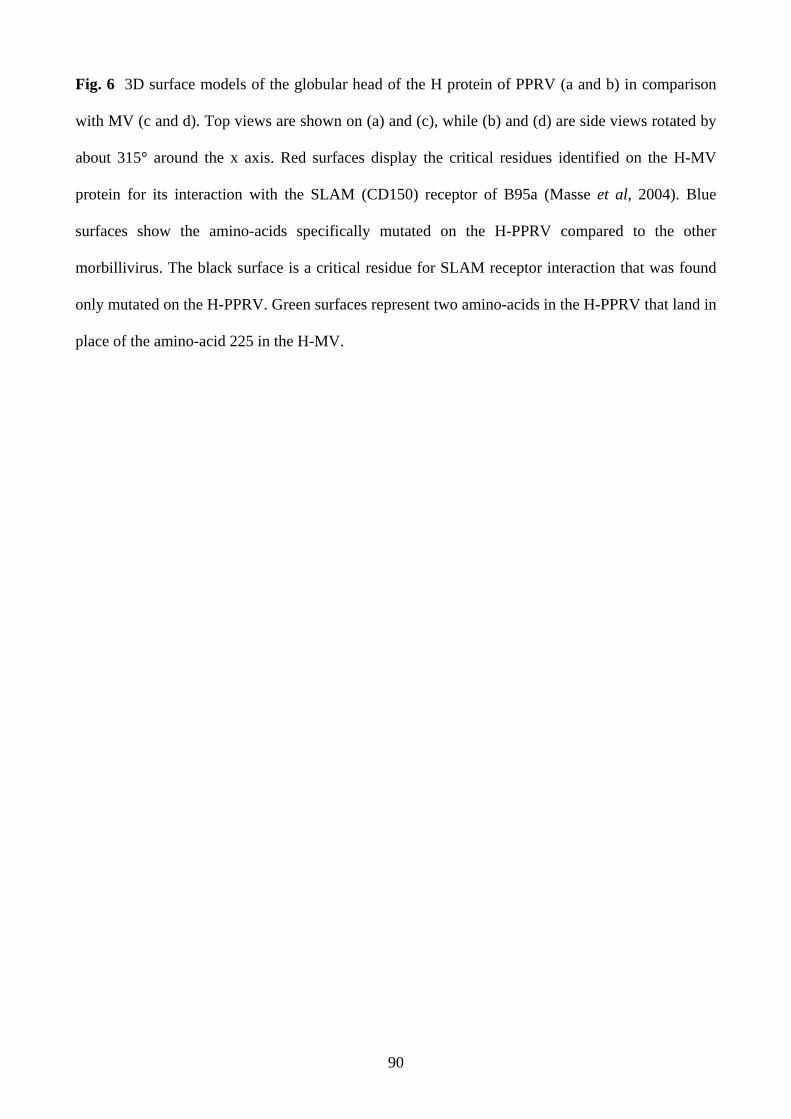
90
Fig. 6 3D surface models of the globular head of the H protein of PPRV (a and b) in comparison
with MV (c and d). Top views are shown on (a) and (c), while (b) and (d) are side views rotated by
about 315° around the x axis. Red surfaces display the critical residues identified on the H-MV
protein for its interaction with the SLAM (CD150) receptor of B95a (Masse et al, 2004). Blue
surfaces show the amino-acids specifically mutated on the H-PPRV compared to the other
morbillivirus. The black surface is a critical residue for SLAM receptor interaction that was found
only mutated on the H-PPRV. Green surfaces represent two amino-acids in the H-PPRV that land in
place of the amino-acid 225 in the H-MV.

91
Difference in virulence may not result exclusively on the host or cell susceptibility. Once
entered in the cells, the virus may not necessarily find the necessary environment for an
efficient replication. This could explain a diffence in the clinical outcome. Inside the cells,
the major biological processes on which relies the virus replication, involve three proteins
tighly associated with the nucleic acid to form the ribonucleic complex. The function of this
complex is dependent on the presence of particular inverted terminal sequences at the
genome extremities. These sequences are named genome promoter (3’ – 5’, genome sens)
and antigenome promoter (5’ – 3’ genome antisens). The first one is responsible for the
mRNA transcription and the production of the positive full genome RNA that serves as a
template for genome replication. The antigenome promoter is responsible for the production
of new genome molecule. Promoter sequences are conserved regions of the gene because of
the essential role they play in transcription and replication. Nucleotide mutations in these
regions may result in a loss of virulence, at least between wild type virus and cell culture
adapted vaccine strain. We were also interested to see if differences in these sequences
could be seen between strains producing disease in goats and strains producing disease in
sheep. In this study, base changes that may contribute to viral attenuation were investigated
by comparative analysis of the GP and AGP from field strains affecting sheep or goats and a
vaccinal. The promoter sequences were also compared with those from other members of
the Morbillivirus genus and the family Paramyxoviridae to assess possible shared critical
residues for virulence and the phylogenetic relationships between all these viruses.

92
Chapter 5
Anticle 3 : Submitted to Journal of General Virology
Sequence analysis of the genome and anti-genome promoters of peste des petits
ruminants virus (PPRV), comparison with other Morbillivirus
G. Abraham1, G. Libeau1*, A. Diallo2, S. Thomassin1,C. Minet1, M Yami1, O. Kwiatek1, O. Friedgut3, E. Albina1,
1 : CIRAD- EMVT, TA30/G, Campus International de Baillarguet, 34398 Montpellier, Cedex, 5, FRANCE. 2 : FAO/IAEA Agriculture and Biotechnology Laboratory, IAEA Laboratories, Animal Production Unit, A-2444, Seibersdorf, Austria 3 : Kimron Veterinary Institute, Beit Dagan 50250, Israel.
Abstract
Genome and antigenome promoters (GP and AGP) of the peste des petits ruminants (PPR) virus,
amplified from different pathological samples of sheep and goat origins, were sequenced and
compared with corresponding sequences of PPR vaccine strain and other morbilliviruses.
Alignment of GP sequences revealed six nucleotide changes at positions 5, 12, 26, 36, 42 and 81,
and one nucleotide mutation in AGP at position 15842 between PPR vaccine and field strains.
Mutation 26 was clearly linked with the attenuated phenotype of the vaccine strains of PPRV,
rinderpest virus (RPV), measles virus (MV) and canine distemper virus (CDV) as well. Mutations 5
and 12 were only seen on the PPRV and RPV vaccine strains and 4 other mutations, 36, 42, 81 and
15842 were seen only on the PPRV vaccine strain sequence. Interestingly, the field strains of PPRV
were the only ones showing a residue U at position 36 instead of C for other morbilliviruses and the
PPR vaccine strain. These nucleotide mutations, or some of them, may influence gene expression
by changing the interaction of the leader with the viral polymerase or with a cellular protein
involved in the modulation of the gene replication/transcription. As such they may be some of the
candidates involved in the attenuation of PPRV. In spite of differences in virulence between goats
and sheep reported in the field, there had been 100% homology of sequences in clinical samples of
nasal swabs, lung and lymph nodes from both species. However, a higher heterogeneity within
strains isolated from different areas was evidenced and accounted for a relevant phylogenetic
clustering according to the geographic origin. It was also shown that the promoter sequences,
although very short compared to the full genome, could make relevant phylogenetic tree of
Paramyxoviridae.
Key words: leader, morbillivirus, peste des petits ruminants (PPR), promoter, rinderpest (RP), trailer *Corresponding author: Dr Genevieve Libeau; [email protected]

93
Introduction Peste des petits ruminants (PPR) is an acute and highly contagious viral disease of domestic and
wild small ruminants, which is characterised by high fever, ocular and nasal discharge, pneumonia,
necrosis and ulceration of the mucous membrane and inflammation of the gastro-intestinal tract
leading to severe diarrhoea and death (Gibbs et al., 1979). PPR is caused by a single strand non-
segmented negative RNA virus which belongs to the family Paramyxoviridae, genus Morbillivirus
which also includes measles virus, rinderpest virus (RPV), canine-distemper virus, phocine-
distemper virus, and dolphin and porpoise morbilliviruses (Barrett et al., 1993a). All morbilliviruses
are related serologically, and sequence data shows that there is a high degree of homology at the
sequence level. The genome contains six tandemly arranged transcription units encoding six
structural proteins, the surface glycoproteins F and H, the nucleocapsid (N), the matrix (M), the
polymerase or large (L) and the polymerase-associated (P) proteins. The cistron directing the
synthesis of this later protein is encoding the virus non-structural proteins C and V by the use of two
other open reading frames (ORF) of the messengers. The gene order is 3’N-P-M-F-H-L5’, as
determined by transcriptional mapping (Dowling et al., 1986). The genome is flanked by extragenic
sequences at the 3’ and 5’ ends, referred to as the leader (52 nucleotides) and trailer (37
nucleotides), respectively. For viruses of the family Paramyxoviridae, the genome promoter (GP)
includes 107 nucleotides comprising the leader sequence and the adjacent non-coding region of the
N gene at the 3’ end of the negative-strand. The antigenome promoter (AGP) includes 109
nucleotides that encompass the trailer sequence and the proximal untranslated region of the L gene.
Both the GP and the AGP contains the polymerase binding sites and the RNA encapsidation signals
for the replication of the full genome while the production of messengers RNA is a function of the
GP (Murphy et al., 1998; Murphy and Parks 1999; Tapparel et al., 1998; Walpita, 2004). Therefore
nucleotide changes in the GP and AGP of the genome which affect the replication/transcription of
the RNA may have an impact on the virulence of the virus. Comparison of sequence data from

94
vaccinal and virulent strains of rinderpest and measles viruses identified nucleotide changes
throughout the genome. Those located in the GP and AGP are among the potential attenuation of
the virus (Parks et al., 2001; Baron et al., 1996; Banyard et al, 2005). In this paper, evidence of base
changes between an attenuated vaccine strain and virulent PPRV strains from the field was obtained
by the comparative analysis of the GP and AGP sequences. We used also those sequences to
identify the relationship between the different samples we analysed and compared these data with
those previously reported for the F protein gene sequence (Shaila et al, 1996; Dhar et al., 2002).
The promoter sequences were also compared with those from other members of the Morbillivirus
genus for the identification of shared attenuation genetic markers, and of the Paramyxoviridae
family for their phylogenetic relationships.
Materials and Methods The samples
The samples were obtained either as nasal swabs or as post-mortem tissues submitted for
confirmatory diagnosis from clinical diseases in goats and sheep (Table 1). A set of samples from
goats and sheep collected in Israel and covering several years of collection was included in order to
assess the molecular variation of the promoters within a given region. All samples were processed
using standard methods (Couacy-Hymann et al., 2002) and one hundred microlitres of the sample
solution were used for the RNA extraction. Vaccine strain of PPR virus (Diallo et al,. 1989) was
grown in Vero cells. Cell growth medium was Eagles MEM supplemented with 10% fœtal bovine
serum (FBS) and 1% of a mixed antibiotic solution (Gibco, Life Technologies, UK). Infected cells
and the supernatant were harvested at 75% of the cytopathic effect (CPE), and frozen at -70°C until
RNA extraction.
RNA extraction
RNA was extracted as described by Couacy-Hymann and others (2002). Briefly, nine hundred µl
lysis solution (containing guanidine isothiocyanate, β-mercaptoethanol, sarcosyl and sodium citrate)

95
and 100 µl sample suspension were mixed. The tube was then centrifuged for 3 minutes at 10,000
rpm. The supernatant was used in subsequent steps. One volume of 70% ethanol was mixed by
pipetting and the mixed solution was extracted by rapid spinning at 10,000 rpm through RNeasy
mini spin column (Qiagen, USA). The supernatant was discarded and the pellet was washed three
times with washing solution (Ethanol 50%, Tris-Hcl 10mM pH7.4-7.6, EDTA 1mM, NaCl 50mM).
The pellet was eluted in 50 µl of DEPC-treated water. One µl of RNase inhibitor (10U/µl)
(Amersham,UK) was added to the RNA solution and frozen at –70°C until used.
Single stranded cDNA synthesis and PCR for virus detection and sequencing
For the detection of PPRV in pathological samples, the RNA was denatured at 65°C for ten
minutes. Reverse transcription (RT) reaction was carried out in a 250 µl PCR tube as follows : 8 µl
of the extracted RNA was mixed with 1µl of DTT, 1µl of random primer pd(N)6 and 5 µl of First
strand cDNA bulk (Amersham, UK). The mixture was incubated at 37°C for 1h. PCR was
conducted using primers designed by Couacy-Hymann et al. (2002) to amplify fragments of the
PPRV N gene. The forward (5’-CAAGCCAAGGATTGCAGAAATGA-3’) and reverse
(5’-AATTGAGTTCTCTAGAATCACCAT-3’) primers were universal Morbillivirus, while the
forward (5’-GTCTCGGAAATCGCCTCACAG-3’) and reverse (5’-CCTCCTCCTG
GTCCTCCAGAA-3’) primers were PPR specific. In a 250µl PCR microtube, cDNA was mixed
with the following reagents: 5 µl of dNTP mixture (200mM), 5µl of 10X Taq buffer, 5 µl of
primers (5 pmol/µl), 36 µl of water and 1 µl of Taq polymerase (1.25U/µl) (Qiagen, USA). Once all
the reagents were mixed, the tube was placed into the Gene Amp PCR system 2400 (Applied
Biosystem, USA). The amplification was carried out according to the following programme: an
initial heating step 95°C for 5 min followed by denaturation at 94°C for 2 min and 35 cycles of
94°C for 30s, annealing at 55°C for 30 s, extension at 72 °C for 2 minutes.
The full genome sequence of PPRV vaccine strain Nigeria 75/1 was determined in our laboratory
(accession numbers: X74443, Z37017, Z47977, Z81358).The 3’ and 5’ ends of the PPRV vaccine

96
strain was amplified from the virus infected cell total RNA using the 5´ RACE System for Rapid
Amplification of cDNA Ends kit following instructions of the supplier (Invitrogen, USA). Primers
used in those reactions were designed based on the N gene
(5’-ATCATCTGTGATCCGCTGTATCAAT-3’) for the genome 3’ end cloning and the L gene
(5’-TCCACCATGATGTTGCCTCAGG-3’) for the genome 5’. For the amplification of the genome
and antigenome promoters of other PPRV strains, the same primers were used in conjunction with
new primers which were designed at the genome extremities based on the vaccine strain leader and
trailer sequences previously determined, 5’-ACCAGACAAAGCTGGGTAAGGATA-3’ and
5’-ACCAGACAAAGCTGGGTAAGGATA-3’, respectively. Two hundred pmol of each primer
were used in a 50 µl PCR reaction mixture. The amplification conditions were the same as indicated
previously with exception of changes in annealing temperature of 52°C for 30 seconds in the trailer
PCR cycle.
Analysis of PCR- amplified products
Ten microlitres of the amplicon were analysed by electrophoresis on a gel which was made of 1.5%
agarose in Tris-borate-EDTA (TBE) buffer (0.089M Tris base, 0.089M boric acid and 0.002M
EDTA, pH 8.3). The gel was stained with ethidium bromide and the DNA was visualised by UV
fluorescence and photographed.
Determination of GP and AGP sequences
PCR products were purified on columns (Qiaquick PCR Purification kit, Qiagen, USA) and directly
sequenced. Cycle sequencing was performed using dye-labeled terminators and Taq DNA
polymerase (Applied Biosystems, USA), followed by analysis on an ABI Prism 377 automatic
sequencer (Applied Biosystems, USA). Analysis of sequence results was performed using Vector
NTI-9 package (Informax Inc., USA). Sequence alignments were done using CLUSTAL W
included in this package. For the analysis of nucleotide changes, 6 PPRV sequences from Genbank
(Table 2), including one vaccine strain sequence, were first considered because the 5’end of their
97
GP and the 3’end of their AGP have been completed. Subsequently, interesting positions identified
previously were confirmed on partial promoter sequences (primers excluded) of our 12 additional
virulent strains detected in field samples (coded strains in Table 1). All Israeli strains were
sequenced but only one from each collection year and the divergent ones within a given year were
used. Phylogenetic analysis was then carried out using a criterion of neighbourhood based on the
principle of parsimony (Saitou and Nei, 1987). Dissimilarities and distances between the sequences
were first determined using the Darwin 5 software (Perrier et al., 2003). Trees were then generated
with the TreeCon MATRIXW program (Van de peer and De Watchter, 1993) included in Darwin5.
Tree construction was based on the unweighted Neighbor-Joining method proposed by Gascuel
(1997). Bootstraps were determined on 1000 replicates.
Results and Discussion
Base substitutions on the GP and AGP between the virulent strains of PPRV and the vaccine
strain
In the cycle of infection, the RNA dependant RNA polymerase (RdRp) of non-segmented negative
viruses as morbilliviruses uses the GP to synthesize the messengers RNA and later full-length
genome copies are made via replication intermediate strands of the genome from both GP and AGP
(Fig. 1a). Therefore changes in these regions may have a significant effect on the rate of
transcription of viral mRNA or full-length genome replication (Mioulet et al., 2001). As a first step
for looking into the molecular basis of PPR pathogenesis, we decided to compare the GP and AGP
of different PPRV strains. We started by determining the leader and trailer sequences of the PPRV
vaccine strain. As demonstrated for the Sendai virus (SeV) (Tapparel et al., 1998, Vuillermoz and
Roux, 2001), the promoters of morbilliviruses are bipartite and are composed of the leader plus the
untranslated 5’ end of the N gene for the GP, the 3’ untranslated gene of the L gene plus the trailer
for AGP (Fig. 1b). In Fig. 2, is presented the alignment of the different morbillivirus promoter
sequences. It can be seen that while the 3’ ends of the genomes are completely uniform in size, they

98
are two gaps in the alignment of morbillivirus AGP alignment. Indeed, for CDV and PDV, one
nucleotide is missing in the 3’ end of the L gene and is compensated by the presence of an extra
nucleotide in the trailer making the figures for both viruses of 71 and 38 nucleotides, respectively,
instead of 72 and 37 for the other morbilliviruses. Except this minor difference, the sequence
alignment generated for GP and AGP of the Morbillivirus genus revealed a high degree of
conservation. For example, the first four nucleotides of the 3’ terminus, UGGU (Fig 1b), are shared
between all viruses. It is believed that these four nucleotides contain the landing site for the viral
RdRp. However, if the intergenic sequence is conserved among all morbilliviruses, being GAA, the
intergenic L-trailer sequence is more variable and is GAU for PPRV, GUU for CDV and marine
mammal morbilliviruses. In Fig. 2, are indicated the two important regions constituting each
promoter according to what was proposed by Vuillermoz and Roux (2001) for the SeV. Both GP
and AGP complement each other on about the first 16 nucleotides for each virus. The promoter
region I is probably involved both in the encapsidation of the genome within the helical structure
made by the N protein and in the interaction between the encapsidated genome and the RNA-
dependant RNA polymerase (Tapparel et al, 1998). The second essential element, promoter region
II, is composed of a series of three hexamer motifs present at position 79-84, 85-90 and 91-96 on
the GP sequence. Here, the existence of the conserved discontinuous elements has been
substantiated by studies with the SeV (Tapparel et al., 1998) and Simian virus (Murphy and Parks,
1999) showing that the three sequences can significantly influence paramyxovirus RNA replication.
In this region, conserved critical C residues are found at positions 79, 85 and 91 and conserved U
residues are found at positions 83 and 89, thus forming a 3’-(CNNNUN)2-CNNNNN-5’ conserved
hexamer motif on the GP sequence and its AGP counterpart 5’-(GNNNAN)2-CNNNNN-3’
(Tapparel et al, 1998, Walpita, 2004). This motif is playing a major role in the promoter activities
and is expected to contain important signals for encapsidation and transcription. The hexamer phase
of this conserved motif is though to be stacked close to the three hexamers of the promoter region I

99
and to expose the conserved nucleotide residues on the same face of the helical nucleocapsid to
form a RNA-dependent RNA polymerase binding site (Tapparel et al, 1998; Mioulet et al, 2001). In
addition to these two conserved promoter regions, a third conserved region is found at position 52-
71 in GP and 59-71 on AGP.
To identify possible attenuation nucleotides in the promoters of PPRV, we have amplified and
sequenced the GP and AGP from 12 virulent strains recovered from field samples and compared the
data with those of the vaccine strain. This later is derived from the virulent Nigeria 75/1 strain and
is known to be highly attenuated for all animals into which it has been inoculated so far (Diallo et
al., 1989). PPRV in field samples was initially detected by a RT-PCR using Morbillivirus universal
primers targeting a highly conserved region of the N gene (positions 790-812 and 988-1012) as
reported by Couacy-Hymann et al. (2002). Virus specific RNA was detected in nasal and eye
swabs, mouth and gum erosions (Table 3). Amplification using universal primers produced 250 bp
fragments from PPRV and also from RPV. PPRV specific primers, targeting the 3’ terminal of the
N gene, detected 390 bp fragments but did not amplify RPV (Fig. 3). Half of the samples positive
with the PPR specific primers were however negative with universal primers thus indicating that the
current Morbillivirus universal N gene based primers have limitations in detecting all strains of
PPRV at the conditions of amplification we used (Table 3). Similar results were obtained by
Forsyth and Barrett (1995) who used P gene primers as Morbillivirus universal primers. Even
though the primers were designed in a zone where the gene sequence is well conserved between
different morbilliviruses, a single base change at the 3’ end of a primer as a result of virus strain
variation can abolish the amplification. The negative results reported by Forsyth and Barrett (1995)
and by us here with primers designed in conserved target sequences indicate again the necessity of
using at least two sets of primers for the diagnosis of pathogens such as RNA viruses known to have
high rate of mutations. Specimens which were positive with the PPRV specific primers were then
submitted to two other PCR tests to amplify both GP and AGP fragments as indicated in Materials

100
and Methods. The amplified products were sequenced. The sequence analysis was first done by
including also the sequences from 2 RPV, 2 MV, 2 CDV and that of the original strain PPRV 75/1
from which derives our vaccine strain, all being available from Genbank. Sequence alignment of the
GP (107 base pairs) of virulent strains of PPRV and the vaccine strain revealed six unique
nucleotide changes at positions 5 (transition U C), 12 (transition A G), 26 (transversion U
A), 36 (transition U C), 42 (transition C U) and 81 (transition C U) in the vaccine strain
(Fig. 4). Nucleotide changes on positions 5 and 12 could not be identified in our 12 field strains
because they were included in the forward primer used for GP amplification. However, positions
26, 36, 42 and 81 were all confirmed to be U, U, C, and C, respectively, thus strengthening the
importance of these mutations in link with the virulent/attenuated phenotype. The original virus
Nigeria 75/1 wild type (Nig75/1*) and its derived vaccine strain (63 passages on cell culture) have
only 2 nt changes in GP. Their AGP sequences are identical. In Nig75/1* one nucleotide change is
on position 12 (transition Awt → Gvac) in link with the virulent genotype. The other change at
position 83 is C which is specific to the Nig75/1* since all other PPR strains and other
morbilliviruses have a U residue at this position. Preliminary results of experimental works on live
animals indicated that, although virulent, PPR Nigeria 75/1 wild type has a much lower virulence
than other PPR strains (Couacy-Hymann, personal communication). This observation would be in
agreement with the conservation of promoter sequences between this low virulent strain and its
attenuated progeny. It also suggests that other genetic determinants for PPRV virulence are
probably present outside the viral promoters as already shown for RPV (Banyard et al, 2005). Non-
segmented negative strand viruses have sequence elements at their genomic RNA ends which
complement to each other (see Fig. 2 for morbillivirus 5’ and 3’ ends). For the SeV, it was
demonstrated that the trailer is a stronger promoter than the leader and that all the 31 first
nucleotides are important (Calain and Roux, 1995; Tapparel and Roux, 1996). Since the trailer
serves exclusively for the replication to produce negative sense genome RNA while the leader

101
directs both transcription and replication, it can be assumed that the minimum sequence shared by
the virus genome termini is the most important for the replication. Therefore, sequences
downstream the shared area may be important for the transcription as suggested by Li and Pattnaik
(1999) for vesicular stomatitis virus (VSV). The nucleotide substitutions occurring during cell
culture passages for vaccine derivation can be located in any part of the genome. Those favouring
specific modifications of the promoters can affect either the replication or mRNA transcription or
both functions. Scanning mutagenesis analysis of the 3’ end of the genome of RPV carried out by
Mioulet et al. (2001) did not distinguish the two functions since it was based on the measurement of
the reporter CAT protein expression by the minigenome construct. This study identified nucleotides
1, 3, 4, 10 and 19 as critical for the expression of the CAT protein. None of these nucleotides is
concerned by the changes identified between the PPRV virulent strains and the vaccine in the work
reported here. The same authors reported also the identification of residues in position 23-26 as
critical. Precisely, for PPRV, RPV, MV and CDV, there is a pyrimidine purine transversion at
position 26 from the wild types (U or C) to the vaccine strains (A) (Fig. 4). Nucleotide 26 is
downstream the sequence of the leader which complement with that of the trailer and it may be
important for the transcription function of the leader promoter in its interaction with the RdRp or a
cellular protein. It is interesting to note that for the SeV, two transversions U A at positions 20
and 24 in the leader sequence, outside the zone identified as critical for the replication/transcription
of the viral genome, contribute to attenuate the virus for mice (Fuji et al., 2002). The transversion
identified at the position 26 in the leader sequence of PPRV, RPV, MV and CDV may be one of the
essential elements involved in the attenuation of morbilliviruses. As it can be seen in Fig. 4, there
are some other nucleotide changes in the promoters sequences between virulent and vaccine strains
of PPRV. The transition (C U) at position 42 was only observed with PPRV. However, MV had
a transversion (U G ) at that position and for this virus it is believed that position 26 and 42
pyrimidine-to-purine transversions belong to a nucleotide stretch between nt 17 and 42 that could

102
serve as contact site for a modified polymerase complex used for mRNA synthesis (Parks et al.,
2001). For PPRV, the conservation of a pyrimidine residue during the base 42 substitution limits
probably the potential impact on the level of mRNA synthesis but may still contribute to an
attenuated phenotype. Transitions at positions 5 and 12 were only identified in both PPRV and
RPV. They occur within the highly conserved complementary hexamer motifs of GP and AGP
starts. They affect residues identified as not critical by the scanning mutagenesis study of the RPV
promoter but their change can reduce the reporter molecule production by about 60%. (Mioulet et
al. 2001). Thus the transition at positions 5 and 12 may contribute also to the attenuated phenotype
of the vaccine strain. The two other transitions of nucleotides 36 and 81 were only observed with
PPRV. The pyrimidine U at position 36 of all PPRV virulent strains is unique among all other wild
type and vaccine strains of morbilliviruses for which a conserved pyrimidine C is systematically
found. Therefore, the transition U C at that position gives the PPRV vaccine strain the
morbillivirus conserved residue. Moreover, block mutation around that nucleotide has no effect in
the promoter function of the RPV leader sequence (Mioulet et al., 2001). Also there is no evidence
that the change in the position 81 has an impact on the PPRV virulence.
In the AGP sequence (109 bp), there was only one unique nucleotide mutation specific to the
vaccine strain at position 15842, 107 nucleotides from the 5’terminus of the genome (Fig. 4). The
nucleotide in that position is variable although nucleotide G is found only in the vaccine strain.
As demonstrated by Banyard et al. (2005) in the case of RPV, the virus genome promoters play a
role in the pathogenicity of morbilliviruses. The 6 nucleotides changes observed at positions
generally well conserved in the PPRV GP are likely attenuation candidates. With the advance of the
reverse technology, it will be possible to generate strains with each individual mutation or group of
mutations and evaluate their impact on the virus virulence. Among all the candidates, the most
interesting is certainly the mutation observed at the position 26 because not only it is found in the
genome of RPV, PPRV, MV and CDV vaccines strains but also it is in the area identified as

103
involved in the pathogenesis of SeV, another paramyxovirus (Fuji et al., 2002). Likely, any
mutation in the promoters involved in the attenuation of PPRV may play in concert with other
critical changes not only within the promoters but also in other virus genes as demonstrated with
RPV. Indeed, as demonstrated Banyard et al., (2005) and Baron et al. (2005) for RPV, the
attenuation of morbillivirus may be a result of changes in different genes of the genome.
Differences in virulence of PPR between goats and sheep reported in Africa were not supported by
our sequence analysis of PPRV promoters. Also, specimen type did not play any role as there was
100% homology between sequences derived from nasal swabs, lung and lymph nodes from the
same goat or sheep (data not shown). In contrast, within a same geographic region like Israel, some
nucleotide changes may occur in a short period. Interestingly, mutations were observed between
1999 and 2000 in both GP and AGP sequences of goat as well as sheep strains, while the sequences
are conserved after 2000 (data not shown). None of these viruses had been passaged on cell culture,
which could have introduced genetic variability. It is however not clear what triggered these
nucleotide changes.
Use of the GP and AGP sequences of PPRV strains to determine the phylogenetic
relationship
Despite the overall high conservation of PPRV promoters, some variations were seen in parts of
their sequence (Fig. 3). Phylogenetic analysis of tandemly linked GP and AGP (Fig. 5a) of PPR
field strains from different areas and of the vaccine strain (Nig 75 vac) showed that these variations
could lead to a geographic clustering of the strains. Earlier, Shaila et al. (1996), and then Dhar et al.
(2002) demonstrated that isolates of PPRV could be grouped into four distinct lineages on the basis
of partial sequence analysis of the fusion (F) protein gene. Relationship deduced from a method
based on the principle of parsimony established that the promoter sequences were also lineage
specific. Similar lineage specific base changes in the GP and AGP were also observed with
rinderpest viruses (Banyard et al., 2005). This illustrates the possibility of using very short sequence

104
regions at the end of the virus genome for molecular typing of isolates, both for virulence and
lineage discrimination. Lineage speciation was identical when using independently the GP or AGP
sequences (data not shown). However, combination of the two allows to increase the bootstrap
values and to strengthen the molecular typing of PPRV isolates next to other morbilliviruses.
Furthermore, phylogenetic analysis of the GP-AGP tandem of Paramyxoviridae listed in Table 2,
confirms that morbilliviruses form a unique cluster within the family (Fig. 5b). Although having
unique features that clearly distinguish them from other paramyxoviruses, Henipa and Tupaia
viruses are significantly closer to morbilliviruses than to any other virus genera (bootstrap values
higher than 80%). The same observation was obtained when grouping the viruses through full-
genome sequences (Lwamba et al, 2005). The closer relationship observed among the genomic
termini of these viruses could be supported by the slightly better identity of the N gene between
Hendra and Morbillivirus (Yu et al., 1998) and of the L gene between Henipa, Tupaia and
Morbillivirus (Harcourt et al, 2001). In particular, the polymerase contains a multifunctional G-rich
motif in the domain VI that is identical in all these virsues. This phylogenetic proximity observed
by independent sequence analysis of the viral promoters and the N and L proteins, may result from
the physical constraints applied to the viral ribonucleoprotein (RNP) in which these three
components have to be intimately linked to form a functional complex. Thus, one can imagine that
co-evolution of these interacting molecules is necessary to maintain a functional RNP and a viable
virus. Although the bootstrapping was not significant, further Paramyxoviridae discrimination was
still possible and in agreement with the classification obtained using the full-genomes (Lwamba et
al, 2005). The low bootstrap values are resulting from the promoter sequence shortness used for
molecular discrimination.
Acknowledgement
The authors acknowledge Mrs. P. Gil for her technical assistance. This study was financially
supported by the French Ministry of Foreign Affairs.

105
Reference
1. Banyard, A.C., Baron, M.D., Barrett, T. (2005) A role for virus promoters in determining
the pathogenesis of rinderpest virus in cattle. Journal of General Virology 86: 1083-92.
2. Barrett, T., Visser, I.K.G., Mamaev, L., Goatley, L., Bressem, M.F., Van Osterhaus, A.D.M.
(1993) Dolphine and porpoise morbilliviruses are genetically distinct from phocine
distemper virus. Virology 193: 1010 – 1012.
3. Baron, M.D, Kamata, Y., Barras, V., Goatley, L. & Barrett, T. (1996). The genome
sequence of the virulent Kabete ‘O’ strain of rinderpest virus: comparison with the derived
vaccine. J Gen Virol, 77, 3041–3046.
4. Baron, M. D, A. C. Banyard, S. Parida, and T. Barrett. (2005) The Plowright vaccine strain
of Rinderpest virus has attenuating mutations in most genes J. Gen. Virol. 86: 1093 – 1101
5. Calain, P. and Roux, L. (1995). Functional characterisation of the genomic and antigenomic
promoters of Sendai virus. Virology, ;212:163-73
6. Couacy-Hymann, E., Roger,F., Hurard, C., Guillou, J.P., Libeau, G., Diallo, A. (2002)
Rapid and sensitive detection of peste des petits ruminants virus by a polymerase chain
reaction assay. Journal of Virological Methods 100: 17-25.
7. Diallo, A. Taylor, W.P. Lefèvre and Provost, A. (1989) Atténuation d’une souche de virus
de la peste des petits ruminants: candidat pour un vaccin homologue vivant. Rev. Elev. Méd.
Vét. Pays Trop., 42: 311-319.
8. Dhar P, Sreenivasa BP, Barrett T, Corteyn M, Singh RP, Bandyopadhyay SK. (2002).
Recent epidemiology of peste des petits ruminants virus (PPRV).Vet Microbiol., 88:153-
159.
9. Dowling, P.C., Blumberg, B.M., Menonna, J., Adamus, J.E., Cook, P., Crowley, J.C.,
Kolakofsky, D., Cook, S.D. (1986) Transcriptional map of the measles virus genome.
Journal of General Virology 67: 1987-1992.

106
10. Forsyth, M.A., Barrett, T. (1995) Evaluation of polymerase chain reaction for the detection
and characterisation of rinderpest and peste des petits ruminants viruses for epidemiological
studies. Virus Research 39: 151-163.
11. Fuji, Y., Takemasa Sakaguchi, Katsuhiro Kiyotani, Cheng Huang, Noriko Fukuhara,
Yoshiko Egi, and Tetsuya Yoshida (2002 ) Involvement of the Leader Sequence in Sendai
Virus Pathogenesis Revealed by Recovery of a Pathogenic Field Isolate from cDNA J.
Virol. 76: 8540-8547
12. Gascuel, O. (1997) Concerning the NJ algorithm and its unweighted version, UNJ. In:
Mathematical Hierachies and Biology. DIMACS workshop, Series in Discrete Mathematics
and Theoretical Computer Science. American Mathematical Society 37: 149-170.
13. Gibbs, P.J.E., Taylor, W.P. Lawman, M.P. and Bryant, J. (1979) Classification of the peste
des petits ruminants virus as the fourth member of the genus Morbillivirus. Intervirology 11:
268 – 274.
14. Harcourt B.H., Tamin A., Halpin K., Ksiazek T.G., Rollin P.E., Bellini W.J., Rota P.A.
(2001). Molecular characterization of the polymerase gene and genomic termini of Nipah
virus. Virology 287:192-201.
15. Li, T and Pattnaik, A.K. (1999) Overlapping Signals for Transcription and Replication at the
3' Terminus of the Vesicular Stomatitis Virus Genome. J. Virol. 73: 444-452.
16. Lwamba HC, Alvarez R, Wise MG, Yu Q, Halvorson D, Njenga MK, Seal BS (2005)
Comparison of the full-length genome sequence of avian metapneumovirus subtype C with
other paramyxoviruses. Virus Research 107: 83-92.
17. Mioulet, V., Barrett,T., Baron, M.D. (2001) Scanning mutagenesis identifies critical
residues in the rinderpest virus genome promoter. Journal of General Virology 82: 2905-
2911.

107
18. Murphy S.K., Parks G.D. (1999) RNA replication for the paramyxovirus simian virus 5
requires an internal repeated (CGNNNN) sequence motif. Journal of Virology 73: 805-9
19. Murphy, S.K., Ito, Y. and Parks, G.D. (1998). Functional Antigenomic Promoter for the
Paramyxovirus Simian Virus 5 Requires Proper Spacing between an Essential Internal
Segment and the 3' Terminus. J. Virol., 72: 10-19
20. Parks, L., Lerch, R.A., Walpita, P., Wang, H., Sidhu, M.S., Udem, S.A. (2001) Analysis of
noncoding regions of measles virus strains in the Edmonston vaccine lineage. Journal of
Virology 75: 921-933.
21. Perrier, X., Flori, A., Bonnet, F. (2003) Data analysis methods IN: Hamos, P., Seguin, M.,
Perrier, X., Glaszmann, J.C. Ed., Genetic diversity of cultivated tropical plants. Gifield
Science Publishers, Montpellier. Pp. 43 -76.
22. Saitou, N. and Nei, M. (1987) The neighbour-Joining method: a new method for
reconstructing phylogenetic trees. Mol. Biol. Evol., 4: 406-425.
23. Shaila, M.S., David S,. M.A. Foryth, A. Diallo, L. Goatley, Kitching, R.P., Barret, T.
(1996): Geographical distribution and epidemiology of PPR viruses. Virus research, 43,
149-153.
24. Tapparel, C. and Roux, . (1996). The efficiency of Sendai virus genome replication: the
importance of the RNA primary sequence independent of terminal complementarity.
Virology. 1996 Nov 1;225(1):163-71
25. Tapparel, C., Maurice, D., Roux, L. (1998) The activity of Sendai virus genomic and
antigenomic promoters requires a second element past the leader template regions: a motif
(GNNNNN)3 is essential for replication. Journal of Virology 1998 72: 3117-28.
26. Van de peer, Y., and De Watchter, R. (1993) Treecon: a software package for the
construction and drawing of evolutionary trees. Comput. Applic. Biosci., 9: 177-182.

108
27. Vulliemoz D., Roux L. (2001) "Rule of six": how does the Sendai virus RNA polymerase
keep count? Journal of Virology 2001 75: 4506-18.
28. Walpita, P. (2004) An internal element of the measles virus antigenome promoter modulates
replication efficiency. Virus Research 100: 199-211.
29. Whelan, S. P. J., Wertz, G. W. (1999a). Regulation of RNA Synthesis by the Genomic
Termini of Vesicular Stomatitis Virus: Identification of Distinct Sequences Essential for
Transcription but Not Replication. J. Virol. 73: 297-306
30. Yu M., Hansson E., Shiell B., Michalski W., Eaton B.T., Wang L.F. (1998) Sequence
analysis of the Hendra virus nucleoprotein gene: comparison with other members of the
subfamilyParamyxovirinae.Journal of General Virology 79: 1775-80.

109
Table 1. Samples processed for RT-PCR and further genome and antigenome promoters sequence analysis No. Identification Sequence
coding Country of
origin Animal species
Specimen Cell culture, cell type and passage no.
1 Ethiopia-1994 Ethio/94 Ethiopia Goat Sheep fibroblast-3
2 Nigeria75/1wildtype Nig75wt Vero-1 3 RP-RBOKvac-1986* - UK, Pirbright RBT-1 4 Israel-1998 Isrl/98 Israel Goat lung Not passaged 5 Israel-1999 Isrl/99 Israel Sheep eye swab « 6 Israel-2000a** Isrl/00a Israel Goat lung « 7 Israel-2000b - Israel Goat lung « 8 Israel-2000c Isrl/00c Israel Sheep eye swab « 9 Israel-2000d - Israel Sheep lung «
10 Israel-2000e - Israel Sheep eye swab « 11 Israel-2000f - Israel Sheep intestine « 12 Israel-2000g - Israel Sheep eye swab « 13 Israel-2001a Irsl/01a Israel Sheep spleen « 14 Israel-2001b Isrl/01b Israel Sheep lung « 15 Israel-2001c - Israel Sheep lung « 16 Israel-2001d - Israel Goat lung « 17 Israel-2001e - Israel Sheep lymph
node «
18 Israel-2001f - Israel Goat lung « 19 Israel-2002 - Israel Sheep eye swab « 20 Israel-2003a Isrl/03a Israel Sheep eye swab « 21 Israel-2003b - Israel Sheep eye swab « 22 Israel-2003c - Israel Sheep eye swab « 23 Israel-2003d - Israel Sheep lung « 24 Cote d’Ivoire-1989 C’Ivo/89 Cote d’Ivoire goat- lung- Sheep-
fibroblast-3 25 India-Calcutta-1995 Ind-C/95 India, Calcutta Goat lymph
node Sheep-
fibroblast26 26 India-Pradesh-1995 Ind-P/95 India, Pradesh Goat lung Sheep-
kidney15 27 Tadjikistan-2005 Tadijk/05 Tadjikistan - - -
* The rinderpest virus strain (RP-RBOKvac) used for RT-PCR control in this study was not
sequenced (the sequence for this virus was collected from GenBank, see table 2).
** letters at the end of the year indicate different localities of specimen collection.

110
Table 2. Selected strains of Paramyxoviridae from Gene bank database for phylogenetic analysis of genome and antigenome promoter sequences No. Virus Sequence
coding
Accession
No.
Author Publication
1 Nigeria75/1vac Nig75vac X74443 Berhé et al, 2005 NCBI
2 PPRV Nigeria 75/1
(wild type)
Nig75/1* AJ879463
AJ879467
Dash and Barrett,
2005
NCBI
3 PPRV Nigeria76/1
(wild type)
Nig76/1 AJ879466
AJ879468
Dash and Barrett,
2005
NCBI
4 PPRV Oman-83 Oman/83 AJ879464
AJ879469
Dash and Barrett,
2005
NCBI
5 PPRV GuineaB-91 Guin-B/91 AJ879465
AJ879470
Dash and Barrett,
2005
NCBI
6 PPRV Turkey-2000 Turk/00 NC-006383 Bailey et al., 2005 Virus Res.
7 Rinderpest virus RPV NC-006296 Baron et al., 1996 J.Gen.Virol.
8 Measles virus MV NC-001498 Takeuchi et al., 2000 Virus Genes
9 Dolphin morbillivirus DMV NC-005283 Rima et al., 2003 NCBI
10 Bovine respiratory
syncitial virus
BRSV NC-001989 Buchholz et al., 1999 J. Virol.
11 Bovine parainfluenza
virus
BPIV NC-002161 Bailly et al., 2000 Virus Genes
12 Human respiratory
syncitial virus
HRSV NC-001781 Karron et al., 1997 Proc.Natl.Acad.
Sci. USA
13 Human parainfluenza
virus
HPIV NC-003461 Newman et al., 2002 Virus Genes
14 Newcastle disease
virus
NDV NC-002617 Sellers and Seal, 2000 NCBI
15 Avian paramyxovirus APMV NC-003043 Chang et al., 2001 NCBI
16 Sendai virus Sendai NC-001552 Itoh et al., 1997 J.Gen.Virol.
17 Mumps virus Mumps NC-002200 Okazaki et al., 1992 Virology
18 Nipah virus Nipah NC-002728 Harcourt et al., 2001 Virology
19 Hendra virus Hendra NC-001906 Wang et al., 2000 J.Virol.
20 Tupaia virus Tupaia NC-002199 Tidona et al., 1999 Virology

111
Table 3. Results of RT-PCR using target gene specific primers. All negative results were confirmed by repeated tests
No. Identification Morbillivirus
specific PCR PPR specific PCR
Genome promoter specific PCR
Antigenome promoter specific PCR
1 Nigeria75/1vac + + + + 2 Nigeria75/1 wt + + + + 3 Ethiopia-1994 + + + + 4 RP-RBOKvac-
1986 + - - -
5 Israel-1998 + + + + 6 Israel-1999 - + + + 7 Israel-2000a - + + + 8 Israel-2000b - + + + 9 Israel-2000c - + + +
10 Israel-2000d - + + + 11 Israel-2001a - + + + 12 Israel-2001b - + + + 13 Israel-2003a + + + + 14 Israel-2003b + + + + 15 Israel-2003c - + + +

112
(a)
(b)
Fig. 1 Morbillivirus genome organisation. (a) Morbillivirus replication pathway. Full-length genome copies are made via replication intermediate strands of the minigenome. (b) Genes and promoters of Morbillivirus (from Parks et al, 2001): the protein coding regions (N, P, V, C, M, F, H, and L), noncoding intergenic regions and the leader and trailer regions along with specialized sequence motifs are shown. The genome promoter includes the leader sequence and the non coding regions N at the 3' end of the genomic RNA. The antigenome promoter includes the trailer sequence and the untranslated regions of the L gene at 5’ end. Gene start (GS) and gene end (GE), enclosing the intergenic trinucleotide motifs are also shown.

113
(a) 1 11 21 31 41 51 MV UGGUUUGUUU CAACCCAUUC CUAUCAAGUU AGUUACUAGU AGAAGAUCAC GUGAAUCCUA RPV UGGUCUGUUU CGACCCAUUC CUAGCAAGAU AGUUACUAAC ACUAAAUCGU GUGAAUCCUA PPRV UGGUCUGUUU CGACCCAUUC CUAUCAAGAA UAUUACUGAU AUCUGACCGU UUGAAUCCUC DMV UGGUCUGUUU CGACCGAUCC CCAUCUUAUU GUCUAUUACU AUUUAAUAGU AUGAAUCCUA CDV UGGUCUGUUU CAACCGAUUC CUAUCUAUUU AAUAACUUAU AAAAUAAUUU UUGAAUCCCA Promoter Region I 61 71 81 91 101 MV AGUUCUAGGA UAAUAGUCCC UGUUCUCGUC CUAAUCCCUA UAGGCUCUAC RPV AGUUCUAGGA UAGCUGACCU CGUCCGAAUC CCGUGUCCAA GAAAUUUUAC PPRV AUUUCUAGGA UGACAGCCCC UCUCCUCCUC CUCGUUCUAG AAACUGGUAC DMV AUUACUAGGA UAGUUAACCG UGUCCUAAAC CUAUUUCCAA GUGUCAGUAC CDV GUUACUAGGA UGGAAUCCCU UGUUCCAGUC CCAAGUCUGG AUGGUUAUAC Promoter Region II (b) 1 11 21 31 41 51 MV AACCAACUUG AGGCCUUGGG AUUAGGACGG GAUCCACCAA UCCGUAAUAA ACGUUAUAUA RPV AAGACGUUGA CAGACGAAGG ACUAGGACUA GACCAGUUAU UUUGACAUCU UUAUUAUAUG PPRV AAGCGCUAUG UAGACGGGGG AAGAGGAGGC GGUACUCUGA GAUGACCGUU AGAUUUUCUA DMV AUUGAUUACU UCACCUGGGG ACGAGGACAG GAACAGUCUC ACUAUAGUCU AAUAUUAAUA CDV ---GUCCAAC CAGACCGAGG AUUGGGAGAA GAUAAGUAAC GAUAACUUAA -AUUAAUAUG PDV ---AUUCGAU UGGCCGGAGG AGGUGGGGGA GAAGACUAGA GACACGCUAU –UUUAAUCUG Promoter Region II 61 71 80 90 100 MV AUUUCUUUUG AA-ACUUUUA UGCUUCAAAG AUAAGGGUCG AAACAGACCA RPV AUUUCUUUUG AA-GUUUCUA CACUUCAAAG AUAGGGGUCG AAACAGACCA PPRV AUUUCUUUUG AU-GUAUAAC CUAUUCAUAG AUAAGGGUCG AAACAGACCA DMV AUUCUUUUUG UU-CUAAGCU AAAUUCAUGG AUAUGGGUCG AAACAGACCA CDV CUUUUUUUUG UUGCCAAUAA UUAUUCAAUA GUAUGGGUCG AAACAGACCA PDV AUUUUUUUUG UUGUCUUCAA UUAUUCAAAG ---------- ---------- Promoter Region I Fig. 2 Alignment of the genomic (a) and antigenomic (b) promoters of the members of the genus Morbillivirus. The boxes delineate the promoter region I, that includes the first 30 nucleotides and the promoter region II composed of the three sequential hexamer residues. Bold letters in grey boxes represent identical residues. The trinucleotide inter-cistronic sequences are underlined, while dashes represent gaps introduced during alignment. Underscores mean no sequence available for that position. Bold white letters in black boxes represent the critical residues that neutralise the GP promoter activity of RPV (Mioulet et al, 2001) and the AGP promoter activity of MV (Walpita, P, 2004). Accession numbers for this figure are CDV (AY466011), DMV (NC_005283), RPV (Z30697), MV (K01711), PPRV (X74443).

114
(a)
(b)
Fig. 3 Agarose gel electrophoresis of PCR products. (a). amplification was carried out using universal primers; Lane 4 is RPV and others were PPRV strains: lane 6 is a negative control (b). amplification was carried out using PPR specific primers. Lane 4 is RPV and others were PPRV strains; lane 12 in 3B is a negative control. M. represents 100bp DNA ladder (Biolabs, USA).

115
(a) Nucleotide position 1 108
A G 5 12 26 36 42 A 81 GG ↓ ↓ ↓ G ↓ ↓ U AGUG C GC A G ↓ A GG GG G G U AGUA C GC A C G U A G GG PPRV(wt) (cons/X4) UGGUUU GUUUCA ACCCAU UCCUAU CU AGAAUAUUAU UGAUAC CUGACC GUUUGA AUCCUC AUUUCU AGGAUG ACAGCC CCCCUC CUCCUC CUCGUU CUAGAA ACUAAU 11 22 33 1144 1155 1166 Nig75/1* (wt) ....C. .....A ...... ...... .A.... .....C .....U ...... ...... ...... ...... ...... ...... ..U.C. ...... ...... ...... ...GGU Nig75vac ....C. .....G ...... ...... .A.... .....C .....U ...... ...... ...... ...... ...... ...... ..U.U. ...... ...... ...... ...GGU --------------------------------------------------------------------------------------------------------------------------------------------------- RPV (wt) U A AG CC C C GUGUGA AUCCUA AGUUCU C U UUU RPV (vac) C G AG CA C C GUGUGA AUCCUA AGUUCU C U UUU MV (wt) U A U C U U U UCU MV (vac) U A A C G U U UCU CDV (wt) C A U C A U U UAU CDV (vac) C A A C A U U UAU Morbillivirus (wt) cons U A U C - U U U-U Morbillivirus (vac) cons C A A C - U U U-U
(b) Nucleotide position ↓15842 15948
A A C U A A U U A A C U A C C A A A U U A A C U A C G C CC CA A A U CUA A A C U A C GG U A CCC CA U A A A UC CUAU AA A CAU A C A C GG PPRV(wt) (cons/X4) A AUCGAU AUGUAG ACGGGG GAAGAG GAGGCG GUACUC UGGGGU GACUGC UAGGUU UUCUAA UUUCUU UUGAUG UAUAAC CUAUUC AUAGAU AAGGGU CGAAAC AGACCA 1166 1155 1144 33 22 11 Nig75/1* . .G..C. ...... ...... ...... ...... ...... ...... ...... ...... ...... ...... ...... ...... ...... ...... ...... ...... ...... Nig75vac . .G..C. ...... ...... ...... ...... ...... ...... ...... ...... ...... ...... ...... ...... ...... ...... ...... ...... ...... ------------------------------------------------------------------------------------------------------------------------------------------------------- RPV (wt) G RPV (vac) G MV (wt) C MV (vac) C CDV (wt) C CDV (vac) C
Mutations
Mutations

116
Fig. 4 Analysis of GP (a) and AGP (b) promoter sequences of PPRV strains and comparison with other morbilliviruses on some interesting positions (shown by number and vertical arrows). Consensus sequences of wild type PPRV are shown as nucleotide hexamers from the ends of GP and AGP, respectively. Above the consensus, are shown the nature and frequency of mutations that were found. Directly below the consensus, the sequence of the vaccine and wild type strain of PPRV Nigeria 75 are aligned (dots mean conservative residue compared to consensus sequence). Below the dash line, the nucleotides of RPV, MV and CDV morbilliviruses at the interesting positions are reported. For GP, consensus nucleotides at the interesting positions for vaccine and wild type strains of morbilliviruses are also indicated. Positions where vaccine and wild type strains differ are emphasized by grey boxes. Pale orange Boxes indicated the blocks found to be critic on RPV by Mioulet et al (2001). Coloured hexamers represent the GP and AGP promoter regions I and II that are essential for transcription and replication activity (Tapparel et al, 1998). Accession numbers used in this figure are PPRV (Table 2), RPV (vac: Z30697, wt: Z33635, AY775545), MV (vac: AF266286, AF266289, wt: AF266288, AB012948), CDV (vac: AF305419, AF378705, wt: AF164967, X74443

117
(a) (b)
(outgroup 1) (outgroup 2)
Lineage 4
Lineage 3
Lineage 1
Lineage 2
Avulavirus
Rubulavirus
Morbillivirus
Henipavirus
Pneumovirus
Respirovirus

118
Fig. 5 Phylogenetic analysis of the genome and antigenome promoters, combined in tandem
sequences. (a) Promoter sequences of PPR field and vaccine (Nig75vac) strains with two
outgroups (MV and RPV) added in order to root the tree. The tree was constructed using
maximum parsimony (Neighbor joining method, Saitou and Nei, 1987) following a
CLUSTAL W alignment of the nucleotide sequences. The numbers indicate the bootstrap
values after 1000 resamplings. Scale represents the degree of similarity. PPRV lineages were
placed according to Shaila et al (1996). In the tree (b), the same procedure was applied on the
corresponding promoter sequences of various Paramyxoviridae. All strains used in this
analysis are described in Table 1 and 2, except PPRV, which is the strain Nig75vac.

119
Chapter 6
General discussion 6.1. Epidemiology of PPR in Ethiopia The observations made on pathogenicity traits of PPRV in the natural biological life cycle
indicate that it is a rather complex and multifaceted phenomenon for such recently classified
virus. Even though it is known to be a single viral strain in the Morbillivirus genus and
closely related to the other five members of the group with their restricted host range
specificity, PPR infects most ruminant animals with varying degree of clinical and
pathological outcome. Both large and small ruminants are readily infected and there is
efficient natural transmission in the field in the large ruminant population from infected small
ruminants as detected by seroconversion. However, infections in this population are generally
asymptomatic and cannot be transmitted to contact animals, as confirmed by animal
experimentation. Determinant factors for difference in PPRV pathogenicity according to the
concerned species are not yet known. The figure is even more complicated in goats and sheep
because in certain regions of Africa, some goat breeds may have subclinical infection while
others manifest severe clinical disease and death. In East Africa (Ethiopia), PPR caused high
mortality in goats, while sheep in contact had not been affected (Roeder et al. 1994). In Asia
and the Middle East both goats and sheep are evenly affected and the virus caused high
mortality in both species of animals (Shaila et al., 1989). Pattern of disease incidence and
severity depends on the immunity level in the population, duration of maternal antibody,
occurrence of other bacterial or parasitic diseases, movement and gathering of large
populations for grazing or marketing purposes, feed stress and seasonal and climatic changes.
To clarify the situation prevailing in Ethiopia, a field observation was conducted. Livestock
production is an integral part of the country's agricultural system. The different ecological
zones allow the production of various species of livestock which represent a major national

120
resource. Small scale and pastoralist farming systems in Ethiopia constitute reserves for
family emergency needs and food security. In addition, animal products give major export
commodities of about 50 millions USD annually. Sheep and goats represent more than 30% of
the domestic meat consumption. Pastoralists and semi-pastoralists sustain their culture, life
style and pride on their livestock. Animal diseases affect productivity by 50 to 60% a year by
reducing production potentials of the indigenous stock and restricting the introduction of more
productive exotic breeds. PPR was confirmed in 1991 and outbreaks were seen in goats while
sheep in the same area were not affected. In the present work, we showed that the antibody
seroprevalence in camel, cattle and sheep confirmed natural transmission of infection in these
animals without clinical disease. The level of virus circulation in sheep was similar to the one
observed in goats which were suffering from the disease. The apparent absence of
pathogenicity in sheep could result from a particular resistance of the local species and/or a
loss of virulence of the Ethiopian PPRV strains for sheep. Low virulence outcome may result
from a reduced capacity of the virus to infect cells or to replicate in these cells, resulting in a
lower viral antigen distribution through different organs and tissues. Such characteristics may
account for the milder clinical disease and lower mortality. Therefore, differences in virulence
can be related to the virus load received by the animals, the efficiency of intercellular
spreading inside the body and capacity to induce damages in infected cells. However, other
factors which determine strain virulence remain essentially unknown and those identified so
far have not been related to a single event. The capability of cells to be infected and support
active virus replication has important implications on the pathogenesis and epidemiology of
the disease. Therefore, we were interested to test the cell susceptibility of PPRV.

121
6.2. Monitoring of PPRV virulence in vitro in cell culture systems
In light of the epidemiological observation in the field, where PPRV expressed different
degrees of pathogenicity in different species of animals. Monitoring of PPRV in vitro, in cell
culture systems was conducted to determine if active replication of PPR and rinderpest viruses
occurred in the same cell lines and thus if virus antigen expression can vary according to the
cell type. The relative capacity of the cell to support active replication of PPRV and rinderpest
virus (RPV) was determined by flow cytometry analysis. This analysis used labelling with
MAbs against N and H proteins. Thus, the use of these virus specific monoclonal antibodies
enabled to detect differences in cell susceptibility of PPRV compared to RPV. Vero and 293T
cells supported productive infection by both viruses. However, comparison of the infections
showed that the kinetics of replication in Vero and 293T cells was different in terms of virus
antigen expression. The production of virus antigen and infectious virus progeny at the earlier
time points post infection was lower in infected Vero cells compared to 293T cells. These
results suggested that replication in 293T cells was initially more efficient. However, at later
stages during the infectious cycle, the infection of Vero cells became more productive. The
reason for the early efficient infection of 293T cells compared to Vero may relate to the
difference levels of cytoplasmic, particularly lysosomal, activity in the cell types or to
differences in endocytic and phagocytic activities. Nevertheless, these differences did not
prevent the virus from eventual spreading to all cells at 120 or 144 hours post infection.
MOCL5 cells did not support replication of PPRV and RPV (data not shown). Although of
ovine origin, these cells may lack a specific attachment protein or an intracellular host protein
necessary for efficient replication. Our results showed that B95a marmoset lymphoblastoid
cells supported RPV replication, but they did not sustain replication of PPRV. Further
attempts to adapt the PPRV to B95a cell line by six blind serial passages failed. This is
contradictory to the report of (Das et al., 2000) in which PPRV could be adapted to B95a cells

122
after five to six blind passages. Nonetheless, PPRV seems to behave differently from all
morbilliviruses tested so far. RPV (Kobune et al., 1991), MV (Kobune et al., 1990) and
canine distemper virus (Seki et al., 2003) replicate and can be isolated successfully in B95a
cell line. It may be that these cells lack an efficient receptor for the attachment H
hemagglutinin protein of PPRV or are deficient for an essential intracellular host protein
necessary for efficient PPRV replication. Cellular receptors are one of the major determinants
of the host range and tissue tropism of a virus (Wild et al., 1991, Tatsuo et al., 2001). MV
uses CD46 receptor for Vero but SLAM for B95a (Takeuchui et al., 2002). The H
(attachment) glycoproteins of morbilliviruses are divergent and this variation may play a role
in host cell specificity (Blixenkrone-Müller et al., 1996). Therefore, we carried out an amino-
acid sequence analysis of the H protein of several cell-adapted vaccine strains and wild type
strains of PPRV, RPV, measles virus (MV) and canine distemper virus (CDV). We observed
that all critical residues identified so far on the H-MV protein for the interaction with the
SLAM (CD150 receptor) were conserved in the H-PPRV. However, we could identify 27
unique mutations on the H-PPRV at conserved positions for MV, RPV and CDV. In order to
anticipate the potential effect of these mutations on the H structure and function, we decided
to use the hypothetical model of Masse et al (2004) established on the H-MV on the H amino-
acid sequence of our H-PPRV. Surface modelling using this hypothetical 3D structural model
confirmed one mutation as critical for interaction with the SLAM of B95a cells (Masse et al,
2004). Thus, the hydrophobic non polar aliphatic residue V525 of H-MV was replaced by the
hydrophobic non polar aliphatic I525. The model also detected two conformational
differences between the H-MV and H-PPRV molecules. First, on top side, the surface of
critical residues was slightly modified, may be as a consequence of the mutation V508
I508. Second, the critical residue L526 on H-MV was replaced by amino-acids Y540 and
I542. Whether these modifications may account for a defective interaction of the H-PPRV

123
with the B95a SLAM receptor will need further analysis, as the non-susceptibility of B95a
cells to PPRV cannot be explained simply by sequence modification of the H protein.
6.3. Sequence analysis of PPRV and other morbilliviruses
Molecular basis for attenuation of the virus vaccine strain was sought with comparative
sequence analysis of the genome and antigenome promoters (GP and AGP). Thus, GP and
AGP of the peste des petits ruminants (PPR) virus, amplified from different pathological
samples of sheep and goat origins, were sequenced and compared with corresponding
sequences of PPR vaccine strain and other morbilliviruses. The promoters of morbilliviruses
are composed of the leader plus the untranslated 3’ end of the N gene for the GP, the 5’
untranslated gene of the L gene plus the trailer for AGP. Both GP and AGP complement each
other on about the first 16 nucleotides for each virus. The promoter region I is probably
involved both in the encapsidation of the genome within the helical structure made by the N
protein and in the interaction between the encapsidated genome and the RNA-dependant RNA
polymerase (Tapparel et al, 1998). The conserved motif is thought to be attached to the three
hexamers of the promoter region I of the helical nucleocapsid to form the polymerase binding
site (Tapparel et al, 1998; Mioulet et al, 2001). The second essential element, promoter region
II, is composed of a series of three hexamer motifs present at position 79-84, 85-90 and 91-96
on the GP sequence. In addition to these two conserved promoter regions, a third conserved
region is found at position 52-71 in GP and 59-71 on AGP. The sequence alignment generated
for GP and AGP of the Morbillivirus genus revealed a high degree of conservation, and so,
the first four nucleotides of the 3’ terminus, UGGU, are shared between all viruses. It is
believed that these four nucleotides contain the landing site for the viral RNA dependent RNA
polymerase. In the cycle of infection, the RNA dependant RNA polymerase of non-segmented
negative viruses as morbilliviruses uses the GP to synthesize the messengers RNA and later
full-length genome copies are made via replication intermediate strands of the genome from

124
both GP and AGP. Therefore, changes in these regions may have a significant effect on the
rate of transcription of viral mRNA or full-length genome replication (Mioulet et al., 2001).
To identify possible attenuation nucleotides in the promoters of PPRV, we have amplified and
sequenced the GP and AGP of 12 virulent strains recovered from field samples and compared
the data with those of the vaccine strain. This later is derived from the virulent Nigeria 75/1
strain and is known to be highly attenuated for all animals into which it has been inoculated so
far (Diallo et al., 1989b). PPRV in field samples was initially detected by a RT-PCR (Couacy-
Hymann et al. (2002). Virus specific RNA was detected in nasal and eye swabs, mouth and
gum erosions. Specimens which were positive with the PPRV specific primers were then
submitted to two other PCR tests to amplify both GP and AGP fragments. The amplified
products were sequenced. The sequence analysis was first done by including also the
sequences from 2 RPV, 2 MV, 2 CDV and that of the original strain PPRV 75/1 from which
derives our vaccine strain, all being available from Genbank. Sequence alignment of the GP
(107 base pairs) of virulent strains of PPRV and the vaccine strain revealed six unique
nucleotide changes at positions 5, 12, 26, 36, 42 and 81 in the vaccine strain. Nucleotide
changes on positions 5 and 12 could not be identified in our 12 field strains because they were
included in the forward primer used for GP amplification. However, mutations at positions
26, 36, 42 and 81 were all confirmed, thus strengthening their importance in link with the
virulent/attenuated phenotype. The original virus Nigeria 75/1 wild type (Nig75/1*) and its
derived vaccine strain (63 passages on cell culture) have only 2 nt changes in GP. Their AGP
sequences are identical. In Nig75/1* one nucleotide change, on position 12 is related to the
virulent genotype as expected, whereas the other at position 83 is specific of Nig75/1* since
all other PPR strains and other morbilliviruses have the same residue at that position.
Preliminary results of experimental works on live animals indicated that, although virulent,
PPR Nigeria 75/1 wild type has a much lower virulence than other PPR strains (Couacy-
125
Hymann, personal communication). This observation would be in agreement with the
conservation of promoter sequences between this low virulent strain and its attenuated
progeny. Mioulet al. (2001) reported residues in position 23-26 as critical. Precisely, for
PPRV, RPV, MV and CDV, there is a pyrimidine purine transversion at position 26 from
the wild type to the vaccine strains (A). Nucleotide 26 is downstream the sequence of the
leader which complement with that of the trailer and it may be important for the transcription
function of the leader promoter in its interaction with the cellular protein. The transversion
identified at the position 26 in the leader sequence of PPRV, RPV, MV and CDV may be one
of the essential elements involved in the attenuation of morbilliviruses. The transition at
position 42 was only observed with PPRV. However, MV had a transversion at that position
and for this virus it is believed that position 26 and 42 pyrimidine-to-purine transversions
belong to a nucleotide stretch between nt 17 and 42 that could serve as contact site for a
modified polymerase complex used for mRNA synthesis (Parks et al., 2001). For PPRV, the
conservation of a pyrimidine residue during the base 42 substitution limits probably the
potential impact on the level of mRNA synthesis but may still contribute to an attenuated
phenotype. Transitions at positions 5 and 12 were only identified in both PPRV and RPV.
They occur within the highly conserved complementary hexamer motifs of GP and AGP
starts. They affect residues identified as not critical by the scanning mutagenesis study of the
RPV promoter but their change can reduce the reporter molecule production by about 60%.
(Mioulet et al., 2001). Thus the transition at positions 5 and 12 may contribute also to the
attenuated phenotype of the vaccine strain. The two other transitions of nucleotides 36 and 81
were only observed with PPRV. The pyrimidine at position 36 of all PPRV virulent strains is
unique among all other wild type and vaccine strains of morbilliviruses for which a conserved
pyrimidine is systematically found. Therefore, the transition at that position gives the PPRV
vaccine strain the same morbillivirus conserved residue. Moreover, block mutation around

126
that nucleotide has no effect in the promoter function of the RPV leader sequence (Mioulet et
al., 2001). Also there is no evidence that the change in the position 81 has an impact on the
PPRV virulence.
In the AGP sequence (109 bp), there was only one unique nucleotide mutation specific to the
vaccine strain at position 15842, 107 nucleotides from the 5’terminus of the genome (Fig. 4).
The nucleotide in that position is variable although nucleotide G is found only in the vaccine
strain.
As demonstrated by Banyard et al. (2005) in the case of RPV, the virus genome promoters
play a role in the pathogenicity of morbilliviruses. The 6 nucleotides changes observed at
positions generally well conserved in the PPRV GP are likely attenuation candidates. Among
all the candidates, the most interesting is certainly the mutation observed at the position 26
because not only it is found in the genome of RPV, PPRV, MV and CDV vaccines strains but
also it is in the area identified as involved in the pathogenesis of SeV, another paramyxovirus
(Fuji et al., 2002). Differences in virulence of PPR between goats and sheep reported in
Africa were not supported by our sequence analysis of PPRV promoters. Also, specimen type
did not play any role as there was 100% homology between sequences derived from nasal
swabs, lung and lymph nodes from the same goat or sheep (data not shown). In contrast,
within a same geographic region like Israel, some nucleotide changes may occur in a short
period. Interestingly, mutations were observed between 1999 and 2000 in both GP and AGP
sequences of goat as well as sheep strains, while the sequences are conserved after 2000 (data
not shown). None of these viruses had been passaged on cell culture, which could have
introduced genetic variability. It is however not clear what triggered these nucleotide changes.
Phylogenetic analysis of tandemly linked GP and AGP of PPR field strains from different
areas and of the vaccine strain (Nig 75 vac) showed that these variations could lead to a
geographic clustering of the strains. Earlier, Shaila et al. (1996), and then Dhar et al. (2002)

127
demonstrated that isolates of PPRV could be grouped into four distinct lineages on the basis
of partial sequence analysis of the fusion (F) protein gene. Relationship deduced from a
method based on the principle of parsimony established that the promoter sequences were also
lineage specific. Similar lineage specific base changes in the GP and AGP were also observed
with rinderpest viruses (Banyard et al., 2005). This illustrates the possibility of using very
short sequence regions at the end of the virus genome for molecular typing of isolates, both
for virulence and lineage discrimination. Lineage speciation was identical when using
independently the GP or AGP sequences (data not shown). However, combination of the two
allows to increase the bootstrap values and to strengthen the molecular typing of PPRV
isolates next to other morbilliviruses. Furthermore, phylogenetic analysis of the GP-AGP
tandem of Paramyxoviridae, confirms that morbilliviruses form a unique cluster within the
family. Although having unique features that clearly distinguish them from other
paramyxoviruses, Henipa and Tupaia viruses are significantly closer to morbilliviruses than to
any other virus genera (bootstrap values higher than 80%). The same observation was
obtained when grouping the viruses through full-genome sequences (Lwamba et al, 2005).
In our work, the initial observation in Ethiopia of a difference in clinical expression after
infection of different species by PPRV supported the hypothesis of variable strain virulence.
To assess this possibility, we have investigated the cell susceptibility of PPRV and tried to
identify the molecular basis for different cell host range and replication capacity of this virus
comparatively to RPV and their implication for understanding the pathology and pathogenesis
of PPRV infection. Sequence analysis was done on two important virus components, the
attachment H protein and the virus promoters. We conclude that the virus has modified
nucleotide sequences either in the H gene (responsible for attachment with cellular receptors)
or its promoters (which are responsible for initiation of transcription and mRNA replication)
virulence/attenuated phenotype.

128
Recommendations
1. Epidemiology and Control: PPR is one of the most important diseases of small
ruminants in Africa, where poor farmers with subsistence agriculture rely on these
animals for their livelihood and food security. It appears that in Africa, goats are
severely affected while sheep undergo a mild form of the disease. PPR is one of the
most important economical diseases in Ethiopia, since it had been confirmed in goats
in 1991. However, its circulation in other animals has never been described. In the
present work, we showed that the antibody seroprevalence in camel, cattle, goat and
sheep confirmed natural transmission in these animals without clinical disease. The
apparent absence of pathogenicity in these animals may have been due to host
resistance or loss of virulence of the virus strain in Ethiopia. The epidemiology of the
disease is much more complex than previously thought with added differences in
pathogenicity and virulence.
Therefore, in our view PPR warrants due attention for control and future eradication.
PPR can be effectively controlled by RPV vaccine, rinderpest eradication programmes
have been launched in many countries and, Office International des Epizooties (OIE)
recommends the cessation of vaccination of all the animals with RPV vaccine so that
any residual foci of RPV could be identified. Under these circumstances, small
ruminants could only be protected against PPR by using homologous attenuated
vaccine. In addition, the successful use of an attenuated PPRV vaccine against RPV
has been reported in goats, opening the possibility to use it as a differentiable vaccine
for cattle. Indeed, after RP is eradicated in cattle, small ruminants may serve as a
reservoir from which RPV could re-emerge. Since there is presently no test for
distinguishing between vaccine and wild-type RPV, one cannot determine the origin

129
(vaccinal or natural infection) of antibodies in goats and sheep. The problem of
differentiating between vaccine and wild-type PPRV may also be posed. This may be
the stimulating force for developing marked vaccines for both rinderpest and PPR.
Future vaccines should be able to incorporate marked vaccines to differentiate natural
infection from vaccine induced antibodies.
Therefore, control programmes of PPR should be supported by field data generated by
rigorous epidemiological surveillance, risk analysis and geo-referenced mapping
systems using GIS.
2. Diagnostic tools: Both antigen detection and cELISAs have been effectively used in
diagnosis and surveillance of PPR. Virus isolation has been used as a gold standard
technique. Initial studies on the relationship among the morbilliviruses were done
using classical serological tests (agar gel precipitation, complement fixation,
hemagglutination and virus neutralization) and cross protection studies. Cross
neutralization has been adopted as a means for differentiating between PPRV and
RPV whose host ranges overlap in small ruminants. Serum raised against one virus
neutralised the homologous virus at a higher titre than the heterologous one. A
practical consequence of serologic cross reactivity between morbilliviruses is that
diagnostic tests based on polyclonal antibody are incapable of distinguishing between
PPRV and RPV.
The capability of cells to be infected and support active virus replication has important
implications on the pathogenesis of the disease. Morbilliviruses use SLAMs of their
respective host species as cellular receptors. However, MV, CDV, and RPV strains
could use SLAMs of nonhost species as receptors, albeit at reduced efficiencies. Thus,
the finding that these three morbilliviruses use SLAMs as cellular receptors suggests

130
that the usage of SLAM as a receptor has been maintained from the ancestral virus,
accounting for an essential part of the pathogenesis of morbillivirus infections.
Differences in sensitivity of cells to PPRV and RPV were observed using monoclonal
antibody based analysis with the Facs scan flow cytometry. Therefore, more works
should be conducted on merits of this technique in terms of sensitivity, specificity and
cost to use as a diagnostic tool in developing countries compared to classical virus
neutralisation test.
3. Molecular tools: Molecular basis for attenuation of the vaccine strain was sought by
comparative sequence analysis of the genome and antigenome promoters. Thus,
promoters of the peste des petits ruminants (PPR) virus, amplified from different
pathological samples of sheep and goat origins, were sequenced and compared with
corresponding sequences of PPR vaccine strain and other morbilliviruses. The
promoters of morbilliviruses are composed of the leader sequence and the non coding
regions of the 3’ end of the N gene, and the 5’ untranslated region of the L gene and the
trailer sequences. Both genome and antigenome promoters complement each other on
about the first 16 nucleotides for each virus. The promoters are involved in interaction
between the genome and the RNA-dependant RNA polymerase for the initiation of
transcription, encapsidation of the genome and mRNA synthesis. It is believed that
promoters contain the binding site for the viral RNA dependent RNA polymerase. In the
cycle of infection, the RNA dependant RNA polymerase of non-segmented negative
viruses as morbilliviruses uses the promoters to synthesize the messengers RNA and
later full-length genome copies are made via replication intermediate strands of the
genome. Therefore, changes in these regions may have a significant effect on the rate of
transcription of viral mRNA or full-length genome replication.

131
Sequence analysis of genome and antigenome promoter regions detected high
similarities between field virus and vaccine strain. There had been vaccine strain
specific nucleotide mutations. Whether these mutations are associated with virulence or
attenuation needs further research. More striking observation from this work is that
promoter regions correlated with lineage specific geographic and phylogenetic analysis
previously conducted using the F gene. Therefore, relatively short sequences of
promoters could be used for virulence and epidemiological studies of PPR
phylogeography and molecular phylogenetic classification of viruses within
Paramyxoviridae.
4. More research: Virulence of a virus to a specific host is multifactorial. From virus
point of view it is multigenic and is not related to a single event. Nucleotide mutations
observed in the promoter regions have not been tested in cellular models and further in
animal models. Therefore, further work is needed to establish the role of these mutations
in virulence and pathogenicity of the virus. Full length genome sequences are available
for MV, RPV, CDV, PPRV and the dolphin morbillivirus (DMV). These data have been
used to establish reverse genetics, a technology critical for negative sense RNA virus
research. Interaction between viral H protein and cellular receptors needs more work in
light of current developments in reverse genetics and molecular modelling. This study
will be helpful to better understand the virus-cell interaction and its receptor effects on
viral pathogenicity and epidemiology.
132
Annexe I
Virus neutralisation protocol
The test can be carried in tissue culture roller tubes as follows:
i) inactivated sera are diluted in a two-fold dilution series and mixed with a stock of virus
suspension containing approximately 103 TCID50/ml;
ii) the virus/serum mixtures are incubated for 1 hour at 37°C;
iii) 0.2 ml of the mixture are inoculated into each of five roller tubes, followed immediately
by 1 ml of vero cell suspension in growth medium at a rate of 2x10⁵ cells/ml;
iv) the sloped tubes are incubated for 3 days at 37°C;
v) the medium is replaced with maintenance medium and the plates are incubated for a further
7 days. The virus challenge dose is acceptable if it falls between 101.8 and 102.8 TCID50/ml.
Any detectable antibody at a dilution of 1/8 is considered to be positive.
The serum neutralization has become more efficient and economical with the use of tissue
culture microplates and micro-pipetting systems. Thus, using multichannel pipettor, the serum
is diluted serially and the constant amount (103 TCID50) of virus is added and incubated at
37°C for 1 h. Still using multichannel pipettors, a constant amount of cells (2x 10⁴ cells/ml) is
added to the virus serum mixture. The plate is incubated at 37°C in the 5% CO2 incubator.
The plate is read under microscope for cytopathic effect (CPE) of the virus. Judgement of the
results is based on positive and negative control wells. No CPE should be detected in the
negative control wells. For the positive controls the record should be similar to ten fold
dilutions of the virus. The serum titre is the last dilution for which the test serum neutralizes
the virus.
133
Annexe II
PCR Protocol
PCR can be used to amplify the virus RNA in gum and lachrymal swabs transported in
phosphate buffer containing penicillin, streptomycin and fungizone, whole blood and
postmortem specimens. In brief, PCR can be carried out as follows: RNA is extracted using
the rapid methods (Forsyth and Barrett, 1995, Couacy-Hymann et al., 2002) and was
conducted in nine hundred µl of lysis solution (6 M guanidine isothiocyanate or 6 M of
sodium iodide) and 100 µl of sample suspension were mixed in a 0.5ml tube. The tube is spun
in a microfuge. The supernatant is discarded and the pellet washed and eluted in DEPC
treated water. The RNA was stored in –70°C until used. The primers were derived from the
nucleoprotein and phosphoprotein gene (Couacy-Hymann et al., 2002). The reverse
transcription reaction (RT) was carried out in a 250 µl tube as follows: 7 µl of the extracted
RNA (denatured at 65°C for 10 min) were mixed with 1 µl of DTT, 1µl of random primer
pdN6 and 5 µl of cDNA synthesis bulk (First strand cDNA synthesis kit) and the mixture was
incubated at 37°C for 1 h. Three microliters of the RT product were used as template for the
PCR. In a 250 µl thin wall tube, the cDNA was mixed with the following reagents: 5 µl of
dNTP mixture (200 mM for each dNTP), 5 µl of 10X Taq buffer, 1µl of forward and reverse
primers mixture (100 pmol/µl for each primer), 34 µl of water and 1 µl of Taq polymerase
(1.25 U/µl). Once all the reagents were mixed, the tube was placed into the PCR machine.
The amplification was carried out according to the following programme: initial heating step
95°C for 5 min followed by 35 cycles of denaturation at 94°C for 30 sec, annealing at 55°C
for 30 sec and the extension at 72°C for 2 min. (Fig. 1-12). Five µl of PCR products were
analysed by electrophoresis on a gel (1.5% Nusieve agarose in Tris-borate-EDTA buffer,
pH8.3). The gel was stained with ethidium bromide and visualized by UV fluorescence for the
presence of DNA bands of the expected size and are photographed.

134
Bibliography
Abraham, G., and Berhan, A. (2001) The use of antigen – capture enzyme linked
immunosorbent assay (ELISA) for the diagnosis of rinderpest and peste des petits
ruminants in Ethiopia. Tropical Animal Health and Production 33 (5): 423-430.
Abu Elzein, E.M.E., Hassanien, M.M., Alfaleg, A.I.A, Abd Elhadi, M.A., Housawi, F.M.T.
(1990) Isolation of PPR virus from goats in Saudi Arabia. Vet. Rec., 127: 309-310.
Abu Elzein, E.M.E., Housawi, F.M.T., Bashareek, Y., Gameel, A.A., Al- Afaleq, A.I.,
Anderson, E.C. (2004) Severe PPR infection in Gazelles kept under semi-free range
conditions in Saudi Arabia. J. Vet. Microbiol. B 51 (2), 68–71.
Anderson, E. C., Hassan, A., Barrett, T.,Anderson, J.(1990) Observation on the pathogenicity
for sheep and goats and the transmissibility of the strain of virus isolated during the
Rinderpest outbreak in Sri Lanka in 1987 . Vet. Microbiology 21: 309-318.
Anderson, J. and McKay (1994) The detection of antibodies against PPR virus in cattle,
sheepand goats and the possible implications to RP control programs. Epid. &Infect. 112:
225-231.
Appel, M. J., and B. A. Summers (1995) Pathogenicity of morbilliviruses for terrestrial
carnivores. Vet. Microbiol. 44: 187–191.
Appel, M. J., R. A. Yates, G. L. Foley, J. J. Bernstein, S. Santinelli, L. H. Spelman, L. D.
Miller, L. H. Arp, M. Anderson, M. Barr, S. Pearce-Kelling, and B. A. Summers (1994)
Canine distemper epizootic in lions, tigers, and leopards in North America. J. Vet. Diagn.
Investig. 6: 277–288.
Appel, M., and J. Gillespie (1972) Canine distemper virus. In Virology. Monographs. 11
(Gard, S. Hallauer, C., Meyer, K.F. eds). Springer Verlag, New York. 1–96.
Appel, M.J.G., W.R. Shek, H. Sheshberadaran and E. Norrby (1984) Measles virus and
inactivated canine distemper virus Arch. Virol. 82: 73-82.

135
Appel, M.J.G.,C. Reggiardo, B.A. Summers, S. Pearce-Kelling, C.J. Mare, T.H. Noon, R.E.
Reed, J.N. Shively, and C.Orvell (1991) Canine distemper virus infection and
encephalitis in javelinas (collared peccaries). Arch.Virol. 119:147-152.
Appel, M.J.G.,E.P.J.Gibbs, S.J.Martin, V.Ter Meulen, B.K. Rima, J.R. Stephensen, and W.P.
Taylor (1981) Morbillivirus diseases of animals and man, p. 235-297 In E. Kurstak and
C. Kurstak (eds.), Comparative Diagnosis of viral diseases, IV. Academic Press, New
York.
Baczko, K., U.G. Liebert, M. Billeter, R. Cattaneo, H. Bridka and V. ter Meulen (1986)
Expression of defective measles virus genes in brain tissues of patients with subacute
sclerosing panencepahalitis. J. Virol. 59: 472-478.
Bailey, D., A. Banyard, P. Dash, A. Ozkul, T. Barrett (2005) Full genome sequence of peste
des petits ruminants virus, a member of the Morbillivirus genus. Virus Research 110:
119–124.
Bandyopadhyay, S.K. (2002) The economic appraisal of PPR control in India. In 14th annual
conference and national seminar on management of viral diseases with emphasis on
global trade and WTO regime, Indian Virological Society, 18-20 January, Hebbal
Bangalore.
Banyard, A.C., Baron, M.D., Barrett, T. (2005) A role for virus promoters in determining the
pathogenesis of rinderpest virus in cattle. Journal of General Virology 86: 1083-92.
Baron, M. D., A. C. Banyard, S. Parida, and T. Barrett. (2005) The Plowright vaccine strain of
Rinderpest virus has attenuating mutations in most genes J. Gen. Virol. 86: 1093 – 1101.
Baron, M.D. and Barrett, T. (1995) Sequencing and analysis of the nucleocapsid (N) and
polymerase (L) genes and the extragenic domains of the vaccine strain of rinderpest
virus. Journal of General Virology 76: 593-603.

136
Baron, M.D., Barrett, T. (2000) Rinderpest viruses lacking the C and V proteins show specific
defects in growth and transcription of viral RNAs. J. Virology 74: 2603–2611.
Baron, M.D., Kamata, Y., Barras, V., Goatley, L., Barrett, T. (1996) The genome sequence of
the virulent Kabete ‘O’ strain of rinderpest virus: comparison with the derived vaccine.
Journal of General Virology 77: 3041-3046.
Barrett, T. (1999) Morbillivirus infection, with special emphasis on morbillivirus of
carnivores. Vet. Microbiology 69: 3-13.
Barrett, T., (2001) Morbilliviruses: dangers old and new. In: Smith, G.L., Mc Cauley, J.W.,
Rowlands, D.J. (Eds.), New Challenges to Health: The Threat of Virus Infection: Society
for General Microbiology, Symposium 60. Cambridge University Press, pp. 155–178.
Barrett, T., and P.B. Rossiter (1999) Rinderpest: the disease and its impact on humans and
animals. Adv. Virus Res. 53: 89-110.
Barrett, T., D.K. Clarke, S.A. Evans and B.K. Rima (1987) The nucleotide sequence of the
gene encoding the F protein of canine distemper virus: A comparison of the deduced
amino acid sequence with other paramyxoviruses. Virus Res. 8: 373-386.
Barrett, T., G.J. Belsham, S.M. Subbarao, and S.A. Evans (1989) Immunization with a
vaccinia recombinant expressing the F protein protects rabbits from challenge with a
lethal dose of rinderpest virus. Virology 170: 11-18.
Barrett, T., S.M. Subbarao, G.J. Belsham and B.W.J. Mahy (1991) The molecular biology of
the morbilliviruses, pp. 83-102. In D.W. Kingsbury (ed.) The paramyxoviruses. Plenum
Press . New York.
Barrett, T., Visser, I.K.G., Mamaev, L., Goatley, L., Bressem, M.F., Van Osterhaus, A.D.M.
(1993a). Dolphine and porpoise morbilliviruses are genetically distinct from phocine
distemper virus. Virology, 193: 1010 – 1012.

137
Barrett, T., Amarel-Doel, C., Kitching, R.P., Gusev, A., (1993b) Use of the polymerase chain
reaction in differentiating rinderpest field virus and vaccine virus in the same animals.
Revue Scientifique et Technique Office International des Epizooties 12, 865-872.
Bellini, W.J., G. Englund, S. Rozenblatt, H. Arnheiter and C.D. Richardson (1985) Measles
virus P gene codes for two proteins. J. Virol. 53: 903-919.
Berhe, G., C. Minet, C. Le Goff, T. Barrett, A. Ngangnou, C. Grillet, G. Libeau, M. Fleming,
D.N. Black and A. Diallo (2003) Development of a dual recombinant vaccine to protect
small ruminants against peste-des-petits-ruminants virus and capripoxvirus infections. J
Virol. 77: 1571-1577.
Billeter, M.A., K. Baczko, A. Schmid and V. ter Meulen (1984) Cloning of DNA
corresponding to four different measles virus genomic regions. Virology 132: 147-159.
Black, F.L., (1991) Epidemiology of paramyxoviride. In: Kingsbury, D. (Ed.), The
Paramyxoviruses, Plenum Press, New York, pp. 509-536.
Blixenkrone-Moller, M. (1993) Biological properties of phocine distemper virus and canine
distemper virus. APMIS Suppl. 36: 1–51.
Blixenkrone-Moller, M., B. Sharma, T.M. Varsanyi, A. Hu., E. Norrbyu and J. Kovamees
(1992) Sequence analysis of the genes encoding the nucleocapsid protein and
phosphoprotein (P) of phocid distemper virus, and editing of the P gene transcript. J. Gen.
Virol. 73: 885-893.
Blixenkrone-Müller, M, Bolt, G. Jensen, T.D. Harder, T. and Svansson, V. (1996)
Comparative analysis of the attachment protein (H) of dolphin morbillivirus. Virus
Research 40: 47-55
Bourdin, P. (1973) La peste des petits ruminants (PPR) et sa prophylaxie au Sénégal et en
Afrique de l’Ouest. Rev.Elev Méd.vét. Pays trop. 26 :71-74.
Braide, V.B. (1981) Peste des petits ruminantss. World anim. Review.39: 25-28.

138
Brinckmann, U.G., B. Bankamp, A. Reich, V. ter Meulen and U.G. Liebert (1991) Efficacy of
individual measles virus structural proteins in the protection of rats from measles
encephalitis. J. Gen. Virol. 72: 2491-2500.
Brown, C.C. and Torres, A. (1994) Distribution of antigen in cattle infected with rinderpest
virus. Veterinary Pathology 31: 194 – 200.
Brown, C.C., Mariner, J.C., Olander, H.J. (1991) An immunohistochemical study of the
pneumonia caused by PPR virus. Vet. Path. 28: 166-170.
Brugger, M., T. W. Jungi, A. Zurbriggen, and M. Vandevelde (1992) Canine distemper virus
increases procoagulant activity of macrophages. Virology 190: 616–623.
Bundza, A., Afshar, A., Dukes, T.W., Myers, D.J., Dulac Susi, G., Becker, A.W.E. (1988)
Experimental PPR (goat plague) in Goats and sheep. Canadian J. Vet. Res. 52, 46-52.
Calain, P., Roux, L. (1993) The rule of six, a basic feature for efficient replication of Sendai
virus defective interfering RNA. J. Virology. 67 : 4822–4830.
Calain, P., Roux, L. (1995) Functional characterisation of the genomic and antigenomic
promoters of Sendai virus. Virology 212: 163–173.
Campbell, J.J., S.L. Cosby, J.K. Scott, B.K. Rima, S.J. Martin and M. Appel (1980) A
comparison of measles and canine distemper virus polypeptides J. Gen. Virol. 48: 149-
159.
Castro, A. G., T. M. Hauser, B. G. Cocks, J. Abrams, S. Zurawski, T. Churakova, F. Zonin,
D. Robinson, S. G. Tangye, G. Aversa, K. E. Nichols, J. E. de Vries, L. L. Lanier, and A.
O’Garra (1999) Molecular and functional characterization of mouse signaling
lymphocytic activation molecule (SLAM): differential expression and responsiveness in
Th1 and Th2 cells. J. Immunol. 163:5860–5870.

139
Cattaneo, R., A. Schmid, M.A. Billetter, R.D. Sheppard, and S.A. Udem (1988) Multiple viral
mutations rather than host factors cause defective measles virus gene expression in a
subacute sclerosing panencephalitis cell line. J. Virol. 62: 1388-1397.
Cattaneo, R., K. Kaelin, K. Baczko and M.A. Billeter (1989) Measles virus editing provides
an additional cystein-rich protein. Cell 56:759-764.
Chattopadhyay, A., Raha, T., Shaila, M.S. (2004) Effect of single amino acid mutations in the
conserved GDNQ motif of L protein of Rinderpest virus on RNA synthesis in vitro and in
vivo. Virus Res. 99 (2): 139–145.
Choppin, P., and A. Scheid (1980) The role of viral glycoproteins in adsorption, penetration
and pathogenicity of viruses. Rev. Infect. Dis. 2: 40-61.
Cocks, B. G., C.-C. J. Chang, J. M. Carballido, H. Yssel, J. E. de Vries, and G. Aversa. (1995)
A novel receptor involved in T-cell activation. Nature 376: 260–263.
Couacy-Hymann, E., Bidjeh, K., Angba, A, Domenech, J., Diallo, A. (1995) Protection of
goats against RP by vaccination with attenuated PPR virus. Res. Vet.Sc.59: 106-109.
Couacy-Hymann, E., Roger,F., Hurard, C., Guillou, J.P., Libeau, G., Diallo, A. (2002) Rapid
and sensitive detection of peste des petits ruminants virus by a polymerase chain reaction
assay. Journal of Virological Methods 100: 17-25.
Crowley, J.C., P.C. Dowling, J. Menonna, J.I. Silverman, D. Schuback, S.D. Cook, and B.M.
Blumberg (1988) Sequence variability and function of measles 3’ and 5’ ends and
intercistronic regions. Virology 164: 498-506.
Curran, M.D. and B.K. Rima (1992) The genes encoding the phosphor and matrix proteins of
phocine distemper virus. J. Gen. Virol. 73: 1587-1591.
Das, S.C., Baron, M.D. and Barrett, T. (2000) Recovery and characterization of a chimeric
rinderpest virus with the glycoproteins of peste des petits ruminants virus: Homologous F
and H proteins are required for virus viability. Journal of Virology 74: 9039-9047.

140
De Vries, P., F.G.C.M. Uytdehaag, and A.D.M.E. Osterhaus (1988) Canine distemper virus
(CDV) immune stimulating complexes (ISCOMs) but not measles virus ISCOMs protect
dogs against CDV infection. J. Gen. Virol. 69: 2071-2083.
Dhar, B.P., Barrett T., Corteyn M., Sing, R.P.; Bandyopadhyay, S.K. (2002) Recent
epidemiology of peste de petits ruminants virus. Veterinary Microbiology 88: 153-159.
Diallo, A. (1988) Peste bovine et peste des petits ruminants: des menaces constantes contre
l’élevage dans beacoup de pays en development. Impact : Science et Société 150: 191-
204.
Diallo, A. (1990) Morbillivirus group: genome organisation and proteins. Vet. Microbiol. 23:
155-163.
Diallo, A., Barret Thomas, Barbon Monique, Shaila M.Subbarao Taylor, William P. (1989a)
Differentiation of rinderpest and peste des petits ruminantss viruses using specific cDNA
clones. J. Virol. Methods. 23: 127-136.
Diallo, A. Taylor, W.P. Lefèvre and Provost, A. (1989b) Atténuation d’une souche de virus de
la peste des petits ruminants: candidat pour un vaccin homologue vivant. Rev. Elev. Méd.
Vét. Pays Trop., 42: 311-319.
Diallo, A., Barret, T., Lefevre, P.C., Taylor, W. P. (1987): Comparison of proteins induced in
cells infected with RP and PPR viruses.J.Gen.Virol.68: 2033-2038.
Diallo, A., Barrett, T., Barbron, M., Meyer, G., Lefevre, P.C. (1994) Cloning of the
nucleocapsid protein gene of peste des petits ruminants virus: relationship to other
Morbilliviruses. Journal of General Virology 75: 233-237.
Diallo, A., Libeau, G., Couacy-Hymann E., Barbron, M. (1995): Recent development in
diagnosis of RP and PPR. Vet. Microbiol. 44, 307-317.

141
Diop, M. Sarr, J., Libeau, G. (2005) Evaluation of novel diagnostic tools for peste des petits
ruminants virus in naturally infected goat herds. Epidemioloy and infection, 133: 4:711-
717.
Domingo, M., L. Ferrer, M. Pumarola, A. Marco, J. Plana, S. Kennedy, M. McAliskey, and
B. K. Rima. (1990) Morbillivirus in dolphins. Nature 348:21.
Dowling, P.C., Blumberg, B.M., Menonna, J., Adamus, J.E., Cook, P., Crowley, J.C.,
Kolakofsky, D., Cook, S.D. (1986) Transcriptional map of the measles virus genome.
Journal of General Virology 67: 1987-1992.
Duignan, P.J., House, C., Geraci, J.R., Early, G., Walsh, M.T., St Aubin, D.J., Koopman, H.,
Rhinelart, H. (1995) Morbillivirus infection in cetaceans of the western Atlantic. Vet.
Microbiol. 44: 241-249.
El Hag Ali, B. and Taylor, W.P. (1988) An investigation of rinderpest virus transmission and
maintenance by sheep, goats and cattle. Bulletin of Animal Health and Production in
Africa 36: 290-294.
El Hag, B., Taylor, T. W. (1984) Isolation of PPR virus from Sudan. Res. Vet.Sci. 36: 1-4.
Evans, S.A., G.J. Belsham, and T. Barrett (1990) The role of the 5’ nontranslated regions of
the fusion protein mRNAs of canine distemper virus and rinderpest virus Virology 177:
317-323.
Forsyth, M.A., Barrett, T. (1995) Evaluation of polymerase chain reaction for the detection
and characterisation of rinderpest and peste des petits ruminants viruses for
epidemiological studies. Virus Research 39: 151-163.
Fuji, Y., Takemasa Sakaguchi, Katsuhiro Kiyotani, Cheng Huang, Noriko Fukuhara, Yoshiko
Egi, and Tetsuya Yoshida (2002) Involvement of the Leader Sequence in Sendai Virus
Pathogenesis Revealed by Recovery of a Pathogenic Field Isolate from cDNA J. Virol76:
8540-8547.

142
Fujinami, R.S., E. Norrby and M.B.A. Oldston (1984) Antigenic modulation induced by
monoclonal antibodies to measles virus hemagglutinin alters expression of other viral
polypeptides in infected cells. J. Immunol. 132: 2618-2621.
Furley, C.W., Taylor, T.W., Obi, T.U. (1987) An outbreak of PPR in a zoological collection.
Vet. Rec. 121: 443-447.
Gargadennec, L., Lalanne, A. (1942) La peste des petits ruminants. Bulletin des Services Zoo
Technique et des Epizootie de l’Afrique Occidentale Française 5: 16 – 21.
Gascuel, O. (1997) Concerning the NJ algorithm and its unweighted version, UNJ. In:
Mathematical Hierachies and Biology. DIMACS workshop, Series in Discrete
Mathematics and Theoretical Computer Science. American Mathematical Society 37:
149-170.
Giavedoni, L., L. Jones, C. Mebus, and T. Yilma (1991) A vaccinia virus double recombinant
expressing the F and H genes of rinderpest virus protects cattle against rinderpest and
causes no pock lesions Proc. Natl. Acad. Sci. USA. 88: 8011-8015.
Gibbs, P.J.E., Taylor, W.P. Lawman, M.P. and Bryant, J. (1979) Classification of the peste
des petits ruminants virus as the fourth member of the genus Morbillivirus. Intervirology
11: 268 – 274.
Gilbert, Y., and J. Monnier (1962) Adaptation du virus de la PPR aux cultures cellulaire.
Rev.Elev. Méd. Vét. Pays trop. 15: 321-335.
Giraudon, P., R. Buckland, and T.F. Wild (1991) The immune response to measles virus in
mice; T-Helper epitopes. Virus Res. 22: 41-54.
Gopilo, A, Roeder, R.L., Kinfe, G. (1991) PPR as a cause of goat mortality. In IAR
proceeding of the 4th National livestock improvement conference 13-15 Nov., pp. 276-
279.

143
Gotoh, B., Komatsu, T., Takeuchi, K., Yokoo, J. (2001) Paramyxovirus accessory proteins as
interferon antagonists. Microbiol. Immunol. 45: 787–800.
Grubman, M.J., C.Mebus, B. Dale, M. Yamanaka and T. Yilma (1988) Analysis of the
polypeptides synthesized in rinderpest virus infected cells. Virology 163: 48-54.
Haffar, A., Libeau, G., Moussa, A., Cècile, M., Diallo, A. (1999) The matrix protein gene
sequence analysis reveals close relationship between PPR virus and dolphin
morbillivirus. Virus Research 64: 69-75.
Hall, W.W., and W.P. Choppin (1981) Measles virus proteins in the brain tissue of patients
with subacute sclerosing panencephalitis: absence of the M protein. N. Eng. J. Med. 394:
1152-1155.
Hall, W.W., R.A. Lamb and P.W. Choppin (1980) The polypeptides of canine distemper
virus: synthesis in infected cells and relatedness to the polypeptides of other
morbilliviruses. Virology 100: 433-449.
Hamaguchi, M., T. Yoshida, K. Nishikawa, H. Naruse, and Y. Nagal (1983) Transcriptive
complex of Newcastle disease virus I. Both L and P proteins are required to constitute an
active complex. Virology 128: 105-117.
Hamdy, F.M., Dardiri, A.H., Nduaka, O, Breese, S.S., Ihemelandu, E.C. (1976) Etiology of
the stomatitis pneumoenteritis complex in Nigerian dwarf goats. Can. J. Comp.Med. 40:
276-284.
Harcourt, B.H., Tamin A., Halpin K., Ksiazek T.G., Rollin P.E., Bellini W.J., Rota P.A.
(2001) Molecular characterization of the polymerase gene and genomic termini of Nipah
virus. Virology 287:192-201
Harder, T.C., V. Moening, I. Greiser-Wilke, T. Barrett, and B. Liess (1991) Analysis of
antigenic differences between sixteen phocine distemper viruses and other
morbilliviruses. Arch. Virol. 118: 261-268.

144
Harrison, M. J., D. T. Oxer, and F. A. Smith. (1968) The virus of canine distemper in cell
culture. II. Effect of serial passage in ferret kidney cell cultures and BS-C-1 cell cultures
on the virulence of canine distemper virus.J. Comp. Pathol. 78: 133–139.
Hirano, A., A.H. Wang, A.F. Gombart and T.C. Wong (1992) The matrix proteins of
neurovirulent subacute sclerosing panencephalitis virus and its acute measles virus
progenitor are functionally different. Proc. Natl. Acad. USA, 89: 8745-8749.
Hirayama, N., M. Senda, N. Nakashima, M. Takagi, M. Sugiyama, Y. Yoshikawa and K.
Yamanouchi (1991) Protective effects of monoclonal antibodies against lethal canine
distemper virus infection in mice J. Gen. Virol. 72: 2827-2830.
Horvath, C.M. (2004) Silencing STATs: lessons from paramyxovirus interferon evasion.
Cytokine Growth Factor Rev. 15: 117–127.
Huber, M., R. Cattaneo, P. Spielhoffer, C. Orvell, E. Norrby, M. Messerli, J-C. Perriard, and
M.A. Billeter (1991) Measles virus phosphoprotein retains the nucleocapsid protein in the
cytoplasm. Virology 185: 299-308.
Ismail, T. H., Yamanaka, M.K., Saliki, J.K., Elkhoy, A., Mebus, C., Yilma, T. (1995) Cloning
and expression of the nucleoprotein of PPR virus in baculovirus for use in serological
diagnosis. Virol. 208: 776-778.
Jones, L., Giavedoni, L., Saliki, J.T., Brown, C., Mebus, C., Yilma, T. (1993) Protection of
goats against peste des petits ruminants with a vaccinia virus double recombinant
expressing the F and H genes of rinderpest virus. Vaccine 13: 36-40.
Kai, C., F. Ochikubo, M. Okita, T. Iinuma, T. Mikami, F. Kobune, and K. Yamanouchi.
(1993) Use of B95a cells for isolation of canine distemper virus from clinical cases. J.
Vet. Med. Sci. 55: 1067–1070.
Kingsbury, D.W. (1990) Paramyxoviridae and their replication pp. 945-962. IN B.N. Fields,
D.M. Knipe et al., (eds.) Virology 2nd edition Vol. I Raven Press, New York.

145
Kingsbury, D.W., M.A. Bratt, P.W. Choppin, R.P. Hanson, Y. Hosaka, V. Meulen, E. Norrby,
W. Plowright, R. Rott and W.H. Wunner (1978) Paramyxoviridae. Intervirology 10: 137-
152.
Kitching, R.P. (1988) The economic significance and control of small ruminant viruses in
North Africa and West Asia. In: Thompson, F.S. (Ed.), Increasing Small Ruminant
Productivity in Semi-arid Areas. ICARDA, pp. 225–236.
Kobune, F. Sakata, H. Sugiura, A. (1990) Marmoset lymphoblastoid cells as a sensitive host
for isolation of measles virus. Journal of Virology 64: 700-705.
Kobune, F. Sakata, H. Sugiyama, M. Sugiura, A. (1991) B95a, a marmoset lymphoblastoid
cell line as a sensitive host for rinderpest virus. Journal of General Virology 72: 687-692.
Kolakofsky, D., Pelet, T., Garcin, D., Hausmann, S., Curran, J., Roux, L., (1998)
Paramyxovirus RNA synthesis and the requirement for hexamer genome length: the rule
of six revisited. J. Virol. 72: 891–899.
Lamb, R.A., Kolakofsky, D. (2001) Paramyxoviridae: the viruses and their replication. In:
Knipe, D.M., Howley, P.M. (Eds.), Fields Virology, third ed. Lippincott Williams and
Wilkins, Philadelphia, pp. 1305–1340.
Laurent, A. (1968) Aspects biologiques de la multiplication du virus de la peste des petits
ruminants sur les cultures cellulaire. Rev. Elev. Méd. Vét. Pays trop. 21: 297-308.
Lefèvre, P.C. (1987) Peste des petits ruminants et infection bovipestique des ovins et caprins
(Synthèse bibliographique), Institute d’Elevage et de Médecine vétérinaire des pays
tropicaux, 94704 Maison-Alfort, France, 99pp.
Lefèvre, P.C. and Diallo, A. (1990) Peste des petites ruminants. Revue Scientifique Office
International des Epizooties 9: 951-965.
Li, T and Pattnaik, A.K. (1999) Overlapping Signals for Transcription and Replication at the
3' Terminus of the Vesicular Stomatitis Virus Genome. J. Virol. 73: 444-452.

146
Libeau, G. Diallo, A., Calvez, D. Lefevre, P.C. (1992) A competitive ELISA using anti-N
monoclonal antibodies for specific detection of RP antibodies in cattle and small
ruminantss. Veterinary Microbiology 31: 147-160.
Libeau, G., and P.C. Lefèvre (1990) Comparison of rinderpest and peste des petits ruminants
viruses using anti-nucleoprotein monoclonal antibodies. Vet. Microbiol. 25: 1-6.
Libeau, G., Diallo, A., Colas, F. and Guerre, L. (1994) Rapid differential diagnosis of RP and
PPR using an immunocapture ELISA.Vet.Rec.134: 300-304.
Libeau, G., Prehaud, C., Lancelot, R., CoLas, F., Guerre, L., Bishop, D.H., Diallo, A. (1995)
Development of a competitive ELISA for detecting antibodies to the PPR virus using a
recombinant nucleoprotein.Res.Vet.Sc. 58: 50-55.
Liermann, H., Harder, T.C., Lochelt, M., Von Messling, V., Baumgartner, W., Moennig, V.,
Haas, L., (1998) Genetic analysis of the central untranslated genome region and the
proximal coding part of the F gene of wild-type and vaccine canine distemper
morbilliviruses. Virus Genes 17: 259-70.
Lund, B. T., and T. Barrett. (2000) Rinderpest virus infection in primary bovine skin
fibroblasts. Arch. Virol. 145:1231–1237.
Lwamba, HC, Alvarez R, Wise MG, Yu Q, Halvorson D, Njenga MK, Seal BS (2005)
Comparison of the full-length genome sequence of avian metapneumovirus subtype C
with other paramyxoviruses. Virus Research 107: 83-92.
Mack, R. (1970) The great African cattle plague epidemic of the 1890s. Trop. Anim. Health
and Prod. 2, 210 - 219.
Mahapatra, M., Parida, S., Egziabher, B.G., Diallo, A., Barrett, T. (2003) Sequence analysis
of the phosphoprotein gene of peste des petits ruminants (PPR) virus: editing of the gene
transcript. Virus Res. 96: 85–98.

147
Masse, N., T. Barrett, C.P. Muller, T.F. Wild, and Buckland (2004) Identification of a second
major site for CD46 binding in the hemagglutinin protein from a laboratory strain of
measles virus (MV): potential consequences for wild-type MV infection. J.Virol. 76:
13034-13038.
McCullough, K.C., H. Sheshberadaran, E. Norrby, T.U. Obi and J.R. Crowther (1986)
Monoclonal antibodies against morbilliviruses. Rev. sci. tech. Off. Int. Epiz. 5: 411-427.
McCullough, S. J., F. McNeilly, G. M. Allan, S. Kennedy, J. A. Smyth, S. L. Cosby, S.
McQuaid, and B. K. Rima (1991) Isolation and characterisation of a porpoise
morbillivirus. Arch. Virol. 118: 247–252.
McIlhatton, M.A., Curran, M.D., Rima, B.K. (1997) Nucleotide sequence analysis of the large
(L) genes of phocine distemper virus and canine distemper virus (corrected sequence). J.
Gen. Virol. 78: 571–576.
Meyer, G. and Diallo, A. (1995) The nucleotide sequence of the fusion protein gene of the
PPR virus: The long untranslated region in the 5-end of the F-protein gene of
morbilliviruses seems to be specific to each virus. Virus Research 37: 23-38.
Mioulet, V., Barrett, T., Baron, M.D. (2001) Scanning mutagenesis identifies critical residues
in the rinderpest virus genome promoter. J. Gen. Virol. 82: 2905–2911.
Murphy S.K. and Parks G.D. (1999) RNA replication for the paramyxovirus simian virus 5
requires an internal repeated (CGNNNN) sequence motif. Journal of Virology 73: 805-9.
Murphy, F.A. E.P.J.Gibbs, M.C. Horzinek and M.J. Studdert (1999) Veterinary virology, 3rd
ed., P. 411- 428. Academic Press, San Diego, Calif.
Murphy, S.K., Ito, Y. and Parks, G.D. (1998) Functional Antigenomic Promoter for the
Paramyxovirus Simian Virus 5 Requires Proper Spacing between an Essential Internal
Segment and the 3' Terminus. J. Virol., 72: 10-19.

148
Nagai, Y. (1999) Paramyxovirus replication and pathogenesis: reverse genetics transforms
understanding. Rev. Med. Virol. 9: 83–99.
Nanda, Y.P., Chatterjee, A., Purohit, A.K., Diallo, A., Innui, K., Sharma, R.N., Libeau, G.,
Thevasagayam, J.A., Bruning, A., Kitching, R.P., Anderson, J., Barrett, T., Taylor, W.P.,
(1996) The isolation of peste des petits ruminants virus from northern India. Vet.
Microbiol. 51.
Neumann, G., Whitt, M., Kawaoka, Y. (2002) A decade after the generation of a negative-
sense RNA virus from cloned cDNA what have we learned? J. Gen. Virol. 83: 2635–
2662.
Ngangnou A., Zoyem, N., Hamet, M., Abdoulkadiri, S. (1996) Evaluation de la protection
vaccinale contre la peste bovine au Cameroun. III. Evaluation globale. Revue d’élevage
et de médecine vétérinaire des pays tropicaux. , 49(1): 18-22.
Norrby, E., and M.N. Oxman (1990) Measles virus, PP; 1013-1044. In B.N. Fields et al.,
Virology, 2nd edition, Vol. I, Raven Press, New York 1: 1013 – 1044.
Norrby, E., H. Sheshberadaran, K.C. McCullough, W.C. Carpenter and C. Orvell (1985) Is
rinderpest virus the archevirus of the morbillivirus genus? Intervirology 23: 228-232.
Obi, T. U. (1984) The detection of PPR virus antigen by agar gel precipitation test and
counter-immunoelectrophoresis.J.Hyg. 93: 579-586.
Olivier, M., Berthon, P., Chastang, J., Cordier, G., Lantier, F. (2001) Establishment and
characterisation of ovine blood monocyte-derived cell lines. Veterinary Immunology and
Immunopathology 82: 139-151.
Opasina, B.A., Putt, S.N.H. (1985) Outbreaks of peste des petits ruminantss in village goat
flocks in Nigeria. Trop. Anim.Hlth. Prod. 17: 219 -224.
Orvell, C. (1980) Structural polypeptides of canine distemper virus. Arch. Virol. 66: 193-206.

149
Orvell, C. and E. Norrby (1974) Further studies on the immunologic relationships among
measles, distemper and rinderpest viruses. J. Immunology 113: 1850-1858.
Osterhaus, A.D. M.E., J. Groen, H.E.M. Spijer, H.W.J. Broeders, F.G.C.M. Uytdehaag (1990)
Mass mortality in seals caused by a newly discovered virus like morbillivirus. Vet.
Microbiol. 23: 343-350.
Osterhaus, A.D.M.E. (1992) Studies on virus infections of wild aquatic mammals, p 1-27 In
the Ciba-Geiger prize for research in animal health, 1991 Ciba Geiger Ltd., Basel.
Ozkul, A, Akca, Y, Alkan, F, Barrett, T, Karaoglu, T, Daglap, S.B., Anderson, J, Yesilbag, K,
Cokcaliskan, C, Genacy, A and Burgu, I. (2002) Prevalence, distribution and host range
of peste des petits ruminants virus, Turkey. Emerg. Infect. Dis. 8: 708-12.
Pandey, K.D., M.D.Baron and T. Barrett (1992) Differential diagnosis of rinderpest and PPR
using biotinylated cDNA probes. Vet. Rec. 131:199-200.
Parks, L., Lerch, R.A., Walpita, P., Wang, H., Sidhu, M.S., Udem, S.A. (2001) Analysis of
noncoding regions of measles virus strains in the Edmonston vaccine lineage. Journal of
Virology 75: 921-933.
Paterson, R.G., Leser, G.P., Shaughnessy, M.A., Lamb, R.A. (1995) The paramyxovirus SV5
V protein binds 2 atoms of zinc and is a structural component of virions. Virology 208:
121–131.
Pegram, R.G. and Tereke, F. (1981) Observation on the health of Afar livestock. Ethiopian
Vet. J. 5: 11-4.
Perrier, X., Flori, A., Bonnet, F. (2003) Data analysis methods IN: Hamos, P., Seguin, M.,
Perrier, X., Glaszmann, J.C. Ed., Genetic diversity of cultivated tropical plants. Gifield
Science Publishers, Montpellier. Pp. 43 -76.
Plowright, W., and R.D. Ferris (1962) Studies with rinderpest virus in tissue culture. The use
of attenuated culture virus as a vaccine for cattle. Res. Vet. Sci. 3: 172-182.

150
Poch, O., Blumberg, B.M., Bougueleret, L., Tordo, N. (1990) Sequence comparison of five
polymerases (L proteins) of unsegmented negativestrand RNA viruses: theoretical
assignment of funcional domains. J. Gen. Virol. 71: 1153–1162.
Polding, J.B. and R.M. Simpson (1957) A possible immunological relationship between
canine distemper and rinderpest. Vet. Rec. 69: 582-584.
Provost, A., Y. Maurice and C. Borredon (1971) Protection antipestic conférée aux bovins par
le virus de la rougéole II. Application aux veaux nés de mères elles-mêmes vaccinées
avec la souche MB 113Y. Rev. Elev.Méd.vét. Pays trop. 24: 167-172.
Radecke, F., Spielhofer, P., Schneider, H., Kaelin, K., Huber, M., Dotsch, C., Christiansen,
G., Billeter, M., (1995) Rescue of measles viruses from cloned DNA. EMBO J. 14:
5773–5784.
Ramji, D.P., Foka, P., (2002) CCAAT/enhancer-binding proteins: structure, function and
regulation. J. Biochem. 365: 561–575.
Rassa, J.C., Wilson, G.M., Brewer, G.A., Parks, G.D. (2000) Spacing constraints on
reinitiation of paramyxovirus transcription: the gene end U tract acts as a spacer to
separate gene end from gene start sites. Virology 274: 438–449.
Ray, J., L. Whitton, and R.S. Fujinami (1991) Rapid accumulation of measles virus leader
RNA in the nucleus of infected HeLa cells and human lymphoid cells J. Virol. 65: 7041-
7045.
Reed, L.J, and Muench, H. (1938) A simple method for estimating 50% endpoints. American
Journal of Hygiene 27: 493 – 497.
Rey Nores, J. E., J. Anderson, R.N. Butcher, G. Libeau, and K.C. McCullough (1995)
Rinderpest virus infection of bovine peripheral blood monocytes. J;Gen. Virol. 76: 2779-
2791.

151
Rey Nores, J.E. and McCullough, K.C. (1996) Relative ability of different bovine leukocyte
populations to support active replication of rinderpest virus. Journal of Virology 70:
4419- 4436.
Rima, B.K. (1983) The proteins of morbilliviruses. J.Gen. Virol. 64: 1205-1219.
Rima, B.K., Collin, A. and Earle, P.J.A (2003) Completion of the sequence of a cetacean
morbillivirus and comparative analysis of the complete genome sequences four
morbilliviruses. National Center for Biotechnology Information (NCBI) NIH, Bethesda,
MD. USA.
Roeder, P.L. and Obi, T.U. (1999) Recognizing peste des petites ruminants: A field manual,
FAO Animal Health Manual (5), 28 pp.
Roeder, P.L., Abraham, G., Kenfe, and Barrett, T. (1994) PPR in Ethiopian goats. Tropical.
Animal Health. Production, 26: 69-73.
Roelke-Parker, M. E., L. Munson, C. Packer, R. Kock, S. Cleaveland, M. Carpenter, S. J.
O’Brien, A. Pospischil, R. Hofmann-Lehmann, H. Lutz, G. L. M. Mwamengele, M. N.
Mgasa, G. A. Machange, B. A. Summers, and M. J. G. Appel.(1996) A canine distemper
virus epidemic in Serengeti lions (Panthera leo). Nature 379: 441–445.
Rogan, W.J. and Gladen, B. (1978) Estimation of prevalence from results of a screening test.
American Journal of Epidemiology, 107: 71-76.
Roger, F., Diallo, A., Yigezu, L.M., Hurard, C., Libeau, G., Mebratu, G.Y., Faye, B. (2000)
Investigation of a new pathological condition of camels in Ethiopia. Journal of Camel
Practice and Research 7: 163-166.
Romero, C.H., Barrett, T., Kitching, R.P., Bostock, C., Black, D.N. (1995) Protection of goats
against peste des petits ruminants with a recmbinant capripox viruses expressing the
fusion and haemagglutinin protein genes of rinderpest virus. Vaccine 13 : 36-40.

152
Rossiter, P.B., (1994) Rinderpest. In: Coetzer, J.A.W., Thompson, G.R., Tustin, R.C., Kriek,
N.P. (Eds.), Infectious Diseases of Livestock with Special Reference to South Africa, vol.
2, Oxford University Press, Cape Town, pp. 735-757.
Rowland, A.C., G.R. Scott, S. Ramachadran, and D.H.Hill (1971) Comparative study of peste
des petits ruminants and kata in West African dwarf goats. Trop. Anim. Health Prod. 3:
241-245.
Saiki, R.K., Gelfand, D.H., Stoffel, S., Scharf, S.J., Higuchi, R., Horn, G.R., Mullis, K.B.,
Erlich, H.A. (1988) Primer-directed enzymatic amplification of DNA with a thermostable
DNA polymerase. Science 239: 487-491.
Saito, H., H. Sat, M. Abe, S. Harata, K. Amano, T. Suto and M. Morita (1992) Isolation and
characterization of measles virus strains with low hemagglutination activity.
Intervirology 33: 57-60.
Saitou, N. and Nei, M. (1987) The neighbour-Joining method: a new method for
reconstructing phylogenetic trees. Mol. Biol. Evol., 4: 406-425.
Saliki, J. Libeau, G. House, J.A. Mebus, A. Dubov, E.J. (1993) Monoclonal antibody based
blocking ELISA for specific detection and titration of PPR virus antibody in caprine and
ovine sera. Journal of Clinical Microbiology 31: 1075 – 1082.
Saliki, Jeremiah T., House James A., Charles A. Mebus, Dubovi, Edward J. (1994)
Comparison of monoclonal antibodies-based sandwich ELISA and virus isolation for
detection of PPR virus in goats tissue and secretion. J. Clin. Microb. 32: 1349-1353.
Sato, T.A., T. Kohama, and A. Sugiura (1988) Intracellular processing of measles virus fusion
protein. Arch. Virol. 98: 39-50.
Scheid, A., and P.W. Choppin (1974) Identification of biological activities of paramyxovirus
glycoproteins. Activation of cell fusion, hemolysis, and infectivity by proteolytic
cleavage of an inactive precursor protein of Sendai virus. Virology 57: 475-490.

153
Schmid, A., P. Spielhoffer, R. Cattaneo, K. Baczko, V. ter Meulen, and M. Billetor (1992)
Subacute sclerosing panencephalitis is typically characterized by alterations in the fusion
protein cytoplasmic domain of the persisting measles virus. Virology 188: 910-915.
Schwede, T., Kopp, J., Guex, N., and Peitsch, M.C. (2003) SWISS-MODEL: an automated
protein homology-modelling server. Nucleic Acids Research 31: 3381-3385.
Scott, G.R., (1981) Rinderpest and peste des petits ruminants. In Gibbs, E.P.J. (Ed.). Virus
Diseases of Food Animals. Vol. II Disease Monographs. Academic Press, New York. PP.
401 – 425.
Seki, F. Ono, N. Yamaguchi, R. and Yanagi, Y. (2003) Efficient isolation of wild strains of
canine distemper virus in vero cells expressing canine SLAM (CD150) and their
adaptability to marmoset B95a cells. Journal of Virology 77: 9943-9950.
Shaila, M.S., David S,. M.A. Foryth, A. Diallo, L. Goatley, Kitching, R.P., Barret, T. (1996)
Geographical distribution and epidemiology of PPR viruses. Virus Research 43: 149-153.
Shaila, M.S., Purushothaman, V., Bhavasar, D., Venugopal, K., and Venkatesan, R.A. (1989)
Peste des petits ruminants of sheep in India. Veterinary Record 125: 602.
Sheshberadaran, H., E. Norrby, K.C. McCullough, W.C. Carpenter and C. Orvell (1986) The
antigenic relationship between measles, canine distemper and rinderpest viruses studied
with monoclonal antibodies. J.Gen.Virol. 67: 1381-1392.
Singh, R.P., Saravanan, B.P, Dhar, P., Shah, L.C., Bandyopadhyay, S.K. (2004) Development
of a monoclonal antibody based competitive-ELISA for detection and titration of
antibodies to peste des petites ruminants (PPR) virus. Vet. Microbiol. 98: 3 – 15.
Sinnathamby, G., G.J. Renukaradhya, M. Rajasekhar, R. Nayak, M.S. Shaila (2001) Immune
responses in goats to recombinant hemagglutinin-neuraminidase glycoprotein of peste
des petits ruminants virus: identification of a T cell determinant. Vaccine 19: 4816-4823.

154
Spehner, D., A. Kirn, and R. Drillien (1991) Assembly of capsid-like structures in animal
cells infected with a vaccinia virus recombinant encoding the measles virus
nucleoproteins J. Virol. 65: 6296-6300.
Stem, C. (1993) An economic analysis of the prevention of PPR in Nigerien goats.
Prev.Vet.Med.16, 141-150.
Sugiyama, M., N. Minamoto, T.H. Kinjo, N.Hirayama, K. Asano, K. Tsukiyama-Kohara, K.
Yoshikawa, and K. Yamanouchi (1991) Antigenic and functional characterization of
rinderpest virus envelope proteins using monoclonal antibodies. J. Gen. Virol. 72: 1863-
1869.
Sumption, k.J.S., Aradom, G., Libeau, G., Wilsmone, A.J. (1998) Detection of PPR virus
antigen in conjunctival smears of goats by indirect immunofluorescence. Vet.Rec.142:
421-424.
Tapparel, C. and Roux, L. (1996) The efficiency of Sendai virus genome replication: the
importance of the RNA primary sequence independent of terminal complementarity.
Virology. 225: 163-71.
Tapparel, C., Maurice, D., Roux, L. (1998) The activity of Sendai virus genomic and
antigenomic promoters requires a second element past the leader template regions: a
motif (GNNNNN)3 is essential for replication. Journal of Virology 72 : 3117-28.
Tatsuo Hironobu, Nobuyuki Ono, and Yusuke Yanagi (2001) Morbilliviruses Use Signaling
Lymphocyte Activation Molecules (CD150) as Cellular Receptors. Journal of Virology
75 : 5842–5850
Tatsuo, H., K. Okuma, K. Tanaka, N. Ono, H. Minagawa, A. Takade, Y.Matsuura, and Y.
Yanagi (2000) Virus entry is a major determinant of cell tropism of Edmonston and wild-
type strains of measles virus as revealed by vesicular stomatitis virus pseudotypes bearing
their envelope proteins. J. Virol.74: 4139–4145.

155
Taylor, W.P. (1979a) Protection of goats against PPR with attenuated RP virus. Res. Vet. Sci.
27: 321-324.
Taylor, W. P. (1979b) Serological studies with the virus of peste des petits ruminants in
Nigeria. Res. Vet. Sc. 26: 236-242.
Taylor, W.P. (1984) The distribution and epidemiology of PPR. Preventive Veterinary
Medicine. 2: 157-166.
Taylor, W.P., and A. Abegunde (1979) The isolation of peste des petits ruminants virus from
Nigerian sheep and goats. Res. Vet. Sci. 26 : 94-96.
Taylor, W. P. Abusaidy, S., Barret, T. (1990) The epidemiology of PPR in the sultanate of
Oman. Vet. Micro. 22: 341-352.
Ter Meulen, V., M.J. Carter (1984) Measles virus persistency and disease. Prog. Med. Virol.
30: 44-61.
Tibbo, M., Woldemeskel, M., and Gopilo, A., (2001) An outbreak of respiratory disease
complex in sheep in central Ethiopia. Tropical Animal Health and Production 33: 355 –
365.
Tounkara, K., Traore, A., Traore, A.P., Sidibe, S., Samake, K., Diallo, B. O., Diallo, A.
(1996) Epidémiologie de la peste des petits ruminants (PPR) et de la peste bovine au
Mali: enquètes sérologiques. Revue d’élevage et de médecine vétérinaire des pays
tropicaux. 49: 273-7.
Van de peer, Y. and De Watchter, R. (1993) Treecon: a software package for the construction
and drawing of evolutionary trees. Comput. Applic. Biosci. 9: 177-182.
Varsanyi, T.M., B. Morein, A. Love, and E. Norrby (1987) Protection against lethal measles
virus infection in mice by immune-stimulating complexes containing the hemagglutinin
or fusion protein. J. Virol. 61: 3896-3901.

156
Vinson, C.R., Sigler, P.B., McKnight, S.L., (1989) Scissors-grip model for DNA recognition
by a family of leucine zipper proteins. Science 246: 911–916.
Visser, I. K., M. F. van Bressem, T. Barrett, and A. D. Osterhaus (1993) Morbillivirus
infections in aquatic mammals. Vet. Res. 24:169–178.
Visser, I.K., V.P. Kumarev, C. Orvell, P. de Vries, H.W.J. Broeders (1990) Comparison of
two morbilliviruses isolated from the seals during outbreaks of distemper in North West
Europe and Siberia. Arch. Virol. 111: 149-164.
Vulliemoz D., Roux L. (2001) "Rule of six": how does the Sendai virus RNA polymerase
keep count? Journal of Virology 2001 75: 4506-18.
Walpita, P. (2004) An internal element of the measles virus antigenome promoter modulates
replication efficiency. Virus Research 100: 199-211.
Wardrop, E.A. and D.J. Briedis (1991) Characterization of V protein in measles virus infected
cells. J. Virol. 65: 3421-3428.
Wild, T.F. Malvoisin, E. and Buckland, R. (1991) Measles virus: both the haemagglutin and
fusion glycoproteins are required for fusion. J. General Virology 72: 439 – 442.
Wild, T.F., A. Bernard, D.Spehner, and R. Drillien (1992) Construction of vaccinia virus
recombinant expressing several measles virus proteins and analysis of their efficacy in
vaccination of mice. J. Gen. Virol. 73: 359-367.
Wild, T.F., D.Naniche, C.Rabourdin-Combe, D.Gerlier, E. Malvoisin, V. Lecouturier, R.
Buckland (1995) Mode of entry of morbilliviruses. Veterinary Microbiology 44: 267-270.
Wilkinson, L. (1984) Rinderpest and mainstream infectious disease concepts in the eighteenth
century. Medical History 28: 129-150.
Wohlsein, P.G. Wamwayi, H.M. Trautwein, G. Pohlenz, J., Liess, B. and Barrett, T. (1995)
Pathomorphological and immunohistological findings in cattle experimentally infected

157
with rinderpest virus isolates of different pathogenicity. Veterinary Microbiology 44:
141 – 147.
Yu M., Hansson E., Shiell B., Michalski W., Eaton B.T., Wang L.F. (1998) Sequence analysis
of the Hendra virus nucleoprotein gene: comparison with other members of the
subfamilyParamyxovirinae. Journal of General Virology 79: 1775-80.
Zurbriggen, A., M. Vandevelde, M. Dumas, C. Griot, and E. Bollo (1987) Oligodendroglial
pathology in canine distemper virus infection in vitro. Acta Neuropathol. 74: 366–373.

RésuméLa peste des petits ruminants (PPR) est une maladie infectieuse, contagieuse des petits ruminants domestiques ou sauvages. Elle se caractérise par une hyperthermie élevée (autour de 41°C), du jetage, des écoulements oculaires, une stomatite nécrosante, de la diarrhée profuse et généralement une forte mortalité. En Afrique, elle peut avoir différentes incidences cliniques sur les moutons ou les chèvres, depuis l'infection subclinique jusqu'à une infection aiguë létale. En Ethiopie, la PPR clinique est rarement décrite et l'étude de la circulation virale était jusqu'à présent peu développée. Dans ce travail, nous montrons la présence d'anticorps contre le virus de la PPR sur un grand nombre de moutons, chèvres, vaches et chameaux éthiopiens et nous confirmons la transmission naturelle du virus PPR chez ces animaux sans manifestation clinique détectable. Cette absence apparente de pathogénicité peut être liée à une résistance génétique particulière des races de petits ruminants présentes en Ethiopie ou à une variation de la virulence des souches de virus PPR. Afin d'étudier ce dernier point, nous avons entrepris des études in vitro sur des souches isolées en Ethiopie et dans différents pays en comparaison avec une souche vaccinale obtenue par atténuation par passages en série sur culture cellulaire et d'autres souches de morbillivirus.Dans un premier temps, nous avons testé la capacité du virus PPR à infecter différents systèmes cellulaires. Nous établissons que les cellules VERO (fibroblastes de rein de singe) et 293T (cellules épithéliales de rein humain) permettent la réplication du virus PPR comme celle du virus de la peste bovine. En revanche, les cellules B95a (cellules lymphoblastoïdes B de singe) ne multiplient que le virus de la peste bovine. La capacité d'une cellule à supporter la réplication du virus est de nature à influer son pouvoir pathogène et l'épidémiologie de la maladie. La différence de sensibilité des cellules au virus PPR peut être lié à l'affinité de la glycoprotéine d'enveloppe virale H pour son ou ses récepteurs cellulaires utilisés notamment par le virus de la peste bovine. Pour aborder cette question, nous avons entrepris des comparaisons de séquences au niveau de la protéine H du virus PPR, en lien avec ce qui a été déjà décrit sur d'autres morbillivirus.Pour compléter cette étude sur la virulence, nous avons séquencé les promoteurs de plusieurs souches de virus PPR et conduit une analyse des mutations pouvant jouer un rôle dans l'atténuation. En effet, les promoteurs viraux des morbillivirus déterminent la transcription des ARNm viraux et la réplication du génome viral : la modification de leur séquence peut donc affecter leur efficacité et influer sur la virulence de la souche concernée. Nous observons 7 mutations sur les promoteurs de la souche vaccinale du virus PPR en comparaison avec les autres souches virulentes. Certaines mutations sont retrouvées sur les autres morbillivirus, d'autres sont spécifiques du virus PPR. De cette approche moléculaire, nous déduisons également l'intérêt d'utiliser les séquences des promoteurs du virus, relativement très variables par rapport au reste du génome, pour mener des études de phylogéographie et de comparaison entre paramyxovirus.Le document de thèse a été organisé en 6 chapitres. Le premier concerne l'histoire naturelle de la PPR avec la description du virus, du génome, de l'épidémiologie, de la transmission, des symptômes, de la pathologie, de l'immunologie, du diagnostic, de la lutte contre la maladie et des aspects économiques en Afrique sub-saharienne. Le deuxième chapitre traite de la biologie comparative du virus PPR avec les autres morbillivirus. Le troisième chapitre concerne les travaux d'épidémiologie de la PPR effectués en Ethiopie. Le quatrième volet de ce travail reprend les études sur la spécificité cellulaire du virus PPR et la comparaison des séquences sur la protéine H. Le cinquième chapitre expose les analyses de séquence des promoteurs génomique et antigénomique du virus PPR. Enfin, la dernière partie comprend une discussion générale et des perspectives.Mots-clés : Ethiopie, épidémiologie, Morbillivirus, pest bovine, phylogéographie, PPR, promoteurs, virulence