epidermal growth factor enhances insulin-like growth factor binding protein-1 synthesis in human...
TRANSCRIPT

.
Vol. 189, No. 2, 1992 BIOCHEMICAL AND BIOPHYSICAL RESEARCH COMMUNICATIONS
December 15, 1992 Pages 1177-1183
EPIDERMAL GROWTH FACTOR ENHANCES INSULIN-LIKE GROWTH FACTOR BINDING PROTEIN-l SYNTHESIS
IN HUMAN HEPATOMA CELLS
Maarit Anget-vo
Departments I and II of Obstetrics and Gynecology, Helsinki University Central Hospital,
SF-00290 Helsinki, Finland
Received November 2, 1992
Epidermal growth factor (EGF) was found to induce a rapid 2-fold increase in the amount of insulin-like growth factor binding protein-l (IGFBP-1) mRNA in human hepatoma Hep2G cells, and this was accompanied by a 2-fold increase in IGFBP-1 secretion. A protein synthesis inhibitor cycloheximide (CHX) caused a 2-3-fold increase in the amount of IGFBP-1 n-RNA, which could be accounted for the observed stabilization in decay of IGFBP-1 mRNA after CHX treatment. In nuclear run-on transcription experiments neither EGF nor CHX affected the transcription rate of the IGFBP-1 gene. It is concluded that EGF increases IGFBP-1 secretion rapidly by enhancing IGFBP-1 mRNA accumulation, and the addition of a protein synthesis inhibitor results in a specific increment of IGFBP-1 mRNA, suggesting that a labile protein repressor protein is involved in the turnover IGFBP- 1 mRNA. 0 1992 Academic
Press, Inc.
The stability, availibility and bioactivity of insulin-like growth factors (IGF-I and IGF-II) are regulated by their receptors and six binding proteins (IGFBPs) (1). The liver is the primary source of IGFs and IGFBP-1 (2,3). Human IGFBP-1 is a nonglycosylated 25 kDa protein translated from a single 1.6 kb mRNA transcript of the IGFBP-1 gene (4). The liver-derived IGFBP-1 is involved in rapid metabolic adaptation to nutritional stimuli. Thus the IGFBP-1 levels are increased in response to hypoglycemia and fasting (5). The circulating IGFBP- 1 levels are suppressed during a euglycemic insulin clamp (6), and studies on Hep2G cells indicate that insulin potently suppresses hepatic IGFBP- 1 production by inhibiting IGFBP- 1 gene transcription (7). Epidermal growth factor (EGF) is a polypeptide growth factor produced by glands of the gastrointestinal tract (8). EGF has a role in liver regeneration, and it also exerts biological activity on carbohydrate metabolism in the liver (9). EGF increases IGFBP- 1 secretion in human granulosa-luteal cells (10) and in an endometrial cancer cell line (11). Considering that the liver is an important site of IGFBP-1 synthesis and an important target for EGF action (8), it was of interest to study the effects of EGF on IGFBP-1 gene regulation in HepG2 cells that exhibit most of the characteristics of normal hepatocytes (12,13).
MATERIALS AND METHODS
Cell culture. HepG2 cells (American Type Culture Collection, ATCC, Rockville, Md) were cultured in Earle’s minimal essential medium (Whittaker Bioproducts Inc.,
0006-291X/92 $4.00
1177 Copyright 0 1992 by Academic Press, Inc.
All rights of reproduction in any form reserved.

.
Vol. 189, No. 2, 1992 BIOCHEMICAL AND BIOPHYSICAL RESEARCH COMMUNICATIONS
Walkersville, Md.) supplemented with 10% fetal calf serum (Flow Laboratories, Strathclyde, U.K.), 2mM L-glutamine, 100 III/ml penicillin and 100 kg/ml streptomycin (Gibco, Grand Island, NY) and incubated in 95% air-5% C02at 37°C. For IGFBP-1 protein analysis 105 cells were plated on 48-well dishes (Costar, Cambridge, NY), and 5x10⁶ cells were plated on 100 mm-dishes (Falcon, Los Angeles, CA) for the RNA studies. The cells were maintained in growth medium for 48 h, after which the subconfluent monolayer cultures were washed twice with phosphate-buffered saline and refed with 0.5 ml and 10 ml of serum-free medium containing 100 j.tg/ml EGF (Sigma, St. Louis, MO) and 0.1% bovine serum albumin for various times up to 24 h. To study the effect of a protein synthesis inhibitor on hormonal induction, 2 kg/ml CHX (Sigma) was added to the cultures 1 h before the addition of EGF. The chosen concentration of CHX did not affect cell viability during the 24 h incubation period as estimated by total cellular protein mass, but it inhibited protein synthesis by 88% as previously described (14). Assays. The concentration of IGFBP-1 was measured by a specific time-resolved immunofluorometric assay essentially as described (11). The sensitivity of the assay was 0.1 p.gJl. The interassay variation was 12% and the intra-assay variation was 6.5% at the level of 4.5 pg/l. To study the possible effect of EGF or CHX on cellular growth, protein content of the cell lysates was measured by the Bradford method (Biorad, Richmond, CA). Extraction of RNA. Total cellular RNA was extracted using the method described by Chomczynski and Sacchi (15). RNA was quantitated by measuring absorbance at 260
%&A probes. The entire 1.5 kb long human IGFBP-1 cDNA probe was used (4). As an internal control for the amount of RNA loaded the rat glyceraldehyde-3- phosphate dehydrogenase (GAPDH) cDNA probe (16) was used. Northern hybridization. Northern blot analysis was performed by electrophoresis of total RNA, 5 pg/lane, on 1.2% agarose formaldehyde denaturating gels and transfered onto nitrocellulose filters (Amersham International plc, Amersham, UK) according to the manufacturer’s instructions. To verify integrity of the RNA and equivalence of the RNA loads per lane, ribosomal RNAs (28s and 18s) were visualized by ethidium bromide staining. The blots were prehybridized at 42’C for 2-6 h in a buffer containing 50% formamide, 5 x SSC (1 x SSC = 0.15 M NaCl and 0.015 M sodium citrate, pH 7.0), 5 x Denhardt’s solution, 0.5% sodium dodecyl sulfate (SDS), and 10 mg/ml heterologous herring sperm DNA (Sigma). The probes were labeled with [32P]dCTP (3000 Ci/mmol; Amersham) by random priming according to the manufacturer’s instructions (Multiprime DNA Labelling Systems, RPN. 1601Y, Amersham). Hybridization was performed overnight at 42°C in the same buffer containing the radioactive probes at a final concentration of 1 x 10⁶cpm/ml. The blots were washed as described (15) and autoradiographed by exposing on KODAK-X- OMAT AR film (Eastman Kodak, Rochester, NY) at -7O’C. The signal intensity of the autoradiograms was analyzed quantitatively by using an LKB Ultro Scan 2202 laser densitometer. The amount of IGFBP-1 mRNA was expressed in relation to GAPDH mRNA. In vitro nuclear run-on transcription assay. The nuclear transcription analyses were performed using minor modifications of the method described by Powell (8). 5 x 10 cells were treated with 100 rig/ml EGF, or 2 /.tg/ml of CHX, for 2 h. The nuclei were prepared by lysis in 0.5% Nonidet-40 buffer (17) at 4’C. The pelleted nuclei were suspended in 100 l.tl glycerol buffer (40% glycerol, 5 mM MgCl , 0.1 mM EDTA, 50 mM Hepes, pH 7.4 ) frozen in liquid nitrogen and stored at -70 ‘3 until use. The nuclei (100 l.tl) were incubated for 15 min at room temperature in 400 ~1 reaction buffer containing 2 l,tl RNAse inhibitor (Boehringer Mannheim, Mannhein, Germany), 100 l.tCi of P2P]UTP (3000 Ci/mmol)(Amersham) and 250 l.tM each of ATP, GTP and CTP; 150 mM KCL, 5 mM MgCl
8 , 1 mM MnCl
was terminated by the addition 2 pg of DNase T , 2.5 mM dithiotreitol. The reaction for 5 min, followed by addition of
0.2 mg proteinase-K at 37’C for 30 min. The nascent RNA was purified and extracted as described (7). The labeled RNA was hybridized to optimized excess of IGFBP-1 cDNA (3 pg plasmid pGem3 containing full-length IGFBP-1 cDNA) denatured and immobilized onto a nitrocellulose membrane using a slot-blot apparatus (Biorad) (17). Each membrane also contained a dot of pGem3 as a negative control and 3 Vg PU13 containing full-length GAPDH cDNA as a positive and quantitative hybridization control. Typical incorporation ranged between 2-6 x 10⁶/cpm. The samples in each
1178
Vol. 189, No. 2, 1992 BIOCHEMICAL AND BIOPHYSICAL RESEARCH COMMUNICATIONS
hybridization experiment were equalized to contain the same final cpm of [32Pl RNA/ml (= lx 106/m& Prehybridization and hybridization were performed at 42°C for 72 h in buffers described above. After hybridization the filters were washed and autoradiographed for 4-6 days. IGFBP-1 mRNA decay. HepG2 cells were incubated with CHX for 6 h, i.e., the time of maximal induction of IGFBP-I mRNA and 5 pg/ml actinomycin D, an inhibitor of DNA transcription, was added to the CHX-treated and control cultures. IGFBP-1 mRNAs isolated up to 24 h incubation were quantified by Northern blot analysis. Statistical analyses. The experiments were repeated at least three times in each case. Statistical analyses were performed by the Student’s unpaired t-test.
RESULTS
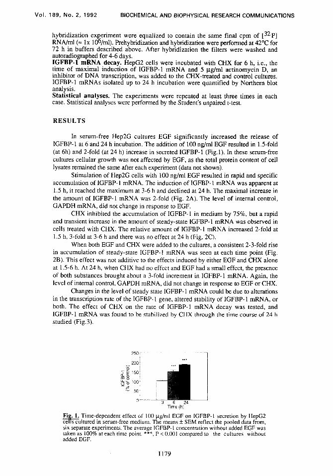
In serum-free Hep2G cultures EGF significantly increased the release of IGFBP-1 at 6 and 24 h incubation. The addition of 100 @ml EGF resulted in 1.5-fold (at 6h) and 2-fold (at 24 h) increase in secreted IGFBP-1 (Fig.1). In these serum-free cultures cellular growth was not affected by EGF, as the total protein content of cell lysates remained the same after each experiment (data not shown).
Stimulation of Hep2G cells with 100 rig/ml EGF resulted in rapid and specific accumulation of IGFBP- 1 mRNA. The induction of IGFBP- 1 mRNA was apparent at 1.5 h, it reached the maximum at 3-6 h and declined at 24 h. The maximal increase in the amount of IGFBP-1 mRNA was 2-fold (Fig. 2A). The level of internal control, GAPDH mRNA, did not change in response to EGF.
CHX inhibited the accumulation of IGFBP-1 in medium by 75%, but a rapid and transient increase in the amount of steady-state IGFBP-1 mRNA was observed in cells treated with CHX. The relative amount of IGFBP-1 mRNA increased 2-fold at 1.5 h, 3-fold at 3-6 h and there was no effect at 24 h (Fig. 2C).
When both EGF and CHX were added to the cultures, a consistent 2-3-fold rise in accumulation of steady-state IGFBP-1 mRNA was seen at each time point (Fig. 2B). This effect was not additive to the effects induced by either EGF and CHX alone at 1.5-6 h. At 24 h, when CHX had no effect and EGF had a small effect, the presence of both substances brought about a 3-fold increment in IGFBP-1 mRNA. Again, the level of internal control, GAPDH mRNA, did not change in response to EGF or CHX.
Changes in the level of steady state IGFBP-1 mRNA could be due to alterations in the transcription rate of the IGFBP- 1 gene, altered stability of IGFBP- 1 mRNA, or both. The effect of CHX on the rate of IGFBP-1 mRNA decay was tested, and IGFBP-1 mRNA was found to be stabilized by CHX through the time course of 24 h studied (Fig.3).
0 3 6 24 Time (h)
Fig. 1. Time-dependent effect of 100 @ml EGF on IGFBP-1 secretion by HepG2 cells cultured in serum-free medium. The means f SEM reflect the oooled data from. six separate experiments. The average IGFBP-I concentration witho& added EGF was taken as 100% at each time point. *** P < 0.001 compared to the cultures without , added EGF.
1179

Vol. 189, No. 2, 1992 BIOCHEMICAL AND BIOPHYSICAL RESEARCH COMMUNICATIONS
1.5 3 6 24 III, <Time (h)
EGF - + + - - + + - - + + - - + + - CHX - - + + - - + + - - + + - - + +
IGFBP-1 GAPDH
1.6 kb 1.3 kb
200 EGF
200 CHX
100
LKl 1.5 3 6 24
Time (h)
Fig. 2. The effects of EGF and CHX on steady-state IGFBP-1 mRNA in HepG2 cellsThe cells were incubated up to 24 h in the presence and absence of 100 rig/ml EGF and 2 ug/ml CHX. Total RNA (5 u&lane) was electrophoresed through a 1.2% a arose-formaldehyde gel and transfered to a nitrocellulose filter and hybridized with 1 5 P] labeled IGFBP-1 cDNA and GAPDH cDNA. The mRNA levels were further quantitated with densitometric scanning in relation to GAPDH mRNA. The values are expressed as percentage of the control. The mean IL SEM is shown for three separate experiments. The autoradiograph is representative of a Northern blot showing 1.6 kb human IGFBP-1 mRNA and 1.3 kb GAPDH mRNA. A) The effect of EGF, B) EGF and CHX and C) CHX on the relative amount of LGFBP-1 mRNA. The average amount of IGFBP-I mRNA without added EGF or CHX was taken as 100% at each time point.
Nuclear run-on experiments were done to assess the effect of EGF and CHX on the transcription rate of IGFBP- 1 gene. In three separate experiments EGF and CHX did not affect the rate of IGFBP- 1 gene transcription at 2 h, neither did they have any significant effect on the transcrption rate of the internal control, GAPDH gene, or on pGem3, the negative control (Fig. 4).
DISCUSSION
EGF was found to increase IGFBP- 1 secretion in human hepatoma cells. This took place concominantly with increased amount of steady-state IGFBP-1 mRNA. EGF has been shown to promote DNA replication in liver (8). The effect of EGF on IGFBP-1 secretion could not have resulted from increased cell number, because
1180

Vol. 189, No. 2, 1992 BIOCHEMICAL AND BIOPHYSICAL RESEARCH COMMUNICATIONS
Control CHX
0 3 6 12 24”O 3 6 12 24’Time(h)
IGFBP-l-
GAPDH-
I I
6 12 18 24 Time (h)
Fig. 3. CHX stabilizes IGFBP-1 mRNA decay. HepC2 cells were grown for 6 h in the absence or presence of 2 lg/ml CHX. Thereafter thegene transcription was blocked by the addition of actinomycin D (5 pg/ml). RNA was isolated at variouQme points after this addition and analyzed by Northern blot hybridization, using [ P] labeled IGFBP-1 cDNA and GAPDH cDNA. The average amount of IGFBP-I mRNA at the time of actinomycin D addition was taken as 100%. The relative amount of IGFBP-1 mRNA remaining is shown for control (0) and CHX-treated cells (0). The autoradiograph is representative of three separate experiments.
HepG2 cells did not proliferate during experiment in serum-free conditions irrespective of whether EGF was added or not. Furthermore, the effect of EGF on IGFBP-1 mRNA accumulation was specific, because there was no change in GAPDH mRNA. The EGF-induced effect on the amount of IGFBP- 1 mRNA was rapid, which could be interpreted as a result from direct stimulation of transcription. Yet in the nuclear run-on transcription analysis, EGF had no detectable effect on IGFBP-1 gene transcription rate.
IGFBP-I-
GAPDH - pGem-
a
IGFBP-l-
GAPDH -
pGem -
IGFBP-l-
_ GAPDH- ”
Fig. 4. EGF or CHX have no significant effect on IGFBP-I gene transcription. Nuclei isolated from control cells (1) or cells incubated with either 100 pg/ml EGF (2) or 2 @ml CHX (3) for 2 h were allowed to continue transcription in the presence of @?P]UTP. The labeled transcripts were hybridized to IGFBP-1 cDNA immobilized to nitrocellulose filters. GAPDH cDNA and pGem3 dots are shown as controls. Densitometric scanning revealed no significant differences.
1181
Vol. 189, No. 2, 1992 BIOCHEMICAL AND BIOPHYSICAL RESEARCH COMMUNICATIONS
The addition of protein synthesis inhibitor did not abolish or decrease the effect of EGF on IGFBP-I mRNA accumulation. Consistent with our previos report (14) CHX itself rapidly and transiently increased the amount of steady-state IGFBP-1 mRNA in relation to GAPDH mRNA. The abudance of the latter did not change in the presence of CHX. In nuclear run-on analysis, CHX did not affect the IGFBP-1 gene transcription rate, but it stabilized the decay of IGFBP-1 mRNA. The observed stabilizing effect could account for the increased amount of steady-state IGFBP-1 mRNA. It is possible that CHX operates at the postranscriptional level and prevents IGFBP-1 mRNA from degradation. This implies that a labile protein repressor is involved in the regulation of IGFBP-1 mRNA degradation (18). It also suggests that the secondary structure of human IGFBP- 1 mRNA, or its association with protective proteins, constitutes an important regulatory mechanism in human IGFBP-1 gene expression.
In conclusion, acute stimulation of IGFBP- 1 by EGF in hepatoma cells implies a role for IGFBP-1 in the liver regeneration and, in view of the possible role of IGFBP-1 in glucose counterregulation, these results indicate a novel mechanism by which EGF may exert its metabolic action in the liver.
ACKNOWLEDGMENTS
I am grateful to Dr. Pekka Leinonen for advice and discussions, to Prof. Markku Seppala for valuable comments about the manuscript, and to Ms. Merja Turunen for expert technical assistance. This work was supported by grants from the Academy of Finland, the Ida Montin Foundation, the Medical Research Foundation Duodecim,‘The Cancer Society of Finland, the Finnish Social Insurance Institution, the Sigrid Juselius Foundation, the University of Helsinki and the Nordisk Insulin Foundation Committee.
REFERENCES
5.
6.
7.
8.
9.
10.
11.
Shimasaki, S. and Ling, N. (1991) Prog. Growth Factor Res. 3, 243-266.
Humbel, R. E. (1990) Eur. J. Biochem. 190,445462.
Baxter, B. C and Martin, J. L. (1989) Prog. Growth Factor Res. 1, 49-68.
Julkunen, M., Koistinen, R., Aalto-Setala, K., Seppala, M., JInne, 0. A. and Kontula, K. (1988) FEBS Lett. 236, 295302.
Lewitt, M. S. and Baxter R. C. (1991) Mol. Cell. Endocrinol. 79, C147-152.
Suikkari, A-M., Koivisto, V. A., Rutanen, E-M., Yki-Jiztrvinen, H., Karonen, S- L. and Sepplll, M. (1988) J. Clin. Endocrinol. Metab. 66, 266-272.
Powell, D. R., Suwanichkul, A., Cubbage, M. L., DePaolis, L. A., Snuggs, M. B. and Lee, P. D. K. (1991) J. Biol. Chem. 266, 18868-18876.
Marti, U., Burwen, S.J. and Jones, A. L. (1989) Hepatology 9, 126-138.
Andus, T., Bauer, J. and Gerok, W. (1991) Hepatology, 13, 364-375.
Angervo, M., Koistinen, R. and Sepplll, M. (1992) J. Endocrinol. 134, 127- 131.
Angervo, M., Koistinen, R. and Seppall M. (1991) Abstract no C 29. 2nd International IGF Symposium, San Francisco, USA.
1182

.
Vol. 189, No. 2, 1992 BIOCHEMICAL AND BIOPHYSICAL RESEARCH COMMUNICATIONS
12.
13.
Knowles, B. B., Howe, C. C. and Aden, D. P. (1980) Science 208,497-499.
Kelly, J. H. and Darlington, G. J. (1989) In Vitro Cell. Dev. Biol. 25, 217- 222.
14. Angervo, M., Leinonen, P., Koistinen, R., Julkunen, M. and Seppala, M. (1992) J. Mol. Endocrinol. In press.
15.
16.
Chomczynski P, Sacchi N (1987) Anal. Biochem. 162: 156-159.
Tso, J. Y., Sun, X-H., Kao, T., Reece, K. S. and Wu, R. (1985) Nucleic Acids Res. 13, 24852502.
17. Nevins, J. R. (1987) Meth. Enzymol. 152; 234-241.
18. Subramaniam, M.,Schmidt, L. J., Crutchfield, C. E. and Getz, M. J. (1989) Nature 340, 64-66.
1183