epigenetic alteration of receptor tyrosine kinases …...epigenetic alteration of receptor tyrosine...
TRANSCRIPT
14
Epigenetic Alteration of Receptor Tyrosine Kinases in Cancer
Anica Dricu et al.,* Department of Biochemistry,
University of Medicine and Pharmacy of Craiova, Romania
1. Introduction
In the process of cellular carcinogenesis, genetic and epigenetic mechanisms contribute to
abnormal expression of genes. Conrad Waddington introduced the term ‘‘epigenetics’’ in
1942 (Goldberg, Allis et al. 2007) to describe heritable changes in gene expression that have
no connection with changes in DNA sequence (Yoo and Jones 2006; Goldberg, Allis et al.
2007). The molecular basis of epigenetics is complex and involves DNA methylation, histone
modifications, chromatin remodeling and microRNAs (Esteller 2006).
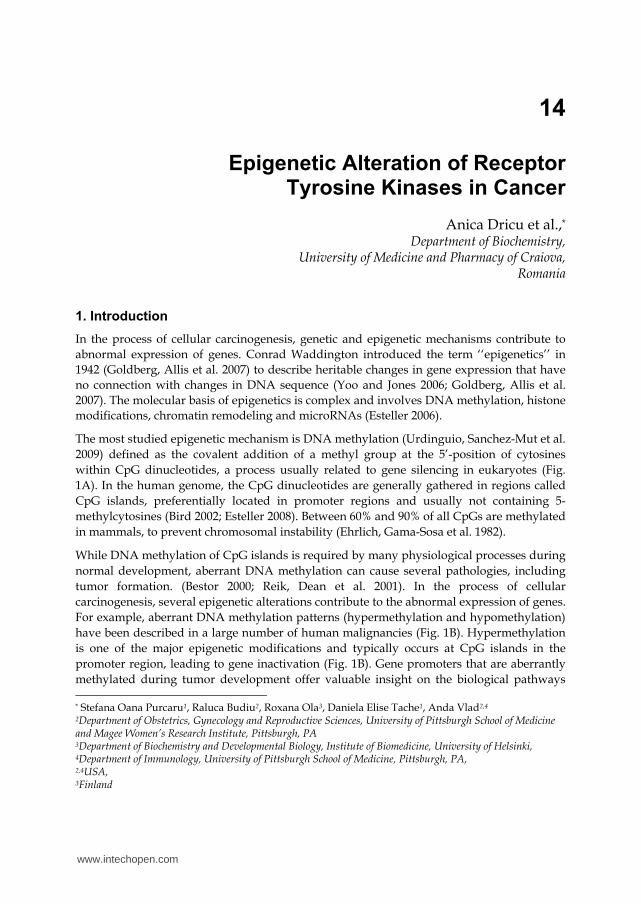
The most studied epigenetic mechanism is DNA methylation (Urdinguio, Sanchez-Mut et al.
2009) defined as the covalent addition of a methyl group at the 5’-position of cytosines
within CpG dinucleotides, a process usually related to gene silencing in eukaryotes (Fig.
1A). In the human genome, the CpG dinucleotides are generally gathered in regions called
CpG islands, preferentially located in promoter regions and usually not containing 5-
methylcytosines (Bird 2002; Esteller 2008). Between 60% and 90% of all CpGs are methylated
in mammals, to prevent chromosomal instability (Ehrlich, Gama-Sosa et al. 1982).
While DNA methylation of CpG islands is required by many physiological processes during
normal development, aberrant DNA methylation can cause several pathologies, including
tumor formation. (Bestor 2000; Reik, Dean et al. 2001). In the process of cellular
carcinogenesis, several epigenetic alterations contribute to the abnormal expression of genes.
For example, aberrant DNA methylation patterns (hypermethylation and hypomethylation)
have been described in a large number of human malignancies (Fig. 1B). Hypermethylation
is one of the major epigenetic modifications and typically occurs at CpG islands in the
promoter region, leading to gene inactivation (Fig. 1B). Gene promoters that are aberrantly
methylated during tumor development offer valuable insight on the biological pathways * Stefana Oana Purcaru1, Raluca Budiu2, Roxana Ola3, Daniela Elise Tache1, Anda Vlad2,4
2Department of Obstetrics, Gynecology and Reproductive Sciences, University of Pittsburgh School of Medicine and Magee Women’s Research Institute, Pittsburgh, PA 3Department of Biochemistry and Developmental Biology, Institute of Biomedicine, University of Helsinki, 4Department of Immunology, University of Pittsburgh School of Medicine, Pittsburgh, PA, 2,4USA, 3Finland
www.intechopen.com
DNA Methylation – From Genomics to Technology 304
that are commonly interrupted during tumorigenesis (Klarmann, Decker et al. 2008; Suzuki,
Toyota et al. 2008) . Gene promoter hypermethylation is related to the inhibition of cancer-
related genes such as tumor suppressor genes and DNA mismatch repair genes (Feinberg
and Tycko 2004; Yoo and Jones 2006) (Fig. 1B). Several key tumor suppressor genes have
been found to exhibit promoter hypermethylation more often than genetic disruption (Chan,
Glockner et al. 2008).
Fig. 1. Aberrant DNA methylation is involved in in the development of cancer.
DNA methylation is a process of covalent addition of a methyl group at the 5’-position of cytosines (A) within CpG dinucleotides. DNA hypermethylation is commonly interrupted during tumorigenesis and is related to the inhibition of tumor suppressor genes and DNA mismatch repair genes while DNA hypometylation appears to be involved in tumor cell development by activation of oncogenes (B).
Global hypomethylation has also been implicated in the development and progression of
cancer, through different mechanisms (Yoo and Jones 2006). DNA hypometylation, which is
the first epigenetic event identified in cancer cell (Feinberg and Tycko 2004), appears to be
involved in tumor cell development by activation of oncogenes, generation of chromosomal
instability and loss of imprinting (Feinberg and Tycko 2004) (Fig. 1B). The low level of DNA
methylation in tumors can increase the expression of several oncoproteins involved in cell
growth and survival, apoptosis and cell cycle regulation (Jun, Woolfenden et al. 2009).
www.intechopen.com
Epigenetic Alteration of Receptor Tyrosine Kinases in Cancer 305
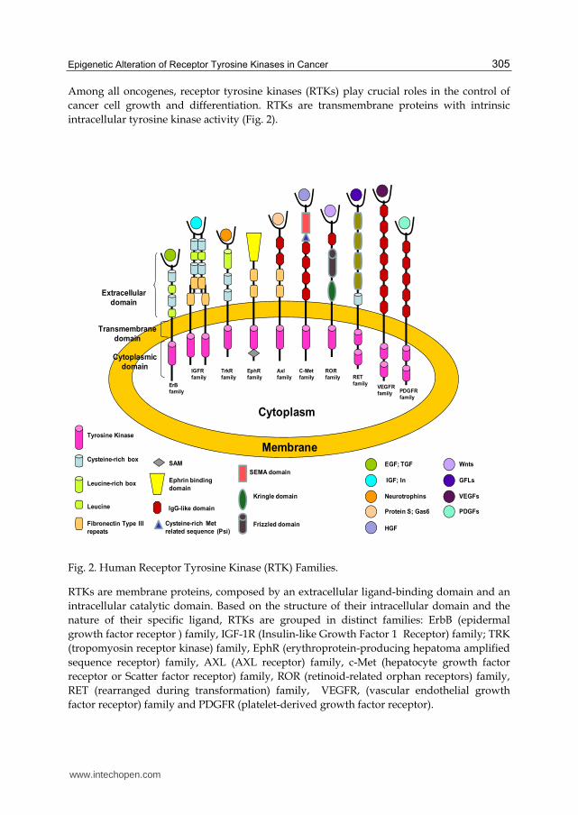
Among all oncogenes, receptor tyrosine kinases (RTKs) play crucial roles in the control of
cancer cell growth and differentiation. RTKs are transmembrane proteins with intrinsic
intracellular tyrosine kinase activity (Fig. 2).
Fig. 2. Human Receptor Tyrosine Kinase (RTK) Families.
RTKs are membrane proteins, composed by an extracellular ligand-binding domain and an
intracellular catalytic domain. Based on the structure of their intracellular domain and the
nature of their specific ligand, RTKs are grouped in distinct families: ErbB (epidermal
growth factor receptor ) family, IGF-1R (Insulin-like Growth Factor 1 Receptor) family; TRK
(tropomyosin receptor kinase) family, EphR (erythroprotein-producing hepatoma amplified
sequence receptor) family, AXL (AXL receptor) family, c-Met (hepatocyte growth factor
receptor or Scatter factor receptor) family, ROR (retinoid-related orphan receptors) family,
RET (rearranged during transformation) family, VEGFR, (vascular endothelial growth
factor receptor) family and PDGFR (platelet-derived growth factor receptor).
VEGFs
PDGFs
Wnts
Extracellular
domain
Membrane
Cytoplasm
Transmembrane
domain
Cytoplasmic
domain
ErB
family
Leucine-rich box
Tyrosine Kinase
Cysteine-rich box
TrkR
family
Leucine
IGFR
family
Fibronectin Type III
repeats
EGF; TGF
IGF; In
Neurotrophins
SAM
Ephrin binding
domain
EphR
family
Axl
family
IgG-like domainProtein S; Gas6
Cysteine-rich Met
related sequence (Psi)
SEMA domain
C-Met
family
HGF
Kringle domain
Frizzled domain
ROR
family RET
family
GFLs
VEGFR
family PDGFR
family
www.intechopen.com
DNA Methylation – From Genomics to Technology 306
RTKs overexpression or overactivation has been described in almost all tumor types. The human genome is reported to contain 58 genes encoding receptor protein kinase, grouped into 20 classes, or subfamilies, based upon their kinase domain sequence (Robinson, Wu et al. 2000; Lemmon and Schlessinger 2010). Many RTK genes contain a typical CpG island and alterations in RTK promoter methylation have been linked to cancer development and progression (Datta, Kutay et al. 2008). Furthermore, emerging data suggests that acquired resistance to conventional cancer therapy results from progressive accumulation of RTK epigenetic modifications.
We review herein the epigenetic alterations of ten RTK families (Fig. 1), discuss their role in tumor development and implications for the response of cancer cells to conventional therapy.
2. ErbB family
ErbB protein family, or epidermal growth factor receptor (EGFR) family, consists of four structurally related receptors, with a well described tyrosine kinase activity: ErbB-1, also called epidermal growth factor receptor (EGFR), ErbB-2, named HER2 in humans and neu in rodents, ErbB-3, also named HER3 and ErbB-4 or HER4 (Bublil and Yarden 2007). ErbB-1 and ErbB-4 bind different polypeptide extracellular ligands including the epidermal growth factor (EGF), transforming growth factor ┙ (TGF┙), amphiregulin, betacellulin, epiregulin, heparin binding EGF, epigen, and neuregulins 1-4, which share a conserved epidermal growth factor (EGF) domain, giving rise to a diverse signaling network (Citri and Yarden 2006). ErbB2 is an “orphan” receptor and ErbB3 lacks the tyrosine kinase activity, hence, both signal through ErbB family heterodimers (Alimandi, Romano et al. 1995).
ErbB receptors/ligands are involved in many fundamental processes during organogenesis and adulthood: cell growth, differentiation, proliferation, apoptosis, motility, invasion, repair, survival and cell-cell interaction (Yarden and Sliwkowski 2001). In tumors, there is abnormal signalling via the ErbB pathway, mostly as a result of receptor overexpression or constitutive activation (Yarden and Sliwkowski 2001).
The egfr proto-oncogene localizes on chromosome 7p12, and the promoter region was
described as a CG rich sequence, which lacks a TATA box and displays a large CpG island
that extends into exon 1 (Kageyama, Merlino et al. 1988). EGFR expression is primarily
regulated at the mRNA levels (Xu, Richert et al. 1984; Merlino, Ishii et al. 1985) and many
human malignancies with epithelial origin including brain, head and neck squamous cell
carcinoma (HNSCC), breast, esophagus, gastric, colon and lung cancers are characterized by
EGFR overexpression. In glioma, lung, ovarian and breast cancers, mutations in the egfr
gene are responsible for the receptor overactivity (Moscatello, Holgado-Madruga et al.
1995). Amplifications at egfr locus were also described in gliomas (Li, Chang et al. 2003),
colorectal cancers, and less frequently in breast cancer (Al-Kuraya, Schraml et al. 2004; Ooi,
Takehana et al. 2004). Genetic polymorphism of egfr in the intron 1 region, involving CA
single sequence repeats (SSR), constitutes another mechanism that may influence egfr gene
transcription (Gebhardt, Zanker et al. 1999). More recently it was shown that DNA
methylation may also be responsible for the aberrant transcription of egfr in neoplastic cells
(Kulis and Esteller 2010). Petrangeli et al., attempted for the first time to reveal the
implication of DNA methylation in the control of EGFR function in breast cancer patients.
www.intechopen.com
Epigenetic Alteration of Receptor Tyrosine Kinases in Cancer 307
However, a methylation profile comparison of egfr promoter in tumoral versus
perineoplastic tissues could not identify any differences in egfr gene methylation (Petrangeli,
Lubrano et al. 1995). In colorectal cancer, Montero et al., identified egfr methylation only in
the adjacent normal colon tissue but not in the corresponding tumor tissue analyzed
(Montero, Diaz-Montero et al. 2006). In contrast with these results, Scartozzi et al., identified
egfr promoter methylation in 39% of the colorectal tumors samples (Scartozzi, Bearzi et al.
2011). Furthermore, in a subsequent study of a larger cohort, the same authors showed that
58% of patients treated with monoclonal antibodies targeting EGFR had egfr, monoallelic or
biallelic methylated (Scartozzi, Bearzi et al. 2011) and revealed a direct correlation between
egfr methylation status and clinical outcome. However, no correlation between the
methylated egfr and absence of protein expression was evident, as EGFR protein was
detected in samples displaying a methylated egfr.
Different methylation density in the egfr promoter was also identified in three breast cancer
cell lines (MB435, CAMA1, and MB453), in SAOS-2 human osteosarcoma cell line, SF-539
glioma cell line, and in two hematopoietic cell lines Raji and Raji Dac where a clear
association between the methylated egfr and the loss of protein expression was evident
(Montero, Diaz-Montero et al. 2006). Furthermore, a low density of methylated cytosines
within the egfr promoter was also identified in 20% of primary breast, 11% of lung and 35%
of head and neck tumors. It is therefore apparent that egfr promoter methylation is one
potential mechanism responsible for the transcriptional control of egfr oncogene in cancer.
The second member of the EGF receptor family, Her-2/neu proto-oncogene, located on
human chromosome 17q21 contains, similarly to EGFR, a large CpG island within the gene
promoter region. Even though the regulatory region of the gene displays more than 10
potential sites of methylation, no aberrant methylation of her2 could be detected in different
tumor samples, including breast, lung, colon, head and neck and cell lines (Montero, Diaz-
Montero et al. 2006). Nonetheless, in epithelial ovarian cancer, the expression of DNA
demethylase (dMTase) correlates with poor methylation of her2, suggesting that dMTase
enzyme is involved in Her2 promoter demethylation (Hattori, Sakamoto et al. 2001). In
addition, overexpression of the rat neu oncogene in mammary tumors of MMTV/c-neu
transgenic mice induced demethylation of the MMTV promoter (Zhou, Chen et al. 2001). In
the same mouse model Kmieciak et al., later showed that Interferon ┛ (IFN-┛) mediates
epigenetic changes in neu oncogene, resulting in neu antigen loss and tumor escape
(Kmieciak, Knutson et al. 2007).
Only a paucity of data describing the transcriptional control by any epigenetic mechanisms
of Her 3 and Her 4 currently exists. DNA methylation of a CpG island in the promoter
region seems to be involved in her4 transcriptional suppression in breast cancer cell lines
and primary breast carcinomas. Moreover, Das, P.M., et al., demonstrated a direct
correlation between the her4 methylation status and HER4 protein expression and proposed
her4 promoter methylation as a negative prognostic indicator at least in breast cancer (Das,
Thor et al. 2010).
Identification of DNA methylation as a regulatory mechanism and a prognostic marker for
three out of four EGF receptors provides promising new alternatives to targeting ERB
signaling with better selectivity, safety and efficacy.
www.intechopen.com
DNA Methylation – From Genomics to Technology 308
3. IGF receptor family
The insulin-like growth factor (IGF) family is composed of two ligands – (IGF-1 and IGF-2),
two receptors – (IGF-1R and IGF-2R), six high-affinity IGF-binding proteins – (IGFBP 1-6)
and several associated IGFBP degrading enzymes (proteases). The IGF family has a critical
role in the development and maintenance of normal tissue homeostasis and it appears to be
involved a number of pathological states, including cancer.
Insulin- like growth factor type-1 receptor (IGF-1R), the main receptor of IGF family, is a
transmembrane heterotetramer, linked to the PI3K/Akt and MAPK signal transduction
pathways. Approximately 75% of the IGF-1R promoter region consists of cytosine and
guanine. The IGF-1R promoter is also TATA-less and CCAAT-less and contains several
potential binding sites for Sp1, ETF and AP-2 nuclear transcription factors (Werner,
Stannard et al. 1990). IGF-1R is overexpressed in several types of cancer and their
involvement in cancer cells’ response to treatment has also been reported (Kanter-
Lewensohn, Dricu et al. 2000; Cosaceanu, Budiu et al. 2007). In the human IGF-1R promoter
region, bioinformatic analysis revealed the presence of multiple CpG dinucleotides
(Schayek, Bentov et al. 2010). The presence of CpG islands in the promoter region of IGF-1R
gene supports the theory that an altered methylation status of the IGF-1R promoter may be
responsible for the IGF-1R oncogene overexpression in various cancers. In 2010, we
described for the first time the partial methylation of IGF-1R promoter in three subtypes of
non-small cell lung cancer (NSCLC): large cell lung cancer, squamous cell carcinoma and
adenocarcinoma. We found the same level of IGF-1R promoter methylation in all NSCLC
subtypes and no correlation between the gene methylation level and receptor protein
expression (Ola 2010).
While several studies suggest IGF-1R overexpresion as a key in prostate cancer initiation and progression (Hellawell, Turner et al. 2002; Turney, Turner et al. 2011), Chott at al. demonstrate that loss of IGF-1R may contribute to prostate cancer progression (Chott, Sun et al. 1999). In line with these findings, Schayek et al., 2010, showed that progression towards metastatic stages in prostate cancer is correlated with a dramatic reduction in both total IGF-1R protein levels and basal phospho-IGF-1R values, which reflects a decrease in IGF-1R activation (Schayek, Bentov et al. 2010). Furthermore, studies on the IGF-1R methylation of six prostate cancer cell lines showed that the IGF-1R promoter is unmethylated in all examined cell lines, suggesting that the gene silencing in metastatic prostate cancer is probably not caused by direct gene promoter methylation (Schayek, Bentov et al. 2010). However, our studies from two glioblastoma lines sugget that IGF-1R is, at least in this setting, partially methylated (Ola 2010).
Many promising agents that inhibit the key enzymes involved in establishing and maintaining the epigenetic changes have been identified and tested in various types of pathologies. One of those agents is S-Adenosylmethionine (SAM), a methyl donor agent. Several studies have reported DNA hyper-methylation, after SAM exposure (Fuso, Cavallaro et al. 2001; Detich, Hamm et al. 2003). In contrast, evidence from our studies on S-Adenosylmethionine-induced cytotoxicity in both glioblastoma and non-small cell lung cancer cell lines demonstrates that SAM does not affects the IGF-1R methylation status (Ola 2010; Ola 2010). While IGF1R epigenetics seems important, the exact mechanisms underlying IGF-1R overexpression in cancer are far from being understood.
www.intechopen.com
Epigenetic Alteration of Receptor Tyrosine Kinases in Cancer 309
Insulin- like growth factor type-2 receptor (IGF-2R, also known as the cation-independent mannose 6-phosphate receptor), the second cell-surface receptor of the IGF family, is a 275 ± 300 kDa glycoprotein, comprised of an extracytoplasmic domain made up of 15 contiguous cysteine-rich repeats, a single transmembrane-spanning domain and a carboxyl -terminal cytoplasmic domain (Ghosh, Dahms et al. 2003). The receptor does not appear to have any protein kinase activity but the carboxyl-terminal cytoplasmic domain has been shown to facilitate endocytosis (Probst, Puxbaum et al. 2009) and intracellular sorting of lysosomal enzymes (Munier-Lehmann, Mauxion et al. 1996). IGF-2R is a maternally expressed gene regulated by epigenetic modifications and one of the classical examples of tissue-specific and species-dependent imprinted genes. There are several studies demonstrating that IGF2 ligand is also an imprinted gene and it has been proved that loss of imprinting causes proliferation of transformed cells in Wilm’s tumors, by elevating the level of available IGF2 ligand (Steenman, Rainier et al. 1994; Vu 1996). Both IGF2 and IGF2R genes possess a CG-rich and TATA-less promoter (Szentirmay, Yang et al. 2003). In mice, methylation of region 2, a region rich in cytosine-guanine doublets in the second intron of IGF-2R, represents the imprinting signal that maintains expression of the maternal allele (Stoger, Kubicka et al. 1993). Wutz and his colleagues tested the role of region 2 and the influence of chromosome location on IGF-2R imprinting, using mouse YAC-T1/P and YAC-T1/PR2 transgenes (Wutz, Smrzka et al. 1997). Their results show that deletion of region 2 of the IGF-2R gene is associated with loss of imprinting and restoration of biallelic IGF2R expression, demonstrating the primary role for the region 2 and the negligible role for chromosomal location in IGF2R imprinting (Wutz, Smrzka et al. 1997).
Increasing evidence from genetic studies currently implicate IGF-2R as a tumour suppressor in a variety of malignancies. Loss of heterozygosity (LOH) at the gene locus on 6q26 ± 27 has been reported for a number of tumour types, including breast (Chappell, Walsh et al. 1997), liver (Oka, Waterland et al. 2002), lung (Kong, Anscher et al. 2000) and head and neck (Jamieson 2003) cancers. Further investigations of the involvement of both IGF-1R and IGF-2R and theirs interaction with the ligands are required to fully understand the role of these complex molecules in cancer.
Unlike genetic changes, epigenetic events do not alter the DNA code and are potentially reversible. Therefore, reactivation of epigenetically silenced genes like IGF2-R could provide attractive therapeutic opportunities.
4. C-MET receptor family
The c-met oncogene is a tyrosine kinase receptor binding to the hepatocyte growth factor (HGF) also known as the scatter factor (SF). Signals through c-Met are required for normal mammalian development and fundamental processes like cell migration, differentiation, proliferation, cell growth, branching and angiogenesis (Birchmeier, Birchmeier et al. 2003). Although HGF is the only known ligand for C-Met, glial cell derived neurotrophic factor (GDNF) also seems to be able to activate C-Met, albeit indirectly (Popsueva, Poteryaev et al. 2003).
C-met gene has been mapped to chromosome 7q31 and its regulatory region is GC-rich. It contains no TATA box, and displays a CpG island with an increased frequency of CpG, suggesting that an aberrant transcriptional regulation may play a critical role in oncogene activation (Seol and Zarnegar 1998).
www.intechopen.com
DNA Methylation – From Genomics to Technology 310
Many studies to date provide evidence that dysregulation of c-Met activity is a key event in
the initiation and progression of carcinogenesis (Maulik, Shrikhande et al. 2002). Mutations
in c-met were identified in papillary renal carcinoma, gastric and liver cancer, small and
non–small cell lung cancers, and head and neck squamous cell carcinomas (Peruzzi and
Bottaro 2006). Furthermore, amplification of c-met in gastric, colorectal and lung carcinoma
seems to inversely correlate with survival (Kuniyasu, Yasui et al. 1992; Zeng, Weiser et al.
2008; Lee, Seo et al. 2011).
The possibility that an altered methylation of c-met promoter induces overexpression of C-
MET protein was, for the first time, addressed in papillary carcinoma although no changes
in c-Met promoter methylation could be detected in tumour tissue compared to normal
tissue (Scarpino, Di Napoli et al. 2004). Evidence from Morozov et al demonstrated that the
death-domain associated protein (Daxx) (a component of a multiprotein repression
complex), preferentially binds to the c-met promoter together with Histone deacetylase 2
(HDAC2), leading to c-met transcriptional repression (Morozov, Massoll et al. 2008).
However, DNA methylation seems not to be involved in the Daxx-mediated repression of
the c-met promoter (Morozov, Massoll et al. 2008). Further studies are needed in order to
elucidate the possible implication of c-met promoter methylation in carcinogenesis.
Despite the transcriptional repression of c-met proto-oncogene by chromatin modifications, direct evidence of DNA methylation as a responsible mechanism for c-met promoter function is missing and needs to be further investigated.
5. Trk receptor family
Tropomyosin-receptor-kinase (Trk) receptors belong to a family of tyrosine kinases that control synaptic strength and plasticity in the mammalian nervous system (Huang and Reichardt 2003). Trk receptors bind neurotrophins and trigger downstream activation of several signaling cascades that affect normal physiological processes, like neuronal survival and differentiation (Segal 2003).
TrkA, TrkB, and TrkC are the three most common types of trk receptors. Each of them has different binding affinity to certain types of neurotrophins. The receptors act through different intracellular pathways and the differences in their signaling can generate a diversity of biological responses (Segal 2003). The Trk oncogene was initially identified in a colon carcinoma and its identification led to the discovery of the first member, TrkA (Huang and Reichardt 2003). The oncogene was produced by a mutation in chromosome 1 that resulted in the fusion of the first seven exons of tropomyosin to the transmembrane and cytoplasmic domains of the then-unknown TrkA receptor (Martin-Zanca, Hughes et al. 1986).
Accumulating evidence now suggests that TrkA, TrkB and TrkC play an important role in
the malignant behavior of cancer cells, like increased metastasis, proliferation and survival
(Sclabas, Fujioka et al. 2005; Jin, Kim et al. 2010). Studies on different tumors show that TrkC
seems to be highly expressed in neuroblastoma, medulloblastoma, (Segal, Goumnerova et
al. 1994; Yamashiro, Liu et al. 1997; Grotzer, Janss et al. 2000) and breast cancer (Bardelli,
Parsons et al. 2003; Wood, Calhoun et al. 2006). Furthermore, overexpression of TrkA was
reported in papillary thyroid and colon carcinoma (Nakagawara 2001), and of TrkB in
malignant keratinocytes (Slominski and Wortsman 2000) and pancreatic cancer (Sclabas,
www.intechopen.com
Epigenetic Alteration of Receptor Tyrosine Kinases in Cancer 311
Fujioka et al. 2005). Recent evidece from Jiin et al show that while Trks might be important
for liver cancer metastasis and are highly expressed during the course of tumor progression,
no aberrant promoter methylation could be detected. In contrast, low or undetectable
expression level of Trk receptors in normal liver cell lines demonstrated a dramatically
increased pattern of methylation (Jin, Lee et al. 2011).
Given their high level of expression in cancer and their roles in metastasis, current studies now attempt to decipher their functions as oncogenic tyrosine kinases during malignant transformation, and to define their potential as attractive targets for therapeutic intervention (Shawver, Slamon et al. 2002).
6. Eph receptor family
The erythroprotein-producing hepatoma amplified sequence (Eph) receptor tyrosine kinase family is the largest family of tyrosine kinases, comprising at least 14 Eph receptors and 8 ligands (Manning, Whyte et al. 2002). Based upon sequence similarities in their extracellular domains and their ability to bind to ligands, Eph receptors are divided into two groups, EphA and EphB. The EphA members (EphA1-A8 and A10) bind to the glycosyl-phospahtidyl-inositol (GPI) - linked ligands EFNA1-5, whereas EphB1-B4 and B6 belonging to the EphB receptor family binds the transmembrane ligands EFNB1-3 (Pasquale 2004). Upon ephrin binding, Eph receptors are clustered, phosphorylated and kinase activated (Bruckner and Klein 1998). Different pathways have been shown to be implicated in Eph signaling, including activation of the MAPK/ERK pathways by EphB2 receptor (Huusko, Ponciano-Jackson et al. 2004), inhibition of the Ras/ERK1/2 signaling cascade by EphA2, and phosphorylation of Src kinases and Akt by EphA2 and EphA4 (Guo, Miao et al. 2006).
Various human tissues differentially express the Eph/ephrin family (Hafner, Schmitz et al.
2004; Fox, Tabone et al. 2006; Hafner, Becker et al. 2006). Eph involvement in developmental
processes, especially in embryonic development, vasculature and nervous system formation
has been reported (Kullander and Klein 2002; Himanen and Nikolov 2003).
A growing body of evidence currently indicates that Eph gene family also plays an important role in carcinogenesis and tumor progression in many types of cancer (Andres, Reid et al. 1994; Walker-Daniels, Coffman et al. 1999; Liu, Ahmad et al. 2002; Kinch, Moore et al. 2003; Fang, Brantley-Sieders et al. 2005; Noren, Foos et al. 2006; Merlos-Suarez and Batlle 2008). Eph receptors were reported to have both tumor promoting activity, by being highly expressed in different human cancers, (i.e. breast, colon, melanomas, lung and prostate cancer) (Zelinski, Zantek et al. 2001; Surawska, Ma et al. 2004; Nakada, Drake et al. 2006; Foubert, Silvestre et al. 2007), and suppressing activity by acting as tumor suppressors (Batlle, Bacani et al. 2005; Noren, Foos et al. 2006). Some tumors are characterized by loss of expression of Eph receptors (EphB2 and B4 receptors in colorectal cancer and EphB6 receptor in breast cancer) (Alazzouzi, Davalos et al. 2005; Davalos, Dopeso et al. 2006; Fox and Kandpal 2006). Gene promotor methylation of epha2, epha3, epha7, ephb2, ephb4 and ephb6 was found in several human solid tumors, including breast, colorectal and prostate cancer, suggesting that an aberrant CpG island methylation, located in the promoter region of tumor related genes, may determine inactivation (Dottori, Down et al. 1999; Alazzouzi, Davalos et al. 2005; Wang, Kataoka et al. 2005; Davalos, Dopeso et al. 2006; Fox and Kandpal 2006; Nosho, Yamamoto et al. 2007). In addition, epigenetic silencing by hypermethylation
www.intechopen.com
DNA Methylation – From Genomics to Technology 312
of EPH/EPRIN genes seems to contribute to the pathogenesis of acute lymphoblastic leukemia, where ephb4 acts as a tumor suppressor (Kuang, Bai et al. 2010).
In breast cancer, EphA2 appears to be important for the progression of the tumor from noninvasive to an invasive phenotype (Fox and Kandpal 2004), potentially via ErbB2 signaling (Brantley-Sieders, Zhuang et al. 2008). Fu et al. demosntrated that EphA5 is frequently down regulated in breast cancer cell lines and tumor tissues via aberrant methylation of its promoter, a finding significantly associated with the clinico-pathologic tumor grade and metastasis to the lymph nodes (Fu, Wang et al. 2010). The authors also reported the presence of a dense GpG island with transcriptional and translational start sites at the 5’ end of the EphA5 gene. Such molecular architecture is entirely consistent with that described for other genes, known as targets for epigenetic silencing during tumorigenesis (Feltus, Lee et al. 2003), an early and frequent event in the development of breast cancer (Lehmann, Langer et al. 2002).
Altogether, these reports suggest that methylation of Eph genes might be used as a potential marker for cancer diagnosis and prognosis and underscore the need for further studies that better define their translational potential.
7. AXL receptor family
The Axl receptors belong to the TAM (Tyro-Axl-Mer) receptor tyrosine kinases family (Lai
and Lemke 1991; O'Bryan, Frye et al. 1991). Axl is a 140-kDa protein, with an extracellular
region comprised of two immunoglobulin-like domains and fibronectin type III repeats, a
transmembrane region and a cytoplasmic domain with kinase activity (O'Bryan, Frye et al.
1991). There are two ligands for Axl: protein S and growth-arrest specific 6 (Gas6), the latter
binding with higher affinity to Axl (Stitt, Conn et al. 1995; Varnum, Young et al. 1995). The
main downstream Axl signaling is triggered through the phosphatidylinositol 3 kinase
(PI3K) pathway (Collett, Sage et al. 2007), although in some circumstances the Janus kinase-
signal transducers and activator of transcription (STAT) (Rothlin, Ghosh et al. 2007) or the
p38 mitogen-activated protein kinase pathway (Allen, Linseman et al. 2002) may also be
induced. The Axl receptor also cooperates with the cytokine receptor signaling network in
order to regulate many biologic functions (Budagian, Bulanova et al. 2005; Gallicchio, Mitola
et al. 2005) like cell survival, proliferation, adhesion and migration (Hafizi and Dahlback
2006). It contributes to vascular smooth muscle homeostasis and regulates endothelial cell
migration and vascular network formation (Korshunov, Mohan et al. 2006; Collett, Sage et
al. 2007).
Axl is overexpressed in several human cancers (Craven, Xu et al. 1995; Ito, Ito et al. 1999; Berclaz, Altermatt et al. 2001; Sun, Fujimoto et al. 2004) where it plays important roles in tumor angiogenesis (Holland, Powell et al. 2005; Li, Ye et al. 2009) and metastasis as demonstrated by studies in lung (Shieh, Lai et al. 2005), prostate (Sainaghi, Castello et al. 2005), breast (Meric, Lee et al. 2002), gastric (Wu, Li et al. 2002) andrenal cell carcinomas (Chung, Malkowicz et al. 2003), as well as glioblastomas (Hutterer, Knyazev et al. 2008). Interestingly, Axl overexpression was reported to induce Imatinib resistance in gastrointestinal stromal tumors (Mahadevan, Cooke et al. 2007), Herceptin resistance in breast cancer (Liu, Greger et al. 2009) or chemotherapy resistance in acute myeloid leukemia (Hong, Lay et al. 2008), supporting its applicability as a potential therapeutic biomarker.
www.intechopen.com
Epigenetic Alteration of Receptor Tyrosine Kinases in Cancer 313
The invasive capacity of cancer cells was shown to be reduced by downregulation of Axl expression, as evidenced in breast and lung cancer (Holland, Powell et al. 2005; Shieh, Lai et al. 2005). Liu R et al. recently showed that Axl is induced in Kaposi sarcoma and Kaposi sarcoma herpesvirus (KSHV) transformed endothelial cells, but also in Kaposi sarcoma cell lines lacking KSHV(Liu, Gong et al. 2010).
Axl is transcriptionally regulated by the Sp1/Sp3 transcription factors and further controlled by CpG island methylation (Mudduluru and Allgayer 2008). Mudduluru et al. showed that transactivation of Axl gene is controled by MZF1, which induces migration, invasion and in vivo metastasis formation (Mudduluru, Vajkoczy et al. 2010). In a subsequent study, focused on the the epigenetic regulation of Axl at post-transcriptional level by micro-RNAs, the same authors showed that three micro-RNAs, miR-34a, miR-199a and miR-199b were frequently methylated and that their expression levels inversely correlates with Axl expression in three types of cancer: non-small cell lung cancer, colorectal cancer and breast cancer (Mudduluru, Ceppi et al. 2011).
TAM receptor inhibitions in animal xenograft tumor models of glioblastoma and breast cancer have provided preliminary validation of this receptor family as a cancer therapy target and further translational studies are needed in order to fully establish its potential.
8. ROR receptor family
Two human Ror RTK-encoding genes, hRor1 and hRor2, the first Ror family members, and
two rat partial complementary DNAs (cDNAs), rRor1 and rRor2 were identified nearly 10
years ago (Masiakowski and Carroll 1992). Ror family members have a conserved domain
structure; the extracellular regions contain immunoglobulin (Ig), cysteine-rich (CRD) and
kringle domains, all of which are thought to mediate protein-protein interactions. The
intracellular domain of Rors contains a tyrosine kinase domain, two regions rich in serine
and threonine separated by a region rich in prolines. There is a high similarity between the
two Ror proteins Ror1 and Ror2 from each species. For instance, hRor1 and hRor2 have a
58% overall amino acid identity and 68% amino acid identity within the kinase domains
(Forrester 2002).
ROR1 belongs to the RTK family of orphan receptors connected with muscle specific kinase and neurotrophin receptors (Masiakowski and Carroll 1992; Valenzuela, Stitt et al. 1995; Glass, Bowen et al. 1996). ROR has a predicted 937 amino acids sequence including an Ig-like domain, cysteine-rich domain, kringle domain, tyrosine kinase domain and proline-rich domain (Yoda, Oishi et al. 2003) .
An alternative spliced form of hRor1, called truncated hRor1 (Reddy, Phatak et al. 1996) has been reported in fetal and adult central nervous system, and was identified in human leukemia and lymphoma cell lines, as well as in a variety of neuroectodermal cancers. However, the function and significance of truncated hRor1 remain unclear (Reddy, Phatak et al. 1996; Forrester 2002).
Although ROR1 is situated on chromosomal region 1p31.3, which is a region where chromosomal aberrations are not frequently found in hematological malignancies, results from gene expression profiling studies show a 43.8-fold increase of ROR1 in chronic lymphocytic leukemia (CLL) cells (Klein, Tu et al. 2001). Furthermore, activation of NF-kB in
www.intechopen.com
DNA Methylation – From Genomics to Technology 314
CLL cells give rise to a functionally active ROR1 protein via Wnt5a and nonclassical Wnt-signaling pathway, supporting a role for ROR1 in the pathogenesis of CLL (Fukuda, Lu et al. 2004). Due to the high level of Ror1 surface expression in leukemic cells and its absence in normal (nonactivated) blood leukocytes (Daneshmanesh, Mikaelsson et al. 2008), ROR1 is now considered a candidate structure for targeted therapy, including monoclonal antibody-based therapies (Reddy, Phatak et al. 1996; Daneshmanesh, Mikaelsson et al. 2008). However, the exact mechanisms behind ROR1 gene regulation are yet to be fully delineated. Despite the recent evidence from fluorescence in situ hybridization (FISH) studies (Daneshmanesh, Mikaelsson et al. 2008) showing that the expression of RORr1 may not be related to genomic aberrations but rather epigenetic regulation, further evidence is needed.
Receptor tyrosine kinase-like orphan receptor 2 (ROR2), a transmembrane protein, is a member of a conserved family of tyrosine kinase receptors implicated in many developmental processes, including chondrogenesis (Sammar, Stricker et al. 2004), osteoblastogenesis (Liu, Bhat et al. 2007) and neural differentiation (Matsuda, Nomi et al. 2001). ROR2 mutations in humans result in dominant brachydactyly type B and Robinow syndrome (Ali, Jeffery et al. 2007; Lara, Calvanese et al. 2010).
ROR2 exerts its role in cell differentiation primarily via the Wnt signalling pathway (Angers and Moon 2009), employed by several extracellular effectors, membrane proteins, intracellular signal transducers and nuclear gene regulators that transmit extracellular signals to the nucleus as precise instructions for regulating specific genes (Aguilera, Munoz et al. 2007). Canonical Wnt is the signalling pathway involving ┚-catenin. Beta catenin-independent signals can also be induced by Wnt effectors via the non-canonical Wnt signalling pathway. The manner in which ROR2 realises its primary role, to mediate WNT5A signals within the Wnt signalling pathway, is still unclear (Lara, Calvanese et al. 2010). It was initially demonstrated in 293 cells that ROR2 mediates WNT5A-dependent inhibition of canonical Wnt signalling downstream of ┚-catenin, at the level of TCF-mediated transcription (Mikels and Nusse 2006). Subsequently it was shown that ROR2 mediates WNT5A dependent JNK (c-JUN NH2-terminal protein kinase) activation and regulates convergent extension movements in Xenopus gastrulation (Schambony and Wedlich 2007) while in osteoblastic cells it enhances WNT1 and antagonise WNT3 activities (Billiard, Way et al. 2005). The inhibition of the ┚-catenin-dependent Wnt signalling pathway is mediated by ROR2 (Billiard, Way et al. 2005; Mikels and Nusse 2006; MacLeod, Hayes et al. 2007). Furthermore, by promoting constitutive Wnt signaling, the aberrant epigenetic repression of other Wnt inhibitors such as WIF-1, DKK1, SFRP1 and SFRP2 directly promotes tumourigenesis in colon cancer cells (Suzuki, Gabrielson et al. 2002; Mazieres, He et al. 2004; Aguilera, Fraga et al. 2006; Aguilera, Munoz et al. 2007). Inhibitor of the canonical Wnt signalling pathway in certain molecular contexts (Mikels and Nusse 2006), the ROR2 extracellular ligand WNT5A, is also aberrantly repressed by promoter hypermethylation in acute lymphoblastic leukaemia (Roman-Gomez, Jimenez-Velasco et al. 2007) and in colon cancer (Ying, Li et al. 2008), and its absence is tumourigenic in these tumour types.
ROR2 positively modulates Wnt3a-activated canonical signaling in the H441 lung carcinoma cell line (Li 2008). The Wnt signalling pathway is essential to cell differentiation and cancer. A primary mechanism of colon cancer development is the genetic and epigenetic changes of components of the canonical Wnt signalling pathway (Aguilera, Munoz et al. 2007). ROR2 is
www.intechopen.com
Epigenetic Alteration of Receptor Tyrosine Kinases in Cancer 315
overexpressed in oral (Kobayashi, Shibuya et al. 2009) and renal cancer (Wright, Brannon et al. 2009) and in osteosarcoma (Enomoto, Hayakawa et al. 2009). In osteosarcoma cells, the same suppressed expression of ROR2 (or its extracellular effector, WNT5A) diminishes invadopodium formation and inhibits cell invasiveness (Enomoto, Hayakawa et al. 2009). These studies underline the complex role of ROR2 in cancer and its function both as a promoter and suppressor of tumor formation, depending on the tumour type and molecular context.
Furthermore, ROR2 can mediate WNT5A-dependent activation of JNK, a member of the non-canonical Wnt pathway in mice (Oishi, Suzuki et al. 2003; Schambony and Wedlich 2007) and it can govern the WNT5A-dependent inhibition of canonical Wnt signalling downstream of ┚-catenin stabilization (Mikels and Nusse 2006). Considering the tumour type, ROR2 signals can therefore reveal a preference for ┚-catenin/TCF-dependent genes or for non-canonical Wnt pathways. There are two possibilities which support the evidences: firstly, in renal cancer and osteosarcoma activation of the non-canonical Wnt signalling kinase JNK mediates the pro-tumourigenic role of ROR2 (Enomoto, Hayakawa et al. 2009; Wright, Brannon et al. 2009) and secondly, restoration of ROR2 activity increased the inhibition of ┚-catenin reporter genes in colon cancer cells with constitutive Wnt signalling activity (MacLeod, Hayes et al. 2007; Lara, Calvanese et al. 2010).
As suggested for ROR2, WNT5A might have both a tumour-promoting and suppressing
role. A tumour-suppressing effect has been reported in many studies, and it is
downregulated in a number of cancers such as colorectal and ductal breast cancer,
neuroblastoma and leukaemia (McDonald and Silver 2009). It was shown that WNT5A
repression in colon and haematopoietic tumours is intervened by aberrant promoter
hypermethylation (Roman-Gomez, Jimenez-Velasco et al. 2007; Ying, Li et al. 2008). WNT5A
also reveals a tumour-promoting role in diseases such as non-small-cell lung cancer,
melanoma, breast, gastric, pancreatic and prostate cancers (McDonald and Silver 2009).
ROR2 knockout mice phenocopy most of the alterations seen in the WNT5A knockout mice,
further supporting the intimate association between ROR2 and WNT5A and their
complementary roles in cancer. While ROR2/WNT5A epigenetic downregulation would
benefit tumours, such as colon and haematopoietic cancers typically driven by canonical
Wnt signaling, their upregulation could be advantageous to cancers which are driven by
non-canonical Wnt signaling (Lara, Calvanese et al. 2010).
Ror2 repression by aberrant promoter hypermethylation was reported in human colon
cancer where epigenetic-dependent loss of ROR2 can promote tumour growth in colon
cancer cells. Furthermore, ROR2 was reported to be overexpressed and have oncogenic
properties in other tumour types such as oral cancer (Kobayashi, Shibuya et al. 2009), renal
cancer (Wright, Brannon et al. 2009) and osteosarcoma (Enomoto, Hayakawa et al. 2009).
9. RET receptor family
Ret gene encodes for a receptor tyrosine kinase shared by four ligands, all members of the glial cell-derived neurotrophic factor (GDNF) family with important roles in neuronal survival: GDNF, artemin (ARTN), neurturin (NRTN) and persephin (PSPN) (Sariola and Saarma 2003). Signaling through RET is required for the development of the enteric nervous system, metanephric kidney and for the process of spermatogenesis (Schuchardt, D'Agati et
www.intechopen.com
DNA Methylation – From Genomics to Technology 316
al. 1994; Meng, Lindahl et al. 2000). The human RET gene is localized on chromosome 10 (10q11.2) and contains 21 exons (Ceccherini, Bocciardi et al. 1993). Nine hundred base pairs within the promoter region of the Ret protooncogene contains 95 5'-CG-3' dinucleotide pairs, suggesting that Ret transcriptional activity might be regulated by DNA methylation (Munnes, Patrone et al. 1998).
Mutations of Ret are frequently reported in thyroid carcinoma and in Hirschsprung’s (HSCR) disease (Pelet, Attie et al. 1994). Germline mutations in Ret induce constitutive RET protein activation and are the cause of multiple endocrine neoplasia type 2 (MEN 2). Also, Ret rearrangements were identified as a frequent pathogenic event that occurs in papillary thyroid carcinoma (PTC) (Jhiang, Sagartz et al. 1996).
The DNA methylation profile of Ret gene promoter was for the first time addressed in human HSCR disease (Munnes, Patrone et al. 1998). By bisulfite sequencing, Munnes, M., et al., reported that the Ret promoter is completely lacking 5-mC in both, malignant and normal samples (Munnes, Patrone et al. 1998). However, the methylation profile of the genomic region upstream of the Ret promoter indicated methylation in some CG sequences, with consequences on promoter activity (Munnes, Patrone et al. 1998). Recent investigations on the DNA methylation profiles at the Ret locus in medullary thyroid carcinoma identified a significantly lower degree of methylation in tumor cells compared with normal thyroid tissues (Angrisano, Sacchetti et al. 2011), providing supporting evidence for Ret epigenetics in thyroid cancer.
Furthermore, both histone and DNA methylation modifications seem to contribute to retinoic acid (RA)-mediated RET activation in neuroblastoma cells (Angrisano, Sacchetti et al. 2011) where several changes in methylation and acetylation profile of the core histones H3K4, H3K9, H3K9 and H3K27 within the Ret promoter region trigger modifications of Ret transcription. In addition, in vitro treatment of neuroblastoma cells (SK-N-BE) with a demethylating agent, seams to trigger an increase of RET expression even in the absence of RA (Angrisano, Sacchetti et al. 2011), indicating the possibility of using the methylation state of Ret proto-oncogene as a tumoral prognostic marker in cancer and raising the prospect of employing demethylating drugs in cancer therapy.
10. VEGF receptor family
Vascular endothelial growth factor receptors – 1 (VEGFR-1) and – 2 (VEGFR-2) play a critical role in physiologic and pathologic angiogenesis, including that associated with cancer (Carmeliet and Jain 2000; Ferrara 2005). Moreover, the expression of VEGF, VEGFR-1 and VEGFR-2 was reported in many solid tumors (Smith, Baker et al. 2010) of the colon (Kobayashi, Sugihara et al. 2008), ovary (Boocock, Charnock-Jones et al. 1995), breast (de Jong, van Diest et al. 2001), lung (Takahama, Tsutsumi et al. 1999) and prostate (Ferrer, Miller et al. 1999).
The 5′-flanking region of the VEGFR-1 gene contains a CpG island, where several putative transcription factor binding sites such as a TATA box and a binding protein/activating transcription factor (CREB/ATF) element have been described (Morishita, Johnson et al. 1995). In 2003, Yamada et al. reported an aberrant promoter methylation of the 5’ region of the VEGFR1 detected by cDNA microarray screening analysis in prostate cancer cell lines compared to primary prostate samples (Yamada, Watanabe et al. 2003). The VEGFR-1
www.intechopen.com
Epigenetic Alteration of Receptor Tyrosine Kinases in Cancer 317
promoter was methylated in 38.1% of primary prostate cancer samples, in contrast to results from benign prostate samples where no promoter methylation could be detected. These results demonstrate that promoter methylation of VEGFR-1 plays a key role in silencing of this gene in prostate cancer cells (Yamada, Watanabe et al. 2003). However, the VEGFR-1 promoter methylation status was not elucidated in other cancer types. In 2009, Kim et al. evaluated the VEGF and VEGFR genes promoter methylation in different cancer cell lines and primary solid cancers (Kim, Hwang et al. 2009). While the VEGF promoter was not methylated in any of the cancer cell lines tested (colon, stomach, lung, melanoma, breast or thyroid cancers) VEGFR-1 as well as VEGFR-2 showed variable hypermethylation in the tested cancer cell lines, with an increased frequency in the stomach and colon cancer (Kim, Hwang et al. 2009). The study also showed a negative correlation between promoter methylation and expression of VEGFR genes, providing new insight on the epigenetic mechanisms underlying VEGFR expression in tumor tissues.
11. PDGFR family
Plateled-derived growth factor family includes four ligands: PDGFA, PDGFB PDGFC and PDGFD that regulate cell proliferation, cellular differentiation, cell growth, development and many diseases including cancer. The PDGFs bind to the protein tyrosine kinase receptors PDGF receptor-┙ and -┚. These two receptor isoforms dimerize upon binding the ligand dimer, leading to three possible receptor combinations: -┙┙, -┚┚ and -┙┚.
PDGFR plays an important role in the normal development of neurons as well as in the
pathogenesis of different disorders of the central nervous system. PDGFs and PDGFR are coexpressed in human gliomas and glioma cell lines (Hermanson, Funa et al. 1992; Lokker, Sullivan et al. 2002). On the other hand, PGFRa was reported to be overexpressed in malignant gliomas (Fleming, Saxena et al. 1992; Joensuu, Puputti et al. 2005; Carapancea, Cosaceanu et al. 2007) which leads to cell proliferation, invasion, and resistance to apoptosis. PDGFR activation initiates the signaling cascades that comprise the Ras/Raf/MAPK and PI3K/AKT pathways (Dong, Jia et al. 2011).
Several other neoplasia have also been associated with disregulated expression of PGFR, in particular ovarian cancer (Lassus, Sihto et al. 2004), osteosarcoma (Sulzbacher, Birner et al. 2003), breast cancer (Carvalho, Milanezi et al. 2005) and laryngeal small cell carcinoma (Kanazawa, Nokubi et al. 2011). Following their intial studies showing that the human
PDGFR contains 10 polymorphic sites that give rise to 5 haplotypes, H1, H2a, H2b, H2g and H2d (Joosten, Toepoel et al. 2001), Joosten and colleagues later reported data on PDGFRa haplotype promoter’s modifications and its consequences for the genetic predisposition of individuals to develop glioblastoma multiforme (Toepoel, Joosten et al. 2008). These studies show that H1 allele has a low activity in glioblastoma cell lines and is associated with allele-specific DNA methylation and histone deacetylation. Furthermore, epigenetic repression causes a low-activity of PDGFRa H1 allele during glial development and provides a reduce risk of glioblastoma development (Toepoel, Joosten et al. 2008).
12. Conclusion
Tumors are characterized by acquired somatic mutations and epigenetic alterations in genes that are crucial for differentiation, proliferation and survival pathways. Receptor tyrosine
www.intechopen.com
DNA Methylation – From Genomics to Technology 318
kinases and their downstream signalling pathways play key roles in cancer development and are widely studied for their potential as therapeutic targets. It is increasingly evident that deciphering the mechanisms of RTK gene regulation in cancer is essential for the future development of new and improved therapies. Cancer epigenetics is one of the most rapidly expanding fields and current comprehensive epigenomic approaches will likely lead to a better understanding of the epigenetic regulations of RTK genes and their roles in proliferation, differentiation and cell growth and will open the door for the development of new treatment strategies based on these mechanisms. The advent of sensitive technology and the increasing evidence from recent studies provide a solid rationale for future exploration of epigenetics in mainstream oncology. Furthermore, the integration of epigenetics with data from genomics and transcriptomics will dramatically increase our understanding of tumorigenesis and will potentially yield better biomarkers for early detection, prognosis and therapy responses. The heterogeneity and genetic complexity of tumors is daunting, but the improvement in our knowledge of the pathogenetic mechanisms underlying RTK-induced transformation, coupled with the increasing availability of agents that target these pathways, offer unique opportunities for cancer research.
13. Acknowledgments
Grant support: 134/2011 UEFISCDI Romania to AD and DOD Ovarian Cancer Academy grant W8IXWH-10-1-0525 to AMV.
14. References
Aguilera, O., M. F. Fraga, et al. (2006). "Epigenetic inactivation of the Wnt antagonist
DICKKOPF-1 (DKK-1) gene in human colorectal cancer." Oncogene 25(29): 4116-
4121.
Aguilera, O., A. Munoz, et al. (2007). "Epigenetic alterations of the Wnt/beta-catenin
pathway in human disease." Endocr Metab Immune Disord Drug Targets 7(1): 13-21.
Al-Kuraya, K., P. Schraml, et al. (2004). "Prognostic relevance of gene amplifications and
coamplifications in breast cancer." Cancer Res 64(23): 8534-8540.
Alazzouzi, H., V. Davalos, et al. (2005). "Mechanisms of inactivation of the receptor tyrosine
kinase EPHB2 in colorectal tumors." Cancer Res 65(22): 10170-10173.
Ali, B. R., S. Jeffery, et al. (2007). "Novel Robinow syndrome causing mutations in the
proximal region of the frizzled-like domain of ROR2 are retained in the
endoplasmic reticulum." Hum Genet 122(3-4): 389-395.
Alimandi, M., A. Romano, et al. (1995). "Cooperative signaling of ErbB3 and ErbB2 in
neoplastic transformation and human mammary carcinomas." Oncogene 10(9): 1813-
1821.
Allen, M. P., D. A. Linseman, et al. (2002). "Novel mechanism for gonadotropin-releasing
hormone neuronal migration involving Gas6/Ark signaling to p38 mitogen-
activated protein kinase." Mol Cell Biol 22(2): 599-613.
Andres, A. C., H. H. Reid, et al. (1994). "Expression of two novel eph-related receptor
protein tyrosine kinases in mammary gland development and carcinogenesis."
Oncogene 9(5): 1461-1467.
www.intechopen.com
Epigenetic Alteration of Receptor Tyrosine Kinases in Cancer 319
Angers, S. and R. T. Moon (2009). "Proximal events in Wnt signal transduction." Nat Rev Mol
Cell Biol 10(7): 468-477.
Angrisano, T., S. Sacchetti, et al. (2011). "Chromatin and DNA methylation dynamics during
retinoic acid-induced RET gene transcriptional activation in neuroblastoma cells."
Nucleic Acids Res 39(6): 1993-2006.
Bardelli, A., D. W. Parsons, et al. (2003). "Mutational analysis of the tyrosine kinome in
colorectal cancers." Science 300(5621): 949.
Batlle, E., J. Bacani, et al. (2005). "EphB receptor activity suppresses colorectal cancer
progression." Nature 435(7045): 1126-1130.
Berclaz, G., H. J. Altermatt, et al. (2001). "Estrogen dependent expression of the receptor
tyrosine kinase axl in normal and malignant human breast." Ann Oncol 12(6): 819-
824.
Bestor, T. H. (2000). "The DNA methyltransferases of mammals." Hum Mol Genet 9(16): 2395-
2402.
Billiard, J., D. S. Way, et al. (2005). "The orphan receptor tyrosine kinase Ror2 modulates
canonical Wnt signaling in osteoblastic cells." Mol Endocrinol 19(1): 90-101.
Birchmeier, C., W. Birchmeier, et al. (2003). "Met, metastasis, motility and more." Nat Rev
Mol Cell Biol 4(12): 915-925.
Bird, A. (2002). "DNA methylation patterns and epigenetic memory." Genes Dev 16(1): 6-21.
Boocock, C. A., D. S. Charnock-Jones, et al. (1995). "Expression of vascular endothelial
growth factor and its receptors flt and KDR in ovarian carcinoma." J Natl Cancer Inst
87(7): 506-516.
Brantley-Sieders, D. M., G. Zhuang, et al. (2008). "The receptor tyrosine kinase EphA2
promotes mammary adenocarcinoma tumorigenesis and metastatic progression in
mice by amplifying ErbB2 signaling." J Clin Invest 118(1): 64-78.
Bruckner, K. and R. Klein (1998). "Signaling by Eph receptors and their ephrin ligands." Curr
Opin Neurobiol 8(3): 375-382.
Bublil, E. M. and Y. Yarden (2007). "The EGF receptor family: spearheading a merger of
signaling and therapeutics." Curr Opin Cell Biol 19(2): 124-134.
Budagian, V., E. Bulanova, et al. (2005). "A promiscuous liaison between IL-15 receptor and
Axl receptor tyrosine kinase in cell death control." EMBO J 24(24): 4260-4270.
Carapancea, M., D. Cosaceanu, et al. (2007). "Dual targeting of IGF-1R and PDGFR inhibits
proliferation in high-grade gliomas cells and induces radiosensitivity in JNK-1
expressing cells." J Neurooncol 85(3): 245-254.
Carmeliet, P. and R. K. Jain (2000). "Angiogenesis in cancer and other diseases." Nature
407(6801): 249-257.
Carvalho, I., F. Milanezi, et al. (2005). "Overexpression of platelet-derived growth factor
receptor alpha in breast cancer is associated with tumour progression." Breast
Cancer Res 7(5): R788-795.
Ceccherini, I., R. Bocciardi, et al. (1993). "Exon structure and flanking intronic sequences of
the human RET proto-oncogene." Biochem Biophys Res Commun 196(3): 1288-1295.
Chan, T. A., S. Glockner, et al. (2008). "Convergence of mutation and epigenetic alterations
identifies common genes in cancer that predict for poor prognosis." PLoS Med 5(5):
e114.
www.intechopen.com

DNA Methylation – From Genomics to Technology 320
Chappell, S. A., T. Walsh, et al. (1997). "Loss of heterozygosity at the mannose 6-phosphate
insulin-like growth factor 2 receptor gene correlates with poor differentiation in
early breast carcinomas." Br J Cancer 76(12): 1558-1561.
Chott, A., Z. Sun, et al. (1999). "Tyrosine kinases expressed in vivo by human prostate cancer
bone marrow metastases and loss of the type 1 insulin-like growth factor receptor."
Am J Pathol 155(4): 1271-1279.
Chung, B. I., S. B. Malkowicz, et al. (2003). "Expression of the proto-oncogene Axl in renal
cell carcinoma." DNA Cell Biol 22(8): 533-540.
Citri, A. and Y. Yarden (2006). "EGF-ERBB signalling: towards the systems level." Nat Rev
Mol Cell Biol 7(7): 505-516.
Collett, G. D., A. P. Sage, et al. (2007). "Axl/phosphatidylinositol 3-kinase signaling inhibits
mineral deposition by vascular smooth muscle cells." Circ Res 100(4): 502-509.
Cosaceanu, D., R. A. Budiu, et al. (2007). "Ionizing radiation activates IGF-1R triggering a
cytoprotective signaling by interfering with Ku-DNA binding and by modulating
Ku86 expression via a p38 kinase-dependent mechanism." Oncogene 26(17): 2423-
2434.
Craven, R. J., L. H. Xu, et al. (1995). "Receptor tyrosine kinases expressed in metastatic colon
cancer." Int J Cancer 60(6): 791-797.
Daneshmanesh, A. H., E. Mikaelsson, et al. (2008). "Ror1, a cell surface receptor tyrosine
kinase is expressed in chronic lymphocytic leukemia and may serve as a putative
target for therapy." Int J Cancer 123(5): 1190-1195.
Das, P. M., A. D. Thor, et al. (2010). "Reactivation of epigenetically silenced HER4/ERBB4
results in apoptosis of breast tumor cells." Oncogene 29(37): 5214-5219.
Datta, J., H. Kutay, et al. (2008). "Methylation mediated silencing of MicroRNA-1 gene and
its role in hepatocellular carcinogenesis." Cancer Res 68(13): 5049-5058.
Davalos, V., H. Dopeso, et al. (2006). "EPHB4 and survival of colorectal cancer patients."
Cancer Res 66(18): 8943-8948.
de Jong, J. S., P. J. van Diest, et al. (2001). "Expression of growth factors, growth factor
receptors and apoptosis related proteins in invasive breast cancer: relation to
apoptotic rate." Breast Cancer Res Treat 66(3): 201-208.
Detich, N., S. Hamm, et al. (2003). "The methyl donor S-Adenosylmethionine inhibits active
demethylation of DNA: a candidate novel mechanism for the pharmacological
effects of S-Adenosylmethionine." J Biol Chem 278(23): 20812-20820.
Dong, Y., L. Jia, et al. (2011). "Selective inhibition of PDGFR by imatinib elicits the sustained
activation of ERK and downstream receptor signaling in malignant glioma cells."
Int J Oncol 38(2): 555-569.
Dottori, M., M. Down, et al. (1999). "Cloning and characterization of EphA3 (Hek) gene
promoter: DNA methylation regulates expression in hematopoietic tumor cells."
Blood 94(7): 2477-2486.
Ehrlich, M., M. A. Gama-Sosa, et al. (1982). "Amount and distribution of 5-methylcytosine in
human DNA from different types of tissues of cells." Nucleic Acids Res 10(8): 2709-
2721.
Enomoto, M., S. Hayakawa, et al. (2009). "Autonomous regulation of osteosarcoma cell
invasiveness by Wnt5a/Ror2 signaling." Oncogene 28(36): 3197-3208.
www.intechopen.com
Epigenetic Alteration of Receptor Tyrosine Kinases in Cancer 321
Esteller, M. (2006). "The necessity of a human epigenome project." Carcinogenesis 27(6): 1121-
1125.
Esteller, M. (2008). "Epigenetics in evolution and disease." Lancet
Fang, W. B., D. M. Brantley-Sieders, et al. (2005). "A kinase-dependent role for EphA2
receptor in promoting tumor growth and metastasis." Oncogene 24(53): 7859-7868.
Feinberg, A. P. and B. Tycko (2004). "The history of cancer epigenetics." Nat Rev Cancer 4(2):
143-153.
Feltus, F. A., E. K. Lee, et al. (2003). "Predicting aberrant CpG island methylation." Proc Natl
Acad Sci U S A 100(21): 12253-12258.
Ferrara, N. (2005). "VEGF as a therapeutic target in cancer." Oncology 69 Suppl 3: 11-16.
Ferrer, F. A., L. J. Miller, et al. (1999). "Expression of vascular endothelial growth factor
receptors in human prostate cancer." Urology 54(3): 567-572.
Fleming, T. P., A. Saxena, et al. (1992). "Amplification and/or overexpression of platelet-
derived growth factor receptors and epidermal growth factor receptor in human
glial tumors." Cancer Res 52(16): 4550-4553.
Forrester, W. C. (2002). "The Ror receptor tyrosine kinase family." Cell Mol Life Sci 59(1): 83-
96.
Foubert, P., J. S. Silvestre, et al. (2007). "PSGL-1-mediated activation of EphB4 increases the
proangiogenic potential of endothelial progenitor cells." J Clin Invest 117(6): 1527-
1537.
Fox, B. P. and R. P. Kandpal (2004). "Invasiveness of breast carcinoma cells and transcript
profile: Eph receptors and ephrin ligands as molecular markers of potential
diagnostic and prognostic application." Biochem Biophys Res Commun 318(4): 882-
892.
Fox, B. P. and R. P. Kandpal (2006). "Transcriptional silencing of EphB6 receptor tyrosine
kinase in invasive breast carcinoma cells and detection of methylated promoter by
methylation specific PCR." Biochem Biophys Res Commun 340(1): 268-276.
Fox, B. P., C. J. Tabone, et al. (2006). "Potential clinical relevance of Eph receptors and ephrin
ligands expressed in prostate carcinoma cell lines." Biochem Biophys Res Commun
342(4): 1263-1272.
Fu, D. Y., Z. M. Wang, et al. (2010). "Frequent epigenetic inactivation of the receptor tyrosine
kinase EphA5 by promoter methylation in human breast cancer." Hum Pathol 41(1):
48-58.
Fukuda, T., D. Lu, et al. (2004). "Restricted Expression of the Orphan Tyrosine Kinase
Receptor ROR1 in Chronic Lymphocytic Leukemia." ASH Annual Meeting Abstracts
104(11): 772-.
Fuso, A., R. A. Cavallaro, et al. (2001). "Gene silencing by S-adenosylmethionine in muscle
differentiation." FEBS Lett 508(3): 337-340.
Gallicchio, M., S. Mitola, et al. (2005). "Inhibition of vascular endothelial growth factor
receptor 2-mediated endothelial cell activation by Axl tyrosine kinase receptor."
Blood 105(5): 1970-1976.
Gebhardt, F., K. S. Zanker, et al. (1999). "Modulation of epidermal growth factor receptor
gene transcription by a polymorphic dinucleotide repeat in intron 1." J Biol Chem
274(19): 13176-13180.
www.intechopen.com
DNA Methylation – From Genomics to Technology 322
Ghosh, P., N. M. Dahms, et al. (2003). "Mannose 6-phosphate receptors: new twists in the
tale." Nat Rev Mol Cell Biol 4(3): 202-212.
Glass, D. J., D. C. Bowen, et al. (1996). "Agrin acts via a MuSK receptor complex." Cell 85(4):
513-523.
Goldberg, A. D., C. D. Allis, et al. (2007). "Epigenetics: a landscape takes shape." Cell 128(4):
635-638.
Grotzer, M. A., A. J. Janss, et al. (2000). "TrkC expression predicts good clinical outcome in
primitive neuroectodermal brain tumors." J Clin Oncol 18(5): 1027-1035.
Guo, H., H. Miao, et al. (2006). "Disruption of EphA2 receptor tyrosine kinase leads to
increased susceptibility to carcinogenesis in mouse skin." Cancer Res 66(14): 7050-
7058.
Hafizi, S. and B. Dahlback (2006). "Signalling and functional diversity within the Axl
subfamily of receptor tyrosine kinases." Cytokine Growth Factor Rev 17(4): 295-304.
Hafner, C., B. Becker, et al. (2006). "Expression profile of Eph receptors and ephrin ligands in
human skin and downregulation of EphA1 in nonmelanoma skin cancer." Mod
Pathol 19(10): 1369-1377.
Hafner, C., G. Schmitz, et al. (2004). "Differential gene expression of Eph receptors and
ephrins in benign human tissues and cancers." Clin Chem 50(3): 490-499.
Hattori, M., H. Sakamoto, et al. (2001). "DNA demethylase is expressed in ovarian cancers
and the expression correlates with demethylation of CpG sites in the promoter
region of c-erbB-2 and survivin genes." Cancer Lett 169(2): 155-164.
Hellawell, G. O., G. D. Turner, et al. (2002). "Expression of the type 1 insulin-like growth
factor receptor is up-regulated in primary prostate cancer and commonly persists
in metastatic disease." Cancer Res 62(10): 2942-2950.
Hermanson, M., K. Funa, et al. (1992). "Platelet-derived growth factor and its receptors in
human glioma tissue: expression of messenger RNA and protein suggests the
presence of autocrine and paracrine loops." Cancer Res 52(11): 3213-3219.
Himanen, J. P. and D. B. Nikolov (2003). "Eph receptors and ephrins." Int J Biochem Cell Biol
35(2): 130-134.
Holland, S. J., M. J. Powell, et al. (2005). "Multiple roles for the receptor tyrosine kinase axl in
tumor formation." Cancer Res 65(20): 9294-9303.
Hong, C. C., J. D. Lay, et al. (2008). "Receptor tyrosine kinase AXL is induced by
chemotherapy drugs and overexpression of AXL confers drug resistance in acute
myeloid leukemia." Cancer Lett 268(2): 314-324.
Huang, E. J. and L. F. Reichardt (2003). "Trk receptors: roles in neuronal signal
transduction." Annu Rev Biochem 72: 609-642.
Hutterer, M., P. Knyazev, et al. (2008). "Axl and growth arrest-specific gene 6 are frequently
overexpressed in human gliomas and predict poor prognosis in patients with
glioblastoma multiforme." Clin Cancer Res 14(1): 130-138.
Huusko, P., D. Ponciano-Jackson, et al. (2004). "Nonsense-mediated decay microarray
analysis identifies mutations of EPHB2 in human prostate cancer." Nat Genet 36(9):
979-983.
Ito, T., M. Ito, et al. (1999). "Expression of the Axl receptor tyrosine kinase in human thyroid
carcinoma." Thyroid 9(6): 563-567.
www.intechopen.com
Epigenetic Alteration of Receptor Tyrosine Kinases in Cancer 323
Jamieson, T. A. (2003). "M6P/IGF2R loss of heterozygosity in head and neck cancer
associated with poor patient prognosis." BMC Cancer 3:4.
Jhiang, S. M., J. E. Sagartz, et al. (1996). "Targeted expression of the ret/PTC1 oncogene
induces papillary thyroid carcinomas." Endocrinology 137(1): 375-378.
Jin, W., G. M. Kim, et al. (2010). "TrkC plays an essential role in breast tumor growth and
metastasis." Carcinogenesis 31(11): 1939-1947.
Jin, W., J. J. Lee, et al. (2011). "DNA methylation-dependent regulation of TrkA, TrkB, and
TrkC genes in human hepatocellular carcinoma." Biochem Biophys Res Commun
406(1): 89-95.
Joensuu, H., M. Puputti, et al. (2005). "Amplification of genes encoding KIT, PDGFRalpha
and VEGFR2 receptor tyrosine kinases is frequent in glioblastoma multiforme." J
Pathol 207(2): 224-231.
Joosten, P. H., M. Toepoel, et al. (2001). "Promoter haplotype combinations of the platelet-
derived growth factor alpha-receptor gene predispose to human neural tube
defects." Nat Genet 27(2): 215-217.
Jun, H. J., S. Woolfenden, et al. (2009). "Epigenetic regulation of c-ROS receptor tyrosine
kinase expression in malignant gliomas." Cancer Res 69(6): 2180-2184.
Kageyama, R., G. T. Merlino, et al. (1988). "A transcription factor active on the epidermal
growth factor receptor gene." Proc Natl Acad Sci U S A 85(14): 5016-5020.
Kanazawa, T., M. Nokubi, et al. (2011). "Atypical carcinoid of the larynx and expressions of
proteins associated with molecular targeted therapy." Auris Nasus Larynx 38(1): 123-
126.
Kanter-Lewensohn, L., A. Dricu, et al. (2000). "Expression of insulin-like growth factor-1
receptor (IGF-1R) and p27Kip1 in melanocytic tumors: a potential regulatory role of
IGF-1 pathway in distribution of p27Kip1 between different cyclins." Growth Factors
17(3): 193-202.
Kim, J. Y., J. H. Hwang, et al. (2009). "The expression of VEGF receptor genes is concurrently
influenced by epigenetic gene silencing of the genes and VEGF activation."
Epigenetics 4(5): 313-321.
Kinch, M. S., M. B. Moore, et al. (2003). "Predictive value of the EphA2 receptor tyrosine
kinase in lung cancer recurrence and survival." Clin Cancer Res 9(2): 613-618.
Klarmann, G. J., A. Decker, et al. (2008). "Epigenetic gene silencing in the Wnt pathway in
breast cancer." Epigenetics 3(2): 59-63.
Klein, U., Y. Tu, et al. (2001). "Gene expression profiling of B cell chronic lymphocytic
leukemia reveals a homogeneous phenotype related to memory B cells." J Exp Med
194(11): 1625-1638.
Kmieciak, M., K. L. Knutson, et al. (2007). "HER-2/neu antigen loss and relapse of mammary
carcinoma are actively induced by T cell-mediated anti-tumor immune responses."
Eur J Immunol 37(3): 675-685.
Kobayashi, H., K. Sugihara, et al. (2008). "Messenger RNA expression of vascular endothelial
growth factor and its receptors in primary colorectal cancer and corresponding
liver metastasis." Ann Surg Oncol 15(4): 1232-1238.
www.intechopen.com

DNA Methylation – From Genomics to Technology 324
Kobayashi, M., Y. Shibuya, et al. (2009). "Ror2 expression in squamous cell carcinoma and
epithelial dysplasia of the oral cavity." Oral Surg Oral Med Oral Pathol Oral Radiol
Endod 107(3): 398-406.
Kong, F. M., M. S. Anscher, et al. (2000). "M6P/IGF2R is mutated in squamous cell
carcinoma of the lung." Oncogene 19(12): 1572-1578.
Korshunov, V. A., A. M. Mohan, et al. (2006). "Axl, a receptor tyrosine kinase, mediates
flow-induced vascular remodeling." Circ Res 98(11): 1446-1452.
Kuang, S. Q., H. Bai, et al. (2010). "Aberrant DNA methylation and epigenetic inactivation of
Eph receptor tyrosine kinases and ephrin ligands in acute lymphoblastic leukemia."
Blood 115(12): 2412-2419.
Kulis, M. and M. Esteller (2010). "DNA methylation and cancer." Adv Genet 70: 27-56.
Kullander, K. and R. Klein (2002). "Mechanisms and functions of Eph and ephrin signalling."
Nat Rev Mol Cell Biol 3(7): 475-486.
Kuniyasu, H., W. Yasui, et al. (1992). "Frequent amplification of the c-met gene in scirrhous
type stomach cancer." Biochem Biophys Res Commun 189(1): 227-232.
Lai, C. and G. Lemke (1991). "An extended family of protein-tyrosine kinase genes
differentially expressed in the vertebrate nervous system." Neuron 6(5): 691-704.
Lara, E., V. Calvanese, et al. (2010). "Epigenetic repression of ROR2 has a Wnt-mediated,
pro-tumourigenic role in colon cancer." Mol Cancer 9: 170.
Lassus, H., H. Sihto, et al. (2004). "Genetic alterations and protein expression of KIT and
PDGFRA in serous ovarian carcinoma." Br J Cancer 91(12): 2048-2055.
Lee, J., J. W. Seo, et al. (2011). "Impact of MET amplification on gastric cancer: possible roles
as a novel prognostic marker and a potential therapeutic target." Oncol Rep 25(6):
1517-1524.
Lehmann, U., F. Langer, et al. (2002). "Quantitative assessment of promoter
hypermethylation during breast cancer development." Am J Pathol 160(2): 605-612.
Lemmon, M. A. and J. Schlessinger (2010). "Cell signaling by receptor tyrosine kinases." Cell
141(7): 1117-1134.
Li, B., C. M. Chang, et al. (2003). "Resistance to small molecule inhibitors of epidermal
growth factor receptor in malignant gliomas." Cancer Res 63(21): 7443-7450.
Li, C. (2008). "Ror2 modulates the canonical Wnt signaling in lung epithelial cells through
cooperation with Fzd2." BMC Mol Biol. 23: 9-11.
Li, Y., X. Ye, et al. (2009). "Axl as a potential therapeutic target in cancer: role of Axl in tumor
growth, metastasis and angiogenesis." Oncogene 28(39): 3442-3455.
Liu, L., J. Greger, et al. (2009). "Novel mechanism of lapatinib resistance in HER2-positive
breast tumor cells: activation of AXL." Cancer Res 69(17): 6871-6878.
Liu, R., M. Gong, et al. (2010). "Induction, regulation, and biologic function of Axl receptor
tyrosine kinase in Kaposi sarcoma." Blood 116(2): 297-305.
Liu, W., S. A. Ahmad, et al. (2002). "Coexpression of ephrin-Bs and their receptors in colon
carcinoma." Cancer 94(4): 934-939.
Liu, Y., R. A. Bhat, et al. (2007). "The orphan receptor tyrosine kinase Ror2 promotes
osteoblast differentiation and enhances ex vivo bone formation." Mol Endocrinol
21(2): 376-387.
www.intechopen.com
Epigenetic Alteration of Receptor Tyrosine Kinases in Cancer 325
Lokker, N. A., C. M. Sullivan, et al. (2002). "Platelet-derived growth factor (PDGF) autocrine
signaling regulates survival and mitogenic pathways in glioblastoma cells:
evidence that the novel PDGF-C and PDGF-D ligands may play a role in the
development of brain tumors." Cancer Res 62(13): 3729-3735.
MacLeod, R. J., M. Hayes, et al. (2007). "Wnt5a secretion stimulated by the extracellular
calcium-sensing receptor inhibits defective Wnt signaling in colon cancer cells." Am
J Physiol Gastrointest Liver Physiol 293(1): G403-411.
Mahadevan, D., L. Cooke, et al. (2007). "A novel tyrosine kinase switch is a mechanism of
imatinib resistance in gastrointestinal stromal tumors." Oncogene 26(27): 3909-3919.
Manning, G., D. B. Whyte, et al. (2002). "The protein kinase complement of the human
genome." Science 298(5600): 1912-1934.
Martin-Zanca, D., S. H. Hughes, et al. (1986). "A human oncogene formed by the fusion of
truncated tropomyosin and protein tyrosine kinase sequences." Nature 319(6056):
743-748.
Masiakowski, P. and R. D. Carroll (1992). "A novel family of cell surface receptors with
tyrosine kinase-like domain." J Biol Chem 267(36): 26181-26190.
Matsuda, T., M. Nomi, et al. (2001). "Expression of the receptor tyrosine kinase genes, Ror1
and Ror2, during mouse development." Mech Dev 105(1-2): 153-156.
Maulik, G., A. Shrikhande, et al. (2002). "Role of the hepatocyte growth factor receptor, c-
Met, in oncogenesis and potential for therapeutic inhibition." Cytokine Growth Factor
Rev 13(1): 41-59.
Mazieres, J., B. He, et al. (2004). "Wnt inhibitory factor-1 is silenced by promoter
hypermethylation in human lung cancer." Cancer Res 64(14): 4717-4720.
McDonald, S. L. and A. Silver (2009). "The opposing roles of Wnt-5a in cancer." Br J Cancer
101(2): 209-214.
Meng, X., M. Lindahl, et al. (2000). "Regulation of cell fate decision of undifferentiated
spermatogonia by GDNF." Science 287(5457): 1489-1493.
Meric, F., W. P. Lee, et al. (2002). "Expression profile of tyrosine kinases in breast cancer."
Clin Cancer Res 8(2): 361-367.
Merlino, G. T., S. Ishii, et al. (1985). "Structure and localization of genes encoding aberrant
and normal epidermal growth factor receptor RNAs from A431 human carcinoma
cells." Mol Cell Biol 5(7): 1722-1734.
Merlos-Suarez, A. and E. Batlle (2008). "Eph-ephrin signalling in adult tissues and cancer."
Curr Opin Cell Biol 20(2): 194-200.
Mikels, A. J. and R. Nusse (2006). "Purified Wnt5a protein activates or inhibits beta-catenin-
TCF signaling depending on receptor context." PLoS Biol 4(4): e115.
Montero, A. J., C. M. Diaz-Montero, et al. (2006). "Epigenetic inactivation of EGFR by CpG
island hypermethylation in cancer." Cancer Biol Ther 5(11): 1494-1501.
Morishita, K., D. E. Johnson, et al. (1995). "A novel promoter for vascular endothelial growth
factor receptor (flt-1) that confers endothelial-specific gene expression." J Biol Chem
270(46): 27948-27953.
Morozov, V. M., N. A. Massoll, et al. (2008). "Regulation of c-met expression by transcription
repressor Daxx." Oncogene 27(15): 2177-2186.
www.intechopen.com
DNA Methylation – From Genomics to Technology 326
Moscatello, D. K., M. Holgado-Madruga, et al. (1995). "Frequent expression of a mutant
epidermal growth factor receptor in multiple human tumors." Cancer Res 55(23):
5536-5539.
Mudduluru, G. and H. Allgayer (2008). "The human receptor tyrosine kinase Axl gene--
promoter characterization and regulation of constitutive expression by Sp1, Sp3
and CpG methylation." Biosci Rep 28(3): 161-176.
Mudduluru, G., P. Ceppi, et al. (2011). "Regulation of Axl receptor tyrosine kinase
expression by miR-34a and miR-199a/b in solid cancer." Oncogene 30(25): 2888-
2899.
Mudduluru, G., P. Vajkoczy, et al. (2010). "Myeloid zinc finger 1 induces migration,
invasion, and in vivo metastasis through Axl gene expression in solid cancer." Mol
Cancer Res 8(2): 159-169.
Munier-Lehmann, H., F. Mauxion, et al. (1996). "Re-expression of the mannose 6-phosphate
receptors in receptor-deficient fibroblasts. Complementary function of the two
mannose 6-phosphate receptors in lysosomal enzyme targeting." J Biol Chem
271(25): 15166-15174.
Munnes, M., G. Patrone, et al. (1998). "A 5'-CG-3'-rich region in the promoter of the
transcriptionally frequently silenced RET protooncogene lacks methylated cytidine
residues." Oncogene 17(20): 2573-2583.
Nakada, M., K. L. Drake, et al. (2006). "Ephrin-B3 ligand promotes glioma invasion through
activation of Rac1." Cancer Res 66(17): 8492-8500.
Nakagawara, A. (2001). "Trk receptor tyrosine kinases: a bridge between cancer and neural
development." Cancer Lett 169(2): 107-114.
Noren, N. K., G. Foos, et al. (2006). "The EphB4 receptor suppresses breast cancer cell
tumorigenicity through an Abl-Crk pathway." Nat Cell Biol 8(8): 815-825.
Nosho, K., H. Yamamoto, et al. (2007). "Genetic and epigenetic profiling in early colorectal
tumors and prediction of invasive potential in pT1 (early invasive) colorectal
cancers." Carcinogenesis 28(6): 1364-1370.
O'Bryan, J. P., R. A. Frye, et al. (1991). "axl, a transforming gene isolated from primary
human myeloid leukemia cells, encodes a novel receptor tyrosine kinase." Mol Cell
Biol 11(10): 5016-5031.
Oishi, I., H. Suzuki, et al. (2003). "The receptor tyrosine kinase Ror2 is involved in non-
canonical Wnt5a/JNK signalling pathway." Genes Cells 8(7): 645-654.
Oka, Y., R. A. Waterland, et al. (2002). "M6P/IGF2R tumor suppressor gene mutated in
hepatocellular carcinomas in Japan." Hepatology 35(5): 1153-1163.
Ola, R. (2010). "CpG Island Promotor of Igf-1r is Partial Methylated in Non Small Cell Lung
Cancer." Journal of Radiotherapy and Medical Oncology XVI(3): 140-147.
Ola, R. (2010). "S-Adenosylmethionine - induced cytotoxicity in glioblastoma cells does not
affect the Igf-1r methylation." Revista Romana de Medicina de Laborator 18(3/4): 51-59.
Ooi, A., T. Takehana, et al. (2004). "Protein overexpression and gene amplification of HER-2
and EGFR in colorectal cancers: an immunohistochemical and fluorescent in situ
hybridization study." Mod Pathol 17(8): 895-904.
Pasquale, E. B. (2004). "Eph-ephrin promiscuity is now crystal clear." Nat Neurosci 7(5): 417-
418.
www.intechopen.com
Epigenetic Alteration of Receptor Tyrosine Kinases in Cancer 327
Pelet, A., T. Attie, et al. (1994). "De-novo mutations of the RET proto-oncogene in
Hirschsprung's disease." Lancet 344(8939-8940): 1769-1770.
Peruzzi, B. and D. P. Bottaro (2006). "Targeting the c-Met signaling pathway in cancer." Clin
Cancer Res 12(12): 3657-3660.
Petrangeli, E., C. Lubrano, et al. (1995). "Gene methylation of oestrogen and epidermal
growth factor receptors in neoplastic and perineoplastic breast tissues." Br J Cancer
72(4): 973-975.
Popsueva, A., D. Poteryaev, et al. (2003). "GDNF promotes tubulogenesis of GFRalpha1-
expressing MDCK cells by Src-mediated phosphorylation of Met receptor tyrosine
kinase." J Cell Biol 161(1): 119-129.
Probst, O. C., V. Puxbaum, et al. (2009). "The mannose 6-phosphate/insulin-like growth
factor II receptor restricts the tumourigenicity and invasiveness of squamous cell
carcinoma cells." Int J Cancer 124(11): 2559-2567.
Reddy, U. R., S. Phatak, et al. (1996). "Human neural tissues express a truncated Ror1
receptor tyrosine kinase, lacking both extracellular and transmembrane domains."
Oncogene 13(7): 1555-1559.
Reik, W., W. Dean, et al. (2001). "Epigenetic reprogramming in mammalian development."
Science 293(5532): 1089-1093.
Robinson, D. R., Y. M. Wu, et al. (2000). "The protein tyrosine kinase family of the human
genome." Oncogene 19(49): 5548-5557.
Roman-Gomez, J., A. Jimenez-Velasco, et al. (2007). "WNT5A, a putative tumour suppressor
of lymphoid malignancies, is inactivated by aberrant methylation in acute
lymphoblastic leukaemia." Eur J Cancer 43(18): 2736-2746.
Rothlin, C. V., S. Ghosh, et al. (2007). "TAM receptors are pleiotropic inhibitors of the innate
immune response." Cell 131(6): 1124-1136.
Sainaghi, P. P., L. Castello, et al. (2005). "Gas6 induces proliferation in prostate carcinoma
cell lines expressing the Axl receptor." J Cell Physiol 204(1): 36-44.
Sammar, M., S. Stricker, et al. (2004). "Modulation of GDF5/BRI-b signalling through
interaction with the tyrosine kinase receptor Ror2." Genes Cells 9(12): 1227-1238.
Sariola, H. and M. Saarma (2003). "Novel functions and signalling pathways for GDNF." J
Cell Sci 116(Pt 19): 3855-3862.
Scarpino, S., A. Di Napoli, et al. (2004). "Papillary carcinoma of the thyroid: methylation is
not involved in the regulation of MET expression." Br J Cancer 91(4): 703-706.
Scartozzi, M., I. Bearzi, et al. (2011). "Epidermal growth factor receptor (EGFR) gene
promoter methylation and cetuximab treatment in colorectal cancer patients." Br J
Cancer 104(11): 1786-1790.
Schambony, A. and D. Wedlich (2007). "Wnt-5A/Ror2 regulate expression of XPAPC
through an alternative noncanonical signaling pathway." Dev Cell 12(5): 779-792.
Schayek, H., I. Bentov, et al. (2010). "Progression to metastatic stage in a cellular model of
prostate cancer is associated with methylation of the androgen receptor gene and
transcriptional suppression of the insulin-like growth factor-I receptor gene." Exp
Cell Res 316(9): 1479-1488.
Schuchardt, A., V. D'Agati, et al. (1994). "Defects in the kidney and enteric nervous system of
mice lacking the tyrosine kinase receptor Ret." Nature 367(6461): 380-383.
www.intechopen.com
DNA Methylation – From Genomics to Technology 328
Sclabas, G. M., S. Fujioka, et al. (2005). "Overexpression of tropomysin-related kinase B in
metastatic human pancreatic cancer cells." Clin Cancer Res 11(2 Pt 1): 440-449.
Segal, R. A. (2003). "Selectivity in neurotrophin signaling: theme and variations." Annu Rev
Neurosci 26: 299-330.
Segal, R. A., L. C. Goumnerova, et al. (1994). "Expression of the neurotrophin receptor TrkC
is linked to a favorable outcome in medulloblastoma." Proc Natl Acad Sci U S A
91(26): 12867-12871.
Seol, D. W. and R. Zarnegar (1998). "Structural and functional characterization of the mouse
c-met proto-oncogene (hepatocyte growth factor receptor) promoter." Biochim
Biophys Acta 1395(3): 252-258.
Shawver, L. K., D. Slamon, et al. (2002). "Smart drugs: tyrosine kinase inhibitors in cancer
therapy." Cancer Cell 1(2): 117-123.
Shieh, Y. S., C. Y. Lai, et al. (2005). "Expression of axl in lung adenocarcinoma and
correlation with tumor progression." Neoplasia 7(12): 1058-1064.
Slominski, A. and J. Wortsman (2000). "Neuroendocrinology of the skin." Endocr Rev 21(5):
457-487.
Smith, N. R., D. Baker, et al. (2010). "Vascular endothelial growth factor receptors VEGFR-2
and VEGFR-3 are localized primarily to the vasculature in human primary solid
cancers." Clin Cancer Res 16(14): 3548-3561.
Steenman, M. J., S. Rainier, et al. (1994). "Loss of imprinting of IGF2 is linked to reduced
expression and abnormal methylation of H19 in Wilms' tumour." Nat Genet 7(3):
433-439.
Stitt, T. N., G. Conn, et al. (1995). "The anticoagulation factor protein S and its relative, Gas6,
are ligands for the Tyro 3/Axl family of receptor tyrosine kinases." Cell 80(4): 661-
670.
Stoger, R., P. Kubicka, et al. (1993). "Maternal-specific methylation of the imprinted mouse
Igf2r locus identifies the expressed locus as carrying the imprinting signal." Cell
73(1): 61-71.
Sulzbacher, I., P. Birner, et al. (2003). "Expression of platelet-derived growth factor-AA is
associated with tumor progression in osteosarcoma." Mod Pathol 16(1): 66-71.
Sun, W., J. Fujimoto, et al. (2004). "Coexpression of Gas6/Axl in human ovarian cancers."
Oncology 66(6): 450-457.
Surawska, H., P. C. Ma, et al. (2004). "The role of ephrins and Eph receptors in cancer."
Cytokine Growth Factor Rev 15(6): 419-433.
Suzuki, H., E. Gabrielson, et al. (2002). "A genomic screen for genes upregulated by
demethylation and histone deacetylase inhibition in human colorectal cancer." Nat
Genet 31(2): 141-149.
Suzuki, H., M. Toyota, et al. (2008). "Frequent epigenetic inactivation of Wnt antagonist
genes in breast cancer." Br J Cancer 98(6): 1147-1156.
Szentirmay, M. N., H. X. Yang, et al. (2003). "The IGF2 receptor is a USF2-specific target in
nontumorigenic mammary epithelial cells but not in breast cancer cells." J Biol Chem
278(39): 37231-37240.
www.intechopen.com
Epigenetic Alteration of Receptor Tyrosine Kinases in Cancer 329
Takahama, M., M. Tsutsumi, et al. (1999). "Expression of vascular endothelial growth factor
and its receptors during lung carcinogenesis by N-nitrosobis(2-
hydroxypropyl)amine in rats." Mol Carcinog 24(4): 287-293.
Toepoel, M., P. H. Joosten, et al. (2008). "Haplotype-specific expression of the human
PDGFRA gene correlates with the risk of glioblastomas." Int J Cancer 123(2): 322-
329.
Turney, B. W., G. D. Turner, et al. (2011). "Serial analysis of resected prostate cancer suggests
up-regulation of type 1 IGF receptor with disease progression." BJU Int 107(9):
1488-1499.
Urdinguio, R. G., J. V. Sanchez-Mut, et al. (2009). "Epigenetic mechanisms in neurological
diseases: genes, syndromes, and therapies." Lancet Neurol 8(11): 1056-1072.
Valenzuela, D. M., T. N. Stitt, et al. (1995). "Receptor tyrosine kinase specific for the skeletal
muscle lineage: expression in embryonic muscle, at the neuromuscular junction,
and after injury." Neuron 15(3): 573-584.
Varnum, B. C., C. Young, et al. (1995). "Axl receptor tyrosine kinase stimulated by the
vitamin K-dependent protein encoded by growth-arrest-specific gene 6." Nature
373(6515): 623-626.
Vu, T. H. (1996). "Alterations in the promoter-specific imprinting of the insulin-like growth
factor-II gene in Wilms' tumor." J Biol Chem.
Walker-Daniels, J., K. Coffman, et al. (1999). "Overexpression of the EphA2 tyrosine kinase
in prostate cancer." Prostate 41(4): 275-280.
Wang, J., H. Kataoka, et al. (2005). "Downregulation of EphA7 by hypermethylation in
colorectal cancer." Oncogene 24(36): 5637-5647.
Werner, H., B. Stannard, et al. (1990). "Cloning and characterization of the proximal
promoter region of the rat insulin-like growth factor I (IGF-I) receptor gene."
Biochem Biophys Res Commun 169(3): 1021-1027.
Wood, L. D., E. S. Calhoun, et al. (2006). "Somatic mutations of GUCY2F, EPHA3, and
NTRK3 in human cancers." Hum Mutat 27(10): 1060-1061.
Wright, T. M., A. R. Brannon, et al. (2009). "Ror2, a developmentally regulated kinase,
promotes tumor growth potential in renal cell carcinoma." Oncogene 28(27): 2513-
2523.
Wu, C. W., A. F. Li, et al. (2002). "Clinical significance of AXL kinase family in gastric
cancer." Anticancer Res 22(2B): 1071-1078.
Wutz, A., O. W. Smrzka, et al. (1997). "Imprinted expression of the Igf2r gene depends on an
intronic CpG island." Nature 389(6652): 745-749.
Xu, Y. H., N. Richert, et al. (1984). "Characterization of epidermal growth factor receptor
gene expression in malignant and normal human cell lines." Proc Natl Acad Sci U S
A 81(23): 7308-7312.
Yamada, Y., M. Watanabe, et al. (2003). "Aberrant methylation of the vascular endothelial
growth factor receptor-1 gene in prostate cancer." Cancer Sci 94(6): 536-539.
Yamashiro, D. J., X. G. Liu, et al. (1997). "Expression and function of Trk-C in favourable
human neuroblastomas." Eur J Cancer 33(12): 2054-2057.
Yarden, Y. and M. X. Sliwkowski (2001). "Untangling the ErbB signalling network." Nat Rev
Mol Cell Biol 2(2): 127-137.
www.intechopen.com
DNA Methylation – From Genomics to Technology 330
Ying, J., H. Li, et al. (2008). "WNT5A exhibits tumor-suppressive activity through
antagonizing the Wnt/beta-catenin signaling, and is frequently methylated in
colorectal cancer." Clin Cancer Res 14(1): 55-61.
Yoda, A., I. Oishi, et al. (2003). "Expression and function of the Ror-family receptor tyrosine
kinases during development: lessons from genetic analyses of nematodes, mice,
and humans." J Recept Signal Transduct Res 23(1): 1-15.
Yoo, C. B. and P. A. Jones (2006). "Epigenetic therapy of cancer: past, present and future."
Nat Rev Drug Discov 5(1): 37-50.
Zelinski, D. P., N. D. Zantek, et al. (2001). "EphA2 overexpression causes tumorigenesis of
mammary epithelial cells." Cancer Res 61(5): 2301-2306.
Zeng, Z. S., M. R. Weiser, et al. (2008). "c-Met gene amplification is associated with advanced
stage colorectal cancer and liver metastases." Cancer Lett 265(2): 258-269.
Zhou, H., W. D. Chen, et al. (2001). "MMTV promoter hypomethylation is linked to
spontaneous and MNU associated c-neu expression and mammary carcinogenesis
in MMTV c-neu transgenic mice." Oncogene 20(42): 6009-6017.
www.intechopen.com
DNA Methylation - From Genomics to TechnologyEdited by Dr. Tatiana Tatarinova
ISBN 978-953-51-0320-2Hard cover, 400 pagesPublisher InTechPublished online 16, March, 2012Published in print edition March, 2012
InTech EuropeUniversity Campus STeP Ri Slavka Krautzeka 83/A 51000 Rijeka, Croatia Phone: +385 (51) 770 447 Fax: +385 (51) 686 166www.intechopen.com
InTech ChinaUnit 405, Office Block, Hotel Equatorial Shanghai No.65, Yan An Road (West), Shanghai, 200040, China
Phone: +86-21-62489820 Fax: +86-21-62489821
Epigenetics is one of the most exciting and rapidly developing areas of modern genetics with applications inmany disciplines from medicine to agriculture. The most common form of epigenetic modification is DNAmethylation, which plays a key role in fundamental developmental processes such as embryogenesis and alsoin the response of organisms to a wide range of environmental stimuli. Indeed, epigenetics is increasingregarded as one of the major mechanisms used by animals and plants to modulate their genome and itsexpression to adapt to a wide range of environmental factors. This book brings together a group of experts atthe cutting edge of research into DNA methylation and highlights recent advances in methodology andknowledge of underlying mechanisms of this most important of genetic processes. The reader will gain anunderstanding of the impact, significance and recent advances within the field of epigenetics with a focus onDNA methylation.
How to referenceIn order to correctly reference this scholarly work, feel free to copy and paste the following:
Anica Dricu, Stefana Oana Purcaru, Raluca Budiu, Roxana Ola, Daniela Elise Tache, Anda Vlad (2012).Epigenetic Alteration of Receptor Tyrosine Kinases in Cancer, DNA Methylation - From Genomics toTechnology, Dr. Tatiana Tatarinova (Ed.), ISBN: 978-953-51-0320-2, InTech, Available from:http://www.intechopen.com/books/dna-methylation-from-genomics-to-technology/epigenetic-alteration-of-receptor-tyrosine-kinases-in-cancer

© 2012 The Author(s). Licensee IntechOpen. This is an open access articledistributed under the terms of the Creative Commons Attribution 3.0License, which permits unrestricted use, distribution, and reproduction inany medium, provided the original work is properly cited.