epimerization of bile acids by of microorganisms of the 7-hydroxy group of bile acids by the...
TRANSCRIPT

Epimerization of the 7-hydroxy group of bile acids by the combination of two kinds of microorganisms with 7a- and 7P-hydroxysteroid dehydrogenase activity, respectively
Seiju Hirano and Noriyuki Masuda Department of Bacteriology, Faculty of Medicine, Kagoshima University, 1208-1 Usuki, Kagoshima, 890, Japan
Abstract An unidentified gram-positive anaerobic organism capable of dehydrogenating the 7P-hydroxy group of ursodeoxycholic acid was recovered from human feces. By combined action of this organism with the ordinary 7a- dehydrogenating bacteria, chenodeoxycholic acid and cholic acid alike were readily converted into their respec- tive 7p-epimers and the reverse reactions were also carried out. The estimated levels of these 7a- and 7p-dehydro- genating organisms among the intestinal microflora give a satisfactory explanation for the frequent appearance of the 7p-hydroxylated bile acids in vivo.-Hirano, S., and N. Masuda. Epimerization of the 7-hydroxy group of bile acids by the combination of two kinds of microorganisms with 7a- and 7p-hydroxysteroid dehydrogenase activity, respec- tive1y.J. Lipid Res. 1981. 22: 1060-1068.
Supplementary key words 78-hydroxy bile acid * 78-hydroxy- steroid dehydrogenase activity . gas-liquid chromatography- mass spectrometry
The 7P-hydroxy epimers of chenodeoxycholic acid (CDCA) and cholic acid (CA), the two prominent primary bile acids in human bile, have frequently been detected in both the bile and feces of man (1 -4). How- ever, the metabolic sequence of biosynthesis of these 7P-hydroxylated bile acids is not completely under- stood. Although it has generally been proposed that 7P-hydroxy bile acids may be derived from 7a- analogues by way of an oxone, the exact site of the stereospecific reduction of the 7-oxo group to a P- hydroxy configuration remains unclear. Certainly, there is a prevailing concept, mainly from the results of in vivo studies, that the epimerizing reaction takes place in the liver catalyzed by hepatic enzymes; however, no decisive enzymatic proof has ever been provided ( 5 - 10). Recently, Fromm et al. (1 1) observed that 7-ketolithocholic acid (7KL) administered intra- venously to man was largely reduced to a 7a-01
1060 Journal of Lipid Research Volume 22, 1981
(CDCA) in the liver, only small portions of 7KL being converted to a 7/3-01 (ursodeoxycholic acid, UDCA). On the other hand, Fedorowski et al. (12) reported on the conversion of CDCA into its 7P- epimer, UDCA, in in vitro cultures of human feces, and we have also demonstrated an extensive inter- conversion between CDCA and UDCA by mixed cul- tures of human fecal flora (13). These results suggest that the epimerization of the 7-hydroxy group can be effected entirely by microbial actions in the intestine, excluding the participation of any liver function. In the course of in vitro testing of a number of micro- bial strains from human feces for their bile acid-trans- forming ability, a gram-positive anaerobic organism, which is not infrequently recovered from human feces, was found to be capable of performing reversible dehydrogenation of the 7P-hydroxy group in UDCA. It was then demonstrated that, by combination of this organism with an ordinary 7a-dehydrogenating bacterium, the hydroxy group at C-7 in CDCA and CA was interconverted between the a- and the P-positions, with an oxo product as a prerequisite intermediate, thus providing a satisfactory explanation for the previous finding by mixed fecal cultures (13).
This report describes these experimental data, together with the bacteriological features of the hitherto unreported 7P-dehydrogenating organism.
Abbreviations: HSDH, hydroxysteroid dehydrogenase; PY, peptone-yeast extract; PYG, peptone-yeast extract-glucose; GLC, gas-liquid chromatography; MS, mass spectrometry; m-TMS, methyl ester trimethylsilyl ether; CA, cholic acid (3a,7a,12a- triol-58-cholanoic acid); CDCA, chenodeoxycholic acid (3a-, 7a-diol-5j3-cholanoic acid); 7KD, 7 - ketodeoxycholic acid (7-oxo-3a,12a-diol-5~-cholanoic acid); 7KL, 7-ketolithocholic add (7-oxo-3a-ol-5~-cholanoic acid); LCA, lithocholic acid (3a-01- 58-cholanoic acid); UDCA, ursodeoxycholic acid (3a,7P-diol-58- cholanoic acid); 78CA, 78-epimer of CA (3a,78,12a-triol-58- cholanoic acid).
by guest, on June 14, 2018w
ww
.jlr.orgD
ownloaded from
MATERIALS AND METHODS
Isolation of bacterial strains from human feces Viable bacterial counts in three fecal samples col-
lected successively from the same healthy adult (one of our staff members) were determined. Portions (0.1 ml) of serial tenfold dilutions were spread on GAM plates (Gifu anaerobic medium, purchased from Nissui Pharmaceutical Co., Tokyo) (14) and incubated at 37°C for 48 hr in an anaerobic jar under an at- mosphere of 90% N2 and 10% COS. The total counts were 5 X 1 09, 2 X lolo, and 2 X 1 O ' O cells per mg (wet weight) of feces, respectively. From plates containing discrete colonies, about 60 colonies from each fecal sample (a total of 18 1 from three samples) were picked and subcultured in tubes of semisolid GAM agar for 48 hr. The resulting cultures were purified by colonial reisolation and maintained in the same medium for the following tests. Before testing, the strains were grown anaerobically overnight in peptone-yeast extract-glucose broth (PYG) (15), and the early sta- tionary phase cultures were used as inocula.
Screening in growing cultures of the isolated strains for bile acid-transforming activity
The 181 isolates were divided into groups of five, and in each group 0.1-ml portions of the five cultures were mixed and inoculated into a tube of buffered 2% peptone-yeast extract broth (PY) containing 200 p M CDCA, the same assay medium as used in the previous report (13). After anaerobic incubation for 4 days, all 36 mixed cultures were analyzed for bile acid metabolites. In cultures in which the ex- pected reaction occurred, the individual constituent strains were tested in a similar way, singly or in combination.
Transformation of bile acids by resting cells The metabolism of bile acids by active strains was
also examined by the use of resting cell suspension. Cells were harvested by centrifugation from a sta- tionary phase culture in GAM broth (with no addi- tion of bile acid) and washed three times with 0.1 M sodium phosphate buffer (pH 7.0) containing 0.1% Na thioglycolate. The reaction mixture contained 20-30 mg (wet weight) of washed whole cells and 500 p g of bile acid in 3.0 ml of the same phosphate buffer at various pH values. After incubation at 37°C for up to 21 hr under anaerobic or aerobic conditions, the re- action was stopped by the addition of HCl, and the acidified samples were submitted to analysis. Anaero- bic incubation was carried out in Thunberg tubes under flushing with pure N2, and aerobic incubation
was in L-shaped tubes held aerobically in a shaker incubator.
Assay of bile acid metabolites
The spent culture medium (growing) or the re- action mixture (resting cells) was analyzed for bile acids by the same GLC and MS procedures as de- scribed previously (13). Briefly, a 4-ml quantity of sample, acidified to lower than pH 2.0 with 6 N HCl, was thoroughly extracted with ethyl acetate, and the bile acids were converted to methyl esters (m), by an acidic methanol method, for GLC analysis on a 3% QF-1 column. A portion of the methylated sample was further trimethylsilylated (m-TMS) by the method of Makita and Wells (16), and the derivatives were chromatographed on both 3% Hi Eff-8B and 2% OV- 17 columns. Individual bile acids were identified by comparative evaluation of the RRT values (reten- tion time relative to that of m- or m-TMS-derivative of deoxycholic acid) from these three GLC pro- cedures. Reference compounds were also treated in a similar way for comparison. Quantities were cal- culated by measuring peak areas as before (13) and expressed in % composition after it had been con- firmed that the total recovery of individual bile acids in each sample was comparable to the substrate bile acid originally added.
The m-TMS derivatives separated on Hi Eff-8B or OV-17 were analyzed by MS, and the mass spectra were compared with those of the corresponding authentic standards, simultaneously recorded andlor previously published by Sjovall, Eneroth, and Ryhage (17).
Examination of bacteriological properties
Biochemical and fermentative reactions of 7p- dehydrogenating strains were examined according to the procedures of Holdeman and Moore (15), using ordinary PY broth as the basal medium. Inoculum was a standard volume of an overnight culture in GAM broth. Final acidity of the carbohydrate broth was determined with a pH meter on the 3rd day of anaerobic incubation. Fatty acids, volatile and non- volatile, produced in 3-day cultures in PYG were analyzed by GLC on an ethylene glycol adipate column with temperature programming (18). For electron microscopy, cells from a 48-hr anaerobic GAM plate were suspended in a small amount of sterile distilled water, and a drop of the suspension was mounted on a Formvar-carbon coated grid. After negative staining with 3% uranyl acetate, the specimen was examined in a Hitachi H-300 electron microscope.
Hirano and Masuda 7-Epimerization by 7a- and 78-dehydrogenating organisms 1061
by guest, on June 14, 2018w
ww
.jlr.orgD
ownloaded from
RESULTS
Conversion of CDCA into UDCA in mixed cultures of fecal isolates and selection of the causative organism
The 181 bacterial strains isolated from the three fecal samples were tested for their ability to transform CDCA in pools of five strains. Three of the 36 mixed cultures, one from each fecal sample, converted large quantities of CDCA into a compound eluting at the same retention time as UDCA, RRT 1.20 (as methyl ester on QF-1) (Table 1). The identity of this com- pound as UDCA was confirmed by the combined data from GLC and MS.
The five strains from each positive mixed culture were all unable to exhibit the same activity against CDCA, when tested in individual pure cultures under the same cultural conditions, but invariably included one or two strains capable of performing 7a-de- hydrogenation of CDCA to give a 7-oxo acid, 7KL (Table 1). Strains a-16, b-52, or c-15 (one of the five strains from each fecal sample) effected an extensive converison of CDCA into UDCA when cocultured with any one of the 7a-dehydrogenating organisms; in the table, data for b-52 and c-15 are omitted. As noted in the table, all three of these strains were gram-positive anaerobic cocci, and all the 7a-dehy- drogenating strains were gram-negative anaerobic bacilli. Similar epimerizing conversion was also caused with Escherichia coli C or Bacteroides fragzlis 2536 as a 7a-dehydrogenating companion (Table 1). These results clearly suggest that the gram-positive cocci may act on the 7-oxo acid which had been produced from CDCA through the action of the 7a- dehydrogenating organism and may convert the oxo group into a 7P-hydroxy group leading to the forma- tion of UDCA. The latter reaction represents the reductive process by a reversible 7P-hydroxysteroid dehydrogenase (HSDH).
Reversible dehydrogenation between UDCA and 7KL
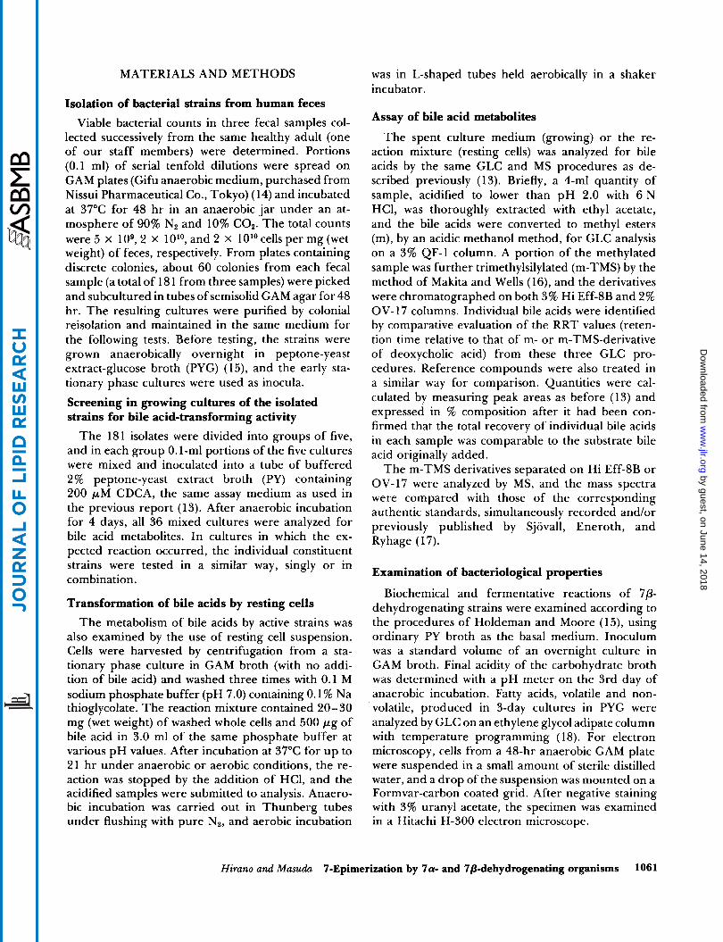
T o confirm the above assumption, one of these strains, b-52, was grown anaerobically in broth in the presence of either UDCA or 7KL (Fig. 1). 7KL was rapidly reduced to and almost quantitatively con- verted into UDCA during the early phase of in- cubation, while the substrate UDCA was scarcely oxi- dized to 7KL and more than 90% of UDCA remained unchanged until the 8th day, presumably because of the reductive environment elaborated by anaerobic bacterial growth. The identity of the respective con-
1062 Journal of Lipid Research Volume 22, 1981
TABLE 1. Transformation of CDCA by intestinal isolates in mixed cultures of five strains, individual pure cultures,
and a combination of two selected strains
Bile Acid Metabolites"
1.126 1.206 1.6P Culture CDCA UDCA 7k1
Mixed culture' of a-16 to a-20
Pure cultured of a-16 (+C) a-17 (+C) a-18 (-R) a-I9 (-R) a-20 (- R)
Combination" of a-16 + a-18 a-16 + a-I9 a-16 + C a-16 + 2536
Mixed culture of b-5 1 to b-55
Pure culture of b-51 (-R) b-52 (+C) b-53 (-R) b-54 (- R) b-55 (- R)
Combination of b-52 + 2536
Mixed culture of c-1 1 to c-15
Pure culture of c- 1 1 (+R) C-12 (-R) C-13 (-R) C-14 (-R) c-15 (+C)
Combination of c-15 + 2536
Pure culture of C 2536
%
57 43
100 100
19 81 86 14
100
100 100 100 100
77
100 100 100 34 69
62
100 95
100 100 100
9 44
23
66 31
99 1
36 2
5
99 1
91 56
The strains were grown in buffered PY broth containing 200 pM CDCA under anaerobic conditions for 4 days. Bile acids ex- tracted from the spent culture medium were separated as methyl esters on QF-I.
* Relative retention times (RRT values). Three mixed cultures consisting of a-16 to a-20, b-51 to b-55,
and c-11 to c-15 were positive for the conversion of CDCA into UDCA.
The individual strains from the positive mixed cultures were tested in pure cultures. Their morphological appearance is given in parentheses: +R, gram-positive rod; +C, gram-positive coccus; -R, gram-negative rod.
e Selected strains a-16, b-52, and c-15 were tested for the trans- formation of CDCA in cocultures with a 7a-dehydrogenating member of the mixed culture (a-I8 or a-19) or a 7a-dehydro- genating stock culture of E . coli, strain C, or Bacteroides fragilis, strain 2536.
version products was established by means of GLC and MS.
Oxidative conversion of UDCA to give 7KL was tested by the use of resting cells, which can be in- cubated under either anaerobic or aerobic conditions. The conversion took place under both conditions,
by guest, on June 14, 2018w
ww
.jlr.orgD
ownloaded from

100 - bp - 0 z
UDCA The same metabolites were obtained from the two bile acids, indicating interconversion between these two epimeric bile acids via 7KL as an intermediate.
The reaction sequence of the interconversion was
ating) and E . coli C (7a-dehydrogenating) (Table 3). Cells of a-16 reduced 7KL to UDCA under anaerobic conditions and oxidized UDCA to 7KL under aerobic incubation, thus mediating the reversible dehydro- genation between the 7P-hydroxy and 7-oxo groups. The 7KL produced through the oxidation of UDCA by a- 16 was reduced to CDCA when incubated anaero- bically with E. coli cells, and the 7KL derived from
UDCA examined with resting cells of a-I6 (7P-dehydrogen-
1 2 4 a 1 2 4 a INCUBATION (DAYS)
Fig. 1. Mutual conversion of UDCA and 7KL by anaerobic CDcA by aerobic action of E . coli was also converted cultures of strain b-52. The strain was grown for 8 days in buffered into UDCA by anaerobic incubation with a- 16 cells. PY broth containing UDCA or 7KL; samples were taken at 1-day intervals for assay.
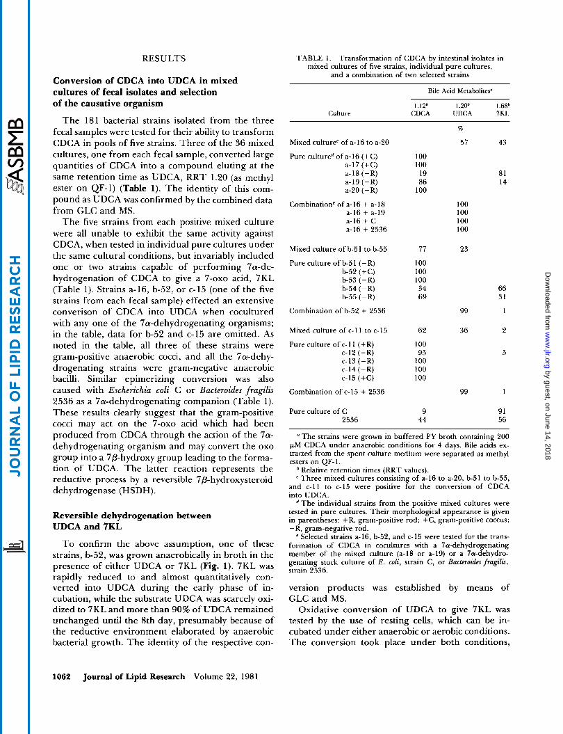
but to a larger extent in an aerobic than an anaerobic environment (Fig. 2A). Aerobic oxo formation as a function of pH was then tested (Fig. 2B). The reaction proceeded significantly over a broad pH range above 6.0, with an optimum of 8.0. Similar findings were obtained with the other strains, a-I6 and c-15. From these observations, it may be concluded that the strains of the b-52 group constitutively synthesize 7P-HSDH which mediates the reversible oxidation-reduction re- actions between the 7P-hydroxy and 7-oxo groups.
Mutual conversion between CDCA and UDCA by collaboration of 7a- and 7P-dehydrogenating organisms
The 7P-dehydrogenating strains were cocultured anaerobically with E. coli strain C (7a-dehydrogen- ating) in the presence of CDCA or UDCA (Table 2).
Conversion of CA into its 7P-epimer by combined action of 7a- and 7P-dehydrogenating organisms
The metabolic bile acids produced from CA in co- cultures of the 7P-dehydrogenating strains with E . coli C are shown in Table 2. Two compounds eluting at RRTs of 0.97 and 3.4 (as silyl ethers on Hi Eff-8B) were found as principal metabolites. The same products were also observed with resting cells (Table 4). In the light of their GLC behavior and MS features, the 0.97 compound was identified as 7PCA, the 7- epimerized product of CA, and the 3.4 compound as 7KD, an oxidation metabolite of CA. The results may be explained by assuming that CA was oxidized by E. coli to 7KD (7a-dehydrogenation), which, in turn, was reduced to the 7P-hydroxy acid by the 7P-de- hydrogenating organism. When 7KD was exposed as a substrate to the latter organism, the keto acid was converted into 7PCA but not into CA. It should be
TABLE 2. Transformation of CDCA, UDCA, and CA in cultures of a 7P-dehydrogenating organism (a-16, b-52, or c-15) combined with 7cx-dehydrogenating E . coli C
Bile Acid Metabolites' from
CDCA UDCA CA
1.09 1.68 5.2 1.09 I .68 5.2 0.65 0.97 3.4 CDCA UDCA 7KL Culture CDCA UDCA 7KL CA 7PCA 7KD
%
a-16 + C 41 30 29 Not determined 43 18 39
b-52 + C 52b 9 39 3 95 2 49 3 48 47 38 15 7 74 19 79 11 10 13 84 3 12 84 4 32 53 15
C-15 + C 26 55 19 Not determined 45 23 32
" Each pair of strains was cocultured anaerobically for 4 days in buffered PY broth with the desired bile acid at 200 pmol. Bile acids extracted from the spent culture medium were identified by GLC of their m-TMS derivatives on Hi Eff-8B. The RRT values of the bile acids are indicated.
Tested in triplicate.
Hirano and Masuda 7-Epimerization by 7a- and 78-dehydrogenating organisms 1063
by guest, on June 14, 2018w
ww
.jlr.orgD
ownloaded from
I " PH 8.0 9.0 7.0
6.0
5.0 21 1 2 3
REACTION PERIOD (HRS)
Fig. 2. 7j3-Dehydrogenating conversion of UDCA into 7KL by resting cells of strain b-52, compared between anaerobic and aerobic conditions (A) and as a function of pH (B). A: a resting cell suspension was incubated with UDCA in phosphate buffer at pH 7.0 in a Thunberg tube under N, (anaerobic) or in an L-shaped tube with continuous shaking in air (aerobic). B: the same reaction mixtures were incubated under aerobic conditions using phosphate buffer at different pH values.
noted in this connection that the two ?-epimeric trihydroxy bile acids, CA and 7pCA, were quite in- distinguishable by the chromatography of their methyl esters on QF-1 because of the similarity of elution times (RRT 2.0 for the two). The 7-epimeri- zation of CA had been overlooked until the silylation procedure was undertaken.
Lastly, preliminary studies have shown that 7p- HSDH activity is extractable and NADP-dependent. A washed cell suspension of strain b-52 was disrupted with a Branson Sonifier B-12 (Branson Sonic Power
TABLE 3. Conversion between CDCA and UDCA via 7KL by resting cells of strain a- 16 in collaboration
with resting cells of E . coli C
Bile Acid Metabolites Substrate Incuha-
Cells Bile Acid tion CDCA UDCA 7KL
%
a-16 + C" CDCA 21 hr, N, 17 68 15
a-16 + C" UDCA 21 hr, N, 18 66 16
a- l6* 7KL 21 hr, N2 0 78 22
a-16 UDCA 5 hr, O2 0 24 76 + CC 16 hr, N, 14 63 23
C CDCA 5 hr, 0, 70 0 30 + a-1W 16 hr, N, 17 62 21
a Washed cells of a-16 and C were incubated with CDCA (or
Cells of a- 16 alone were incubated with 7KL under N, for 2 1 hr. Cells of a-16 (or C) were incubated with UDCA (or CDCA)
under aerobic shaking (0,) for 5 hr. The atmosphere in the reaction tube was made anaerobic by flushing with N,, and C (or a-16) cells were added to react under N, for 16 hr.
UDCA) under an atmosphere of N2 for 2 1 hr.
TABLE 4. Conversion of CA into its 7p-epimer by collaboration of resting cells of strains a- 16 and E . coli C
Bile Acid Metabolites Incuha-
Cells tion CA 7PCA 7KD ~~
%
a-16 + C 21 hr, N2 17 67 16
C 5 hr, 0, 85 0 15 + a-16 16 hr, NZ 27 55 22
For procedures, see Table 3.
Co., Stamford, CT) at 80 W for 4 min and centrifuged at 6,000 g for 30 min to remove cell debris. A 0.5-ml portion of the supernatant (equivalent to 10 ml of the original GAM broth culture) was incubated with 1 pmol of UDCA in 3.0 ml of glycine-NaOH buffer at pH 9.5, with the addition of 2.5 pmol of NAD or NADP (Sigma Chemical Co., St Louis, MO). After 30 min at 20°C, about 80% of UDCA was converted into 7KL in the presence of NADP but the conversion was negligible in the presence of NAD.
Bacteriological characterization of 7P-dehydrogenating strains
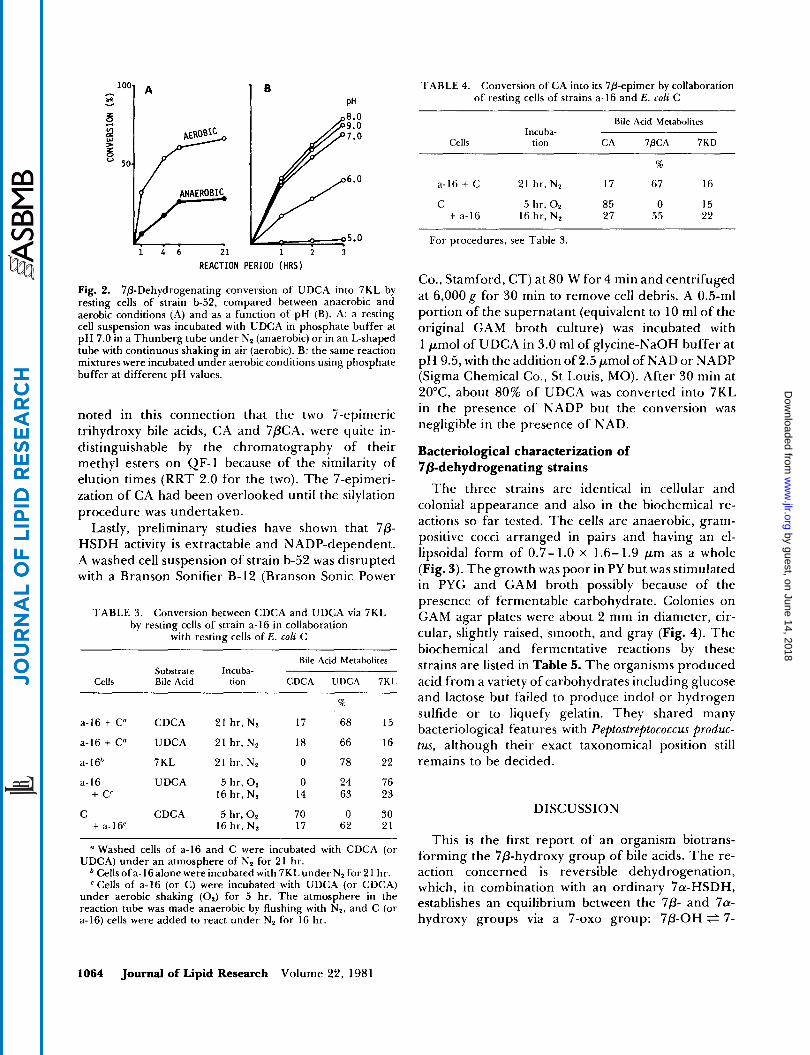
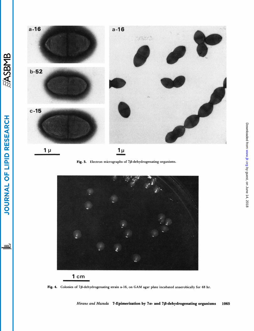
The three strains are identical in cellular and colonial appearance and also in the biochemical re- actions so far tested. The cells are anaerobic, gram- positive cocci arranged in pairs and having an el- lipsoidal form of 0.7- 1.0 X 1.6- 1.9 pm as a whole (Fig. 3). The growth was poor in PY but was stimulated in PYG and GAM broth possibly because of the presence of fermentable carbohydrate. Colonies on GAM agar plates were about 2 rnm in diameter, cir- cular, slightly raised, smooth, and gray (Fig. 4). The biochemical and fermentative reactions by these strains are listed in Table 5. The organisms produced acid from a variety of carbohydrates including glucose and lactose but failed to produce indol or hydrogen sulfide or to liquefy gelatin. They shared many bacteriological features with Peptostreptococcus produc- tus, although their exact taxonomical position still remains to be decided.
DISCUSSION
This is the first report of an organism biotrans- forming the 7p-hydroxy group of bile acids. The re- action concerned is reversible dehydrogenation, which, in combination with an ordinary Ya-HSDH, establishes an equilibrium between the 7p- and 7a- hydroxy groups via a 7-oxo group: 7p-OH e 7-
1064 Journal of Lipid Research Volume 22, 1981
by guest, on June 14, 2018w
ww
.jlr.orgD
ownloaded from
I C ( - 1cI
Fig. 3. Electron micrographs of 78-dehydrogenating organisms.
1 cm
Fig. 4. Colonies of 78-dehydrogenating strain a-16, on GAM agar plate incubated anaerobically for 48 hr.
Hirano and Masuda 7-Epimerization by 7a- and 7pdehydrogenating organisms 1065
by guest, on June 14, 2018w
ww
.jlr.orgD
ownloaded from
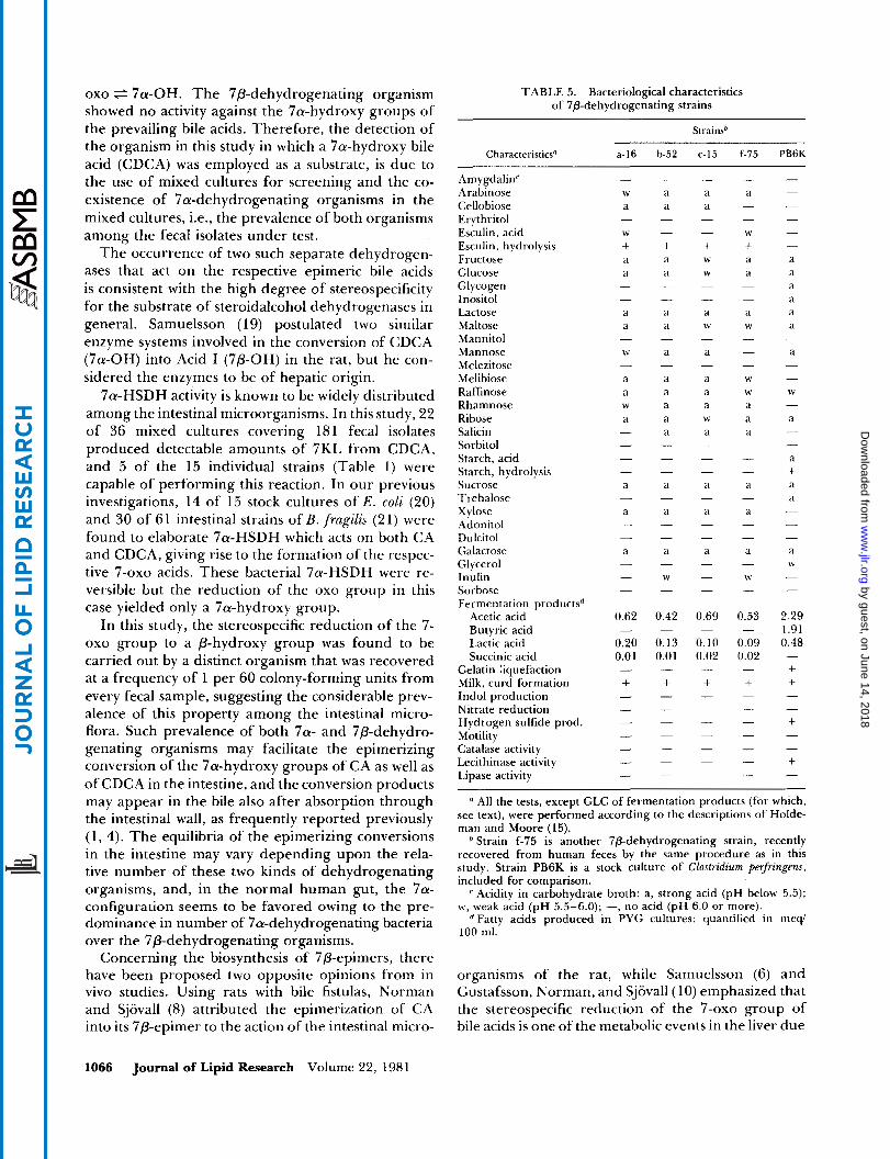
oxo 7a-OH. The 7p-dehydrogenating organism TABLE 5. Bacteriological characteristics showed no activity against the 7a-hydroxy groups of of 7P-dehydrogenating strains
the prevailing bile acids. Therefore, the detection of Strainsb the organism-in this study in which a 7a-hydroxy bile acid (CDCA) was employed as a substrate, is due to the use of mixed cultures for screening and the co- existence of 7a-dehydrogenating organisms in the mixed cultures, i.e., the prevalence of both organisms among the fecal isolates under test.
The occurrence of two such separate dehydrogen- ases that act on the respective epimeric bile acids is consistent with the high degree of stereospecificity for the substrate of steroidalcohol dehydrogenases in general. Samuelsson (19) postulated two similar enzyme systems involved in the conversion of CDCA (7a-OH) into Acid I (7P-OH) in the rat, but he con- sidered the enzymes to be of hepatic origin.
7a-HSDH activity is known to be widely distributed among the intestinal microorganisms. In this study, 22 of 36 mixed cultures covering 18 1 fecal isolates produced detectable amounts of 7KL from CDCA, and 5 of the 15 individual strains (Table 1) were capable of performing this reaction. In our previous investigations, 14 of 15 stock cultures of E . coli (20) and 30 of 61 intestinal strains of B.fragilzs (21) were found to elaborate 7a-HSDH which acts on both CA and CDCA, giving rise to the formation of the respec- tive 7-oxo acids. These bacterial 7a-HSDH were re- versible but the reduction of the oxo group in this case yielded only a 7a-hydroxy group.
In this study, the stereospecific reduction of the 7- oxo group to a P-hydroxy group was found to be carried out by a distinct organism that was recovered at a frequency of 1 per 60 colony-forming units from every fecal sample, suggesting the considerable prev- alence of this property among the intestinal micro- flora. Such prevalence of both 7a- and 7P-dehydro-
CharacteristicsU - f-75 PB6K a-16 b-52 c-15
Amygdalinr Arabinose Cellobiose Erythritol Esculin, acid Esculin, hydrolysis Fructose Glucose Glycogen Inositol Lactose Maltose Mannitol Mannose Melezitose Melibiose Raffinose Rhamnose Ribose Salicin Sorbitol Starch, acid Starch, hydrolysis Sucrose Trehalose Xylose Adonitol Dulcitol Galactose Glycerol Inulin Sorbose Fermentation productsd
Acetic acid Butyric acid Lactic acid Succinic acid
Gelatin Ziquefaction Milk, curd formation Indol production Nitrate reduction Hydrogen sulfide prod. Motilitv
W
+ a a
- + +
a a
W
W
- a a
- a W
a
a a a a a -
a a a W
a -
0.62
0.20 0.01
- 0.42
0.13 0.01
- 0.69
0.10 0.02
- 0.53
0.09 0.02
- 2.29 1.91 0.48
+ + + + + - +
genating organisms may facilitate the epimerizing Catalaie activity - - - - - conversion of the 7a-hydroxy groups of CA as well as ~ ~ ~ ~ ~ ~ ~ v ~ t i v i t Y of CDCA in the intestine, and the conversion products
- - - - + - - - - -
may appear in the bile also after absorption through the intestinal wall, as frequently reported previously (1, 4). The equilibria of the epimerizing conversions in the intestine may vary depending upon the rela- tive number of these two kinds of dehydrogenating organisms, and, in the normal human gut, the 7a- configuration seems to be favored owing to the pre- dominance in number of 7a-dehydrogenating bacteria over the 7p-dehydrogenating organisms.
Concerning the biosynthesis of 7P-epimers, there have been proposed two opposite opinions from in vivo studies. Using rats with bile fistulas, Norman and Sjovall (8) attributed the epimerization of CA into its 7p-epimer to the action of the intestinal micro-
" All the tests, except GLC of fermentation products (for which, see text), were performed according to the descriptions of Holde- man and Moore (15).
Strain f-75 is another 7p-dehydrogenating strain, recently recovered from human feces by the same procedure as in this study. Strain PB6K is a stock culture of Clostrulium perfringew, included for comparison.
Acidity in carbohydrate broth: a, strong acid (pH below 5.5); w, weak acid (pH 5.5-6.0); -, no acid (pH 6.0 or more).
"atty acids produced in PYG cultures: quantified in meqi 100 ml.
organisms of the rat, while Samuelsson (6) and Gustafsson, Norman, and Sjovall(l0) emphasized that the stereospecific reduction of the 7-oxo group of bile acids is one of the metabolic events in the liver due
1066 Journal of Lipid Research Volume 22, 1981
by guest, on June 14, 2018w
ww
.jlr.orgD
ownloaded from
to hepatic enzymes. It seems difficult to draw any decisive conclusion from these in vivo studies for the hepatointestinal relationship of the epimerizing reaction.
Samuelsson (22) observed the conversion of 3a,7P,- 12a-trihydroxycholanoate into deoxycholate in the rat cecum and suggested that the microbial removal of the 7P-hydroxy group is analogous with 7a-de- hydroxylation. A similar idea was presented by Fedorowski et al. (23) as to the formation of LCA from UDCA in man. However, no microorganism has so far been shown to be capable of performing the direct removal of these 7P-hydroxy groups, and it seems more likely that the 7P-hydroxy groups were transformed into an a-orientation by the con- version system described here before the ordinary 7a-dehydroxylation.
The concept that two kinds of microorganisms take part in the epimeric conversion of the hydroxy group at C-7 contrasts with the idea of a similar reaction at C-3 being carried out by one and the same or- ganism. Hayaishi et al. (24) extracted from Escherichia (Citrobacter) freundii an NAD-dependent dehydrogen- ase preparation that was capable of oxidizing LCA to a 3-oxo acid and also of reducing the oxo group to both CY- and P-hydroxy groups. On the other hand, Marcus and Talalay (25) and Talalay and Marcus (26) demonstrated that Pseudomonas testosteroni elaborates two distinct oxidoreductases specifically affecting 3a- and 3P-hydroxysteroids, respectively, to perform the epimerizing conversion of the 3-hydroxy group. Simi- lar conversion at C-3 has also been reported in cer- tain strains of Clostridium perfringens (27), Bacillus cereus (27), Eubacterium lentum (28), and anaerobic streptococci (29), although it is undecided at present whether these organisms synthesize an ambivalent enzyme, as suggested by the work of Hayaishi et al. (24), or two specific dehydrogenases, as was the case with P. testosteroni. However, whether the reaction is caused by one enzyme or two separate enzymes, all of these 3-hydroxy epimerizations are carried out by a single bacterium. The involvement of two dif- ferent organisms has never been reported until the present study of 7-hydroxy epimerization.m
Manuscript received 2 9 December 1980 and in revised form 22 April 1981.
REFERENCES
1. Sjovall, J. 1959. The occurrence of 7P-hydroxylated bile acids in human bile. Acta Chem. Scand. 13: 711-716.
2. Eneroth, P., B. Gordon, and J. Sjovall. 1966. Charac- terization of trisubstituted cholanoic acids in human feces. J . Lipid Res. 7: 524-530.
3. Gleich, G. J., and A. F. Hofmann. 1971. Use of choles- tyramine to control diarrhea associated with acquired hypogammaglobulinemia. Am. J. Med. 51: 281-286.
4. Igimi, M. 1976. Ursodeoxycholate-a common bile acid in gallbladder bile of Japanese subjects. L f e Sci.
5 . Mahowald, I. A., M. W. Yin, J. T . Matschiner, S. L. Hsia, E. A. Doisy, Jr., W. H. Elliott, and E. A. Doisy. 1958. Bile acids. VIII. Metabolism of 7-ketolitho- cholic acid-24-C14 in the rat. J . Biol. Chem. 230: 58 1-588.
6. Samuelsson, B. 1959. The metabolism of 7-ketolitho- cholic acid-24-I4C in the rat. Acta Chem. S c a d 13:
7. Hellstrom, K., and J. Sjovall. 1960. Metabolism of chenodeoxycholic acid in the rabbit. Acta Chem. Scand. 14: 1763- 1769.
8. Norman, A., and J. Sjovall. 1958. Microbial trans- formation products of cholic acid in the rat. Biochim. Biophys. Acta. 29: 467-468.
9. Norman, A., and J. Sjovall. 1958. On the transformation and enterohepatic circulation of cholic acid in the rat. J . Biol. Chem. 233: 872-885.
10. Gustafsson, B. E., A. Norman, and J. Sjovall. 1960. Influence of E. coli infection on turnover and metabo- lism of cholic acid in germ-free rats. Arch. Biochem. Biophys. 91: 93- 100.
1 I . Fromm, H., G. L. Carlson, A. F. Hofmann, S. Farivar, and P. Amin. 1980. Metabolism in man of 7-keto- lithocholic acid: precursor of cheno- and ursodeoxy- cholic acids. Am. J . Physiol. 239: GI61 -G166.
12. Fedorowski,T., G. Salen, G. S. Tint, and E. H. Mosbach. 1979. Transformation of chenodeoxycholic acid and ursodeoxycholic acid by human intestinal bacteria. Gastroenterology. 77: 1068- 1073.
13. Hirano, S., N. Masuda, and H. Oda. 1981. In vitro transformation of chenodeoxycholic acid and urso- deoxycholic acid by human intestinal flora, with particular reference to the mutual conversion between the two bile acids. J . Lipid Res. 22: 735-743.
14. Sutter, V. L., D. M. Citron, and S. M. Finegold. 1980. Wadsworth Anaerobic Bacteriology Manual. 3rd ed. C. V. Mosby Co. , St. Louis, MO. 98.
15. Holdeman, L. V., and W. E. C. Moore. 1973. Anaerobe Laboratory Manual. 2nd ed. Virginia Polytechnic In- stitute and State University Anaerobe Laboratory, Blacksburg, VA. 108- 110.
16. Makita, M., and W. W. Wells. 1963. Quantitative analysis of fecal bile acids by gas-liquid chromatog- raphy. Anal. Biochem. 5: 523-530.
17. Sjiivall, J., P. Eneroth, and R. Ryhage. I97 1. Mass spectra of bile acids. In The Bile Acids. Vol. I . P. P. Nair and D. Kritchevsky, editors. Plenum Press, New York. 233-244.
18. Hirakawa, K. 1979. Resting cell studies on fermenta- tion of sugar and peptone by Clostridium. Acta Med. Univ. Kagoshima. 21: 1 17- 130.
19. Samuelsson, B. 1959. On the metabolism of cheno- deoxycholic acid in the rat. Actn Chem. Scand. 13: 976-983.
20. Imamura, T., N. Sakamoto, M. Tamaki, and S. Hirano. 1979. Transformation of bile acids by members of the
Enterobacteriaceae. J p n . J . Bacteriol. 34: 5 13-520.
18: 993-999.
236-240.
Hirano and Masuda 7-Epimerization by 7a- and 7P-dehydrogenating organisms 1067
by guest, on June 14, 2018w
ww
.jlr.orgD
ownloaded from
21. Hirano, S., N. Masuda, H. Mukai, K. Hirakawa, and T. Imamura. 1979. Transformation of bile acids by Bacteroides fragilis strains isolated from the human in- testine. J p n . J . Bacteriol. 34: 403-4 1 1.
22. Samuelsson, B. 1960. Metabolism of 3a,7P,l2a-tri- hydroxycholanoic acid in the rat. Acta Chem. Scand. 14: 21-47.
23. Fedorowski, T., G . Salen, F. G. Zaki, S. Shefer, and E. H. Mosbach. 1978. Comparative effects of urso- deoxycholic acid and chenodeoxycholic acid in the rhesus monkey. Biochemical and ultrastructural stud- ies. Gastroenterology. 74: 75-8 1.
24. Hayaishi, O., Y. Sato, W. B. Jakoby, and E. F. Stohlman. 1955. Reversible enzymatic oxidation of bile acids. Arch. Biochem. Biophys. 56: 554-555.
25. Marcus, P. I . , and P. Talalay. 1956. Induction and purification of a- and 0-hydroxysteroid dehydrogen- ases. J . Biol. Chem. 218: 661-674.
26. Talalay, P., and P. I. Marcus. 1956. Specificity, kinetics and inhibition of a- and P-hydroxysteroid dehydrogen- ases. J . Biol. Chem. 218: 675-691.
27. Midtvedt, T., and A. Norman. 1967. Bile acid trans- formations by microbial strains belonging to genera found in intestinal contents. Acta Pathol. Microbiol. Scand. 71:.629-638.
28. Bokkenheuser, V. D., J. Winter, P. Dehazya, and W. G. Kelly. 1977. Isolation and characterization of human fecal bacteria capable of 2 1-dehydroxylating corticoids. Appl. Environ. Microbiol. 34: 571 -575.
29. Dickinson, A. B., B. E. Gustafsson, and A. Norman. 197 1. Determination of bile acid conversion potencies of intestinal bacteria by screening in vitro and sub- sequent establishment in germfree rats. Acta Pathol. Microbiol. Scand. 79: 691 -698.
1068 Journal of Lipid Research Volume 22, 1981
by guest, on June 14, 2018w
ww
.jlr.orgD
ownloaded from