epistaxis - intech - open science open minds | intechopen
TRANSCRIPT
2
Epistaxis
Jin Hee Cho and Young Ha Kim College of Medicine, The Catholic University of Korea
South Korea
1. Introduction
Epistaxis occur due to trauma, disorders in mucosa or vessels, or coagulopathy. It is a very common disease, as 10% of all population experience severe epistaxis, about 30% of children aged 0~5, 56% of children aged 6~10 and 64% of children aged 11~15 are reported to experience one or more episode of epistaxis. (Cho, 2009 )
Although most cases of epistaxis are mild, that can be self-managed, life-threatening condition can be also possible. When encounter patient with severe epistaxis, it is important to find the bleeding focus and to analyse the causes of epistaxis fast and accurately, to treat the patient promptly to avoid complications such as hypotension, hypoxia, anemia, aspiration or death.
2. Anatomy
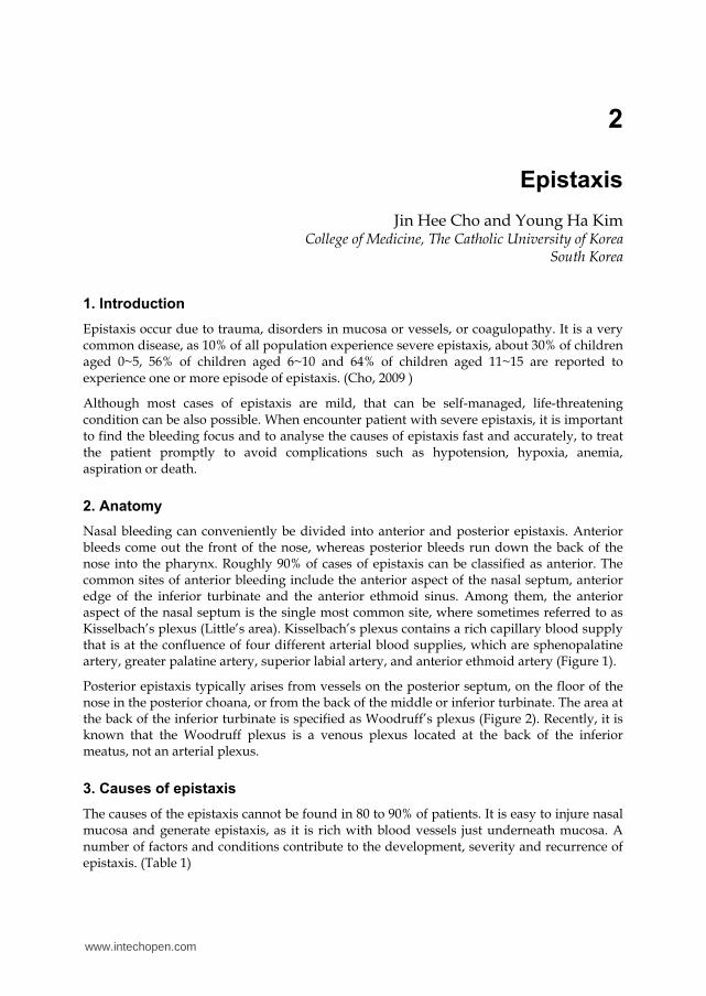
Nasal bleeding can conveniently be divided into anterior and posterior epistaxis. Anterior bleeds come out the front of the nose, whereas posterior bleeds run down the back of the nose into the pharynx. Roughly 90% of cases of epistaxis can be classified as anterior. The common sites of anterior bleeding include the anterior aspect of the nasal septum, anterior edge of the inferior turbinate and the anterior ethmoid sinus. Among them, the anterior aspect of the nasal septum is the single most common site, where sometimes referred to as Kisselbach’s plexus (Little’s area). Kisselbach’s plexus contains a rich capillary blood supply that is at the confluence of four different arterial blood supplies, which are sphenopalatine artery, greater palatine artery, superior labial artery, and anterior ethmoid artery (Figure 1).
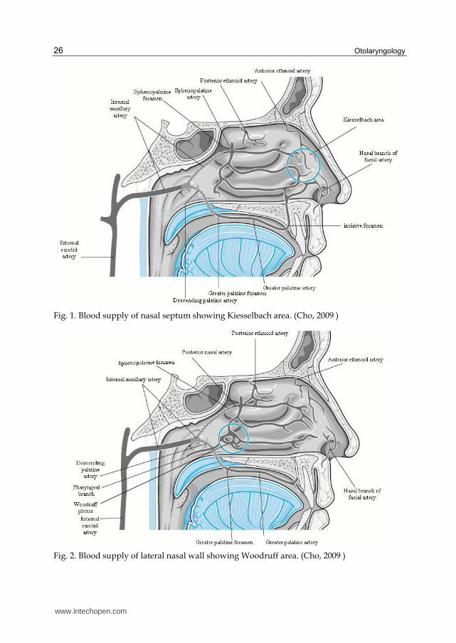
Posterior epistaxis typically arises from vessels on the posterior septum, on the floor of the nose in the posterior choana, or from the back of the middle or inferior turbinate. The area at the back of the inferior turbinate is specified as Woodruff’s plexus (Figure 2). Recently, it is known that the Woodruff plexus is a venous plexus located at the back of the inferior meatus, not an arterial plexus.
3. Causes of epistaxis
The causes of the epistaxis cannot be found in 80 to 90% of patients. It is easy to injure nasal mucosa and generate epistaxis, as it is rich with blood vessels just underneath mucosa. A number of factors and conditions contribute to the development, severity and recurrence of epistaxis. (Table 1)
www.intechopen.com
Otolaryngology
26
Fig. 1. Blood supply of nasal septum showing Kiesselbach area. (Cho, 2009 )
Fig. 2. Blood supply of lateral nasal wall showing Woodruff area. (Cho, 2009 )
www.intechopen.com
Epistaxis
27
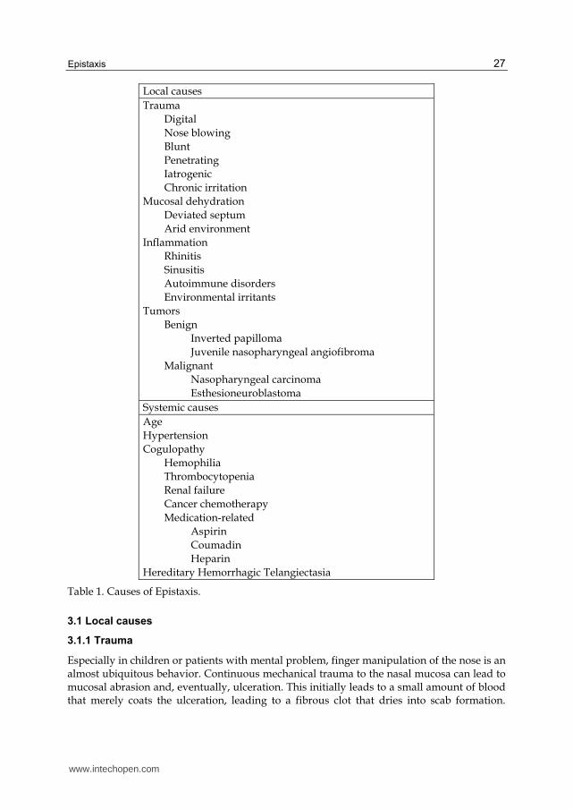
Local causes
Trauma
Digital
Nose blowing
Blunt
Penetrating
Iatrogenic
Chronic irritation
Mucosal dehydration
Deviated septum
Arid environment
Inflammation
Rhinitis
Sinusitis
Autoimmune disorders
Environmental irritants
Tumors
Benign
Inverted papilloma
Juvenile nasopharyngeal angiofibroma
Malignant
Nasopharyngeal carcinoma
Esthesioneuroblastoma
Systemic causes
Age
Hypertension
Cogulopathy
Hemophilia
Thrombocytopenia
Renal failure
Cancer chemotherapy
Medication-related
Aspirin
Coumadin
Heparin
Hereditary Hemorrhagic Telangiectasia
Table 1. Causes of Epistaxis.
3.1 Local causes
3.1.1 Trauma
Especially in children or patients with mental problem, finger manipulation of the nose is an almost ubiquitous behavior. Continuous mechanical trauma to the nasal mucosa can lead to mucosal abrasion and, eventually, ulceration. This initially leads to a small amount of blood that merely coats the ulceration, leading to a fibrous clot that dries into scab formation.
www.intechopen.com
Otolaryngology
28
Removal of the scab causes further injury to the mucosa, which can result in more significant bleeding.
It is possible to cause rupture of superficial vessels of the mucosa by violent nose blowing. Nose blowing is an especially prominent source of trauma in patients who have undergone recent surgery on the nose or sinuses or who have preexisting bleeding sites.
The trauma may be in the form of blunt trauma to the nose or sinuses as a result of traffic accident or during sports, resulting in fracture of the septum, lateral wall, or one of the sinuses. The fracture leads to disruption of the mucosal lining, tearing of blood vessels, and bleeding.
Chronic irritation of nasal mucosa can also cause epistaxis. For example, nasal abuse of cocaine, nasal smoke or misuse of nasal spray can cause nasal irritation and dehydration which can lead to epistaxis or even septal perforation.
3.1.2 Dehydration
Drying of the nasal mucosa is one of the common factors contributing to epistaxis. The possibility of epistaxis increases when the nasal humidifying function falls as the nasal secretion decreases, or when nasal mucosa expose to the cold, dry environmental air as a seasonal factor. Also, when the septum is significantly deviated, or when nasal airway is altered as a result of surgery, there can be an abnormally high airflow that are no longer able to humidify the air adequately and as a result, epistaxis can occur.
Number of epistaxis patients who visit emergency department increases as temperature and humidity decrease. Also, the number of patients who admit to hospital increases in winter. Comparing in-patients number with air temperature, admission increases 30% in days with average temperature under 5°C than days with average temperature over 5°C. (Viducich et al., 1995)
3.1.3 Inflammation
Inflammatory conditions such as acute upper respiratory infection, allergic rhinitis, sinusitis and nasal foreign bodies can often lead to nasal bleeding. Nasal decongestant or intranasal steroid spray also can cause nasal dryness and epistaxis. Any factors that cause nasal inflammation can make the mucosa more fragile and make patients to blow the nose more frequently, weak vessels can be damaged easily. Nasal granulomatous diseases such as Wegener’s granulomatosis, sarcoidosis, nasal tuberculosis and nasal syphilis lead to mucosal ulceration or extreme inflammation that may predispose the patient to crusting, abrasion, and eventually, bleeding.
3.1.4 Tumors and aneurysms
Juvenile nasopharyngeal angiofibroma classically presents as recurrent epistaxis in adolescents or young adult men, malignant tumors such as malignant melanoma and squamous cell carcinoma present as unilateral nasal stuffiness with epistaxis in adults. Intracavernous aneurysm of internal carotid artery after trauma can cause severe epistaxis. This posttraumatic aneurysm occurs about 7 weeks after trauma, and mortality rate reaches 50%.
www.intechopen.com
Epistaxis
29
3.2 Systemic causes
3.2.1 Age
In elderly, changes in vessel wall, especially in arterial wall fibrosis, are related with epistaxis. In children, previously mentioned mechanical trauma, nasal foreign body and nasal mucosal inflammation are the causes of epistaxis.
3.2.2 Hypertension
Hypertension and epistaxis commonly occur simultaneously among adults of general population. It is uncertain whether the hypertension is an etiologic factor in all of these patients. It is known that hypertension in epistaxis patients is caused by anxiety. However, one study that analyzed 200 epistaxis patients reported that 75% showed elevated blood pressure during nose bleeding and 30% was severe hypertension patients. (Herkner et al., 2000)
Elevated blood pressure can contribute to epistaxis in two different ways. First, the high pressure causes chronic damage of a blood vessel wall in the nasal or sinus mucosa. Second, 20% of epistaxis patients experience elevated blood pressure because the natural response to seeing blood from one’s nose is to get agitated, which can directly lead to elevation of the blood pressure. Practically, active bleeding patients in emergency department were related to hypertension and patients without active nasal bleeding had less related to hypertension.
3.2.3 Coagulopathy
Coagulopathy leads to unwanted bleeding due to the absence or inactivity of one of the clotting factors. These conditions are rare, but affected patients tend to have severe nosebleeds from an early age. There are also patients with inherent disorders of platelet function. Conditions such as hemophilia, von Willebrand disease, thrombocytopenia, AIDS or liver disease can often cause epistaxis. And among them, von Willebrand disease is most common.
Patients with chronic renal failure commonly have problems with epistaxis. This is due to the two-prolonged problem of regularly receiving heparin during dialysis and having poor clotting secondary to the renal failure. In these patients, epistaxis tends to occur either while the patient is undergoing dialysis or shortly after the dialysis. Patients with septic shock develop a condition of poor clotting that may progress to disseminated intravascular coagulation (DIC). This starts out as uncontrolled clotting of the blood within the vascular system and progresses to a coagulopathy secondary to consumption of all available clotting factors.
Finally, there are patients who acquire clotting deficiencies as a result of cancer therapy. This may occur secondary to high-dose chemotherapy, leading to transient decrease in the platelet count. Alternatively, the coagulopathy may be caused by depletion of bone marrow reserves of platelets due to bone marrow transplant. In both of these cases, thrombocytopenia becomes a clinical reality and epistaxis may result.
3.2.4 Medications
There are several medications that interfere normal blood clotting process, for example, warfarin, heparin and nonsteroid anti-inflammatory drugs(NSAIDs). Most commonly used
www.intechopen.com
Otolaryngology
30
medication is NSAIDs, including aspirin. Aspirin, by inhibiting the enzyme cyclooxygenase, interferes with platelet function. This results in significant increases in bleeding time, but should not increase the incidence of nosebleeds. Millions of patients are currently taking a regular dose of aspirin, as prescribed by their doctors, for prevention of stroke, heart attack, and clotting in prosthetic arteries. For this reason, aspirin use is becoming an increasingly important risk factor for epistaxis in adults.
3.2.5 Hereditary hemorrhagic telangiectasia
Hereditary hemorrhagic telangiectasia (HHT), also known as Rendu-Osler-Weber disease, is a rare systemic fibrovascular dysplasia with autosomal dominant inheritance. Multiple telangiectasic vascular malformations can be seen on the skin and in the mucosa of the digestive tract and respiratory airways. 20% of patients have family history and the incidence is 1~2 per 100,000. Several elevated small cherry red spots in lip and oral cavity mucosa can be seen and they become pale when pressed. Dilatation of arterioles under basement membrane is referred to as telangiectasia, and these arterioles can easily be damaged as they do not have elastic tissue under endothelial layer. If the patient is diagnosed of HHT, arteriovenous malformation should be checked in other organs. Other manifestations of the disease occur in the internal organs such as the lungs, liver, or the central nervous system. It is estimated that at least 30% of HHT patients have pulmonary, 30% hepatic, and 10–20% cerebral involvement. (Guttmacher et al., 1995)
Diagnosis is made according to the Curaçao Criteria: telangiectasia on the face, hands and in oral cavity, recurrent epistaxis, arteriovenous malformations with visceral involvement, family history. Diagnosis is confirmed upon the presence of at least three of these manifestations. HHT is an uncommon cause of epistaxis, but an important cause owing to the severity of the condition and the special measures required for treatment. In any patient with recurrent epistaxis, a careful examination of the mucosal surfaces in the nose should be performed to rule out HHT lesions. The presence of three or more suggestive vascular lesions should alert the physician to the possibility that the patient may have HHT. (Fuchizaki et al., 2003)
4. Diagnosis
When the patient with epistaxis initially presents for treatment, it is important to perform a systematic evaluation. One may be tempted to proceed directly to managing the patient’s symptoms without performing a careful history and physical examination. Indeed, in cases of heavy bleeding, this may be necessary. For most patients, nose with Neo-Synephrine-soaked cotton pledgets, should control the bleeding sufficiently to allow the physician to perform a proper evaluation before initiating definitive treatment.
4.1 History
During the initial inquiry, it is important to investigate the duration of bleeding, frequency of bleeding, and amount of bleeding. If not in an emergency situation, it is also important to determine the side of the bleeding and its primary site of origin and flow: out the front of the nose, down the back of the nose or a combination of the two. It may be possible during history taking to elicit information that will provide clues to the underlying cause of the bleeding, such as trauma, surgery, history of coagulopathy or medication history.
www.intechopen.com
Epistaxis
31
4.2 Physical examination
After stabilize the patient, the initial examination of the nose should be performed to find the origin site by anterior rhinoscopy after removal of the blood clot and minimize the edema using decongestant, using adequate light source to visualize whole nasal cavity. If the bleeding has stopped after the removal of clots, additional immediate treatment is not needed. Packing of the nasal cavity without evidence of continuous bleeding can damage the nasal mucosa and cause epistaxis rather than stopping it. The most likely finding is a superficial vessel that has eroded on anterior nasal septum, or medial portion of the turbinate in patients with no specific cause, so special cautions are needed in those parts of the nose.
4.3 Endoscopy
In cases of either acute or recurrent epistaxis without an obvious bleeding source on anterior rhinoscopy, an endoscopic examination is indicated to attempt to identify the site. This is usually performed bilaterally and with thorough decongestion and topical anesthesia. Either a flexible or rigid endoscope can be used. The flexible scope is perhaps easier to use and less uncomfortable. However, the rigid scopes have superior optics, with better image resolution and are easy to use instruments.
4.4 Radiologic evaluation
Routine radiologic studies have little role in the initial diagnosis of epistaxis. However, in patients with recurrent epistaxis without a known source or cause, imaging studies have important role in diagnosis. The imaging study of choice for initial evaluation of most nasal or sinus pathologic conditions, including epistaxis, is the CT scan and tumors that cause epistaxis are often found.
5. Management
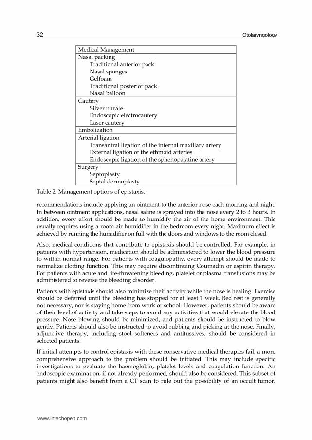
It is important to evaluate epistaxis patients thoroughly because the method of controlling epistaxis depends largely on the specifics of an individual case, with an entire menu of options available. (Table 2)
Anterior flexion of head can prevent nausea or airway obstruction, as blood does not flow back to pharynx. It is important to keep the blood pressure low and keep the airway clean. Also, fluid replacement can be considered according to the amount of blood loss. Systemic diseases can cause multiple bleeding sites or frequent recurrent epistaxis so blood testing should be performed in patients with those findings. Patients with posterior epistaxis, coagulopathy, coronary artery disease, uncontrolled hypertension, severe anemia or old age should be considered to treat as in-patient basis.
5.1 Medical treatment
On the first visit of a patient with minimal or moderate history of nasal bleeding, an empiric trial of medical therapy is advised. This includes several measures designed to increase humidification of the mucosa to allow the bleeding site to heal. This approach is based on the assumption that dryness is one of the most important factors causing epistaxis. The
www.intechopen.com
Otolaryngology
32
Medical Management
Nasal packing Traditional anterior pack Nasal sponges Gelfoam Traditional posterior pack Nasal balloon
Cautery Silver nitrate Endoscopic electrocautery Laser cautery
Embolization
Arterial ligation Transantral ligation of the internal maxillary artery External ligation of the ethmoid arteries Endoscopic ligation of the sphenopalatine artery
Surgery Septoplasty Septal dermoplasty
Table 2. Management options of epistaxis.
recommendations include applying an ointment to the anterior nose each morning and night. In between ointment applications, nasal saline is sprayed into the nose every 2 to 3 hours. In addition, every effort should be made to humidify the air of the home environment. This usually requires using a room air humidifier in the bedroom every night. Maximum effect is achieved by running the humidifier on full with the doors and windows to the room closed.
Also, medical conditions that contribute to epistaxis should be controlled. For example, in patients with hypertension, medication should be administered to lower the blood pressure to within normal range. For patients with coagulopathy, every attempt should be made to normalize clotting function. This may require discontinuing Coumadin or aspirin therapy. For patients with acute and life-threatening bleeding, platelet or plasma transfusions may be administered to reverse the bleeding disorder.
Patients with epistaxis should also minimize their activity while the nose is healing. Exercise should be deferred until the bleeding has stopped for at least 1 week. Bed rest is generally not necessary, nor is staying home from work or school. However, patients should be aware of their level of activity and take steps to avoid any activities that would elevate the blood pressure. Nose blowing should be minimized, and patients should be instructed to blow gently. Patients should also be instructed to avoid rubbing and picking at the nose. Finally, adjunctive therapy, including stool softeners and antitussives, should be considered in selected patients.
If initial attempts to control epistaxis with these conservative medical therapies fail, a more comprehensive approach to the problem should be initiated. This may include specific investigations to evaluate the haemoglobin, platelet levels and coagulation function. An endoscopic examination, if not already performed, should also be considered. This subset of patients might also benefit from a CT scan to rule out the possibility of an occult tumor.
www.intechopen.com
Epistaxis
33
Second-line therapy is then indicated, including nasal packing, cauterization, and vessel ligation.
5.2 General treatment
5.2.1 General treatment for anterior epistaxis
If the bleeding site is identified, it is recommended to use pledgets impregnated with
decongestant containing bosmin or phenylephrine solution. Compress both nasal dorsum
directly for at least 5 minutes after packing the nasal cavity with gauzes impregnated with
decongestant and anesthetic agent.
In cases with coagulopathy, packing with pledget can damage the mucosa so hemostatic
agents such as Avitene, Surgicel or gelfoam can be used. Electric cautery should
be performed carefully because it can damage surrounding normal tissue. Also,
physician should keep in mind that electric cautery on bilateral septum can lead to septal
perforation.
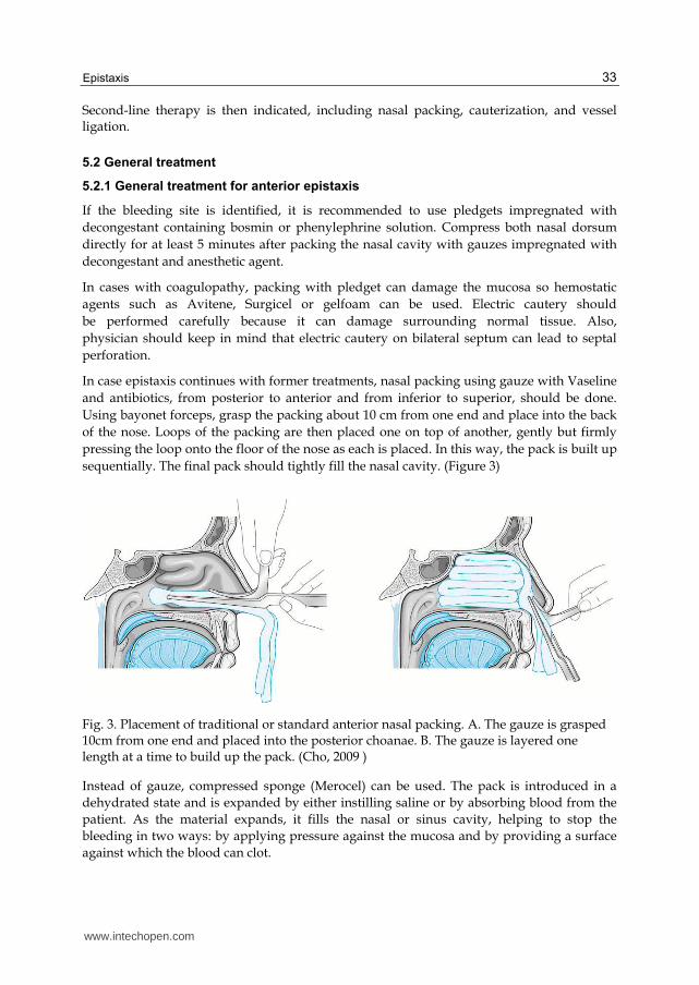
In case epistaxis continues with former treatments, nasal packing using gauze with Vaseline
and antibiotics, from posterior to anterior and from inferior to superior, should be done.
Using bayonet forceps, grasp the packing about 10 cm from one end and place into the back
of the nose. Loops of the packing are then placed one on top of another, gently but firmly
pressing the loop onto the floor of the nose as each is placed. In this way, the pack is built up
sequentially. The final pack should tightly fill the nasal cavity. (Figure 3)
Fig. 3. Placement of traditional or standard anterior nasal packing. A. The gauze is grasped 10cm from one end and placed into the posterior choanae. B. The gauze is layered one length at a time to build up the pack. (Cho, 2009 )
Instead of gauze, compressed sponge (Merocel) can be used. The pack is introduced in a
dehydrated state and is expanded by either instilling saline or by absorbing blood from the
patient. As the material expands, it fills the nasal or sinus cavity, helping to stop the
bleeding in two ways: by applying pressure against the mucosa and by providing a surface
against which the blood can clot.
www.intechopen.com
Otolaryngology
34
The packs are maintained for a period of 2 to 5 days depending on the circumstances. The
patient can be sent home with an anterior pack in the nose. It is recommended to coat
sponge with antistaphyloccocal ointment to prevent toxic shock syndrome, which is a side
effect of prolonged packing. It is advisable to prescribe antistaphylococcal antibiotics for the
duration of time the pack is in place for same reason. The patient should be advised to
restrict activity. Absolute bed rest is not necessary, but the patient should avoid strenuous
activity and should not attempt to maintain normal activity levels.
5.2.2 General treatment for posterior epistaxis
If posterior epistaxis does not stop with anterior packing, balloon insertion or posterior
packing should be done. Posterior epistaxis is typically heavy and cannot be controlled with
an anterior pack or by anterior compression of the nose. Posterior packing is the traditional
first-line therapy for posterior epistaxis. However, with the many options available today,
many otolaryngologists prefer not to use the traditional posterior pack.
The benefits of the posterior pack are that, if properly placed, this method almost always
results in control of the bleeding. Also, it can be created with minimal supplies available in
all emergency rooms and on all nursing units. However, there are significant disadvantages
to this method. The most important is that it is very uncomfortable to place as well as to
maintain for any length of time. And these packs tend to cause significant injury to the nasal
mucosa. Ulcerations are common and abrasions are universal. Finally, there is a significant
risk for development of hypoxia with this pack. Nasal pulmonary reflex is suspected to be
the cause of unexplained hypoxia, but it is still controversial.
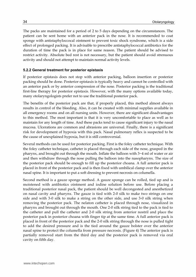
Several methods can be used for posterior packing. First is the foley catheter technique. With
the foley catheter technique, catheter is placed through each side of the nose, grasped in the
pharynx, and brought out through the mouth. Inflate the balloon with 3~4 ml of water or air
and then withdraw through the nose pulling the balloon into the nasopharynx. The size of
the posterior pack should be enough to fill up the posterior choana. A full anterior pack is
placed in front of the posterior pack and is then fixed with umbilical clamp over the anterior
nasal spine. It is important to put a soft dressing to prevent necrosis on columella.
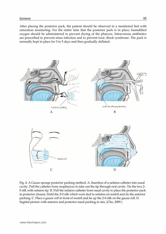
Second method is a gauze sponge method. A gauze sponge can be rolled, tied up and is moistened with antibiotics ointment and iodine solution before use. Before placing a traditional posterior nasal pack, the patient should be well decongested and anesthetized on nasal cavity and pharynx. The gauze is tied with 2-0 silk to make two strings on one side and with 3-0 silk to make a string on the other side, and use 3-0 silk string when removing the posterior pack. The nelaton catheter is placed through nose, visualized in pharynx and brought out through the mouth. One 2-0 silk string tied to the pack is tied to the catheter and pull the catheter and 2-0 silk string from anterior nostril and place the posterior pack in posterior choana with finger tip at the same time. A full anterior pack is placed in front of the posterior pack and the 2-0 silk string through the nose is pulled tight to add the desired pressure and is the tied around the gauze bolster over the anterior nasal spine to protect the columella from pressure necrosis. (Figure 4) The anterior pack is partially removed start from the third day and the posterior pack is removed via oral cavity on fifth day.
www.intechopen.com
Epistaxis
35
After placing the posterior pack, the patient should be observed in a monitored bed with saturation monitoring. For the entire time that the posterior pack is in place, humidified oxygen should be administered to prevent drying of the pharynx. Intravenous antibiotics are prescribed to prevent sinus infection and to prevent toxic shock syndrome. The pack is normally kept in place for 3 to 5 days and then gradually deflated.
A B
C D
Fig. 4. A Gauze sponge posterior packing method. A. Insertion of a nelaton catheter into nasal cavity. Pull the catheter from oropharynx to take out the tip through oral cavity. Tie the two 2-0 silk with nelaton tip. B. Pull the nelaton catheter from nasal cavity to place the posterior pack in posterior choana. Hold the 2-0 silk which were tied to nelaton on nostril and do the anterior packing. C. Place a gauze roll in front of nostril and tie up the 2-0 silk on the gauze roll. D. Sagittal picture with anterior and posterior nasal packing in situ. (Cho, 2009 )
www.intechopen.com
Otolaryngology
36
5.3 Cautery
Cauterization is an effective and simple way of controlling epistaxis. There are currently two
methods of applying cautery in the nose. The first is the use of chemical agent, such as silver
nitrate and is primarily applicable to anterior epistaxis. The second is electrocautery, which
can be used throughout the nose.
5.3.1 Chemical cautery
Usually silver nitrate is used for chemical cautery. The most convenient and usual method
of delivery is via a silver nitrate applicator stick. When touched to the mucosa, this causes a
chemical burn that coagulates the tissue. This method should be applied to selected cases of
epistaxis from the small superficial vessels. The best indication for use of silver nitrate is for
active bleeding from an anterior nasoseptal vessel. The depth of coagulation is both an
advantage and a limitation of this technique. The advantage is that there is minimal tissue
damage and, therefore, minimal discomfort and rapid healing. The limitation is that only
superficial, small vessels can be coagulated.
For chemical cauterization, the nose is first decongested and anesthetized with topical
anesthesia. For actively bleeding vessels, one or two sticks are pressed against the vessel and
slowly rolled to release the chemical. A piece of moistened cotton is then pressed over the
area, pushing the sticks onto the vessel. After 15 to 30 seconds, the cotton and sticks are
removed and the area of the cautery should be reinforced with Gelfoam or other suitable
packing.
5.3.2 Endoscopic cautery
Endoscopic cauterization is rising as a valuable management tool for intractable epistaxis. Posterior endoscopic cauterization can replace arterial ligation method in many cases. Endoscopic cautery is indicated for controlling posterior epistaxis and epistaxis that stems from the septum and lateral wall, both of which lie beyond the reach of a silver nitrate stick. This procedure is very effective if the exact location of the bleeding vessel is known or the bleeding is active at the time of the procedure. The result of endoscopic cautery is largely dependent upon the experience on sinus endoscopy of the surgeon. To cauterize the bleeding on lateral wall of nasal cavity, a greater palatine block is needed to minimize the pain. In case of severe posterior epistaxis, cauterization in operating room under general anesthesia is recommended for safety.
The nose is examined using a 0-degree or 30-degree to find the bleeding focus. When doing this, use of decongestant should be avoided, as the agent can stop bleeding temporarily. If the bleeding focus is not found, it is helpful to irritate suspicious site of bleeding with suction tip. Mucosal ulceration, mucosal injury, fresh blood clot or prominent blood capillaries are common findings to suspect recent bleeding. Cautery is performed on the bleeding focus using a suction cautery unit with the power of 15 to 20 watt. If a suction cautery is not equipped, monopolar cautery is used after cover all raw surface of suction except tip with insulating tape. Although nasal packing is usually not necessary, decision is made by the surgeon after consideration of the amount of bleeding and the status of patients.
www.intechopen.com
Epistaxis
37
Endoscopic cautery is widely used as an effective treatment method for epistaxis, as success rate is up to 89% and also, this method can reduce the rate of nasal packing and admission.
5.4 Arterial ligation
At the era before internal maxillary artery ligation via transantral approach was introduced,
external carotid artery ligation was commonly used to treat intractable epistaxis. Although
transantral approach for internal maxillary artery ligation attained less morbidity than
external carotid artery ligation, it still has had weak points. Transantral approach showed
high failure rate as it cannot identify the divergences of internal maxillary artery and had
several complication that cannot be bypassed, such as infraorbital nerve paresthesia,
oroantraol fistula, teeth injury, sinusitis or rarely blindness.
5.4.1 Transantral ligation of the internal maxillary artery
This procedure is the traditional procedure of choice for controlling posterior epistaxis.
When comparing this procedure traditional posterior packing for posterior epistaxis in
terms of cost-effectiveness and length of hospitalization, surgical procedure was superior to
the packing. Nevertheless, most patients would prefer not to have a surgical procedure, and
the morbidity of the transantral procedure is significant.
Under general anesthesia, the sublabial incison is made with a No. 15 scalpel through the
mucosa. The incision is located 5mm above the gingivolabial sulcus and extends from the
midline to the first molar. The periosteum is incised and a subperiosteal elevation is
performed over the anterior wall of the maxillary sinus. The maxillary sinus is entered over
the canine fossa using a 4mm osteotome and mallet. The opening is enlarged with a
Kerrison rongeur. The opening should be maximized by removing the anterior wall
medially to the medial buttress, laterally to the lateral wall, inferiorly to the floor of the
sinus, and superiorly to the infraorbital nerve. Then, maxillary posterior wall is drilled or
removed with chisel. The initial bone removal of the posterior wall should start 1 to 2 cm
below the floor of the orbit. A 4mm osteotome can be used in a controlled fashion with very
light taps to fracture the thin bone of this area. Incision is made on periosteum after
cauterization with a bayonet type bipolar forceps in a cruciate fashion. vessels in
pterygopalatine fossa is divided and proximal vessel and distal vessels, such as
sphenopalatine artery and descending palatine artery, are double ligated using hemoclips.
The anatomy of the vessel is somewhat variable. However, there are several branches that
can usually be identified, including the descending palatine, the superior alveolar, the
infraorbital, and the sphenopalatine. Nasal packing materials are removed after the
procedure and the nose is suctioned. Sinus packing with strip gauze is performed and the
sublabial incision is then closed using interrupted 3-0 Vicryl suture.
This method has a high probability of failure because of anatomic variation in arterial
positions. Recurrent bleeding is possible but unusual after this operation. The primary cause
of rebleeding is misdiagnosis of the source of bleeding, the other cause is failure to ligate the
appropriate vessels. The problem in the case of rebleeding is that it is difficult to distinguish
between these two possibilities based on examination or history. In the case of rebleeding
after transantral ligation of the sphenopalatine vessel, the recommended management is to
www.intechopen.com
Otolaryngology
38
perform an arteriogram. This will reliably distinguish between these situations, and also
affords the option of embolization if amenable.
5.4.2 External ligation of the ethmoid artery
Epistaxis from anterior ethmoid artery is rare, however, trauma patients with fractures
through the skull base that avulse or lacerate the anterior ethmoid artery can occur. Anterior
ethmoid artery ligation via external approach is usually used in cases of epistaxis after facial
trauma. Also, following endoscopic sinus surgery, some patients may develop heavy
bleeding from the anterior or posterior ethmoid arteries that may not respond to packing.
These can be treated with endoscopic cautery in most of cases. In certain cases, it may be
preferable to ligate the vessel. Finally, patients with HHT may require vessel ligation to
minimize the frequency or severity of epistaxis.
Under general anesthesia, operation starts with incision. In most cases, the nose is packed
and there may be a posterior pack or nasal balloon. If possible, it is best to leave this in place
until after the procedure is finished. Lynch incision is made on epistaxis side and the
subcutaneous tissue is divided down through the periosteum to the bone. The angular
artery will be encountered in the inferior aspect of the wound and should be controlled with
either ligation or bipolar cautery. The soft tissues are retracted and the eye is elevated from
the orbital bone with a Freer elevator. Lacrimal fossa and frontoethmoidal suture line can be
identified. While advancing posteriorly along the frontoethmoid suture line, anterior
ethmoid artery is found 20 to 24 mm posterior to the anterior lacrimal crest and this should
be double ligated or cauterized. The preferred technique is to use bipolar cautery, as the
clips tend to fall off and can lead to orbital hematoma. It is important to make sure that the
full diameter of the artery is cauterized. Once the artery is fully cauterized or clipped,
Jameson scissors are used to transect the artery in the middle, making sure that there is
room to cauterize each stump if necessary. The dissection is then carried posteriorly for
another 10 to 12mm until reaching the posterior ethmoid artery. Posterior ethmoid artery
goes close to optic nerve and is not a major vessel that causes epistaxis, so ligation is usually
not considered. After the arteries are divided, the wound is closed. Hemostasis should be
achieved using bipolar cautery on the orbital tissues and monopolar cautery on the
subcutaneous tissues.
All patients should be observed at least overnight after this surgery for two significant
concerns: orbital hematoma and rebleeding. Orbital edema and ecchymosis are expected.
However, there should not be any chemosis, true proptosis, or limitation of extraocular
movements. Any of these signs suggests orbital hematoma and should be managed
immediately according to the management protocol for orbital hematoma.
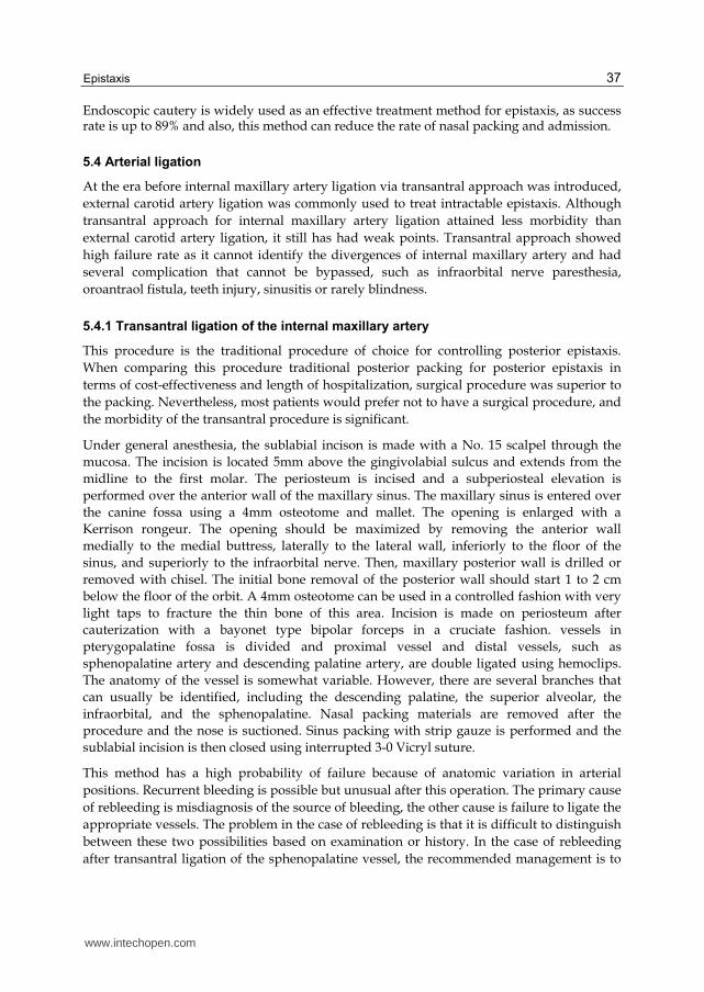
5.4.3 Endoscopic ligation of sphenopalatine artery
Endoscopic ligation of the internal maxillary artery is the newest addition to the surgical arsenal of the rhinologist for posterior epistaxis that can lower the complication of transantral approach and pterygomaxillary fossa dissection, and can prevent the collateral vessel formation as the most distal part of the artery is ligated, compared to other methods.
www.intechopen.com
Epistaxis
39
General anesthesia is achieved with orotracheal intubation. Before the nasal pack is
removed, a transpalatal injection of the greater palatine foramen is performed using local
anesthetic agent to minimize the bleeding after removal of the posterior pack. After removal
of posterior pack, using 0 degree endoscope, thorough suction in the nasal cavity is
performed for visualization. The posterior attachment of the middle turbinate is identified
and an incision is made in the mucosa about 1 cm anterior to this point, extending from the
inferior turbinate to the ethmoid sinus. The mucoperiosteal flap is elevated
posterosuperiorly then surgeon can identify bundle of vessels and nerves originated from
sphenopalatine foramen. The vessels are then carefully dissected with Freer elevator to
identify sphenopalatine artery and ligation of the artery with surgical clip is performed. The
mucoperiosteal flap is then repositioned and the procedure is concluded simply by placing a
Surgicel on the incision site.
Fig. 5. Endoscopic ligation of sphenopalatine artery. Left side. A.The mucosa immediately posterior to the attachment of the middle turbinate is elevated to expose the branches of the sphenopalatine artery. B. The bone of the sphenopalatine foramen is removed to expose the main artery. C. The artery and its main branches are ligated with microclips. D. The mucosal flap is covered for the last step of the surgery. (Snyderman & Carau, 1997) MT; middle turbinate, IT; inferior turbinate, ST; superior turbinate.
www.intechopen.com
Otolaryngology
40
After the operation, the patient’s activity should be restricted for several days. After
discharge from the hospital, activity and diet are gradually returned to normal. Full activity,
including exercise, should be avoided for 2 weeks after the procedure. Extreme care must be
exercised when suctioning over the area of the ligation during dressing, to avoid
accidentally suctioning off one of the clips. All of the complications associated with
endoscopic sinus surgery are possible with this endoscopic ligation procedure, though the
risks of those are lower in this setting than for routine endoscopic sinus surgery.
5.4.4 External carotid artery ligation
External carotid artery ligation is performed at site afar from nasal cavity, so rebleeding
from collateral vessels from internal carotid artery or contralateral carotid artery is reported
upto 45% of patients. Therefore, limited cases of epistaxis from peripheral collateral vessels
from external carotid artery are indicated for external carotid artery ligation.
Horizontal incision between hyoid and superior border of thyroid cartilage is made,
subplatysmal flap elevation is performed and sternocleidomastoid muscle is retracted
posteriorly to visualize internal jugular vein, which is retracted to identify external carotid
artery. More than two branches should be identified to confirm external carotid artery from
internal carotid artery, and then external carotid artery is ligated.
5.5 Arterial embolization
One of the options for severe posterior epistaxis is intraarterial embolization. Selective arterial embolization has advantages that duration of in-hospital stay is short, procedure can be performed under local anesthesia, vessels that are laborious for surgical approach can be treated, and success rate is high in surgical failure cases of arteriovenous communication. This procedure is usually performed under local anesthesia in the arteriography suite of the hospital, a catheter is placed into the groin and threaded through the femoral artery, into the aorta, into the carotid artery and finally into the internal maxillary artery. A test injection is performed to identify the bleeding site. If there is no communication between internal maxillary artery and internal carotid artery or vertebral artery, various materials are used for embolization, including gelfoam, coils, balloons, and polyvinyl alcohol particles. Injection of the materials is repeated until blood flow at the bleeding sites decreases or stops. If bleeding have recurred short after the embolization, the source of the bleeding is anterior or posterior ethmoid artery, which are branches of internal carotid artery, so surgical approach to these vessels might be needed, and collateral vessel bleeding from facial artery is occurred, reembolization should be performed. More than half of cases of rebleeding long after the treatment are reported to be hereditary hemorrhagic telangiectasia.
Embolization is a useful and highly successful technique for controlling posterior epistaxis.
However, in all large series of carotid artery catherization, there is an incidence of serious
cerebrovascular accident (CVA) resulting in permanent neurologic deficit. The incidence of
this varies with the skill of the radiologist in performing the procedure, with an overall
average of about 4%. As it is a relatively high rate for such a major complication, the use of
embolization for control of epistaxis is still controversial.
www.intechopen.com
Epistaxis
41
5.6 Septoplasty
Septoplasty is helpful to manage epistaxis in selected cases with severe septal deviation or
septal mass. This procedure is needed in cases that bleeding focus is behind the septal spur
or management approach is hindered by septal deviation. As most haemorrhages occur
from the septum, raising a mucoperichondral flap during septal surgery can be beneficial as
this will decrease blood flow to the mucosa, which often in itself stems bleeding. It is also
performed to block the blood supply in Kiesselbach plexus, or to remove hemorrhagic
nodule or septal turbinate. Surgery is also used to correct a deviated septum or remove a
septal spur, which may be the cause of epistaxis. This occurs either by altering air flow
through the nose or in severe cartilage deformities, by persistent mucosal irritation. As it is
one of the most common procedures that are performed by rhinologists, detailed operation
skills are not described in this chapter. (Pope & Hobbs, 2005)
5.7 Anonymous
5.7.1 Fibrin glue
Fibrin glue is developed from human plasma cryoprecipitate and binds itself to damaged
vessels. The technique entails spraying a thin layer of glue over the bleeding site and can be
repeated as needed. On one randomized trial has reported that complications of local
swelling, nasal mucosa atrophy, and excessive nasal discharge were lower than the
electrocautery, silver nitrate, and nasal packing group. The rebleed rate was 15%, which is
comparable to electrocautery. (Pope & Hobbs, 2005)
5.7.2 Hot water irrigation
Hot water irrigation was first described by Guice in 1878 as an effective method of treating
severe, life-threatening epistaxis. Hot water irrigation is performed in continuous posterior
epistaxis cases. The affected nasal cavity is continuously irrigated during 3 minutes with 500
ml water heated to 50°C after packing the posterior choanae with balloon catheter. Tap
water is administered via a water irrigator. The patient is then seated upright with their face
pointed downward over a basin, to allow outflow of water from the affected nasal cavity
into the basin. This method produces vasodilation and edema of the nasal mucosa without
the risk of necrosis. This mucosal edema leads to local compression of the bleeding vessels,
while at the same time triggering and probably accelerating the clotting cascade. (Novoa &
Schlegel-Wagner, 2011)
5.8 Management of Hereditary Hemorrhagic Telangiectasia
The management of HHT depends on several independent factors. These include the
number and location of the intranasal lesions, the severity and frequency of epistaxis, the
number and location of gastrointestinal, brain and pulmonary lesions, the number of blood
transfusions attributable to the epistaxis, the effect of the epistaxis on the patient’s quality of
life and the previous treatments attempted.
Medical therapy for HHT, in addition to that outlined earlier for epistaxis in general, has
focused on the use of estrogen and progesterone. This treatment was based on the
www.intechopen.com
Otolaryngology
42
observation that epistaxis tended to decrease during pregnancy among affected individuals.
Antifibrinolytic therapy using aminocaproic acid has also been discussed. None of these
medical treatments has proven effective in completely relieving epistaxis in the long term,
but some benefits have been claimed.
Because of the failure of these methods, numerous other modalities have been attempted,
including sclerotherapy, brachytherapy, electrocautery, arterial ligation, embolization,
septodermoplasty, closure of the nasal cavities, and laser coagulation. The clear implication
of all of these reports is that there remains no cure for HHT and, despite all treatment
attempts, recurrent epistaxis is possible. In light of these facts, the author suggests that
treatment should be designed to minimize the bleeding while avoiding complications. For
this reason, extreme measures, such as brachytherapy, embolization, and invasive surgery,
should be avoided.
As regards laser treatment of HHT, there is controversy over which type of laser is best.
Carbon dioxide, ND:YAG, pulsed dye laser, diode and KTP lasers have all been used for this
purpose, with the KTP laser now gaining consensus as the most popular device. It has been
suggested that laser can easily ablate the periphery of larger lesions to reduce central blood
flow, but that direct laser focused on the center of a lesion causes extensive bleeding and
makes further treatment difficult. (Joshi et al., 2011)
Septodermoplasty involves removal of nasal mucosa from the anterior part of the nasal
cavity and replacement by a split skin graft. This technique has been found to have good
initial outcomes in patients with HHT patients, which unfortunately decline over time due
to contraction and revascularization of the graft. The technique has also been found to be
associated with nasal crusting and halitosis.
Closure of the nasal cavity, the so called Young’s procedure is a radical technique involving
closure of the nasal vestibule. Although it provides long term relief in patients with
moderate to severe epistaxis secondary to HHT, the disadvantages (dry mouth, loss of smell
and complete nasal obstruction) are often not tolerated by patients. Upon reviewing the
procedure, Young himself found that many patients had to have the procedure reversed
because of these problems. Young’s procedure is best reserved for cases of HHT
unresponsive to other treatment modalities.
Radiofrequency coblation is a relatively new technique which is being increasingly used in
ENT surgery. Coblation has been demonstrated to promote good healing and to preserve
surrounding normal tissue. Despite low temperatures, small blood vessels are sealed by this
process. Radiofrequency coblation can therefore theoretically achieve both ablation and
hemostasis of telangiectatic and arteriovenous malformations, using the same instrument.
The use of coblation for hereditary haemorrhagic telangiectasia epistaxis is a much more
conservative procedure, which can be safely repeated without significant complications.
(Joshi et al., 2011)
Elevated plasma levels of vascular endothelial growth factor (VEGF) play a key pathogenic
role in HHT. Bevacizumab, an anti-VEGF monoclonal antibody, prevents the binding of
VEGF to VEGF receptor on endothelial cells, thus blocking cellular proliferation and
www.intechopen.com
Epistaxis
43
angiogenesis. As a result, bevacizumab has been used as a potential treatment of recurrent
epistaxis. Previous studies have demonstrated marked improvement of epistaxis with
intravenous administration of bevacizumab. Intranasal treatment with bevacizumab, by
either submucosal injection or topical nasal spray, has recently been reported to be a safe
alternative to intravenous treatment. (Brinkerhoff et al., 2011)
6. Conclusion
Over the past decade, there has been a significant development in the options available for
the management of epistaxis. Traditional strategies like nasal packing have been
supplemented by modern technology using the latest optic and electrical devices. Treatment
should ideally use a systematic protocol; starting with simple procedures that can be
undertaken in the clinic environment and proceeding to endoscopic techniques for more
difficult cases.
7. References
Brinkerhoff, BT, Choong, NW, Treisman, JS & Poetker, DM (2011). Intravenous and
topical intranasal bevacizumab (Avastin) in hereditary hemorrhagic
telangiectasia. American Journal of Otolaryngology Vol. No., (Sep 12), pp., ISSN 0196-
0709
Cho, JS (2009 ).Epistaxis. In: Otorhinolarngology head and neck surgery, surgery, KsoO-Han, pp.
1048-1057, Ilchokak, ISBN 978-89-337-0559-9, Seoul
Fuchizaki, U, Miyamori, H, Kitagawa, S, Kaneko, S & Kobayashi, K (2003). Hereditary
haemorrhagic telangiectasia (Rendu-Osler-Weber disease). The Lancet Vol. 362, No.
9394, (Nov 1), pp. 1490-1494, ISSN 0140-6736
Guttmacher, AE, Marchuk, DA & White, RI, Jr. (1995). Hereditary hemorrhagic
telangiectasia. The New England Journal of Medicine Vol. 333, No. 14, (Oct 5), pp. 918-
924, ISSN 0028-4793
Herkner, H, Laggner, AN, Mullner, M, Formanek, M, Bur, A, Gamper, G, Woisetschlager, C
& Hirschl, MM (2000). Hypertension in patients presenting with epistaxis. Annals of
Emergency Medicine Vol. 35, No. 2, (Feb), pp. 126-130, ISSN 0196-0644
Joshi, H, Woodworth, BA & Carney, AS (2011). Coblation for epistaxis management in
patients with hereditary haemorrhagic telangiectasia: a multicentre case series. The
Journal of Laryngology & Otology Vol. No., (Aug 16), pp. 1-5, ISSN 1748-5460
Novoa, E & Schlegel-Wagner, C (2011). Hot water irrigation as treatment for intractable
posterior epistaxis in an out-patient setting. The Journal of Laryngology & Otology
Vol. No., (Sep 5), pp. 1-3, ISSN 1748-5460
Pope, LE & Hobbs, CG (2005). Epistaxis: an update on current management. Postgraduate
Medical Journal Vol. 81, No. 955, (May), pp. 309-314, ISSN 0032-5473
Snyderman, CH & Carau, RL (1997). Endoscopic Ligation of the Sphenopalatine Artery For
Epistaxis. Operative Techniques in Otolaryngology Head and Neck Surgery Vol. 8, No. 2,
(Jun 1997), pp. 85-89, ISSN 1043-1810
www.intechopen.com

Otolaryngology
44
Viducich, RA, Blanda, MP & Gerson, LW (1995). Posterior epistaxis: clinical features and
acute complications. Annals of Emergency Medicine Vol. 25, No. 5, (May), pp. 592-
596, ISSN 0196-0644
www.intechopen.com

OtolaryngologyEdited by Prof. Balwant Singh Gendeh
ISBN 978-953-51-0624-1Hard cover, 198 pagesPublisher InTechPublished online 23, May, 2012Published in print edition May, 2012
InTech EuropeUniversity Campus STeP Ri Slavka Krautzeka 83/A 51000 Rijeka, Croatia Phone: +385 (51) 770 447 Fax: +385 (51) 686 166www.intechopen.com
InTech ChinaUnit 405, Office Block, Hotel Equatorial Shanghai No.65, Yan An Road (West), Shanghai, 200040, China
Phone: +86-21-62489820 Fax: +86-21-62489821
This book emphasizes on different aspects of otolaryngology - the medical sciences of diagnosis andtreatment of ENT disorders. "Otolaryngology" is divided into various clinical sub-specialities, namely otology,rhinology, laryngology, and head and neck. This book incorporates new developments, as well as futureperspectives in otolaryngology. I would like to dedicate this book to those of you who will pick up the torch andby continued research, close clinical observation and the highest quality of clinical care, as well as bypublication and selfless teaching, further advance knowledge in otolaryngology from this point forward. It isintended to be a guide to other books to follow. Otolaryngologists, researches, specialists, trainees, andgeneral practitioners with interest in otolaryngology will find this book interesting and useful.
How to referenceIn order to correctly reference this scholarly work, feel free to copy and paste the following:
Jin Hee Cho and Young Ha Kim (2012). Epistaxis, Otolaryngology, Prof. Balwant Singh Gendeh (Ed.), ISBN:978-953-51-0624-1, InTech, Available from: http://www.intechopen.com/books/otolaryngology/epistaxis

© 2012 The Author(s). Licensee IntechOpen. This is an open access articledistributed under the terms of the Creative Commons Attribution 3.0License, which permits unrestricted use, distribution, and reproduction inany medium, provided the original work is properly cited.