epithelia and glands
TRANSCRIPT
Prelab Exercise 1 –EPITHELIA
1
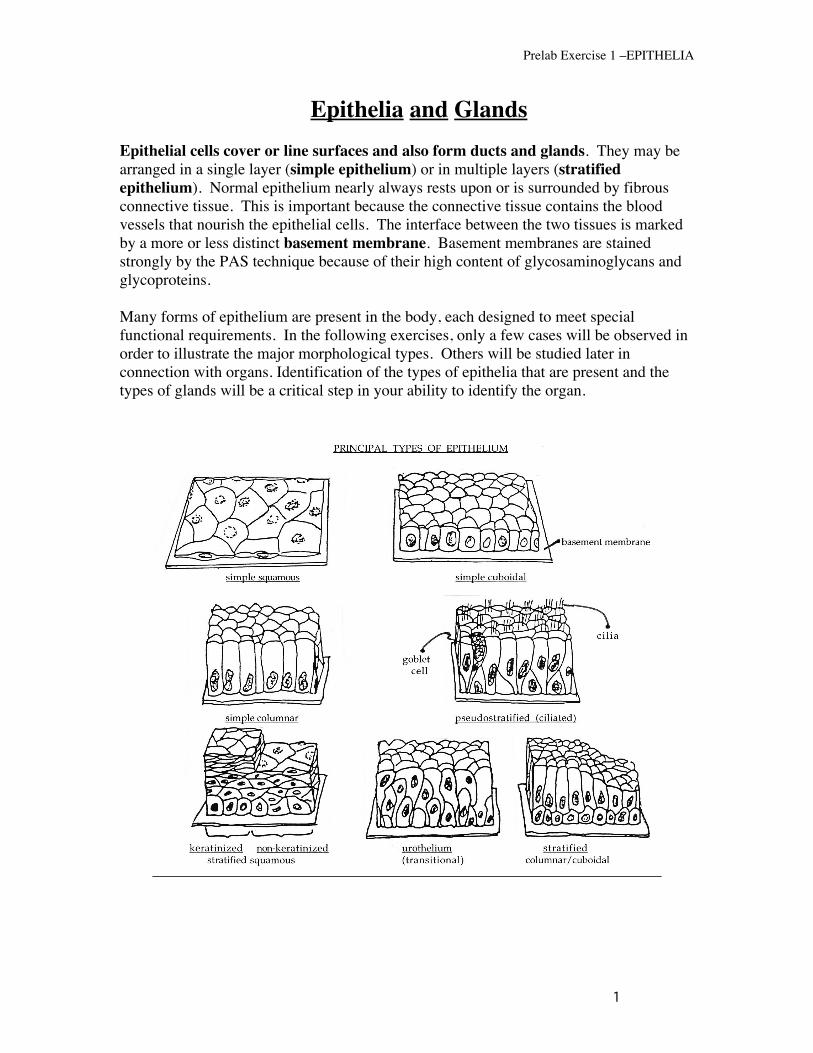
Epithelia and Glands Epithelial cells cover or line surfaces and also form ducts and glands. They may be arranged in a single layer (simple epithelium) or in multiple layers (stratified epithelium). Normal epithelium nearly always rests upon or is surrounded by fibrous connective tissue. This is important because the connective tissue contains the blood vessels that nourish the epithelial cells. The interface between the two tissues is marked by a more or less distinct basement membrane. Basement membranes are stained strongly by the PAS technique because of their high content of glycosaminoglycans and glycoproteins. Many forms of epithelium are present in the body, each designed to meet special functional requirements. In the following exercises, only a few cases will be observed in order to illustrate the major morphological types. Others will be studied later in connection with organs. Identification of the types of epithelia that are present and the types of glands will be a critical step in your ability to identify the organ.

Prelab Exercise 1 –EPITHELIA
2
SIMPLE (SINGLE-LAYERED) TYPES.
Simple epithelia are a single layer of cells thick. Each of the cells contacts the basement membrane. Most can be seen to have a single layer of nuclei although pseudostratified epithelium has nuclei at multiple levels (despite the fact that all of the cells have their bases resting on the basement membrane).
Simple squamous
Simple squamous epithelium consists of a single layer of flat cells. The cell may be thicker in the location of the nucleus, giving the cell the appearance of a fried egg. In some places the simple squamous epithelium has a special name. The simple squamous lining of blood vessels is called the endothelium, and simple squamous cells covering the mesentaries inside the thorax and abdomen are called mesothelial cells.
Simple cuboidal epithelium
Simple cuboidal cells line many ducts. Depending on whether these cells are involved in absorption or secretion, they may have infoldings of the cell membrane (particularly at the base of the cell) or finger-like projections from the surface side of the cell (microvilli). These increase surface membrane area.
Simple columnar epithelium
Simple columnar epithelia are found mostly in the GI tract, where they are heavily involved in absorption. They also line some of the GU tract (for example, the uterine tubes). Most simple columnar cells have microvilli, which appear as a “brush border” at the light microscopic level. You can see microvilli in figs. 1 & 6 of the “EM of epithelium” module of virtual histology.
Pseudostratified columnar epithelium
This term is commonly applied to certain epithelia which, when sectioned, appear to be stratified, mostly because there are nuclei at various levels. However, observation of cells separated by teasing would show that all of the cells maintain an attachment to the underlying basement membrane. On the other hand, only some reach the free surface of the epithelium. This kind of epithelium is most commonly found lining the passages of the respiratory system (e.g., trachea and bronchi). It is also found in some ducts in the male reproductive system that we will study later in the course.
STRATIFIED (MULTI-LAYERED) EPITHELIA
It is imperative to understand that the different types of stratified epithelia are identified on the basis of the morphology of the most superficial layer of cells (i.e. cells of the luminal or free surface). Deeper lying cells in these epithelia may have a different appearance.

Prelab Exercise 1 –EPITHELIA
3
Stratified squamous epithelia covers internal and external surfaces (moist or dry) that are prone to abrasion and need the added protection that this type of epithelium affords. Areas lined with this type of epithelium include the skin, oral cavity, esophagus, and vagina among others. Stratified squamous epithelia are characterized by several layers (strata) of cells. The surface cells have one of two appearances that define the stratified squamous epithelium as being either kertinized or non-kerainized. In mostly dry areas, such as the skin, the nuclei of the surface layers shrink and become pyknotic (and in some cases disappear altogether). In addition, the cytoplasm undergoes varying degrees of keratinization (sometimes referred to as cornification) during which keratin and associated matrix proteins increase in abundance in the cytoplasm, eventually filling the cell. Keratin is also the protein that comprises finger and toe-nails. Toward the surface of the skin, cells undergo transformation from living cells to dead scales, which are then sloughed from the epithelium. In moist areas (such as the mouth, esophagus and vagina) non-keratinized epithelial cells retain their nuclei all the way to the surface. This epithelium is kept moist by the presence of mucus or other fluids.
Consider the structural changes that occur in the nucleus and cytoplasm as living cells move from the basal layer to the free surface during their conversion to a dead scale. Observe pyknosis, which is the shrinkage of the nucleus to form a small, irregularly shaped, compact mass. Loss of normal nuclear structure is one of the most useful criteria for determination of loss of cellular vitality due to trauma, disease, or as in this case, natural processes. This criterion for evaluation of cellular vitality will be used repeatedly in CTO and in pathology.
Stratified cuboidal or columnar epithelium. These types of stratified epithelia are uncommon and have a very limited distribution in the body. When present, they usually occur in large glandular ducts (e.g. pancreatic duct, salivary ducts). They occur occasionally in zones of transition between two different varieties of epithelium (e.g., transition between simple columnar and stratified squamous).
Transitional epithelium (urothelium). The distribution of this epithelium is limited to lining the different parts of the urinary tract (renal pelvis, ureter, urinary bladder). This epithelium is specifically adapted for stretching and its morphology may correspondingly change. For example, in the distended urinary bladder, the epithelium appears only two or three layers thick, whereas, in the contracted bladder, it may be four to six or even more layers thick. This type of multilayered epithelium (when not under tension) is characterized by its surface layer of dome, pillow-like, or umbrella-like cells. Subjacent layers contain cells that may be cuboidal, columnar or polyhedral in shape.
SURFACE SPECIALIZATIONS OF EPITHELIAL CELLS While these structures may be discerned by light microscopy, their detailed morphology can only be appreciated by electron microscopy. You should correlate your observations

Prelab Exercise 1 –EPITHELIA
4
here with the electron micrograph illustrations in your textbooks. Also, keep in mind that similar surface specializations may occur in the cells of other tissues besides epithelium.
Microvilli
As described above microvilli are projections of the surface membrane of columnar and some cuboidal cells that are designed to increase surface area. The actin filament cores of the microvilli extend down into the apical cytoplasm of the cell where they are crosslinked by myosin and other actin-associated proteins. This dense meshwork of filaments creates the terminal web and can be see as a denser, eosinophilic line immediately beneath the microvilli. Also observe microvilli in figs 1 & 6 of the “epithelium” module of virtual histology. Observe the columnar surface epithelial cells in Slide #37 [small intestine, monkey, P.A.S. + H]. The brush borders of these cells are distinctly “P.A.S. positive” (stain red). Why?
Cilia Cilia are long, motile projections from the surface of the epithelium. They are anchored to ciliary basal bodies, which are located in a row beneath the apical epithelium. Review the ultrastructure of cilia from the illustrations in your textbook and from figs. 3 & 4 of the “EM of epithelium” module of virtual histology.
Stereocilia Stereocilia are immotile structures and basically represent very long, somewhat less-rigid microvilli.
Intercellular Junctions Generally, junctional specializations of the opposed lateral surfaces of epithelial cells (e.g., tight junctions) are below the resolution of the light microscope. However, pre-EM light microscopists found some evidence of their existence in the so-called “terminal bars” located apically between contiguous epithelial cells. Examine these specializations in figs. 1, 6, 7 & 8 of the “EM of epithelium” module of virtual histology. Figure 6 shows the entire junctional complex of zonula occludens (tight junction), zonula adherens (intermediate junction) and macua adherens (desmosome).
EPITHELIAL GLANDS
Glands may be classified in a variety of ways. Perhaps the most basic is classifying glands as either “exocrine” (secretion into a lumen or onto a surface) or “endocrine” (secretion into the circulation via capillaries). Although these glands all originate as an epithelium, the exocrine glands maintain a connection with the surface via a duct, and the endocrine glands lose this connection. You will examine exocrine glands in this lab while endocrine glands will be studied later in the course. There are three methods for classifying exocrine glands: 1. According to structural organization; 2. Based on type of secretion; 3. Based on their mode of secretion.

Prelab Exercise 1 –EPITHELIA
5
EXOCRINE GLANDS - Classification based on the STRUCTURAL organization of cells
Unicellular glands: The goblet cells are examples of unicellular (single cell) glands. They
secrete mucin onto the surface of the epithelium. Multicellular glands: Simple glands: Such a gland consists of a secretory unit (collection of secretory cells)
connected to the surface epithelium directly or by a single unbranched duct.
Types of simple glands: (A) Simple tubular (B) simple coiled tubular (C) simple branched
tubular (D) simple acinar or alveolar (E) simple branched acinar
Compound glands: consist of a varying number of simple glands whose small excretory ducts join to form progressively larger and larger ducts which carry the secretion onto an epithelial surface. The pattern of the structure and arrangement of the ducts is often characteristic in a specific compound gland. Large compound glands, such as salivary glands (compound tubuloacinar) and the pancreas (compound acinar) will be studied in detail later in conjunction with the specific systems to which they belong.
(A) compound tubular (B) compound acinar (C) compound tubuloacinar
Prelab Exercise 1 –EPITHELIA
6
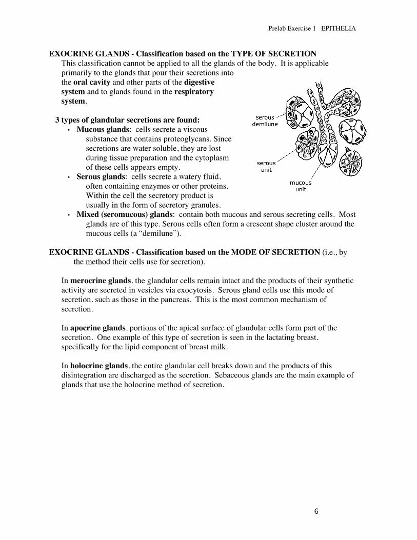
EXOCRINE GLANDS - Classification based on the TYPE OF SECRETION This classification cannot be applied to all the glands of the body. It is applicable primarily to the glands that pour their secretions into the oral cavity and other parts of the digestive system and to glands found in the respiratory system.
3 types of glandular secretions are found: • Mucous glands: cells secrete a viscous
substance that contains proteoglycans. Since secretions are water soluble, they are lost during tissue preparation and the cytoplasm of these cells appears empty.
• Serous glands: cells secrete a watery fluid, often containing enzymes or other proteins. Within the cell the secretory product is usually in the form of secretory granules.
• Mixed (seromucous) glands: contain both mucous and serous secreting cells. Most glands are of this type. Serous cells often form a crescent shape cluster around the mucous cells (a “demilune”).
EXOCRINE GLANDS - Classification based on the MODE OF SECRETION (i.e., by
the method their cells use for secretion).
In merocrine glands, the glandular cells remain intact and the products of their synthetic activity are secreted in vesicles via exocytosis. Serous gland cells use this mode of secretion, such as those in the pancreas. This is the most common mechanism of secretion.
In apocrine glands, portions of the apical surface of glandular cells form part of the
secretion. One example of this type of secretion is seen in the lactating breast, specifically for the lipid component of breast milk.
In holocrine glands, the entire glandular cell breaks down and the products of this
disintegration are discharged as the secretion. Sebaceous glands are the main example of glands that use the holocrine method of secretion.

Prelab Exercise 1 –EPITHELIA
7
CHECK LIST FOR EPITHELIUM AND GLANDS
Be able to identify the seven listed “principal types of epithelium”: -simple squamous, -simple cuboidal, -simple columnar, -pseudostratified, -keratinized and non-keratinized stratified squamous -urothelium. -stratified columnar/cuboidal
Know the terms:
-basement membrane, -endothelium -mesothelium (visceral peritoneum), -pyknosis
Understand each of the components of surface specializations:
-microvilli/terminal web -cilia/basal bodies -stereocilia -intercellular junctions, terminal bars -occluding (tight) junctions; zonula occludens -adherent (anchoring) junctions; zonula adherens -desmosomes; macula densa -hemidesmosomes -gap (communicating) junctions
Classify epithelial glands based on the structural organization - unicellular, multicellular, simple and compound.
Know the morphology of the chemistry of secretion:
-mucous -serous -mixed (seromucous) [with demilunes]
Recognize the mode of secretion of glandular cells, including examples of each: -holocrine -merocrine -apocrine
Understand the difference between exocrine and endocrine secretion.