ercp: pancreatobiliary tree without the surgeon€¦ · 1 ercp: pancreatobiliary tree without the...
TRANSCRIPT
1
ERCP: Pancreatobiliary tree without the surgeon
Kamran Ayub, MD, MRCPVisiting Clinical Associate Professor, UIC
Silver Cross Hospital
Advocate Christ Medical Center
Provena St Joseph Medical Center
Little Company of Mary Hospital
Advocate Good Samaritan Hospital
Disclosures
• No conflict of interest for this presentation
• This conference supported by an independent educational grant from Cook Medical
• No political affiliations
ERCP and Advanced Endoscopy: ERCP and Advanced Endoscopy: Team EffortTeam Effort
• Strong team required for successful program:
• Endoscopist, and well trained:– Nurses
– Technicians
– Cytopathologist
2
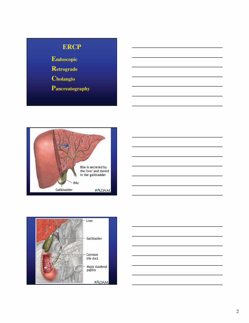
ERCP
Endoscopic
Retrograde
Cholangio
Pancreatography

3
4

5
Estimated Procedural Volume -2004
♦ United States - 445,000 cases per year
♦ Worldwide - 1.3-1.5 million cases per year
Sources: Timely Data ResourcesSources: Timely Data Resources
PSPSF PSPSF –– Medicare claimsMedicare claims
SolutionsSolutions

6
ERCP’s – United States 2004
♦ Diagnostic ERCP’s - 69,000
− Decreasing ~ 4% per year
♦ Therapeutic ERCP’s - 376,000
− Increasing ~ 12% per year
ERCP
♦ Historical contraindications
− Pancreatitis
− Cholangitis
ERCP-Current Indications
♦ Biliary Tract:
− Choledocholithiasis
− Benign obstruction
− Malignant obstruction
− Complications of laparoscopic
cholecystectomy

7
ERCP-Current Indications
♦ Pancreatic disease
− Acute pancreatitis with complications
− Evaluation and management of recurrent pancreatitis
− Chronic pancreatitis with complications
ERCP-Current Indications
♦ Evaluation of imaging abnormalities -e.g. MRCP, CT, EUS
♦ Evaluation of abdominal pain ? SOD
♦ Excision of ampullary tumors
Case from Seattle
• 68 y/o MSM with h/o CAD, HTN, Diabetes, CRI presents with ~ 6 month upper abdominal pain, deep seated, vague, 3-5/10. Two attacks of mild pancreatitis.
• MRI scan: 3 cm mass ampullary region, 1.4 cm area of signal abnormality in HOP. Normal CBD and PD.

8
Case
• P/Hx:– CAD: CABG in ’96
– IDDM
– HTN
– CRI, creatinine around 2.2
– Obesity
Physical Exam
• VSS
• Obese, ht 5.6”, wt 235 lbs
• Otherwise unremarkable
Laboratory
• Hct 30
• Cr 2.2
• LFT’s Normal
• CA 19-9 Normal
• EUS: ampullary mass, no deep invasion
• Biopsies: Carcinoid

9
Endoscopy

10
Ampullary NeoplasmsAmpullary Neoplasms
• Familial Polyposis– Disease prevalence: 1/5000 to 1/7500– 50% to 80% will have adenomatous change of the
papilla– 4% to 12% lifetime incidence of duodenal cancer
• Sporadic– Prevalence: 0.04% to 0.12% in autopsy series– Patients are usually > 40 years old (usually in 70s)
Bussey et al. Gastroenterology 1978;74:1325, Burt Semin Gastrointest Dis 1992;3:13, Debinski et al. Eur J Cancer 1995;31A:1149,Sato et al. Hepatogastroenterology 1999;46:1959, Shapiro & Lifvendahl Ann Surg 1931;94:61, Baker & Caldwell Surgery 1947;21:523,Rosenberg et al. Cancer 1986;58:1563, Sobol & Cooperman Gastroenterology 1978;75:107
PresentationPresentation
• No symptoms• Obstructive jaundice
– 50% to 75% of symptomatic patients– Usually either painless or with a dull midepigastric
ache– Up to 25% will have associated CBD stones due
to cholestasis
• Abdominal pain• Pancreatitis• Bleeding and/or anemia
Treitschke & Beger Ann Oncol 1999;10 Suppl 4:212, Ashkar et al. Digestion 1999;60:583, Sharp & Brandes Am Surg 1990;56:214,Taxier et al. Gastrointest Endosc 1979;25:155, Binmoeller et al. Gastrointest Endosc 1993;39:127, Shemesh et al. Surg GynecolObstet 1989;169:445, Sato et al. Gastrointest Endosc 1999;50:672, Ohmori et al. Am J Surg 1976;132:662

11
StagingStaging
• Accurate staging is important to determine the appropriate intervention
• Methods:– U/S– CT– ERCP– EUS– IDUS
Treatment OptionsTreatment Options
• Endoscopic ampullectomy• Surgery
– Wide local excision– Pylorus-preserving resection of pancreatic
head– Whipple

12
Surgical managementSurgical management
• Carcinoma• Extension into CBD/PD
• Indeterminate staging• Large >2 cm ??
• HGD: young, good health
Complications of Endoscopic Complications of Endoscopic AmpullectomyAmpullectomy
• Pancreatitis: 8 to 15% (typically mild)• Bleeding: 4 to 6%
• Perforation: 4%• Stenosis:
Endoscopic surveillanceEndoscopic surveillance
• Tubular histology– 2 to 3 years
• Unfavorable histology: Villous, HGD– 1 year

13
Critical Influences on ERCP
♦ Technology
− Accessory development-e.g. guidewires, multilumen catheters, dilating balloons
− “smart” cautery – e.g. ERBE− Smaller therapeutic scopes− Stent technology-plastic, SEMS
Critical Influences on ERCP
♦♦ TechnologyTechnology
−− Laparoscopic surgeryLaparoscopic surgery
−− EUSEUS
−− MRCPMRCP
−− High resolution CTHigh resolution CT
Case
• 55 y/o woman presents with 3 day h/o worsening abdominal pain and abdominal distention. Lap Chole 4 days ago.
• LFT’s mildly elevated
• Diagnosis:
• Next step???
14
Case
• 55 y/o woman presents with 3 day h/o worsening abdominal pain and abdominal distention.
• LFT’s mildly elevated
• Diagnosis:
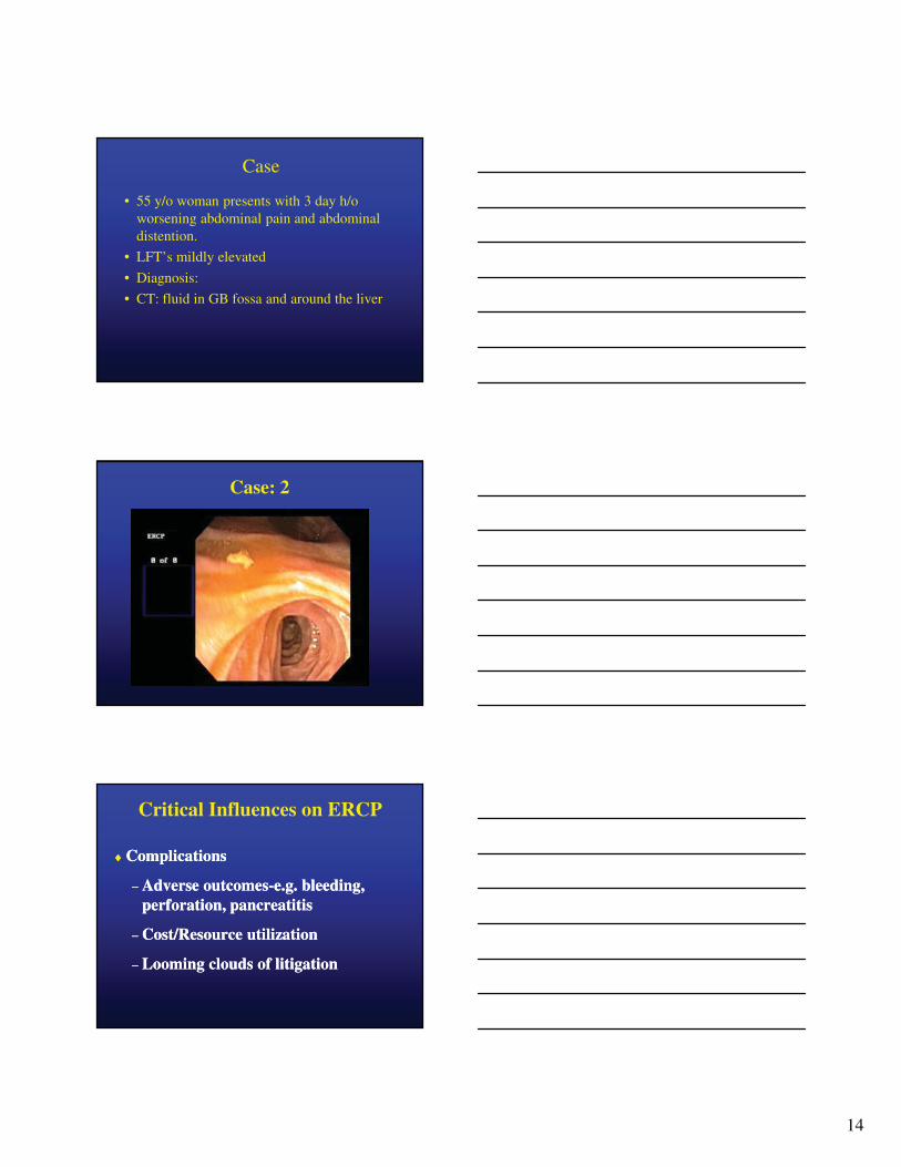
• CT: fluid in GB fossa and around the liver
Case: 2
Critical Influences on ERCP
♦♦ ComplicationsComplications
−− Adverse outcomesAdverse outcomes--e.g. bleeding, e.g. bleeding, perforation, pancreatitisperforation, pancreatitis
−− Cost/Resource utilizationCost/Resource utilization
−− Looming clouds of litigationLooming clouds of litigation

15
Tissue Acquisition
♦♦ Future directions/technologiesFuture directions/technologies
−− Expanded use of EUS/FNAExpanded use of EUS/FNA
−− SPYGLASS biopsiesSPYGLASS biopsies
−− Pilot Balloon Pilot Balloon CholangioscopyCholangioscopy
−− IDUS probesIDUS probes−− Tissue analysisTissue analysis--e.g. flow e.g. flow cytometrycytometry, ,
molecular genetics, genotyping, FISHmolecular genetics, genotyping, FISH
CaseA 24 y/o woman presents with c/o severe 10/10
RUQ abdominal pain. She has 1 year hx of similar attacks of pain lasting few hours.
Lap chole 14 months ago. Has seen her PCP on several occasions with same complaint, a KUB, CT scan and routine labs have been normal. On percocet for pain.
Examination is unremarkable except slight voluntary guarding in RUQ.
Case
• Bouts of severe RUQ pain
• Labs normal
• CT normal

16
Case
• Bouts of severe RUQ pain
• Labs normal
• CT normal
• Treated as IBS
Case
• Bouts of severe RUQ pain
• Labs normal
• CT normal
• Treated as IBS
• ERCP: Slightly generous bile duct, manometry: high basal pressure 70 mmHg
• Sphincterotomy provided pain relief
Case
• Bouts of severe RUQ pain
• Labs normal
• CT normal
• Treated as IBS
• ERCP: Slightly generous bile duct, manometry: high basal pressure 70 mmHg
• Sphincterotomy provided pain relief
• 18 months later, doing well

17
Biliary Dyskinesia orSphincter of Oddi Dysfunction
• Approx 750,000 lap cholecystectomies performed every year
• 15 to 30% have recurrent or persistent pain
• Majority have biliary dyskinesia (or sphincter of oddi dysfunction)
Sphincter of Oddi Dysfunction
Two groups
• Biliary group: RUQ pain/epigastric, 1-2 hrs after food, abn LFTs, dilated ducts
• Pancreatic group: epigastric pain, radiating to back, alleviation on stooping, raised amylase levels
Sphincter of Oddi Dysfunction
Biliary group classification:Type 1: biliary-type pain
abn LFT >2N on at least 2 occasions
CBD dilated >12mmdelayed drainage of CBD >45min
Type 2: biliary-type pain and one or two ofabove criteria
Type 3: biliary-type pain only
18
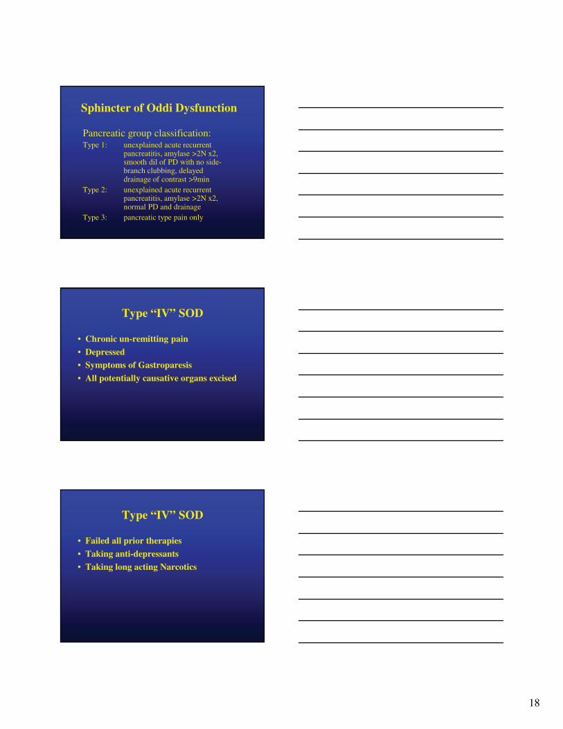
Sphincter of Oddi Dysfunction
Pancreatic group classification:Type 1: unexplained acute recurrent
pancreatitis, amylase >2N x2, smooth dil of PD with no side-branch clubbing, delayed drainage of contrast >9min
Type 2: unexplained acute recurrent pancreatitis, amylase >2N x2, normal PD and drainage
Type 3: pancreatic type pain only
Type “IV” SOD
• Chronic un-remitting pain
• Depressed• Symptoms of Gastroparesis
• All potentially causative organs excised
Type “IV” SOD
• Failed all prior therapies
• Taking anti-depressants• Taking long acting Narcotics

19
Type “IV” SOD
• Normal physical exam
• Normal lab values• Normal imaging studies
• Difficult to manage!!
Sphincter of Oddi Dysfunction(SOD)
The typical patient with SOD• female, 20-50 years old
• s/p cholecystectomy
• symptoms similar to pre-cholecystectomy
• episodic (or constant) pain
Sphincter of Oddi Dysfunction
Clinical evaluation:• History and Physical• CXR, EKG• Abdo U/S, LFTs, Amylase/Lipase• EGD• CAT scan• Secretin EUS or Secretin MRCP.• HIDA• ERCP with Manometry
20
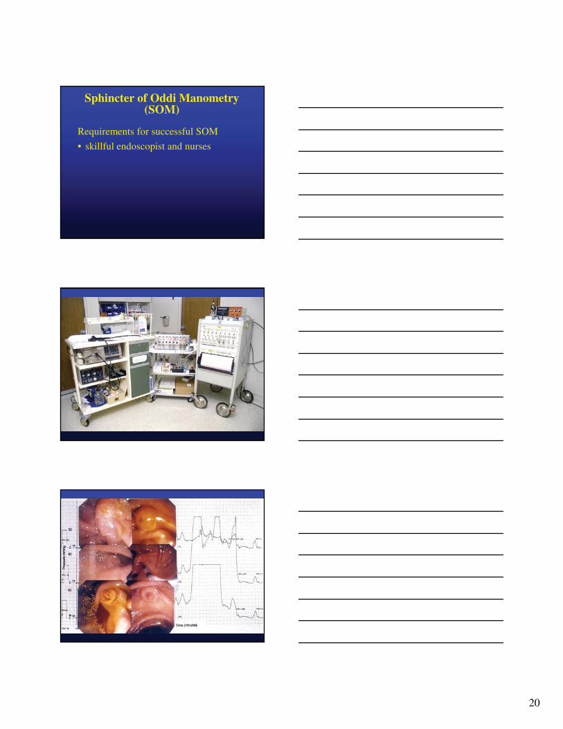
Sphincter of Oddi Manometry (SOM)
Requirements for successful SOM
• skillful endoscopist and nurses
21
Sphincter of Oddi Manometry
Abnormal valuesBasal sphincter pressure > 40mmHg
Phasic contractions
• amplitude >220mmHg
• duration > 8 sec
• frequency > 10/min
• retrograde > 50%
Sphincter of Oddi Manometry
Increased basal sphincter pressure is
the most reproducible and predictive
of positive therapeutic outcomes
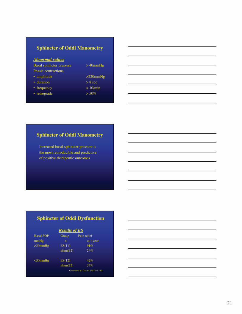
Sphincter of Oddi Dysfunction
Results of ESBasal SOP Group Pain relief
mmHg n at 1 year
>30mmHg ES(11) 91%
sham(12) 24%
<30mmHg ES(12) 42%
sham(12) 33%
Geenen et al. Gastro 1987;92:1401
22
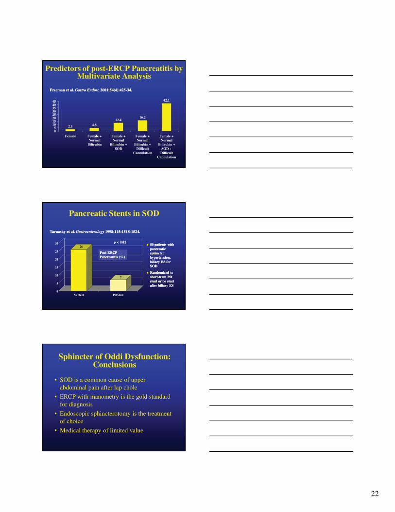
2.5 4.812.4
16.2
42.1
05
1015202530354045
Female Female +Normal
Bilirubin
Female +Normal
Bilirubin +SOD
Female +Normal
Bilirubin +Difficult
Cannulation
Female +Normal
Bilirubin +SOD +
DifficultCannulation
Freeman et al. Freeman et al. Gastro Gastro EndoscEndosc 2001;54(4):4252001;54(4):425--34.34.
Predictors of post-ERCP Pancreatitis by Multivariate Analysis
Pancreatic Stents in SOD
26
7
0
5
10
15
20
25
30
No Stent PD Stent
p p < 0.01< 0.01
PostPost--ERCP ERCP Pancreatitis (%)Pancreatitis (%)
80 patients with 80 patients with pancreatic pancreatic sphincter sphincter hypertension, hypertension, biliary ES for biliary ES for SODSOD
Randomized to Randomized to shortshort--term PD term PD stent or no stent stent or no stent after biliary ESafter biliary ES
Tarnasky et al. Tarnasky et al. GastroenterologyGastroenterology 1998;115:15181998;115:1518--1524. 1524.
Sphincter of Oddi Dysfunction:Conclusions
• SOD is a common cause of upper abdominal pain after lap chole
• ERCP with manometry is the gold standard for diagnosis
• Endoscopic sphincterotomy is the treatment of choice
• Medical therapy of limited value

23
Sphincter of Oddi Dysfunction:Conclusions
• SOD manometry is technically difficult and hazardous procedure
• Success rate still 50 to 85%• Properly trained and skilled endoscopist and
nurses required
ERCP: Complications
• Pancreatitis
• Bleeding
• Perforation
• Infection
Can ≠≠≠≠ Should
Richard Kozarek, M.D.Richard Kozarek, M.D.
24
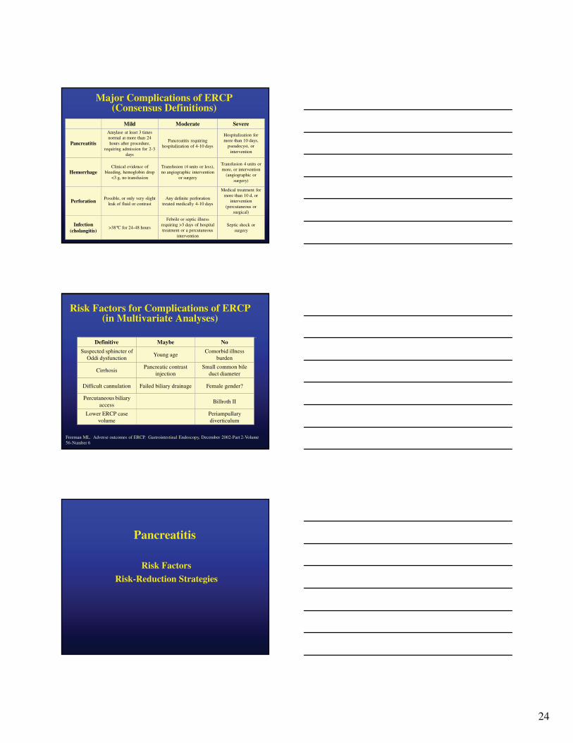
Major Complications of ERCP (Consensus Definitions)
Septic shock or surgery
Febrile or septic illness requiring >3 days of hospital treatment or a percutaneous
intervention
>38°C for 24-48 hoursInfection
(cholangitis)
Medical treatment for more than 10 d, or
intervention (percutaneous or
surgical)
Any definite perforation treated medically 4-10 days
Possible, or only very slight leak of fluid or contrastPerforation
Transfusion 4 units or more, or intervention
(angiographic or surgery)
Transfusion (4 units or less), no angiographic intervention
or surgery
Clinical evidence of bleeding, hemoglobin drop
<3 g, no transfusionHemorrhage
Hospitalization for more than 10 days,
pseudocyst, or intervention
Pancreatitis requiring hospitalization of 4-10 days
Amylase at least 3 times normal at more than 24 hours after procedure,
requiring admission for 2-3 days
Pancreatitis
SevereModerateMild
Risk Factors for Complications of ERCP (in Multivariate Analyses)
Periampullary diverticulum
Lower ERCP case volume
Billroth IIPercutaneous biliary
access
Female gender?Failed biliary drainageDifficult cannulation
Small common bile duct diameter
Pancreatic contrast injection
Cirrhosis
Comorbid illness burden
Young ageSuspected sphincter of
Oddi dysfunction
NoMaybeDefinitive
Freeman ML. Adverse outcomes of ERCP. Gastrointestinal Endoscopy, December 2002-Part 2-Volume 56-Number 6
Pancreatitis
Risk Factors
Risk-Reduction Strategies

25
Risk Factors for Post-ERCP Pancreatitis
Balloon dilation of biliary sphincter
Pre-cut sphincterotomy
Pancreatic sphincterotomy
Pancreatic duct injection
Lower ERCP case volumeHistory of post-ERCP
pancreatitis
Biliary SphincterotomyAbsence of common bile duct
stoneNormal bilirubin
Sphincter of Oddi manometryAcinarizationYoung Age
Small Common Bile Duct diameter
Female GenderSuspected sphincter of Oddi
dysfunction
NoMaybe Definite
Freeman ML. Adverse outcomes of ERCP. Gastrointestinal Endoscopy, December 2002-Part 2-Volume 56-Number 6
Risk Factors for Procedure-Induced Pancreatitis
Several factors may act independently or in combination to induce post-ERCP pancreatitis:
Mechanical Injury
Hydrostatic Injury
Enzymatic Injury
Infection
Pancreatic Ductal Edemaor Perforation
Chemical or Allergic Injury
Thermal Injury
Freeman ML, Guda NM. Prevention of post-ERCP pancreatitis: A comprehensive review. In: Gastrointestinal Endoscopy, June 2004 – Volume 59 – Number 7
Pancreatitis Risk-Reduction Strategies
Strategies are broken out into two groups*:
1. Patient Selection
2. Procedural Technique
*Listed In no particular order
26
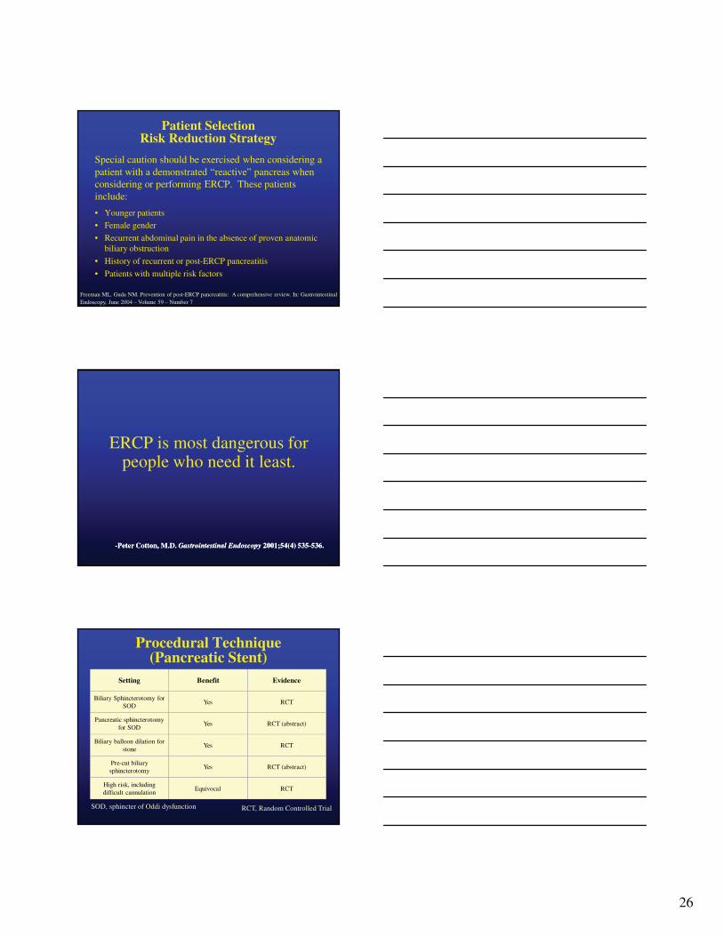
Patient SelectionRisk Reduction Strategy
Special caution should be exercised when considering a patient with a demonstrated “reactive” pancreas when considering or performing ERCP. These patients include:
• Younger patients
• Female gender
• Recurrent abdominal pain in the absence of proven anatomic biliary obstruction
• History of recurrent or post-ERCP pancreatitis
• Patients with multiple risk factors
Freeman ML, Guda NM. Prevention of post-ERCP pancreatitis: A comprehensive review. In: Gastrointestinal Endoscopy, June 2004 – Volume 59 – Number 7
ERCP is most dangerous for people who need it least.
--Peter Cotton, M.D. Peter Cotton, M.D. Gastrointestinal Endoscopy Gastrointestinal Endoscopy 2001;54(4) 5352001;54(4) 535--536.536.
Procedural Technique (Pancreatic Stent)
RCTEquivocalHigh risk, including difficult cannulation
RCT (abstract)YesPre-cut biliary sphincterotomy
RCTYesBiliary balloon dilation for
stone
RCT (abstract)YesPancreatic sphincterotomy
for SOD
RCTYesBiliary Sphincterotomy for
SOD
EvidenceBenefitSetting
RCT, Random Controlled TrialSOD, sphincter of Oddi dysfunction
27
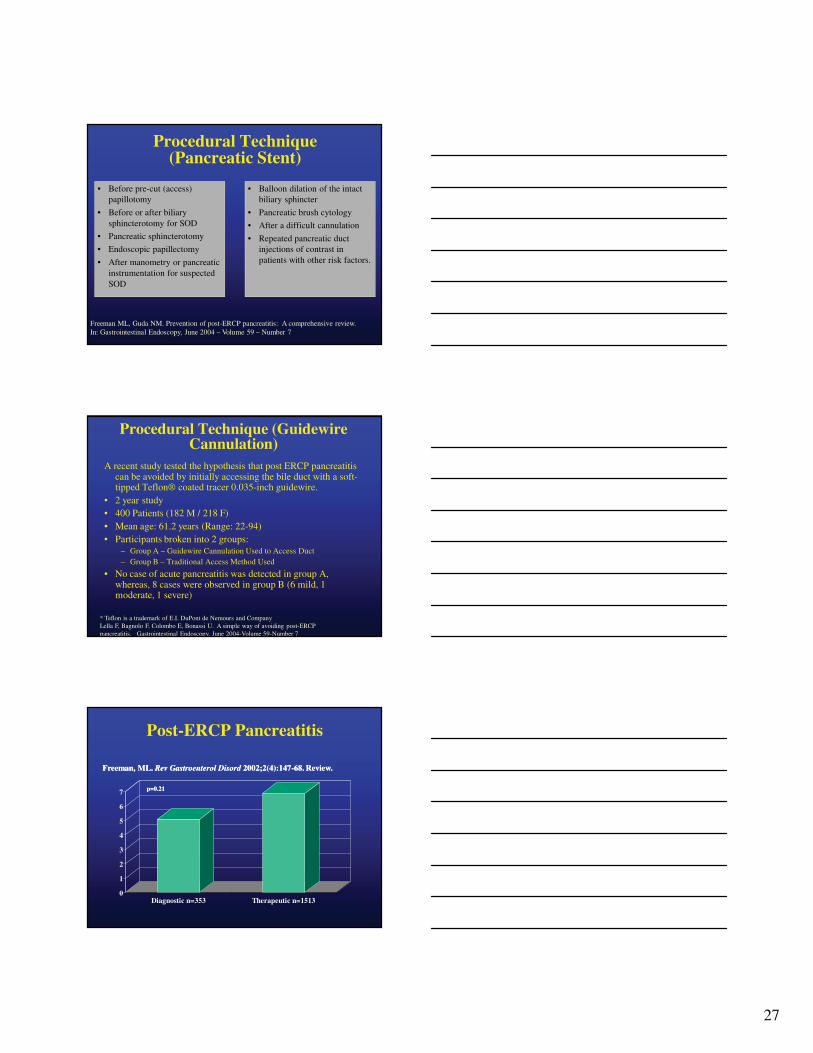
Procedural Technique (Pancreatic Stent)
• Before pre-cut (access) papillotomy
• Before or after biliary sphincterotomy for SOD
• Pancreatic sphincterotomy
• Endoscopic papillectomy
• After manometry or pancreatic instrumentation for suspected SOD
• Balloon dilation of the intact biliary sphincter
• Pancreatic brush cytology
• After a difficult cannulation
• Repeated pancreatic duct injections of contrast in patients with other risk factors.
Freeman ML, Guda NM. Prevention of post-ERCP pancreatitis: A comprehensive review.In: Gastrointestinal Endoscopy, June 2004 – Volume 59 – Number 7
Procedural Technique (GuidewireCannulation)
A recent study tested the hypothesis that post ERCP pancreatitis can be avoided by initially accessing the bile duct with a soft-tipped Teflon® coated tracer 0.035-inch guidewire.
• 2 year study• 400 Patients (182 M / 218 F)• Mean age: 61.2 years (Range: 22-94)• Participants broken into 2 groups:
– Group A – Guidewire Cannulation Used to Access Duct– Group B – Traditional Access Method Used
• No case of acute pancreatitis was detected in group A, whereas, 8 cases were observed in group B (6 mild, 1 moderate, 1 severe)
* Teflon is a trademark of E.I. DuPont de Nemours and CompanyLella F, Bagnolo F, Colombo E, Bonassi U. A simple way of avoiding post-ERCP pancreatitis. Gastrointestinal Endoscopy, June 2004-Volume 59-Number 7
Post-ERCP Pancreatitis
0
1
2
3
4
5
6
7
Diagnostic n=353 Therapeutic n=1513
p=0.21p=0.21
Freeman, ML. Freeman, ML. Rev Gastroenterol DisordRev Gastroenterol Disord 2002;2(4):1472002;2(4):147--68. Review.68. Review.
28
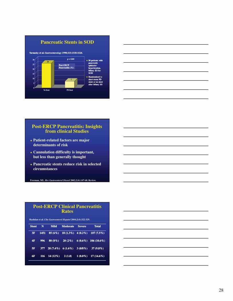
Pancreatic Stents in SOD
26
7
0
5
10
15
20
25
30
No Stent PD Stent
p p < 0.01< 0.01
PostPost--ERCP ERCP Pancreatitis (%)Pancreatitis (%)
80 patients with 80 patients with pancreatic pancreatic sphincter sphincter hypertension, hypertension, biliary ES for biliary ES for SODSOD
Randomized to Randomized to shortshort--term PD term PD stent or no stent stent or no stent after biliary ESafter biliary ES
Tarnasky et al. Tarnasky et al. GastroenterologyGastroenterology 1998;115:15181998;115:1518--1524. 1524.
Post-ERCP Pancreatitis: Insights from clinical Studies
♦ Patient-related factors are major determinants of risk
♦ Cannulation difficulty is important, but less than generally thought
♦ Pancreatic stents reduce risk in selected circumstances
Freeman, ML. Rev Gastroenterol Disord 2002;2(4):147-68. Review.
Post-ERCP Clinical Pancreatitis Rates
17 (14.6%)17 (14.6%)1 (0.0%)1 (0.0%)2 (1.8)2 (1.8)14 (12%)14 (12%)1161166F6F
37 (9.8%)37 (9.8%)3 (0/8%)3 (0/8%)6 (1.6%)6 (1.6%)28 (7.4%)28 (7.4%)3773775F5F
106 (10.6%)106 (10.6%)6 (0.6%)6 (0.6%)20 (2%)20 (2%)80 (8%)80 (8%)9969964F4F
107 (7.5%)107 (7.5%)4 (0.2%)4 (0.2%)18 (1.3%)18 (1.3%)85 (6%)85 (6%)145114513F3F
TotalTotalSevereSevereModerateModerateMildMildNNStentStent
Rashdan et al. Clin Gastroenterol Hepatol 2004;2(4):322-329.

29
Spontaneous Pancreatic Polyethylene Stents Dislodgment
86
7367 65
0
10
20
30
40
50
60
70
80
90
100
3F(a) 4F(b) 5F(c) 6F(d)
Stent diameterStent diameter
Spon
tane
ous
disl
odgm
ent
rate
Spon
tane
ous
disl
odgm
ent
rate
Rashdan et al. Clin Gastroenterol Hepatol 2004;2(4):322-329.
Polyethylene Stent-Induced Pancreatic Ductal Changes
13
36
8375
0
10
20
30
40
50
60
70
80
90
3F 4F 5F 6F
Stent diameterStent diameter
Fre
quen
cy o
f st
ent
Fre
quen
cy o
f st
ent--
indu
ced
duct
al c
hang
esin
duce
d du
ctal
cha
nges
Smith et al. Gastrointest Endosc 1996;44:268-275.
Post ERCP pancreatitis: Post ERCP pancreatitis: IndomethacinIndomethacin suppositorysuppository
30
Summary: Risk Reduction for Post-ERCP Pancreatitis
• Careful patient selections
• Meticulous endoscopic technique
• Insertion of a pancreatic stent in selected patients
• Endoscopist who maintains ERCP procedural volume
• Well trained (high volume) nurses/techs
Freeman ML, Guda NM. Prevention of post-ERCP pancreatitis: A comprehensive review. In:Gastrointestinal Endoscopy, June 2004 – Volume 59 – Number 7
Hemorrhage
Risk Factors
Risk Reduction Strategies
31
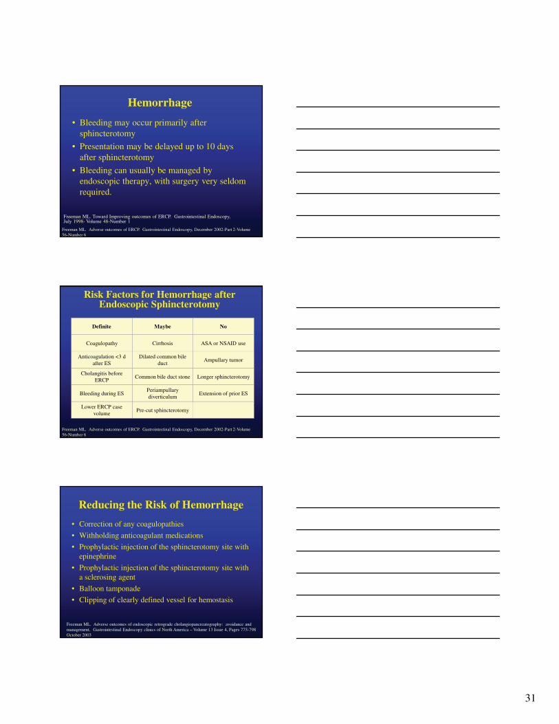
Hemorrhage
• Bleeding may occur primarily after sphincterotomy
• Presentation may be delayed up to 10 days after sphincterotomy
• Bleeding can usually be managed by endoscopic therapy, with surgery very seldom required.
Freeman ML. Toward Improving outcomes of ERCP. Gastrointestinal Endoscopy, July 1998- Volume 48-Number 1
Freeman ML. Adverse outcomes of ERCP. Gastrointestinal Endoscopy, December 2002-Part 2-Volume 56-Number 6
Risk Factors for Hemorrhage after Endoscopic Sphincterotomy
Pre-cut sphincterotomyLower ERCP case
volume
Extension of prior ESPeriampullary diverticulum
Bleeding during ES
Longer sphincterotomyCommon bile duct stoneCholangitis before
ERCP
Ampullary tumorDilated common bile
ductAnticoagulation <3 d
after ES
ASA or NSAID useCirrhosisCoagulopathy
NoMaybeDefinite
Freeman ML. Adverse outcomes of ERCP. Gastrointestinal Endoscopy, December 2002-Part 2-Volume 56-Number 6
Reducing the Risk of Hemorrhage
Freeman ML. Adverse outcomes of endoscopic retrograde cholangiopancreatography: avoidance and management. Gastrointestinal Endoscopy clinics of North America – Volume 13 Issue 4, Pages 775-798 October 2003
• Correction of any coagulopathies
• Withholding anticoagulant medications
• Prophylactic injection of the sphincterotomy site with epinephrine
• Prophylactic injection of the sphincterotomy site with a sclerosing agent
• Balloon tamponade
• Clipping of clearly defined vessel for hemostasis

32
Hemorrhage
Hemorrhage
Perforation
Risk Factors
Risk Reduction Strategies
33
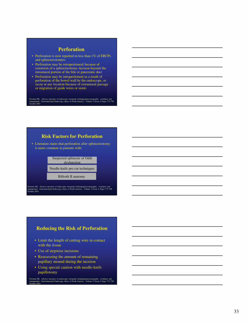
Perforation• Perforation is now reported in less than 1% of ERCPs
and sphincterotomies.• Perforation may be retroperitoneal because of
extension of a sphincterotomy incision beyond the intramural portion of the bile or pancreatic duct
• Perforation may be intraperitoneal as a result of perforation of the bowel wall by the endoscope, or occur at any location because of extramural passage or migration of guide wires or stents
Freeman ML. Adverse outcomes of endoscopic retrograde cholangiopancreatography: avoidance and management. Gastrointestinal Endoscopy clinics of North America – Volume 13 Issue 4, Pages 775-798 October 2003
Risk Factors for Perforation• Literature states that perforation after sphincterotomy
is more common in patients with:
Billroth II anatomy
Needle-knife pre-cut techniques
Suspected sphincter of Oddidysfunction
Freeman ML. Adverse outcomes of endoscopic retrograde cholangiopancreatography: avoidance and management. Gastrointestinal Endoscopy clinics of North America – Volume 13 Issue 4, Pages 775-798 October 2003
Reducing the Risk of Perforation
• Limit the length of cutting wire in contact with the tissue
• Use of stepwise incisions
• Reassessing the amount of remaining papillary mound during the incision
• Using special caution with needle-knife papillotomy
Freeman ML. Adverse outcomes of endoscopic retrograde cholangiopancreatography: avoidance and management. Gastrointestinal Endoscopy clinics of North America – Volume 13 Issue 4, Pages 775-798 October 2003
34
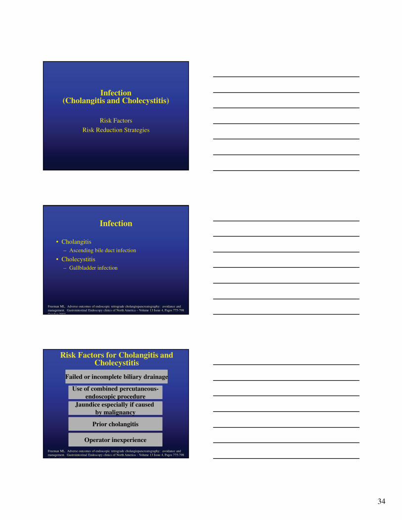
Infection(Cholangitis and Cholecystitis)
Risk Factors
Risk Reduction Strategies
Infection
• Cholangitis – Ascending bile duct infection
• Cholecystitis – Gallbladder infection
Freeman ML. Adverse outcomes of endoscopic retrograde cholangiopancreatography: avoidance and management. Gastrointestinal Endoscopy clinics of North America – Volume 13 Issue 4, Pages 775-798 October 2003
Risk Factors for Cholangitis and Cholecystitis
Failed or incomplete biliary drainage
Use of combined percutaneous-endoscopic procedure
Jaundice especially if caused by malignancy
Prior cholangitis
Operator inexperience
Freeman ML. Adverse outcomes of endoscopic retrograde cholangiopancreatography: avoidance and management. Gastrointestinal Endoscopy clinics of North America – Volume 13 Issue 4, Pages 775-798 October 2003

35
Reducing the Risk of Cholangitis and Cholecystitis
• The principle recommendation regarding prevention and treatment of cholangitis is obtaining successful and complete biliary drainage.
Freeman ML. Adverse outcomes of endoscopic retrograde cholangiopancreatography: avoidance and management. Gastrointestinal Endoscopy clinics of North America – Volume 13 Issue 4, Pages 775-798 October 2003
SummaryComplications of ERCP
• Major complications of ERCP are:– Pancreatitis, Hemorrhage, Perforation and
Infection
• Risk factors related to ERCP are:– Pre-procedural (related to patient selection)– Technique-related; operator inexperience
• Complications may be reduced if risk-reduction strategies are understood and employed
Case
• A 46 y/o Asian lady from Juneau, with 5 week h/o severe attacks of abdominal pain and vomiting
• Pain severe, 10/10 during episode, vague constant discomfort in between episodes
• Examination reveal mild abdominal tenderness
• CT/USS scan unremarkable
• Treated by GI as SOD, with analgesics, nifedipine and reassurance
36
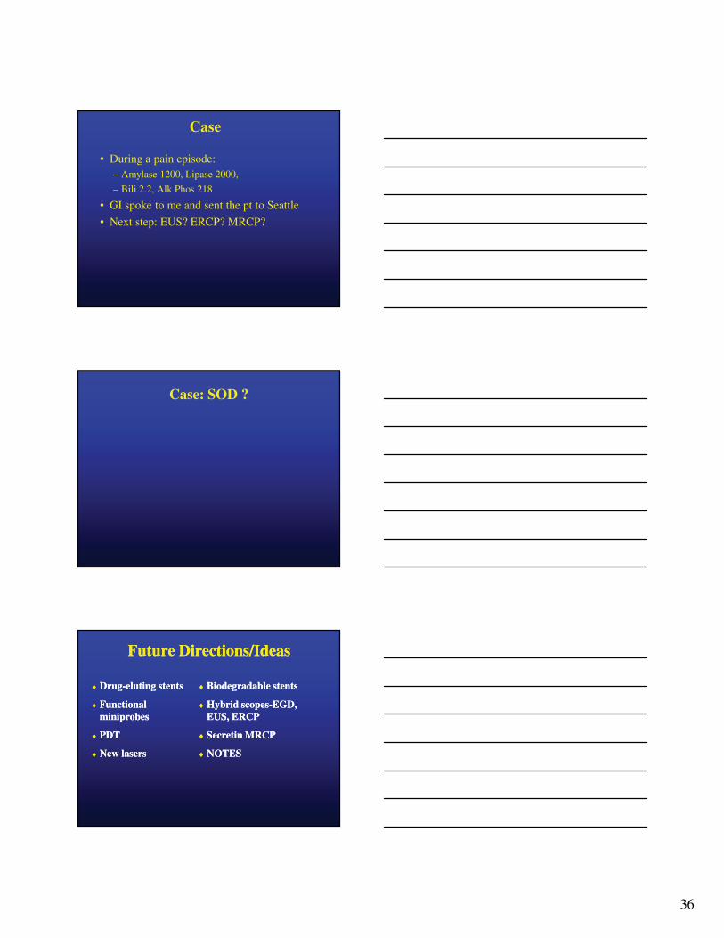
Case
• During a pain episode:– Amylase 1200, Lipase 2000,
– Bili 2.2, Alk Phos 218
• GI spoke to me and sent the pt to Seattle
• Next step: EUS? ERCP? MRCP?
Case: SOD ?
Future Directions/Ideas Future Directions/Ideas
♦♦ DrugDrug--eluting stentseluting stents
♦♦ Functional Functional miniprobesminiprobes
♦♦ PDTPDT
♦♦ New lasersNew lasers
♦♦ Biodegradable stentsBiodegradable stents
♦♦ Hybrid scopesHybrid scopes--EGD, EGD, EUS, ERCPEUS, ERCP
♦♦ SecretinSecretin MRCPMRCP
♦♦ NOTESNOTES

37
The sole purpose of human existence is to kindle a light in the darkness of mere being
C G Jung