escholarship uc item 3476s5cs
TRANSCRIPT

eScholarship provides open access, scholarly publishingservices to the University of California and delivers a dynamicresearch platform to scholars worldwide.
Electronic Thesis and DissertationsUCLA
Peer Reviewed
Title:Novel regulators of social motility in the African trypanosome, Trypanosoma brucei
Author:Jahanbakhsh, Shahriyar
Acceptance Date:2016
Series:UCLA Electronic Theses and Dissertations
Degree:M.S., Biochemistry, Molecular and Structural Biology 0090UCLA
Advisor(s):Hill, Kent L, Bouchard, Louis-Serge
Committee:Hill, Kent L, Bouchard, Louis-Serge, Gober, James
Permalink:http://escholarship.org/uc/item/3476s5cs
Abstract:
Copyright Information:All rights reserved unless otherwise indicated. Contact the author or original publisher for anynecessary permissions. eScholarship is not the copyright owner for deposited works. Learn moreat http://www.escholarship.org/help_copyright.html#reuse
UNIVERSITY OF CALIFORNIA
Los Angeles
Novel regulators of social motility in the
African trypanosome, Trypanosoma brucei
A thesis submitted in partial satisfaction
of the requirements for the degree Master of Science in
Biochemistry, Molecular and Structural Biology
by
Shahriyar Jahanbakhsh
2016

© Copyright by
Shahriyar Jahanbakhsh
2016

ii
ABSTRACT OF THE THESIS
Novel regulators of social motility in the
African trypanosome, Trypanosoma brucei
by
Shahriyar Jahanbakhsh
Master of Science in Biochemistry, Molecular and Structural Biology
University of California, Los Angeles, 2016
Professor Kent L. Hill, Co-Chair
Professor Louis-Serge Bouchard, Co-Chair
Trypanosoma brucei is the causative agent of sleeping sickness, a major threat
to economic stability and public health across sub-Saharan Africa. This protozoan
parasite features a complex digenetic life cycle that alternates between the tsetse fly
(Glossina spp.) vector and the mammalian host. Infections in both the fly and vector are
characterized by intimate contact with tissue surfaces. Recent studies have shown that
when cultivated on semisolid agarose surfaces, T. brucei engages in social behavior,
termed social motility, which manifests as parasites assembling into groups that move
collectively to form symmetrical arrays of radial projections. Herein we describe two

iii
novel modulators of social motility in T. brucei, the zinc finger protein ZC3H34 and the
prolyl isomerase cyclophilin A (CyPA). Using RNA interference, we show that
knockdown of either ZC3H34 or CyPA delays social motility. The delay in social motility
observed with CyPA knockdown is not due to decreased propulsive motility, although
ZC3H34-depleted parasites do show reduced propulsive motility. Our study provides
new insights into the biochemistry of trypanosome social behavior and identifies
potential targets for chemical inhibition of vector-to-host parasite transmission.

iv
The thesis of Shahriyar Jahanbakhsh is approved.
James Gober
Louis-Serge Bouchard, Committee Co-Chair
Kent L. Hill, Committee Co-Chair
University of California, Los Angeles
2016

v
DEDICATION
I dedicate this work to Justin Thayer,
the brilliant educator and scientist who profoundly
shaped my course in science.

vi
TABLE OF CONTENTS
Abstract.........................................................................................................................ii–iii
Committee Page..............................................................................................................iv
Dedication.........................................................................................................................v
Table of Contents.............................................................................................................vi
Acknowledgments.....................................................................................................vii–viii
CHAPTER 1: Background and Introduction.......................................................1–5
References...................................................................................6–8
CHAPTER 2: Materials and Methods...............................................................9–11
References.....................................................................................12
CHAPTER 3: Results.....................................................................................13–31
References...............................................................................32–37
CHAPTER 4: Discussion................................................................................38–42
References...............................................................................43–44
SUPPLEMENTAL INFORMATION...........................................................................45–46

vii
ACKNOWLEDGEMENTS
The scientific growth I have enjoyed over these last four years at UCLA has been
catalyzed by excellent mentorship from a number of individuals.
Foremost, I am grateful to Kent Hill and the members of the Hill laboratory for
providing me with this exciting research opportunity in the biosciences. I especially
thank Stephanie DeMarco, whose comprehensive and patient mentorship has greatly
increased my proficiency with the process and techniques of modern biochemical and
cell biological research. Stephanie challenges students to think critically and truly
develop as scientists, and the extent of her commitment to her mentees is remarkable. I
acknowledge the superb mentorship and insight of Sunayan Ray. Finally, I thank Edwin
Saada and Michelle Shimogawa for many exciting and often humorous discussions on
matters of science and non-science alike. I benefited greatly from Ed’s firm support of
my commitment to advancing the mission of the laboratory of Dr. Kent Hill, and I extend
much thanks to Ed for his critical review of this work.
I am further indebted to Louis Bouchard and the members of the Bouchard
laboratory for the opportunity to perform research at the interface between biology and
physical chemistry. My standing as a freshman with no prior experience did not deter
Louis from graciously inviting me into his laboratory in 2013. I am especially grateful for
the extensive mentorship I received from Jeff McCormick and Michael Lake. Jeff and
Michael vastly improved my proficiency with quantitative analysis and modeling and my
ability to work and troubleshoot independently, in situ sensu stricto. I also thank Brian
Archer for many memorable discussions.
viii
I thank James Gober for many quarters of excellent and entertaining instruction
in biochemistry and microbiology, as well as for his service on my thesis committee. Jim
has the distinction of having taught me more times than any other Professor at UCLA.
Jim also deserves credit for correctly predicting that “if something goes wrong [in
Biochemistry lab], you ain’t goin’ to medical school!”
Finally, I gratefully acknowledge the funding sources for my research activities at
UCLA: The Kivelson Endowment, the Naumburg Endowment, the Irving and Jean Stone
Endowment, the Oppenheimer Endowment, the Aerospace Corporation, and the
American Vacuum Society.
The RNAi cell lines described in sections (I)(A)(i)-(iii) and (II)(A)(i) of Chapter 3
were generated and characterized in collaboration with Alexandra Stream.

1
CHAPTER 1
Background and Introduction
Social behavior is ubiquitous among microbes1. Multicellular groups of interacting
microbes possess emergent properties not possible in isolated contexts. The
biochemical mechanisms underlying bacterial social behavior and the consequences of
this behavior have been extensively studied. Among the best characterized social
phenomena are quorum sensing2-5, biofilm formation2-4, and social motility2-4,6. These
cooperative behaviors provide important advantages to participants. Quorum sensing
enables populations of bacteria to coordinately regulate gene expression and efficiently
utilize exoenzymes and other extracellular “public goods”2-5. Biofilms confer protection
against predation, phagocytosis, and antimicrobial compounds and facilitate nutrient
acquisition and genetic exchange2-4. In addition, the coordinated movement that occurs
during social motility has been shown to enhance nutrient uptake, facilitate colonization
of and adaptation to differing physiological and ecological environments, protect against
macrophages and antibiotics, and promote toxin secretion2-4,6,7. The secondary
messenger cyclic dimeric GMP (c-di-GMP) has been shown to have important roles in
many of these social behaviors and is a key regulator of biofilm formation, swarming
behavior, virulence, and cell cycle progression in bacteria8.
Social behaviors have also been described for eukaryotic microorganisms.
Perhaps the best recognized is the starvation response in the slime mold Dictyostelium
discoideum, which initiates an oscillatory cyclic AMP (cAMP) relay signal that results in

2
the aggregation of individual cells into an organized, multicellular fruiting body9,10.
Additional examples include flocculin-mediated self-recognition and biofilm-like
cooperation in Saccharomyces cerevisiae11 and biofilm formation in the fungus Candida
albicans12. Despite well-documented instances of social behaviors among bacteria and
many eukarya, however, the existence of, and mechanisms underlying such behaviors
among protozoan parasites remain largely unexplored13,14.
The protozoan parasite Trypanosoma brucei is the causative agent of sleeping
sickness in humans and Nagana in cattle and other mammals. T. brucei is endemic to
sub-Saharan Africa, where it poses a major threat to public health and agriculture in 36
countries15. All three subspecies of T. brucei, Trypanosoma brucei (T. b.) gambiense, T.
b. rhodesiense, and T. b. brucei, are able to infect non-primate mammals, whereas only
T. b. gambiense and T. b. rhodesiense are human-infective. T. brucei is transmitted to
mammals through the bite of an infected tsetse fly (Glossina spp.) vector. Thus, the
lifecycle of T. brucei is digenetic, alternating between infection of the mammalian host
and transmission through an insect vector15,16.
T. brucei transitions through many distinct morphological states during the course
of its lifecycle. After ingestion, short-stumpy bloodstream trypomastigotes differentiate
into procyclic form trypomastigotes in the tsetse fly midgut17. To be capable of re-
infecting the mammalian host, these cells must then undergo a series of ordered
developmental changes and directional migrations from the midgut through the
proventriculus, foregut, proboscis, and into the salivary gland ducts17. Throughout this
process, the parasites are in intimate contact with fly tissue epithelia, colonizing and
migrating along these epithelial surfaces. Surface-induced changes in organism

3
behavior and motility are known to occur among diverse groups of bacteria and
protists10-12,18-23, raising the intriguing possibility that surface contact may also induce
and modulate parasite behavior in T. brucei.
In support of this hypothesis, recent work discovered that when cultivated on
semisolid agarose surfaces, procyclic form (insect midgut stage) trypanosomes form
communities that undergo polarized coordinated movements outward from the site of
inoculation.24 These coordinated movements yield symmetrical arrays of radial
projections reminiscent of those formed by Pseudomonas aeruginosa18,19, Myxococcus
xanthus22, and Paenibacillus dendritiformis23 during swarming. This form of coordinated
group movement was termed social motility, by analogy to social motility in bacteria.
Coordinated movement of groups of cells requires signaling, as individuals cells in the
group must respond to external cues and to each other. Notably, knockdown of several
T. brucei receptor-type adenylate cyclases (AC1, combined AC1/AC2, and AC6) was
found to increase the number and density of radial projections advancing from the site
of inoculation, whereas knockdown or chemical inhibition of the cAMP-specific
phosphodiesterase PDEB1 abolished social motility25,26. These results indicate that
social motility is controlled by a signaling pathway that modulates cAMP levels within
the trypanosome flagellum25,26. The architecture of this signaling pathway shows
parallels to that of the c-di-GMP signaling pathway that modulates swarming motility
and biofilm formation in bacteria26.
A growing body of evidence indicates that social motility is physiologically
relevant in the Glossina vector. Foremost, social motility is a feature of “early” procyclic
form cells, a specific T. brucei developmental stage found in the fly midgut during the

4
first week of infection; only cells staining positive for GPEET, a glycophosphatidylinositol
(GPI)-anchored surface procyclin and a marker for early procyclic forms, exhibit social
motility27. Proteomic analyses have identified a number of other proteins differentially
expressed between early and late procyclic forms, although whether or not these
proteins function as regulators of social motility in T. brucei has not been established27.
As further evidence of a physiological role for social motility, null mutants lacking Rft1, a
protein required for translocation of lipid-linked oligosaccharides across the ER
membrane28, display attenuated social motility and a four-fold decrease in efficiency in
establishing fly midgut infections29. Despite this recent progress, however, additional
genes involved in the control of social motility in T. brucei have yet to be identified.
Given that social behavior and cell-cell communication depend on signaling systems,
the relevance of social motility in T. brucei extends beyond parasite transmission
through the tsetse fly to include opportunities for studying signal transduction, which
represents a critical yet poorly understood aspect of parasite biology14.
Herein we describe two novel genes involved in regulation of social motility in T.
brucei. A prior transcriptome-wide RNA-seq screen revealed that certain genes are
differentially upregulated in trypanosomes cultivated on semisolid agarose compared to
parasites cultured in suspension. We use RNA interference (RNAi) to probe these
candidate genes for a potential functional role in social motility. We demonstrate that
stable knockdown of a zinc finger protein, ZC3H34, or of cyclophilin A delays the onset
of social motility. We further show that the social motility phenotype of CyPA-depleted
cells is not the result of defective propulsive motility. The phenotype of ZC3H34-
depleted cells is correlated with decreased propulsive motility in suspension culture,

5
though a causal connection between these phenotypes has not been established. Our
work elucidates new regulators of social motility in T. brucei and identifies potential
targets for chemical inhibition of parasite transmission.

6
References
1. West, S.A.; Diggle, S.P.; Buckling, A.; Gardner, A.; Griffin, A.S. “The social lives of microbes.” Annu. Rev. Ecol. Evol. Syst. 2007, 38, 53–77.
2. Shapiro, J.A. “Thinking about bacterial populations as multicellular organisms.”
Annu. Rev. Microbiol. 1998, 52, 81–104.
3. Bassler, B.L.; Losick, R. “Bacterially speaking.” Cell 2006, 125, 237–246.
4. Xavier, J.B. “Social interaction in synthetic and natural microbial communities.” Mol. Syst. Biol. 2011, 7, 483.
5. Shi, W.; Zusman, D.R. “The two motility systems of Myxococcus xanthus show
different selective advantages on various surfaces.” Proc. Natl. Acad. Sci. U.S.A. 1993, 90, 3378–3382.
6. Kearns, D.B. “A field guide to bacterial swarming motility.” Nat. Rev. Microbiol.
2010, 8, 634–644.
7. Josenhans, C.; Suerbaum, S. “The role of motility as a virulence factor in bacteria.” Int. J. Med. Microbiol. 2002, 291, 605–614.
8. Hengge, R. “Principles of c-di-GMP signalling in bacteria.” Nat. Rev. Microbiol.
2009, 7, 263–273.
9. Kessin, R.H. Dictyostelium, Evolution, Cell Biology, and the Development of Multicellularity. Cambridge University Press, 2010.
10. Queller, D.C.; Ponte, E.; Bozzaro, S.; Strassmann, J.E. “Single-gene greenbeard
effects in the social amoeba Dictyostelium discoideum.” Science 2003, 299, 105–106.
11. Veelders, M.; Brückner, S.; Ott, D.; Unverzagt, C.; Mösch, H.U., Essen, L.O.
“Structural basis of flocculin-mediated social behavior in yeast.” Proc. Natl. Acad. Sci. U.S.A. 2010, 107, 22511–22516.
12. Murillo, L.A.; Newport, G.; Lan, C.Y.; Habelitz, S.; Dungan, J.; Agabian, N.M.
“Genome-wide transcription profiling of the early phase of biofilm formation by Candida albicans.” Eukaryotic Cell 2005, 4, 1562–1573.
13. Lopez, M.A.; Nguyen, H.T.; Oberholzer, M.; Hill, K.L. “Social parasites.” Curr.
Opin. Microbiol. 2011, 14, 642–648.

7
14. Saada, E.A.; DeMarco, S.F.; Shimogawa, M.M.; Hill, K.L. “‘With a little help from my friends’—social motility in Trypanosoma brucei.” PLoS Pathog. 2015, 11, e1005272.
15. Kennedy, P.G. “The continuing problem of human African trypanosomiasis
(sleeping sickness).” Ann. Neurol. 2008, 64, 116–126.
16. Langousis, G.; Hill, K.L. “Motility and more: the flagellum of Trypanosoma brucei.” Nat. Rev. Microbiol. 2014, 12, 505–518.
17. Van Den Abbeele, J.; Claes, Y.; van Bockstaele, D.; Le Ray, D.; Coosemans, M. “Trypanosoma brucei spp. development in the tsetse fly: characterization of the post-mesocyclic stages in the foregut and proboscis.” Parasitology 1999, 118, 469–478.
18. Rashid, M.H.; Kornberg, A. “Inorganic polyphosphate is needed for swimming,
swarming, and twitching motilities of Pseudomonas aeruginosa.” Proc. Natl. Acad. Sci. U.S.A. 2000, 97, 4885–4890.
19. Kohler, T.; Curty, L.K.; Barja, F.; van Delden, C.; Pechere, J.C. “Swarming of
Pseudomonas aeruginosa is dependent on cell-to-cell signaling and requires flagella and pili.” J. Bacteriol. 2000, 182, 5990–5996.
20. Rauprich, O.; Matsushita, M.; Weijer, C.J.; Siegert, F.; Esipov, S.E.; Shapiro, J.A.
“Periodic phenomena in Proteus mirabilis swarm colony development.” J. Bacteriol. 1996, 178, 6525–6538.
21. Alberti, L.; Harshey, R.M. “Differentiation of Serratia marcescens 274 into
swimmer and swarmer Cells.” J. Bacteriol. 1990, 172, 4322–4328.
22. Velicer, G.J.; Yu, Y.T. “Evolution of novel cooperative swarming in the bacterium Myxococcus xanthus.” Nature 2003, 425, 75–78.
23. Ingham, C.J.; Jacob, E.B. “Swarming and complex pattern formation in
Paenibacillus vortex studied by imaging and tracking cells.” BMC Microbiol. 2008, 8, 36.
24. Oberholzer, M.; Lopez, M.A.; McLelland, B.T.; Hill, K.L. “Social motility in African
trypanosomes.” PLoS Pathog. 2010, 6, e1000739.
25. Lopez, M.A.; Saada, E.A.; Hill, K.L. “Insect stage-specific adenylate cyclases regulate social motility in African trypanosomes”. Eukaryotic Cell 2015, 14, 104–112.
26. Oberholzer, M.; Saada, E.A.; Hill, K.L. “Cyclic AMP regulates social behavior in
African trypanosomes.” MBio 2015, 6, e01954-14.

8
27. Imhof, S.; Knüsel, S.; Gunasekera, K.; Vu, X.L.; Roditi, I. “Social motility of
African trypanosomes is a property of a distinct life-cycle stage that occurs early in tsetse fly transmission.” PLoS Pathog. 2014, 10, e1004493.
28. Helenius, J.; Ng, D.T.; Marolda, C.L.; Walter, P.; Valvano, M.A.; Aebi, M.
“Translocation of lipid-linked oligosaccharides across the ER membrane requires Rft1 protein.” Nature 2002, 415, 447–450.
29. Imhof, S.; Vu, X.L.; Bütikofer, P.; Roditi, I. “A glycosylation mutant of
Trypanosoma brucei links social motility defects in vitro to impaired colonization of tsetse flies in vivo.” Eukaryotic Cell 2015, 14, 588–592.

9
CHAPTER 2
Materials and Methods
Cell culture. Procyclic form 29–13 cells1, which stably express T7 RNA
polymerase and the tetracycline repressor, were cultured in Cunningham’s semi-defined
medium (SM) supplemented with 10% heat-inactivated fetal calf serum (Gibco), 15
µg/mL G418 (Gibco), and 50 µg/mL hygromycin B (Gibco), as described previously2.
RNAi lines were cultured in media also containing 10 µg/mL blasticidin (Gibco). Cells
were passaged to maintain early- to mid-logarithmic growth. Cultures were maintained
at 28°C and 5% CO2.
Generation of RNAi lines. All gene sequences were obtained from the TriTrypDB
kinetoplastid parasite genome database3. RNAi target regions were selected using the
TrypanoFAN RNAit algorithm4. These regions were PCR-amplified and cloned into the
p2T7-177 RNAi vector5. The primers used to amplify and clone the genes of interest are
listed in Supplemental Table 1. The resulting constructs were linearized with NotI and
stably transfected into 29–13 cells using electroporation, as described previously6,7.
Transfectants were selected by addition of 10 µg/mL blasticidin to the culture medium.
Clonal lines were obtained via limiting dilution.
Growth assays. RNAi was induced by addition of 1 µg/mL tetracycline. Cells
were passaged to an initial concentration of 5×10⁵/mL (early logarithmic phase). Cells
density was monitored daily or twice daily using a Z1 Coulter Particle Counter
(Beckman Coulter). Cells were passaged back to a density of 5×10⁵/mL every ~24 h.

10
Cumulative cell densities at time points beyond 24 h after the assay startpoint were
estimated as: (⍴t*×⍴t-1)/(⍴t-2), where ⍴t* is the measured density, ⍴t-1 is the cumulative cell
density at the most recent prior time point, and ⍴t-2 is the cumulative cell density at the
second most recent prior time point (or post-dilution density [i.e. ⍴t-2 = 5×10⁵/mL], if the
cells were passaged).
RT-qPCR. RNAi lines were incubated in suspension culture for 72 h in presence
and absence of 1 µg/mL tetracycline. Following this period, total RNA was isolated from
mid-logarithmic cells using an RNeasy Mini Kit (Qiagen) and treated with amplification
grade DNase (Thermo Fisher). cDNA was then synthesized from 2 µg total RNA using
SuperScript II reverse transcriptase (Thermo Fisher) and oligo (dT) primers to select for
the poly(A)+ RNA fraction. qPCR primers were designed using NCBI Primer-BLAST8
and the sequences are listed in Supplemental Table 2. qPCR was performed in
duplicate for both induced (with tetracycline) and uninduced (without tetracycline)
conditions, and on 1 to 3 independent RNA preparations. Samples were analyzed using
a CFX Connect Real-Time PCR Detection System (Bio-Rad). Fluorescence values were
normalized against values from two genes (PFR2 and TERT) abundantly expressed in
both procyclic and bloodstream form T. brucei. Relative gene expression between the
induced and uninduced conditions was quantified using the 2−ΔΔCT method9.
Social motility assays. Cultivation on semisolid agarose plates was performed
essentially as described in Oberholzer et al.2 A sterile solution of 4% (w/v) agarose
(SeaPlaque GTG Agarose, Lonza) was diluted into prewarmed (42°C for 20 min) SM
supplemented with 15 µg/mL G418 and 50 µg/mL hygromycin B to a final concentration
of 0.4%. Ethanol and methanol were added to a final concentration of 1%, and

11
tetracycline to a final concentration of 1 µg/mL (addition of tetracycline was omitted for
control plates). The resulting solution was poured in aliquots of 11.5 mL into Petri dishes
(85 mm × 15 mm) and allowed to cool, with the Petri dishes uncovered, for 1 h in a
laminar flow hood at room temperature. RNAi lines were cultured in suspension at mid-
logarithmic phase (with and without 1 µg/mL tetracycline) for 72 h prior to plating on
agarose. For inoculation onto the plates, 5 µL of cells from a suspension culture at a
density of 2×10⁷/mL were added to the center of the agarose surface. The plates were
incubated at 28°C and 3.5% CO2 and imaged at 24 h intervals post-inoculation.
Radial projection formation was quantified using ImageJ10. Given the
considerable heterogeneity in projection length, only those projections at or extending
past a defined radial distance from the site of inoculation were included in the count.
These threshold radii were set as 7.5 mm, 10.8 mm, 19.9 mm, and 24.9 mm for time
points 48 h, 72 h, 96 h, and 120 h post-inoculation, respectively, for both ZC3H34 and
CyPA.
Motility traces. RNAi lines were cultured in suspension (with and without 1 µg/mL
tetracycline) for 72 h prior to analysis. Logarithmic-phase cells were diluted to 2×10⁶/mL
and observed in polyglutamate-coated slide chambers11. Cells were viewed under dark-
field illumination on a Zeiss Axioskop II compound microscope using a 10× objective.
Approximately 30 s of video from separate regions on each slide was captured. Motility
traces were generated using MetaMorph software (Molecular Devices). Cells that could
not be tracked for the full 30 s, as a result of leaving the focal plane or field of view,
were excluded from the analysis.
12
References
1. Wirtz, E.; Leal, S.; Ochatt, C.; Cross, G.A. “A tightly regulated inducible expression system for conditional gene knock-outs and dominant-negative genetics in Trypanosoma brucei.” Mol. Biochem. Parasitol. 1999, 99, 89–101.
2. Oberholzer, M.; Lopez, M.A.; McLelland, B.T.; Hill, K.L. “Social motility in African
trypanosomes.” PLoS Pathog. 2010, 6, e1000739.
3. Aslett, M.; Aurrecoechea, C.; Berriman, M.; Brestelli, J.; Brunk, B.P.; Carrington, M.; Depledge, D.P.; Fischer, S.; Gajria, B.; Gao, X. et al. “TriTrypDB: a functional genomic resource for the Trypanosomatidae.” Nucleic Acids Res. 2010, 38, 457–462.
4. Redmond, S.; Vadivelu, J.; Field, M.C. “RNAit: an automated web-based tool for
the selection of RNAi targets in Trypanosoma brucei.” Mol. Biochem. Parasitol. 2003, 128, 115–118.
5. LaCount, D.J.; Barrett, B.; Donelson, J.E. “Trypanosoma brucei FLA1 is required
for flagellum attachment and cytokinesis.” J. Biol. Chem. 2002, 277, 17580–17588.
6. Vaidya, T.; Bakhiet, M.; Hill, K.L.; Olsson, T.; Kristensson, K.; Donelson, J.E.
“The gene for a T lymphocyte triggering factor from African trypanosomes.” J. Exp. Med. 1997, 186, 433–438.
7. Hill, K.L.; Hutchings, N.R.; Russell, D.G.; Donelson, J.E. “A novel protein
targeting domain directs proteins to the anterior cytoplasmic face of the flagellar pocket in African trypanosomes.” J. Cell Sci. 1999, 112, 3091–3101.
8. Ye, J.; Coulouris, G.; Zaretskaya, I.; Cutcutache, I.; Rozen, S.; Madden, T.L.
“Primer-BLAST: a tool to design target-specific primers for polymerase chain reaction.” BMC Bioinformatics 2012, 13, 134.
9. Livak, K.J.; Schmittgen, T.D. “Analysis of relative gene expression data using
real-time quantitative PCR and the 2−ΔΔCT Method.” Methods 2001, 25, 402–408.
10. Schneider, C.A.; Rasband, W.S.; Eliceiri, K.W. "NIH Image to ImageJ: 25 years of image analysis." Nat. Methods. 2012, 9, 671–675.
11. Gadelha, C.; Wickstead, B.; McKean, P. G.; Gull, K. “Basal body and flagellum
mutants reveal a rotational constraint of the central pair microtubules in the axonemes of trypanosomes.” J. Cell Sci. 2005, 119, 2405–2413.

13
CHAPTER 3
Results
We sought to identify and characterize additional regulators of social motility in T.
brucei. To this end, we assayed social motility and propulsive motility characteristics in
cell lines stably expressing dsRNA against the transcripts of selected candidate genes.
A previous RNA-seq screen (Figure 1) had identified 2,141 genes differentially
regulated (p < 0.01) in trypanosomes cultured on semisolid agarose versus in
suspension (21.8 percent of total hits). Of these, 1,240 genes were transcribed above
50 RPKM (reads per kilobase per million reads mapped1) in both suspension- and
agarose-cultured trypanosomes. Our candidate genes were among a 22-member
subset of these 1,240 genes for which RPKMplate/RPKMsuspension ≥ 2.

14
Figure 1: Summary of RNA-seq screen of suspension- and semisolid agarose-
cultivated T. brucei and experimental workflow. The subscripts P and S denote plate
and suspension culture RPKM values, respectively.
I. Genes essential for viability in vitro.
A. Translation elongation factor-1β, histone H3, universal minicircle sequence-
binding protein 2, S-adenosylhomocysteine hydrolase, and RNA-binding protein 11 are
essential for viability.
Of the 22 genes found to be upregulated 2-fold or more in trypanosomes
cultivated on semisolid agarose, seven genes with physiologically distinct functions
were selected for analysis for a possible role in social motility (Table 1). We began by
generating transfected T. b. brucei cell lines harboring tetracycline-inducible RNAi
constructs against these seven candidate genes: translation elongation factor-1β (EF-
1β, GeneDB accession Tb927.10.5840), histone H3 (H3, Tb927.1.2510), universal
minicircle sequence-binding protein 2 (UMSBP2, Tb927.10.6060), S-
adenosylhomocysteine hydrolase (SAHH, Tb927.11.9590), RNA-binding protein 11
(RBP11, Tb927.8.4450), zinc finger protein ZC3H34 (ZC3H34, Tb927.10.12330), and
cyclophilin A (CyPA, Tb927.11.880). In all cases, knockdown of the target gene was
verified using reverse transcription–quantitative PCR (Figure 2).

15
Table 1: List of candidate genes investigated in this study and their known major
functions.
Figure 2: Knockdown of candidate social motility genes. RT-qPCR analysis of
transcript levels in cell lines induced for RNAi normalized to levels in uninduced
controls. For ZC3H34, (S) denotes expression levels in cells in suspension and (P)
denotes expression levels in cells obtained from radial projections on semisolid

16
agarose. Expression levels for all other genes are for cells grown in suspension.
Normalized transcript expression levels, expressed as mean ± standard deviation, for
RNAi-induced cells are: 24.53% ± 22.45% (EF-1β), 29.14% ± 12.18% (Histone H3),
24.14% ± 3.64% (UMSBP2), 18.02% ± 14.03% (SAHH), 19.13% ± 12.52% [ZC3H34
(S)], 14.86% ± 1.13% [ZC3H34 (P)], and 6.92% ± 5.12% (CyPA). n=3 biological
replicates for CyPA, 2 for ZC3H34 (S) and ZC3H34 (P), and 1 for the other genes. Two
technical replicates were performed for all genes. For genes for which only a single
biological replicate was performed, error bars indicate standard deviation of technical
replicates. Otherwise, error bars indicate standard deviation of biological replicates.
i. Translation elongation factor-1β
Peptide elongation during protein biosynthesis in eukaryotes is a GTP-dependent
process promoted by the elongation factor-1 (EF-1) complex (comprised of three
subunits, α, β, and γ) and elongation factor (EF-2). The elongation step involves binding
of aminoacyl-tRNA to the ribosomal A site (catalyzed by EF-1α), formation of the amide
bond, GTP hydrolysis and ejection of EF-1α•GDP, peptide bond formation (catalyzed by
the peptidyl transferase center in the large (60S) ribosomal subunit), and subsequent
translocation of the newly-formed peptidyl-tRNA to the P site (catalyzed by EF-2). The
guanine nucleotide exchange factor EF-1β regenerates the GTP-bound EF-1α
necessary for each elongation cycle and is therefore a key regulator of protein
elongation2. The γ subunit not essential for viability in yeast3 and is thought to anchor
the EF-1βγ complex to the endoplasmic reticulum4, a major site of protein synthesis. In
addition to its conserved role in translation, the tetrameric EF-1 holocomplex ([αβ(γ2)]4)

17
exhibits trypanothione S-transferase and peroxidase activities in Leishmania5,6, another
kinetoplastid parasite.
We first evaluated the effect of EF-1β depletion on cell viability. RT-qPCR
confirmed knockdown of EF-1β transcript expression to 24.5 ± 22.5 percent (mean ±
standard deviation) of levels in uninduced controls (Figure 2). EF-1β knockdown lines
displayed severe growth defects beginning ~24 h after tetracycline induction (Figure
3A). The lethality of EF-1β knockdown in procyclic form T. brucei is consistent with the
observation that this factor is also essential for growth in S. cerevisiae7. Given the lethal
phenotype observed with EF-1β depletion, this gene was not explored further.
ii. Histone H3
We next evaluated the effect of histone H3 depletion on cell viability. H3 was one
of several histone variants identified in our RNA-seq analysis as being highly
upregulated in T. brucei cultured on semisolid agarose. Histone H3 is one of four core
histone proteins that comprise the nucleosome, the fundamental unit of DNA
organization in eukaryotes8. The N-terminal tail of histone H3 is subject to extensive
post-translational modifications that regulate gene expression. The T. brucei histone H3
lacks the serine 10 residue found in other eukaryotes. Phosphorylation of this residue is
required for chromosome condensation and segregation in other eukaryotes, and the
absence of Ser10 in T. brucei histone H3 may be functionally significant, as mitotic
chromosome condensation does not occur in trypanosomatids9. Furthermore, deletion
of the H3 lysine 76 dimethyltransferase Dot1A results in premature progression through
mitosis without DNA replication, and H3K76 trimethylation is essential for the
developmental transition from the bloodstream to procyclic forms10.

18
In addition to its roles in cell cycle control, histone H3 is a key regulator of
antigenic variation in T. brucei. H3 depletion in bloodstream form T. brucei de-represses
variable surface glycoprotein (VSG) genes found adjacent to telomeres at polycistronic
expression sites (ESs)11. Moreover, several of the chromatin-associated factors
required for maintenance of VSG silencing at ESs act on histone H3, including the
chromatin remodeler ISWI12, the H3K76 methyltransferase Dot1B13, the DAC3 histone
deacetylase14, and the FACT chromatin-remodeling factor15.
Knockdown of histone H3 transcript expression in procyclic stage T. brucei to
29.1 ± 12.2 percent relative to uninduced controls (Figure 2) produced a rapid and
severe growth defect (Figure 3B), consistent with the essential nature of the core
histone proteins in all eukaryotes16. Due to this lethal phenotype, histone H3 was not
investigated further for a potential role in social motility.
iii. Universal minicircle sequence-binding protein 2
We proceeded to evaluate the effect of UMSBP2 depletion on cell viability. A
distinctive feature of T. brucei and other kinetoplastid protozoans is the organization of
the cell’s mitochondrial DNA into a unique catenated DNA network called kinetoplast
DNA (kDNA). This kDNA network consists of several thousand topologically-linked DNA
minicircles located within the cell’s single mitochondrion. Replication of kDNA
minicircles initiates at a single-stranded dodecamer, termed the universal minicircle
sequence (UMS, 5’-GGGGTTGGTGTA-3’) that is conserved in all trypanosomatid
species. The UMS-binding protein (UMSBP) specifically recognizes and binds this
origin-associated sequence. In addition to binding ssDNA, UMSBP also binds ssRNA,
but is unable to bind double-stranded or quadruplex DNA structures17.

19
The T. brucei genome encodes two UMSBP orthologs, UMSBP1 and UMSBP2,
which contain five and seven CCHC-type zinc fingers, respectively18. Silencing of
UMSBP2 yields significant changes in the cell’s dimensions and inhibits nuclear mitosis.
Combined knockdown of UMSBP1 and UMSBP2 arrests growth, inhibits minicircle
replication initiation, causes abnormal cell ploidy and morphology, and impairs the
segregation of the kDNA network and the flagellar basal body18.
Consistent with the results of Milman et al.18, knockdown of UMSBP2 expression
to 22.1 ± 3.64 percent of transcript levels in uninduced controls (Figure 2) resulted in a
moderate growth defect that became apparent ~40 h post-induction (Figure 3C). Given
the confirmed essential role of UMSBP2 in maintenance of the kDNA network18, this
gene was not examined further for a potential role in social motility.
iv. S-adenosylhomocysteine hydrolase
We then evaluated the effect of S-adenosylhomocysteine (SAHH) depletion on
cell viability. SAHH catalyzes the conversion of S-adenosylhomocysteine (SAH) to
homocysteine and adenosine. SAH is a toxic byproduct of methyl group transfer from S-
adenosylmethionine (SAM), and a critical feature of SAM metabolism is the need to
rapidly remove SAH. The toxicity of SAH arises from its potent inhibition of methylation
reactions mediated by SAM19. In T. brucei treated with 35S-methionine, SAH levels do
not exceed 10 percent of the total soluble 35S incorporated, even in the presence of a
small molecule inhibitor of ornithine decarboxylase that diverts SAM from the polyamine
synthesis pathway to use in methylation events20. This non-accumulation of SAH under
conditions of increased transmethylation activity indicates that SAHH is highly active in

20
T. brucei. Given its critical role in SAM metabolism, SAHH has emerged as a target for
antitrypanosomal agents21.
Knockdown of SAHH transcript expression to 18.0 ± 14.0 percent of levels in
uninduced controls (Figure 2) produced a rapid and severe growth defect (Figure 3D),
consistent with the effects of SAHH depletion or inhibition in yeast22, Arabidopsis23,
zebrafish24, and mammals25,26. Due to the lethal phenotype observed following induction
of RNAi, SAHH was not investigated further for a potential role in social motility.
v. RNA-binding protein 11
We next assessed the effect of RNA-binding protein 11 (RBP11) knockdown on
cell viability. A whole-cell stable isotope labeling by amino acids in culture (SILAC)
proteomic analysis revealed a 2.7-fold increase in abundance of RBP11 in procyclic
form T. brucei over bloodstream form cells27. The function of this putative RNA-binding
protein is not known, although the domain architecture suggests involvement in rRNA
processing28.
Depletion of RBP11 produced a rapid and severe growth defect (Figure 3E). Our
result contradicts that reported by Wurst et al.28, who observed no growth defect upon
RNAi of RBP11. Wurst et al. did not assess knockdown, however, and this discrepancy
in growth phenotype may be attributed to poor knockdown of RBP11 in Wurst et al. Our
qPCR analysis yielded an expression value for RBP11 of 141.7 ± 77.1 percent relative
to uninduced control cells (n=1 biological replicate). This result is inconsistent with the
drastic decline in growth observed in the presence of tetracycline, indicating that the
qPCR result may be in error and that additional replicates should be performed. In any

21
case, given the lethality observed upon induction of RNAi, RBP11 was not investigated
for a possible role in social motility.
A B
C D
E

22
Figure 3: Knockdown of EF-1β, Histone H3, UMSBP2, SAHH, and RBP11 yields
severe growth defects. Growth curves for (A) EF-1β, (B) Histone H3, (C) UMSBP2,
(D) SAHH, and (E) RBP11 RNAi lines. Each color corresponds to an independent clonal
line. Closed and open symbols indicate presence and absence of 1 μg/mL tetracycline,
respectively. Error bars are ± 1 standard deviation. Three technical replicates were
performed per data point. Tet, tetracycline.
II. Genes dispensable for viability in vitro.
A. Zinc finger protein ZC3H34 and cyclophilin A knockdown lines maintain
logarithmic growth.
i. Zinc finger protein ZC3H34
The genome of T. brucei is organized into polycistronic gene clusters containing
multiple genes that are co-transcribed from a single promoter29. Unlike operons in
prokaryotes, these co-transcribed genes are not functionally related. Because of this
genome arrangement, most gene regulation in T. brucei occurs at the post-
transcriptional (RNA processing, stability, and translation) and post-translational
levels29. T. brucei ZC3H34 is a CCCH-type zinc finger protein with a molecular weight of
22.4 kDa and a single C–X7–C–X5–C–X3–H finger motif, where X is any amino acid30.
Most of the characterized CCCH-type zinc finger proteins bind to RNA and are
associated with RNA metabolism, including RNA cleavage, RNA degradation, RNA
polyadenylation, or RNA export30, although DNA-binding CCCH motifs have been
reported31-33. ZC3H34 has been shown to increase the stability of target transcripts in T.

23
brucei; transcript abundance increased 20.9-fold when ZC3H34 was attached to the
UTR of an mRNA reporter in a “tethering” assay34.
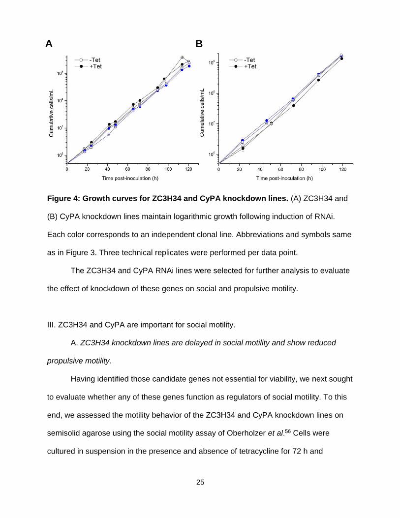
Knockdown of ZC3H34 to 19.1 ± 12.5 percent of expression levels in uninduced
controls (Figure 2) did not affect viability, as these cell lines maintained logarithmic
growth throughout the time course of the assay (Figure 4A).
ii. Cyclophilin A
The cyclophilins are a family of evolutionarily well-conserved proteins that are
present in all prokaryotes and eukaryotes. Cyclophilins possess peptidyl-prolyl
isomerase activity, catalyzing the cis-trans isomerization of peptide bonds preceding
proline residues and thereby facilitating protein folding35. In addition to their role as
molecular chaperones, cyclophilins participate in a diverse array of other biological
processes, including protein trafficking and maturation36, receptor complex
stabilization37, receptor signaling38, RNA processing39, detoxification of reactive oxygen
species40, immune response41, spliceosome assembly42, miRNA activity43, RNA-
induced silencing complex (RISC) assembly44, and chemotaxis45,46.
Cyclophilin A (CyPA) is the most abundantly expressed cyclophilin, comprising
0.1 to 0.6 percent of total cytosolic protein content47. In T. brucei, CyPA (MW = 18.7
kDa) is 1.65 times more abundant in bloodstream form than in procyclic form cells27,
and 1.55 times more abundant in the intact flagellum of procyclic form cells than in the
cellular debris fraction48. Consistent with these proteomic analyses, CyPA localizes to
the cytoplasm and also along the flagellum in immunofluorescence micrographs,
although the staining is not uniform throughout the flagellum, indicating differences in
distribution and density49.
24
Proteins involved in folding and degradation are major components of the T.
brucei secretome, and CyPA was recently shown to be among the proteins secreted by
T. brucei50. Moreover, antisera obtained from cattle immunized with cell lysate from T.
congolense reacted strongly with an 18 kDa band on a western blot of trypanosome
whole-cell lysate as well as with recombinant CyPA49. CyPA is a modulator of the
immune system of mammalian hosts51 and is known to stimulate macrophages52.
Moreover, extracellular CyPA has a potent chemotactic effect on leukocytes45,
monocytes46, and lymphocytes46. Finally, CyPA has been implicated in the intracellular
replication cycle of the kinetoplastid parasite Leishmania, as CyPA siRNA interference
or sequestration by cyclosporin A reduced parasite burden in murine macrophages53.
Taken together, these observations raise the intriguing possibility that CyPA is important
to the pathogenesis African trypanosomiasis. CyPA’s role as a paracrine factor capable
of mediating intercellular communication also makes it an especially promising
candidate regulator of trypanosome social motility. `
Knockdown of CyPA expression to 6.9 ± 5.1 percent of levels in uninduced
controls (Figure 2) did not affect viability, as these cell lines maintained logarithmic
growth throughout the time course of the assay (Figure 4B), consistent with the
dispensability of cyclophilins for viability in yeast54 and mammals55.
25
Figure 4: Growth curves for ZC3H34 and CyPA knockdown lines. (A) ZC3H34 and
(B) CyPA knockdown lines maintain logarithmic growth following induction of RNAi.
Each color corresponds to an independent clonal line. Abbreviations and symbols same
as in Figure 3. Three technical replicates were performed per data point.
The ZC3H34 and CyPA RNAi lines were selected for further analysis to evaluate
the effect of knockdown of these genes on social and propulsive motility.
III. ZC3H34 and CyPA are important for social motility.
A. ZC3H34 knockdown lines are delayed in social motility and show reduced
propulsive motility.
Having identified those candidate genes not essential for viability, we next sought
to evaluate whether any of these genes function as regulators of social motility. To this
end, we assessed the motility behavior of the ZC3H34 and CyPA knockdown lines on
semisolid agarose using the social motility assay of Oberholzer et al.56 Cells were
cultured in suspension in the presence and absence of tetracycline for 72 h and
A B
26
subsequently inoculated on corresponding plates ± tetracycline. The motility of
individual cells obtained from the same suspension culture was also analyzed.
To evaluate the effect of ZC3H34 knockdown on social motility, we first assessed
the number of radial projections formed by ZC3H34-depleted cells and non-depleted
controls at fixed time points post-inoculation on semisolid agarose. Figure 5A shows
representative images of projection formation by RNAi-induced (+ tetracycline) and
uninduced (- tetracycline) control groups over the time course of the assay. Knockdown
of ZC3H34 significantly delayed the onset of social motility. Cells depleted of ZC3H34
had formed significantly fewer radial projections at 2, 3, and 4 days post-inoculation on
semisolid agarose compared with uninduced controls at the same time points (Figure
5B). Moreover, the projections that did form in the presence of tetracycline were less
extensive than those formed by controls, only occasionally reaching the edge of the
plate (Figure 5A). qPCR analysis verified that formation of radial projections on the +
tetracycline plates was not the result of poor transcript knockdown (Figure 2).
Cells that lack propulsive (directional) motility are unable to perform social
motility56. We therefore examined the propulsive motility of individual cells in suspension
culture to better understand the basis for the delayed social motility phenotype. Of note,
ZC3H34-depleted cell lines also displayed significantly reduced propulsive motility in
suspension compared to uninduced controls, as measured by total distance traveled
(Figure 6A) and mean-square displacement (Figure 6B). Whether the observed delay in
social motility onset represents a bona fide defect in social motility or is the result of
slower single-cell motility is unclear (see Discussion).

27
Figure 5: Knockdown of ZC3H34 delays social motility. (A) Representative images
of radial projections on semisolid agarose formed by uninduced, ZC3H34-expressing
control cells (upper panel) and cells depleted of ZC3H34 (lower panel) at the indicated
times post-inoculation (TPI). (B) Distribution of number of radial projections per plate at
the indicated times point post-inoculation. Vertical axis indicates number of projections
extending ≥ 7.5 mm, 10.8 mm, and 19.9 mm from the inoculation site for time points 48
A
B

28
h, 72 h, and 96 h post-inoculation, respectively. Boxes indicate the 25th, 50th, and 75th
percentiles. Closed squares (▪) indicate the mean. Error bars are + 2 standard
deviations. n=16 plates for each time/condition. ***p < 0.001; ****p < 0.0001,
*****p < 0.00001 (two-tailed Student’s t-test).
Figure 6: ZC3H34 knockdown decreases propulsive motility. (A) Total distance
traveled and (B) mean-square displacement (<x2>) of ZC3H34 RNAi knockdown lines in
presence and absence of 1 μg/mL tetracycline. Center line in (A) is the mean. Error bars
are ± 1 SEM. n=52 cells tracked per condition. **p < 0.01 (two-tailed Student’s t-test).
B. Knockdown of CyPA delays social motility, and this delay is not attributable to
a defect in propulsive motility.
We evaluated the effect of CyPA knockdown on social motility in a manner
analogous to our assessment of ZC3H34 knockdown. Figure 7A shows representative
images of projection formation by RNAi-induced and uninduced control groups over the
time course of the assay. As with ZC3H34 depletion, knockdown of CyPA significantly
delayed the onset of social motility. Cells depleted of CyPA had formed significantly
fewer radial projections at 3, 4, and 5 days post-inoculation on semisolid agarose
A B

29
compared with uninduced controls at the same time points (Figure 7B). This delay was
not attributable to decreased propulsive motility, as the total distance traveled (Figure
8A) and mean-square displacement (Figure 8B) of CyPA knockdown cells was slightly
or even significantly greater than CyPA-expressing controls. Interestingly, the number of
radial projections formed by CyPA-depleted parasites increased rapidly between days 4
and 5, as reflected by the 105-fold decrease in p-value.
A
B

30
Figure 7: Knockdown of CyPA delays social motility. (A) Representative images of
radial projections on semisolid agarose formed by uninduced, CyPA-expressing control
cells (upper panel) and cells depleted of CyPA (lower panel) at the indicated times post-
inoculation (TPI). (B) Distribution of number of radial projections per plate at the
indicated time points post-inoculation. Graph and box plot elements same as in Figure
5; radial projections at 120 h are those extending ≥ 24.9 mm from site of inoculation.
n=16 plates for each time/condition. ns, p > 0.05; **p < 0.01; ***p < 0.001; ********p <
0.00000001 (two-tailed Student’s t-test).
Figure 8: Depletion of CyPA does not reduce general propulsive motility. (A) Total
distance traveled and (B) mean-square displacement (<x2>) of CyPA RNAi knockdown
A
B

31
lines in presence and absence of 1 μg/mL tetracycline. Two biological replicates were
performed, and each column shows results from a given biological replicate. Plot
elements same as in Figure 6. n=52 cells tracked per condition per replicate. ns, p >
0.05; *p < 0.05; **p < 0.01; ***p < 0.001 (two-tailed Student’s t-test).

32
References
1. Mortazavi, A.; Williams, B.A.; McCue, K.; Schaeffer, L.; Wold, B. “Mapping and quantifying mammalian transcriptomes by RNA-Seq.” Nat. Methods 2008, 5, 621–628.
2. Hershey, J.W. “Translational control in mammalian cells.” Annu. Rev. Biochem.
1991, 60, 717–755.
3. Kinzy, T.G.; Ripmaster, T.L.; Woolford, J.L. “Multiple genes encode the translation elongation factor EF-1 gamma in Saccharomyces cerevisiae.” Nucleic Acids Res. 1994, 22, 2703–2707.
4. Sanders, J.; Brandsma, M.; Janssen, G.M.; Dijk, J.; Möller, W.
“Immunofluorescence studies of human fibroblasts demonstrate the presence of the complex of elongation factor-1 beta gamma delta in the endoplasmic reticulum.” J. Cell Sci. 1996, 109, 1113–1117.
5. Vickers, T.J.; Fairlamb, A.H. “Trypanothione S-transferase activity in a
trypanosomatid ribosomal elongation factor 1B.” J. Biol. Chem. 2004, 279, 27246–27256.
6. Vickers, T.J.; Wyllie, S.; Fairlamb, A.H. “Leishmania major elongation factor 1B
complex has trypanothione S-transferase and peroxidase activity.” J. Biol. Chem. 2004, 279, 49003–49009.
7. Hiraga, K.; Suzuki, K.; Tsuchiya, E.; Miyakawa, T. “Cloning and characterization
of the elongation factor EF-1 beta homologue of Saccharomyces cerevisiae. EF-1 beta is essential for growth.” FEBS Lett. 1993, 316, 165–169.
8. Kornberg, R.D. “Chromatin structure: a repeating unit of histones and DNA.”
Science 1974, 184, 868–871.
9. Wei, Y.; Yu, L.; Bowen, J.; Gorovsky, M.A.; Allis, C.D. “Phosphorylation of histone H3 is required for proper chromosome condensation and segregation.” Cell 1999, 97, 99–109.
10. Janzen, C.J.; Hake, S.B.; Lowell, J.E.; Cross, G.A. “Selective di- or trimethylation
of histone H3 lysine 76 by two DOT1 homologs is important for cell cycle regulation in Trypanosoma brucei.” Mol. Cell 2006, 23, 497–507.
11. Alsford, S.; Horn, D. “Cell-cycle-regulated control of VSG expression site
silencing by histones and histone chaperones ASF1A and CAF-1b in Trypanosoma brucei.” Nucleic Acids Res. 2012, 40, 10150–10160.
33
12. Stanne, T.M.; Kushwaha, M.; Wand, M.; Taylor, J.E.; Rudenko, G. “TbISWI regulates multiple polymerase I (Pol I)-transcribed loci and is present at Pol II transcription boundaries in Trypanosoma brucei.” Eukaryotic Cell 2011, 10, 964–976.
13. Figueiredo, L.M.; Janzen, C.J.; Cross, G.A. “A histone methyltransferase
modulates antigenic variation in African trypanosomes.” PLoS Biol. 2008, 6, e161.
14. Wang, Q.P.; Kawahara, T.; Horn, D. “Histone deacetylases play distinct roles in
telomeric VSG expression site silencing in African trypanosomes.” Mol. Microbiol. 2010, 77, 1237–1245.
15. Denninger, V.; Fullbrook, A.; Bessat, M.; Ersfeld, K.; Rudenko, G. “The FACT
subunit TbSpt16 is involved in cell cycle specific control of VSG expression sites in Trypanosoma brucei.” Mol. Microbiol. 2010, 78, 459–474.
16. van Holde, K. Chromatin. Berlin, Germany: Springer-Verlag KG; 1988.
17. Tzfati, Y.; Abeliovich, H.; Avrahami, D.; Shlomai, J.; “Universal minicircle
sequence binding protein, a CCHC-type zinc finger protein that binds the universal minicircle sequence of trypanosomatids. Purification and characterization.” J. Biol. Chem. 1995, 270, 21339–21345.
18. Milman, N.; Motyka, S.A.; Englund, P.T.; Robinson, D.; Shlomai, J.
“Mitochondrial origin-binding protein UMSBP mediates DNA replication and segregation in trypanosomes.” Proc. Natl. Acad. Sci. U.S.A. 2007, 104, 19250–19255.
19. Kredich, N.M.; Hershfield, M.S. “S-adenosylhomocysteine toxicity in normal and
adenosine kinase-deficient lymphoblasts of human origin.” Proc. Natl. Acad. Sci. U.S.A. 1979, 76, 2450–2454.
20. Bacchi, C.J.; Goldberg, B.; Garofalo-Hannan, J.; Rattendi, D.; Lyte, P.; Yarlett, N.
“Fate of soluble methionine in African trypanosomes: effects of metabolic inhibitors.” Biochem. J. 1995, 309, 737–743.
21. Seley, K.L.; Schneller, S.W.; Rattendi, D.; Lane, S.; Bacchi, C.J. “Synthesis and
antitrypanosomal activities of a series of 7-deaza-5’-noraristeromycin derivatives with variations in the cyclopentyl ring substituents.” Antimicrob. Agents. Chemother. 1997, 41, 1658–1661.
22. Malanovic, N.; Streith, I.; Wolinski, H.; Rechberger, G.; Kohlwein, S.D.; Tehlivets,
O. “S-adenosyl-L-homocysteine hydrolase, key enzyme of methylation metabolism, regulates phosphatidylcholine synthesis and triacylglycerol

34
homeostasis in yeast: implications for homocysteine as a risk factor of atherosclerosis.” J. Biol. Chem. 2008, 283, 23989–23999.
23. Rocha, P.S.; Sheikh, M.; Melchiorre, R.; Fagard, M.; Boutet, S.; Loach, R.;
Moffatt, B.; Wagner, C.; Vaucheret, H.; Furner, I. “The Arabidopsis HOMOLOGY-DEPENDENT GENE SILENCING1 gene codes for an S-adenosyl-L-homocysteine hydrolase required for DNA methylation-dependent gene silencing.” Plant Cell 2005, 17, 404–417.
24. Matthews, R.P.; Lorent, K.; Mañoral-Mobias, R.; Huang, Y.; Gong, W.; Murray,
I.V.; Blair, I.A.; Pack, M. “TNFalpha-dependent hepatic steatosis and liver degeneration caused by mutation of zebrafish S-adenosylhomocysteine hydrolase.” Development 2009, 136, 865–875.
25. Miller, M.W.; Duhl, D.M.; Winkes, B.M.; Arredondo-Vega, F.; Saxon, P.J.; Wolff,
G.L.; Epstein, C.J.; Hershfield, M.S.; Barsh, G.S. “The mouse lethal nonagouti (a(x)) mutation deletes the S-adenosylhomocysteine hydrolase (Ahcy) gene.” EMBO J. 1994, 13, 1806–1816.
26. Baric, I.; Fumic, K.; Glenn, B.; Cuk, M.; Schulze, A.; Finkelstein, J.D.; James,
S.J.; Mejaski-Bosnjak, V.; Pazanin, L.; Pogribny, I.P. et al. “S-adenosylhomocysteine hydrolase deficiency in a human: a genetic disorder of methionine metabolism.” Proc. Natl. Acad. Sci. U.S.A. 2004, 101, 4234–4339.
27. Urbaniak, M.D.; Guther, M.L.; Ferguson, M.A. “Comparative SILAC proteomic
analysis of Trypanosoma brucei bloodstream and procyclic lifecycle stages.” PLoS ONE 2012, 7, e36619.
28. Wurst, M.; Robles, A.; Po, J.; Luu, V.D.; Brems, S.; Marentije, M.; Stoitsova, S.;
Quijada, L.; Hoheisel, J.; Stewart, M. et al. “An RNAi screen of the RRM-domain proteins of Trypanosoma brucei.” Mol. Biochem. Parasitol. 2009, 163, 61–65.
29. Berriman, M.; Ghedin, E.; Hertz-Fowler, C.; Blandin, G.; Renauld, H.;
Bartholomeu, D.C.; Lennard, N.J.; Caler, E.; Hamlin, N.E.; Haas, B.; Böhme, U. et al. “The genome of the African trypanosome Trypanosoma brucei.” Science 2005, 309, 416–422.
30. Kramer, S.; Kimblin, N.C.; Carrington, M. “Genome-wide in silico screen for
CCCH-type zinc finger proteins of Trypanosoma brucei, Trypanosoma cruzi and Leishmania major.” BMC Genomics 2010, 11, 283.
31. Hwang, E.S.; Choi, A.; Ho, I.C. “Transcriptional regulation of GATA-3 by an
intronic regulatory region and fetal liver zinc finger protein 1.” J. Immunol. 2002, 169, 248–253.

35
32. Wang, L.; Xu, Y.; Zhang, C.; Ma, Q.; Joo, S.H.; Kim, S.K.; Xu, Z.; Chong, K. “OsLIC, a novel CCCH-type zinc finger protein with transcription activation, mediates rice architecture via brassinosteroids signaling.” PLoS ONE 2008, 3, e3521.
33. Pomeranz, M.C.; Hah, C.; Lin, P.C.; Kang, S.G.; Finer, J.J.; Blackshear, P.J.;
Jang, J.C. “The Arabidopsis tandem zinc finger protein AtTZF1 traffics between the nucleus and cytoplasmic foci and binds both DNA and RNA.” Plant Physiol. 2010, 152, 151–165.
34. Erben, E.D.; Fadda, A.; Lueong, S.; Hoheisel, J.D.; Clayton, C. “A genome-wide
tethering screen reveals novel potential post-transcriptional regulators in Trypanosoma brucei.” PLoS Pathog. 2014, 10, e1004178.
35. Kumari, S.; Roy, S.; Singh, P.; Singla-Pareek, S.L.; Pareek, A. “Cyclophilins:
proteins in search of function.” Plant Signal Behav. 2013, 8, e22734.
36. Ferreira, P.A.; Nakayama, T.A.; Pak, W.L.; Travis, G.H. “Cyclophilin-related protein RanBP2 acts as chaperone for red/green opsin.” Nature 1996, 383, 637–640
37. Leverson, J.D.; Ness, S.A. “Point mutations in v-Myb disrupt a cyclophilin-
catalyzed negative regulatory mechanism.” Mol. Cell 1998, 1, 203–211.
38. Brazin, K.N.; Mallis, R.J.; Fulton, D.B.; Andreotti, A.H. “Regulation of the tyrosine kinase Itk by the peptidyl-prolyl isomerase cyclophilin A.” Proc. Natl. Acad. Sci. U.S.A. 2002, 99, 1899–1904.
39. Krzywicka, A.; Beisson, J.; Keller, A.M.; Cohen, J.; Jerka-Dziadosz, M.; Klotz, C.
“KIN241: a gene involved in cell morphogenesis in Paramecium tetraurelia reveals a novel protein family of cyclophilin-RNA interacting proteins (CRIPs) conserved from fission yeast to man.” Mol. Microbiol. 2001, 42, 257–267.
40. Hong, F.; Lee, J.; Song, J.W.; Lee, S.J.; Ahn, H.; Cho, J.J. Ha, J.; Kim, S.S.
“Cyclosporin A blocks muscle differentiation by inducing oxidative stress and inhibiting the peptidyl-prolyl-cis-trans isomerase activity of cyclophilin A: cyclophilin A protects myoblasts from cyclosporin A-induced cytotoxicity.” FASEB J. 2002, 16, 1633–1635.
41. Wiederrecht, G.; Hung, S.; Chan, H.K.; Marcy, A.; Martin, M.; Calaycay, J.;
Boulton, D.; Sigal, N.; Kincaid, R.L.; Siekierka, J.J. “Characterization of high molecular weight FK-506 binding activities reveals a novel FK-506-binding protein as well as a protein complex.” J. Biol. Chem. 1992, 267, 21753–21760.
42. Horowitz, D.S.; Lee, E.J.; Mabon, S.A.; Misteli, T. “A cyclophilin functions in pre-
mRNA splicing.” EMBO J. 2002, 21, 470–480.

36
43. Smith, M.R.; Willmann, M.R.; Wu, G.; Berardini, T.Z.; Möller, B.; Weijers, D.; Poethig, R.S. “Cyclophilin 40 is required for microRNA activity in Arabidopsis.” Proc. Natl. Acad. Sci. U.S.A. 2009, 106, 5424–5429.
44. Lki, T.; Yoshikawa, M.; Meshi, T.; Ishikawa, M. “Cyclophilin 40 facilitates HSP90-mediated RISC assembly in plants.” EMBO J. 2012, 31, 267–278.
45. Xu, Q.; Leiva, M.C.; Fischkoff, S.A.; Handschumacher, R.E.; Lyttle, C.R. “Leukocyte chemotactic activity of cyclophilin.” J. Biol. Chem. 1992, 267, 11968–11971.
46. Sherry, B.; Yarlett, N.; Strupp, A.; Cerami, A. “Identification of cyclophilin as a
proinflammatory secretory product of lipopolysaccharide-activated macrophages.” Proc. Natl. Acad. Sci. U.S.A. 1992, 89, 3511–3515.
47. Nigro, P.; Pompilio, G.; Capogrossi, M.C. “Cyclophilin A: a key player for human
disease.” Cell Death Dis. 2013, 4, e888.
48. Subota, I.; Julkowska, D.; Vincensini, L.; Reeg, N.; Buisson, J.; Blisnick, T.; Huet, D.; Perrot, S.; Santi-Rocca, J.; Duchateau, M. et al. “Proteomic analysis of intact flagella of procyclic Trypanosoma brucei cells identifies novel flagellar proteins with unique sub-localization and dynamics.” Mol. Cell. Proteomics. 2014, 13, 1769–1786.
49. Geiger, A.; Hirtz, C.; Bécue, T.; Bellard, E.; Centeno, D.; Gargani, D.; Rossignol,
M.; Cuny, G.; Peltier, J.B. “Exocytosis and protein secretion in Trypanosoma.” BMC Microbiol. 2010, 10, 20.
50. Pellé, R.; McOdimba, F.; Chuma, F.; Wasawo, D.; Pearson, T.W.; Murphy, N.B.
“The African trypanosome cyclophilin A homologue contains unusual conserved central and N-terminal domains and is developmentally regulated.” Gene 2002, 290, 181–191.
51. Darji, A.; Beschin, A.; Sileghem, M.; Heremans, H.; Brys, L.; De Baetselier, P. “In
vitro simulation of immunosuppression caused by Trypanosoma brucei: active involvement of gamma interferon and tumor necrosis factor in the pathway of suppression.” Infect. Immun. 1996, 64, 1937–1943.
52. Kim, H.; Kim, W.J.; Jeon, S.T.; Koh, E.M.; Cha, H.S.; Ahn, K.S.; Lee, W.H.
“Cyclophilin A may contribute to the inflammatory processes in rheumatoid arthritis through induction of matrix degrading enzymes and inflammatory cytokines from macrophages.” Clin. Immunol. 2005, 116, 217–224.
37
53. Hoerauf, A.; Rascher, C.; Bang, R.; Pahl, A.; Solbach, W.; Brune, K.; Rollinghoff, M.; Bang, H. “Host-cell cyclophilin is important for the intracellular replication of Leishmania major.” Mol. Microbiol. 1997, 24, 421–429.
54. Dolinski, K.; Muir, S.; Cardenas, M.; Heitman, J. “All cyclophilins and FK506
binding proteins are, individually and collectively, dispensable for viability in Saccharomyces cerevisiae.” Proc. Natl. Acad. Sci. U.S.A. 1997, 94, 13093–13098.
55. Colgan, J.; Asmal, M.; Luban, J. “Isolation, characterization and targeted
disruption of mouse Ppia: cyclophilin A is not essential for mammalian cell viability.” Genomics 2000, 68, 167–178.
56. Oberholzer, M.; Lopez, M.A.; McLelland, B.T.; Hill, K.L. “Social motility in African
trypanosomes.” PLoS Pathog. 2010, 6, e1000739.

38
CHAPTER 4
Discussion
African trypanosomiasis ranks among the three most neglected tropical
diseases1. Despite the considerable medical and economic burden of this disease, only
four drugs are available for the treatment of sleeping sickness, and no new treatments
have been developed in over a quarter century2. Moreover, existing therapeutics are
toxic, increasingly ineffective against early-stage, hemolymphatic infection, and limited
in their ability to treat late-stage, central nervous system infection2.
Social motility in Trypanosoma brucei was first described in 20103, and was
recently selected as one of the seven most influential discoveries in parasitology of the
past decade by the editors of PLoS Pathogens4, a leading microbial pathogenesis
journal. Numerous studies since then have shown that social motility is controlled by
cAMP signaling in the trypanosome flagellum5,6 and have linked social motility to
establishment of tsetse fly midgut infection and vector-to-host transmission7. Despite
this progress, many genetic modulators of social motility have yet to be discovered, and
the mechanisms underlying this phenomenon remain poorly understood. Biochemical
studies of genes that control social motility in T. brucei have been limited to a few
flagellar adenylate cyclases5,6, a flagellar phosphodiesterase6, and the ER membrane
protein Rft17. Additional regulators of social motility were unknown prior to this work.
Although direct evidence implicating social motility in the transmission of T.
brucei from vector to mammalian host is lacking, the observation that Rft1-/- cell lines

39
defective in social motility are also impaired in their ability to establish midgut infection
in the Glossina vector7 supports the idea that social motility may play a role in promoting
trypanosome colonization and infection of tsetse flies. Thus, chemical inhibition of
regulators of social motility is a potentially promising approach for blocking the
transmission of trypanosomiasis. More broadly, social motility requires both intra- and
intercellular signaling events, including the ability of cells to alter their movement in
response to external stimuli. Identification and characterization of the genes required for
social motility in T. brucei will therefore also advance our understanding of parasite
signaling, an important yet highly understudied aspect of parasite biology.
The present work identifies two novel modulators of social motility in T. brucei,
the zinc finger RNA-binding protein ZC3H34 and cyclophilin A. Knockdown of ZC3H34
delayed social motility, although the ZC3H34 knockdown parasites also exhibited
reduced propulsive motility of individual cells in liquid culture. A complete block of
directional motility causes a complete block of social motility3. Thus, it is possible that
the delayed social motility phenotype of ZC3H34 knockdowns is due to reduced
propulsive motility. However, T. brucei depleted of the AC4 receptor-type adenylate
cyclase show a decrease in propulsive motility comparable to that observed with
ZC3H34 knockdowns (~35 µm decrease in total distance traveled relative to non-
depleted controls) but do not exhibit any apparent differences in social motility
compared to wild type controls5. It therefore remains to be determined whether reduced
propulsive motility is the cause of the delayed social motility seen with the ZC3H34
knockdowns. A direct parallel time course analysis of radial projection formation by
ZC3H34- and AC4-depleted cells will help distinguish between these possibilities.
40
Additional work is needed to define the mechanism by which ZC3H34
influences social motility. ZC3H34 is an RNA-binding protein that associates with
DRDB38, another RNA-binding protein that functions in stabilization and transport of
mRNAs9. As noted in Chapter 3, gene expression in T. brucei is regulated almost
exclusively at the post-transcriptional level by RNA-binding proteins10. This makes
ZC3H34 a particularly interesting candidate, as it could function in controlling the
expression of other genes involved in social motility. The RNA substrates of ZC3H34
are unknown. Epitope tagging of ZC3H34 followed by RNA-immunoprecipitation
sequencing (RIP-seq) would provide a means to identify the RNA targets of ZC3H34.
Subsequent analysis of social motility phenotype upon knockdown or overexpression of
the identified ZC3H34 target will clarify the mechanism through which ZC3H34
regulates social motility. Given the predicted role of ZC3H34 as an RNA-binding protein
and its ability to stabilize RNA transcripts when fused to the UTR of an mRNA
reporter11,12, we speculate that ZC3H34 may function in stabilizing the transcripts of
genes involved in social motility and/or individual cell motility.
As with ZC3H34 depletion, knockdown of cyclophilin A resulted in a delayed
social motility phenotype. Unlike ZC3H34, however, this delay in social motility was not
accompanied by reduced motility of individual cells. In fact, CyPA knockdown lines were
found to have slightly or even significantly elevated propulsive motility compared to non-
depleted controls (Figure 8). Cyclophilin A is secreted by T. brucei13,14 and has been
shown to elicit immunosuppression in an in vitro model of T-cell proliferation15.
Moreover, CyPA stimulates the migration of eosinophils and neutrophils at nanomolar
concentrations16, and also elicits monocyte chemotaxis17. Our observation that CyPA

41
knockdown lines are defective in social motility, combined with CyPA’s possible role as
a cytokine-like protein involved in interactions with infected species and previous work
implicating social motility in tsetse fly infection7, suggest that CyPA might be important
to parasite transmission. Experiments that assess the ability of CyPA (and ZC3H34) null
mutants to infect the tsetse fly are needed to determine whether the social motility
defects observed in vitro are indeed physiologically relevant.
The other five genes analyzed in this study were not evaluated for a potential role
in social motility owing to the lethality observed in suspension culture after tetracycline-
induced knockdown of transcript expression. The finding that a gene upregulated in
trypanosomes cultured on semisolid agarose is essential for viability does not exclude a
possible role for that gene in social motility, and the genetic system used in this study
did not allow for control over dsRNA expression levels. Thus, low-level knockdown of
the genes whose depletion resulted in lethality might yield detectable differences in
social motility phenotype while maintaining cell viability. Conveniently, an inducible
expression system that enables precise control over expression of both genes and
dsRNAs in T. brucei was recently developed by Sunter18. A more practical and perhaps
more informative approach, however, would be to assay social motility in parasites
overexpressing these genes. Future studies should also explore the potential functional
relevance to social motility of the remaining genes among the list of 22 candidates
upregulated in trypanosomes cultivated on semisolid agarose using a workflow similar
to that employed in this study. Overexpression of the 38 genes found to be
downregulated in T. brucei plated on semisolid agarose followed by assays of social
motility will also prove insightful.

42
In summary, elucidating and characterizing regulators of social motility in
Trypanosoma brucei not only has the potential to alleviate the burden of African
trypanosomiasis by informing the development of chemical inhibitors of vector-to-host
parasite transmission, but will also advance our understanding of signaling pathways in
protozoan parasites and improve the relevance of T. brucei as a model organism for the
study of eukaryotic signaling in general.
43
References
1. Sutherland, C.S.; Yukich, J.; Goeree, R.; Tediosi, F. “A literature review of
economic evaluations for a neglected tropical disease: human African trypanosomiasis (“sleeping sickness”).” PLoS Negl. Trop. Dis. 2015, 9, e0003397.
2. Steverding. D. “The development of drugs for treatment of sleeping sickness: a
historical review.” Parasit. Vectors 2010, 3, 15.
3. Oberholzer, M.; Lopez, M.A.; McLelland, B.T.; Hill, K.L. “Social motility in African trypanosomes.” PLoS Pathog. 2010, 6, e1000739.
4. Haldar, K.; McFadden, G. “PLoS Pathogens at 10 years.”
http://collections.plos.org/pathogens-10th-anniversary (accessed June 12, 2016).
5. Lopez, M.A.; Saada, E.A.; Hill, K.L. “Insect stage-specific adenylate cyclases regulate social motility in African trypanosomes.” Eukaryotic Cell 2015, 14, 104–112.
6. Oberholzer, M.; Saada, E.A.; Hill, K.L. “Cyclic AMP regulates social behavior in
African trypanosomes.” MBio 2015, 6, e01954-14.
7. Imhof, S.; Vu, X.L.; Bütikofer, P.; Roditi, I. “A glycosylation mutant of Trypanosoma brucei links social motility defects in vitro to impaired colonization of tsetse flies in vivo.” Eukaryotic Cell 2015, 14, 588–592.
8. Fernández-Moya, S.M.; García-Pérez, A.; Kramer, S.; Carrington, M.; Estévez,
A.M. “Alterations in DRBD3 ribonucleoprotein complexes in response to stress in Trypanosoma brucei.” PLoS ONE 2012, 7, e48870.
9. Stern, M.Z.; Gupta, S.K.; Salmon-Divon, M.; Haham, T.; Barda, O.; Levi, S.;
Wachtel, C.; Nilsen, T.W.; Michaeli, S.; Salmon-Divon, M. “Multiple roles for polypyrimidine tract binding (PTB) proteins in trypanosome RNA metabolism.” RNA 2009, 15, 648–665.
10. Berriman, M.; Ghedin, E.; Hertz-Fowler, C.; Blandin, G.; Renauld, H.;
Bartholomeu, D.C.; Lennard, N.J.; Caler, E.; Hamlin, N.E.; Haas, B.; Böhme, U. et al. “The genome of the African trypanosome Trypanosoma brucei.” Science 2005, 309, 416–422.
11. Kramer, S.; Kimblin, N.C.; Carrington, M. “Genome-wide in silico screen for
CCCH-type zinc finger proteins of Trypanosoma brucei, Trypanosoma cruzi and Leishmania major. BMC Genomics 2010, 11, 283.

44
12. Erben, E.D.; Fadda, A.; Lueong, S.; Hoheisel, J.D.; Clayton, C. “A genome-wide tethering screen reveals novel potential post-transcriptional regulators in Trypanosoma brucei.” PLoS Pathog. 2014, 10, e1004178.
13. Pellé, R.; McOdimba, F.; Chuma, F.; Wasawo, D.; Pearson, T.W.; Murphy, N.B.
“The African trypanosome cyclophilin A homologue contains unusual conserved central and N-terminal domains and is developmentally regulated.” Gene 2002, 290, 181–191.
14. Geiger, A.; Hirtz, C.; Bécue, T.; Bellard, E.; Centeno, D.; Gargani, D.; Rossignol,
M.; Cuny, G.; Peltier, J.B. “Exocytosis and protein secretion in Trypanosoma.” BMC Microbiol. 2010, 10, 20.
15. Xu, Q.; Leiva, M.C.; Fischkoff, S.A.; Handschumacher, R.E.; Lyttle, C.R.
“Leukocyte chemotactic activity of cyclophilin.” J. Biol. Chem. 1992, 267, 11968–11971.
16. Sherry, B.; Yarlett, N.; Strupp, A.; Cerami, A. “Identification of cyclophilin as a
proinflammatory secretory product of lipopolysaccharide-activated macrophages.” Proc. Natl. Acad. Sci. U.S.A. 1992, 89, 3511–3515.
17. Darji, A.; Beschin, A.; Sileghem, M.; Heremans, H.; Brys, L.; De Baetselier, P. “In vitro simulation of immunosuppression caused by Trypanosoma brucei: active involvement of gamma interferon and tumor necrosis factor in the pathway of suppression.” Infect. Immun. 1996, 64, 1937–1943.
18. Sunter, J.D. “A vanillic acid inducible expression system for Trypanosoma
brucei.” Mol. Biochem. Parasitol. 2016, 207, 45–48.

45
SUPPLEMENTAL INFORMATION
Supplemental Table 1: Primers used for generation of RNAi amplicons. Restriction
sites are italicized.
GeneDB Accession (Gene Description)
Primer Sequence
Tb927.10.12330 (ZC3H34)
FWD: ATATTCTAGAACCCGCCTCAACAGTATCAC REV: ATATAAGCTTCACATCATGTTTTCCATCGG
Tb927.11.880 (CyPA)
FWD: AGTCGATCTAGAATGTGAGCATTGCAGGTCAG REV: AGTCGACTCGAGTAGTCGGTGTTCGTCTGTGC
Tb927.10.5840 (EF-1β)
FWD: ATATTCTAGATAAAGGAAATCAACGGTCGC REV: ATATAAGCTTTGTGGTCACCCCACAGTAGA
Tb927.1.2510 (Histone H3)
FWD: ATATTCTAGAAGGCCTCAAAGGGTTCTGAT REV: ATATAAGCTTTTGTCTCAACCCTGACCCTC
Tb927.10.6060 (UMSBP2)
FWD: ATATTCTAGATTGAAACGTCTCCAACCCTC REV: ATATAAGCTTCTTTTCCATCCCTCCTCTCC
Tb927.11.9590
(SAHH)
FWD: AGTCGATCTAGACTCGTGCAACATCTTCTCCA REV: AGTCGAAAGCTTTATCCACAGACACATGCGGT
Tb927.8.4450 (RBP11)
FWD: ATATTCTAGAGGAGTGACGACTTTGGTGGT REV: ATATAAGCTTGCGGCTATGGGATTCTTGTA
Supplemental Table 2: Primers used for qPCR quantification of gene products.
GeneDB Accession (Gene Description)
Primer Sequence
Tb927.10.12330 (ZC3H34)
FWD: GCTGCCGAATCCCCCTAATG REV: ATGCAAGAGGACGGTCGAGA
Tb927.11.880 (CyPA)
FWD: ATGGACGTCGTCAAGGCAAT REV: TAGTCGGTGTTCGTCTGTGC
Tb927.10.5840 (EF-1β)
FWD: GTCGTTTGGAATGTGGCGAG REV: TTTACGGTTGAGGAGGCTGC

46
Tb927.1.2510 (Histone H3)
FWD: ACCACAACTCTCAAACCAAGCA REV: ACCCTTTGAGGCCTTCTTGC
Tb927.10.6060 (UMSBP2)
FWD: CTTTCGCCATAAAGCGGTGC REV: CACTACGACCACCACCCAC
Tb927.11.9590
(SAHH)
FWD: GTTTTCGTAACCGCACGCTG REV: GCGTATCGGTAACACCCTCC
Tb927.8.4450 (RBP11)
FWD: ATGCAAACGTGTCAATGCGT REV: AGGAGAAACTCCCTACGGCT
Tb427.08.5010
(PFR2)
FWD: GAAGTTGAAGGTGTTGTGAGTCC REV: CCTCCAGCGTGATATCTGTTACC
Tb927.11.10190
(TERT)
FWD: GAGCGTGTGACTTCCGAAGG REV: AGGAACTGTCACGGAGTTTGC