essential cardiac anatomy and...
TRANSCRIPT

Essential Cardiac Anatomy and Physiology
Tobias Schaeffter Division of Imaging Sciences and Biomedical Engineering
King’s College London

Learning objectives
• Provide an overview on Heart Anatomy, Conducting System and Physiology
• Provide terminology of the external and internal features of the heart
• Show imaging examples of cardiac anatomy and physiology

Heart Anatomy • Approximately the size of your fist
– Weight = 250-300 grams • Location
– In the mediastinum between the lungs – Superior surface of diaphragm – ⅔ of it to the left of midsternal line
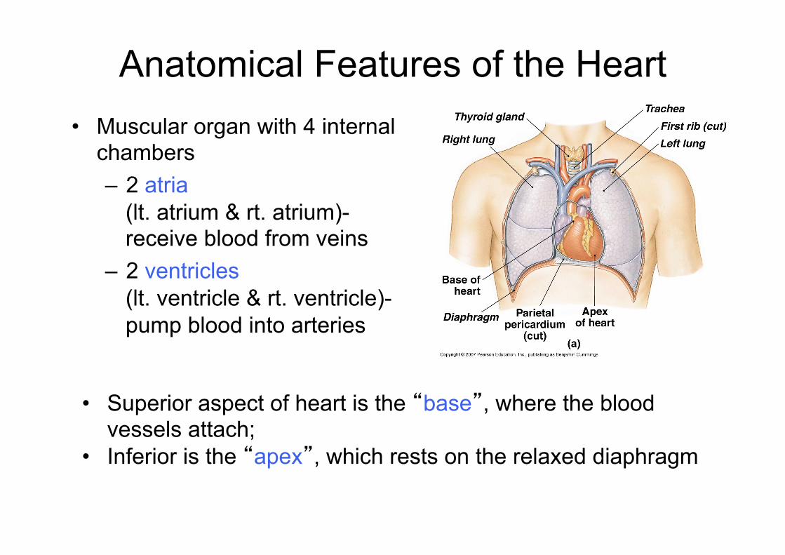
Anatomical Features of the Heart • Muscular organ with 4 internal
chambers – 2 atria
(lt. atrium & rt. atrium)- receive blood from veins
– 2 ventricles (lt. ventricle & rt. ventricle)- pump blood into arteries
• Superior aspect of heart is the “base”, where the blood vessels attach;
• Inferior is the “apex”, which rests on the relaxed diaphragm
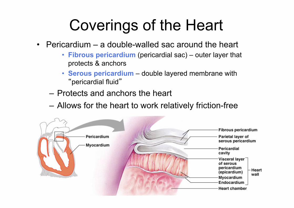
Coverings of the Heart • Pericardium – a double-walled sac around the heart
• Fibrous pericardium (pericardial sac) – outer layer that protects & anchors
• Serous pericardium – double layered membrane with “pericardial fluid”
– Protects and anchors the heart – Allows for the heart to work relatively friction-free
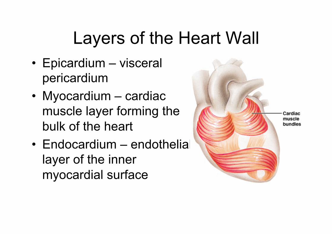
Layers of the Heart Wall • Epicardium – visceral
pericardium • Myocardium – cardiac
muscle layer forming the bulk of the heart
• Endocardium – endothelial layer of the inner myocardial surface
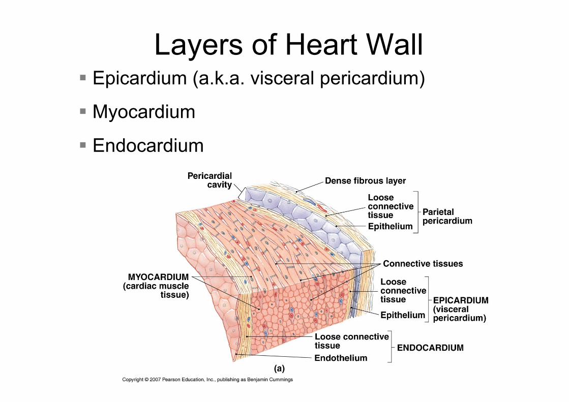
Layers of Heart Wall § Epicardium (a.k.a. visceral pericardium)
§ Myocardium
§ Endocardium
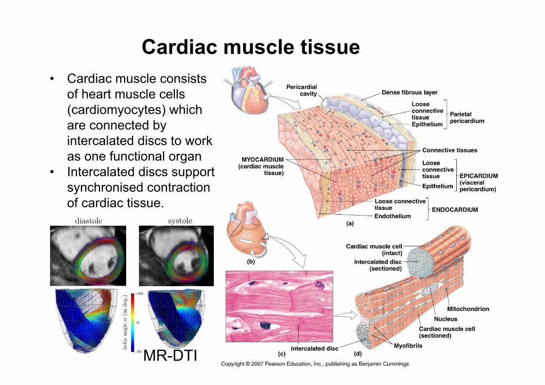
Cardiac muscle tissue • Cardiac muscle consists
of heart muscle cells (cardiomyocytes) which are connected by intercalated discs to work as one functional organ
• Intercalated discs support synchronised contraction of cardiac tissue.
MR-DTI
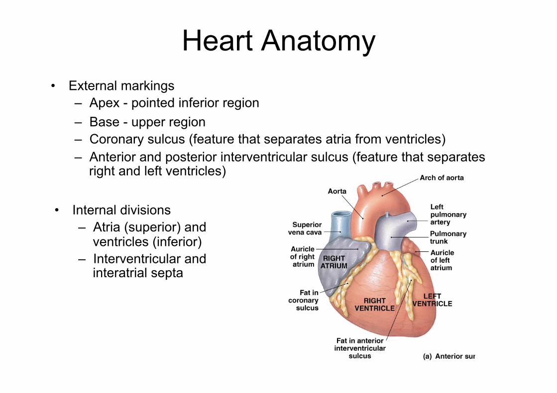
Heart Anatomy • External markings
– Apex - pointed inferior region – Base - upper region – Coronary sulcus (feature that separates atria from ventricles) – Anterior and posterior interventricular sulcus (feature that separates
right and left ventricles)
• Internal divisions – Atria (superior) and
ventricles (inferior) – Interventricular and
interatrial septa

External Features
§ Auricles
§ Coronary sulcus – contains the coronary sinus
§ Anterior interventricular sulcus – contains coronary vessels
§ Posterior interventricular sulcus – contains coronary vessels
Cardiac CT

Atria of the Heart • Atria - receiving chambers of the heart
– Receive venous blood returning to heart – Separated by an interatrial septum (wall)
• Fossa ovalis - remnant of foramen ovale (opening in interatrial septum in fetus)
• Pectinate muscles mark atrial walls • Pump blood into ventricles • Blood enters right atria from superior and inferior
venae cavae and coronary sinus • Blood enters left atria from pulmonary veins
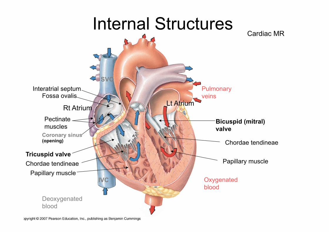
Rt Atrium Lt Atrium
Pectinate muscles
SVC
IVC
Coronary sinus (opening)
Deoxygenated blood
Pulmonary veins
Oxygenated blood
Tricuspid valve
Bicuspid (mitral) valve
Chordae tendineae Papillary muscle
Chordae tendineae
Papillary muscle
Interatrial septum Fossa ovalis
Internal Structures Cardiac MR
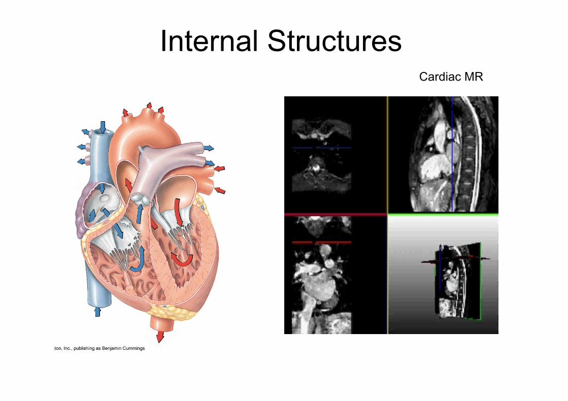
Internal Structures Cardiac MR

Ventricles of the Heart • Ventricles are the pumping chambers of the heart • Papillary muscles and trabeculae carneae muscles mark
ventricular walls • Separated by an interventricular septum
– Contains components of the conduction system • Right ventricle pumps blood into the pulmonary trunk • Left ventricle pumps blood into the aorta
– 3xThicker myocardium due to greater work load • Pulmonary circulation supplied by
right ventricle is a much low pressure system requiring less energy
• Systemic circulation supplied by left ventricle is a higher pressure system and thus requires more forceful contractions
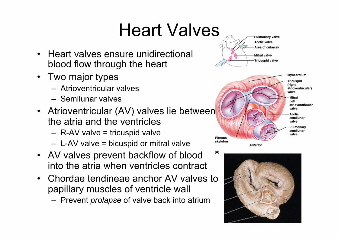
Heart Valves • Heart valves ensure unidirectional
blood flow through the heart • Two major types
– Atrioventricular valves – Semilunar valves
• Atrioventricular (AV) valves lie between the atria and the ventricles – R-AV valve = tricuspid valve – L-AV valve = bicuspid or mitral valve
• AV valves prevent backflow of blood into the atria when ventricles contract
• Chordae tendineae anchor AV valves to papillary muscles of ventricle wall – Prevent prolapse of valve back into atrium
Fibrous Skeleton
• Surrounds all four valves – Composed of dense connective tissue
• Functions – Anchors valve cusps – Prevents overdilation of valve openings – Main point of insertion for cardiac muscle – Blocks direct spread of electrical impulses
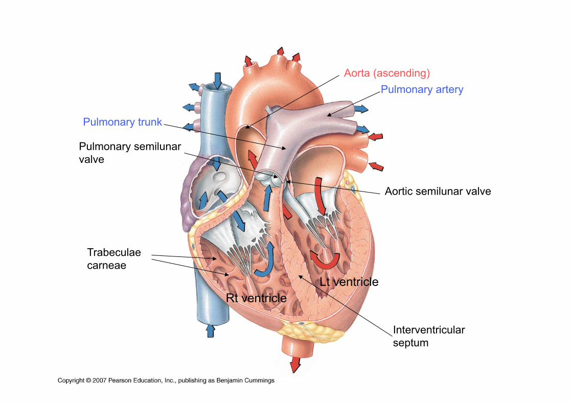
Rt ventricle Lt ventricle
Interventricular septum
Trabeculae carneae
Aorta (ascending)
Aortic semilunar valve
Pulmonary semilunar valve
Pulmonary trunk
Pulmonary artery
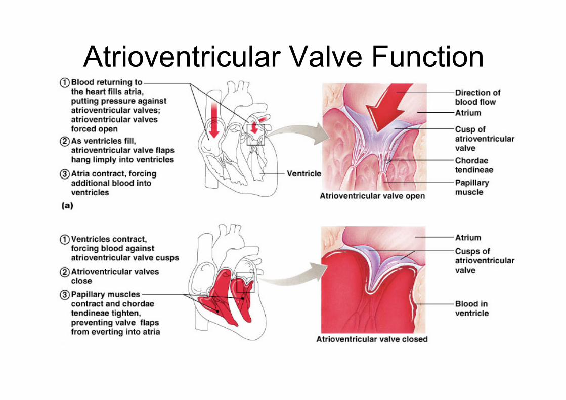
Atrioventricular Valve Function
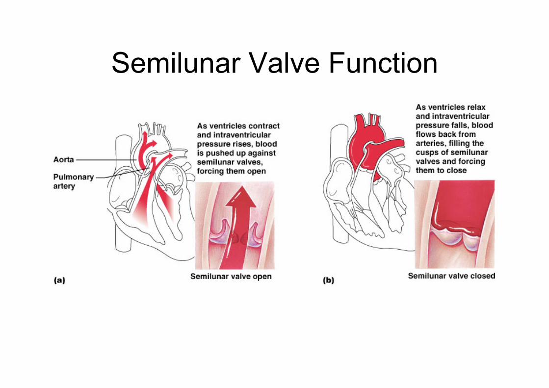
Semilunar Valve Function
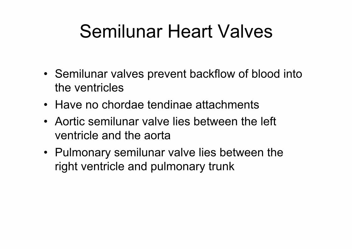
Semilunar Heart Valves
• Semilunar valves prevent backflow of blood into the ventricles
• Have no chordae tendinae attachments • Aortic semilunar valve lies between the left
ventricle and the aorta • Pulmonary semilunar valve lies between the
right ventricle and pulmonary trunk
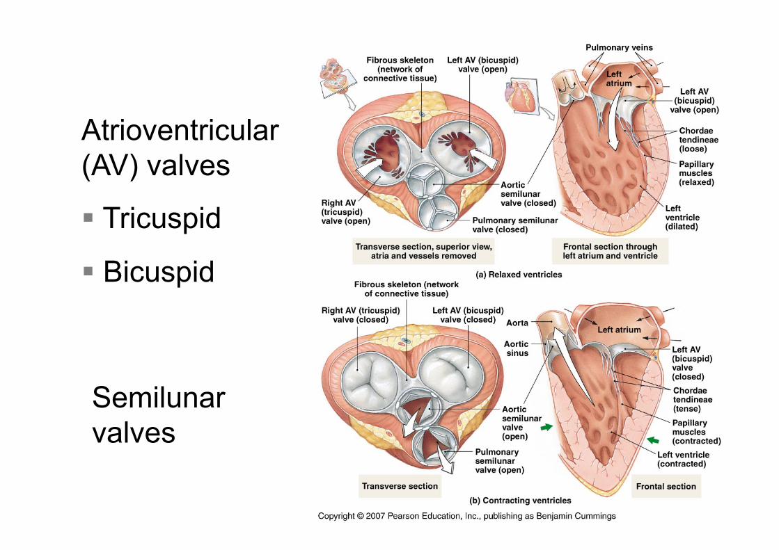
Atrioventricular (AV) valves
§ Tricuspid
§ Bicuspid
Semilunar valves

3D reconstruction of the heart. To the left 2D US-images from the same dataset, showing tricuspid and mitral valves (above) and aortal and mitral valve (below) (source Wikipedia)
Heart Valves
Heart sounds (“lub-dup”) due to valves closing “Lub” - closing of atrioventricular valves “Dub”- closing of semilunar valves
Cardiac US

Heart Defects

Conducting System
• Cardiac muscle tissue has intrinsic ability to: – Generate and conduct impulses – Signal these cells to contract rhythmically
• Conducting system – A series of specialized cardiac muscle cells – Sinoatrial (SA) node sets the inherent rate of
contraction • “Autorhythmic (conducting) cells”
– spontaneously generate action potentials
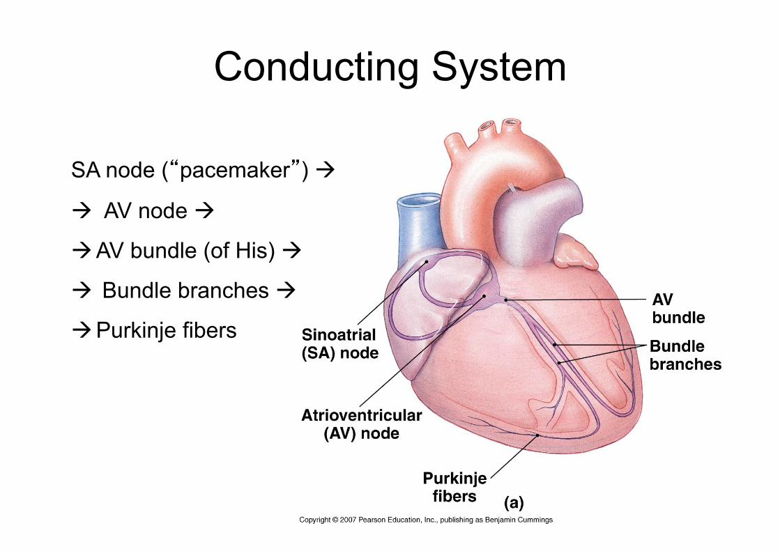
Conducting System
SA node (“pacemaker”) à
à AV node à
à AV bundle (of His) à
à Bundle branches à
à Purkinje fibers

Innervation • Heart rate is altered by
external controls • Nerves to the heart
include: – Visceral sensory fibers – Parasympathetic
branches of the vagus nerve
– Sympathetic fibers – from cervical and upper thoracic chain ganglia
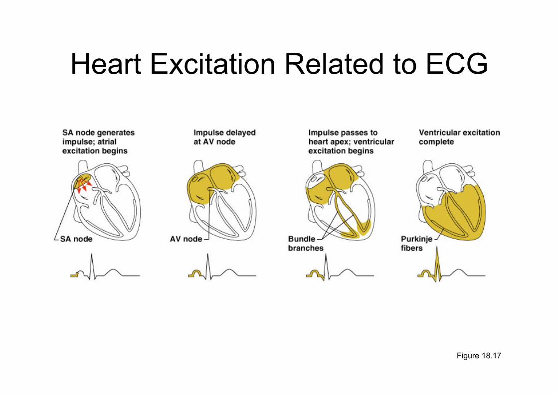
Heart Excitation Related to ECG
Figure 18.17
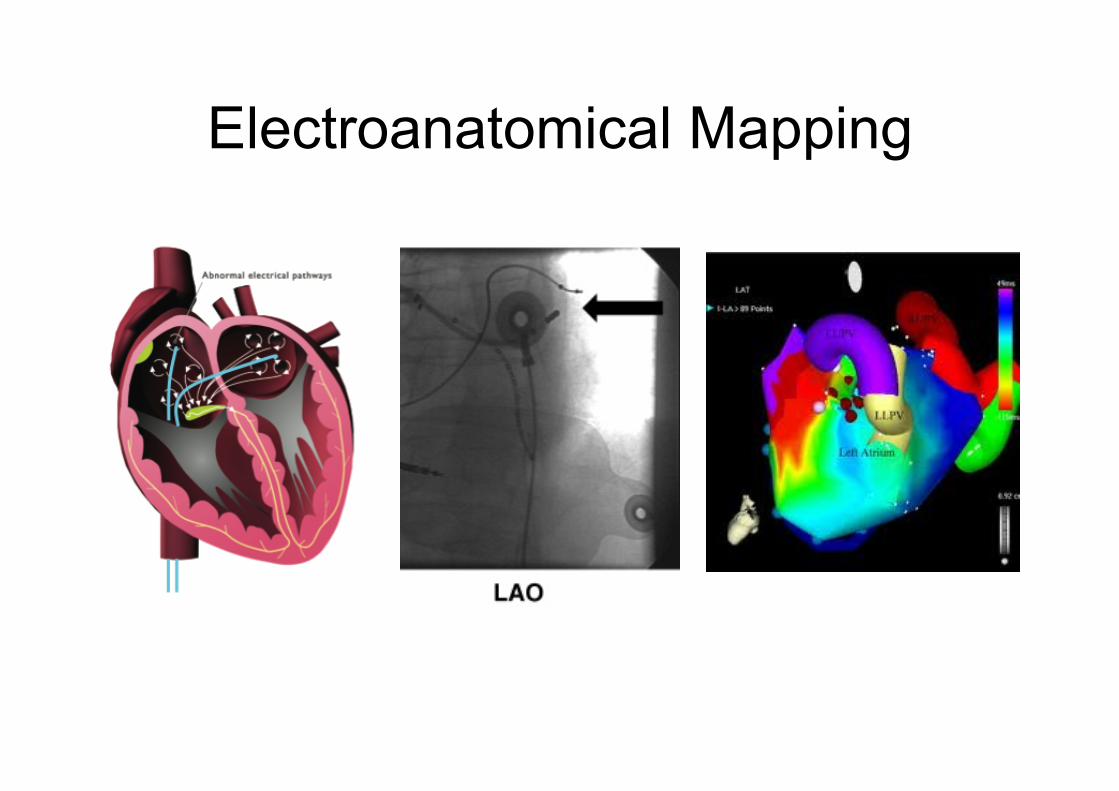
Electroanatomical Mapping
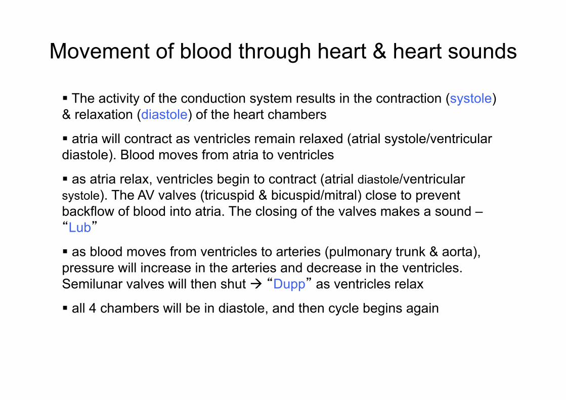
§ The activity of the conduction system results in the contraction (systole) & relaxation (diastole) of the heart chambers
§ atria will contract as ventricles remain relaxed (atrial systole/ventricular diastole). Blood moves from atria to ventricles
§ as atria relax, ventricles begin to contract (atrial diastole/ventricular systole). The AV valves (tricuspid & bicuspid/mitral) close to prevent backflow of blood into atria. The closing of the valves makes a sound – “Lub”
§ as blood moves from ventricles to arteries (pulmonary trunk & aorta), pressure will increase in the arteries and decrease in the ventricles. Semilunar valves will then shut à “Dupp” as ventricles relax
§ all 4 chambers will be in diastole, and then cycle begins again
Movement of blood through heart & heart sounds

Electrocardiography
Cardiac Cycle
• Cardiac cycle refers to all events associated with blood flow through the heart – Systole – contraction of heart muscle – Diastole – relaxation of heart muscle
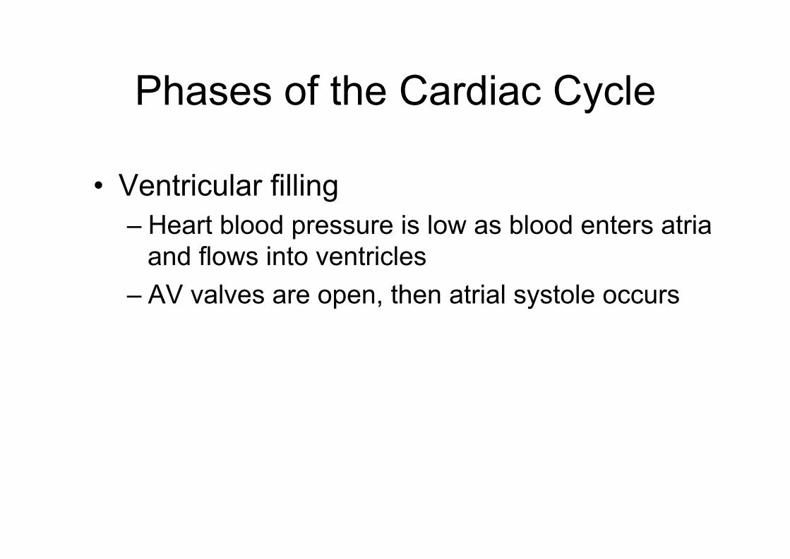
Phases of the Cardiac Cycle
• Ventricular filling – Heart blood pressure is low as blood enters atria
and flows into ventricles – AV valves are open, then atrial systole occurs
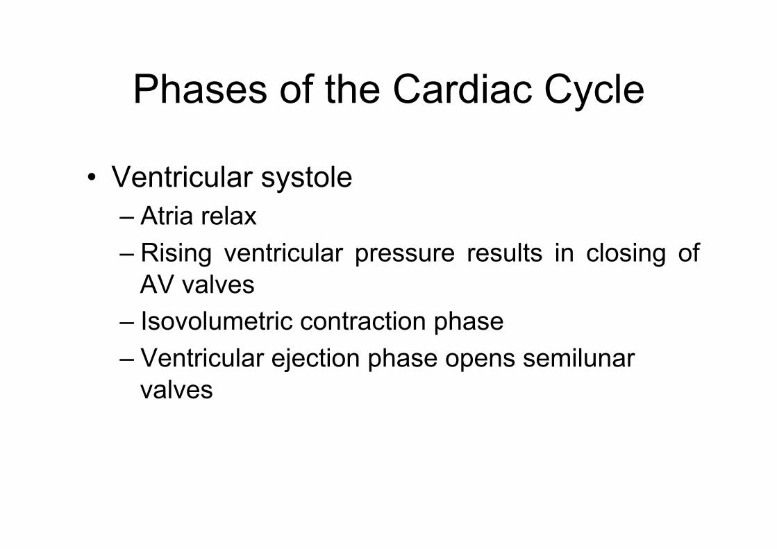
Phases of the Cardiac Cycle
• Ventricular systole – Atria relax – Rising ventricular pressure results in closing of
AV valves – Isovolumetric contraction phase – Ventricular ejection phase opens semilunar
valves
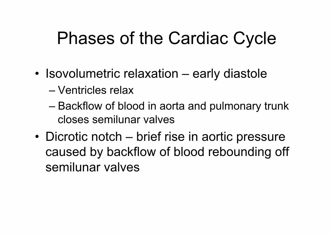
Phases of the Cardiac Cycle
• Isovolumetric relaxation – early diastole – Ventricles relax – Backflow of blood in aorta and pulmonary trunk
closes semilunar valves • Dicrotic notch – brief rise in aortic pressure
caused by backflow of blood rebounding off semilunar valves

Phases of the Cardiac Cycle
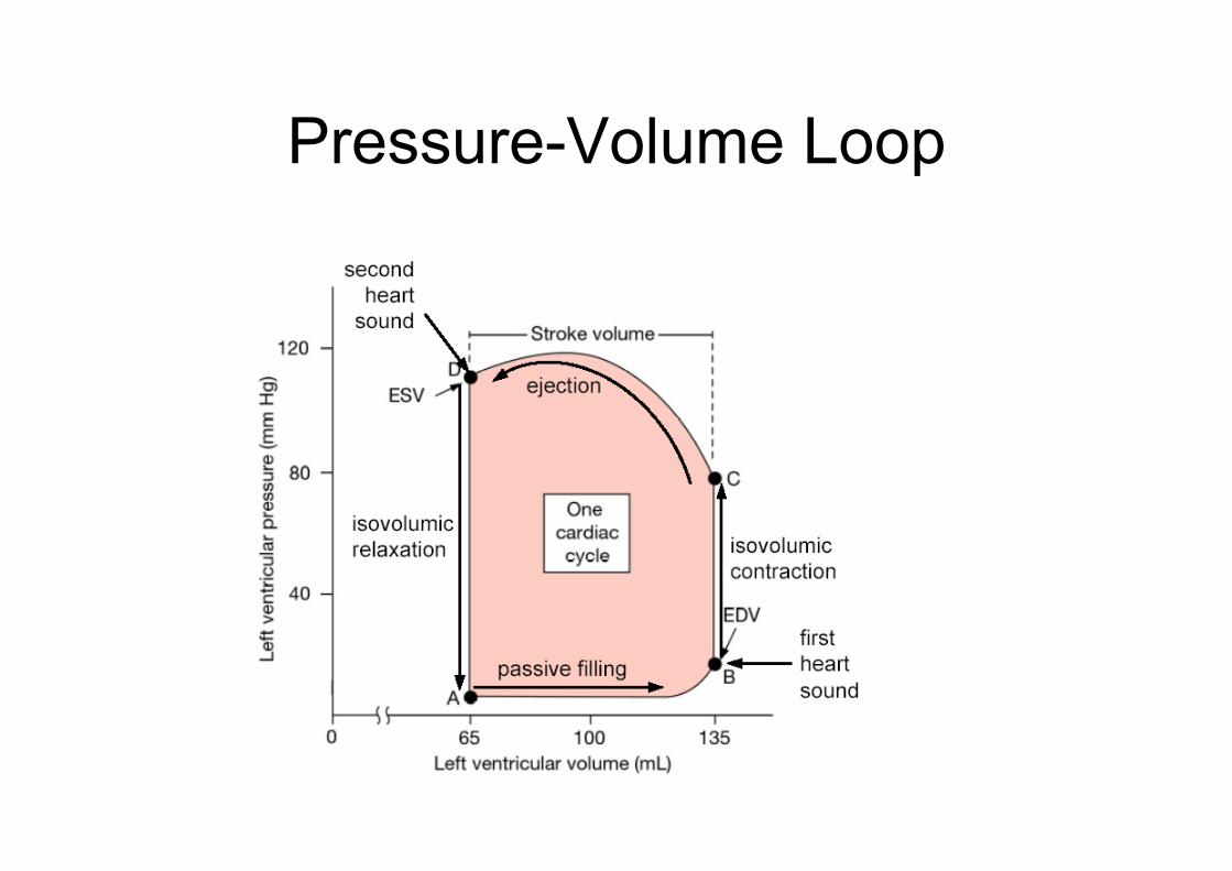
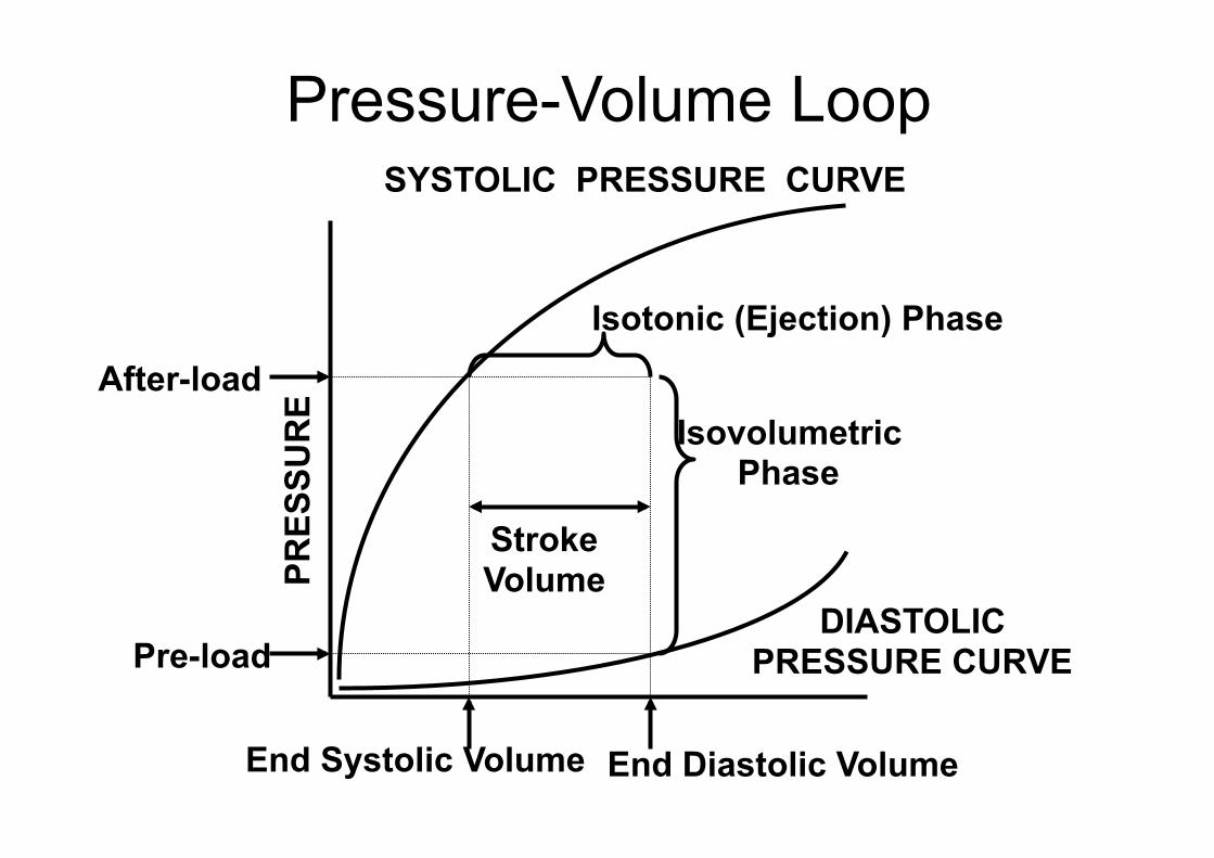
Pressure-Volume Loop
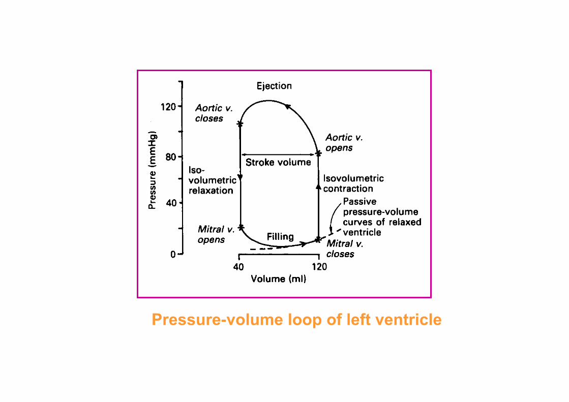
Pressure-volume loop of left ventricle
Cardiac Output (CO) and Reserve
• CO is the amount of blood pumped by each ventricle in one minute
• CO is the product of heart rate (HR) and stroke volume (SV)
• HR is the number of heart beats per minute • SV is the amount of blood pumped out by a
ventricle with each beat • Cardiac reserve is the difference between
resting and maximal CO
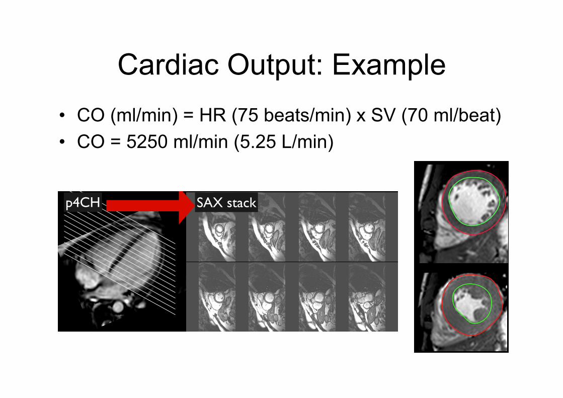
Cardiac Output: Example • CO (ml/min) = HR (75 beats/min) x SV (70 ml/beat) • CO = 5250 ml/min (5.25 L/min)
Left ventricular chamber assessment
quantitative: contiguous stack of short axis slices
• LV Volumes
• Papillary muscles: either included or excluded in LV volumes (report to specify)
• LVOT included in LV volume: the contour is drawn to include the outflow tract to the level of the aortic valve cusps.
• The highest basal slice: can be identified when more than 50% of the blood volume is surrounded by myocardium
Basal descent: As a result of systolic motion of the mitral valve toward the apex (basal descent) care must be taken with the one or two most basal slices. A slice, which contains blood volume at end-diastole may include only left atrium (LA) without LV blood volume at end-systole. The LA can be identified when less than 50% of the blood volume is surrounded by myocardium and the blood volume cavity is seen to be expanding. Some software packages automatically adjust for systolic atrioventricular ring descent.
Simpsons rule of discs Short axis cine stack
���������� �� ������������������������
����� ����� ���p4CH SAX stack
bbb every slice has a defined thickness (8mm)
gap between the slices = 0mm

Regulation of Stroke Volume
• SV = end diastolic volume (EDV) minus end systolic volume (ESV)
• EDV = amount of blood collected in a ventricle during diastole
• ESV = amount of blood remaining in a ventricle after contraction
Factors Affecting Stroke Volume
• Preload – amount ventricles are stretched by contained blood
• Contractility – cardiac cell contractile force due to factors other than EDV
• Afterload – back pressure exerted by blood in the large arteries leaving the heart
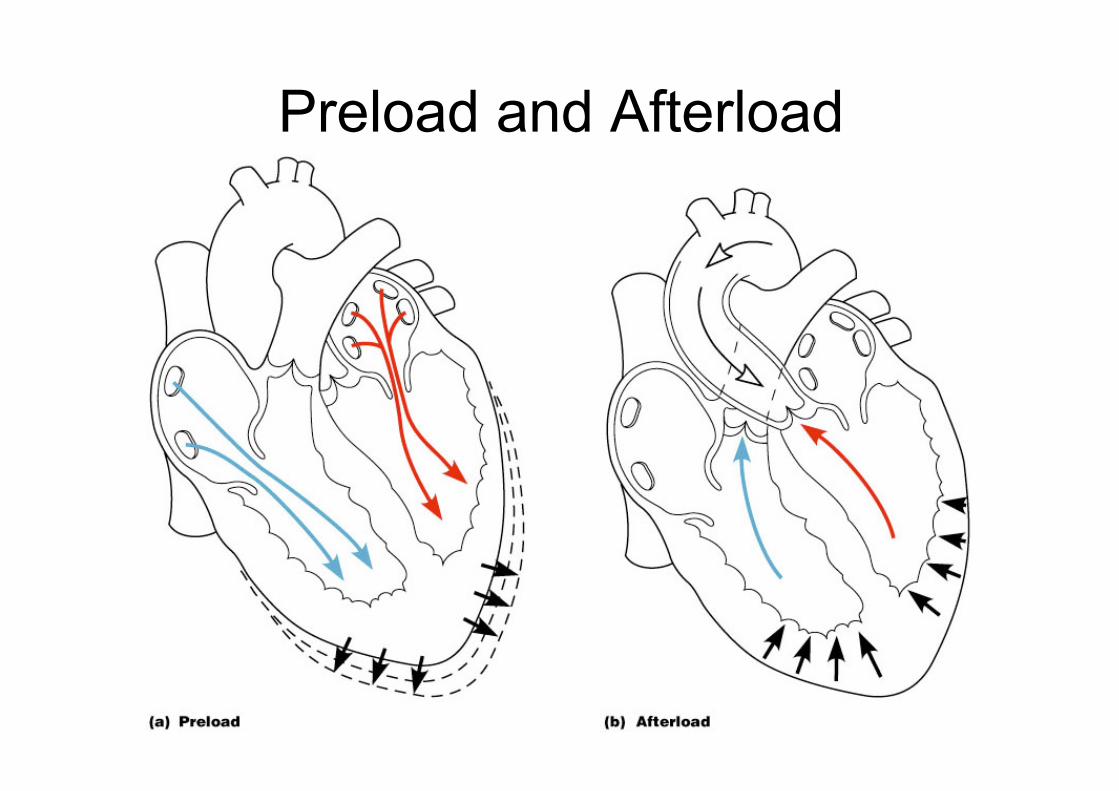
Preload and Afterload
PRES
SUR
E
DIASTOLIC PRESSURE CURVE
SYSTOLIC PRESSURE CURVE
End Diastolic Volume End Systolic Volume
Isovolumetric Phase
Isotonic (Ejection) Phase
Stroke Volume
Pre-load
After-load
Pressure-Volume Loop

Extrinsic Factors Influencing Stroke Volume
• Contractility is the increase in contractile strength, independent of stretch and EDV
• Increase in contractility comes from: – Increased sympathetic stimuli – Certain hormones – Ca2+ and some drugs

• Vessels returning blood to the heart include: – Superior and inferior venae cavae
• Open into the right atrium • Return deoxygenated blood from body cells
– Coronary sinus • Opens into the right atrium • Returns deoxygenated blood from heart muscle (coronary veins)
– Right and left pulmonary veins • Open into the left atrium • Return oxygenated blood from lungs
Major Vessels of the Heart
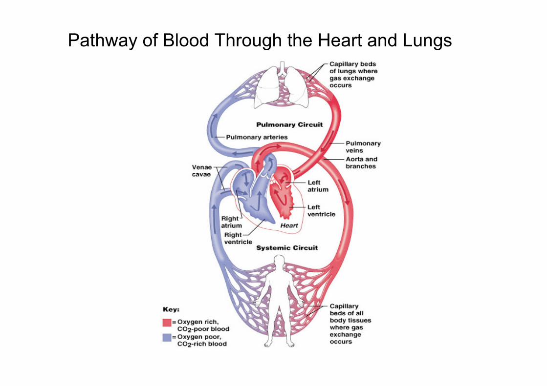
Pathway of Blood Through the Heart and Lungs

Blood Flow Through the Heart

Vessels conveying blood away from the heart include: – Pulmonary trunk
• Carries deoxygenated blood from right ventricle to lungs • Splits into right and left pulmonary arteries
– Ascending aorta • Carries oxygenated blood away from left atrium to body organs • Three major branches
– Brachiocephalic – Left common carotid, – Left subclavian artery
Major Vessels of the Heart
4D MR-Flow

Coronary Circulation
• Coronary circulation – The functional blood supply to the heart
muscle itself – R and L Coronary arteries are 1st branches
off the ascending aorta – Coronary sinus (vein) empties into R. atrium
• Collateral routes ensure blood delivery to heart even if major vessels are occluded
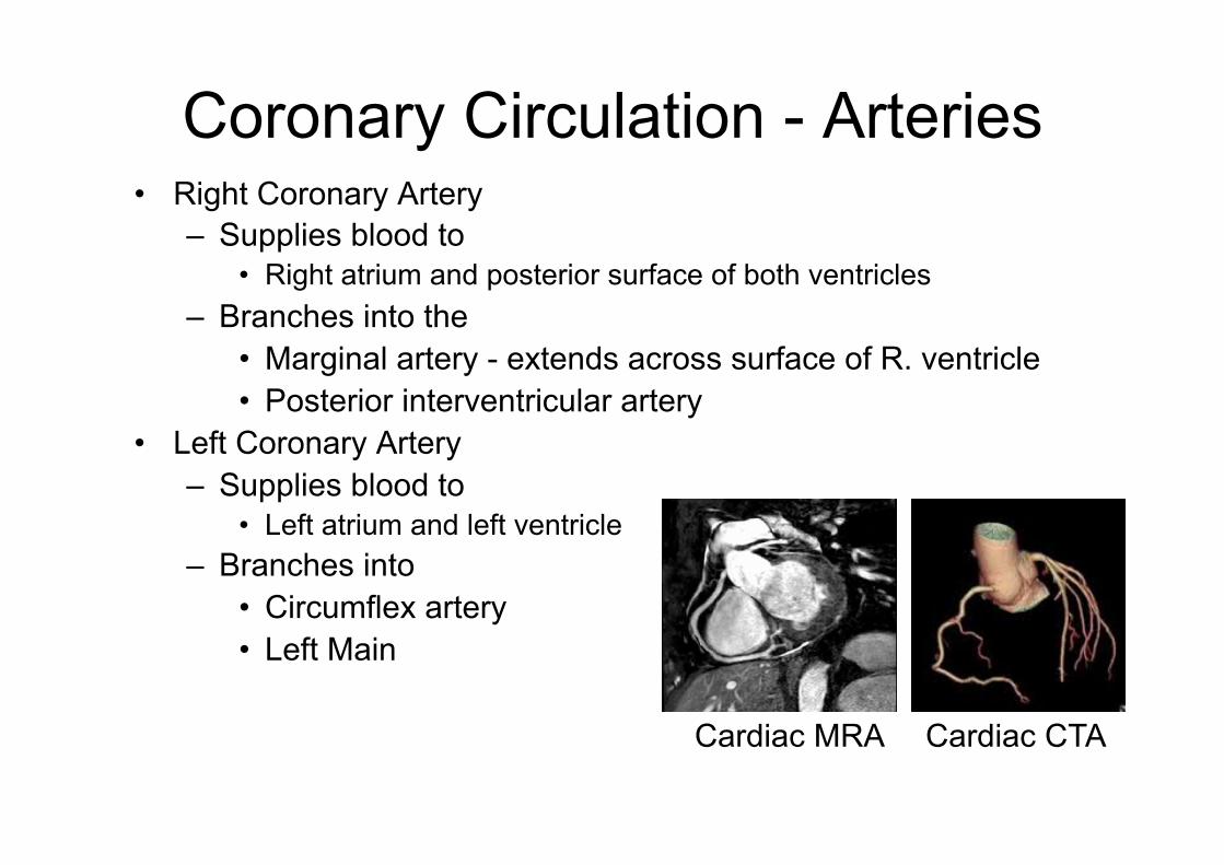
Coronary Circulation - Arteries • Right Coronary Artery
– Supplies blood to • Right atrium and posterior surface of both ventricles
– Branches into the • Marginal artery - extends across surface of R. ventricle • Posterior interventricular artery
• Left Coronary Artery – Supplies blood to
• Left atrium and left ventricle – Branches into
• Circumflex artery • Left Main
Cardiac CTA Cardiac MRA

Coronary Circulation: Arterial Supply
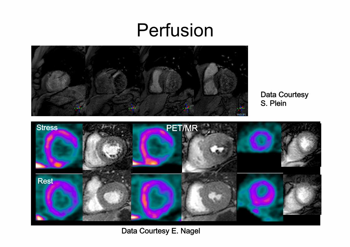
Perfusion
Stress
Rest
Data Courtesy E. Nagel
Data Courtesy S. Plein
PET/MR

Coronary Circulation - Veins Coronary sinus: • Vein that empties into right atrium • Receives deoxygenated blood from:
– Great cardiac vein - on anterior surface – Posterior cardiac vein
• Drains area served by circumflex – Middle cardiac vein
• Drains area served by posterior interventricular artery – Small cardiac vein
• Drains blood from posterior surfaces of right atrium and ventricle
Coronary Vein MRA
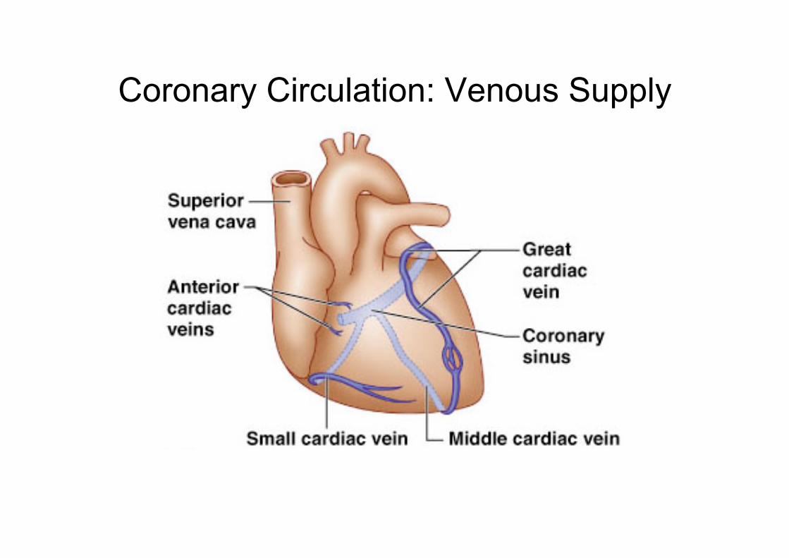
Coronary Circulation: Venous Supply
Learning objectives
• Provide an overview on Heart Anatomy, Conducting System and Physiology
• Provide terminology of the external and internal features of the heart
• Show imaging examples of cardiac anatomy and physiology

Readings • Achilles J. Pappano, Withrow Gil Wier, Matthew
N. Levy; Cardiovascular Physiology; Mosby; 9th edition 2006
• Elaine N. Marieb, Essentials of Human Anatomy and Physiology, Pearson Education ; 10th edition 2010
• Lauralee Sherwood. Human Physiology: From Cells to Systems; Brooks Cole; 8th edition, 2012
• Robert F. Schmidt, Gerhard Thews, Human Physiology, Springer 1989
• Many slides from www.slideworld.org