ethylpyrrole: adding insight to experiment with ab initio ... · of electronic structure...
TRANSCRIPT

Green, J. A., Makhov, D. V., Cole-Filipiak, N. C., Symonds, C.,Stavros, V. G., & Shalashilin, D. V. (2019). Ultrafast photodissociationdynamics of 2-ethylpyrrole: adding insight to experiment with ab initiomultiple cloning. Physical Chemistry Chemical Physics, 21(7), 3832-3841. https://doi.org/10.1039/c8cp06359a
Publisher's PDF, also known as Version of recordLicense (if available):CC BYLink to published version (if available):10.1039/c8cp06359a
Link to publication record in Explore Bristol ResearchPDF-document
This is the final published version of the article (version of record). It first appeared online via Royal Society ofChemistry at10.1039/C8CP06359A . Please refer to any applicable terms of use of the publisher.
University of Bristol - Explore Bristol ResearchGeneral rights
This document is made available in accordance with publisher policies. Please cite only thepublished version using the reference above. Full terms of use are available:http://www.bristol.ac.uk/pure/user-guides/explore-bristol-research/ebr-terms/

3832 | Phys. Chem. Chem. Phys., 2019, 21, 3832--3841 This journal is© the Owner Societies 2019
Cite this:Phys.Chem.Chem.Phys.,
2019, 21, 3832
Ultrafast photodissociation dynamics of2-ethylpyrrole: adding insight to experimentwith ab initio multiple cloning†
James A. Green, *a Dmitry V. Makhov,ab Neil C. Cole-Filipiak, cd
Christopher Symonds,a Vasilios G. Stavros c and Dmitrii V. Shalashilin*a
The ultrafast photodissociation dynamics of 2-ethylpyrrole (2-EP) is simulated in a fully quantum manner
on the S1 and S2 ps* states by the ab initio multiple cloning (AIMC) method. AIMC treats electrons with
accurate electronic structure methods ‘‘on the fly’’, and nuclear dynamics with wavefunction propagation via
a basis set of Ehrenfest trajectory guided Gaussian wavepackets. Total kinetic energy release (TKER) spectra
are produced, as well as velocity map images and N–H dissociation times. These are compared to results
from time-resolved velocity map imaging studies, and the AIMC method is able to provide quantitative
reproduction of experimental data, including dissociation times of 50–80 fs. Novel insight into the dissocia-
tion mechanism is then obtained, with the experimentally obtained time constant shown to be composed of
two components. Firstly, there is a contribution in o50 fs from 2-EP molecules that have sufficient energy
in the N–H stretch coordinate to dissociate almost immediately over the barrier, and this is followed by
a second slower contribution from 2-EP molecules that must sample the potential energy surface
before finding a way around the barrier to dissociate. This two component mechanism is not observed
experimentally due to the temporal widths of the laser pulses obscuring the dynamics in the o50 fs
window, and is shown for the first time via theory. Calculations are also performed on selectively
deuterated 2-EP, demonstrating that AIMC is able to produce a kinetic isotope effect for the dissociation
time constant, and correctly predict a shift to lower energy in the TKER spectrum. The S2 ps* state is
also shown to be unstable with respect to the S1 ps* state, with the N–H dissociation proceeding along
S1 when initially excited to S2. This work demonstrates that the combination of state of the art theory
and experiments can provide unprecedented novel insight into the N–H dissociation mechanism, with
the tantalising prospect of providing insight into more general heteroatom hydride bond dissociation.
1 Introduction
A number of fundamental processes in chemistry and biologyinvolve ultrafast excited state dynamics following photo-absorption,including light harvesting in plants,1 UV photodamage in DNA,2
and photoprotection in melanin pigments.3 Nitrogen containingaromatic heterocycles are found in molecules responsible forthe above processes, and in particular pyrrole is a component ofchlorophyll as well as being present in chromophores of other
important natural compounds such as: vitamin B12, heme,bilirubin, biliverdin, and tryptophan. Due to the prevalence ofpyrrole, in recent years there has been significant interest in itsexcited state dynamics both experimentally4–20 and theoretically.19–40
Whilst a large component of effort has been directed tounderstanding the excited state dynamics of pyrrole, much less wellstudied are its derivatives. This is also an important area to explorehow modification alters its function. Recently, 2-ethylpyrrole (2-EP)has been studied experimentally by H (Rydberg) atom photo-fragment translational spectroscopy (HRA-PTS),37 time-resolvedvelocity map imaging (TR-VMI) and time-resolved ion yield (TR-IY)mass spectrometry41 to examine the effects of ring-substitution onpyrrole. The only theoretical investigations on 2-EP have consistedof electronic structure calculations,37 however dynamics calcula-tions are desirable to further elucidate the photodissociationprocess. Fully quantum dynamics calculations still remain a hugechallenge, however they are possible with ab initio multiplecloning (AIMC),42,43 and in this work we use the method to offer
a School of Chemistry, University of Leeds, Leeds, LS2 9JT, UK.
E-mail: [email protected], [email protected] School of Mathematics, University of Bristol, Bristol, BS8 1TW, UKc Department of Chemistry, University of Warwick, Library Road,
Coventry CV4 7AL, UKd Combustion Research Facility, Sandia National Laboratories, Mail Stop 9055,
Livermore, CA 94551-0969, USA
† Electronic supplementary information (ESI) available. See DOI: 10.1039/c8cp06359a
Received 12th October 2018,Accepted 2nd January 2019
DOI: 10.1039/c8cp06359a
rsc.li/pccp
PCCP
PAPER
Ope
n A
cces
s A
rtic
le. P
ublis
hed
on 3
0 Ja
nuar
y 20
19. D
ownl
oade
d on
2/2
5/20
19 4
:33:
59 P
M.
Thi
s ar
ticle
is li
cens
ed u
nder
a C
reat
ive
Com
mon
s A
ttrib
utio
n 3.
0 U
npor
ted
Lic
ence
.
View Article OnlineView Journal | View Issue

This journal is© the Owner Societies 2019 Phys. Chem. Chem. Phys., 2019, 21, 3832--3841 | 3833
insight to experiment on 2-EP.41 The effect of selective deuterationat the N–H bond has also been studied experimentally,41 so weconsider modelling this as well to illustrate the capability of AIMCto reproduce experimental kinetic isotope effects.
Initially, it is worthwhile to briefly summarise the currentunderstanding of the ultrafast excited state dynamics ofpyrrole, and to contrast and compare to 2-EP. Early electronicstructure work on pyrrole21–24 helped to characterise the lowestenergy excited states, with the seminal work by Sobolewskiet al.22,23 identifying the importance of the low lying ps* stateswith regards to biological photoprotection. These states aredissociative along the N–H stretch coordinate, and provide anultrafast radiationless transfer route through a conical inter-section with the ground state, deactivating potentially reactiveexcited species. There are two low lying ps* states in pyrrolethat have spin symmetry labels 11A2 and 11B1 due to its C2v
molecular symmetry. Transitions from the X1A1 ground state tothe lower energy of these two, the 11A2(ps*) state, are formallyelectric dipole forbidden. However, it can become directlypopulated through vibronic mixing with nearby higher lyingpp* states 11A1 and 11B2 (albeit with low transition cross-sections), or indirectly via internal conversion from the11B2(pp*) state at shorter wavelengths. Electronic structurecalculations for 2-EP revealed an increase in oscillator strengthfor the ground to first excited ps* state transition compared topyrrole,37 which is to be expected due to the reduction inmolecular symmetry to Cs in the anti conformation and C1
in the lowest energy gauche conformer.Early experimental studies on pyrrole observed two H atom
dissociation channels in total kinetic energy release (TKER)spectra: one with a sharp high kinetic energy distribution of Hatoms emitted perpendicular to the transition dipole moment;and one with a broader lower kinetic energy distribution, and amore isotropic emission.4–9 It was posited that the former highkinetic energy dissociation channel was due to rapid N–Hdissociation along the 11A2(ps*) state, followed by conicalintersection with the ground state to produce a pyrrolyl radicalin the 12A2 ground state. The latter lower kinetic energydissociation channel was thought to be due to internal conver-sion to a vibrationally ‘‘hot’’ ground state, followed by dissocia-tion. The high kinetic energy channel is believed to bedominant at longer pump wavelengths, with Wei et al.5 observingapproximately 76% following this route at l = 243.1 nm via theintegration of the kinetic energy spectrum. At shorter pumpwavelengths (l o 218 nm) the lower kinetic energy channel isdominant,7 with 11B2(pp*) populated initially before passingthrough conical intersections and dissociating, with the pyrrolylradical produced in the ground state. The addition of a s donatingethyl group in 2-EP causes the onset of H atom dissociation toappear at longer wavelengths: 267 nm in 2-EP37 compared to254 nm in pyrrole.7 Excitation in the range 248 r l r 263 nmleads to dissociation from the lowest energy ps* state.37
Time-resolved studies on pyrrole provided quantitative measuresof the H atom appearance lifetimes in each of these dissociationchannels.15–20 Lippert et al. conducted the first of these studies byusing TR-IY to obtain time-constants of t1 = 110� 80 fs for the high
kinetic energy channel and t2 = 1.1 � 0.5 ps for the lower kineticenergy channel at l = 250 nm.15 Subsequently, Roberts et al. usedboth TR-VMI and TR-IY to observe a single time constant of 126�28 fs for the high kinetic energy channel at l = 250 nm.18 It waspostulated that tunnelling out of the quasi-bound region of the11A2(ps*) state plays a role at this excitation wavelength, as a timeconstant of 1.4 � 0.3 ps for selectively deuterated pyrrole-d1
dissociation gave a kinetic isotope effect (KIE) of B11. Furtherpump wavelengths were used by Roberts et al. in this study, withexcitation at l = 238 nm yielding a time constant of 46 � 22 fs forundeuterated pyrrole, and 136 � 38 fs for pyrrole-d1. Due to thesmaller KIE (B3), this was attributed to faster, over the barrierdissociation with negligible contribution from tunnelling. Finally,at l = 200 nm a time constant of 52 � 12 fs was observed,attributed to initial population of the 11B2(pp*) state andsubsequent rapid internal conversion to the 11A2(ps*) stateand dissociation. A low kinetic energy feature was observed atthis wavelength, however it had a time-constant of 1.0 � 0.4 nsrather than the picosecond timescale observed by Lippert et al.15 Itwas suggested that the picosecond timescale observed by Lippertet al. could be due to undesired multiphoton dissociative ionisa-tion events, and the nanosecond time-constant was due to C–Hdissociation following internal conversion to the ground state,with the analogous process for N–H having an even longertimescale.18
TR-VMI and TR-IY studies on 2-EP in the 248 r l r 265 nmrange found high kinetic energy H atom appearance lifetimeson the order of 50–80 fs, and selectively deuterated 2-EP-d1 atl = 257 nm had an appearance lifetime of 140 � 20 fs giving aKIE of B2, suggesting that tunnelling does not play a signifi-cant role in the dissociation mechanism. At excitation wave-lengths shorter than 248 nm, an additional low kinetic energyfeature appeared in the H atom TKER spectrum, similarto pyrrole. However, the appearance lifetime was B1.5 ps,intimating it was not due to dissociation following internalconversion to the ground state, and the explanation of a seconddissociation channel involving population of higher-lying singletstates was offered once undesired multiphoton dissociationevents and clustering in the molecular beam were ruled out.41
Previous quantum dynamics calculations on pyrrole haveused reduced dimensionality potential energy surfaces,25–27
surface hopping,29–33 and multiconfigurational time-dependentHartree (MCTDH) with parameterised potential energysurfaces.19,20,34–36 Our own ab initio multiconfigurationalEhrenfest (AI-MCE)44 and ab initio multiple cloning42,43 methodshave also previously been used for dynamics studies onpyrrole.38–40 In the former, using the AI-MCE method, conicalintersections of the 12A2 and 12B1 radical states following Hdissociation were observed, helping to explain the lack ofexperimentally observed pyrrolyl radicals in the 12B1 state.38
In the latter, using the AIMC method, it was suggested thatsome low kinetic energy H atoms are formed in an ultrafastmanner as a result of dissociation where the radical does nottransfer to the ground state immediately.39,40 Both methods usefrozen/fixed width Gaussian basis functions (or equivalentlycoherent states) guided by Ehrenfest trajectories to describe the
Paper PCCP
Ope
n A
cces
s A
rtic
le. P
ublis
hed
on 3
0 Ja
nuar
y 20
19. D
ownl
oade
d on
2/2
5/20
19 4
:33:
59 P
M.
Thi
s ar
ticle
is li
cens
ed u
nder
a C
reat
ive
Com
mon
s A
ttrib
utio
n 3.
0 U
npor
ted
Lic
ence
.View Article Online

3834 | Phys. Chem. Chem. Phys., 2019, 21, 3832--3841 This journal is© the Owner Societies 2019
nuclear wavepacket, and calculate electronic potential energy sur-faces at the centre of these Gaussian basis functions as the simula-tion is running (‘‘on the fly’’). This provides a fully dimensional andaccurate description of nonadiabatic quantum dynamics.
Fully quantum simulations of multiatomic species stillremain a huge challenge in chemical dynamics, and in thepresent work we seek to provide a full description of theultrafast photodissociation dynamics of 2-EP using the AIMCmethod, for rigorous comparison to previously publishedexperimental results.41 The computational details for the AIMCcalculations carried out in this work are described in Section 3,and it is used to study the ultrafast dynamics of 2-EP in Section4, alongside its selectively deuterated form. Initially however, abrief overview of the experimental method used to compare tothe theoretical results is presented in the following section.
2 Experimental details
The experiments for 2-EP used TR-VMI apparatus with tempo-rally delayed femtosecond pump and probe laser pulses toprovide ultrafast time-resolved photochemical measurements.A full description of the apparatus used may be found in ref. 41.
The pump laser pulse was tuned over the wavelength range238 r lpump r 265 nm and the probe pulse was produced at awavelength of lprobe = 243.1 nm to facilitate the 2 + 1 resonanceenhanced multiphoton ionisation (REMPI) of H/D atom photofrag-ments via the two photon allowed 2s ’ 1s transition, andsubsequent one photon ionisation. A cross-correlation/instrumentresponse function at the temporal overlap of pump and probepulses (Dt = 0) was obtained via the TR-IY of Xe+ (by 2 + 10
ionisation). This measurement was fitted to a Gaussian distribu-tion, which yielded a Gaussian instrument response functionGIRF(t) with width parameter sXC = 38 fs. The laser beams werethen focused to intersect a molecular beam containing 2-EP, andthe resulting cationic photoproducts accelerated down a time-of-flight tube and impacted onto a detector selectively gated forthe exclusive detection of H+ (or D+) ions. The design andelectric potentials of the ion optics and detector projected thethree-dimensional photofragment velocity distribution ontothe two-dimensional imaging detector, resulting in a velocitymap image. From the velocity map image, the original three-dimensional photofragment velocity distribution was recon-structed and integrated using the polar onion peeling method,45
resulting in one-dimensional photofragment velocity and angulardistributions. After calibration to the known dissociation energeticsof HBr at 200 nm,46 the velocity distribution was transformed intoa one-dimensional TKER spectrum. Multiple TKER spectra wererecorded at various time delays Dt to produce H/D transients,which were used to obtain appearance lifetimes.
3 Computational details
As mentioned in the introduction, the AIMC method uses frozenGaussian basis functions guided by Ehrenfest trajectories todescribe the nuclear wavepacket, and calculates electronic
potential energy surfaces at the centre of these Gaussian basisfunctions ‘‘on the fly’’ to provide a fully quantum description ofnonadiabatic dynamics. The full numerical details of the methodhas been described elsewhere,42,43 and brief overview of theworking equations is given in the ESI.† For the discussion inthe main text, it suffices to say that the Ehrenfest trajectories thatguide the Gaussian basis incorporate components from allelectronic states involved in the calculation, and hence includenonadiabatic effects at all times. In regions of strong non-adiabatic coupling when an Ehrenfest trajectory is comprisedof an unphysical average of multiple electronic states, a‘‘cloning’’ procedure is applied to expand the basis, mimickingwavepacket splitting. This usually occurs following a non-adiabatic transition with incomplete population transfer, whenthe electronic states with significant population also havediffering gradients.
As with previous AIMC studies,39,40,42 dynamics were simulatedusing a modified version of AIMS-MOLPRO47–49 that incorporatesEhrenfest trajectories. Electronic structure calculations were per-formed using the complete active space self-consistent field(CASSCF) method. The electronic basis set used was Dunning’scc-PVDZ set,50 with one additional diffuse s function, oneadditional set of p functions, and one additional set of dfunctions added to the nitrogen atom; as well as one additionaldiffuse s function, and one additional set of p functions addedto the dissociative hydrogen atom. The active space used has 8electrons in 7 orbitals: three ring p orbitals and two corres-ponding p* orbitals, and the N–H s and corresponding s*orbital. State averaging was performed over three states anddynamics were performed on three states: the ground and twolowest excited singlet states. The width of the Gaussian basisfunctions g was taken to be 4.7 Bohr�2 for hydrogen, 6.6 Bohr�2
for deuterium, 22.7 Bohr�2 for carbon and 19.0 Bohr�2 fornitrogen, as suggested previously.51
Initial positions and momenta for the nuclei were sampledfrom the ground state vibrational Wigner distribution in theharmonic approximation using vibrational frequencies andnormal modes calculated at the same level of CASSCF theoryas above. As in previous AIMC works,39,40,42 excitation fromground to excited state is approximated by projecting thewavefunction onto the desired excited state, which amountsto setting the normalised electronic state amplitude for thedesired excited state to unity for each trajectory, whilst theother electronic state amplitudes are set to zero. It is noted thatthe finer details of initial photoexcitation are not accountedfor completely by this approximation, and there may be somesmall energetic sampling deficiencies that are discussed in thefollowing section. The temporal widths of the pump and probelaser pulses from the photoexcitation are taken into accountwhen calculating time-constants for H dissociation however,the details of which shall also be explained in the followingsection.
The initial number of trajectories, starting adiabatic electronicstate, and number of cloning events for each calculation carriedout in this work are shown in Table 1. Undeuterated 2-EPcalculations are performed with dynamics starting on the
PCCP Paper
Ope
n A
cces
s A
rtic
le. P
ublis
hed
on 3
0 Ja
nuar
y 20
19. D
ownl
oade
d on
2/2
5/20
19 4
:33:
59 P
M.
Thi
s ar
ticle
is li
cens
ed u
nder
a C
reat
ive
Com
mon
s A
ttrib
utio
n 3.
0 U
npor
ted
Lic
ence
.View Article Online

This journal is© the Owner Societies 2019 Phys. Chem. Chem. Phys., 2019, 21, 3832--3841 | 3835
S1 and S2 states, and deuterated 2-EP calculations are per-formed with dynamics starting on only the S1 state. Hereafter,they are referred to with the starting adiabatic electronic statein parenthesis afterwards. The parameters used to determine acloning event are the same for each system, and are explainedin more detail in the ESI.† The smaller number of cloningevents for 2-EP(S2) compared to 2-EP(S1) is due to more completepopulation transfer at conical intersections. The smaller numberof cloning events for 2-EP-d1(S1) compared to 2-EP(S1) is due to thelower frequency of the N–D vibration compared to N–H, meaningfewer conical intersections are encountered along this coordinate.Furthermore, the cloning procedure incorporates a description ofwavepacket splitting which is less likely to occur for D than H as itis heavier. Calculations were run for 350 fs, or until the N–H bondexceeded 4 Å, which was defined as the point of dissociation.All calculations used a timestep of B0.6 fs (2.5 a.u.).
4 Results
In the following section, calculated H/D atom TKER spectra arepresented and compared to experiment. The dissociation energiesobtained from calculation are smoothed with Gaussian functions(with width s = 200 cm�1) to create a curve for the TKER spectrum,as opposed to a stick spectrum of delta functions. This allowsbetter comparison to experiment, and this procedure has beenused before to obtain simulated TKER spectra.39,40 An experi-mental velocity map image relative to the electric component ofthe laser field is reproduced by the 2-EP(S1) calculation using acalculated velocity distribution relative to the molecular axes.
Calculated dissociation times are also presented, in terms ofraw dissociation times from the trajectory data, and following asmoothing procedure to take into account experimental laserpump and probe temporal widths (this procedure has also beencarried out recently52). The smoothing procedure is requiredbecause the simulation has well defined start and end points,however, experimentally these points are ‘‘blurred’’ by thetemporal widths of the laser pulses. The process for thissmoothing procedure is as follows: the pump and probe laserpulses are assumed to be Gaussian in time, with widthsobtained from the experimental GIRF(t) measurement (whichhas width sXC, equal to the cross-correlation/convolution ofpump and probe pulses). The pump and probe laser pulses areboth produced in the same manner, so it is expected that theirtemporal widths will be similar. However, as the probe consists
of a 2 + 1 REMPI of H atom photofragments, this simultaneousabsorption of 3 photons will reduce the width of the Gaussian
that models it by a factor offfiffiffi3p
so that sprobe ¼ spump
� ffiffiffi3p
.
Therefore we have spump = 33 fs and sprobe ¼ 33� ffiffiffi
3p¼ 19 fs,
which reproduce well the experimentally obtained sXC = 38 fs.The raw dissociation times from the trajectory data are then
represented as Gaussian probability distributions, centredaround each raw dissociation time, with width parameterspump. For each point in this distribution, the dissociatingtrajectory will have a probability of being ‘‘probed’’ by anotherGaussian probability distribution with width sprobe. These Gaussianprobability distributions are convoluted and summed for eachtrajectory with associated weights from calculated amplitudes,enabling a smoothed transient to be produced that can be com-pared to experimental data. This smoothed transient is then fitusing the kinetic model employed in experiment18,41 to obtain timeconstants, and these fits are analysed relative to the raw dissocia-tion times to add insight to experimental results. The kinetic modelis given by an exponential rise with H/D-atom appearancelifetime t and time zero correction t0, convoluted with theGaussian instrument response function GIRF(t), multiplied byamplitude A and Heaviside unit step function u(t) (which isequal to 1 for t Z 0 and 0 otherwise)
S(t) = A�GIRF(t) � [(1 � e�(t�t0)/t)u(t)]. (1)
4.1 TKER spectra
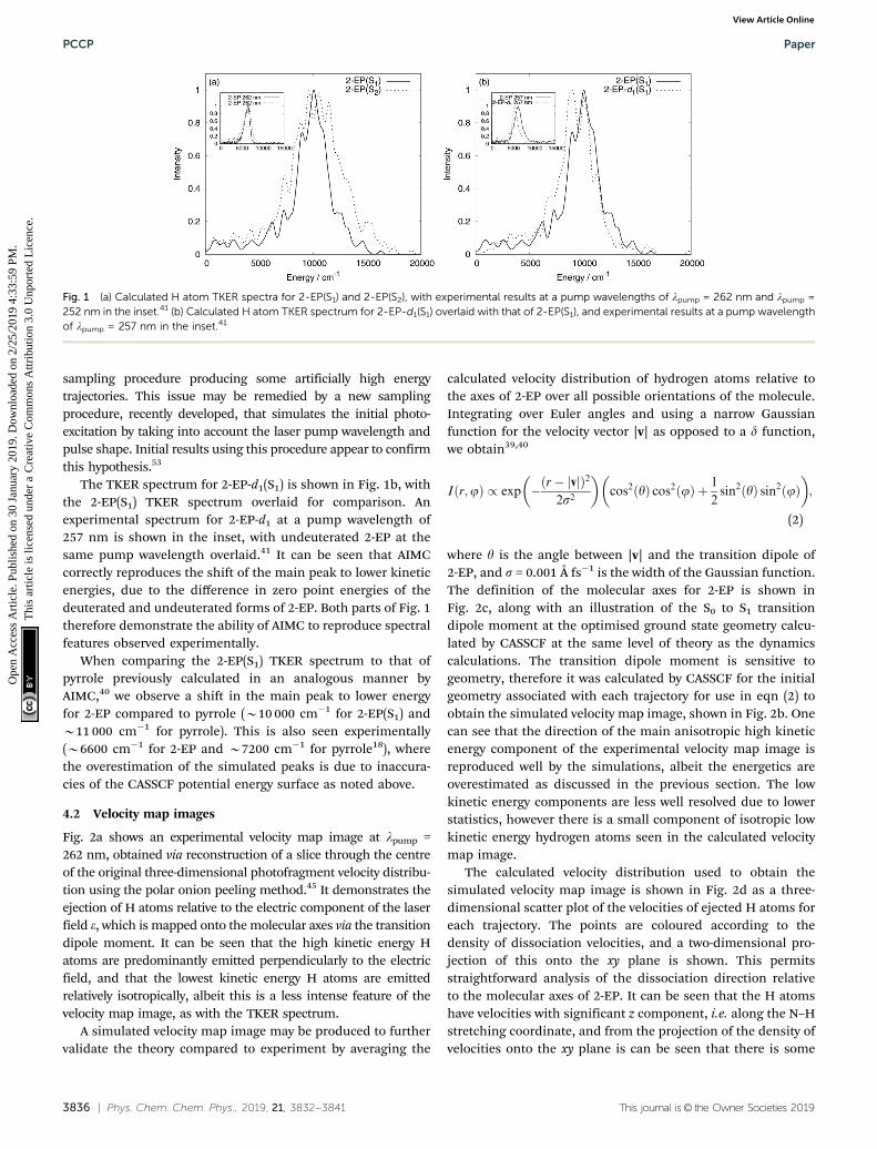
Calculated TKER spectra are shown in Fig. 1, with experimentalTKER spectra shown in the insets,41 and intensity of the mainpeaks maxima set to 1. The theoretical spectra show goodqualitative agreement to those obtained by experiment, withone main peak and some smaller features at lower kineticenergies. Comparing the spectra from S1 and S2 dynamics inFig. 1a, a broader profile of the main peak from 2-EP(S2) than2-EP(S1) is observed. This is also seen experimentally upondecreasing the pump wavelength, where the effect of goingfrom a 262 to 252 nm pump pulse is illustrated in the inset.This indicates that excitation at shorter wavelengths mayinvolve some population of the S2 state initially, rather thanjust the S1 state. Quantitatively, the energy of the main peak is3000–4000 cm�1 larger from simulation than experiment, dueto inaccuracies in the CASSCF potential energy surface. Toattempt to quantify these inaccuracies, we performed multi-reference perturbation theory calculations with multistatecorrections (MS-CASPT2) at the Franck–Condon region of theS1 and S2 potential energy surfaces, and the asymptotic regionof the S0 state (when the N–H distance is equal to 4 Å). Wecalculated the difference in potential energy between these tworegions, and noted that CASSCF overestimated the difference by3750 cm�1 for S1 and 2228 cm�1 for S2 compared to MS-CASPT2.This causes the shift in peak in the TKER spectra, and if it werecomputationally feasible to use a MS-CASPT2 potential energysurface for the dynamics, then the energies would be much morecomparable to experiment. A similar effect has been noted pre-viously with pyrrole.38,39 There are also slightly larger shoulders onthe high energy side than experiment that can be ascribed to the
Table 1 Molecule labels, initial number of trajectories, starting electronicstate, number of cloning events for each calculation carried out in thiswork, calculation time, and percentage of trajectories dissociated by theend of the calculation time
Moleculelabel
Initialtrajectories
Initialadiabaticelectronicstate
Cloningevents
Calculationtime (fs)
Percentagedissociated
2-EP(S1) 600 S1 142 350 62%2-EP(S2) 600 S2 51 350 81%2-EP-d1(S1) 600 S1 18 350 55%
Paper PCCP
Ope
n A
cces
s A
rtic
le. P
ublis
hed
on 3
0 Ja
nuar
y 20
19. D
ownl
oade
d on
2/2
5/20
19 4
:33:
59 P
M.
Thi
s ar
ticle
is li
cens
ed u
nder
a C
reat
ive
Com
mon
s A
ttrib
utio
n 3.
0 U
npor
ted
Lic
ence
.View Article Online
3836 | Phys. Chem. Chem. Phys., 2019, 21, 3832--3841 This journal is© the Owner Societies 2019
sampling procedure producing some artificially high energytrajectories. This issue may be remedied by a new samplingprocedure, recently developed, that simulates the initial photo-excitation by taking into account the laser pump wavelength andpulse shape. Initial results using this procedure appear to confirmthis hypothesis.53
The TKER spectrum for 2-EP-d1(S1) is shown in Fig. 1b, withthe 2-EP(S1) TKER spectrum overlaid for comparison. Anexperimental spectrum for 2-EP-d1 at a pump wavelength of257 nm is shown in the inset, with undeuterated 2-EP at thesame pump wavelength overlaid.41 It can be seen that AIMCcorrectly reproduces the shift of the main peak to lower kineticenergies, due to the difference in zero point energies of thedeuterated and undeuterated forms of 2-EP. Both parts of Fig. 1therefore demonstrate the ability of AIMC to reproduce spectralfeatures observed experimentally.
When comparing the 2-EP(S1) TKER spectrum to that ofpyrrole previously calculated in an analogous manner byAIMC,40 we observe a shift in the main peak to lower energyfor 2-EP compared to pyrrole (B10 000 cm�1 for 2-EP(S1) andB11 000 cm�1 for pyrrole). This is also seen experimentally(B6600 cm�1 for 2-EP and B7200 cm�1 for pyrrole18), wherethe overestimation of the simulated peaks is due to inaccura-cies of the CASSCF potential energy surface as noted above.
4.2 Velocity map images
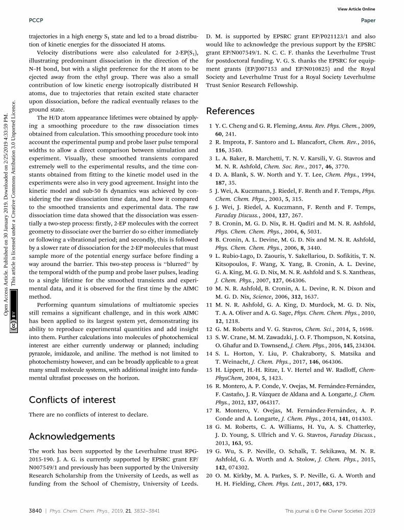
Fig. 2a shows an experimental velocity map image at lpump =262 nm, obtained via reconstruction of a slice through the centreof the original three-dimensional photofragment velocity distribu-tion using the polar onion peeling method.45 It demonstrates theejection of H atoms relative to the electric component of the laserfield e, which is mapped onto the molecular axes via the transitiondipole moment. It can be seen that the high kinetic energy Hatoms are predominantly emitted perpendicularly to the electricfield, and that the lowest kinetic energy H atoms are emittedrelatively isotropically, albeit this is a less intense feature of thevelocity map image, as with the TKER spectrum.
A simulated velocity map image may be produced to furthervalidate the theory compared to experiment by averaging the
calculated velocity distribution of hydrogen atoms relative tothe axes of 2-EP over all possible orientations of the molecule.Integrating over Euler angles and using a narrow Gaussianfunction for the velocity vector |v| as opposed to a d function,we obtain39,40
Iðr;jÞ / exp �ðr� jvjÞ2
2s2
� �cos2ðyÞ cos2ðjÞ þ 1
2sin2ðyÞ sin2ðjÞ
� �;
(2)
where y is the angle between |v| and the transition dipole of2-EP, and s = 0.001 Å fs�1 is the width of the Gaussian function.The definition of the molecular axes for 2-EP is shown inFig. 2c, along with an illustration of the S0 to S1 transitiondipole moment at the optimised ground state geometry calcu-lated by CASSCF at the same level of theory as the dynamicscalculations. The transition dipole moment is sensitive togeometry, therefore it was calculated by CASSCF for the initialgeometry associated with each trajectory for use in eqn (2) toobtain the simulated velocity map image, shown in Fig. 2b. Onecan see that the direction of the main anisotropic high kineticenergy component of the experimental velocity map image isreproduced well by the simulations, albeit the energetics areoverestimated as discussed in the previous section. The lowkinetic energy components are less well resolved due to lowerstatistics, however there is a small component of isotropic lowkinetic energy hydrogen atoms seen in the calculated velocitymap image.
The calculated velocity distribution used to obtain thesimulated velocity map image is shown in Fig. 2d as a three-dimensional scatter plot of the velocities of ejected H atoms foreach trajectory. The points are coloured according to thedensity of dissociation velocities, and a two-dimensional pro-jection of this onto the xy plane is shown. This permitsstraightforward analysis of the dissociation direction relativeto the molecular axes of 2-EP. It can be seen that the H atomshave velocities with significant z component, i.e. along the N–Hstretching coordinate, and from the projection of the density ofvelocities onto the xy plane is can be seen that there is some
Fig. 1 (a) Calculated H atom TKER spectra for 2-EP(S1) and 2-EP(S2), with experimental results at a pump wavelengths of lpump = 262 nm and lpump =252 nm in the inset.41 (b) Calculated H atom TKER spectrum for 2-EP-d1(S1) overlaid with that of 2-EP(S1), and experimental results at a pump wavelengthof lpump = 257 nm in the inset.41
PCCP Paper
Ope
n A
cces
s A
rtic
le. P
ublis
hed
on 3
0 Ja
nuar
y 20
19. D
ownl
oade
d on
2/2
5/20
19 4
:33:
59 P
M.
Thi
s ar
ticle
is li
cens
ed u
nder
a C
reat
ive
Com
mon
s A
ttrib
utio
n 3.
0 U
npor
ted
Lic
ence
.View Article Online

This journal is© the Owner Societies 2019 Phys. Chem. Chem. Phys., 2019, 21, 3832--3841 | 3837
preference for the negative x and y directions, i.e. away from theethyl group. The small contribution of trajectories to the lowkinetic energy isotropic component of Fig. 2b may also be seenfrom those with low velocity in the z-direction. Analysis of theseindividual trajectories reveals that they undergo cloning events andretain some character on the excited states as dissociation occurs.
This effect has also been noted to be responsible for the similarweak intensity low kinetic energy portion of the pyrrole TKERspectrum,39 and the radical formed does eventually relax to theground state.38 This individual analysis of trajectories is possiblebecause they are run separately and combined at the end of thecalculation, see Section S1 in the ESI† for further details.
Fig. 2 (a) Experimental velocity map image at lpump = 262 nm. (b) Simulated velocity map image for 2-EP(S1) with respect to laser pulse polarisation,using calculated transition dipole moments. (c) Definition of molecular axes for 2-EP with an illustration of the S0 to S1 transition dipole moment. (d)Three-dimensional velocity distribution of ejected H atoms from calculation, with points coloured according to their density. A projection of this pointdensity on the xy plane is shown via a contour map.
Paper PCCP
Ope
n A
cces
s A
rtic
le. P
ublis
hed
on 3
0 Ja
nuar
y 20
19. D
ownl
oade
d on
2/2
5/20
19 4
:33:
59 P
M.
Thi
s ar
ticle
is li
cens
ed u
nder
a C
reat
ive
Com
mon
s A
ttrib
utio
n 3.
0 U
npor
ted
Lic
ence
.View Article Online

3838 | Phys. Chem. Chem. Phys., 2019, 21, 3832--3841 This journal is© the Owner Societies 2019
A greater isotropic emission of low kinetic energy H atomsappears experimentally when a second N–H dissociationchannel appears at lpump o 248 nm, with a longer (B1.5 ps)time constant.41 This feature begins to appear predominantlyin the TKER spectrum after 200 fs, and is the main source ofejected H atoms at lpump = 238 nm. The calculations showed nolarge increase in the low kinetic energy feature in the 200–350 fsrange, and is unlikely to appear if the calculations werecontinued beyond 350 fs, as the majority of trajectories havedissociated by this point. This, combined with the fact that theabsorption edge of the higher lying pp* states begins to appearat lpump o 248 nm,41 suggests that initial excitation to the S3 orS4 pp* states occurs at these pump wavelengths rather than S1
(or S2). From that point, the mechanism of the low kineticenergy H dissociation is unclear, as currently dynamics calcula-tions involving additional excited states (hence greater activespace for the CASSCF calculation) for longer times are prohibi-tively expensive for our AIMC method. However, it is possible thatthe mechanism will involve 2-EP molecules that retain some S3/S4
excited state character as they dissociate, similar to the much lessintense low kinetic energy feature seen in 2-EP(S1) dynamics,where the trajectories retain some S1/S2 excited state characteras they dissociate.
4.3 Dissociation kinetics
Turning to the kinetics of dissociation, a cumulative sum of rawdissociation times, alongside smoothed H/D atom appearancetransients with associated fits, and experimental data arepresented in Fig. 3 for 2-EP(S1), 2-EP(S2), and 2-EP-d1(S1). Thesmoothing procedure was performed as outlined in the intro-duction to this section, taking into account the temporal widthsof the pump and probe laser pulses. The smoothed transientsobtained from these probability distributions were then fitusing the kinetic model in eqn (1). A time zero correction wasapplied to the experimental data as determined in ref. 41,whilst it is not necessary (t0 = 0) for the AIMC data. Theexperimental data show non-zero baseline at negative timesdue to ‘‘reverse dynamics’’. In these reverse dynamics, theprobe pulse acts as a pump to photoexcite 2-EP, and thensubsequently provides two photons in a 2 + 10 REMPI schemefor the dissociated H atom, with the third photon providedby the ‘‘pump’’ pulse. This phenomenon has been explainedpreviously in ref. 15 and 18.
Visually, it can be seen that the smoothed transients com-pare well to experimental data, albeit with a slight lifetime shiftfor 2-EP-d1(S1). The lifetimes obtained of 68.2 � 0.5 fs, 72.6 �0.4 fs, and 104.6 � 1.1 fs for 2-EP(S1), 2-EP(S2), and 2-EP-d1(S1),respectively, also compare well to the experimentally obtained55 � 13 fs and 70 � 20 fs at lpump = 262 nm and 252 nm for2-EP, and 140 � 20 fs at lpump = 257 nm for 2-EP-d1. The KIEfrom simulation of B1.5 is slightly less than the experimentallyobtained KIE of B2, however if the calculation was extended fora longer period of time so that more trajectories dissociate it isexpected that this value will increase as undissociated trajec-tories are still in the S1 state. Furthermore, there may be sometunnelling effects that are not accounted for by the calculation
which would give rise to the larger experimental KIE. The effectof tunnelling with regards to dissociation energetics has beenexplored by calculation previously with pyrrole,40 however it hasnot been considered with regards to dissociation kinetics, and
Fig. 3 Raw cumulative sum of dissociation times from trajectories, along-side smoothed H atom appearance transients with associated fits andexperimental data for (a) 2-EP(S1) with experimental data at a pumpwavelength of lpump = 262 nm, (b) 2-EP(S2) with experimental data at apump wavelength of lpump = 252 nm, (c) 2-EP-d1(S1) with experimentaldata at a pump wavelength of lpump = 257 nm.
PCCP Paper
Ope
n A
cces
s A
rtic
le. P
ublis
hed
on 3
0 Ja
nuar
y 20
19. D
ownl
oade
d on
2/2
5/20
19 4
:33:
59 P
M.
Thi
s ar
ticle
is li
cens
ed u
nder
a C
reat
ive
Com
mon
s A
ttrib
utio
n 3.
0 U
npor
ted
Lic
ence
.View Article Online

This journal is© the Owner Societies 2019 Phys. Chem. Chem. Phys., 2019, 21, 3832--3841 | 3839
this could be a subject of a future avenue of research to determinethe influence on the calculated KIE, with the expectation that theKIE would increase to more closely match experiment.
From the lifetimes obtained from the fit, it would appearthat the kinetics of dissociation are similar from the S1 and S2
surfaces. However, when considering the proportion of totaltrajectories that are dissociated by 350 fs (shown in Table 1) it isobserved that a greater proportion have dissociated whendynamics are started on the S2 surface (81%) than the S1 surface(62%). The reason for both of these effects may be seen fromthe averaged electronic state populations for 2-EP(S2) trajec-tories in the first 50 fs, illustrated in Fig. 4. This figure showsthat there is an immediate and significant transfer of popula-tion from S2 to S1 as the calculation begins, indicating that theS2 state is unstable with respect to S1. Around 10 fs later sometrajectories reach the S1–S0 conical intersection, there is steadypopulation transfer, and trajectories begin to dissociate. Theimmediate S2 to S1 transfer produces trajectories in the S1 statewith high energy, and is the reason why a larger percentage of2-EP(S2) trajectories dissociate compared to 2-EP(S1). Further-more, this may also explain the broader profile of the mainpeak in the 2-EP(S2) TKER spectrum compared to 2-EP(S1) inFig. 1a, as the higher energy trajectories result in a broaderdistribution of kinetic energies for the emitted H atoms.
Returning to the transients in Fig. 3, further insight into thedissociation kinetics may be obtained by considering the rawcumulative sum of dissociation times from the trajectories.Examining these a few things may be noticed, particularly inthe sub 50 fs regime, that are masked by the temporal widths ofthe laser pulses experimentally. Firstly, no trajectories dissoci-ate until 14.6 fs for 2-EP(S1), 12.3 fs for 2-EP(S2), and 24.8 fs for2-EP-d1(S1). This is merely a consequence of defining the pointof dissociation as 4 Å, with the delay the time taken for theN–H/D bond to stretch to this distance. Experimentally this willalso occur, as the N–H bond extends over the ps* surfacefollowing the pump laser pulse. However, lack of temporalresolution does not permit this to be observed in the experi-mental transient, and the exact point of dissociation is less easy
to define. The longer time for 2-EP-d1(S1) trajectories to begin todissociate compared to 2-EP(S1) is due to the lower vibrationalfrequency of the N–D bond compared to N–H.
More interestingly, from the initial dissociation pointonwards for the next B40 fs a rapid increase in the numberof trajectories dissociating is observed, as those prepared ingeometries with the correct orientation and sufficient energyin the N–H stretching coordinate to dissociate over the barrierdo so either immediately or following a vibrational period.Fig. S1 in the ESI† illustrates the tendency for trajectories witha large amount of energy in the N–H stretching coordinate todissociate rapidly.
The majority of trajectories that dissociate within the calcu-lation time do so by this mechanism in the first 50 fs for2-EP(S1) and 2-EP(S2), and in the first 70 fs for 2-EP-d1(S1). Thisrapid increase of dissociating molecules is not seen to the sameextent experimentally, instead there is a much smoother risedue to the ‘‘blurring’’ effect of the laser pulses. Following thisinitial rapid rise, the rate of dissociation slows for the rest of thecalculation as the remaining trajectories do not have enoughenergy in the stretching coordinate to immediately dissociate, andmust first sample more of the potential energy surface to find away around the barrier. See Fig. S2 (ESI†) for an example dynamicpotential energy of a trajectory that (a) dissociates via the fastmechanism, and (b) dissociates via the slow mechanism. Itshould be noted that the initial rapid dissociation process maybe slightly overestimated by the sampling technique producingtoo many high energy trajectories, as noted in Section 4.1 with theadditional high energy shoulder on the calculated TKER spectrumrelative to experiment, and this is a further possibility of the causethe time shift of the 2-EP-d1(S1) transient relative to experimentalresults. The recently developed sampling procedure alsomentioned in Section 4.1 may remedy this.53
5 Conclusions
We have used AIMC to simulate the ultrafast photodissociationof 2-EP in a fully quantum manner with dynamics startingon the S1 and S2 ps* states, and deuterated 2-EP with dynamicsstarting on the S1 state. TKER spectra and H/D-atom appear-ance lifetimes from the N–H/D dissociation have beenproduced and compared to time-resolved velocity mapimaging experimental results.41 The TKER spectra reproducethe structure of the main peak and less intense low kineticenergy features from experiment. Comparative featuresobserved experimentally have also been reproduced in eachof the spectra, such as the shift of the main peak to lowerenergies due to deuteration, the lower energy of the peak for2-EP compared to previous AIMC calculations on pyrrole,40 andthe broadening of the main peak due to shorter pump wave-lengths. The latter effect was observed in the 2-EP(S2) calculationscompared to 2-EP(S1), and was explained by considering theaveraged electronic state populations of 2-EP(S2) trajectories.These populations demonstrated that the S2 state is unstable withrespect to S1, leading to rapid transfer from S2 to S1 that producedFig. 4 Averaged electronic state populations for 2-EP(S2) trajectories.
Paper PCCP
Ope
n A
cces
s A
rtic
le. P
ublis
hed
on 3
0 Ja
nuar
y 20
19. D
ownl
oade
d on
2/2
5/20
19 4
:33:
59 P
M.
Thi
s ar
ticle
is li
cens
ed u
nder
a C
reat
ive
Com
mon
s A
ttrib
utio
n 3.
0 U
npor
ted
Lic
ence
.View Article Online
3840 | Phys. Chem. Chem. Phys., 2019, 21, 3832--3841 This journal is© the Owner Societies 2019
trajectories in a high energy S1 state and led to a broad distribu-tion of kinetic energies for the dissociated H atoms.
Velocity distributions were also calculated for 2-EP(S1),illustrating predominant dissociation in the direction of theN–H bond, but with a slight preference for the H atom to beejected away from the ethyl group. There was also a smallcontribution of low kinetic energy isotropically distributed Hatoms, due to trajectories that retain excited state characterupon dissociation, before the radical eventually relaxes to theground state.
The H/D atom appearance lifetimes were obtained by apply-ing a smoothing procedure to the raw dissociation timesobtained from calculation. This smoothing procedure took intoaccount the experimental pump and probe laser pulse temporalwidths to allow a direct comparison between simulation andexperiment. Visually, these smoothed transients comparedextremely well to the experimental results, and the time con-stants obtained from fitting to the kinetic model used in theexperiments were also in very good agreement. Insight into thekinetic model and sub-50 fs dynamics was achieved by con-sidering the raw dissociation time data, and how it comparedto the smoothed transients and experimental data. The rawdissociation time data showed that the dissociation was essen-tially a two-step process: firstly, 2-EP molecules with the correctgeometry to dissociate over the barrier do so either immediatelyor following a vibrational period; and secondly, this is followedby a slower rate of dissociation for the 2-EP molecules that mustsample more of the potential energy surface before finding away around the barrier. This two-step process is ‘‘blurred’’ bythe temporal width of the pump and probe laser pulses, leadingto a single lifetime for the smoothed transients and experi-mental data, and it is observed for the first time by the AIMCmethod.
Performing quantum simulations of multiatomic speciesstill remains a significant challenge, and in this work AIMChas been applied to its largest system yet, demonstrating itsability to reproduce experimental quantities and add insightinto them. Further calculations into molecules of photochemicalinterest are either currently underway or planned; includingpyrazole, imidazole, and aniline. The method is not limited tophotochemistry however, and can be broadly applicable to a greatmany small molecule systems, with additional insight into funda-mental ultrafast processes on the horizon.
Conflicts of interest
There are no conflicts of interest to declare.
Acknowledgements
The work has been supported by the Leverhulme trust RPG-2015-190. J. A. G. is currently supported by EPSRC grant EP/N007549/1 and previously has been supported by the UniversityResearch Scholarship from the University of Leeds, as well asfunding from the School of Chemistry, University of Leeds.
D. M. is supported by EPSRC grant EP/P021123/1 and alsowould like to acknowledge the previous support by the EPSRCgrant EP/N007549/1. N. C. C. F. thanks the Leverhulme Trustfor postdoctoral funding. V. G. S. thanks the EPSRC for equip-ment grants (EP/J007153 and EP/N010825) and the RoyalSociety and Leverhulme Trust for a Royal Society LeverhulmeTrust Senior Research Fellowship.
References
1 Y. C. Cheng and G. R. Fleming, Annu. Rev. Phys. Chem., 2009,60, 241.
2 R. Improta, F. Santoro and L. Blancafort, Chem. Rev., 2016,116, 3540.
3 L. A. Baker, B. Marchetti, T. N. V. Karsili, V. G. Stavros andM. N. R. Ashfold, Chem. Soc. Rev., 2017, 46, 3770.
4 D. A. Blank, S. W. North and Y. T. Lee, Chem. Phys., 1994,187, 35.
5 J. Wei, A. Kuczmann, J. Riedel, F. Renth and F. Temps, Phys.Chem. Chem. Phys., 2003, 5, 315.
6 J. Wei, J. Riedel, A. Kuczmann, F. Renth and F. Temps,Faraday Discuss., 2004, 127, 267.
7 B. Cronin, M. G. D. Nix, R. H. Qadiri and M. N. R. Ashfold,Phys. Chem. Chem. Phys., 2004, 6, 5031.
8 B. Cronin, A. L. Devine, M. G. D. Nix and M. N. R. Ashfold,Phys. Chem. Chem. Phys., 2006, 8, 3440.
9 L. Rubio-Lago, D. Zaouris, Y. Sakellariou, D. Sofikitis, T. N.Kitsopoulos, F. Wang, X. Yang, B. Cronin, A. L. Devine,G. A. King, M. G. D. Nix, M. N. R. Ashfold and S. S. Xantheas,J. Chem. Phys., 2007, 127, 064306.
10 M. N. R. Ashfold, B. Cronin, A. L. Devine, R. N. Dixon andM. G. D. Nix, Science, 2006, 312, 1637.
11 M. N. R. Ashfold, G. A. King, D. Murdock, M. G. D. Nix,T. A. A. Oliver and A. G. Sage, Phys. Chem. Chem. Phys., 2010,12, 1218.
12 G. M. Roberts and V. G. Stavros, Chem. Sci., 2014, 5, 1698.13 S. W. Crane, M. M. Zawadzki, J. O. F. Thompson, N. Kotsina,
O. Ghafur and D. Townsend, J. Chem. Phys., 2016, 145, 234304.14 S. L. Horton, Y. Liu, P. Chakraborty, S. Matsika and
T. Weinacht, J. Chem. Phys., 2017, 146, 064306.15 H. Lippert, H.-H. Ritze, I. V. Hertel and W. Radloff, Chem-
PhysChem, 2004, 5, 1423.16 R. Montero, A. P. Conde, V. Ovejas, M. Fernandez-Fernandez,
F. Castano, J. R. Vazquez de Aldana and A. Longarte, J. Chem.Phys., 2012, 137, 064317.
17 R. Montero, V. Ovejas, M. Fernandez-Fernandez, A. P.Conde and A. Longarte, J. Chem. Phys., 2014, 141, 014303.
18 G. M. Roberts, C. A. Williams, H. Yu, A. S. Chatterley,J. D. Young, S. Ullrich and V. G. Stavros, Faraday Discuss.,2013, 163, 95.
19 G. Wu, S. P. Neville, O. Schalk, T. Sekikawa, M. N. R.Ashfold, G. A. Worth and A. Stolow, J. Chem. Phys., 2015,142, 074302.
20 O. M. Kirkby, M. A. Parkes, S. P. Neville, G. A. Worth andH. H. Fielding, Chem. Phys. Lett., 2017, 683, 179.
PCCP Paper
Ope
n A
cces
s A
rtic
le. P
ublis
hed
on 3
0 Ja
nuar
y 20
19. D
ownl
oade
d on
2/2
5/20
19 4
:33:
59 P
M.
Thi
s ar
ticle
is li
cens
ed u
nder
a C
reat
ive
Com
mon
s A
ttrib
utio
n 3.
0 U
npor
ted
Lic
ence
.View Article Online

This journal is© the Owner Societies 2019 Phys. Chem. Chem. Phys., 2019, 21, 3832--3841 | 3841
21 M. H. Palmer, I. C. Walker and M. F. Guest, Chem. Phys.,1998, 238, 179.
22 A. L. Sobolewski and W. Domcke, Chem. Phys., 2000, 259, 181.23 A. L. Sobolewski, W. Domcke, C. Dedonder-Lardeux and
C. Jouvet, Phys. Chem. Chem. Phys., 2002, 4, 1093.24 B. O. Roos, P.-Å. Malmqvist, V. Molina, L. Serrano-Andres
and M. Merchan, J. Chem. Phys., 2002, 116, 7526.25 V. Vallet, Z. Lan, S. Mahapatra, A. L. Sobolewski and
W. Domcke, Faraday Discuss., 2004, 127, 283.26 V. Vallet, Z. Lan, S. Mahapatra, A. L. Sobolewski and
W. Domcke, J. Chem. Phys., 2005, 123, 144307.27 Z. Lan, A. Dupays, V. Vallet, S. Mahapatra and W. Domcke,
J. Photochem. Photobiol., A, 2007, 190, 177.28 M. Barbatti, M. Vazdar, A. J. A. Aquino, M. Eckert-Maksic
and H. Lischka, J. Chem. Phys., 2006, 125, 164323.29 M. Vazdar, M. Eckert-Maksic, M. Barbatti and H. Lischka,
Mol. Phys., 2009, 107, 845.30 B. Sellner, M. Barbatti and H. Lischka, J. Chem. Phys., 2009,
131, 024312.31 M. Barbatti, J. Pittner, M. Pederzoli, U. Werner, R. Mitric,
V. Bonacic-Koutecky and H. Lischka, Chem. Phys., 2010,375, 26.
32 M. Barbatti and K. Sen, Int. J. Quantum Chem., 2016,116, 762.
33 M. Sapunar, A. Ponzi, S. Chaiwongwattana, M. Malis, A. Prlj,P. Decleva and N. Doslic, Phys. Chem. Chem. Phys., 2015,17, 19012.
34 S. P. Neville and G. A. Worth, J. Chem. Phys., 2014, 140, 034317.35 H. Koppel, E. Gromov and A. Trofimov, Chem. Phys., 2004,
304, 35.36 S. Faraji, M. Vazdar, V. S. Reddy, M. Eckert-Maksic,
H. Lischka and H. Koppel, J. Chem. Phys., 2011, 135, 154310.37 T. N. V. Karsili, B. Marchetti, R. Moca and M. N. R. Ashfold,
J. Phys. Chem. A, 2013, 117, 12067.
38 K. Saita, M. G. D. Nix and D. V. Shalashilin, Phys. Chem.Chem. Phys., 2013, 15, 16227.
39 D. V. Makhov, K. Saita, T. J. Martınez and D. V. Shalashilin,Phys. Chem. Chem. Phys., 2015, 17, 3316.
40 D. V. Makhov, T. J. Martınez and D. V. Shalashilin, FaradayDiscuss., 2016, 194, 81.
41 N. C. Cole-Filipiak, M. Staniforth, N. d. N. Rodrigues,Y. Peperstraete and V. G. Stavros, J. Phys. Chem. A, 2017,121, 969.
42 D. V. Makhov, W. J. Glover, T. J. Martınez and D. V.Shalashilin, J. Chem. Phys., 2014, 141, 054110.
43 D. V. Makhov, C. Symonds, S. Fernandez-Alberti and D. V.Shalashilin, Chem. Phys., 2017, 493, 200.
44 K. Saita and D. V. Shalashilin, J. Chem. Phys., 2012,137, 22A506.
45 G. M. Roberts, J. L. Nixon, J. Lecointre, E. Wrede andJ. R. R. Verlet, Rev. Sci. Instrum., 2009, 80, 053104.
46 P. M. Regan, S. R. Langford, A. J. Orr-Ewing andM. N. R. Ashfold, J. Chem. Phys., 1999, 110, 281.
47 B. G. Levine, J. D. Coe, A. M. Virshup and T. J. Martınez,Chem. Phys., 2008, 347, 3.
48 H.-J. Werner, P. J. Knowles, G. Knizia, F. R. Manby andM. Schutz, WIREs Comput. Mol. Sci., 2012, 2, 242.
49 H.-J. Werner, P. J. Knowles, G. Knizia, F. R. Manby andM. Schutz, et al., Molpro, version 2010.1, a package of ab initioprograms, 2010, see http://www.molpro.net.
50 T. H. Dunning Jr., J. Chem. Phys., 1989, 90, 1007.51 A. L. Thompson, C. Punwong and T. J. Martınez, Chem.
Phys., 2010, 370, 70.52 N. d. N. Rodrigues, N. C. Cole-Filipiak, K. N. Blodgett,
C. Abeysekera, T. S. Zwier and V. G. Stavros, Nat. Commun.,2018, 9, 5188.
53 D. V. Makhov and D. V. Shalashilin, Chem. Phys., 2018,515, 46.
Paper PCCP
Ope
n A
cces
s A
rtic
le. P
ublis
hed
on 3
0 Ja
nuar
y 20
19. D
ownl
oade
d on
2/2
5/20
19 4
:33:
59 P
M.
Thi
s ar
ticle
is li
cens
ed u
nder
a C
reat
ive
Com
mon
s A
ttrib
utio
n 3.
0 U
npor
ted
Lic
ence
.View Article Online