eucaryotic cell surface components
TRANSCRIPT

Vol. 165, No. 1JOURNAL OF BACTERIOLOGY, Jan. 1986, p. 13-200021-9193/86//010013-08$02.00/0Copyright © 1986, American Society for Microbiology
Identification and Properties of Chlamydial Polypeptides That BindEucaryotic Cell Surface Components
TED HACKSTADTDepartment of Health and Human Services, National Institutes of Health, National Institute ofAllergy and InfectiousDiseases, Laboratory of Microbial Structure and Function, Rocky Mountain Laboratories, Hamilton, Montana 59840
Received 28 June 1985/Accepted 20 September 1985
An electroblotting technique was used to identify proteins of Chiamydia that bound surface-radioiodinatedand Triton X-100-solubilized HeLa cell extracts. Two proteins, with apparent molecular masses of 18 and 32kilodaltons (kDa), that bound HeLa cell surface components were identified on Chlamydia trachomatis L2elementary bodies (EBs). Radioiodinated heparin, which disrupts chlamydial association with cultured cells,was also bound by these proteins. These two proteins were found on EBs but were absent or were present inreduced amounts on the noninfectious reticulate bodies. All C. trachomatis strains tested displayed two suchproteins, although the apparent molecular weight of the larger protein varied with serotype in correlation withbiotype and the disease that it caused. Two Chlamydia psittaci strains examined displayed only a single bindingprotein in the range of 17 to 19 kDa. All of the binding proteins stained intensely and distinctively onsilver-stained sodium dodecyl sulfate-polyacrylamide gels and displayed an unusual sensitivity to reducingagents. The 32-kDa protein was not seen and did not bind 12'I-labeled HeLa cell components if the EBs weresolubilized in the presence of 2-mercaptoethanol. The 32-kDa protein was not affected by dithiothreitol,however. Similar to the effect of 2-mercaptoethanol, the 32-kDa protein was not visualized after treatment ofEBs with the protease inhibitors tosyl-phenylalanine chloromethyl ketone (TPCK) or tosyl-lysine chloromethylketone (TLCK). TPCK and TLCK also abolished infectivity as did the alkylating agents N-ethylmaleimide andiodoacetamide, yet the latter two agents did not affect the appearance of the 32-kDa protein. These proteinswere not detected in immunoblots with either rabbit antisera to C. trachomatis L2 EBs or by serum from apatient with lymphogranuloma venereum. The role of these proteins in the interaction of chlamydiae with hostcells is not clear, but the binding of eucaryotic cell surface components and heparin, presence only during theinfectious stage of the life cycle, variation between serotypes in correlation with disease, and sensitivity toreducing agents or protease inhibitors, collectively, suggest a role for these proteins in parasite-hostinteractions.
Chlamydia species are procaryotic obligate intracellularparasites of eucaryotes. Characteristics that distinguishthese bacteria from other obligate intracellular parasites are(i) a complex life cycle that includes an infectious extracel-lular cell type, the elementary body (EB), and a noninfec-tious, intracellular, and replicating cell type, the reticulatebody (RB) (5, 14, 35); (ii) disulfide bonding of outer mem-brane proteins as a mechanism of maintaining structuralstability (4, 18, 19, 30) in the absence of peptidoglycan (2, 16,27, 40); and (iii) replication of the parasite within phago-somes apparently modified by the bacteria to inhibit fusionwith lysosomes (5, 14, 35).There are two species of Chlamydia, C. trachomatis and
C. psittaci. The two species share a number of biologicalproperties but differ antigenically (10) and exhibit only about10% DNA homology (21, 31). There are 15 serotypes withinthe species C. trachomatis (35). These may be grouped intotwo biovars, the strains that cause lymphogranulomavenereum (LGV strains; serotypes Li, L2, and L3) and thenon-LGV-causing strains (non-LGV strains; serotypes A toK) (35). The LGV strains are more invasive pathogens, whilethe infections caused by serotypes A through K are usuallylocalized to mucous membranes. The non-LGV strains canbe subdivided on the basis of the diseases that they cause;serotypes A, B, Ba, and C are associated with endemicblinding trachoma while the remaining serotypes, D to K,are usually associated with urogenital infections or inclusion
conjunctivitis, although more serious infections may occur(35).
In additon to their epidemiology, the C. trachomatisbiovars differ in biological properties observed in vitro. LGVand non-LGV strains differ in their associations with hostcells. Attachment and inclusion formation by non-LGVstrains is greatly enhanced by polycations in the medium orcentrifugation of inocula onto cells (24), while such treat-ments have little effect on LGV association with host cells.The mechanism(s) of chlamydial attachment and internaliza-tion is not yet clear. It has been suggested recently thatchlamydiae, like some viruses and polypeptide hormones,may use a constitutive cellular process such as receptor-mediated endocytosis to gain entry into eucaryotic cells (38).However, in other recent studies it has been found thatendosomes containing chlamydia were not coated withclathrin (43). The internalization of chlamydia, therefore,was felt to differ from the receptor-mediated endocytosis ofviruses and hormones into clathrin-coated vesicles (43).The nature of the ligand on the EB surface that recognizes
and interacts with the host cell surface is also unknown. Iused an electroblotting procedure to demonstrate the asso-ciation of radiolabeled eucarytoic cell components withseparated chlamydial proteins. The chlamydial polypeptidesidentified in this manner exhibited several intriguing biolog-ical properties. These polypeptides were associated onlywith the infectious form of the parasite and varied in subunitmolecular weight among chlamydial serotypes. Variability in
13
14 HACKSTADT
size of these polypeptides directly correlated with the viru-lence properties of the organism.(A preliminary account of this work was presented at the
84th Annual Meeting of the American Society for Microbi-ology, St. Louis, Mo., March 1984).
MATERIALS AND METHODS
Organisms. C. trachomatis strains LGV-434, serotype L2;LGV-404, serotype L3; B/Tw-5/OT, serotype B; D/UW-3/CX, serotype D; G/UW-57/CX, serotype G; H/UW-4/CX,serotype H; and I/UW-12/UR, serotype I, and C. psittacistrains that cause meningopneumonitis and guinea piginclusion conjunctivitis were grown and purified from HeLa229 or L-929 cells as described previously (9). Intrinsicradiolabeling of EBs with 14C-labeled amino acids or[35S]cysteine was also as described previously (11).PAGE. Polyacrylamide gel electrophoresis (PAGE) was
carried out as described by Laemmli (25), except that boththe stacking gel and resolving gel contained 2 mM EDTA.Immunoblotting procedures have been described previously(18).
Binding of eucaryotic cell components. A monolayer ofHeLa 229 cells was rinsed once with Hanks balanced saltsolution and scraped from a 150-cm2 flask with a rubberpoliceman. The cells were washed twice with Hanks bal-anced salt solution by low-speed centrifugation (250 x g, 10min) and suspended in Hanks balanced salt solution andsurface radioiodinated by the lactoperoxidase procedure(29). The 125I-labeled cells were suspended in 50 mM sodiumphosphate-150 mM NaCl-phosphate-buffered saline (PBS;pH 7.4)-1% (vol/vol) Triton X-100 and incubated with con-stant mixing for 2 h at 37°C. The suspension was thencentrifuged at 12,000 rpm for 10 min in a Beckman Microfuge12 (Beckman Instruments, Inc., Fullerton, Calif.), and thesupernatant was saved.Chlamydial EBs were suspended in 2% sodium dodecyl
sulfate (SDS)-10% glycerol-62.5 mM Tris hydrochloride(pH 6.8) and immediately solubilized by immersion in aboiling water bath for 10 min. For some experiments, 4%2-mercaptoethanol (2-ME) was included in this mixture. Thesolubilized EBs were subjected to SDS-PAGE and electro-phoretically transferred to nitrocellulose paper (HAHY;Millipore Corp., Bedford, Mass.) at 27 V/cm and 1.0 A for 2h at 17°C in 25 mM sodium phosphate (pH 7.2). Blocking waswith PBS-0.05% Tween 20 or with PBS plus 3% bovineserum albumin. The nitrocellulose sheets to which chlamyd-ial polypeptides were transferred were placed in plastic bagsand incubated for 2 h in the presence of 2 x 10⁶ cpm of125I-labeled HeLa cell extracts diluted in PBS-0.05% Tween20. This dilution resulted in a final Triton X-100 concentra-tion of about 0.01 to 0.04%. The nitrocellulose was thenremoved from the bag, washed once with PBS-0.05% Tween20 and several times with water, dried, and subjected toautoradiography. In some cases the nitrocellulose sheet wasstained with amido black 10B (18) before autoradiography.
Infectivity determinations. The effect of various proteaseinhibitors and sulfhydryl active agents on infectivity wasdetermined as follows. C. trachomatis L2 EBs were sus-pended in 250 mM sucrose-10 mM sodium phosphate-5 mMglutamate, (SPG; pH 7.2). Soy trypsin inhibitor, chymo-statin, dithiothreitol (DTT), iodoacetamide, and N-ethylmaleimide were made up in sterile distilled water at 10times the final concentrations tested (Table 1) and diluted1:10 in the EB suspension. The remaining inhibitors were
solubilized in absolute ethanol and diluted 1:40 in the EBsuspension to give a final concentration of 2 mM. An equalvolume of absolute ethanol was added to control suspen-sions. This amount of ethanol had no effect on infectivity.The suspensions were incubated for 2 h at 37°C and thenpelleted and washed once with SPG. The EBs were thensuspended and diluted in SPG prior to the inoculation ofHeLa cell monolayers on cover slips for determination ofinclusion-forming units as described previously (15, 18).
RESULTSBinding of HeLa cell components by chlamydial proteins.
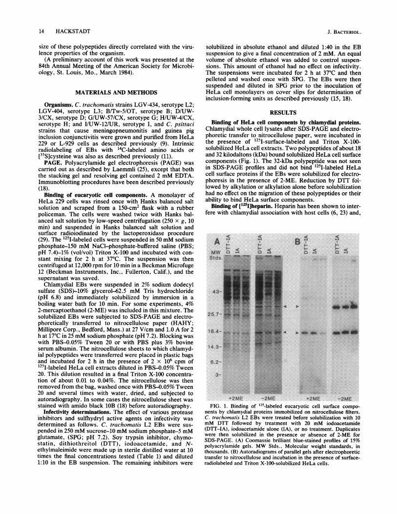
Chlamydial whole cell lysates after SDS-PAGE and electro-phoretic transfer to nitrocellulose paper, were incubated inthe presence of 1251-surface-labeled and Triton X-100-solubilized HeLa cell extracts. Two polypeptides of about 18and 32 kilodaltons (kDa) bound solubilized HeLa cell surfacecomponents (Fig. 1). The 32-kDa polypeptide was not seenin SDS-PAGE profiles and did not bind 125I-labeled HeLacell surface proteins if the EBs were solubilized for electro-phoresis in the presence of 2-ME. Reduction by DTT fol-lowed by alkylation or alkylation alone before solubilizationhad no effect on the migration of these polypeptides or theirability to bind HeLa surface components.
Binding of [12SI]heparin. Heparin has been shown to inter-fere with chlamydial association with host cells (6, 23) and,
A
4-
18.4-
14.3-
B I I
I-~~~~~~-
a4 4
.4 U- SI ES
< > _ ~~~~~~~~~~~...... :.. *l
6.2-
3-
.is. 41t
+2ME -2ME +2ME -2MEFIG. 1. Binding of 125-labeled eucaryotic cell surface compo-
nents by chlamydial proteins immobilized on nitrocellulose filters.C. trachomatis L2 EBs were treated before solubilization with 10mM DTT followed by treatment with 20 mM iodoacetamide(DTT-IA), iodoacetamide alone (IA), or no treatment. Duplicateswere then solubilized in the presence or absence of 2-ME forSDS-PAGE. (A) Coomassie brilliant blue-stained profiles of 15%polyacrylamide gels. MW Stds., Molecular weight standards, inthousands. (B) Autoradiograms of parallel gels after electrophoretictransfer to nitrocellulose and incubation in the presence of surface-radiolabeled and Triton X-100-solubilized HeLa cells.
J. BACTERIOL.
1-41
a 054
CHLAMYDIAL BINDING PROTEINS 15
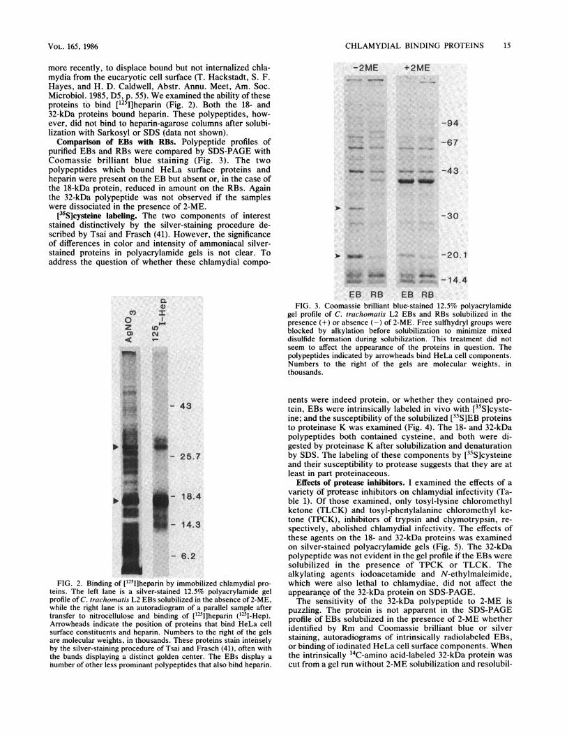
more recently, to displace bound but not internalized chia-mydia from the eucaryotic cell surface (T. Hackstadt, S. F.Hayes, and H. D. Caldwell, Abstr. Annu. Meet, Am. Soc.Microbiol. 1985, D5, p. 55). We examined the ability of theseproteins to bind [125I]heparin (Fig. 2). Both the 18- and32-kDa proteins bound heparin. These polypeptides, how-ever, did not bind to heparin-agarose columns after solubi-lization with Sarkosyl or SDS (data not shown).Comparison of EBs with RBs. Polypeptide profiles of
purified EBs and RBs were compared by SDS-PAGE withCoomassi,e brilliant blue staining (Fig. 3). The twopolypeptides which bound HeLa surface proteins andheparin were present on the EB but absent or, in the case ofthe 18-kDa protein, reduced in amount on the RBs. Againthe 32-kDa polypeptide was not observed if the sampleswere dissociated in the presence of 2-ME.
[35S]cysteine labeling. The two components of intereststained distinctively by the silver-staining procedure de-scribed by Tsai and Frasch (41). However, the significanceof differences in color and intensity of ammoniacal silver-stained proteins in polyacrylamide gels is not clear. Toaddress the question of whether these chlamydial compo-
-2ME +2ME
-94
-67
OW. -43VW _m
-30
-1. -20.1
'I))CO, I
0z lOZ u)
CD CY~
~~~14
-25.7
*18.4
5-14.3
I. 6.2
of ['251]he.arin
teins. The left lane is a silver-stained 12.5% polyacrylamide gel
profile of C. trachomatis L2 EBs solubilized in the absenice of 2-ME,
while the right lane 'is an autoradiogramn of a parallel sample after
transfer to nitrocellulose and binding of [1251]heparin (1251I-Hep).Arrowheads indicate the position of proteins that bind HieLa cell
surface constituents and heparin. Numbers to the right of the gels
are molecular weights, in thousands. These proteins stain intensely
by the silver-staining procedure of Tsai and Frasch (41), oft'en with
the bands displaying a distinct golden ceniter. The EBs display a
number of other less prominant polypeptides that also bihd heparin.
,_0 F- 1 4. 4EB RB EB RB
FIG. 3. Coomassie brilliant blue-stained 12.5% polyacrylamidegel profile of C. trachomatis L2 EBs and RBs solubilized in thepresence (+) or absence (-) of 2-ME. Free sulfhydryl groups wereblocked by alkylation before solubilization to minimize mixeddisulfide formation during solubilization. This treatment did notseem to affect the appearance of the proteins in question. Thepolypeptides indicated by arrowheads bind HeLa cell components.Numbers to the right of the gels are molecular weights, inthousands.
nents were indeed protein, or whether they contained pro-tein, EBs were intrinsically labeled in vivo with [35S]cyste-ine; and the susceptibility of the solubilized [35S]EB proteinsto proteinase K was examined (Fig. 4). The 18- and 32-kDapolypeptides both contained cysteine, and both were di-gested by proteinase K after solubilization and denaturationby SDS. The labeling of these components by [35S]cysteineand their susceptibility to protease suggests that they are atleast in part proteinaceous.
Effects of protease inhibitors. I examined the effects of avariety &f protease inhibitors on chlamydial infectivity (Ta-ble 1). Of those examined, only tosyl-lysine chloromethylketone (TLCK) and tosyl-phertylalanine chloromethyl ke-tone (TPCK), inhibitors of trypsin atid chymotrypsin, re-spectively, abolished chlamydial infectivity. The effects ofthese agents on the 18- and 32-kDa proteins was examinedon silver-stained polyacrylamide gels (Fig. 5). The 32-kDapolypeptide was not evident in the gel profile if the EBs weresolubilized in the presence of TPCK or TLCK. Thealkylating agents iodoacetamide and N-ethylmaleimide,which were also lethal to chlamydiae, did not affect theappearance of the 32-kDa protein on SDS-PAGE.The sensitivity of the 32-kDa polypeptide to 2-ME is
puzzling. The protein is not apparent in the SDS-PAGEprofile of liBs solubilized in the presence of 2-ME whetheridentified by Rm and Coomassie brilliant blue or silverstaining, autoradiograms of intrinsically radiolabeled EBs,or binding of iodinated HeLa cell surface components. Whenthe intrinsically "'C-amino acid-labeled 32-kDa protein wascut from a gel run without 2-ME solubilization and resolubil-
VOL. 165, 1986
'r.
4044urp

16 HACKSTADT
PK: - + - +
4-we
43..
25.7-
18.4-
i14.3-..:,;.. ......
gt.'S6.2-~
de
J. BACTERIOL.
involved in the interaction of chlamydia with host cells, onemight predict that they would be exposed on the EB outersurface. Intact C. trachomatis LGV-434 EBs were thereforesurface iodinated by the lodogen procedure (13) and sub-jected to SDS-PAGE and autoradiography. The 32-kDapolypeptide was not labeled by this radioiodination proce-dure, although the 18-kDa peptide was weakly radiolabeled(Fig. 6).
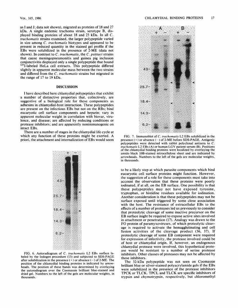
Antigenicity of the 18- and 32-kDa polypeptides. Neither ofthe two polypeptides reacted by immunoblotting with hyper-immune antisera prepared in rabbits against Formalin-killed(or viable; data not shown) C. trachomatis LGV-434 EBs orwith a human LGV convalescent serum (Fig. 7).
Identification of binding proteins on other chiamydialstrains. There are 15 serotypes of C. trachomatis (35). These15 serotypes may be subdivided into three biovars on thebasis of biological properties and diseases caused. I com-pared representative serotypes from each of the three C.trachomatis biovars and two C. psittaci strains (Fig. 8). Allthree of the C. trachomatis biovars exhibited two poly-peptides that bound 125I-labeled HeLa cell surface compo-nents, while the two C. psittaci strains showed only a singlebinding protein. The binding protein of the LGV strains,serotypes L2 and L3, migrated to positions of about 18 and32 kDa, while the equivalent polypeptides of the strains thatcause urogenital infections, serotypes D, G, and H (as well
+2ME -2MEFIG. 4. Autoradiogam of intrinsically [35S]cysteine-labeled C.
trachomatis L2 EBs solubilized for SDS-PAGE in the presence orabsence of 2-ME. Solubilized EBs were treated (+) or not treated(-) with proteinase K (PK; 0.5 mg/ml) for 1 h at 56°C beforeelectrophoresis. Arrowheads indicate the position of chlamydialproteins that bind HeLa cell constituents. Numbers to the left of thegels are molecular weights, in thousands.
ized and run in the presence of 2-ME, a diffuse radioemittingpattern resulted over the molecular mass range of 25 to 32kDa. However, the 32-kDa protein was not affected by DTT.Besides being a reducing agent, 2-ME can also act as achelating agent for divalent cations (28). It seems unlikelythat this activity is responsible for these observations since 5mM EDTA or EGTA [ethylene glycol-bis(P-aminoethylether)-N,N,N',N'-tetraacetic acid] in the solubilizationbuffer had no effect on the 32-kDa proteins (data not shown).
Surface exposure? If the 32- and 18-kDa proteins are
TABLE 1. Effects of protease inhibitors on C. trachomatisinfectivity
Inhibitor (conc.) IFUs (mean ± SEM, x 1o6)"
Control 2.41 ± 0.34Tosyl-arginine methyl ester (2 mM) 2.18 ± 0.09TLCK (2 mM) 0TPCK (2 mM) 0Diazo-norleucine methyl ester (2 mM) 1.91 ± 0.10Phenylmethyl sulfonyl fluoride (2 mM) 2.50 ± 0.06Soy trypsin inhibitor (100 ,ug/ml) 2.92 ± 0.12Chymostatin (100 ,ug/ml) 2.92 ± 0.15DTT (5 mM) 0.35 ± 0.04lodoacetamide (5 mM) 0N-Ethylmaleimide (5 mM) 0
" IFUs, Inclusion-forming units.
o0C.Y-H
MW
_ D
-5z 0
wa~ ~ ~ ~-.43
25.7- ii
-49
18.4 - _
,. lo.,1 4.3 - I"IW.
6.2
--_'t#B-
'.x$
*:,. .:
;X-
_-
+2ME -2 MEFIG. 5. Silver-stained 12.5%o polyacrylamide gel profile of C.
trachomatis L2 EBs solubilized in the presence (+)-or absence (-)of 2-ME, or without 2-ME but with the following: 2 mM TLCK, 2mM TPCK, 5 mM iodoacetamide (IA), 5 mM N-ethylmaleimide(NEM), or 5 mM DTT. Arrowheads indicate the position of theproteins that bind HeLa cell surface components. Numbers to theleft of the gels are molecular weights (MW), in thousands.
CHLAMYDIAL BINDING PROTEINS 17
as I and J; data not shown), migrated as proteins of 18 and 27kDa. A single endemic trachoma strain, serotype B, dis-played binding proteins of about 18 and 23 kDa. In all C.trachomatis strains examined, the larger polypeptide variedin size among C. trachomatis biotypes and appeared to bepresent in reduced quantity in the stained gel profile if theEBs were solubilized in the presence of 2-ME (data notshown). In contrast to C. trachomatis, the C. psittaci strainsthat cause meningopneumonitis and guinea pig inclusionconjunctivitis displayed only a single polypeptide that boundI251-labeled HeLa cell extracts. This polypeptide differedslightly in apparent molecular mass between the two strainsand differed from the C. trachomatis strains but migrated inthe rahge of 17 to 19 kDa.
DISCUSSION
I have described here chlamydial polypeptides that exhibita number of distinctive properties that, collectively, aresuggestive of a biological role for these components asadhesins in chlamydial-host interaction. These polypeptidesare present on the infectious EBs but not on the RBs; bindeucaryotic cell surface components and heparin; vary inapparent molecular weight in correlation with biovar, viru-lence, and disease; are affected by reducing conditions orprotease inhibitors; and are apparently nonimmunogenic onintact EBs.There are a number of stages in the chlamydial life cycle at
which any function of these proteins might be exerted. Apriori, the attachment and internalization of EBs would seem
43-
25.7-X
18.4-
14.3-i
6.2-
FIG. 6. Autoradiogram of C. trachomatis L2 EBs surface la-beled by the lodogen procedure (13) and subjected to SDS-PAGEafter solubilization in the presence (+) or absence (-) of 2-ME. Theposition of the chlamydial binding proteins is indicated by arrow-heads. The position of these bands was determined by overlayingthe autoradiogram over the Coomassie brilliant blue-stained anddried gel. Numbers to the left of the gels are molecular weights, inthousands.
A
4- ...I25.7-~~* .:
B+ -
18.4-
14.3-
6.2- a SB
FIG. 7. Immunoblot of C. trachomatis L2 EBs solubilized in thepresence (+) or absence (-) of 2-ME before SDS-PAGE. Antigenicpolypeptides were detected with rabbit polyclonal antisera to C.trachomatis L2 EBs (A) or human LGV patient serum (B). Positionsof the chlamydial binding proteins were localized by overlaying theamido black 1OB-stained nitrocellulose sheet and are indicated byarrowheads. Numbers to the left of the gels are molecular weights,in thousands.
to be a likely step at which parasite components which bindeucaryotic cell surface proteins might function. However,the suggestion of a role for these components must take intoaccount the observation that these proteins were poorlyiodinated, if at all, on the EB surface. One possibility is thatthese polypeptides may not have exposed tyrosine,tryptophan, or histidine residues available for iodination.Another consideration is that these polypeptides may not besurface exposed until triggered by some close associationwith the host. The resistance of extracellular EBs to theeffects of a number of proteases led us previously to considerthat proteolytic cleavage of some inactive precursor on theEB surface might be required to expose active sites involvedin attachment or penetration (17). Analogy was drawn to theFO protein of paramyxoviruses, of which proteolytic cleav-age is required to activate the hemagglutinating and cellfusion activities of the cleavage product (36, 37). Ifproteolytic cleavage of some EB component were requiredfor expression of infectivity, the protease involved could beof host or chlamydial origin. If, however, an endogenouschlamydial protease were involved, this hypothetical prote-ase would be resistant to a number of serine proteaseinhibitors. Other classes of proteases may not be affected bythese inhibitors.The 32-kDa polypeptide was not seen on Coomassie
brilliant blue or silver-stained polyacrylamide gels if the EBswere solubilized in the presence of the protease inhibitorsTPCK or TLCK. TPCL and TLCK are specific inhibitors oftrypsin and chymotrypsin, respectively, but chloromethyl
VOL. 165, 1986

18 HACKSTADT
B. 125I- Hela
-J j lD a Imco .)E
WbA..
43-
25.7-
18.4-
14.3-
6.2-j
FIG. 8. Strain heterogeneity of the putative chlamydial adhesins.(A) A silver-stained gel profile of C. trachoinatis serotypes L3, L2,D, G, H, and B, and C. psitt-aci strains that cause
meningopneumonitis (Mn) and guinea pig inclusion conjunctivitis(GPIC). Samples were solubilized for electrophoresis without 2-ME.(B) A parallel gel transferred to nitrocellulose, and the potentialadhesins were detected by adsorption of I251-labeled HeLa cellcompotnents (1251-HeLa). Numbers to the left of the gels are molec-ular weights, in thousands.
ketones can also act nonspecifically as alkylating agents (32).However, the alkylating agents N-ethylmaleimide andiodoacetamide did not have this effect under identical con-ditions. It may be that this reflects some difference insolubility or other property that allows access of chloro-methyl ketones, but not other alkylating agents, to essentialsulfhydryl groups. It should also be remembered that theEBs are cells with a number of proteins and other compo-nents exposed on their surface. It is probably not surprisingthat inhibitors with different specificities might decreaseinfectivity by acting at different sites on the same or differentproteins which may function independently or in concert tomediate the interactions of parasite and host.The 18- and 32-kDa proteins share some properties, yet
the precise relationship between the two is not yet under-stood. Several observations are inconsistent with the 32-kDaprotein being a disulfide-linked dimer of the 18-kDa protein,including the following: (i) the apparent molecular mass ofthe larger band varied among serogroups while that of the18-kDa protein varied little, (ii) re-electrophoresis of the32-kDa protein after excision from polyacrylamide gels andsolubilization with 2-ME did not give a fragment of 18 kDa(data not shown), (iii) the amount of the 18-kDa protein onSDS-PAGE did not obviously increase after 2-ME solubili-zation, and (iv) the effect was limited to 2-ME and not DTT.Differential effects of dithiols versus monothiols on enzyme
activity have been described previously (26). I, too, haveseen differential effects of 2-ME versus DTT on the 32-kDaprotein as well as on the 'Oxidation of glutamate by EBs invitro (18). It is unclear, however, whether these differencesare due to some effect on an enzymatic activity or surfacestructure in general.
The sensitivity of the binding activity of the 32-kDaprotein to 2-ME cannot be adequately explained from theresults presented here, but, the fact that it occurs in anorganism that depends on disulfide bonding for structuralstability and therefore requires reduction or disulfide ex-change for differentiation suggests a possible mechanism ofregulating activity through disulfide interactions.The polypeptides described here were found not to be
reactive by immunoblot analysis or radioimmunoprecipita-tion with rabbit antisera to Formalin-killed EBs (data notshown). The rabbit antisera used here did not neutralizeinfectivity. In most cases, antisera produced in rabbits toEBs have neutralized poorly or not at all (8, 20, 33; H. D.Caldwell, personal communication; T. Hackstadt, unpub-lished data). Assigning a role for these proteins awaits theproduction of specific antibodies to address function. Whilethe function of these proteins is still not known, it should beconsidered that it would be to the parasites advantage ifproteins important in its interaction with the host werenonimmunogenic. Protection when it does occur is typespecific (1, 7, 42). At least two surface proteins of C.trachomatis have been shown to have type-specific epitopes:the major outer membrane protein (12) and a 27- to 32-kDaprotein described by Sacks et al. (34). The latter protein isclose to the size of one of the proteins that I describe here.The polypeptides described here varied in apparent molec-ular weight with biovar or epidemiology, but antigenic orstructural analysis of these proteins will be required todetermine the extent of variation that occurs.The ability of SDS and heat-denatured chlamydial
polypeptides, after transfer to nitrocellulose, to bind HeLacell surface proteins is surprising in itself. If these proteinsdo indeed have a function in the chlamydial life cycle andassociation with eucaryotic cell components is part of thatfunction, the domains of these polypeptides which mediateinteraction with host proteins remain active after denatur-ation by heat and SDS and transfer to nitrocellulose.Whether primary structure alone is enough to mediate theinteractions seen here or Whether some higher order struc-ture remains after these treatments is unknown. An addi-tional possibility is that these proteins might possess othermoieties, carbohydrates or lipids, for example, that mayfunction in the recognition by HeLa cell components. Thenature of the interaction is therefore open to question. Theobservation that the polyanionic heparin also binds to theseproteins suggests that charge may be involved. Indeedsurface charge and hydrophobicity have been shown to varyamong C. trachomatis serotypes (39). The variation of thesebinding proteins among groups of C. trachomatis that vary inbiological properties make the localization of these proteinseven more necessary. If these are surface exposed, theycould conceivably contribute to the biological differencesamong strains.
Adsorption of radiolabeled, detergent-solubilized bacterialproteins to eucaryotic cells have identified binding proteinsof Mycoplasma pneumoniae (22) and Treponema pallidum(3). The procedure described here differs in that the bacterialproteins were separated by SDS-PAGE and electroelutedonto nitrocellulose before incubation in the presence ofradiolabeled, Triton X-100-solubilized eucaryotic cell com-ponents. Using this technique, I was successful in identify-ing two polypeptides that vary in quantity between theinfectious and noninfectious forms of the life cycle and inapparent molecular weight between serotypes that differ inbiological properties. Whether these proteins, indeed, rep-resent chlamydial adhesins awaits more detailed analysis,
A. AgNO30
J. BACTERIOL.

CHLAMYDIAL BINDING PROTEINS 19
but they possess enough properties that correlate withbiology and virulence to suggest that they are at least worthyof further study.
ACKNOWLEDGMENTS
I thank John Swanson for suggesting the electroblotting techniqueused here. Helpful discussions with Harlan Caldwell are greatlyappreciated. Thanks also to Jim Simmons and Bob Cole for technicalassistance and to Susan Smaus for preparing the manuscript.
LITERATURE CITED
1. Alexander, E. R., S. P. Wang, and J. T. Grayston. 1967. Furtherclassification of TRIC agents from ocular trachoma and othersources by the mouse toxicity prevention test. Am. J.Ophthalomol. 63:1469-1478.
2. Barbour, A. G., K.-I. Amano, T. Hackstadt, L. Perry, and H. D.Caldwell. 1982. Chlainydia trachornatis has penicillin-bindingproteins but not detectable muramic acid. J. Bacteriol.151:420-428.
3. Baseman, J. B., and E. C. Hayes. 1980. Molecular characteriza-tion of receptor binding proteins and immunogens of virulentTreponema palliduin. J. Exp. Med. 151:573-586.
4. Bavoil, P., A. Ohlin, and J. Schachter. 1984. Role of disulfidebonding in outer membrane structure and permeability in Chla-mydia trachomatis. Infect. Immun. 44:479-485.
5. Becker, Y. 1978. The chlamydiae: molecular biology of procar-yotic obligate parasites of eucaryotes. Microbiol. Rev. 42:274-306.
6. Becker, Y., E. Hochberg, and Z. Rakay-Jones. 1969. Interactionof trachoma elementary bodies with host cells. Israel J. Med.Sci. 5:121-124.
7. Bell, S. D., J. C. Snyder, and E. S. Murray. 1959. Immunizationof mice against toxic doses of homologous elementary bodies oftrachoma. Science 130:626-627.
8. Blyth, W. A., P. Reeve, D. M. Graham, and J. Taverne. 1962.The production of antisera that neutralize inclusion blennor-rhoea virus. Br. J. Exp. Pathol. 43:340-343.
9. Caldwell, H. D., J. Kromhout, and J. Schachter. 1981. Purifica-tion and partial characterization of the major outer membraneprotein of Chlamydia tracliolnatis. Infect. Immun. 31:1161-1176.
10. Caldwell, H. D., C. C. Kuo, and G. E. Kenny. 1975. Antigenicanalysis of chlamydiae by two-dimensional immunoelectropho-resis. I. Antigenic heterogeneity between C. trachoinatiis and C.psittaci. J. Immunol. 115:963-968.
11. Caldwell, H. D., and L. J. Perry. 1982. Neutralization ofChlamydia trach/omatis infectivity with antibodies to the majorouter membrane protein. Infect. Immun. 38:745-754.
12. Caldwell, H. D., and J. Schachter. 1982. Antigenic analysis ofthe major outer membrane protein of C/llamvdia spp. Infect.Immun. 35:1024-1031.
13. Fraker, P. J., and J. C. Speck, Jr. 1978. Protein and cellmembrane iodinations with a sparingly soluble chloramide.1,3,4,6-tetrachloro-3a, 6ca-diphenylglycoluril. Biochem. Bio-phys. Res. Commun. 80:849-857.
14. Friis, R. 1972. Interaction of L-cells and Chlalnydia psittlaci:entry of the parasite and host repsonses to its development. J.Bacteriol. 110:706-721.
15. Furness, G., D. M. Graham, and P. Reeve. 1960. The titration oftrachoma and inclusion blennorrhoea viruses in cell cultures. J.Gen. Microbiol. 23:613-619.
16. Garrett, A. J., M. J. Harrison, and G. P. Manire. 1974. A searchfor the bacterial mucopeptide component, muramic acid, in
Ch/amnydia. J. Gen. Microbiol. 80:315-318.17. Hackstadt, T., and H. D. Caldwell. 1985. Effect of proteolytic
cleavage of surface-exposed proteins on infectivity of Chla-mnydia tr-acho,nahtis. Infect. Immun. 48:546-551.
18. Hackstadt, T., W. J. Todd, and H. D. Caldwell. 1985. Disulfide-mediated interaction of the chlamydial major outer membraneprotein: role in the differentiation of chlamydiae? J. Bacteriol.161:25-31.
19. Hatch, T. P., I. Allen, and J. H. Pearce. 1984. Structural andpolypeptide differences between envelopes of infective andreproductive life cycle forms of Chllamiivdia spp. J. Bacteriol.157:13-20.
20. Hilleman, M. R. 1945. Immunological studies on the psittacosis-lymphogranuloma group of viral agents. J. Infect. Dis.76:96-114.
21. Kingsbury, D. T., and E. Weiss. 1968. Lack of deoxyribonucleicacid homology between species of the genus Chlanzydia. J.Bacteriol. 96:1421-1423.
22. Krause, D. C., and J. B. Baseman. 1982. Mvcoplasmna pneuiemo-niae protein that selectively bind to host cells. Infect. Immun.37:382-386.
23. Kuo, C.-C., and J. T. Grayston. 1976. Interaction of Chlamiivdiatrachlomatis organisms and HeLa 229 cells. Infect. Immun.13:1103-1109.
24. Kuo, C.-C., S.-P. Wang, and J. T. Grayston. 1973. Effect ofpolycations, polyanions, and neuraminidase on the infectivity oftrachoma-inclusion conjunctivitis and lymphogranulomavenereum organisms in HeLa cells: sialic acid residues aspossible receptors for trachoma-inclusion conjunctivitis. Infect.Immun. 8:74-79.
25. Laemmli, U. K. 1970. Cleavage of structural proteins during theassembly of the head of bacteriophage T4. Nature (London)227:680-685.
26. Lee, J. J., and M. J. Fasco. 1984. Metabolism of vitamin K andvitamin K 2,3-epoxide via interaction with a common disulfide.Biochemistry 23:2246-2252.
27. Manire, G. P., and A. Tamura. 1967. Preparation and chemicalcomposition of the cell walls of mature infectious dense forms ofmeningopneumonitis organisms. J. Bacteriol. 94:1178-1183.
28. McMichael, J. C., and J. T. Ou. 1977. Metal ion dependence ofa heat-modifiable protein from the outer membrane of Eshlie-richia coli upon sodium dodecyl sulfate-gel electrophoresis. J.Bacteriol. 132:314-320.
29. Morrison, M. 1974. The determination of the exposed proteinson membranes by the use of lactoperoxidase. MethodsEnzymol. 32:103-109.
30. Newhall, W. J., V, and R. B. Jones. 1983. Disulfide-linkedoligomers of the major outer membrane of chlamydiae. J.Bacteriol. 154:998-1001.
31. Peterson, E. M., and L. M. de la Maza. 1983. Characterization ofchlamydia DNA by restriction endonuclease cleavage. Infect.Immun. 41:604-608.
32. Redelman, D., and D. Hudig. 1980. The mechanism of cell-mediated cytoxicity. 1. Kiling of murine cytotoxic T lympho-cytes requires cell surface thiols and activated proteases. J.Immunol. 124:870-878.
33. Reeve, P., and D. M. Graham. 1962. A neutralizaion test fortrachoma and inclusion blennorrhoea viruses grown in HeLacell cultures. J. Gen. Microbiol. 27:177-180.
34. Sacks, D. L., T. R. Rota, and A. B. McDonald. 1978. Separationand partial characterization of a type-specific antigen fromChl/invdia trachomtiauis. J. Immunol. 121:204-208.
35. Schachter, J., and H. D. Caldwell. 1980. Chlamydiae. Annu.Rev. Microbiol. 34:285-309.
36. Scheid, A., and P. W. Choppin. 1974. Identification of biologicalactivities of Paramyxovirus glycoproteins. Activation of cellfusion, hemolysis, and infectivity by proteolytic cleavage of aninactive precursor protein of Sendai virus. Virology 57:475-490.
37. Scheid, A., and P. WV. Choppin. 1976. Protease activationmutants of Sendai virus. Activation of biological properties byspecific proteases. Virology 69:265-277.
38. Soderlund, G., and E. Kihlstrom. 1982. Physicochemical surfaceproperties of different serotypes of Clhlamvisdia Irachomaiitis andtheir interaction with mouse fibroblasts. Infect. Immun.36:893-899.
39. Soderlund, G., and E. Kihlstrom. 1983. Effect of methylamineand monodansylcadaverine on the susceptibility of McCoy cellsto Chlamnvdia Irachoinalis infection. Infect. Immun. 40:534-541.
40. Tamura, A., and G. P. Manire. 1967. Preparation and chemicalcomposition of the cell membranes of developmental reticulate
VOL. 165, 1986

20 HACKSTADT
forms of meningopneumonitis organisms. J. Bacteriol.94:1184-1188.
41. Tsai, C.-M., and C. E. Frasch. 1982. A sensitive silver stain fordetecting lipopolysaccharides in polyacrylamide gels. Anal.Biochem. 119:115-119.
42. Wang, S. P., J. T. Grayston, and E. R. Alexander. 1%7.
Trachoma vaccine studies in monkeys. Am. J. Ophthalmol.63:1615-1630.
43. Ward, M. E., and A. Murray. 1984. Control mechanismsgoverning the infectivity of Chlamydia trachomatis for HeLacells: mechanisms of endocytosis. J. Gen. Microbiol.130:1765-1780.
J. BACTERIOL.