eunethta joint action 3 wp4 · figure 6.4: 3d print vs. standard; outcome: proportion of outliers...
TRANSCRIPT

Dec2015 ©EUnetHTA, 2015. Reproduction is authorised provided EUnetHTA is explicitly acknowledged 1
EUnetHTA Joint Action 3 WP4
Version 1.4, 17th
April 2019
This report is part of the project / joint action ‘724130 / EUnetHTA JA3’ which has
received funding from the European Union’s Health Programme (2014-2020)
Rapid assessment of other technologies using the HTA Core Model® for Rapid Relative Effectiveness Assessment
Custom-made or customisable 3D printed implants and cutting guides versus non-3D printed standard implants and cutting guides for improving outcome in patients
undergoing knee, maxillofacial, or cranial surgery
PROJECT ID: OTCA11

Custom-made 3D printed implants and cutting guides in knee, maxillofacial, or cranial surgery
EUnetHTA Joint Action 3 WP4 2
DOCUMENT HISTORY AND CONTRIBUTORS
Version Date Description
V1.0 01/12/2018 First draft.
V1.1 20/12/2018 Input from co-author has been processed.
V1.2 01/02/2019 Input from dedicated reviewers has been processed.
V1.3 06/03/2019 Input from external experts and manufacturer(s) has been processed.
V1.4 15/04/2019 Input from medical editor has been processed.
Disclaimer
The assessment represents a consolidated view of the EUnetHTA assessment team members
and is in no case the official opinion of the participating institutions or individuals.
EUnetHTA Joint Action 3 is supported by a grant from the European Commission. The sole re-
sponsibility for the content of this document lies with the authors, and neither the European
Commission nor EUnetHTA are responsible for any use that may be made of the information
contained therein.
Assessment team
Author(s) DEFACTUM – Social & Health Services and Labour Market, Denmark
Lotte Groth Jensen
Claus Løvschall
Anne Marie Ladehoff Thomsen
Gitte Valentin
Bettina Wulff Risør
Co-Author(s) OSTEBA - Office for Health Technology Assessment, Basque Country, Spain
Iñaki Gutierrez-Ibarluzea
Lorea Galnares-Cordero
Gaizka Benguria-Arrate
Dedicated Reviewer(s)
Belgian Health Care Knowledge Centre (KCE), Belgium
Former Agency for Quality and Accreditation in Health Care and Social Welfare (AAZ), Croatia - from 01/01/2019 Ministry of Health (MoH), Croatia
National Institute of Pharmacy and Nutrition (NIPN), Hungary
Gesundheit Österreich (GÖG), Austria
Project manager
Former Agency for Quality and Accreditation in Health Care and Social Welfare (AAZ), Croatia - from 01/01/2019 Ministry of Health (MoH), Croatia

Custom-made 3D printed implants and cutting guides in knee, maxillofacial, or cranial surgery
EUnetHTA Joint Action 3 WP4 3
Consultation of the draft Rapid Assessment
External experts Dirk Leonhardt, Chief Technician at Department of Dentistry and Oral Health, at Aarhus University, Denmark.
Constantinus Politis, Full Professor & Chairperson Oral & Maxillofacial Surgery, at University Hospitals Leuven, Belgium.
Manufacturer(s) V 1.2
(factual accuracy check)
Johnson & Johnson, Materialise and Raomed.
Medical editor V1.3 Nextgenediting
Conflicts of interest
All authors, co-authors, dedicated reviewers and external experts involved in the production of this
assessment have declared they have no conflicts of interest in relation to the technology and
comparator assessed according to the EUnetHTA Declaration of Interest and Confidentiality Un-
dertaking (DOICU) statement except one external expert, Professor Constantinus Politis. He de-
clares a financial or another relationship with a Developing and/or Producing and/or Distributing
Organisation (DPDO) for the technology or comparators undergoing assessment, and thus has a
conflict of interest according to the EUnetHTA guidelines for handling conflicts of interest. Profes-
sor Constantinus Politis acted as Head and Chair of a research group working on development
and validation of surgical tools and image-based solutions in oromaxillofacial surgery. Among
others, KLS Martin funds this research group. There is no single contract between professor Con-
stantinus Polits and KLS Martin, and there is no commercial relationship between the surgical
department and KLS Martin. He has no other conflicts of interest related to the topic of 3D printed
custom-made and customisable implants and surgical guides. According to the EUnetHTA guide-
lines for handling conflicts of interest, the involvement of Professor Constantinus Polits as an ex-
ternal expert is acceptable for commenting on the draft assessment.
How to cite this assessment
Please, cite this assessment as follows:
DEFACTUM, Osteba. Custom-made or customisable 3D printed implants and cutting guides ver-
sus non-3D printed standard implants and cutting guides for improving outcome in patients un-
dergoing knee, maxillofacial, or cranial surgery. EUnetHTA Project ID: OTCA11. 2019.
This document is available on the EUnetHTA website.

Custom-made 3D printed implants and cutting guides in knee, maxillofacial, or cranial surgery
EUnetHTA Joint Action 3 WP4 4
TABLE OF CONTENTS
DOCUMENT HISTORY AND CONTRIBUTORS ............................................................................ 2
TABLE OF CONTENTS ................................................................................................................... 4
LIST OF TABLES AND FIGURES .................................................................................................. 5
LIST OF ABBREVIATIONS ............................................................................................................. 6
1 SUMMARY OF RELATIVE EFFECTIVENESS OF 3D PRINTED CUSTOM-MADE OR CUSTOMISABLE IMPLANTS AND CUTTING GUIDES VERSUS NON-3D PRINTED STANDARD IMPLANTS AND CUTTING GUIDES .................................................. 9 1.1 SCOPE ................................................................................................................................. 9 1.2 INTRODUCTION ...................................................................................................................... 9 1.3 METHODS ............................................................................................................................. 9 1.4 RESULTS ............................................................................................................................ 10 1.5 DISCUSSION ....................................................................................................................... 13 1.6 CONCLUSION ...................................................................................................................... 13
2 SCOPE ...................................................................................................................................... 14
3 METHODS AND EVIDENCE INCLUDED ................................................................................ 16 3.1 ASSESSMENT TEAM ............................................................................................................ 16 3.2 SOURCE OF ASSESSMENT ELEMENTS ................................................................................... 16 3.3 SEARCH ............................................................................................................................. 16 3.4 STUDY SELECTION .............................................................................................................. 18 3.5 DATA EXTRACTION AND ANALYSES ....................................................................................... 18 3.6 QUALITY RATING ................................................................................................................. 19 3.7 DEVIATIONS FROM PROJECT PLAN ........................................................................................ 19
4 DESCRIPTION AND TECHNICAL CHARACTERISTICS OF TECHNOLOGY (TEC) ............ 20 4.1 RESEARCH QUESTIONS ....................................................................................................... 20 4.2 RESULTS ............................................................................................................................ 20
5 HEALTH PROBLEM AND CURRENT USE OF THE TECHNOLOGY (CUR) ......................... 33 5.1 RESEARCH QUESTIONS ......................................................................................................... 33 5.2 RESULTS ............................................................................................................................ 33
6 CLINICAL EFFECTIVENESS (EFF) ......................................................................................... 39 6.1 RESEARCH QUESTIONS ....................................................................................................... 39 6.2 RESULTS ............................................................................................................................ 39
7 SAFETY (SAF) .......................................................................................................................... 50 7.1 RESEARCH QUESTIONS ....................................................................................................... 50 7.2 RESULTS ............................................................................................................................ 50
8 DISCUSSION ............................................................................................................................ 53
9 CONCLUSION .......................................................................................................................... 57
10 REFERENCES ......................................................................................................................... 58
APPENDIX 1: METHODS AND DESCRIPTION OF THE EVIDENCE USED .............................. 63
APPENDIX 2: CHECKLIST FOR POTENTIAL ETHICAL, ORGANISATIONAL, PATIENT AND SOCIAL AND LEGAL ASPECTS .................................................................. 79
APPENDIX 3: TEC DOMAIN - 3D PRINTERS FOR IMPLANTS AND CUTTING GUIDES.................................................................................................................................... 81

Custom-made 3D printed implants and cutting guides in knee, maxillofacial, or cranial surgery
EUnetHTA Joint Action 3 WP4 5
LIST OF TABLES AND FIGURES
Tables
Table 1.1: Summary of findings table for 3D printed implants and cutting guides ......................... 12
Table 2.1: Scope according to population, intervention, comparison, outcomes, and study
design analysis ......................................................................................................................... 14
Table 4.1: Commonly used biomaterials in clinical use. Modified from Williams (11) ................... 23
Table 4.2: Type of materials and comparators* included in this report (based on the evidence
from the included articles) ........................................................................................................ 24
Table 4.3: Custom-made implants and custom-made surgical guides for cranio-maxillofacial
surgery and orthopaedic traumatology surgery ....................................................................... 25
Table 4.4: Custom-made or customisable 3D printed implants and cutting guides in patients
undergoing knee, maxillofacial, or cranial surgery. Shown are the manufacturers, products,
locations, type of products and materials used ........................................................................ 28
Table 6.1: ROBIS results on selected systematic reviews ............................................................. 40
Table 6.2: Cochrane risk of bias results on RCTs ......................................................................... 40
Table 8.1: Meta-analysis regarding alignment in 3D surgery vs. standard. Results from RCTs
and SRs (Thienpont et al. (55)) ................................................................................................ 54
Table A.1: Characteristics and extraction table of randomised controlled studies ........................ 68
Table A.2: Characteristics and extraction table of other relevant studies ...................................... 73
Table A.3: List of on-going studies with custom-made or customisable 3D printed implants and
cutting guides for knee, maxillofacial or cranial surgery .......................................................... 75
Table A.4: GRADE profile. 3D-print technology compared to standard instrumentation in knee
surgery ..................................................................................................................................... 76
Table A.5: Summary table characterising the applicability of a body of studies ............................ 78 Table A.6: 3D printing solutions and use ....................................................................................... 83
Figures
Figure 3.1: Flow chart of systematic literature search ................................................................... 18 Figure 4.1: Process of 3D guide or prosthesis production. Modified from KCE, 2018 (3) ............. 22 Figure 6.1: Hip-knee-ankle angle, mechanical axis, coronal femoral angle, and coronal tibial
angle. Source: Jan Hejle, anatomi.dk ...................................................................................... 41 Figure 6.2: 3D print vs. standard; outcome: proportion of outliers (>3°), hip-knee-ankle angle .... 42 Figure 6.3: 3D print vs. standard; outcome: proportion of outliers (>3°), coronal femur ................ 42 Figure 6.4: 3D print vs. standard; outcome: proportion of outliers (>3°), coronal tibial angle ........ 43 Figure 6.5: Tibial slope angle. Source: Jan Hejle, anatomi.dk ....................................................... 43 Figure 6.6: 3D print vs. standard; outcome: proportion of outliers (>3°), tibial slope ..................... 44 Figure 6.7: Femoral rotational angle. Source: Jan Hejle, anatomi.dk ............................................ 44 Figure 6.8: 3D print vs. standard; outcome: proportion of outliers (>3°), femoral rotation ............. 44 Figure 6.9: 3D print vs. standard; outcome: absolute deviation in degrees, hip-knee-ankle
angle ......................................................................................................................................... 45 Figure 6.10: 3D print vs. standard; outcome: operating time, patients undergoing knee
reconstruction ........................................................................................................................... 46 Figure 6.11: 3D print vs. standard; outcome: Oxford Knee Score, patients undergoing knee
reconstruction ........................................................................................................................... 48
Custom-made 3D printed implants and cutting guides in knee, maxillofacial, or cranial surgery
EUnetHTA Joint Action 3 WP4 6
LIST OF ABBREVIATIONS
ABS Acrylonitrile butadiene styrene
AM Additive manufacturing
CAD Computer-aided design
CDSR Cochrane Database of Systematic Reviews
CI Confidence interval
CT Computed tomography
CUR Current use of the technology
DARE The Database of Abstracts of Reviews of Effects
DC Decompressive craniectomy
DLF Direct laser forming
DMLS Direct metal laser sintering
DOICU Declaration of interest and confidentiality undertaking
EBM Electron beam melting
EDP Environmentally degradable polymer
EFF Clinical effectiveness
ETH Ethical
EULAR European League Against Rheumatism
EUnetHTA European Network for Health Technology Assessment
FDA Food and Drug Administration
FDM Fused deposition modelling
GCS Glasgow coma scale
GRADE Grading of Recommendations, Assessments, Development and Evaluation
HA-reinforced Hydroxyapatite reinforced
HDPE High-density polyethylene
HIPS High-impact polystyrene
HRQoL Health-related quality of life
ICD International Classification of Diseases
ICH Intracranial hypertension
ICU Intensive care unit
KSS Knee Society Score
LEG Legal
MeSH Medical Subject Headings
MJM Multi-jet modelling
NRS Non-randomised studies
NT Navigation template
OA Osteoarthritis
OHIP Oral Health Impact Profile
ORG Organisational
OSCC Oral squamous cell carcinoma

Custom-made 3D printed implants and cutting guides in knee, maxillofacial, or cranial surgery
EUnetHTA Joint Action 3 WP4 7
P[PF-co-EG] Poly(propylene fumarate-co-ethylene glycol)
P(AN-co-allyl sulfonate)
Poly(acrylonitrile-co-allyl sulfonate)
P(GEMA- sulfate) Poly(glucosyloxyethyl methacrylate) sulfate
P(MMA-co-HEMA) Poly(methyl methacrylate-co-2-hydroxyethyl methacrylate)
P(NIPAAm-co-AAc) Poly(N-isopropylacrylamide-co-acrylic acid)
P(NIPAAm-co-EMA) Poly (N-isopropyl acrylamide-co-ethyl methacrylate)
PAAm Polyacrylamide
PBO Polybutylene oxide
PC Polycarbonate
PCL Polycaprolactone
PEEK Polyether ether ketone
PEG Polyethylene glycol
PEG±CDs Polyethylene glycol-modified carbonaceous dots
PEG-g-P(AAm-co- vamine)
Polyethylene glycol-grafted-polyacrylamide-co-vamine
PES Polyester
PHB Polyhydroxybutyrate
PICO Patient-Intervention-Comparison-Outcome
PLA Polylactic acid
PLGA Poly(lactic acid-co-glycolic acid)
PLLA Poly-L-lactic acid
PMMA Poly(methyl methacrylate)
PMPGs Patient-matched positioning guides
PNVP Poly(N-vinylpyrrolidone)
PP Polypropylene
PPSF Polyphenylsulfone
PRISMA Preferred Reporting Items for Systematic Reviews and Meta-analyses
PS Polystyrene
PSI Patient-specific instruments
PTFE Polytetrafluoroethylene
PVA Poly(vinyl alcohol)
PVAc Poly(vinyl acetate)
QALYs Quality-adjusted life years
QoL Quality of life
RA Rheumatoid arthritis
RCT Randomised controlled trial
REA Relative Effectiveness Assessment
RevMan Review Manager
ROBIS Risk of Bias in Systematic Reviews
RP system Rapid prototyping system
SAF Safety

Custom-made 3D printed implants and cutting guides in knee, maxillofacial, or cranial surgery
EUnetHTA Joint Action 3 WP4 8
SIGN Scottish Intercollegiate Guidelines Network
SLA Stereolithography
SLM Selective laser melting
SLS Selective laser sintering
SOC Social
SR Systematic review
SSRO Sagittal split ramus osteotomy
TBI Traumatic brain injury
TEC Technical characteristics of the technology
TKA Total knee arthroplasty
TIJ Thermal inkjet printing
TNM Tumour-node-metastasis
UHMWPE Ultra-high molecular weight polyethylene
UV Ultraviolet
VAS Visual analogue scale
WOMAC Western Ontario and McMaster Universities Osteoarthritis Index

Custom-made 3D printed implants and cutting guides in knee, maxillofacial, or cranial surgery
EUnetHTA Joint Action 3 WP4 9
1 SUMMARY OF RELATIVE EFFECTIVENESS OF 3D PRINTED CUSTOM-
MADE OR CUSTOMISABLE IMPLANTS AND CUTTING GUIDES VERSUS
NON-3D PRINTED STANDARD IMPLANTS AND CUTTING GUIDES
1.1 Scope
The scope of this assessment is 3D printed custom-made or customisable implants and cutting
guides used in adult patients (>18 years) undergoing knee, maxillofacial, or cranial surgery. Com-
parators of interest are standard non-3D printed implants or cutting guides. 3D printing offers the
opportunity to treat complex clinical cases with no alternative treatments available due to their
complexity. In these cases, where no standard solutions are available, the comparison is "no
treatment" or "usual care”. The scope can be found here.
1.2 Introduction
Description of technology and comparators
The technology described in this assessment is the use of 3D print technology to produce custom-
made or customisable 3D printed implants and cutting guides versus non-3D printed standard
implants and cutting guides for improving outcomes in patients undergoing knee, maxillofacial, or
cranial surgery. The claimed benefit of 3D printed devices is the production of customisable and
personalised guides and implants that subsequently improve safety, performance, and effective-
ness.
Health problem
3D printed medical devices are currently most frequently applied in knee, maxillofacial, and crani-
al surgery. The most frequent diseases represented in the included studies are knee osteoarthritis
(OA) and secondary rheumatoid arthritis treated with total knee arthroplasty (TKA), oral cancer
treated by mandibular reconstruction, and traumatic brain injury (TBI) with intracranial hyperten-
sion (ICH) treated with decompressive craniectomy (DC) and later cranioplasty. Each year world-
wide, approximately 1% of the population contacts a doctor with symptoms of knee arthritis, oral
cancer affects over 300,000 people, and TBI over 10 million people. In Europe, 3D printing tech-
nologies are currently used in only ~1.3% of 1,324,000 annual TKAs (1,2). There are currently no
published data on the utilization of 3D printing technologies in mandibular reconstruction and cra-
nioplasty in Europe, but its use is known to be increasing in these clinical areas.
1.3 Methods
A systematic literature search was performed for the effectiveness domain (EFF) of this assess-
ment. The search met the inclusion and exclusion criteria described in the Scope of this assess-
ment. The search was performed in two steps. First, a search for systematic reviews (SRs) was
performed with a time limit of five years (April 2013-2018). Second, a search for primary studies
was performed with a time limit of ten years (April 2008-2018) including controlled clinical trials,
randomised controlled trials (RCTs), and observational studies. For the technical characteristics
(TEC), current use (CUR), and safety (SAF) domains, information was identified through the sys-
tematic literature search, clinical and technical experts, manufacturer submission files, and inter-
net searches on the topic. Literature selection and data extraction were performed independently
by two researchers.
The quality of the included reviews was assessed using the Risk of Bias in Systematic Reviews
(ROBIS) tool. The Cochrane risk-of-bias (RoB2) tool was used to assess the quality in the inclu-
Custom-made 3D printed implants and cutting guides in knee, maxillofacial, or cranial surgery
EUnetHTA Joint Action 3 WP4 10
ded RCTs. Risk of bias in cohort and case-control studies was assessed using Scottish Intercol-
legiate Guidelines Network (SIGN) methodology checklists. The quality of the body of evidence
was assessed using Grading of Recommendations, Assessment, Development and Evaluations
(GRADE). Quality assessment was performed independently by two researchers. For the EFF
domain, statistical summary estimates of associations across studies were where possible de-
rived through a random effects meta-analysis using Review Manager (RevMan version 5.3, The
Cochrane Collaboration).
1.4 Results
The findings are summarised in Table 1.1.
Available evidence
Thirteen studies met the inclusion criteria: six RCTs and two SRs reported on patients undergoing
knee reconstruction; three RCTs and one prospective study reported on maxillofacial patients
(specifically, patients undergoing mandibular reconstruction); and one prospective study exa-
mined patients undergoing cranioplasty. The study characteristics are detailed in Table A.1 and
Table A.2. Six studies were included in the quantitative meta-analysis. All studies from the EFF
domain were included in the SAF domain together with three additional studies regarding safety
concerns in maxillofacial and cranial surgery.
Clinical effectiveness
Overall, the evidence level for the included studies was very low to moderate, mainly due to the
risk of bias and the imprecision of the estimates in the included studies. Therefore, the robustness
of the findings may be limited. There was very low or low quality evidence showing that 3D sur-
gery using 3D printed implants and cutting guides in TKA compared with standard instrumentation
was more precise, as demonstrated through outcomes such as malalignment (hip-knee-ankle
angle, coronal femoral alignment, and coronal tibial alignment) or absolute deviation. There were
no other clinical relevant or significant results or outcomes in favour of 3D print technology or
standard surgery. Consequently, 3D surgery requires further evaluation. Until higher quality evi-
dence is generated, no final decision on the continued use of 3D print technology can be made.
Safety
Safety issues related to 3D printed implants and cutting guides compared with standard implants
and cutting guides were examined in a few of the included studies. There was no overall diffe-
rence in complications between the technologies in TKA, mandibular reconstruction, and cranio-
plasty. There was a difference in ischaemic time in mandibular reconstruction, with a decrease in
ischaemic time in the group using individual 3D printed surgical guides compared to the standard
reconstruction group. The data in these studies described only short-term outcomes such as in-
fection, venous thromboembolism, haemarthrosis, ischaemia, and operating time.
Organisational and legal aspects
Organisational changes
Organisational changes are inevitable if 3D printed implants and surgical guides are implemented
as a supplement to or as a replacement for standard implants and surgical guides. These chan-
ges will mainly consist of workflow changes in the hospital department and competency changes
for personnel. The impact of these changes will depend on the organisational scenario imple-
mented.

Custom-made 3D printed implants and cutting guides in knee, maxillofacial, or cranial surgery
EUnetHTA Joint Action 3 WP4 11
Requirements for market access
Currently, customisable devices are regarded as prescription devices made for individual patients,
even though they sometimes have the potential to be mass produced. As a consequence, they
are usually classified together with custom-made devices with respect to market access. In con-
trast to "standard" medical devices, manufacturers of custom-made medical devices, regardless
of the risk profile and according to the Medical Device Directive, do not apply CE marking to their
product.
Liability
According to the principles of product liability, the producer is liable for any defect in its product.
3D printing deviates from the traditional chain of production, distribution, and use. On the one
hand, the producer is difficult to definitively identify, since in most cases many parties are involved
in 3D printed device production. On the other, the legalities of custom-made or customisable 3D
printed implants and cutting guides remain unclear due to regulatory gaps. Manufacturer’s state-
ments are devoted to single or short series production of medical devices. In the case of 3D print-
ers, large-scale production is an option, but current regulation does not take this issue into ac-
count. Although the principles of liability are applicable to 3D printed implants and cutting guides,
this does not cover the case of large-scale production.
Protection of person data
The 3D printing process unavoidably also involves the processing of the health data of the indi-
vidual patient. Privacy legislation protects the processing of personal data and has rules for this. It
is very important to know who is regarded as "responsible for processing" by law.
Upcoming evidence
Three ongoing studies are detailed in Table A.3: two RCTs investigating custom-made models for
bending implants (not yet recruiting) and personalised maxillary fixation plates (recruiting), and
one intervention study without randomisation investigating the use of patient-specific titanium
plates for jaw surgery (not yet recruiting).

Custom-made 3D printed implants and cutting guides in knee, maxillofacial, or cranial surgery
EUnetHTA Joint Action 3 WP4 12
Table 1.1: Summary of findings table for 3D printed implants and cutting guides
Outcome Anticipated absolute effects (95% CI) Relative effect (95% CI)
Number of participants (studies)
Quality
Risk with comparison Risk with 3D print technology
Proportion of outliers (>3°) - hip-knee-ankle alignment
303 per 1.000 112 per 1.000 (65 to 185)
OR 0.29 (CI 0.16-0.52)
319 (4 RCTs)
⨁⨁◯◯
LOW
Absolute deviation in degrees -
The mean absolute deviation in degrees in the intervention group was 1.28 degrees lower (3.29 lower to 0.74 higher)
- 159 (2 RCTs)
⨁◯◯◯
VERY LOW
Operating time -
The mean operating time in the intervention group was 9.47 minutes lower (18.1 lower to 0.84 lower)
- 239 (3 RCTs)
⨁⨁◯◯
LOW
Oxford knee score (OKS) (1-year follow-up)
-
The mean OKS (1-year follow-up) in the intervention group was 1.29 points higher (0.84 lower to 3.41 higher)
- 289 (2 RCTs)
⨁⨁⨁◯
MODERATE
Knee Society function score (3-months follow-up)
-
The mean Knee Society function score (3-months follow-up) in the intervention group was 0 (0 to 0)
- 240 (2 RCTs)
⨁⨁◯◯
LOW
Proportion of outliers (>3°) - coronal femur
214 per 1.000 61 per 1.000 (24 to 156)
OR 0.24 (CI 0.09- 0.68)
319 (4 RCTs)
⨁⨁◯◯
LOW
Proportion of outliers (>3°) - coronal tibia
172 per 1.000 57 per 1.000
(24 to 126) OR 0.29
(CI 0.12-0.69) 319
(4 RCTs) ⨁⨁◯◯
LOW
Proportion of outliers (>3°) - tibial slope
209 per 1.000 194 per 1.000 (117 to 305)
OR 0.91 (CI 0.50-1.66)
249 (3 RCTs)
⨁◯◯◯
VERY LOW
Custom-made 3D printed implants and cutting guides in knee, maxillofacial, or cranial surgery
EUnetHTA Joint Action 3 WP4 13
1.5 Discussion
This assessment compared 3D surgery using 3D printed implants and cutting guides and stan-
dard surgery in three areas: knee, maxillofacial, and cranial surgery. The analysis showed signifi-
cantly greater precision with 3D surgery compared to standard surgery in TKA as demonstrated
through outcomes such as malalignment or absolute deviation. However, the quality of evidence
was only very low to moderate. These results are both statistically significant and also clinically
significant in relation to the magnitude of change. There were no other statistically nor clinically
significant results in favour of 3D or standard surgery. Relevant outcomes (alignment) are proxy
outcomes, and do not necessarily indicate a future direct effect on the patient such as increased
pain or decreased quality of life (QoL). Future studies need to establish a firm association be-
tween malalignment and long-term patient-relevant outcomes, and methods used to measure
alignment need further validation. Although there were no overall differences in complications
between the technologies, long-term complications such as implant failure, prosthesis problems,
and continued pain need further, more extensive evaluation in order to recognize which safety
issues this new technology could introduce.
The main legal issues regarding 3D printed technology concern whether or not customisable de-
vices must be CE marked. New EU regulations impose stricter requirements for 3D printed medi-
cal devices made in larger quantities, but in many cases customisable devices are considered
individual custom-made devices and therefore do not need to bear the CE mark even though they
are often produced using standard production processes. This means that customisable medical
devices may need to comply with the same conditions as standard medical devices for market
access. Despite these new regulations, current (and future) different legal requirements exist be-
tween the different types of 3D printed medical devices and between 3D printed medical devices
and the comparators (standard medical devices). Challenges remain in identifying who is respon-
sible for 3D printed devices, as the manufacturer of the 3D printer and the devices differ in most
cases, notwithstanding that the liability under current law is clear and applies to all involved par-
ties.
1.6 Conclusion
Evidence of very low or low quality shows significant differences in precision in terms of
malalignment and deviation between 3D printed technology and standard instrumentation in TKA.
Evidence of higher quality is needed to validate these significant results and draw final conclu-
sions. No firm conclusions can be made in mandibular reconstruction and cranioplasty, since no
outcomes were significant in favour of either technology. Regarding safety, while a few short-term
outcomes such as infection, venous thromboembolism, and haemarthrosis were reported, there
were no overall differences except from ischaemic time in mandibular reconstruction between the
assessed technologies.

Custom-made 3D printed implants and cutting guides in knee, maxillofacial, or cranial surgery
EUnetHTA Joint Action 3 WP4 14
2 SCOPE
Table 2.1: Scope according to population, intervention, comparison, outcomes, and study design analysis
Description Project Scope
Population Adult patients (>18 years) undergoing knee, maxillofacial, or cranial surgery.
Intervention
The intervention under assessment is 3D printed custom-made or customisable im-plants and cutting guides used in patients undergoing knee, maxillofacial, or cranial surgery (for product names see Table 4.3).
The following MeSH terms are applied: Printing, Three-Dimensional; Stereolithography; Computer-Aided Design.
Comparison
Comparators of interest are standard non-3D printed implants or cutting guides. In some cases, 3D printing offers the opportunity to treat complex cases that have no alternative treatment due to complexity. In these cases, where no standard solutions are available, the comparison will be "no treatment" or "usual care".
Outcomes Outcomes for patients undergoing knee arthroplasty
Primary outcomes of interest:
Patient Reported Outcome Measures (PROMs):
o Pain measured by the Visual Analogue Scale (VAS) or Numerical Pain Ranking Scale (NPRS)
o Health-related QoL (generic or disease-specific)
o Patient satisfaction
Post-operative function/performance measured by validated tests, i.e., Timed-Up-and-Go Test, Stair Climb Test, or 6-Minute Walk Test.
Function measured by validated clinical outcome scores, i.e., Knee injury and Osteoarthritis Outcome Score or Lower Extremity Functional Scale
Secondary outcomes of interest:
Operation time (in relation to minimising risk of infection, ischaemia, and blood loss)
Overall limb alignment (of functional relevance)
Durability of the device
Longevity of the device
Adverse events
Outcomes for patients undergoing maxillofacial surgery
Primary outcomes of interest:
PROMs:
o Oral health measured by validated specific outcome scales, i.e., Oral Health Impact Profile (OHIP-14) or the United Kingdom Oral Health-Related Quality of Life measure (OHQoL-UK)
o Health-related QoL (generic or disease-specific)
o Pain measured by VAS or NPRS
o Patient satisfaction
Custom-made 3D printed implants and cutting guides in knee, maxillofacial, or cranial surgery
EUnetHTA Joint Action 3 WP4 15
Description Project Scope
Secondary outcomes of interest:
Operating time (in relation to minimising risk of infection, ischaemia, and blood loss)
Amount of bone harvest used in surgery
Durability of the device
Longevity of the device
Adverse events
Outcomes for patients undergoing cranial surgery
Primary outcomes of interest:
PROMs:
o Health-related QoL (generic or disease-specific)
o Pain measured by VAS or NPRS
Precision/accuracy (of cosmetic/aesthetic and functional relevance)
Patient satisfaction
Secondary outcomes of interest:
Operating time (in relation to minimising risk of infection, ischaemia and blood loss)
Durability of the device
Longevity of the device
Adverse events
Study design For the EFF and SAF domains, the following study types were eligible for inclusion:
High-quality SRs or meta-analyses of RCTs or controlled trials published with the last 5 years and RCTs or controlled trials published with the last 10 years
If the subject under assessment does not allow the possibility of an RCT or other controlled trial (e.g., the comparator is "no treatment"), evidence of lower quality was included in the assessment
Studies that compared different types of 3D printed implants or cutting guides were excluded. Studies addressing 3D printing of products incorporating bio-materials like drugs, xenogenic cell therapy preparations, 3D printed drugs, or 3D bioprinting (3D fabrication technology involving biological tissues, organs, and cells for medical and biotechnology applications) were also excluded
For the TEC and CUR domains, the completed EUnetHTA submission files from the manufacturers were used as a starting point. Furthermore, information for these do-mains was obtained from external experts with knowledge of the technology and litera-ture (i.e., descriptive publications), the grey literature, and anecdotal information from general internet searches. Potential social, ethical, legal, and organisational aspects were identified through clinical experts and legal documents.

Custom-made 3D printed implants and cutting guides in knee, maxillofacial, or cranial surgery
EUnetHTA Joint Action 3 WP4 16
3 METHODS AND EVIDENCE INCLUDED
3.1 Assessment Team
Description of the distribution of the work between Authors and Co-authors:
DEFACTUM - Social & Health Services and Labour Market (DEFACTUM) (Author):
Developed the first draft of the EUnetHTA project plan and amended the draft as necessary
Performed the literature search
Carried out the assessment of the health problem and current use of the technology (CUR),
clinical effectiveness (EFF) and safety (SAF) domains
Completed the checklist regarding potential “Ethical, organisational, patient and social, and
legal aspects” of the HTA Core Model® for rapid REAs
Sent “draft versions” to reviewers and compiled feedback from reviewers and performed
changes according to reviewers' comments on the CUR, EFF, and SAF domains
Prepared the final assessment and wrote a final summary of the assessment
Basque Office for Health Technology Assessment (OSTEBA) (Co-author):
Reviewed draft of EUnetHTA project plan. Checked and approved all steps (e.g., literature
selection, data extraction, assessment of risk of bias)
Carried out the assessment of the TEC domain and performed changes according to re-
viewers' comments on the TEC domain
Reviewed draft assessment, proposed amendments where necessary, and provided feed-
back on: information retrieval; sources and search terms for locating domain-specific infor-
mation; and inclusion/exclusion criteria for studies or other information in terms of content,
methods and quality
3.2 Source of assessment elements
The selection of assessment elements was based on the HTA Core Model4® Application for Ra-
pid Relative Effectiveness Assessments (REA) (4.2). The assessment elements were translated
to research questions that would be addressed in the assessment. Additionally, assessment ele-
ments from other HTA Core Model® Applications (for medical and surgical interventions, diagnos-
tic technologies, or screening) were screened and included/merged with the existing questions if
deemed relevant. Furthermore, the checklist for potential ethical, organisational, patient and so-
cial, and legal aspects of the HTA Core Model® for rapid REA was completed.
3.3 Search
A systematic literature search was performed for the effectiveness domain (EFF) of this assess-
ment. The search was performed to meet the inclusion and exclusion criteria described in the
Scope of this assessment. The search was performed in two steps. First, a search for SRs was
performed with a time limit of five years (April 2013-2018). Second, a search for primary studies
was performed with a time limit of ten years (April 2008-2018) including controlled clinical trials,
RCTs, and observational studies. No language restrictions were used in any of the searches.

Custom-made 3D printed implants and cutting guides in knee, maxillofacial, or cranial surgery
EUnetHTA Joint Action 3 WP4 17
The search strategy is detailed in Appendix 1.
The following sources of information were used in the search:
The Cochrane Library (including The Cochrane Database of Systematic Reviews (CDSR),
The Database of Reviews of Effects (DARE), The Cochrane Central Register of Controlled
Trials, and The Cochrane Methodology Register)
EMBASE
PubMed
Manual searches (in the reference lists of relevant studies)
In addition, the following clinical trial databases were searched to identify on-going studies on
custom-made or customisable 3D printed implants and/or cutting guides:
ClinicalTrials.gov
EU Clinical Trials Register
In addition to these systematic searches, clinical and technical experts were consulted to identify
additional studies.
For the TEC, CUR, and SAF domains, information was identified through the systematic literature
search, clinical and technical experts, manufacturers’ submission files, and through internet
searches on the topic.
After removal of duplicates, literature selection was performed independently by two researchers
from DEFACTUM using the inclusion/exclusion criteria and according to the research question
and PICO scheme. Disagreements were resolved by consensus. The PRISMA flow diagram
(Figure 3.1) display the phases of literature selection.
Custom-made 3D printed implants and cutting guides in knee, maxillofacial, or cranial surgery
EUnetHTA Joint Action 3 WP4 18
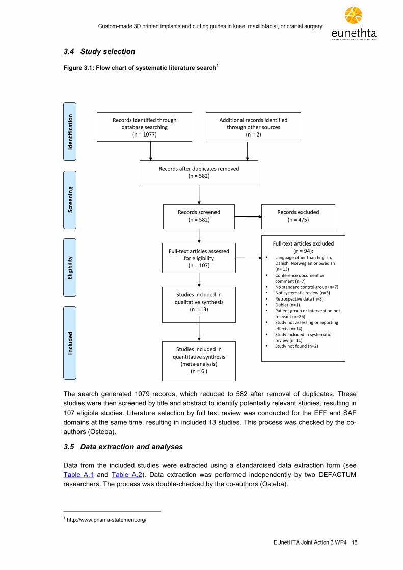
3.4 Study selection
Figure 3.1: Flow chart of systematic literature search1
Records identified through database searching
(n = 1077)
Scre
en
ing
Incl
ud
ed
Elig
ibil
ity
Ide
nti
fica
tio
n
Additional records identified through other sources
(n = 2)
Records after duplicates removed (n = 582)
Records screened (n = 582)
Records excluded (n = 475)
Full-text articles assessed for eligibility
(n = 107)
Full-text articles excluded (n = 94):
Language other than English, Danish, Norwegian or Swedish (n= 13)
Conference document or comment (n=7)
No standard control group (n=7) Not systematic review (n=5) Retrospective data (n=8) Dublet (n=1) Patient group or intervention not
relevant (n=26) Study not assessing or reporting
effects (n=14) Study included in systematic
review (n=11) Study not found (n=2)
Studies included in qualitative synthesis
(n = 13)
Studies included in quantitative synthesis
(meta-analysis) (n = 6 )
The search generated 1079 records, which reduced to 582 after removal of duplicates. These
studies were then screened by title and abstract to identify potentially relevant studies, resulting in
107 eligible studies. Literature selection by full text review was conducted for the EFF and SAF
domains at the same time, resulting in included 13 studies. This process was checked by the co-
authors (Osteba).
3.5 Data extraction and analyses
Data from the included studies were extracted using a standardised data extraction form (see
Table A.1 and Table A.2). Data extraction was performed independently by two DEFACTUM
researchers. The process was double-checked by the co-authors (Osteba).
1 http://www.prisma-statement.org/

Custom-made 3D printed implants and cutting guides in knee, maxillofacial, or cranial surgery
EUnetHTA Joint Action 3 WP4 19
For each outcome, an evidence profile was generated using the GRADEpro software2. Results
from high-quality studies were given the most emphasis in the synthesis. Results were presented
as a narrative synthesis. For the EFF domain, statistical summary estimates of associations
across studies were if possible derived using random effects meta-analysis, anticipating clinical
heterogeneity and with modelling allowing for differences in associations from study to study.
Heterogeneity across studies was statistically assessed using the Q-test and quantified by the
inconsistency (I2) index, where I
2 represents the percentage of total variation across studies
attributable to heterogeneity rather than (statistical) chance. In cases with substantial
heterogeneity across studies (I2>50%), the robustness of the results was checked using a fixed
effects model. A result was considered robust if the point estimate based on the fixed effects
analysis was within the confidence interval of the random effects analysis. Meta-analyses were
performed using Review Manager (RevMan, the Cochrane Collaboration). A two-sided p-value of
<0.05 was considered to be statistically significant in all analyses.
3.6 Quality rating
Study and outcome validity and level of evidence were assessed according to EUnetHTA guide-
lines. In the EFF and SAF domains, the review was prepared in accordance with the Preferred
Reporting Items for Systematic Reviews and Meta-Analysis (PRISMA) statement3. The quality of
the included reviews was assessed using the Risk of Bias in Systematic Reviews (ROBIS) tool4.
This tool assesses four domains to cover key review processes: study eligibility criteria; study
identification and selection; data collection and study appraisal; and synthesis and findings. The
Cochrane risk-of-bias tool was used to assess the quality of the included RCTs according to the
EUnetHTA Guidelines on medical devices for study and outcome level. Risk of bias in cohort and
case-control studies was assessed using Scottish Intercollegiate Guidelines Network (SIGN)5
methodology checklists. The quality of the body of evidence was assessed using Grading of Re-
commendations, Assessment, Development and Evaluation (GRADE). The quality assessment
was performed independently by two DEFACTUM researchers. The process was double-checked
by the co-authors (Osteba). Any disagreement was resolved by consensus. For the TEC and
CUR domains, no quality assessments were applied, but multiple sources were used to validate
potentially biased sources. Descriptive analyses of different information sources were applied.
3.7 Deviations from project plan
In relation to outcomes, function in knee patients was measured using the Knee Society Score
(KSS) and Oxford Knee Scale. No results were found regarding the durability and longevity of the
devices, and patient satisfaction was not specified as an outcome in any study and was not re-
ported in any of the included RCTs or cohort studies.
2 https://gradepro.org/
3 http://www.prisma-statement.org/
4 https://www.bristol.ac.uk/population-health-sciences/projects/robis/
5 https://www.sign.ac.uk/checklists-and-notes.html
Custom-made 3D printed implants and cutting guides in knee, maxillofacial, or cranial surgery
EUnetHTA Joint Action 3 WP4 20
4 DESCRIPTION AND TECHNICAL CHARACTERISTICS OF TECHNOLOGY
(TEC)
The research questions for this assessment refer to two types of technologies: implants and cut-
ting guides for guiding surgical interventions. The intervention is 3D printed custom-made or cus-
tomisable implants and cutting guides, and the comparator is standard produced implants and
cutting guides. The difference between the intervention and the comparator is related to the way
in which the guides and the implants are produced: by moulding in the case of standard care and
by 3D printing in the case of the intervention. 3D printing is a process by which 3D objects are
created layer-by-layer from raw materials guided by a digital file.
4.1 Research questions
Element ID Research question
B0001 What are 3D printed implants and cutting guides versus standard implants and cutting guides?
B0002 What is the claimed benefit of 3D printed implants and cutting guides compared to stand-ard implants and cutting guides?
B0003 What is the phase of development and implementation of 3D printed implants and cutting guides?
B0004 Who administers 3D printed implants and cutting guides and standard implants and cutting guides and in what context and level of care are they provided?
B0008 What kind of special premises are needed to use 3D printed implants and cutting guides and standard implants and cutting guides?
B0009 What equipment and supplies are needed to use 3D printed implants and cutting guides and standard implants and cutting guides?
A0020 For which indications have 3D printed implants and cutting guides received marketing au-thorisation (FDA or CE marking)?
Definitions
Custom-made manufacturer - The natural or legal person who undertakes the design of the
product and manufactures the device to a predefined specification (i.e., a prescription).
Custom-made medical device - Any device specifically made in accordance with a duly qualified
medical practitioner’s prescription which gives, under their responsibility, specific design charac-
teristics and is intended for the sole use of a particular patient.
Customisable medical device - Medical devices that are standard and are customised or
adapted to the characteristics of a particular patient.
Cutting guide/surgical guide - A surgical guide is a small customised tool made from a sterili-
sable material that can be used short-term in a patient and that guides the saw and/or drill in the
planned direction (https://www.xilloc.com/products_services/surgical-guides).
4.2 Results
3D printed and standard implants and cutting guides for knee, maxillofacial, and cranial replace-
ments do not differ very much with respect to materials and final product characteristics. The main
difference is related to the production process and the possibilities for customisation offered by 3D
printing.

Custom-made 3D printed implants and cutting guides in knee, maxillofacial, or cranial surgery
EUnetHTA Joint Action 3 WP4 21
Features of the technology and comparators
[B0001] – What are 3D printed implants and cutting guides versus standard implants and
cutting guides?
Figure 4.1 shows the production flow and the seven basic steps required when using 3D printed
guides or implants for tissue replacement (3). The only difference in the process compared to the
standard procedure for developing the mould is in steps 2 and 4. In steps 2 and 4, software de-
sign and printing are used instead of the usual standard device moulds. The number of steps and
difficulties in printing a 3D device are dependent on device complexity. A general sequence is
described below and in Figure 4.1:
Step 1. Device Design: This step consists of creating the most accurate model of the surfaces to
be replicated and the volumes they refer to. In the case of 3D printing, this is achieved using ima-
ging modalities such as computerised tomography (CT), ultrasonography, and/or magnetic reso-
nance imaging (MRI). In the case of standard guides and implants, this is achieved using wax and
soap models.
Step 2. Software Design: The second step consists of software design for 3D printing or moul-
ding production in the case of standard implants and cutting guides.
Step 3. Material Control: All manufacturing processes require high-quality materials that meet
consistent specifications to build consistent, high-quality devices. This step is identical in both
types of production of implants and guides.
Step 4. Printing: When printing the implants or guides, different printers and materials are used
depending on the material and the intended location. Different techniques are used as described
below. In the case of standard devices, the mould is used to generate the implant guide using
melted materials placed in the mould and hardened by different techniques, for example, freezing
or using hardeners for some chemical substances.
Step 5. Post-Processing: This step can also be the same in both standard and 3D printed devi-
ces. The design is tested and improved in terms of imperfections before being sterilised.
Step 6. Validation and Verification: This step is also the same in both processes, and it requires
tests of usability and adequacy for the purpose for which it was designed.
Step 7. Testing: The two processes are again the same in this step. The processes in this step
relate to the possible different requirements by regulation and the regulatory bodies in charge of
certifying product safety and efficacy/performance (4,5)6.
6 3D Print Exchange. National Institutes of Health; Available at: http://3dprint.nih.gov. Accessed July 12, 2018.

Custom-made 3D printed implants and cutting guides in knee, maxillofacial, or cranial surgery
EUnetHTA Joint Action 3 WP4 22
Figure 4.1: Process of 3D guide or prosthesis production. Modified from KCE, 2018 (3)
Although the materials used (6-10) and the 3D printing techniques are diverse, and a number of
them can be used for the indications in this assessment, we only describe those that have been
found and analysed in the included studies. Detail about the products and materials used in the
studies is low, so it has been difficult to define the type of printer and material (powder, resin,
droplet, or extrusion) used in production. Each material requires a different type of 3D printer and
defines the final product characteristics and performance. Powder materials are 3D printed by
selective laser sintering (SLS), resins by stereolithography (SLA), extrusion materials by fused
deposition modelling (FDM), and droplets by multi-jet modelling (MJM). For more details see Ap-
pendix 3. The materials used for implants and cutting guides in 3D printing are the same as those
that are normally used for cutting guides and implants when produced by standards methods.
Table 4.1 details the materials and indications for commonly used materials in clinical use.
1.Device design
2.Software design
3.Material control
4.Printing 5.Post-
processing 6.Validation - verification
7.Testing
Custom-made 3D printed implants and cutting guides in knee, maxillofacial, or cranial surgery
EUnetHTA Joint Action 3 WP4 23
Table 4.1: Commonly used biomaterials in clinical use. Modified from Williams (11)
Material Use
Metals Titanium alloys Dental implants, femoral stems, pacemaker containers, heart valves, fracture plates, spinal cages
Cobalt–chromium alloys Bearing surfaces, heart valves, stents, pacemaker leads
Platinum group alloys Electrodes
Nitinol (nickel and Titanium alloy)
Shape memory applications
Stainless steel Stents, orthopaedic implants
Magnesium and iron Biodegradable metals for implants
Bioceramics Alumina Bearing surfaces
Calcium phosphates Bioactive surfaces, bone substitutes
Carbon Heart valves
Zirconia Bearing surfaces
Polymers Ultra-high molecular weight polyethylene
Bearing surfaces
PEEK Spinal cages, cranial
PMMA Bone cement, intraocular lenses
Polyurethane Pacemaker lead insulation
Expanded PTFE Vascular grafts, heart valves
Polyester textile Vascular grafts, heart valves
Hydrogel Silicones Soft tissue augmentation, insulating leads, ophthalmological devices
Abbreviations: PEEK=polyether ether ketone; PMMA=poly(methyl methacrylate); PTFE=polytetrafluoroethylene
The materials and comparators included in this assessment are listed in Table 4.2.

Custom-made 3D printed implants and cutting guides in knee, maxillofacial, or cranial surgery
EUnetHTA Joint Action 3 WP4 24
Table 4.2: Type of materials and comparators* included in this report (based on the evidence from the included articles)
Product Indication Reference Material Type of printer Comparator
Guides Maxillofacial Mazzoni 2013 Cobalt-chrome-molybdenum Standard reconstruction with indirect CAD / computer aided manufacturing procedure
Brandao 2016 Acrylic resin Standard surgery Without guides
Ayoub 2014 PolyMide Laser sintering Standard surgery Without guides
Al-Ahmad 2013 Acrylic resin Zcorp Standard SSRO Without guides
Cranial
Knee Huijbregts 2016 Standard instrumentation Legion systems or Genesis II
Boonen 2016 Standard instruments
Pfitzer 2014 Standard instruments Journey
Gan 2015 Acrylate resin Stereolithography Standard instruments Scorpio posterior stabilised system
Qiu 2017 Standard instruments
Zhang 2016 SPSS 350B solid laser prototyping
Standard instruments Triathlon
Thienpont 2017 Standard instruments
Mannan 2016 Standard instruments
Implants Cranial Chrzan 2012 Polypropylene-polyester or aluminium-silicon
Milling Arrow 500 Standard instrumentation Manually adjusted prosthesis
Knee
Maxillofacial
* No further data were obtained from the articles on comparators Abbreviations: CAD=computer-aided design; SSRO=sagittal split ramus osteotomy

Custom-made 3D printed implants and cutting guides in knee, maxillofacial, or cranial surgery
EUnetHTA Joint Action 3 WP4 25
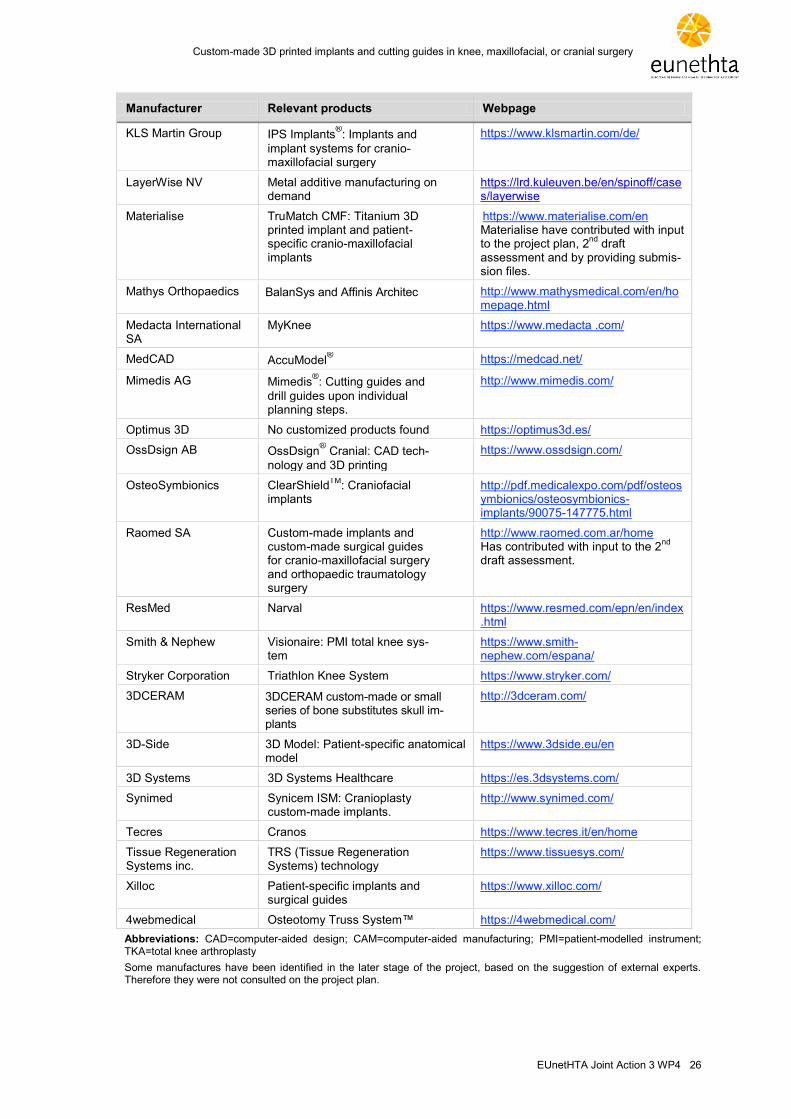
Regarding the manufacturers producing 3D printed implants and cutting guides in patients under-
going knee, maxillofacial, or cranial surgery, efforts were made to identify all relevant manufactu-
rers and their products. However, it is a diverse market, so the list may not be exhaustive. Only a
few manufacturers responded to direct inquiries made by the assessment team to help identify
relevant products.
Table 4.3: Custom-made implants and custom-made surgical guides for cranio-maxillofacial surgery and orthopaedic traumatology surgery
Manufacturer Relevant products Webpage
Anatomics AnatomicsC3D: Custom im-plants(cranial)
http://www.anatomics.com/
Arcam EBM® for Orthopaedic Implants http://www.arcam.com/solutions/ortho
pedic-implants/
Autodesk Within Medi-cal
Novax DMA CEIT-KE
https://www.autodesk.com/products/within-medical/overview
Avinent Personalised CAD/CAM im-plants and prostheses
https://www.avinent.com/eng/default.cfm
Bespokemedical Bespoke solutions: 3D custom-made prostheses
https://bespokemedical.com.au/
Zimmer Biomet The Signature™ System https://www.zimmerbiomet.com.es/
CADskills BVBA CADCAMise: Anatomical mod-els, cutting/drilling guides and 3D print implants
https://www.cadskills.be/en
Cerhum SA Medical ceramic 3D printing https://www.cerhum.com/
CUSMED No information found No information found
EOS (3D printer pro-ducer)
3D printers for other manufac-turers (e.g., Autodesk Within Medical)
https://www.eos.info/en
evonos GmbH & Co. Evo-Shape: Skull implants http://www.evonos.de/
Finceramica CustomBone: Custom-made implant for cranioplasty
http://www.finceramica.it/
FIT Production FIT production: Custom-made implants
http://www.fit-production.de/
Gsell Gsell Medical: Implants http://www.gsell.ch/en/home.html
implantcast GmbH C-Fit 3D®: Patient-specific in-
struments and implants
https://www.implantcast.de/
Johnson & Johnson Medical (DePuySyn-thes)
TruMatch® cutting guides, for
use with standard TKA (i.e., non-customised) implants (Sigma & Attune)
https://www.depuysynthes.com/ Johnson & Johnson have contributed with input to the project plan, 2
nd draft
assessment and by providing EU-netHTA submission files.
Kelyniam Global Inc. Kelyniam Implants: Cranial implants
https://www.kelyniam.com/
Custom-made 3D printed implants and cutting guides in knee, maxillofacial, or cranial surgery
EUnetHTA Joint Action 3 WP4 26
Manufacturer Relevant products Webpage
KLS Martin Group
IPS Implants®: Implants and
implant systems for cranio-maxillofacial surgery
https://www.klsmartin.com/de/
LayerWise NV Metal additive manufacturing on demand
https://lrd.kuleuven.be/en/spinoff/cases/layerwise
Materialise TruMatch CMF: Titanium 3D printed implant and patient-specific cranio-maxillofacial implants
https://www.materialise.com/en Materialise have contributed with input to the project plan, 2
nd draft
assessment and by providing submis-sion files.
Mathys Orthopaedics BalanSys and Affinis Architec http://www.mathysmedical.com/en/homepage.html
Medacta International SA
MyKnee https://www.medacta .com/
MedCAD AccuModel® https://medcad.net/
Mimedis AG Mimedis®: Cutting guides and
drill guides upon individual planning steps.
http://www.mimedis.com/
Optimus 3D No customized products found https://optimus3d.es/
OssDsign AB OssDsign® Cranial: CAD tech-
nology and 3D printing
https://www.ossdsign.com/
OsteoSymbionics ClearShieldTM
: Craniofacial implants
http://pdf.medicalexpo.com/pdf/osteosymbionics/osteosymbionics-implants/90075-147775.html
Raomed SA Custom-made implants and custom-made surgical guides for cranio-maxillofacial surgery and orthopaedic traumatology surgery
http://www.raomed.com.ar/home Has contributed with input to the 2
nd
draft assessment.
ResMed Narval https://www.resmed.com/epn/en/index.html
Smith & Nephew Visionaire: PMI total knee sys-tem
https://www.smith-nephew.com/espana/
Stryker Corporation Triathlon Knee System https://www.stryker.com/
3DCERAM 3DCERAM custom-made or small series of bone substitutes skull im-plants
http://3dceram.com/
3D-Side 3D Model: Patient-specific anatomical model
https://www.3dside.eu/en
3D Systems 3D Systems Healthcare https://es.3dsystems.com/
Synimed Synicem ISM: Cranioplasty custom-made implants.
http://www.synimed.com/
Tecres Cranos https://www.tecres.it/en/home
Tissue Regeneration Systems inc.
TRS (Tissue Regeneration Systems) technology
https://www.tissuesys.com/
Xilloc Patient-specific implants and surgical guides
https://www.xilloc.com/
4webmedical Osteotomy Truss System™ https://4webmedical.com/
Abbreviations: CAD=computer-aided design; CAM=computer-aided manufacturing; PMI=patient-modelled instrument; TKA=total knee arthroplasty
Some manufactures have been identified in the later stage of the project, based on the suggestion of external experts. Therefore they were not consulted on the project plan.

Custom-made 3D printed implants and cutting guides in knee, maxillofacial, or cranial surgery
EUnetHTA Joint Action 3 WP4 27
After identifying the manufacturers and their products, two main issues needed clarifying: 1) which
manufacturers made 3D printers for different purposes and with applications for medical purpo-
ses; and 2) which manufacturers used those 3D printers for medical purposes, especially those
relevant to the scope of this report (6,7). Although this difference might be considered irrelevant, it
is, however, relevant from the safety and legal points of view, i.e., whether the manufacturer is
simply selling 3D printers for different purposes which can be used for medical purposes or if they
are also involved in the design and production of medical devices. In fact, some manufacturers do
either or both.
Table 4.4 shows the manufacturers, their products, and their characteristics. Most manufacturers
did not directly provide information despite several attempts to make contact. Therefore, the ana-
lysis presented in Table 4.4 is based on publicly available information obtained from manufactu-
rers' webpages, brochures, and elsewhere. The information is not to be considered exhaustive
and might contain inaccuracies.

Custom-made 3D printed implants and cutting guides in knee, maxillofacial, or cranial surgery
EUnetHTA Joint Action 3 WP4 28
Table 4.4: Custom-made or customisable 3D printed implants and cutting guides in patients undergoing knee, maxillofacial, or cranial surgery. Shown are the manufacturers, products, locations, type of products and materials used
Relevant products Anatomical location
Guide Prosthesis Material 1 Material 2 Material 3
3D Ceram Cranial X Ceramics
Maxillofacial X Ceramics
3D-Side SkullPT Cranial X Bone cement
Adaptive SkullPT Cranial Bone cement
Anatomics Durashield Cranial X Silicone
Biomodel Stereotaxy Cranial X
Cranial X Acrylic (polymethyl metha-crylate)
Porestar (porous poly-ethylene)
Titanium
Facial X Porestar (porous poly-ethylene)
Titanium
Biomodels X
Mandible templates & guides Facial X X
Cranial templates Cranial X X
Bespokemedical Knee X
Zimmer Biomet Persona®; Vanguard; NexGen Knee X Bearing materials: Vivacit-E
vitamin E highly crosslinked
polyethylene; E1® antioxidant-
infused polyethylene; Pro-
long® highly crosslinked
polyethylene; Durasul® highly
crosslinked polyethylene
OsseoTi® Porous Metal
Technology
EncompassTM
, Midface Maxillofacial X X PEEK Polymer Titanium
HTR PEEK, HTR PMMA, Cranio-
curve®, ThinFlap
TM, SterileTrac
TM,
RapidFire®
Cranial X X Idem Idem Idem
CADskills BVBA Cranial X X Titanium, PEEK CADskills has developed a new UHMWPE specifi-cally treated to increase wear resistance. The method is undisclosed. The UHMWPE is en-riched with vitamin E, a potent antioxidant. These characteristics make this
Cutting guides are 3D printed in a class 1 bio-compatible resin that is autoclava-ble. The material is specially devel-oped for printing precise surgical

Custom-made 3D printed implants and cutting guides in knee, maxillofacial, or cranial surgery
EUnetHTA Joint Action 3 WP4 29
Relevant products Anatomical location
Guide Prosthesis Material 1 Material 2 Material 3
material especially suited for articulating surfaces
guides and similar devices
Maxillofacial X X Idem Idem Idem
Cerhum SA (for confi-dentiality reasons, final product not disclose)
Maxillofacial X Alumina Zirconia
Cranial X
Knee X
Evonos Cranial X Titanium
Finceramica Customized Bone (USA only) / CustomBone
Cranial X Biomimetic ceramic
Fit-production (Does not specify the type of implants)
Cranial X Titanium (EBM technology)
Maxillofacial X
Knee X
Gsell Knee X UHMWPE (Ultra high molecu-lar weight polyethylene), PEEK, PEKK, CF, pyrolytic graphite
Implantcast Knee X Implavit: The majority of implants are made of a cobalt chrome molybdenum (CoCrMo)
Implatan: The raw titani-um (TiAl6V4) alloy mate-rial
UHMWPE
Johnson & Johnson Medical (DePuySyn-thes)
TruMatch® cutting guides, for
use with standard TKA (i.e., non-customised) implants (Sigma & Attune)
Knee X
Kelyniam Cranial, cranio-facial
X PEEK, lightweight Bio-material
KLS Martin Cranial X Titanium Resorbable materials
Maxillofacial X
Materialise (Johnson & Johnson)
TruMatch® Cranio-
maxillofacial X X Titanium and polyamide
(guides)
TruMatch® Cranial X X Titanium and polyamide
(guides)
Knee X Polyamide
Mathys Orthopaedics Ltd.
BalanSys Knee X

Custom-made 3D printed implants and cutting guides in knee, maxillofacial, or cranial surgery
EUnetHTA Joint Action 3 WP4 30
Relevant products Anatomical location
Guide Prosthesis Material 1 Material 2 Material 3
Medacta International SA
GMK Knee X X Cobalt-chrome UHMWPE
MedCAD AccuShape® Cranial X PEEK
Craniofacial X
Mimedis (Medartis) Craniofacial X Flexible
OssDSIGN Craniofacial X Titanium
Osteosymbionics ST-temporalis, PK-Shield Craniofacial X
Raomed Cranial X X Titanium PEEK PMMA, polyamide
Maxillofacial X X Titanium PEEK Cr-Co-Mo, UHMWPE, PMMA, polyamide
Stryker Corporation Cranial X MEDPOR and PEEK Titanium
Maxillofacial X X MEDPOR and PEEK Titanium
Knee X Titanium
Synimed Synicem ISM Craniofacial Cements
Tecres Cranos Cranial X Polymethylmethacrylate
Tissue Regeneration Systems
TRS Scaffold Technology Cranio-maxillofacial
X
Xilloc Cranio-maxillofacial
X PEEK-OPTIMA®: polymeric
biomaterial
TI6Al4V: Titanium alloy PP/PES knitted yarn: polymeric knitted from yarn
4WEB Medical Craniofacial X Polyamide Metallic
Abbreviations: EBM=electron beam melting; PEEK=polyether ether ketone; PES=polyester; PMMA=poly(methyl methacrylate); PP=polypropylene; TKA=total knee arthroplasty; UHMWPE=ultra-high molecular weight polyethylene; X=available information about guide or prosthesis; Empty space=no information.
Custom-made 3D printed implants and cutting guides in knee, maxillofacial, or cranial surgery
EUnetHTA Joint Action 3 WP4 31
[B0002] – What is the claimed benefit of 3D printed implants and cutting guides compared
to standard implants and cutting guides?
The claimed benefit of the technology in relation to the comparator(s) is the personalisation and
the customisation of cutting guides and implants and the possibility of “in-house” producing 3D
printed solutions (12,13,14). Based on this personalisation and customisation, the manufacturers
claim that safety, performance, and efficacy are improved compared with standard practice inclu-
ding the use of customisable implants. Some manufacturers have also claimed that in the case of
certain guides and implants, especially maxillofacial and cranial implants in which standardisation
is more complicated or costly, 3D printers could be a viable economic solution for single implants
or cutting guides.
[B0003] – What is the phase of development and implementation of 3D printed implants
and cutting guides?
3D printing technology is not yet fully or widely implemented, and the comparator is standard
practice. Some commercialised solutions such as TruMatch® from Johnson & Johnson and Tru-
Match® CMF from Materialise have received US Food and Drug Administration (FDA) clearance
through the 510(k) procedure (see Table 4.3). However, in most cases, 3D printing technology
has been introduced as a research or innovative technology without clear moderate or high-level
clinical evidence on its effects, and should therefore be considered under research premises. The
comparator in the included studies is not always well described, making it difficult to describe the
standard solutions in detail.
[B0004] – Who administers 3D printed implants and cutting guides and standard implants
and cutting guides and in what context and level of care are they provided?
In general, the comparators and 3D printing technology are administered at the tertiary level of
provision. This is not due to production requirements, rather the necessary implementation of the
comparators in those premises or under this frame of provision (15). The use of standard implants
and cutting guides requires skilled professionals and learning curves that guarantee quality im-
plementation of the technology. Similarly, 3D printed implants and cutting guides are administered
by certified professionals in accredited centres to ensure high quality provision of care, although
the level of certification differs from country to country and in some countries any centre can pro-
vide the service and use the technology. The same level of expertise is required in those centres
implementing or researching 3D printing solutions as those using standard implants or cutting
guides. In cases where no regulatory approval of 3D printed solutions is needed, the use of 3D
printed solutions is considered under research circumstances and thus details of the technology
must be communicated to the patient and his/her caregiver as required as part of informed con-
sent.
[B0008] – What kind of special premises are needed to use 3D printed implants and cutting
guides and standard implants and cutting guides?
No special premises are required for the use of custom-made 3D printed devices compared to
standard implants and cutting guides. The main differences are related to device production and
not to their application and use in clinical practice.

Custom-made 3D printed implants and cutting guides in knee, maxillofacial, or cranial surgery
EUnetHTA Joint Action 3 WP4 32
[B0009] – What equipment and supplies are needed to use 3D printed implants and cutting
guides and standard implants and cutting guides?
The equipment needed for using 3D printed devices is dependent on the organisation of the entire
production process and how much of this production is in-house. The main equipment required for
the 3D printing process is imaging processing systems and 3D printers. If the organisation is
simply contracting production from external manufacturer, the equipment needed is similar to the
equipment required to use standard implants and cutting guides.
[A0020] – For which indications have 3D printed implants and cutting guides received mar-
keting authorisation (FDA or CE marking)?
Here it is necessary to differentiate between 3D printing market authorisation and CE marking of
the final products/devices. 3D printers in themselves are not normally considered a medical de-
vice and receive CE marks as other technologies on the market. In the case of implants and cut-
ting guides, it is unclear what authorisation is required, since the implants and cutting guides
could be classified as custom-made or customisable. As all the devices in this assessment could
be considered custom-made, the issue is that even as class III devices they do not need to bear
the CE mark because they are custom-made. This does not, however, mean that they are not
subject to regulatory control through post-market surveillance via competent authorities of the
member states, where manufacturers are required to report incidents and maintain post-market
surveillance.
However, some manufacturers, for example Johnson & Johnson and Materialise, have received
CE or FDA authorisation, but the performance at the individual implant/cutting guide level still
needs to be evaluated to establish the technology’s value. So, the consideration of custom-made
or customisable must be taken into account and their differentiation, when 3D printed, is not well
defined. Until there is conclusive evidence of safety and efficacy, patients should be informed of
the alternatives and the uncertainties of their use, with data being collected. Procedures such as
those proposed by EUnetHTA JA3, WP5 for data collection post introduction should be followed
to ensure data quality in those registries.

Custom-made 3D printed implants and cutting guides in knee, maxillofacial, or cranial surgery
EUnetHTA Joint Action 3 WP4 33
5 HEALTH PROBLEM AND CURRENT USE OF THE TECHNOLOGY (CUR)
5.1 Research questions
Element ID Research question
A0002 Which diseases or health conditions most frequently lead to knee, maxillofacial, or cranial surgery?
A0004 What is the natural course of the disease or health condition?
A0005 What are the symptoms and the burden of disease or health condition for the patient?
A0006 What are the consequences of the disease or health condition for society?
A0024 How is the disease or health condition currently diagnosed according to published guidelines and in practice?
A0025 How is the disease or health condition currently managed according to published guidelines and in practice?
A0007 What is the target population in this assessment?
A0023 How many people belong to the target population?
A0011 How much are the technologies utilised?
5.2 Results
3D printed medical devices are used in a wide variety of indications. This assessment focuses on
the following clinical areas in which 3D printed custom-made or customisable implants and cutting
guides are most frequently applied and where most of the published evidence lies: knee,
maxillofacial, and cranial surgery. The most frequent diseases in these clinical areas include knee
osteoarthritis treated with total knee arthroplasty, oral cancer treated with mandibular
reconstruction, and traumatic brain injury with intracranial hypertension treated with
decompressive craniectomy (DC) and later cranioplasty (3, 16).
Overview of the diseases or health conditions
[A0002] – Which diseases or health conditions most frequently lead to knee, maxillofacial,
or cranial surgery?
Knee osteoarthritis
The primary indication for total knee arthroplasty (TKA) is the relief of significant and disabling
pain and improvement of functional status in patients with end-stage knee osteoarthritis (OA) (17).
Two types of knee OA are recognised: primary knee OA is caused by progressive joint cartilage
destruction over time without any apparent underlying cause, while secondary OA can be caused
by trauma or surgery to the joint structures, congenital limb malformations, malposition (va-
rus/valgus), or abnormal articular cartilage such as that found in end-stage rheumatoid arthritis
(RA) (18).
Oral cancer
Invasive oral cancer is the primary indication for mandibular reconstruction, but it is also used for
other diseases such as osteomyelitis and bisphosphonate-related osteonecrosis (19). Oral cancer
includes a group of neoplasms affecting any region of the oral cavity, pharyngeal regions, and
salivary glands. Oral squamous cell carcinoma (OSCC) is the most frequent (90%) oral neoplasm.

Custom-made 3D printed implants and cutting guides in knee, maxillofacial, or cranial surgery
EUnetHTA Joint Action 3 WP4 34
Traumatic brain injury
Cranial surgery with cranioplasty after decompressive craniectomy (DC) is widely used to treat
intracranial hypertension (ICH) following traumatic brain injury (TBI), stroke, and other conditions
associated with raised intracranial pressure (20).
[A0004] – What is the natural course of the disease or health condition?
Knee osteoarthritis
Knee OA is characterised by progressive destruction of the cartilage that lines the knee joints, the
subchondral bone surfaces, and synovium. It is accompanied by pain, immobility, muscle weak-
ness, and reduced function and activities of daily living (21). Knee OA is chronic and progressive
and worsens over time with pain as the primary symptom. It starts with intermittent weight-bearing
pain and develops to more persistent pain, especially if contributing factors (obesity, misalign-
ment, and occupation) are not properly modified. Without treatment, OA eventually leads to signi-
ficant pain and disability requiring surgical intervention (22). Knee RA is a chronic inflammatory
disease characterised by synovial hyperplasia, bone loss, and joint deformity, which eventually
leads to pain and disability requiring surgical intervention (23).
Oral cancer
Oral squamous cell carcinoma (OSCC) often presents as an ulcer with fissuring or raised margins
or may present as a lump, a red, white or mixed white-red lesion, a non-healing wound, or with
cervical lymph node enlargement (23). Despite advances in therapeutic approaches, OSCC prog-
nosis is poor due to frequent aggressive local invasion, metastasis, and recurrence. The five-year
survival rate ranges from 20-90% depending on which part of the oral cavity is involved and the
stage. Patients diagnosed early have a better long-term survival (60-90%), whereas when diag-
nosis is late the long-term survival ranges from 20-50% (24).
Traumatic brain injury
TBI describes an injury to the head arising from blunt or penetrating trauma or accelera-
tion/deceleration forces. It is associated with one or more of the following features: decreased
level of consciousness, amnesia, objective neurological abnormalities, and skull fractures (25).
The Glasgow Coma Scale (GCS) is used to triage patients early after TBI as an indicator of sever-
ity, with TBI graded as mild, moderate, or severe (25). After primary brain injury, there is a risk of
secondary injury from intracranial hypertension (ICH) as a consequence of brain swelling or
haemorrhage. ICH is the most frequent cause of death and disability following severe TBI (25)
and is often treated with DC and later cranioplasty.
Effects of the disease or health condition
[A0005] – What are the symptoms and the burden of disease or health condition
for the patient?
Knee osteoarthritis
For the individual, the burden of knee OA includes pain, limited activity, and markedly reduced
quality of life. The intensity of clinical symptoms and rate of progression vary greatly from person
to person. However, symptoms typically become more frequent and severe and cause progres-
sive limitations in function over time, eventually leading to disability. Patients experience knee
Custom-made 3D printed implants and cutting guides in knee, maxillofacial, or cranial surgery
EUnetHTA Joint Action 3 WP4 35
pain that is gradual in onset and that worsens with activity, swelling and knee stiffness, and pain
after prolonged sitting or resting (18). In a population in New Zealanders aged 40-84 years, knee
OA accounted for 3.44 quality-adjusted life years (QALYs) lost per person, corresponding to
467,240 QALYs across the population. These QALY losses were higher for females than males
due to a greater prevalence of knee OA and higher life expectancy (26).
Oral cancer
The initial stages are often painless. In later and larger lesions, symptoms vary from mild discom-
fort to severe pain. Other symptoms include ear pain, bleeding, mobile teeth, breathing problems,
speech difficulties, dysphagia, problems using prostheses, trismus, and paraesthesia (27). Many
oral cancer patients suffer from anxiety and depression which negatively impact their quality of
life, and the suicide incidence is high in this patient group (28).
Traumatic brain injury
After TBI of any severity, many patients display cognitive and emotional difficulties and require
assistance in their activities of daily living (25,29). Reduced health-related quality of life (HRQoL)
has been identified in individuals with TBI compared with a healthy population (30).
[A0006] – What are the consequences of the disease or health condition for society?
Knee osteoarthritis
The knee is the most frequently affected joint in OA, and the societal and economic burden of this
disease at a population level is substantial. Most of the direct costs are usually attributed to the
healthcare system including hospital stays, orthopaedic surgery, medications, doctor visits, and
other health professional visits. Indirect costs are also incurred that are attributable to productivity
losses from absenteeism, presenteeism, early retirement, and premature death (31).
Oral cancer
Oral cancer is a significant public health concern. According to global estimates, oral cancer
causes approximately 130,000 deaths per year (32). There is a heavy financial burden to society
in treating oral cancer that includes direct costs for diagnosis, treatment, and hospitalisation and
indirect costs including loss of productivity due to morbidity and disability (33).
Traumatic brain injury
TBI is a major public health problem worldwide and is the most common cause of death and disa-
bility in children and young adults. Over 50% of patients with severe TBI are moderate to severely
disabled after one year, and many never recover to full independence (33). The costs for treating
severe TBI result in a heavy financial burden to society, where the indirect costs of disability, lost
wages, and lost productivity outweigh the direct medical costs including hospitalisation and reha-
bilitation (34).

Custom-made 3D printed implants and cutting guides in knee, maxillofacial, or cranial surgery
EUnetHTA Joint Action 3 WP4 36
Current clinical management of the disease or health condition
[A0024] – How is the disease or health condition currently diagnosed according to pub-
lished guidelines and in practice?
Knee osteoarthritis
The diagnosis of knee OA can usually be made by clinical judgment and imaging performed by
clinical experts, but these are not the only markers of knee OA. The European League Against
Rheumatism (EULAR) has developed evidence-based recommendations for the diagnosis of
knee OA (34). These include typical symptoms and signs, the use of imaging and laboratory tests,
and differential diagnosis. Three symptoms (persistent knee pain, limited morning stiffness, and
reduced function) and three signs (crepitus, restricted movement, and bony enlargement) were
found to be most useful. The severity of knee OA can be staged using the Western Ontario and
McMaster Universities Osteoarthritis Index (WOMAC), which reflects clinical symptoms divided
into three subscales: pain, stiffness, and physical function (35).
Oral cancer
The diagnosis of OSCC is based on a thorough clinical examination of the oral mucosa and pal-
pation of the lymphoid tissue of the neck to detect masses that represent metastases. The clinical
findings should be verified with a biopsy to confirm the diagnosis histopathologically (27). The
tumour-node-metastasis (TNM) classification is used to stage oral cancers (36).
Traumatic brain injury
There is no consensus on definitive diagnostic measures, but there is general agreement that a
clear mechanism and suspicion of injury plus a minimum of one common clinical feature define a
TBI. The diagnosis is based on injury history, clinical examination including GCS, and CT scans of
the head (37).
[A0025] – How is the disease or health condition currently managed according to
published guidelines and in practice?
Knee osteoarthritis
Knee OA is not currently curable, and the goal of non-operative treatment is to alleviate the signs
and symptoms of the disease and to slow its progression. Conservative treatment is generally
indicated in patients with generalised knee pain who wish to delay undergoing a surgical proce-
dure. According to EULAR recommendations, conservative treatment of knee OA should proceed
in a stepwise fashion including physical and physiotherapeutic measures, orthopaedic aids,
weight loss, and drug therapy (34). Despite conservative treatment, many with severe knee OA
require a TKA as the definitive treatment (35).
Oral cancer
The treatment of OSCC consists of surgery, radiotherapy, and chemotherapy. OSCC is typically
treated by one or a combination of these modalities according to tumour location and stage (38).
Custom-made 3D printed implants and cutting guides in knee, maxillofacial, or cranial surgery
EUnetHTA Joint Action 3 WP4 37
Traumatic brain injury
DC is used as a therapeutic strategy for refractory cerebral oedema in cases when first-line non-
invasive methods fail (29). Cranioplasty is then performed not only for aesthetic reasons but also
to protect the underlying neural tissue and improve its perfusion and metabolism (39).
Target population
[A0007] – What is the target population of this assessment?
Knee osteoarthritis
The incidence and prevalence of knee OA have increased over recent decades at least in part
due to an aging population and the rising prevalence of obesity (40). Knee OA affects at least
19% of adults aged 45 years, and >50% of patients over 65 years have radiographic changes in
the knee indicating arthritis, although many patients are asymptomatic until after age 65 years
(41,42). Moreover, the occurrence of knee OA in younger active people is reported to be increa-
sing (40). The global prevalence of radiographically confirmed symptomatic knee OA in 2010 was
estimated to be 3.8% (40).
Oral cancer
OSCC is most common in older males with a history of tobacco and alcohol consumption, in lower
socio-economic groups, and in ethnic minority groups (36). However, some studies have shown a
high frequency of OSCC in younger adults (aged <40 years), probably due to changes in lifestyle
habits in this age group such as changes in tobacco and alcohol consumption as well as exposure
to biological agents such as human papillomavirus (HPV) (43).
Traumatic brain injury
TBI is most common in children and young adults (25).
[A0023] – How many people belong to the target population?
Knee osteoarthritis
In Denmark, which has approximately 5.5 million inhabitants, 60,000 people each year with symp-
toms of knee arthritis seek medical advice7. In the USA, knee OA accounts for over 80% of all OA
and affects at least 19% of adults aged 45 years and older (42). The number of people in the tar-
get population with symptomatic OA is likely to increase due to the aging population and the rise
in the prevalence of obesity (44).
Oral cancer
Worldwide, oral cancer accounts for 2-4% of all cancer cases, corresponding to 300,000 new
cases diagnosed in 2012 (45). Age-standardised oral cancer incidence rates vary between 0.9 per
100,000 women in Eastern Asia to 24.0 per 100,000 men in Melanesia.
7 [www.sst.dk/da/udgivelser/2012/nkr-og-faglige-visitationslinjer-knaeartrose]

Custom-made 3D printed implants and cutting guides in knee, maxillofacial, or cranial surgery
EUnetHTA Joint Action 3 WP4 38
Traumatic brain injury
It is estimated that TBI affects over 10 million people annually worldwide, leading to either mortali-
ty or hospitalisation (46).
[A0011] – How much are the technologies utilised?
Knee osteoarthritis
The total number of primary and revision TKAs in Europe was estimated to be 1,324,000 each
year in 2011, whereas the number of patient-specific instrument (PSI) procedures was 17,515,
corresponding to 1.3% of total (1,2).
Oral cancer
The mandibular reconstruction with free fibula flap is currently regarded as the gold standard re-
construction of large segmental mandibular defects caused by benign or malignant disease (47).
Mandibular reconstruction has changed significantly over the years and continues to evolve with
the introduction of newer technologies and techniques. 3D printing is a rapidly growing technology
in medicine, and when mandibular reconstruction is performed with a fibula free flap with 3D
printed models, the long bone can be reconfigured into a 3D angular structure (48). There are no
available published data on the utilisation of these technologies in Europe.
Traumatic brain injury
There are no available published data on the utilisation of either standard cranioplasty or with 3D
printed materials.

Custom-made 3D printed implants and cutting guides in knee, maxillofacial, or cranial surgery
EUnetHTA Joint Action 3 WP4 39
6 CLINICAL EFFECTIVENESS (EFF)
6.1 Research questions
Element ID Research question
D0001 What is the expected benefit of 3D printed implants and cutting guides on mortality?
D0005 How does use of 3D printed implants and cutting guides affect symptoms and findings (severity, frequency) of patients undergoing surgery?
D0006 How do 3D printed implants and cutting guides affect progression (or recurrence) of the disease or health condition?
D0011 What is the effect of 3D printed implants and cutting guides on patients’ body functions?
D0016 How does the use of 3D printed implants and cutting guides affect activities of daily living?
D0012 What is the effect of 3D printed implants and cutting guides on generic health-related quality of life?
D0013 What is the effect of 3D printed implants and cutting guides on disease-specific quality of life?
D0017 Were patients satisfied with the use of 3D printed implants and cutting guides?
6.2 Results
Included studies
Thirteen studies reported clinical effectiveness outcomes (19,39,49-59). Eight studies (six RCTs
(49-54) and two SRs (55,56) reported on patients undergoing knee reconstruction; four studies
(three RCTs (19,58,59) and one prospective study (57) reported on maxillofacial patients (more
specifically patients undergoing mandibular reconstruction); and one prospective study examined
patients undergoing cranioplasty (39). Details of the studies are provided in Table A1 and Table
A2. Studies published between 2012 and 2017 had follow-up periods of between one and 44
months.
Results are presented according to the research questions categorised in relation to specific out-
comes and finally patient group. Not all patient groups are represented under a specific outcome,
since some outcomes relate to particular patient groups, e.g., the outcome 'number of outliers'
only includes results from knee arthroplasty patients.
The quality of the two included cohort studies by Chrzan et al. (39) and Mazzoni et al. (57) was
low. However, the quality of the two SRs by Thienpont et al. (55) and Mannan et al. (55,56) was
high. Studies included in the two SRs were of moderate to high quality. The quality of the included
RCTs was in general moderate, although one study by Qui et al. (53) was low quality. Blinding
was the criterion most often described unevenly. Reporting of the blinding status of study partici-
pants, personnel, and assessors was in general incomplete, making the risk of bias unclear. Ta-
bles 6.1 and 6.2 present the risk of bias of the RCTs and SRs.
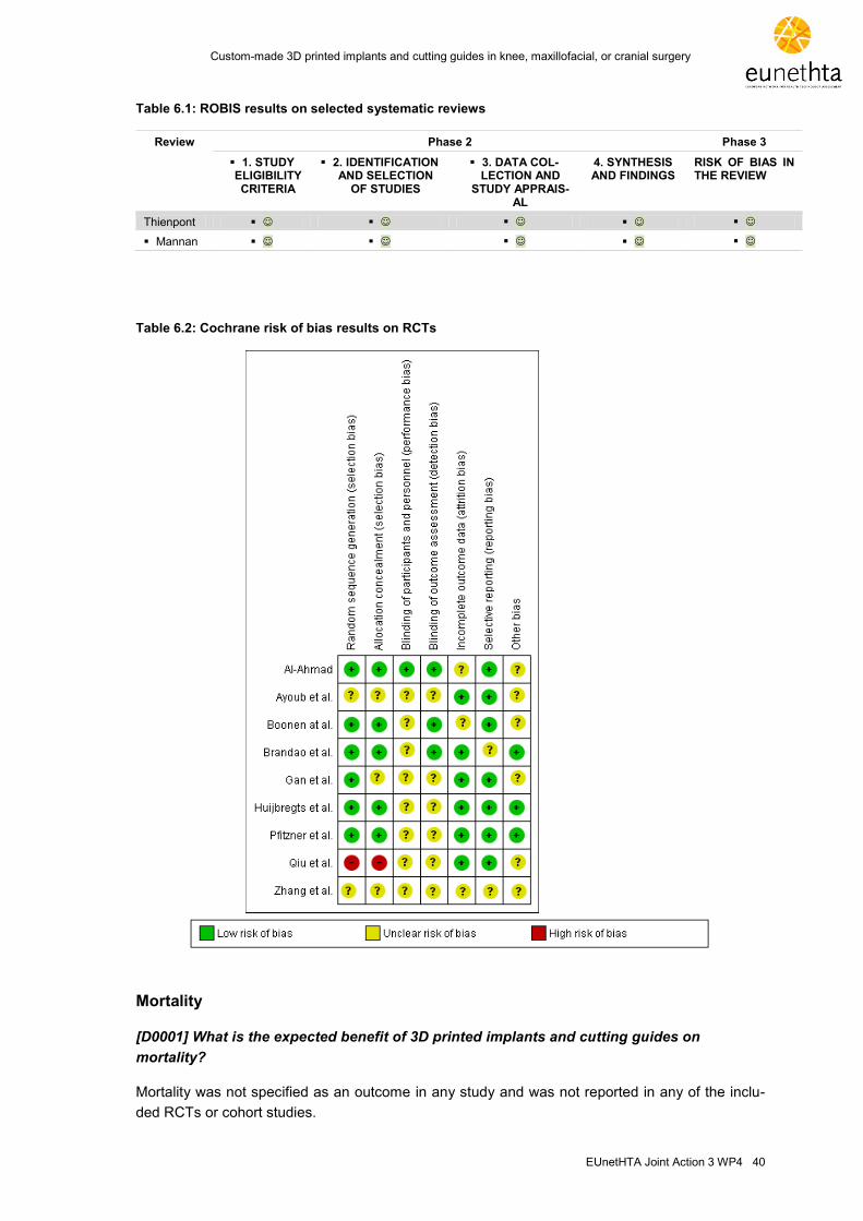
Custom-made 3D printed implants and cutting guides in knee, maxillofacial, or cranial surgery
EUnetHTA Joint Action 3 WP4 40
Table 6.1: ROBIS results on selected systematic reviews
Review Phase 2 Phase 3
1. STUDY ELIGIBILITY CRITERIA
2. IDENTIFICATION AND SELECTION
OF STUDIES
3. DATA COL-LECTION AND
STUDY APPRAIS-AL
4. SYNTHESIS AND FINDINGS
RISK OF BIAS IN THE REVIEW
Thienpont
Mannan
Table 6.2: Cochrane risk of bias results on RCTs
Mortality
[D0001] What is the expected benefit of 3D printed implants and cutting guides on
mortality?
Mortality was not specified as an outcome in any study and was not reported in any of the inclu-
ded RCTs or cohort studies.

Custom-made 3D printed implants and cutting guides in knee, maxillofacial, or cranial surgery
EUnetHTA Joint Action 3 WP4 41
Morbidity
[D0005] – How does use of 3D printed implants and cutting guides affect symptoms and
findings (severity, frequency) of the patients undergoing surgery?
[D0006] – How do 3D printed implants and cutting guides affect progression (or
recurrence) of the disease or health condition?
Number of outliers (by >3°) in TKA: 3D print vs. standard implants and cutting guides in patients
undergoing knee reconstruction
Hip-knee-ankle angle (limb), alignment, or mechanical axis (Figure 6.1)
The number of outliers in TKA was reported in four RCTs (49,51-53) and one SR (55). Data from
all four primary studies were included in the meta-analysis. The odds ratio calculated using the
number of events in the two groups was 0.28 (95% CI 0.12-0.66; p=0.003) favouring 3D print
technology (cutting guides; low quality of evidence). There was a low degree of heterogeneity
between the studies (I2=33%, p=0.21) (Figure 6.2).
The SR by Thienpont et al. (55) reported a pooled relative risk of 0.79 (95% CI 0.65-0.95;
p=0.013) favouring PSI/3D print technology. Mechanical axis deviation (by >3°) was the primary
endpoint in the SR, which included 44 RCTs and cohort studies.
Figure 6.1. Hip-knee-ankle angle, mechanical axis, coronal femoral angle, and coronal tibial angle. Source: Jan Hejle, anatomi.dk

Custom-made 3D printed implants and cutting guides in knee, maxillofacial, or cranial surgery
EUnetHTA Joint Action 3 WP4 42
Figure 6.2: 3D print vs. standard; outcome: proportion of outliers (>3°), hip-knee-ankle angle
Coronal femoral angle
Coronal femoral angle, which describes implant alignment measured between the mechanical
femoral axis and the tangent of the distal femoral condyles (51) (Figure 6.1), was assessed in four
RCTs (49,51-53) and one SR (55). The odds ratio was statistically significant at 0.24 (95% CI
0.09-0.68; p=0.007) and favoured 3D print technology (low quality of evidence). There was a low
degree of heterogeneity between studies (I2=32%, p=0.22) (Figure 6.3). The SR by Thienpont et
al. (55) reported a relative risk of 0.74 (95% CI 0.55-0.99; p=0.043) in favour of PSI/3D print tech-
nology.
Figure 6.3: 3D print vs. standard; outcome: proportion of outliers (>3°), coronal femur
Coronal tibial angle
The coronal tibial angle, which describes the angle between the mechanical tibial axis and the
tangent at the implant-bone interface of the tibia (51) (Figure 6.1), was assessed in four RCTs
(49,51-53) and one SR (55). The odds ratio was statistically significant at 0.29 (95% CI 0.12-0.69;
p=0.005) and favoured 3D print technology (low quality of evidence). There was no heterogeneity
between studies (I2=0%, p=0.40) (Figure 6.4). Thienpont et al. (55) reported an insignificant rela-
tive risk of 1.30 (95% CI 0.92-1.83; p=0.13).

Custom-made 3D printed implants and cutting guides in knee, maxillofacial, or cranial surgery
EUnetHTA Joint Action 3 WP4 43
Figure 6.4: 3D print vs. standard; outcome: proportion of outliers (>3°), coronal tibial angle
Tibial slope
The tibial slope or sagittal tibial alignment is defined as the angle between the anterior tibial cortex
and the tangent of the implant’s inferior surface (Figure 6.5) (51). It was assessed in three RCTs
(49,51,53) and one SR (55). As with the coronal alignment outliers, the tibial slope outlier was
defined as deviation of >3° from the planned alignment, which is normally intended to be a poste-
rior slope of 3°. The analysis showed inconsistent results, and the odds ratio was not statistically
significant at 0.55 (95% CI 0.07-4.43; p=0.57) (very low quality of evidence). Also, there was high
heterogeneity between studies (I2=86%, p<0.001) (Figure 6.6). However, the point estimate of
0.55 was found to be robust based on the fixed effects analysis, which showed a point estimate of
0.91, well within the confidence interval of the random effects analysis. The SR by Thienpont et al.
(55) reported a relative risk of 1.32 (95% CI 1.12-1.56; p=0.001) in favour of standard surgery.
Figure 6.5: Tibial slope angle. Source: Jan Hejle, anatomi.dk
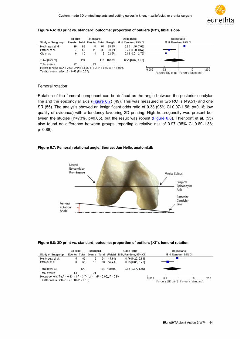
Custom-made 3D printed implants and cutting guides in knee, maxillofacial, or cranial surgery
EUnetHTA Joint Action 3 WP4 44
Figure 6.6: 3D print vs. standard; outcome: proportion of outliers (>3°), tibial slope
Femoral rotation
Rotation of the femoral component can be defined as the angle between the posterior condylar
line and the epicondylar axis (Figure 6.7) (49). This was measured in two RCTs (49,51) and one
SR (55). The analysis showed an insignificant odds ratio of 0.33 (95% CI 0.07-1.56; p=0.16; low
quality of evidence) with a tendency favouring 3D printing. High heterogeneity was present be-
tween the studies (I2=73%, p=0.05), but the result was robust (Figure 6.8). Thienpont et al. (55)
also found no difference between groups, reporting a relative risk of 0.97 (95% CI 0.69-1.38;
p=0.88).
Figure 6.7: Femoral rotational angle. Source: Jan Hejle, anatomi.dk
Figure 6.8: 3D print vs. standard; outcome: proportion of outliers (>3°), femoral rotation
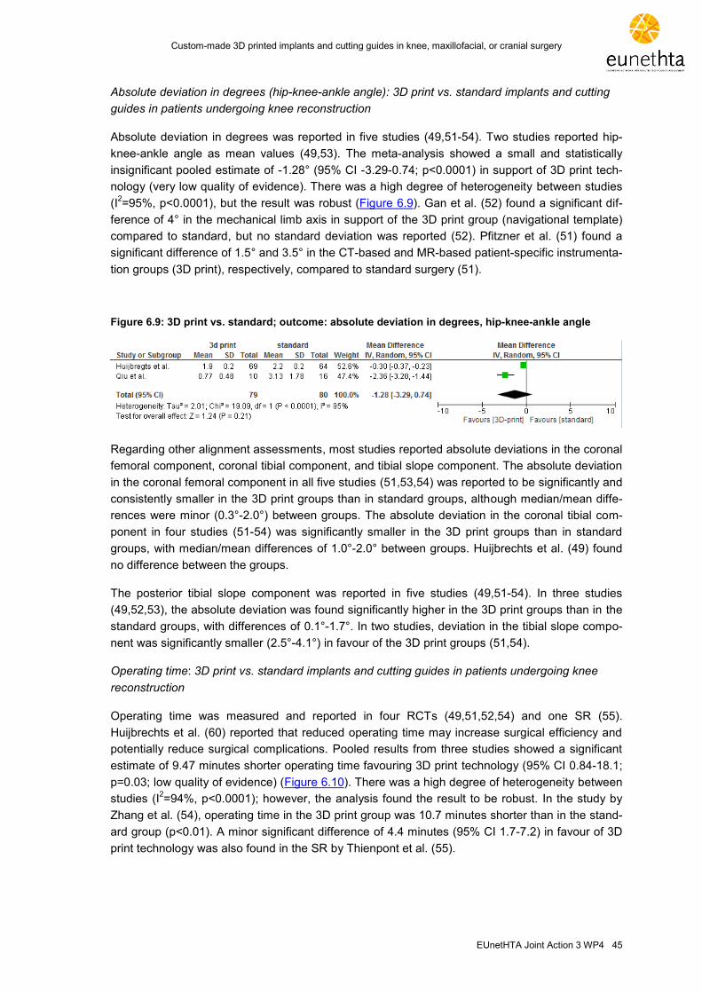
Custom-made 3D printed implants and cutting guides in knee, maxillofacial, or cranial surgery
EUnetHTA Joint Action 3 WP4 45
Absolute deviation in degrees (hip-knee-ankle angle): 3D print vs. standard implants and cutting
guides in patients undergoing knee reconstruction
Absolute deviation in degrees was reported in five studies (49,51-54). Two studies reported hip-
knee-ankle angle as mean values (49,53). The meta-analysis showed a small and statistically
insignificant pooled estimate of -1.28° (95% CI -3.29-0.74; p<0.0001) in support of 3D print tech-
nology (very low quality of evidence). There was a high degree of heterogeneity between studies
(I2=95%, p<0.0001), but the result was robust (Figure 6.9). Gan et al. (52) found a significant dif-
ference of 4° in the mechanical limb axis in support of the 3D print group (navigational template)
compared to standard, but no standard deviation was reported (52). Pfitzner et al. (51) found a
significant difference of 1.5° and 3.5° in the CT-based and MR-based patient-specific instrumenta-
tion groups (3D print), respectively, compared to standard surgery (51).
Figure 6.9: 3D print vs. standard; outcome: absolute deviation in degrees, hip-knee-ankle angle
Regarding other alignment assessments, most studies reported absolute deviations in the coronal
femoral component, coronal tibial component, and tibial slope component. The absolute deviation
in the coronal femoral component in all five studies (51,53,54) was reported to be significantly and
consistently smaller in the 3D print groups than in standard groups, although median/mean diffe-
rences were minor (0.3°-2.0°) between groups. The absolute deviation in the coronal tibial com-
ponent in four studies (51-54) was significantly smaller in the 3D print groups than in standard
groups, with median/mean differences of 1.0°-2.0° between groups. Huijbrechts et al. (49) found
no difference between the groups.
The posterior tibial slope component was reported in five studies (49,51-54). In three studies
(49,52,53), the absolute deviation was found significantly higher in the 3D print groups than in the
standard groups, with differences of 0.1°-1.7°. In two studies, deviation in the tibial slope compo-
nent was significantly smaller (2.5°-4.1°) in favour of the 3D print groups (51,54).
Operating time: 3D print vs. standard implants and cutting guides in patients undergoing knee
reconstruction
Operating time was measured and reported in four RCTs (49,51,52,54) and one SR (55).
Huijbrechts et al. (60) reported that reduced operating time may increase surgical efficiency and
potentially reduce surgical complications. Pooled results from three studies showed a significant
estimate of 9.47 minutes shorter operating time favouring 3D print technology (95% CI 0.84-18.1;
p=0.03; low quality of evidence) (Figure 6.10). There was a high degree of heterogeneity between
studies (I2=94%, p<0.0001); however, the analysis found the result to be robust. In the study by
Zhang et al. (54), operating time in the 3D print group was 10.7 minutes shorter than in the stand-
ard group (p<0.01). A minor significant difference of 4.4 minutes (95% CI 1.7-7.2) in favour of 3D
print technology was also found in the SR by Thienpont et al. (55).

Custom-made 3D printed implants and cutting guides in knee, maxillofacial, or cranial surgery
EUnetHTA Joint Action 3 WP4 46
Figure 6.10: 3D print vs. standard; outcome: operating time, patients undergoing knee reconstruction
3D print vs. usual care in maxillofacial surgery patients
Operating time was assessed in one randomised study of four studies evaluating computer-
assisted mandibular reconstruction (19). Operating time was the main outcome. The operating
time was presented in terms of six outcomes: a) shaping time at the donor site, significant diffe-
rence in favour of standard group, 37.8 min vs. 62.1 min; b) shaping time at the defect site, signi-
ficant difference in favour of 3D print group, 6.2 min vs. 20.3 min; c) time for osteosynthesis, sig-
nificant difference in favour of 3D print group, 10.1 min vs. 18.2 min; d) overall reconstruction time
(which includes time to shape the transplant and perform the osteosynthesis), significant diffe-
rence in favour of 3D print group, 16.4 min vs. 38.5 min (p<0.001); e) ischaemic time (time from
dissection of the transplant until perfusion is restored), significant difference in favour of 3D print
group, 96.1 min vs. 122.9 min (p<0.005); and f) overall operating time, no difference between the
groups, 498.5 min vs. 525.2 min (p=0.527). Further, no difference was found in ICU time and
postoperative hospitalisation time.
3D print vs. usual care in cranioplasty patients
Using 3D print imaging and print technology reduced the time of cranioplasty neurosurgery by
16.2 minutes (p<0.001) in a group of 19 patients compared to a control group of 20 patients (very
low quality of evidence) (39).
Visual analogue scale (VAS)
One study reported pain one and two years postoperatively in patient undergoing TKA and found
no difference between 3D print and standard groups (p=0.227) (50).
Blood Loss
Blood loss or blood transfusion was assessed in two RCTs (52,54) and one SR (55) on patients
undergoing knee reconstruction and in one RCT of maxillofacial surgery patients (19). In two stu-
dies (19,54), there was no difference in blood loss between the groups, and in two other studies
(52,55), there was a significant difference in blood loss in favour of the 3D print groups, with a
mean difference of 38 and 90 ml, respectively. Although significant, these differences were of no
clinically relevant value.
Precision
Precision or accuracy was evaluated in one small prospective study of low quality in patients un-
dergoing mandibular reconstruction (57). The study results can only be indicative of the effect of
3D printing technology in this area. Mazzoni et al. (57) found no difference between groups in
patients undergoing prosthetically guided mandibular reconstruction with respect to midline devia-
tion (vertical and horizontal), mandible angle-shift right, angular deviation of the mandibular arch,

Custom-made 3D printed implants and cutting guides in knee, maxillofacial, or cranial surgery
EUnetHTA Joint Action 3 WP4 47
and condyle position left. Significant differences in favour of the 3D intervention were reported for
the outcomes mandible angle-shift left (mean 1.4 mm vs. 5.042 mm; p<0.006) and condyle posi-
tion right (mean 1.297 mm vs. 4.458 mm; p=0.035).
Changes in sensory function
Changes in sensory function were evaluated in only one small RCT of moderate quality in patients
undergoing bilateral sagittal split ramus osteotomy (SSRO) in a split-mouth design (59). SSRO
can sometimes result in postsurgical neurosensory disturbance. Al-Ahmad et al. (59) investigated
whether or not a computer-assisted surgical guide for SSRO more effectively reduced the inci-
dence and severity of neurosensory complications than standard SSRO. With respect to tactile
threshold, 67% of patients in the computer-assisted SSRO group had an abnormal threshold after
one week in the lower lip and chin vs. 83% in the standard SSRO group (p<0.05). The tactile
threshold was also significantly different in the chin after three months and the lower lip after six
months. Likewise, two-point discrimination was significantly different in favour of the computer-
assisted SSRO group in the lower lip at one week and the chin at six months (p<0.05). Other out-
come measures, including direction of brush stroke and intraoperative parameters assessed by
the surgeon, were equivalent between groups.
[D0011] – What is the effect of 3D printed implants and cutting guides on patients’ body
functions?
[D0016] – How does the use of 3D printed implants and cutting guides affect activities of
daily living?
Function and activities of daily living were examined using two knee assessment scores, an oste-
oarthritis index, and clinical examination (in Brandao et al. (58)). Three RCTs (49-51) and two
SRs (55,56) examined patients undergoing knee reconstruction, and one RCT examined mandib-
ular reconstruction patients.
Knee Society Score (KSS)
The KSS was developed as a simple, objective way to measure a patient’s function before and
after TKA. KSSs range from 0 to 100, with 0 being the worst outcome and 100 being the best
possible outcome. KSSs were reported in two RCTs and two SRs. In the two RCTs by Pfitzner et
al. (51) and Boonen et al. (50), the scores were almost equivalent between groups in follow-up
ranging from three months to two years, although pre- to post-operative scores increased radically
(50,51). The two SRs by Thienpont et al. (55) and Mannan et al. (56) did not report any diffe-
rences in knee scores, but Thienpont et al. (55) reported a mean difference in KSS in favour of
the 3D print group of 4.3 points (95% CI 1.5-7.2; p=0.003). This difference is not considered to be
of clinical significance (50).
Oxford Knee Scale
The Oxford Knee Scale was reported in three studies (49,50,56). The meta-analysis showed a
small and statistically insignificant pooled mean difference of 1.29 (95% CI -0.84-3.41; p=0.24)
points in support of 3D print technology (moderate quality of evidence). There was a low degree
of heterogeneity between studies (I2=0%, p=0.68; Figure 6.11). Mannan et al. (56) reported a
statistically insignificant pooled mean difference of 0.48 points in support of standard surgery.
Thus, the results can be regarded as imprecise and inconsistent using this scale (56).

Custom-made 3D printed implants and cutting guides in knee, maxillofacial, or cranial surgery
EUnetHTA Joint Action 3 WP4 48
Figure 6.11: 3D print vs. standard; outcome: Oxford Knee Score, patients undergoing knee recon-struction

Custom-made 3D printed implants and cutting guides in knee, maxillofacial, or cranial surgery
EUnetHTA Joint Action 3 WP4 49
Quality of life
[D0012] - What is the effect of 3D printed implants and cutting guides on generic health-
related quality of life?
[D0013] - What is the effect of 3D printed implants and cutting guides on disease-specific
quality of life?
Quality of life: SF12, EQ5D, and OHIP14 scores
Health-related quality of life (HRQoL) was evaluated in three RCTs (49,50,58): two in patients
undergoing knee reconstruction (49,50) and one in mandibular reconstruction patients (58). Stu-
dies were of moderate quality. HRQoL was measured using the generic scores SF12 (Physical
and Mental Component) and EQ5D, and the disease-specific OHIP-14 score (Oral Health Impact
Profile) to evaluate the impact of interventions on oral HRQoL.
Huijbrechts et al. (49) reported that physical and mental SF12 scores were not significantly diffe-
rent at three months (p=0.418 and p=0.267, respectively) or at one year (p=0.114 and p=0.569)
post-operatively. Likewise, Boonen et al. (50) reported equal progressions in the intervention and
standard groups using the EQ5D score. OHIP-14 scores in mandibular reconstruction patients
showed a significant difference in favour of the 3D printed group (p=0.027) (58).
Patient satisfaction
[D0017] - Were patients satisfied with the use of 3D printed implants and cutting guides?
Satisfaction was not reported in any study as an outcome and was not reported in any of the in-
cluded RCTs or cohort studies.

Custom-made 3D printed implants and cutting guides in knee, maxillofacial, or cranial surgery
EUnetHTA Joint Action 3 WP4 50
7 SAFETY (SAF)
7.1 Research questions
Element ID Research question
C0008 How safe is the use of 3D printed implants and cutting guides in relation to standard implants and cutting guides?
C0004 How does the frequency or severity of harm change over time or in different settings?
C0005 What are the susceptible patient groups that are more likely to be harmed through the use of 3D printed implants and cutting guides?
B0010 What kind of data/records and/or registry is needed to monitor the use of 3D printed implants and cutting guides and standard implants and cutting guides?
7.2 Results
Included studies
This domain included all studies from the EFF domain as well as three additional studies reporting
safety concerns in maxillofacial and cranial surgery (61-63). Other safety issues not covered in
these studies may exist as short and not long-term outcomes were generally examined.
Patient safety
[C0008] – How safe is the use of 3D printed implants and cutting guides in relation to standard implants and cutting guides?
Knee surgery
Few of the included studies included data on complications occurring in TKA comparing those
undergoing patient-specific instruments (PSIs), patient-matched positioning guides (PMPGs), and
navigation templates (NTs) and those using standard interventions. In these short-term studies,
no additional complications associated with 3D printed implants and cutting guides in TKA were
reported. In Huijbregts et al. (49), some of the most frequent complications were infection (10.1%
vs. 10.9%), manipulation under anaesthetic (1.4% vs. 7.8%), venous thromboembolism (0% vs.
1.6%) and haemarthrosis (1.4% vs. 0%) in the PSI group compared with the standard instrumen-
tation group, respectively. In Boonen et al. (50), some of the most frequent complications were
infection (0% vs. 1.2%), manipulation under anaesthetic (4.9% vs. 2.5%), venous thromboembo-
lism (1.2% vs. 0%) and haemarthrosis (2.4% vs. 0%) in the PMPG group compared with standard
instrumentation. In Zhang et al. (54), there was no difference in infection or venous thromboembo-
lism in the NT group compared with the standard intramedullary positioning group.
Maxillofacial surgery
One study examined complications in mandibular reconstruction comparing those assigned to
computer-assisted mandibular reconstruction and those assigned to standard reconstruction. In
Ayoub et al. (19), there was no significant difference in complication rate such as operating time
or duration in the intensive care unit between the computer-assisted group and the standard
group. However, there was a difference in ischaemic time, with a decrease in the computer-
assisted group. Two retrospective studies of small sample size which were not included in the

Custom-made 3D printed implants and cutting guides in knee, maxillofacial, or cranial surgery
EUnetHTA Joint Action 3 WP4 51
EFF domain evaluated operating times between 3D printing groups (5.5 ± 0.5 hours) and stan-
dard groups (6.5 ± 0.7 hours) and found significant differences (61).
Cranial surgery
Chrzan et al. (63) examined complication rates in cranioplasty between those undergoing com-
puter-aided design (CAD) modelling and those undergoing standard cranioplasty. There was a
significant difference in mean operating time between the CAD group (120.3 minutes, 95% CI
110-140) and the control group (136.5 minutes, 95% CI 120-150; p<0.000004). In a study not
included in the EFF domain, only complications from standard cranioplasty were evaluated. The
most frequently observed complications were wound infection (11.3%), bone resorption (6.5%), or
sunken bone plates (1.6%).
[C0004] – How does the frequency or severity of harm change over time or in different
settings?
Knee, maxillofacial surgery, or cranial surgery
The included studies had some data on short-term outcomes in hospital settings but no infor-
mation on long-term complications to examine safety outcomes such as implant survival. There-
fore, it is not possible to further evaluate harm changes over time or to evaluate harm increases or
decreases in different organisational settings such as implant failure, prosthesis problems, and
continuing pain.
[C0005] – What are the susceptible patient groups that are more likely to be harmed
through the use of 3D printed implants and cutting guides?
Knee, maxillofacial surgery or cranial surgery
3D printed implants and cutting guides in TKA are not recommended in patients with previous
knee replacement of the same knee. Caution should be exerted in patients with any metal device
that affects image quality of the knee, angular deformities greater than 15° of fixed varus, valgus,
or flexion, tibial slope exceeding 15°, or with moderate to severe bony deformities, Charcot knee,
or patients with severe patella tendon calcification that may prevent patella eversion8. No infor-
mation is available concerning susceptible patient groups in mandibular reconstruction and crani-
oplasty.
[B0010] - What kind of data/records and/or registry is needed to monitor the use of 3D
printed implants and cutting guides and standard implants and cutting guides?
Knee, maxillofacial surgery or cranial surgery
In the developmental phase, the use of high-risk 3D-printed medical devices would only be al-
lowed in a selection of specialist centres (phase I and D). This is to avoid having high-risk medical
devices widely adopted in many hospitals without a thorough evaluation of their safety and added
value. Before an innovation becomes widespread, a formal scientific evaluation should have taken
place within an appropriate study design in which a comparison is made with existing alternatives
(phase A). The government can (co-)finance a system to use existing administrative databanks.
8
[http://synthes.vo.llnwd.net/o16/LLNWMB8/INT%20Mobile/Synthes%20International/Product%20Support%20Material/le
gacy_DePuy_PDFs/DSUS-JRC-0115-0698-1_LR.pdf]

Custom-made 3D printed implants and cutting guides in knee, maxillofacial, or cranial surgery
EUnetHTA Joint Action 3 WP4 52
The producer should first finance a study to evaluate outcomes that are not present in the admini-
strative databanks (such as comparative effectiveness in QoL) (3).
Regarding long-term performance, all new procedures must be reported to a follow-up database/
registry to provide data on complication rates of 3D printed implants and cutting guides in TKA
compared with standard instrumentation group(s).
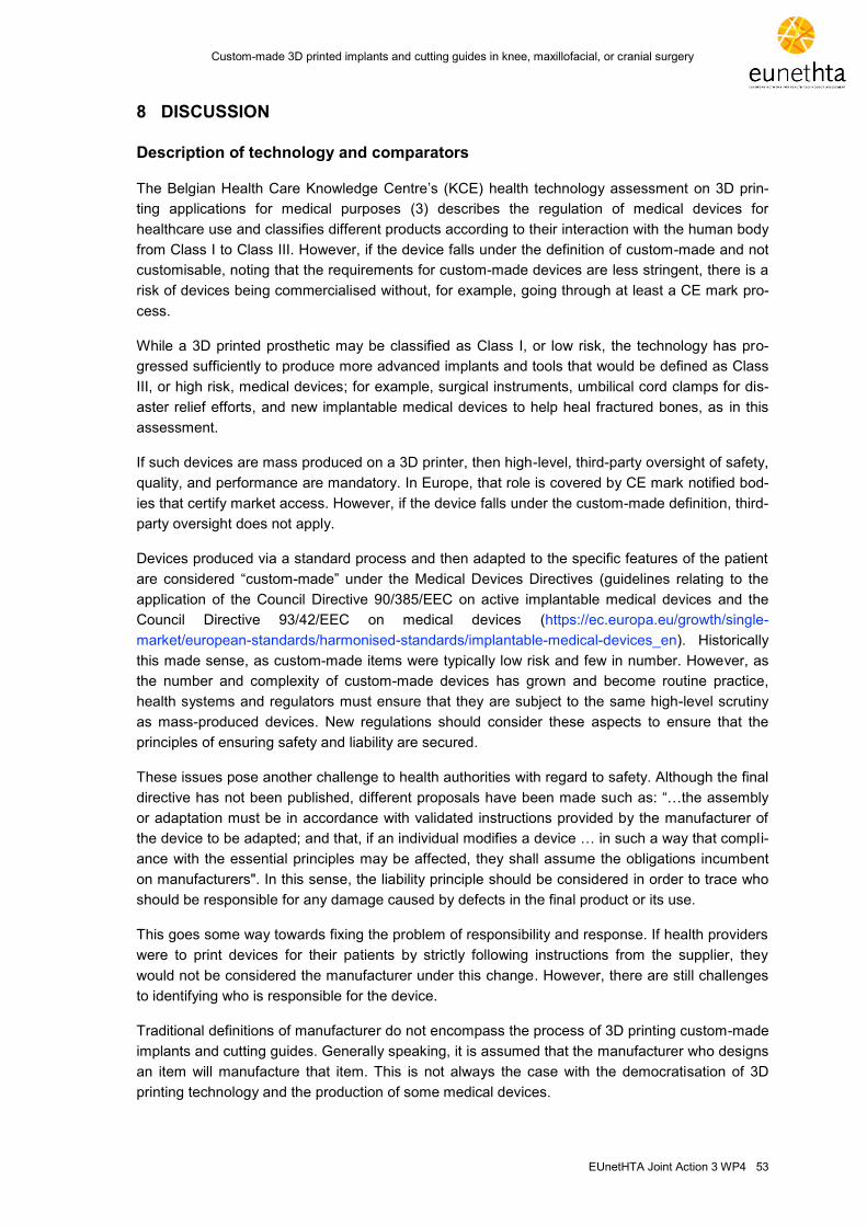
Custom-made 3D printed implants and cutting guides in knee, maxillofacial, or cranial surgery
EUnetHTA Joint Action 3 WP4 53
8 DISCUSSION
Description of technology and comparators
The Belgian Health Care Knowledge Centre’s (KCE) health technology assessment on 3D prin-
ting applications for medical purposes (3) describes the regulation of medical devices for
healthcare use and classifies different products according to their interaction with the human body
from Class I to Class III. However, if the device falls under the definition of custom-made and not
customisable, noting that the requirements for custom-made devices are less stringent, there is a
risk of devices being commercialised without, for example, going through at least a CE mark pro-
cess.
While a 3D printed prosthetic may be classified as Class I, or low risk, the technology has pro-
gressed sufficiently to produce more advanced implants and tools that would be defined as Class
III, or high risk, medical devices; for example, surgical instruments, umbilical cord clamps for dis-
aster relief efforts, and new implantable medical devices to help heal fractured bones, as in this
assessment.
If such devices are mass produced on a 3D printer, then high-level, third-party oversight of safety,
quality, and performance are mandatory. In Europe, that role is covered by CE mark notified bod-
ies that certify market access. However, if the device falls under the custom-made definition, third-
party oversight does not apply.
Devices produced via a standard process and then adapted to the specific features of the patient
are considered “custom-made” under the Medical Devices Directives (guidelines relating to the
application of the Council Directive 90/385/EEC on active implantable medical devices and the
Council Directive 93/42/EEC on medical devices (https://ec.europa.eu/growth/single-
market/european-standards/harmonised-standards/implantable-medical-devices_en). Historically
this made sense, as custom-made items were typically low risk and few in number. However, as
the number and complexity of custom-made devices has grown and become routine practice,
health systems and regulators must ensure that they are subject to the same high-level scrutiny
as mass-produced devices. New regulations should consider these aspects to ensure that the
principles of ensuring safety and liability are secured.
These issues pose another challenge to health authorities with regard to safety. Although the final
directive has not been published, different proposals have been made such as: “…the assembly
or adaptation must be in accordance with validated instructions provided by the manufacturer of
the device to be adapted; and that, if an individual modifies a device … in such a way that compli-
ance with the essential principles may be affected, they shall assume the obligations incumbent
on manufacturers". In this sense, the liability principle should be considered in order to trace who
should be responsible for any damage caused by defects in the final product or its use.
This goes some way towards fixing the problem of responsibility and response. If health providers
were to print devices for their patients by strictly following instructions from the supplier, they
would not be considered the manufacturer under this change. However, there are still challenges
to identifying who is responsible for the device.
Traditional definitions of manufacturer do not encompass the process of 3D printing custom-made
implants and cutting guides. Generally speaking, it is assumed that the manufacturer who designs
an item will manufacture that item. This is not always the case with the democratisation of 3D
printing technology and the production of some medical devices.

Custom-made 3D printed implants and cutting guides in knee, maxillofacial, or cranial surgery
EUnetHTA Joint Action 3 WP4 54
Nevertheless, the directive is clear that all the producers involved in the production process are
liable as well as all actors in the supply chain.
Health problem
3D printed medical devices are most frequently applied to knee, maxillofacial, and cranial surgery.
The most frequent diseases in the included studies were primarily knee OA and secondary RA
treated with TKA, oral cancer treated with mandibular reconstruction, and traumatic brain injury with
intracranial hypertension treated with decompressive craniectomy and later cranioplasty. These are
serious and global conditions, with oral cancer affecting over 300,000 people and TBI over 10 mil-
lion people each year, and approximately 1% of the population make contact with a doctor with
symptoms of knee arthritis (45,46). The introduction of 3D print technology does not significantly
change standard instrumentation procedures in TKA, mandibular reconstruction, or cranioplasty but
it may improve outcomes. In Europe, 3D print technology only accounts for ~1.3% of the 1,324,000
annual TKAs (1,2). In mandibular reconstruction and cranioplasty, there are no published data on
the utilisation of the technologies in Europe, but 3D print technology is also growing in these clinical
areas.
Clinical effectiveness
Thirteen studies reported on the use of surgical interventions using 3D print technology compared
with standard interventions on patients undergoing knee, maxillofacial, or cranioplastic surgery.
This assessment provided both a meta-analysis and a narrative summary on outcomes when
meta-analysis was not possible. Overall, the evidence level for the included studies was very low
to moderate, mainly due to the risk of bias and the imprecision of the estimates in the included
studies, which might limit the robustness of our findings.
Results by outcome
Alignment
This outcome was only reported in patients undergoing TKA.
Number of outliers (by >3°) in TKA
The number of outliers in relation to TKA was reported in six RCTs and one SR with respect to
five separate outcomes: hip-knee-ankle angle (limb), coronal femoral angle, coronal tibial angle,
tibial slope, and femoral rotation. Results from the meta-analysis and SR are presented in Table
8.1.
Table 8.1: Meta-analysis regarding alignment in 3D surgery vs. standard. Results from RCTs and SRs (Thienpont et al. (55))
Endpoints Results (OR) P-value Results SR* (RR)
Hip-knee-ankle angle 0.29 in favour of 3D print <0.0001 0.79 in favour of 3D print (p=0.013)
Coronal femur 0.24 in favour of 3D print <0.001 0.74 in favour of 3D print (p=0.043)
Coronal tibial 0.29 in favour of 3D print 0.005 1.30 (p=0.13)
Tibial slope 0.55 0.57 1.32 in favour of standard (p=0.001)
Femoral rotation 0.33 0.16 0.97 (p=0.88)

Custom-made 3D printed implants and cutting guides in knee, maxillofacial, or cranial surgery
EUnetHTA Joint Action 3 WP4 55
Operating time
In patients undergoing knee surgery, only minor differences in operating time were observed;
however, differences were statistically significant, favouring 3D surgery patients compared with
standard instrumentation. Estimates ranged from 4.4 to 10.7 minutes. The quality of the evidence
was low and heterogeneity was high. No difference in overall operating time was found in maxilla-
facial patients. A significant difference of 16.2 minutes was found in one small study of craniopla-
sty patients favouring 3D surgery (very low quality of evidence).
Other outcomes
No significant or clinically relevant differences were found in pain scores and blood loss. In rela-
tion to function and activities of daily living, no differences in the two groups were found in knee
scores between patients undergoing knee reconstruction. Only one of three health-related quality
of life scores showed a significant difference in favour of the intervention group.
In summary, 3D surgery using 3D printed implants and cutting guides in TKA compared with
standard instrumentation resulted in greater precision, as demonstrated through outcomes such
as malalignment (hip-knee-ankle angle, coronal femoral angle, and coronal tibial angle) or abso-
lute deviation. No other outcomes showed clinical or statistically significant results in favour of 3D
surgery or standard surgery. Until further evidence is generated, these results may support the
use of 3D print technology as an alternative to standard surgery, with the caveat that the quality of
current evidence varies from very low to low. Use should be restricted to reference centres, and
data on safety and efficacy must be collected. Patients should be informed about alternatives,
possible benefits, and the risks. Earlier studies also support the use of 3D surgery until additional
high-quality studies are produced (1). However, it is necessary to establish what if any relevant
improvements 3D printed surgery offers before any final decision is made on continuing use of the
technology. Proposed data collection systems by EUnetHTA JA3, WP5 offer a good starting point
to standardise evidence generation while ensuring quality.
Based on the available literature, it is challenging to weigh which outcomes/results are most im-
portant. From a patient perspective, patient-related outcomes such as functionality, QoL, and
changes in sensory function seem to be most relevant. Unfortunately, it may be difficult to com-
ment on these effects on the basis of the literature, since these results are not particularly well
quantified or established. It should also be mentioned that it is often not possible to determine
which differences in effect sizes are clinically relevant when comparing groups. The outcomes
suggestive of a relevant effect (alignment) are proxy outcomes, such that direct patient effects
such as pain or QoL are not immediate, although larger alignment problems in TKA patients may
result in functional difficulties. It has been shown that between 6 and 12% of TKAs fail as a result
of malalignment of the components (49), and since malalignment may contribute to instability,
aseptic loosening, and unexplained pain (49), precision in these procedures is of great im-
portance. As in this assessment and most studies concerning TKA patients, precision is demon-
strated through outcomes such as outliers (often defined as a deviation by >3° from the planned
positioning of the implant) or absolute deviation in degrees. In support of this approach, Gan et al.
(52) commented that biomechanics research has demonstrated that precisely restoring the lower
extremity can be a very effective method to avoid polyethylene wear. The same authors also sug-
gested that increased contact pressure will lead to increased polyethylene wear, prosthesis loos-
ening, and TKA failure. Gan et al. (52) also reported 3° varus–valgus alignment of the lower limb
in the frontal plane to be optimal, and Pfitzner et al. (51) pointed out that in the absence of long-
term studies, a neutral mechanical axis remains the gold standard. However, Huijbrechts et al.
(49) said: "Future correlation with aseptic loosening is required to determine if the cumulative

Custom-made 3D printed implants and cutting guides in knee, maxillofacial, or cranial surgery
EUnetHTA Joint Action 3 WP4 56
deviation has clinical rather than mathematical value", and Thienpont et al. (55) said that "authors
have concluded that the impact of mechanical coronal plane malalignment may be smaller than
originally believed which may cast doubt on the premise of PSI". Future studies will need to
demonstrate a relevant long-term impact of malalignment in TKA patients. Furthermore, it is im-
portant to note that the above results reflect the use of different 3D systems (Table 4.4, Table A1,
Table A.2) and imaging modalities (MR and CT) and that intra- and inter-observer variability may
influence alignment measurements (49).
Safety
Safety issues related to 3D print technology and standard instrumentation were examined in few
of the included studies. There was no overall difference in complications between the technolo-
gies in TKA, mandibular reconstruction, or cranioplasty. A difference was found in ischaemic time
in mandibular reconstruction, with a decrease in the 3D print group using individual surgical
guides. The included studies only included data on short-term outcomes such as infection, venous
thromboembolism, haemarthrosis, ischaemia, and operating time. Long-term complications such
as harm change over time, implant failure, prosthesis problems, and continued pain are needed to
fully evaluate which safety issues are relevant to this new technology (51,52,58). Another concern
not accounted for is the associated exposure to ionising radiation triggered by the repeat CT-
scans acquired in many of the 3D print technologies. Although the risk for any person is small, the
increased exposure to radiation when using 3D print technology is not negligible (64).
Need for research/evidence gaps
Well-prepared RCTs or prospective cohort studies with relevant comparisons in all areas of 3D
print technology are required. Accordingly, there is still a need to illustrate whether or not the ef-
fects and complications of 3D print technology are different to standard surgery. Also, future stu-
dies need to better utilise standardised assessment tools and gold-standard assessments and
explicitly present criteria when necessary in alignment studies. Furthermore, it will be relevant to
assess patient-related outcomes such as functional outcomes and QoL as well as long-term out-
comes and complications such as implant survival and persistent pain. There also needs to be a
focus on evaluation methods for 3D print technologies. The availability of CT and MRI and the
recent developments in CAD software provide opportunities to update the evaluation methods for
3D printing technologies and thereby deliver more reliable and accurate results.
Another strategy for collecting information about the use of 3D printed implants and cutting guides
could be to limit the use of 3D technology to a limited number of hospitals acting as reference
centres. This offers a number of advantages, including better patient outcomes through concen-
trated expertise and, for devices without evidence of efficacy, opportunities for data collection.

Custom-made 3D printed implants and cutting guides in knee, maxillofacial, or cranial surgery
EUnetHTA Joint Action 3 WP4 57
9 CONCLUSION
3D printed technology showed greater precision in terms of malalignment than standard instru-
mentation in TKA. However, the evidence was of very low or low quality. No firm conclusions can
be drawn for mandibular reconstruction and cranioplasty, since no other outcomes showed clinical
or statistically significant results in favour of either technology. The clinical implications of the fin-
dings in TKA are uncertain, and further research is needed to assess patient-related outcomes
from the use of 3D print technology before any final decision on continued use of the technology.
Regarding safety, while a few short-term outcomes such as infection, venous thromboembolism,
and haemarthrosis were reported, there were no overall differences except from ischaemic time in
mandibular reconstruction between the assessed technologies.

Custom-made 3D printed implants and cutting guides in knee, maxillofacial, or cranial surgery
EUnetHTA Joint Action 3 WP4 58
10 REFERENCES
(1) Kurtz SM, Ong KL, Lau E, et al. International survey of primary and revision total knee re-placement. Int Orthop 2011;35(12):1783-1789.
(2) Thienpont E, Bellemans J, Delport H, et al. Patient-specific instruments: industry's innovation with a surgeon's interest. Knee Surg Sports Traumatol Arthrosc 2013;21(10):2227-2233.
(3) Vinck I, Vijverman A, Vollebregt E, et al. Responsible use of high-risk medical devices: the example of 3D printed medical devices https://kce.fgov.be/sites/default/files/atoms/files/KCE_297C_3D_printing_Synthesis.pdf (accessed 21 Mar 2019)
(4) Ursan ID, Chiu L, Pierce A. Three-dimensional drug printing: a structured review. J Am Pharm Assoc 2013;53(2):136-144.
(5) Bartlett S. Printing organs on demand. Lancet Respir Med 2013;1(9):684-2600(13)70239-X.
(6) Hao L, Savalani MM, Zhang Y, Tanner KE, Harris RA. Selective laser sintering of hydroxyap-atite reinforced polyethylene composites for bioactive implants and tissue scaffold development. Proc Inst Mech Eng H 2006;220(4):521-531.
(7) Kinstlinger IS, Bastian A, Paulsen SJ, et al. Open-Source Selective Laser Sintering (OpenSLS) of Nylon and Biocompatible Polycaprolactone. PLoS One 2016;11(2):e0147399.
(8) Chen CH, Lee MY, Shyu VB, et al. Surface modification of polycaprolactone scaffolds fabri-cated via selective laser sintering for cartilage tissue engineering. Mater Sci Eng C Mater Biol Appl 2014;40:389-397.
(9) Tan KH, Chua CK, Leong KF, et al. Selective laser sintering of biocompatible polymers for applications in tissue engineering. Biomed Mater Eng 2005;15(1-2):113-124.
(10) Rimell JT, Marquis PM. Selective laser sintering of ultra high molecular weight polyethylene for clinical applications. J Biomed Mater Res 2000;53(4):414-420.
(11) Williams DF. On the mechanisms of biocompatibility. Biomaterials 2008;29(20):2941-2953.
(12) Mertz L. Dream it, design it, print it in 3-D: what can 3-D printing do for you? IEEE Pulse 2013;4(6):15-21.
(13) Banks J. Adding value in additive manufacturing: researchers in the United Kingdom and Europe look to 3D printing for customization. IEEE Pulse 2013;4(6):22-26.
(14) Schaub DA, Chu K, Montgomery DC. Optimizing stereolithography throughput. J Manuf Syst 1997;16(4):290.
(15) Hoy MB. 3D printing: making things at the library. Med Ref Serv Q 2013;32(1):94-99.
(16) Tack P, Victor J, Gemmel P, Annemans L. 3D-printing techniques in a medical setting: a systematic literature review. Biomed Eng Online 2016;15(1):115-016-0236-4.
(17) Weinstein AM, Rome BN, Reichmann WM, et al. Estimating the burden of total knee re-placement in the United States. J Bone Joint Surg Am 2013;95(5):385-392.
(18) Hsu H, Siwiec RM. Osteoarthritis, Knee. StatPearls Treasure Island (FL): StatPearls Publish-ing LLC; 2018.
(19) Ayoub N, Ghassemi A, Rana M, et al. Evaluation of computer-assisted mandibular recon-struction with vascularized iliac crest bone graft compared to conventional surgery: a randomized prospective clinical trial. Trials 2014;15:114.

Custom-made 3D printed implants and cutting guides in knee, maxillofacial, or cranial surgery
EUnetHTA Joint Action 3 WP4 59
(20) Kunze E, Meixensberger J, Janka M, Sorensen N, Roosen K. Decompressive craniectomy in patients with uncontrollable intracranial hypertension. Acta Neurochir Suppl 1998;71:16-18.
(21) Newberry SJ, FitzGerald J, SooHoo NF, Booth M, Marks J, Motala A, et al. Agency for Healthcare Research and Quality (US) 2017; Report No.: 17-EHC011-EF.
(22) Neogi T. The epidemiology and impact of pain in osteoarthritis. Osteoarthritis Cartilage 2013;21:1145-1153.
(23) Yan D, Yang J, Pei F. Total knee arthroplasty treatment of rheumatoid arthritis with severe versus moderate flexion contracture. J Orthop Surg Res 2013;8:41.
(24) Palasz P, Adamski L, Gorska-Chrzastek M, Starzynska A, Studniarek M. Contemporary Di-agnostic Imaging of Oral Squamous Cell Carcinoma - A Review of Literature. Pol J Radiol 2017;82:193-202.
(25) Alvis-Miranda H, Castellar-Leones SM, Moscote-Salazar LR. Decompressive Craniectomy and Traumatic Brain Injury: A Review. Bull Emerg Trauma 2013;1(2):60-68.
(26) Abbott JH, Usiskin IM, Wilson R, Hansen P, Losina E. The quality-of-life burden of knee os-
teoarthritis in New Zealand adults: A model-based evaluation. PLoS One 2017;12(10):e0185676.
(27) Bagan J, Sarrion G, Jimenez Y. Oral cancer: clinical features. Oral Oncology 2010;46(6):414-7.
(28) Moubayed SP, L'Heureux-Lebeau B, Christopoulos A, et al. Osteocutaneous free flaps for mandibular reconstruction: systematic review of their frequency of use and a preliminary quality of life comparison. J Laryngol Otol 2014;128(12):1034-1043.
(29) Grindlinger GA, Skavdahl DH, Ecker RD, Sanborn MR. Decompressive craniectomy for se-vere traumatic brain injury: clinical study, literature review and meta-analysis. Springerplus 2016;5(1):1605-016-3251-9. eCollection 2016.
(30) Gopaul R, Wei ZD, Yan J, Gong FY, Xiao SW. Clinical study of quality of life of traumatic brain injury patients after decompressive craniectomy and related influencing factors. Chinese Neurosurg J 2016;2(1):33.
(31) Hunter DJ, Schofield D, Callander E. The individual and socioeconomic impact of osteoarthri-tis. Nat Rev Rheumatol 2014;10(7):437-441.
(32) Parkin DM, Bray F, Ferlay J, Pisani P. Global cancer statistics, 2002. CA Cancer J Clin 2005;55(2):74-108.
(33) Han S, Chen Y, Ge X, et al. Epidemiology and cost analysis for patients with oral cancer in a university hospital in China. BMC Public Health 2010;10:196.
(34) Humphreys I, Wood RL, Phillips CJ, Macey S. The costs of traumatic brain injury: a literature review. Clinicoecon Outcomes Res 2013;5:281-287.
(35) Michael JW, Schluter-Brust KU, Eysel P. The epidemiology, etiology, diagnosis, and treat-ment of osteoarthritis of the knee. Dtsch Arztebl Int 2010;107(9):152-162.
(36) Markopoulos AK. Current aspects on oral squamous cell carcinoma. Open Dent J
2012;6:126-130.
(37) Chung Pearl KF. Traumatic Brain Injury (TBI): Overview of Diagnosis and Treatment. J Neu-rol Neurolphysiol 2013;5(1).
(38) Rivera C. Essentials of oral cancer. Int J Clin Exp Pathol 2015;8(9):11884-11894.
(39) Chrzan R, Urbanik A, Karbowski K, Moskala M, Polak J, Pyrich M. Cranioplasty prosthesis manufacturing based on reverse engineering technology. Med Sci Monit 2012;18(1):MT1-6.

Custom-made 3D printed implants and cutting guides in knee, maxillofacial, or cranial surgery
EUnetHTA Joint Action 3 WP4 60
(40) Yu D, Peat G, Bedson J, Jordan KP. Annual consultation incidence of osteoarthritis estimat-ed from population-based health care data in England. Rheumatol 2015;54(11):2051-2060.
(41) Van Manen MD, Nace J, Mont MA. Management of primary knee osteoarthritis and indica-tions for total knee arthroplasty for general practitioners. J Am Osteopath Assoc 2012;112(11):709-715.
(42) Vos T, Flaxman AD, Naghavi M, et al. Years lived with disability (YLDs) for 1160 sequelae of 289 diseases and injuries 1990-2010: a systematic analysis for the Global Burden of Disease Study 2010. Lancet 2012;380(9859):2163-2196.
(43) de Morais EF, Mafra RP, Gonzaga AKG, de Souza DLB, Pinto LP, da Silveira EJD. Prognos-tic Factors of Oral Squamous Cell Carcinoma in Young Patients: A Systematic Review. J Oral Maxillofac Surg 2017;75(7):1555-1566.
(44) Zhang Y, Jordan JM. Epidemiology of osteoarthritis. Clin Geriatr Med 2010;26(3):355-369.
(45) Torre LA, Bray F, Siegel RL, Ferlay J, Lortet-Tieulent J, Jemal A. Global cancer statistics, 2012. CA Cancer J Clin 2015;65(2):87-108.
(46) Moon JW, Hyun DK. Decompressive Craniectomy in Traumatic Brain Injury: A Review Arti-cle. Korean J Neurotrauma 2017;13(1):1-8.
(47) Okay D, Al Shetawi AH, Moubayed SP, Mourad M, Buchbinder D, Urken ML. Worldwide 10-Year Systematic Review of Treatment Trends in Fibula Free Flap for Mandibular Reconstruction. J Oral Maxillofac Surg 2016;74(12):2526-2531.
(48) Kumar BP, Venkatesh V, Kumar KA, Yadav BY, Mohan SR. Mandibular Reconstruction: Overview. J Maxillofac Oral Surg 2016;15(4):425-441.
(49) Huijbregts HJ, Khan RJ, Fick DP, et al. Component alignment and clinical outcome following total knee arthroplasty: a randomised controlled trial comparing an intramedullary alignment sys-tem with patient-specific instrumentation. Bone Joint J 2016;98-B(8):1043-1049.
(50) Boonen B, Schotanus MG, Kerens B, van der Weegen W, Hoekstra HJ, Kort NP. No differ-ence in clinical outcome between patient-matched positioning guides and conventional instru-mented total knee arthroplasty two years post-operatively: a multicentre, double-blind, random-ised controlled trial. Bone Joint J 2016;98-B(7):939-944.
(51) Pfitzner T, Abdel MP, von Roth P, Perka C, Hommel H. Small improvements in mechanical axis alignment achieved with MRI versus CT-based patient-specific instruments in TKA: a ran-domized clinical trial. Clin Orthop Relat Res 2014;472(10):2913-2922.
(52) Gan Y, Ding J, Xu Y, Hou C. Accuracy and efficacy of osteotomy in total knee arthroplasty with patient-specific navigational template. Int J Clin Exp Med 2015;8(8):12192-12201.
(53) Qiu B, Liu F, Tang B, et al. Clinical Study of 3D Imaging and 3D Printing Technique for Pa-tient-Specific Instrumentation in Total Knee Arthroplasty. J Knee Surg 2017;30(8):822-828.
(54) Zhang YZ, Lu S, Zhang HQ, et al. Alignment of the lower extremity mechanical axis by com-puter-aided design and application in total knee arthroplasty. Int J Comput Assist Radiol Surg 2016;11(10):1881-1890.
(55) Thienpont E, Schwab PE, Fennema P. Efficacy of Patient-Specific Instruments in Total Knee Arthroplasty: A Systematic Review and Meta-Analysis. J Bone Joint Surg Am 2017 15;99(6):521-530.
(56) Mannan A, Akinyooye D, Hossain F. A Meta-analysis of Functional Outcomes in Patient-Specific Instrumented Knee Arthroplasty. J Knee Surg 2017;30(7):668-674.
(57) Mazzoni S, Marchetti C, Sgarzani R, Cipriani R, Scotti R, Ciocca L. Prosthetically guided maxillofacial surgery: evaluation of the accuracy of a surgical guide and custom-made bone plate in oncology patients after mandibular reconstruction. Plast Reconstr Surg 2013;131(6):1376-1385.

Custom-made 3D printed implants and cutting guides in knee, maxillofacial, or cranial surgery
EUnetHTA Joint Action 3 WP4 61
(58) Brandao TB, Vechiato Filho AJ, Prado Ribeiro AC, et al. Evaluation of use of acrylic resin-based surgical guide in the function and quality of life provided by mandibular prostheses with microvascular free fibula flap: A four-year, randomized, controlled trial. J Prosthet Dent 2016;116(3):457-463.e2.
(59) Al-Ahmad HT, M Saleh MW, Hussein AM. Evaluation of an innovative computer-assisted sagittal split ramus osteotomy to reduce neurosensory alterations following orthognathic surgery: a pilot study. Int J Med Robot 2013;9(2):134-141.
(60) Huijbregts HJ, Khan RJ, Fick DP, et al. Component alignment and clinical outcome following total knee arthroplasty: a randomised controlled trial comparing an intramedullary alignment sys-tem with patient-specific instrumentation. Bone Joint J 2016;98-B(8):1043-1049.
(61) Ren WH, Gao L, Li SM, et al. Virtual planning and 3D printing modeling for mandibular re-construction with fibula free flap. Zhonghua Yi Xue Za Zhi 2018;98(33):2666-2670.
(62) Jacek B, Maciej P, Tomasz P, et al. 3D printed models in mandibular reconstruction with bony free flaps. J Mater Sci Mater Med 2018;29(3):23-018-6029-5.
(63) Gooch MR, Gin GE, Kenning TJ, German JW. Complications of cranioplasty following de-compressive craniectomy: analysis of 62 cases. Neurosurg Focus 2009;26(6):E9.
(64) Brenner DJ, Doll R, Goodhead DT, et al. Cancer risks attributable to low doses of ionizing radiation: assessing what we really know. Proc Natl Acad Sci U S A 2003;100(24):13761-13766.
(65) Halldorsson S, Lucumi E, Gomez-Sjoberg R, Fleming RMT. Advantages and challenges of microfluidic cell culture in polydimethylsiloxane devices. Biosens Bioelectron 2015;63:218-231.
(66) Cui X, Boland T, D'Lima DD, Lotz MK. Thermal inkjet printing in tissue engineering and re-generative medicine. Recent Pat Drug Deliv Formul 2012;6(2):149-155.
(67) Vaezi M, Seitz H, Yang S. A review on 3D micro-additive manufacturing technologies. Int J Adv Manufact Technol 2013;67(5-8):1721-1754.
(68) Cazón A, Morer P, Matey L. PolyJet technology for product prototyping: Tensile strength and surface roughness properties. Proc Inst Mech Eng Pt B: J Eng Manuf 2014;228(12):1664-1675.
(69) Kruth J, Wang X, Laoui T, Froyen L. Lasers and materials in selective laser sintering. Assem Autom 2003;23(4):357-371.
(70) Williams JM, Adewunmi A, Schek RM, et al. Bone tissue engineering using polycaprolactone scaffolds fabricated via selective laser sintering. Biomaterials 2005;26(23):4817-4827.
(71) Schmidt M, Pohle D, Rechtenwald T. Selective laser sintering of PEEK. CIRP Ann Manufact Technol 2007;56(1):205-208.
(72) Traini T, Mangano C, Sammons RL, Mangano F, Macchi A, Piattelli A. Direct laser metal sintering as a new approach to fabrication of an isoelastic functionally graded material for manu-facture of porous titanium dental implants. Dent Mater 2008;24(11):1525-1533.
(73) de Beer N, van der Merwe A. Patient-specific intervertebral disc implants using rapid manu-facturing technology. Rapid Prototyping Journal 2013;19(2):126-139.
(74) Hollander DA, von Walter M, Wirtz T, Sellei R, Schmidt-Rohlfing B, Paar O, et al. Structural, mechanical and in vitro characterization of individually structured Ti-6Al-4V produced by direct laser forming. Biomaterials 2006;27(7):955-963.
(75) Mullen L, Stamp RC, Brooks WK, Jones E, Sutcliffe CJ. Selective Laser Melting: a regular unit cell approach for the manufacture of porous, titanium, bone in-growth constructs, suitable for orthopedic applications. J Biomed Mater Res B Appl Biomater 2009;89(2):325-334.

Custom-made 3D printed implants and cutting guides in knee, maxillofacial, or cranial surgery
EUnetHTA Joint Action 3 WP4 62
(76) Olakanmi Et, Cochrane R, Dalgarno K. A review on selective laser sintering/melting (SLS/SLM) of aluminium alloy powders: Processing, microstructure, and properties. Prog Mater Sci 2015;74:401-477.
(77) Cox SC, Thornby JA, Gibbons GJ, Williams MA, Mallick KK. 3D printing of porous hydroxy-apatite scaffolds intended for use in bone tissue engineering applications. Mater Sci Eng C Mater Biol Appl 2015;47:237-247.

Custom-made 3D printed implants and cutting guides in knee, maxillofacial, or cranial surgery
EUnetHTA Joint Action 3 WP4 63
APPENDIX 1: METHODS AND DESCRIPTION OF THE EVIDENCE USED
Documentation of the Search Strategies
PubMed - primary studies:
Date: 03/04/2018
ID Search
#1 Printing, Three-Dimensional, [Mesh]
#2 Stereolithography, [Mesh]
#3 Computer-Aided Design, [Mesh]
#4 #1 OR #2 OR #3
#5 “rapid prototyping”
#6 “patient specific instruments”
#7 “Patient specific instrument”
#8 “Patient specific implant”
#9 “Patient specific implants”
#10 “Surgical guide”
#11 “Surgical guides”
#12 “Additive manufacturing”
#13 “Medical additive manufacturing”
#14 “Subtractive manufacturing”
#15 “Computer numerical machine”
#16 “3d printing”
#17 “Three dimensional printing”
#18 “3d-printing”
#19 “three-dimensional printing”
#20 #4 OR #5 OR #6 OR #7 OR #8 OR #9 OR #10 OR #11 OR #12 OR #13 OR #14 OR #15 OR #16 OR #17 OR #18 OR #19
#21 #20 AND (Controlled Clinical trail [ptyp] OR Observational study [ptyp] OR Randomized Controlled trail [ptyp])
Custom-made 3D printed implants and cutting guides in knee, maxillofacial, or cranial surgery
EUnetHTA Joint Action 3 WP4 64
#22 #21 AND last 10 years [PDat]
#23 #22 AND Humans [Mesh]
PubMed – Reviews
Date: 06/04/2018
ID Search
#1 Printing, Three-Dimensional, [Mesh]
#2 Stereolithography, [Mesh]
#3 Computer-Aided Design, [Mesh]
#4 #1 OR #2 OR #3
#5 “rapid prototyping”
#6 “patient specific instruments”
#7 “Patient specific instrument”
#8 “Patient specific implant”
#9 “Patient specific implants”
#10 “Surgical guide”
#11 “Surgical guides”
#12 “Additive manufacturing”
#13 “Medical additive manufacturing”
#14 “subtractive manufacturing”
#15 “Computer numerical machine”
#16 “3d printing”
#17 “Three dimensional printing”
#18 “3d-printing”
#19 “three-dimensional printing”
#20 #4 OR #5 OR #6 OR #7 OR #8 OR #9 OR #10 OR #11 OR #12 OR #13 OR #14 OR #15 OR #16 OR #17 OR #18 OR #19
#21 #20 AND (Systematic Reviews [ptyp] OR Meta-Analysis[ptyp])
#22 #21 AND last 5 years [PDat]
#23 #22 AND Humans [Mesh]

Custom-made 3D printed implants and cutting guides in knee, maxillofacial, or cranial surgery
EUnetHTA Joint Action 3 WP4 65
EMBASE – Primary studies
Date: 06/04/2018
ID Search
#1 Three dimensional printing/exp
#2 Stereolithography/exp
#3 Computer aided design/exp
#4 #1 OR #2 OR #3
#5 “rapid prototyping”
#6 “patient specific instruments”
#7 “Patient specific instrument”
#8 “Patient specific implant”
#9 “Patient specific implants”
#10 “Surgical guide”
#11 “Surgical guides”
#12 “Additive manufacturing”
#13 “Medical additive manufacturing”
#14 “subtractive manufacturing”
#15 “Computer numerical machine”
#16 “3d printing”
#17 “Three dimensional printing”
#18 “3d-printing”
#19 “three-dimensional printing”
#20 #4 OR #5 OR #6 OR #7 OR #8 OR #9 OR #10 OR #11 OR #12 OR #13 OR #14 OR #15 OR #16 OR #17 OR #18 OR #19
#21 #20 AND ([controlled clinical trail]/lim OR [randomized controlled trail]/lim)
#22 [2008-2018]/py
#23 #22 AND [Humans]/lim

Custom-made 3D printed implants and cutting guides in knee, maxillofacial, or cranial surgery
EUnetHTA Joint Action 3 WP4 66
EMBASE – Reviews
Date: 06/04/2018
ID Search
#1 Three dimensional printing/exp
#2 Stereolithography/exp
#3 Computer aided design/exp
#4 #1 OR #2 OR #3
#5 “rapid prototyping”
#6 “patient specific instruments”
#7 “Patient specific instrument”
#8 “Patient specific implant”
#9 “Patient specific implants”
#10 “Surgical guide”
#11 “Surgical guides”
#12 “Additive manufacturing”
#13 “Medical additive manufacturing”
#14 “subtractive manufacturing”
#15 “Computer numerical machine”
#16 “3d printing”
#17 “Three dimensional printing”
#18 “3d-printing”
#19 “three-dimensional printing”
#20 #4 OR #5 OR #6 OR #7 OR #8 OR #9 OR #10 OR #11 OR #12 OR #13 OR #14 OR #15 OR #16 OR #17 OR #18 OR #19
#21 #20 AND ([systematic review]/lim OR [meta analysis]/lim)
#22 [2013-2018]/py
#23 #22 AND [Humans]/lim

Custom-made 3D printed implants and cutting guides in knee, maxillofacial, or cranial surgery
EUnetHTA Joint Action 3 WP4 67
Cochrane
Date: 06/04/2018
ID Search
#1 MeSH descriptor: [Printing, Three-Dimensional] explode all trees
#2 MeSH descriptor: [Stereolithography] explode all trees
#3 MeSH descriptor: [Computer-Aided Design] explode all trees
#4 #1 or #2 or #3 Publication Year from 2008 to 2018
#5 "rapid prototyping" Publication
#6 "patient specific instruments"
#7 "patient specific instrument"
#8 "patient specific implants"
#9 "patient specific implant"
#10 "Surgical guides"
#11 "Surgical guide"
#12 "additive manufacturing"
#13 "medical additive manufacturing"
#14 "subtractive manufacturing"
#15 "computer numerical machine"
#16 "3d printing"
#17 "Three dimensional printing"
#18 "3d-printing"
#19 "Three-dimensional printing"
#20 #5 or #6 or #7 or #8 or #9 or #10 or #11 or #12 or #13 or #14 or #15 or #16 or #17 or #18 or #19
#21 #4 or #20 Publication Year from 2008 to 2018
A search on on-going clinical trials has been performed on ClinicalTrails.gov and Clinicaltrailsregister.eu. In both databases the search word was 3D. The search was
performed the 19th of February 2018.

Custom-made 3D printed implants and cutting guides in knee, maxillofacial, or cranial surgery
EUnetHTA Joint Action 3 WP4 68
Evidence tables of individual studies included for clinical effectiveness and safety
Table A.1: Characteristics and extraction table of randomised controlled studies
Authors and year
Study type Number of patients/ patient
characteristics
Intervention(s) vs. comparison(s) and characteristics Main endpoint(s) Results
Huijbregts et al., 2016
RCT 140 knees in patients with end-stage rheumatoid or osteoarthritis (75 in PSI group with mean age of 66.7 and 65 in standard group with mean age of 69.0)
Intervention: PSI Comparator: Standard instrumentation Application type: TKA Anatomic location of implant: Knee Description of the surgical procedure: Medial parapatellar approach with implanting a cemented knee arthroplasty and patellar resurfacing when necessary Material of 3D printed device: No information Type of printer used: No information Software used: No information PSI imaging modality: MRI PSI System: Visionaire® PSI Implant: Legion systems or Genesis II Standard implant: Legion systems or Genesis II
Accuracy of component positioning and alignment in TKA Operating time, rate of early complications, OKS, and SF-12 scores. Measurement tools: Radiographs, CT scans and questionnaires Length of follow-up: 1 year Loss to follow-up: n=2 in PSI group and n=1 in standard group
Outliers >3⁰ deviation from planned alignment: Hip Knee Ankle-angle (limb): ND Coronal femur: ND Coronal tibia: ND Tibial slope: Significant different in favour of standard care with 13% vs .20% outliers (p= 0.032) Femoral rotation: ND Operating time: ND OKS: ND Physical and mental SF-12: ND
Boonen et al., 2016
Multicentre, double-blind RCT
180 patients with knee osteoarthritis (90 in PMPG group with mean age of 69 and 90 in the standard group with mean age of 65)
Intervention: PMPG Comparator: Standard instrumentation. Application type: TKA Anatomic location of implant: Knee Description of the surgical procedure: Medial parapatellar approach with implanting a cemented knee arthroplasty and patellar resurfacing where necessary. Material of 3D printed device: No information Type of printer used: No information Software used: Materialise NV, Leuven, Belgium PMPG imaging modality: MRI PMPG System: Signature PMPG Implant: Vanguard Standard implant: No information
KSS, OKS, WOMAC, VAS, EQ-5D-3L index score, EQ-5D-3L VAS health and rate of complications Measurement tools: Questionnaires Mean follow-up: 44 months Loss to follow-up: n=8 in PMPG group and 9 in the standard group
KSS: ND OKS: ND WOMAC: ND VAS: ND EQ-5D: ND EQ-5D VAS: ND

Custom-made 3D printed implants and cutting guides in knee, maxillofacial, or cranial surgery
EUnetHTA Joint Action 3 WP4 69
Authors and year
Study type Number of patients/ patient
characteristics
Intervention(s) vs. comparison(s) and characteristics Main endpoint(s) Results
Pfitzner et al., 2014
RCT 90 patients with primary knee osteoarthritis (30 in CT-based PSI group with a mean age of 63, 30 in the MRI-based PSI group with a mean age of 65, and 30 in the standard instrumentation group with a mean age of 64)
Intervention: CT- or MRI-based PSI Comparator: Standard instrumentation Application type: TKA Anatomic location of implant: Knee Description of the surgical procedure: Posterior stabilized cemented TKA Material of 3D printed device: No information Type of printer used: No information Software used: No information PSI imaging modality: CT or MRI CT-based PSI System: TruMatch® CT-based PSI Implant: Sigma® (Press-fit condylar) MRI-based PSI System: Visionaire® MRI-based PSI Implant: Journey® Standard implant: Journey®
Accuracy of component positioning and alignment in TKA KSS, WOMAC, and operating time Measurement tools: Long leg radiographs, CT scans and questionnaires Mean follow-up time: 3 months Loss to follow-up: None
Outliers >3⁰ deviation from planned alignment: Hip Knee Ankle-angle (limb): CT-based group: ND MRI-based group: Significant different in favour of PSI with 7% vs. 43% outliers (p= 0.002) Coronal femur: ND in both groups Coronal tibia: ND in both groups Tibial slope: ND in both groups Femoral rotation: CT-based group: Significant different in favour of PSI with 13% vs 50% outliers (p= 0.01) MRI-based group: ND Operating time: CT-based group: Significant different in favour of PSI with 63 min vs. 76 min (p< 0.001) MRI-based group: Significant different in favour of PSI with 58 min vs. 76 min (p< 0.001) KSS: No difference WOMAC: No difference
Gan et al., 2015
RCT 70 patients with serious knee osteoarthritis (35 in the NT group with mean age of 68.5, and 35 in the standard group with mean age of 67.8)
Intervention: Patient-specific navigational template Comparator: Standard instrumentation Application type: TKA Anatomic location of implant: Knee Description of the surgical procedure: Medial parapatellar approach with patellar eversion. Material of 3D printed device: Acrylate resin Type of printer used: Stereolithography, a rapid prototyping technique (Hen Tong Company, China) Software used: Materialise NV, Leuven, Belgium Imaging modality: CT NT System: No information NT implant: Scorpio Posterior Stabilized System Standard implant: Scorpio Posterior Stabilized System
Accuracy of component positioning and alignment in TKA Operating time and degree of blood loss. Loss to follow-up: None
Outliers >3⁰ deviation from planned alignment: Hip Knee Ankle-angle (limb): Significant different in favour of NT group with 1 vs. 8 outliers (p< 0.001) Coronal femur: Significant different in favour of NT group with 1 vs. 9 outliers (p< 0.001) Coronal tibia: Significant different in favour of NT group with 1 vs. 10 outliers (p< 0.001) Operating time: Significant different in favour of NT group with 63 min vs. 76 min (p< 0.001) Blood loss: Significant different in favour of NT group with 200 ml vs. 290 ml (p< 0.001)

Custom-made 3D printed implants and cutting guides in knee, maxillofacial, or cranial surgery
EUnetHTA Joint Action 3 WP4 70
Authors and year
Study type Number of patients/ patient
characteristics
Intervention(s) vs. comparison(s) and characteristics Main endpoint(s) Results
Qiu et al., 2017
RCT 26 patients with end-stage knee osteoarthritis (10 in PSI group with mean age of 67.6 and 16 in the standard instrumentation group with mean age of 65.5)
Intervention: PSI Comparator: Standard instrumentation group. Application type: TKA Anatomic location of implant: Knee Description of the surgical procedure: Medial parapatellar approach with implanting a cemented knee arthroplasty and patellar resurfacing where necessary. Material of 3D printed device: No information Type of printer used: No information Software used: Arigin 3D Surgical Templating Imaging modality: CT PSI System: Arigin 3D Surgical Templating PSI Implant: No information Standard implant: No information
Accuracy of component positioning and alignment in TKA
Deviation from planned alignment: Hip Knee Ankle-angle (limb): Significant
different in favour of PSI with 0.77⁰ vs. 3.13⁰ (p< 0.05) Coronal femur: Significant different in
favour of PSI with 0.37⁰ vs. 2.35⁰ (p< 0.05) Coronal tibia: Significant different in favour
of PSI with 0.11⁰ vs. 1.09⁰ (p< 0.05)
Zhang el. at., 2016
Single-blind RCT
40 patients with knee osteoarthritis (20 in the NT-group with mean age of 63, and 20 in the standard intramedullary positioning group with mean age of 62.1)
Intervention: NT Comparator: Standard intramedullary positioning Application type: TKA Anatomic location of implant: Knee Description of the surgical procedure: No information Material of 3D printed device: No information Type of printer used: SPSS 350B solid laser rapid prototyping machine (Shanxi Hengtong Intelligent Machine Co., China) Software used: Imageware 12.0 Imaging modality: CT NT System: No information NT implant: Triathlon Standard implant: Triathlon
Accuracy of component positioning and alignment in TKA Operating time, intraoperative haemorrhage volume Follow-up time: 12 months Loss to follow-up: : n=2 in NT group and 2 in CIP group
Deviation from planned alignment: Hip Knee Ankle-angle (limb) : Significant
different in favour of NT group with 0.6⁰ vs 2.7 (p= 0.0435) Sagittal femoral: ND Coronal tibia: Significant different in favour
of NT group with 0.7⁰ vs. 1.9 (p= 0.0456) Operating time: Significant different in favour of NT group with 46.8 min vs. 57.5 min (p= 0.0086) Blood loss: ND

Custom-made 3D printed implants and cutting guides in knee, maxillofacial, or cranial surgery
EUnetHTA Joint Action 3 WP4 71
Authors and year
Study type Number of patients/ patient
characteristics
Intervention(s) vs. comparison(s) and characteristics Main endpoint(s) Results
Brandão et al., 2016
Single-centre, double-blind RCT
40 patients (22 in surgical guide group and 18 in control group) with median age of 43.5 requiring mandibular reconstruction
Intervention: Surgical guides Comparator: Standard surgery Application type: Mandibular reconstruction Anatomic location of implant: Mandible Description of the surgical procedure: No information Material of 3D printed device: Acrylic resin Type of printer used: No information Software used: No information Imaging modality: CT Surgical guide: Custom made Standard surgery: Without surgical guides
Occlusion pattern and stability Opening deviation Maxillomandibular relationship Loss of prosthetic space Esthetic preparation Diet, speech, oral competence and QoL Length of follow-up: 18 months Loss to follow-up: 4 patients Measurement tools: Clinical examination, CT scans and questionnaires
Surgical guide group vs control group Occlusion pattern change: Significant different in favour of surgical guide group with 0% vs. 66.7% (p= 0.032) Occlusion stability: ND Opening deviation: ND Favourable maxillomandibular relationship: Significant different in favour of surgical guide group with 77.3% vs. 44.4% (p= 0.035) Loss of prosthetic space: ND Diet, speech and oral competence: ND QoL: Significant different in favour of surgical guide group (p=0.027)
Ayoub et al., 2014
RCT
20 patients (10 in rapid prototyping group with mean age of 52.3 and 10 in control group with a mean age of 54.7) requiring mandibular reconstruction
Intervention: CAD and rapid prototyping of individually pre-operative adjusted cutting guide Comparator: Standard treatment Application type: Mandibular reconstruction Anatomic location of implant: Mandible Description of the surgical procedure: No information Material of 3D printed device: Polyamide Type of printer used: Laser sintering Software used: 3matic-Software, Materialise NV, Leuven, Belgium Imaging modality: CT Surgical guides: Custom made Standard surgery: Without surgical guides
Time for harvesting and shaping the transplant, reconstruction, osteosynthesis, ischemia and overall operating time Size of harvested bone ICU time Postoperative hospitalization time Blood transfusion Condyle position Intercondylar distance Transplant fail Loss to follow-up: None Measurement tools: Clinical examination and CT scan
Rapid prototyping group vs. standard Time shaping donor site: Significant different in favour of standard group with 37.8 min vs. 62.1 min (p< 0.005) Time shaping defect site: Significant different in favour of rapid prototyping group 6.2 min vs. 20.3 min (p<0.001) Time for the osteosynthesis: Significant different in favour of rapid prototyping group with 10.1 min vs. 18.2 min (p<0.005) Overall reconstruction time: Significant different in favour of rapid prototyping group with 16.4 min vs. 38.5 min (p<0.001) Ischemic time: Significant different in favour of rapid prototyping group with 96.1 min vs. 122.9 min (p<0.005) Overall operating time: ND ICU time: ND Postoperative hospitalisation time: ND Blood transfusion: ND Intercondylar distance (pre- vs. post-surgery): Significant different in favour of rapid prototyping group with 1.3mm vs. 5.5 mm (p<0.001)

Custom-made 3D printed implants and cutting guides in knee, maxillofacial, or cranial surgery
EUnetHTA Joint Action 3 WP4 72
Authors and year
Study type Number of patients/ patient
characteristics
Intervention(s) vs. comparison(s) and characteristics Main endpoint(s) Results
Al-Ahmad et al., 2013
Double-blind, RCT, split-mouth design
8 patients scheduled for bilateral sagittal split ramus osteotomy (SSRO) (8 sides in Computer-assisted SSRO group and 8 sides in the standard SSRO group) with a mean age of 23.
Intervention: Computer-assisted SSRO Comparator: Standard SSRO Application type: Mandibular SSRO Anatomic location of implant: Mandible Description of the surgical procedure: Obwegeser-Dal Pont technique Material of 3D printed device: Acrylic resin Type of printer used: ZCorp Z310 Software used: Solid Planner, Solid Model Co. Imaging modality: CT Surgical guides: Custom-made Standard surgery: Without surgical guides
On four cutaneous points: Tactile threshold, two-point discrimination and direction of brush stroke. Subjective changes in sensory function Intraoperative parameters assessed by surgeon Length of follow-up: 6 months Measurement tools: Semmes-Wein-stein monofilaments
Computer-assisted SSRO vs. standard SSRO (time after surgery): Tactile threshold: Significant different in favour of computer-assisted SSRO group with 67% abnormal threshold after 1 week at lower lip and chin vs. 83% abnormal threshold (p< 0.05). Significant difference also at the chin after 3 months and lower lip after 6 months. Two-point discrimination: Significant different in favour of computer-assisted SSRO group at lower lip at 1 week and chin at 6 months (p< 0.05). Direction of brush stroke: ND Subjective changes in sensory function: Significant different in favour of computer-assisted SSRO group at 1 week Intraoperative parameters assessed by surgeon: ND
Abbreviations: CT=Computerised tomography; EQ-5D-3L index score=EuroQol-5D-3L VAS health; FFC=Frontal femoral component angle; FTC=Frontal tibial component; HKA angle-Hip-Knee-Ankle angle; KSS=Knee Society Score; LTC=Lateral tibial component; ND=No difference; NT group=Navigation Template group; OKS=Oxford Knee Scores; PMPGs=Patient-matched positioning guides; PSI=Patient-specific instrumentation; RCT=Randomised controlled trial; SF-12=ShortForm-12 scores; TKA=Total knee arthroplasty; VAS=Visual analogue scale; WOMAC=Western Ontario and McMaster osteoarthritis index.

Custom-made 3D printed implants and cutting guides in knee, maxillofacial, or cranial surgery
EUnetHTA Joint Action 3 WP4 73
Table A.2: Characteristics and extraction table of other relevant studies
Authors and year
Study type Number of patients/ patient
characteristics
Intervention(s) vs. comparison(s) and characteristics Main endpoint(s) Results
Thienpont et al., 2017
Systematic Review and Meta-Analysis Search date: 2011 through 2015 Databases: PubMed and Embase Included studies: 44 (20 RCT and 24 cohorts)
5,822 knees (2,866 PSI group and 2,956 standard care group) Eligibility criteria: PSI and standard instrumentation compared, primary TKA, a least 1 of the study outcomes reported, English, French, German or Dutch language studies, follow-up >6 month
Intervention: PSI Comparator: Standard instrumentation Application type: TKA Anatomic location of implant: Knee Description of the surgical procedure: No information Material of 3D printed device: No information Type of printer used: No information Software used: No information Imaging modality: CT or MRI PSI System: Visionaire, Sinature, Zimmer PSI, TruMatch or MyKnee PSI Implant: No information Standard implant: No information
Malalignment coronal/sagittal (mechanical axis, tibia or femur) and KSS (knee and function) Operative time, tourniquet time, and blood loss
PSI vs. standard (Relative risk of axis malalignment): Mechanical axis: 0.79 (CI 0.65-0.95) p=0.013 Coronal tibia: ND Sagittal tibia: 1.32 (CI 1.12-1.56) p=0.001 Coronal femur: 0.74 (CI 0.55-0.99) p=0.043 Sagittal femur: ND Operating time (mean difference in minutes): -4.4 (CI -7.2- -1.7) p=0.002 Blood loss: (mean difference in ml): -37.9 (CI -68.4- -7.4) p=0.015 Tourniquet time: ND KSS (Knee): ND KSS (Function) (mean difference): 4.3 (CI 1.5-7.2) p=0.003
Mannan et al., 2016
Systematic Review and Meta-analysis Search date: 2000-2015 Databases: PubMed, Cochrane Collaboration Trial registry and library, MEDLINE and Embase Included studies: 8 (5 RCT´s and 3 prospective-comparative)
828 knees (418 in PSI group and 412 in standard instrumentation group) Inclusion criteria: Level of evidence 1 or 2, PSI and standard instrumentation compared ≥10 patients in each group, reporting mean and sd, and English language studies. Exclusion criteria: Fracture deformity, tumor, animal, cadaveric studies.
Intervention: PSI Comparator: Standard instrumentation Application type: TKA Anatomic location of implant: Knee Description of the surgical procedure: No information Material of 3D printed device: No information Type of printer used: No information Software used: No information Imaging modality: CT or MRI PSI System: Visionaire, TruMatch Sinature or MyKnee PSI implant: Genesis II, Nexgen, Press-fit condylar, Journey, Vanguard or GMK Standard implant: No information
Postoperative KSS (knee and function) and range of movement Postoperative OKS and WOMAC scores
PSI vs. standard: KSS (Knee): ND KSS (Function): ND ROM: ND OKS: ND WOMAC: ND

Custom-made 3D printed implants and cutting guides in knee, maxillofacial, or cranial surgery
EUnetHTA Joint Action 3 WP4 74
Authors and year
Study type Number of patients/ patient
characteristics
Intervention(s) vs. comparison(s) and characteristics Main endpoint(s) Results
Mazzoni et al., 2013
Prospective Comparative Study
12 oncology patients with tumor lesions (7 in prosthetically guided surgery group and 5 in control group) requiring mandibular reconstruction
Intervention: Prosthetically guided maxillofacial surgery to produce custom made guides and reconstructive plate Comparator: Standard reconstruction with indirect CAD/computer-aided manufacturing procedure Application type: Mandibular reconstruction Anatomic location of implant: Mandible Description of the surgical procedure: No information Material of 3D printed device: Cobolt-chrome-molybdenum (cutting guide) and Titanium (bone plate) Type of printer used: No information Software used: CMF-Software, version 6.0, Materialise, Leuven, Belgium Imaging modality: CT
Midline deviation (vertical and horizontal) Variation in planes of the mandibular angle Angular deviation of the mandibular arch Condyle position Measurement tools: Clinical examination and radiographs Follow-up time: 1 month
Surgery accuracy evaluation test group vs control group : Midline deviation (vertical and horizontal): ND Mandible angle-shift right: ND Mandible angle-shift left: Significant different in favour of test group with 1.4 mm vs. 5.042 mm (p< 0.006) Angular deviation of the mandibular arch: ND Condyle position right: Significant different in favour of test group with mean 1.297 mm vs..4.458 mm (p= 0.035) Condyle position left: ND
Chrzan et al., 2012
Prospective Comparative Study
39 patients (19 in rapid prototyping group at the age of 21-54 and 20 in control group at the age of 18-60) requiring cranioplasty
Intervention: CAD and rapid prototyping of individually pre-operative adjusted prosthesis Comparator: Standard instrumentation with manually adjusted prosthesis Application type: Cranioplasty Anatomic location of implant: Skull Description of the surgical procedure: No information Material of 3D printed device: Polypropylene-polyester or aluminium-silicon Type of printer used: Milling Arrow 500 Software used: CATIA (Dassault Systémes, Vélizy-Villacoublay, France) Imaging modality: CT CAD implant: Codubix Standard implant: Manually adjusted prosthesis
Operating time Surgeons opinion
Rapid prototyping group vs. standard Operating time: Significant different in favour of rapid prototyping group with 120.3 min vs. 136.5 min (p< 0.001)
Abbreviations: CT scans Computerised tomography, EQ-5D-3L index score EuroQol-5D-3L VAS health, FFC Frontal femoral component angle, FTC Frontal tibial component, HKA angle Hip-Knee-Ankle angle, KSS Knee Society Score, LTC Lateral tibial component, ND No difference, NT group Navigation Template, group OKS Oxford Knee Scores, PMPGs Patient-matched positioning guides, PSI Patient-specific instrumentation, RCT Randomised controlled trial, SF-12 ShortForm-12 scores, TKA Total knee arthroplasty, VAS Visual Analogue scale, WOMAC Western Ontario and McMaster osteoarthritis index.

Custom-made 3D printed implants and cutting guides in knee, maxillofacial, or cranial surgery
EUnetHTA Joint Action 3 WP4 75
List of on-going and planned studies
Table A.3: List of on-going studies with custom-made or customisable 3D printed implants and cutting guides for knee, maxillofacial or cranial surgery
Study Identifier Estimated completion date
Study type Number of patients
Intervention Comparator Patient population Endpoints
NTC03292679
Craniofacial Applications of 3D printing
September 2019
Status: Not yet recruiting
RCT 60 Subjects that are randomised into Arm B will have custom models of relevant portions of their facial skeleton printed and used as templates for bending and shaping plates for stabilising the fracture(s).
Subjects that are randomized into Arm A will have their fractures repaired in the usual fashion i.e. using plates that are bent by free hand.
12 years and older (Child, Adult, Older Adult)
Sex: All
Operative time
NCT03057223
Three-Dimensional Printing of Patient-Specific Titanium Plates in Jaw Sur-gery: A Pilot Study
July 2021
Status: Recruiting
Interventional no randomisation
48 3D-printed patient-specific titanium plates will be used in patients.
No comparator 18 years and older (Adult, Older Adult)
Intraoperative success rate
Incidence of postoperative adverse events (Safety)
NCT02914431
Personalised Titanium Plates vs CAD/CAM Surgical Splints in Maxillary Repositioning of Orthognathic Surgery
December 2019
Status: Not yet recruiting
RCT 72 The cutting guides will be placed into the planned position. The 11 screw holes will be drilled using the predetermined screw holes on the guides. The osteotomy / ostectomy will then start. Next, the 3D printing personalised maxillary fixation plates will be adapted to reposition the Le Fort I segment to the planned position. The 11 screw holes on the bones defined by the cutting guides will be used again as the bony reference. The personalised plate will be first firmly installed on the maxilla above the osteotomy line by aligning the corresponding 5 screw holes on the plate to the bone. Afterwards, the osteotomised Le Fort I segment will be moved and rotated until all the remaining corresponding 6 screw holes on bone and plate are aligned.
After the LeFort I osteotomy, the intraoperative repositioning of the maxilla will be accomplished using CAD/CAM surgical splints and the fixation of the maxilla is accomplished using commercial titanium plates.
18 years to 35 years Difference of the maxillary position
Operative time
Intraoperative blood loss
Cost of treatment
Abbreviations: RCT=Randomised controlled trial; CAD: Computer-aided design; CAM=Computer-aided manufacturing

Custom-made 3D printed implants and cutting guides in knee, maxillofacial, or cranial surgery
EUnetHTA Joint Action 3 WP4 76
Table A.4: GRADE profile. 3D-print technology compared to standard instrumentation in knee surgery
Certainty assessment № of patients Effect
Certainty Importance № of
studies Study design
Risk of bias
Inconsistency Indirectness Imprecision Other
considerations 3D-print
technology usual care
Relative (95% CI)
Absolute (95% CI)
Hip-knee-ankle alignment (degrees)
4 randomised trials
serious a not serious not serious serious
d none 21/174
(12.1%) 44/145 (30.3%)
OR 0.29 (CI 0.16-
0.52)
191 fewer per 1.000 (119-238)
⨁⨁◯◯
LOW
IMPORTANT
Absolute deviation in degrees
2 randomised trials
very serious
a
not serious not serious serious d none 79 80 - mean 1.28
degrees lower
(3.29-0.74)
⨁◯◯◯
VERY LOW
IMPORTANT
Operating time
3 randomised trials
serious b not serious not serious serious
d none 122 117 - mean 9.47
minutes lower
(18.1-0.84)
⨁⨁◯◯
LOW
IMPORTANT
Oxford Knee Score (OKS) (1 year follow-up)
2 randomised trials
serious a not serious not serious not serious none 148 141 - mean 1.29
points higher
(0.84-3.41)
⨁⨁⨁◯
MODERATE
IMPORTANT
Knee Society function score (3 month follow-up)
2 randomised trials
serious a not serious not serious serious
d none 120 120 - median 0
(0) ⨁⨁◯◯
LOW
IMPORTANT
Proportion of outliers (> 3°) - coronal femur
4 randomised trials
serious a not serious not serious serious
d none 10/174 (5.7%) 31/145
(21.4%) OR 0.24
(0.09-0.68) 153 fewer per 1.000 (58-190)
⨁⨁◯◯
LOW
IMPORTANT

Custom-made 3D printed implants and cutting guides in knee, maxillofacial, or cranial surgery
EUnetHTA Joint Action 3 WP4 77
Certainty assessment № of patients Effect
Certainty Importance № of
studies Study design
Risk of bias
Inconsistency Indirectness Imprecision Other
considerations 3D-print
technology usual care
Relative (95% CI)
Absolute (95% CI)
Proportion of outliers (> 3°) - coronal tibia
4 randomised trials
serious a not serious not serious serious
d none 9/174 (5.2%) 25/145
(17.2%) OR 0.29
(0.12-0.69) 115 fewer per 1.000 (47-148)
⨁⨁◯◯
LOW
IMPORTANT
Proportion of outliers (> 3°) - tibial slope
3 randomised trials
serious a serious
c not serious serious
d none 27/139
(19.4%) 23/110 (20.9%)
OR 0.91 (0.50-1.66)
15 fewer per 1.000
(92-96)
⨁◯◯◯
VERY LOW
Abbreviations: CI=confidence interval; OR=odds ratio
Explanations a. Unclear risk of bias related to blinding in included studies. High risk of bias related to randomisation and allocation in study by Qui et al., 2017 b. Studies affected by unclear risk of bias in many domains. c. Down-graded to serious because of high heterogeneity between studies and confidence intervals of point estimates do not overlap. d. downgraded to serious because of broad confidence intervals.

Custom-made 3D printed implants and cutting guides in knee, maxillofacial, or cranial surgery
EUnetHTA Joint Action 3 WP4 78
Applicability tables
Table A.5: Summary table characterising the applicability of a body of studies
Domain Description of applicability of evidence
Population The target population for this assessment is adult patients (>18 years) undergoing knee, maxillofacial, or cranial surgery. The target population of this assessment did not differ from the population enrolled in the included studies. The most frequent diseases in the included studies are primarily knee osteoarthritis and secondary rheumatoid arthritis treated with total knee arthroplasty (TKA), oral cancer treated with mandibular reconstruction, and traumatic brain injury with intracranial increased pressure treated with decompressive craniectomy and later cranioplasty. Patients undergoing TKA were generally older than other patient groups.
Intervention The technology described in this assessment is related to the use of 3D printers for the production of custom-made or customisable 3D printed implants and cutting guides for improving outcomes in patients undergoing knee, maxillofacial, or cranial surgery.
The interventions in the included studies cover a wide range of 3D technologies concerning materials, printers, and software and different ways of combining these parts in the process.
Comparators The comparator is standard produced implants and cutting guides which also reflect a variety of procedures not always well described in the included studies.
Outcomes Effectiveness outcomes most frequently reported in the included studies were alignment and precision, but also functional outcomes were reported. Regarding safety issues, the follow-up period was often too short to report on durability/longevity in relation to 3D print technology.
Setting The studies included enrolled patients in Germany, Brazil, China, Italy, Poland, Australia, England, Jordan, Switzerland, and The Netherlands in a hospital setting.

Custom-made 3D printed implants and cutting guides in knee, maxillofacial, or cranial surgery
EUnetHTA Joint Action 3 WP4 79
APPENDIX 2: CHECKLIST FOR POTENTIAL ETHICAL, ORGANISATIONAL,
PATIENT AND SOCIAL AND LEGAL ASPECTS
1. Ethical
1.1. Does the introduction of the new technology and its potential use/non-use instead of the defined, existing comparator(s) give rise to any new ethical is-sues?
Yes/No
1.2. Does comparing the new technology to the defined, existing comparators point to any differences that may be ethically relevant?
Yes/No
2. Organisational
2.1. Does the introduction of the new technology and its potential use/non-use instead of the defined, existing comparator(s) require organisational changes?
Yes/No
Organisational changes are inevitable if one should decide to implement the use of 3D printed implants and surgical guides as a supplement to or as a replacement for standard implants and surgical guides. The whole process of creating data for image editing and producing the device will change and involve the hos-pital and the individual health professional in new ways. These changes will mainly consist of changes in work flow at the hospital department and changes in competences for the personal. The impact of these changes depends on the organisational scenario implemented. The use of 3D printed implants and surgical guides could be organised in many ways and with different consequences.
The implants and surgical guides could be printed locally in each department where they are used
The implants and surgical guides could be printed at each hospital in a central printing department and then distributed to local departments afterwards
The implants and surgical guides could be printed in a central printing department established by all hospitals in a region or a country
The implants and surgical guides could be printed by a private external manufacturer and send to the hospital departments afterwards. These manufacturers could both be national and international
These different ways of organising the printing process for 3D printed implants and surgical guides will place different demands on the organisation, and there is a huge differences in the skills required in house. The diffe-rent scenarios for organising the 3D printing process also come with different legal requirements. See the para-graph below.
2.2. Does comparing the new technology to the defined, existing comparator(s) point to any differences that may be organisationally relevant?
Yes/No
See above (section 2.1)
3. Social
3.1. Does the introduction of the new technology and its potential use/non-use instead of the defined, existing comparator(s) give rise to any new social is-sues?
Yes/No
3.2. Does comparing the new technology to the defined, existing comparator(s) point to any differences that may be socially relevant?
Yes/No
4. Legal
4.1. Does the introduction of the new technology and its potential use/non-use instead of the defined, existing comparator(s) give rise to any legal issues?
Yes/No
The following legal issues have been identified by KCE in their report on "Responsible use of high-risk med-ical devices: The example of 3D printed medical devices" (3) and may be relevant to address in this as-
sessment as well. For a thorough discussion of more legal aspects of the issue, please see the above-mentioned report:
Requirements for market access: In the current EU regulations, the requirements for putting 3D prin-
ted medical devices on the market depends on their classification as a "standard", "customisable", or "custom-made" device. "Custom-made" devices are unique devices fitted to an individual patient, whereas "customisable" medical devices are devices that can be (mass) produced via a standard pro-cess and individualised according to individual parameters. Currently, customisable devices are regard-ed as prescription devices that are made once for a certain patient. As a consequence, they are usually classified with the custom-made devices. In contrast to "standard" medical devices, manufacturers of custom-made medical devices, regardless of the risk profile, do not need to apply any CE marking to their product, there are no specific quality system requirements and, for the higher risk classes, there is

Custom-made 3D printed implants and cutting guides in knee, maxillofacial, or cranial surgery
EUnetHTA Joint Action 3 WP4 80
no prior external evaluation of the device by a notified body. Manufacturers do have to draw up a state-
ment (Annex VIII MDD) with identification data and characteristics of the device, the identity of the pa-tient (coded or not), the prescribing physician, and as applicable the hospital concerned. They must in addition declare that the essential requirements of Annex I MDD (among others, justification of material choice, biocompatibility requirements, and sterility requirements)
are fulfilled. However, they need not
demonstrate that the 3D printed device is safer or more effective than (possibly) existing alternatives. According to the new EU regulations, stricter requirements for 3D printed medical devices made in larg-er quantities will be imposed. This means that customisable medical devices will have to comply with the same conditions as standard medical devices for market access. An exception to the stricter legisla-tion for standard medical devices was made for medical devices that are made in hospitals. Aside from the essential requirements of Annex I, the requirements of the Medical Devices Regulation (MDR) (among others, CE marking, assessment by a notified body for certain risk classes) are not applicable under a number of conditions. The new regulations took effect on May the 25th 2017 and will be directly applicable in spring 2020 for the MDR and spring 2022 for the In Vitro Diagnostics Regulation (IVDR). Thus, based on the above, there are currently (and in the future) different legal requirements between the different types of 3D printed medical devices and between 3D printed medical devices and the com-parators (standard medical devices).
Liability: According to the principles of product liability, the producer is liable for any defect in its pro-
duct. In 3D printing, however, there is a deviation from the traditional chain of production, distribution and use. Who is the producer here? Many parties are involved in the production of 3D devices: the sur-geon who makes the initial design, the software engineer who develops the 3D design, the producers of the 3D printer, material, software, and implant, the implanting surgeon, the hospital, etc. The Product Liability Directive (PLD)
9 states that member states ‘must impose strict liability on’ producers when their
products are defective and cause bodily injury, without the need for the victim to demonstrate that the producer has committed an error. The PLD also encompasses all medical devices that are made in the EU or imported. This strict liability is however only applicable to ‘industrially made products'. It has not yet been determined by the EU whether 3D printed medical devices fall under this PLD, and no EU case law yet exists on the concept 'industrially produced'.
Protection of person data: The 3D printing process unavoidably also involves the processing of health
data of the individual patient. In addition, these data can be used for other than therapeutic purposes, e.g. for scientific research or reimbursement purposes (see below). Privacy legislation protects the pro-cessing of personal data and has developed rules for this.
10 It is very important to know who is regarded
as "responsible for processing" by law. This person is in fact charged with almost all the legal obliga-tions to guarantee protection of the processed data. Hospitals will generally be regarded as responsible for processing the personal data of the patient required for the 3D printing process. If hospitals out-source 3D printing to an external producer, they will have to conclude a processing agreement with it. If the conditions of the privacy legislation are met, no specific problems arise in 3D printing.
Patients' rights: Patients have the right to be properly informed about alternatives. This could be an is-
sue if only one alternative is reimbursed in the health care system.
4.2. Does comparing the new technology to the defined, existing comparator(s) point to any differences that may be legally relevant?
Yes/No
See above (section 4.1)
9
Council Directive 85/374/EEC of 25 July 1985 on the approximation of the laws, regulations and administrative provi-
sions of the Member States concerning liability for defective products, PB L 210 of 7/8/1985, pp. 29–33.
10
EU: Directive 95/46/EC of the European Parliament and of the Council of 24 October 1995 on the protection of indivi-
duals with regard to the processing of personal data and on the free movement of such data, PB L 281 of 23/11/1995
pp. 0031 - 0050 and Regulation (EU) 2016/679 of the European Parliament and of the Council of 27 April 2016 on the
protection of natural persons with regard to the processing of personal data and on the free movement of such data, and
repealing Directive 95/46/EC, PB L 119 of 4/5/2016, pp. 1–88; Belgium: Law of 8 December 1992 on the protection of
privacy in relation to the processing of personal data, Belgian Official Gazette, 18 March 1992 and its implementation
decrees.

Custom-made 3D printed implants and cutting guides in knee, maxillofacial, or cranial surgery
EUnetHTA Joint Action 3 WP4 81
APPENDIX 3: TEC DOMAIN - 3D PRINTERS FOR IMPLANTS AND CUTTING
GUIDES
The technology
Additive manufacturing (AM), or 3D printing, can no longer be considered as a ‘technique for pro-
totyping’. Recent advances in AM have resulted in its application in several sectors including
healthcare. AM offers the capacity to engineer complex topography into materials with specific
chemical, physical, and mechanical properties.
This has allowed the production of personalised prosthesis, implants, and devices for medical
applications. 3D printed medical implants and devices that are in clinical use today are construc-
ted predominantly with metals, ceramics, and organic polymers.
Common types of 3D printers The type of 3D printer chosen for an application often depends on the application to which the
final product will be used, that also defines the materials to those printers use and how the layers
in the finished product are bonded (15). Although a wide range of 3D printing systems have been
developed for industrial use; stereolithography (SLA), multijet modelling (MJM), selective laser
sintering (SLS), and fused deposition modelling (FDM) are the main approaches that have been
explored for medical applications (65). They can also be classified into: a) resin-based systems;
b) powder-based systems; c) extrusion-based systems; and d) droplet-based systems. Each
technique differs in the manner in which layers are built and printing materials used. 3D bioprin-
ting spans between laser-based, extrusion-based, and droplet-based systems. An overview of
each printer technology is given below and summarised in Table A.6 (15,66).
Stereolithography (SLA). Resin-based systems
Amongst the photopolymerisation systems, stereolithography (SLA) was the first RP system
known. In this process, a photopolymer is cured by a low-powder ultraviolet (UV) laser that solidi-
fies specific areas on the surface of the liquid through a chain reaction initiated by reactive spe-
cies generated by UV exposure. An SLA printer uses resin-based materials. Photocuring as a
methodology for RP is particularly attractive for several reasons: high levels of build resolution,
smooth part surfaces that do not typically require finishing processes, a good z axis strength due
to chemical bonding between layers, fast builds possible, and the ability to print clear objects.
Once the planar sections are completed, the prototype is then post-cured in a controlled furnace,
or an ultraviolet curing apparatus, for a designated period of time, to allow final polymerisation
(14).
MultiJet modelling (MJM). Droplet-based systems
Another cluster of 3D printing techniques includes droplet-based systems, where the liquid mate-
rial is deposited in a droplet form instead of a continuous flow. The material often turns solid after
deposition via cooling (e.g., by crystallisation or vitrification), chemical changes (e.g., through the
cross-linking of a polymer), or solvent evaporation (67). In the MultiJet printing or PolyJet techno-
logy, the heads are placed on a jetting head that deposits tiny droplets of ultraviolet (UV)-curable
resin onto the build tray. After building each layer, UV bulbs alongside the jetting head harden the
layer, and the tray moves down in the z direction a certain distance so that the next layer can be
printed (68). The main advantage of MJM techniques is the high resolution comparable with laser-
based systems. However, printing materials used by jetting-based processes are limited and the
high price of these printers make this technology more suitable for large-scale production.
Custom-made 3D printed implants and cutting guides in knee, maxillofacial, or cranial surgery
EUnetHTA Joint Action 3 WP4 82
Selective laser sintering (SLS). Powder-based systems
An SLS printer uses powdered material as the substrate for printing new objects. A laser draws
the shape of the object in the powder, fusing it together. Then a new layer of powder is laid down
and the process repeats, building each layer, one by one, to form the object. Laser sintering can
be used to create metal, plastic, and ceramic objects. The degree of detail is limited only by the
precision of the laser and the fineness of the powder, so it is possible to create especially detailed
and delicate structures with this type of printer (15).
One of the major advantages of the SLS technology is the ability to process about any material in
a powdered form: polymers, metals, ceramics, and a variety of composite materials such as glass
reinforced polymers, metal/polymer composite, and metal/metal composites (69). Moreover, SLS
does not require the use of organic solvents and can be used to make intricate biphasic scaffold
geometries at both the macro and micro scale (70). These possibilities have opened the way for
many medical applications, ranging from the fabrication of high-performance biomaterials such as
HA-reinforced polyethylene composites for bioactive bone implants, to biodegradable polymers
including polycaprolactone (PCL) (7,8,70) and poly-L-lactic acid (PLLA) (9) or nonbiodegradable
polymers such as ultrahigh molecular weight polyethylene (UHMWPE) (10) and polyether ether
ketone (PEEK) (71).
Other powder-based technologies include direct metal laser sintering (DMLS), selective laser
melting (SLM), and direct laser forming (DLF), all of which use concepts comparable to the SLS
except that the material is fully melted rather than sintered. Electron beam melting (EBM) is an-
other powder-based system which differs from SLM only by the use of an electron beam as its
power source instead of a high-power laser beam. DMLS and DLF has been investigated for the
fabrication of porous titanium dental implants (67-69) (71-72) and Ti–6Al–4V scaffolds for bone
tissue engineering and orthopaedic applications (73-75). The main disadvantages of SLS/SLM
techniques are poor surface and dimensional accuracy, as well as low material properties that do
not meet the prerequisite for industrial applications in terms of microstructure and mechanical
strength. To address these drawbacks, post-processing treatments like depowdering, polishing,
painting, heat-treatment, and furnace-infiltration can be employed (76). However, these steps are
considered critical in direct RP for complex and controlled porous interconnected architectures
(77).
Fused Deposition Modelling (FDM). Extrusion-based technology
Fuse deposition modelling (FDM) is a common material extrusion process and is trademarked by
the company Stratasys (https://www.stratasys.com/). FDM is an affordable extrusion-based tech-
nology. A spool of thermoplastic filament feeds into an FDM extrusion head heated above the
melting temperature of the material. FDM printers are much more common and inexpensive than
the SLS type (12). An FDM printer uses a printhead similar to an inkjet printer (12). However,
instead of ink, beads of heated plastic are released from the printhead as it moves, building the
object in thin layers (12,15). This process is repeated over and over, allowing precise control of
the amount and location of each deposit to shape each layer. Since the material is heated as it is
extruded, it fuses or bonds to the layers below (15). As each layer of plastic cools, it hardens,
gradually creating the solid object as the layers build. Depending on the complexity and cost of an
FDM printer, it may have enhanced features such as multiple printheads (12). FDM printers can
use a variety of plastics. In fact, 3D FDM printed parts are often made from the same thermoplas-
tics that are used in traditional injection moulding or machining, so they have similar stability, du-
rability, and mechanical properties (15).

Custom-made 3D printed implants and cutting guides in knee, maxillofacial, or cranial surgery
EUnetHTA Joint Action 3 WP4 83
Table A.6: 3D printing solutions and use
Technology Best suited for
SLA Best suited for smaller models with a very smooth surface finish. Able to produce very intricate details and features. Colours are limited.
Material jetting The optimal solution for high detail, multi-colour, multi-material prints. Can also pro-duce transparent parts. Surface finish is very smooth and models can be larger in size than FDM or SLA. More expensive than other AM technologies.
SLS SLS can produce parts with very complex geometries and good strength. Parts are typically white with a matte-like grainy surface finish. Excellent for replicating bone.
FDM FDM is ideal for geometrically basic surgical models that do not require a high level of detail or include intricate features. A large range of colours are available. Print layer lines will be visible.
Thermal Inkjet Printing (TIJ)
Inkjet printing is a “noncontact” technique that uses thermal, electromagnetic, or piezoelectric
technology to deposit tiny droplets of “ink” (actual ink or other materials) onto a substrate accor-
ding to digital instructions. In inkjet printing, droplet deposition is usually done by using heat or
mechanical compression to eject the ink drops. In TIJ printers, heating the printhead creates small
air bubbles that collapse, creating pressure pulses that eject ink drops from nozzles in volumes as
small as 10 to 150 picoliters. Droplet size can be varied by adjusting the applied temperature gra-
dient, pulse frequency, and ink viscosity.
TIJ printers are particularly promising for use in tissue engineering and regenerative medicine.
Because of their digital precision, control, versatility, and benign effect on mammalian cells, this
technology is already being applied to print simple 2D and 3D tissues and organs (also known as
bioprinting). TIJ printers may also prove ideal for other sophisticated uses, such as drug delivery
and gene transfection during tissue construction (66).