european journal of radiology · 18 c. martinoli et al. / european journal of radiology 82 (2013)...
TRANSCRIPT
I
CAa
b
c
d
e
a
ARA
KCELLFSOSP
1
lasStrdieidiwb(i
0d
European Journal of Radiology 82 (2013) 17– 26
Contents lists available at ScienceDirect
European Journal of Radiology
jo ur n al hom epage: www.elsev ier .com/ locate /e j rad
maging of neuropathies about the hip
arlo Martinoli a,∗, Maribel Miguel-Perezb, Luca Paduac, Nicola Gandolfod, Anna Ziccaa,lberto Tagliaficoe
Radiologia – DISC, Università di Genova, Largo Rosanna Benzi 8, I-16132 Genoa, ItalyUnit of Human Anatomy and Embryology, Department of Pathology and Experimental Therapy, Faculty of Medicine (C Bellvitge), University of Barcelona, Barcelona, SpainFondazione Don Gnocchi Onlus and Department of Neurology, Policlinico “A. Gemelli”, Università Cattolica del Sacro Cuore, Rome, ItalyIM2S – Institut Monégasque de Médecine & Chirurgie Sportive, Montecarlo, MonacoRadiologia – National Institute for Cancer Research, Genoa, Italy
r t i c l e i n f o
rticle history:eceived 15 March 2011ccepted 29 March 2011
eywords:ompression neuropathiesntrapment neuropathiesower extremity
a b s t r a c t
Neuropathies about the hip may be cause of chronic pain and disability. In most cases, these conditionsderive from mechanical or dynamic compression of a segment of a nerve within a narrow osteofibroustunnel, an opening in a fibrous structure, or a passageway close to a ligament or a muscle. Althoughthe evaluation of nerve disorders primarily relies on neurological examination and electrophysiology,diagnostic imaging is currently used as a complement to help define the site and aetiology of nerve com-pression and exclude other disease possibly underlying the patient’ symptoms. Diagnosis of entrapmentneuropathies about the hip with US and MR imaging requires an in-depth knowledge of the normal
ateral femoral cutaneous nerveemoral nerveciatic nervebturator nerveuperior and inferior gluteal nerveudendal nerve
imaging anatomy and awareness of the anatomic and pathologic factors that may predispose or cause anerve injury. Accordingly, the aim of this article is to provide a comprehensive review of hip neuropathieswith an emphasis on the relevant anatomy, aetiology, clinical presentation, and their imaging appear-ance. The lateral femoral cutaneous neuropathy (meiralgia paresthetica), femoral neuropathy, sciaticneuropathy, obturator neuropathy, superior and inferior gluteal neuropathies and pudendal neuropathywill be discussed.
. Introduction
Diagnostic imaging of peripheral nerves about the hip is a chal-enging task due to the complex regional anatomy, the small sizend intricate course of many nerves as well as the variety of clinicalituations leading to local disturbances in the nerve function [1–4].ix are the nerves in the hip area that are amenable to imaging:he lateral femoral cutaneous, the femoral, the sciatic, the obtu-ator, the superior and inferior gluteal and the pudendal. In theiagnostic workup, physicians may have difficulties in distinguish-
ng neuropathies affecting them from other clinical entities andlectrophysiological studies do not often yield reliable functionalnformation because of their complexity, deep location and variableistribution. Diagnostic imaging is, therefore, becoming increas-
ngly crucial to diagnose hip neuropathies and allow decision onhether surgical or percutaneous or conservative treatment should
e instituted. Although ultrasound (US) and magnetic resonanceMR) imaging are able to identify a variety of abnormalities affect-ng hip nerves, this examination remains difficult to perform and
∗ Corresponding author. Tel.: +39 3355614449; fax: +39 010 555 6620.E-mail address: [email protected] (C. Martinoli).
720-048X/$ – see front matter © 2011 Published by Elsevier Ireland Ltd.oi:10.1016/j.ejrad.2011.04.034
© 2011 Published by Elsevier Ireland Ltd.
requires competence, spatial resolution and in-depth knowledgeof anatomy. In nerve imaging, US provides speed of performanceand some advantages over MR imaging, including a higher spatialresolution, the ability to explore long nerve segments in a singlesweep and dynamic capabilities. Its role to image nerves about thehip seems however more limited than in other body areas. In fact,US is unable to explore long segments of these nerves owing to theirtoo deep course or problem of access of the US beam, both factorsmaking the examination of their intrapelvic course unfeasible. MRimaging has therefore to be regarded as the imaging modality ofchoice in this setting. This article describes the anatomy and theclinical and imaging features of peripheral nerve disorders aboutthe hip.
2. Lateral femoral cutaneous neuropathy
The lateral femoral cutaneous nerve (arising from the dorsaldivisions of L2–L3) is a purely sensory nerve which emerges fromthe external border of the psoas major at about its middle third
and crosses the iliacus muscle obliquely, beneath its fascia towardthe anterior superior iliac spine. After crossing the deep circum-flex iliac vessels, the nerve exits the pelvis passing underneath orin a split of the lateral end of the inguinal ligament, approximately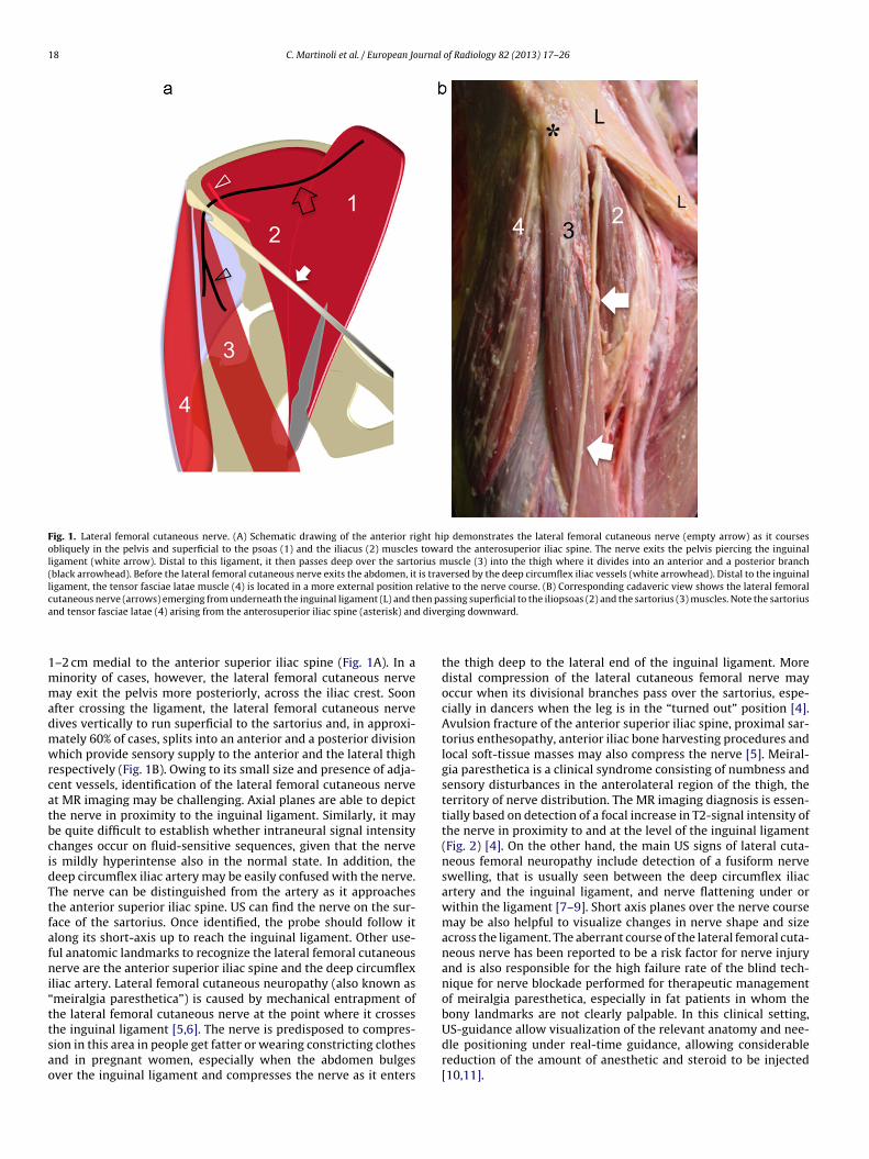
18 C. Martinoli et al. / European Journal of Radiology 82 (2013) 17– 26
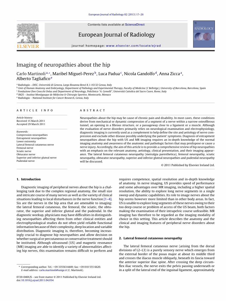
Fig. 1. Lateral femoral cutaneous nerve. (A) Schematic drawing of the anterior right hip demonstrates the lateral femoral cutaneous nerve (empty arrow) as it coursesobliquely in the pelvis and superficial to the psoas (1) and the iliacus (2) muscles toward the anterosuperior iliac spine. The nerve exits the pelvis piercing the inguinalligament (white arrow). Distal to this ligament, it then passes deep over the sartorius muscle (3) into the thigh where it divides into an anterior and a posterior branch(black arrowhead). Before the lateral femoral cutaneous nerve exits the abdomen, it is traversed by the deep circumflex iliac vessels (white arrowhead). Distal to the inguinalligament, the tensor fasciae latae muscle (4) is located in a more external position relative to the nerve course. (B) Corresponding cadaveric view shows the lateral femoralc hen paa dive
1mmadmwrcatbcidTtfafni“ttsao
utaneous nerve (arrows) emerging from underneath the inguinal ligament (L) and tnd tensor fasciae latae (4) arising from the anterosuperior iliac spine (asterisk) and
–2 cm medial to the anterior superior iliac spine (Fig. 1A). In ainority of cases, however, the lateral femoral cutaneous nerveay exit the pelvis more posteriorly, across the iliac crest. Soon
fter crossing the ligament, the lateral femoral cutaneous nerveives vertically to run superficial to the sartorius and, in approxi-ately 60% of cases, splits into an anterior and a posterior divisionhich provide sensory supply to the anterior and the lateral thigh
espectively (Fig. 1B). Owing to its small size and presence of adja-ent vessels, identification of the lateral femoral cutaneous nervet MR imaging may be challenging. Axial planes are able to depicthe nerve in proximity to the inguinal ligament. Similarly, it maye quite difficult to establish whether intraneural signal intensityhanges occur on fluid-sensitive sequences, given that the nerves mildly hyperintense also in the normal state. In addition, theeep circumflex iliac artery may be easily confused with the nerve.he nerve can be distinguished from the artery as it approacheshe anterior superior iliac spine. US can find the nerve on the sur-ace of the sartorius. Once identified, the probe should follow itlong its short-axis up to reach the inguinal ligament. Other use-ul anatomic landmarks to recognize the lateral femoral cutaneouserve are the anterior superior iliac spine and the deep circumflex
liac artery. Lateral femoral cutaneous neuropathy (also known asmeiralgia paresthetica”) is caused by mechanical entrapment ofhe lateral femoral cutaneous nerve at the point where it crosses
he inguinal ligament [5,6]. The nerve is predisposed to compres-ion in this area in people get fatter or wearing constricting clothesnd in pregnant women, especially when the abdomen bulgesver the inguinal ligament and compresses the nerve as it entersssing superficial to the iliopsoas (2) and the sartorius (3) muscles. Note the sartoriusrging downward.
the thigh deep to the lateral end of the inguinal ligament. Moredistal compression of the lateral cutaneous femoral nerve mayoccur when its divisional branches pass over the sartorius, espe-cially in dancers when the leg is in the “turned out” position [4].Avulsion fracture of the anterior superior iliac spine, proximal sar-torius enthesopathy, anterior iliac bone harvesting procedures andlocal soft-tissue masses may also compress the nerve [5]. Meiral-gia paresthetica is a clinical syndrome consisting of numbness andsensory disturbances in the anterolateral region of the thigh, theterritory of nerve distribution. The MR imaging diagnosis is essen-tially based on detection of a focal increase in T2-signal intensity ofthe nerve in proximity to and at the level of the inguinal ligament(Fig. 2) [4]. On the other hand, the main US signs of lateral cuta-neous femoral neuropathy include detection of a fusiform nerveswelling, that is usually seen between the deep circumflex iliacartery and the inguinal ligament, and nerve flattening under orwithin the ligament [7–9]. Short axis planes over the nerve coursemay be also helpful to visualize changes in nerve shape and sizeacross the ligament. The aberrant course of the lateral femoral cuta-neous nerve has been reported to be a risk factor for nerve injuryand is also responsible for the high failure rate of the blind tech-nique for nerve blockade performed for therapeutic managementof meiralgia paresthetica, especially in fat patients in whom thebony landmarks are not clearly palpable. In this clinical setting,
US-guidance allow visualization of the relevant anatomy and nee-dle positioning under real-time guidance, allowing considerablereduction of the amount of anesthetic and steroid to be injected[10,11].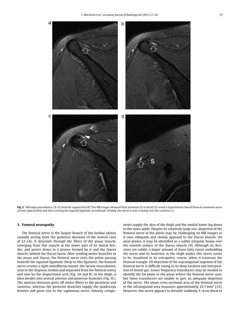
C. Martinoli et al. / European Journal of Radiology 82 (2013) 17– 26 19
Fig. 2. Meiralgia paresthetica. (A–D) Axial fat-suppressed tSE T2w MR images obtained from proximal (A) to distal (D) reveal a hyperintense lateral femoral cutaneous nerve( , the n
3
(oedmtbnnatTsf
arrow) approaching and then crossing the inguinal ligament (arrowhead). Distally
. Femoral neuropathy
The femoral nerve is the largest branch of the lumbar plexususually arising from the posterior divisions of the ventral ramif L2–L4). It descends through the fibers of the psoas muscle,merging from this muscle at the lower part of its lateral bor-er, and passes down in a groove formed by it and the iliacususcle, behind the iliacus fascia. After sending motor branches to
he psoas and iliacus, the femoral nerve exits the pelvis passingeneath the inguinal ligament. Deep to this ligament, the femoralerve crosses a rigid osteofibrous tunnel, the lacuna musculorum,ext to the iliopsoas tendon and separated from the femoral arterynd vein by the iliopectineal arch (Fig. 3A and B). In the thigh, it
hen divides into several anterior and posterior branches (Fig. 3C).he anterior divisions gives off motor fibers to the pectineus andartorius, whereas the posterior branches supply the quadricepsemoris and gives rise to the saphenous nerve. Sensory compo-erve is seen crossing over the sartorius (s).
nents supply the skin of the thigh and the medial lower leg downto the inner ankle. Despite its relatively large size, depiction of thefemoral nerve in the pelvis may be challenging on MR images asit runs obliquely and closely apposed to the iliacus muscle. Onaxial planes, it may be identified as a subtle irregular bump overthe smooth surface of the iliacus muscle [4]. Although its divi-sions are subtle, a larger amount of loose fatty tissue embeddingthe nerve and its branches in the thigh makes this nerve easierto be visualized in its extrapelvic course, when it traverses thefemoral triangle. US depiction of the suprainguinal segment of thefemoral nerve is difficult owing to its deep location and interposi-tion of bowel gas. Lower frequency transducers may be needed toidentify the fat plane in the psoas where the femoral nerve runs,
but these transducers are unable to give an adequate depictionof the nerve. The mean cross-sectional area of the femoral nervein the infrainguinal area measures approximately 22.7 mm2 [12].However, this nerve appears to dissolve suddenly 3–4 cm distal to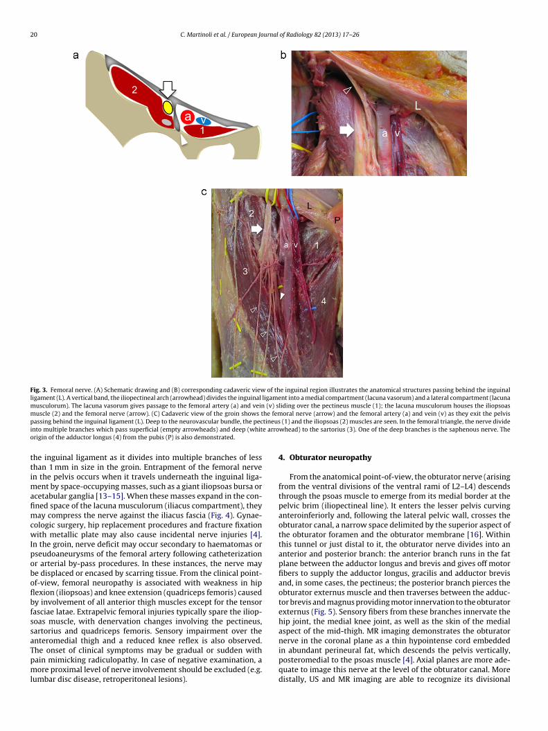
20 C. Martinoli et al. / European Journal of Radiology 82 (2013) 17– 26
Fig. 3. Femoral nerve. (A) Schematic drawing and (B) corresponding cadaveric view of the inguinal region illustrates the anatomical structures passing behind the inguinalligament (L). A vertical band, the iliopectineal arch (arrowhead) divides the inguinal ligament into a medial compartment (lacuna vasorum) and a lateral compartment (lacunamusculorum). The lacuna vasorum gives passage to the femoral artery (a) and vein (v) sliding over the pectineus muscle (1); the lacuna musculorum houses the iliopsoasmuscle (2) and the femoral nerve (arrow). (C) Cadaveric view of the groin shows the femoral nerve (arrow) and the femoral artery (a) and vein (v) as they exit the pelvisp tineusi e arroo
ttimafimcwIpoboflbfssaTpml
assing behind the inguinal ligament (L). Deep to the neurovascular bundle, the pecnto multiple branches which pass superficial (empty arrowheads) and deep (whitrigin of the adductor longus (4) from the pubis (P) is also demonstrated.
he inguinal ligament as it divides into multiple branches of lesshan 1 mm in size in the groin. Entrapment of the femoral nerven the pelvis occurs when it travels underneath the inguinal liga-
ent by space-occupying masses, such as a giant iliopsoas bursa orcetabular ganglia [13–15]. When these masses expand in the con-ned space of the lacuna musculorum (iliacus compartment), theyay compress the nerve against the iliacus fascia (Fig. 4). Gynae-
ologic surgery, hip replacement procedures and fracture fixationith metallic plate may also cause incidental nerve injuries [4].
n the groin, nerve deficit may occur secondary to haematomas orseudoaneurysms of the femoral artery following catheterizationr arterial by-pass procedures. In these instances, the nerve maye displaced or encased by scarring tissue. From the clinical point-f-view, femoral neuropathy is associated with weakness in hipexion (iliopsoas) and knee extension (quadriceps femoris) causedy involvement of all anterior thigh muscles except for the tensorasciae latae. Extrapelvic femoral injuries typically spare the iliop-oas muscle, with denervation changes involving the pectineus,artorius and quadriceps femoris. Sensory impairment over thenteromedial thigh and a reduced knee reflex is also observed.
he onset of clinical symptoms may be gradual or sudden withain mimicking radiculopathy. In case of negative examination, aore proximal level of nerve involvement should be excluded (e.g.umbar disc disease, retroperitoneal lesions).
(1) and the iliopsoas (2) muscles are seen. In the femoral triangle, the nerve dividewhead) to the sartorius (3). One of the deep branches is the saphenous nerve. The
4. Obturator neuropathy
From the anatomical point-of-view, the obturator nerve (arisingfrom the ventral divisions of the ventral rami of L2–L4) descendsthrough the psoas muscle to emerge from its medial border at thepelvic brim (iliopectineal line). It enters the lesser pelvis curvinganteroinferiorly and, following the lateral pelvic wall, crosses theobturator canal, a narrow space delimited by the superior aspect ofthe obturator foramen and the obturator membrane [16]. Withinthis tunnel or just distal to it, the obturator nerve divides into ananterior and posterior branch: the anterior branch runs in the fatplane between the adductor longus and brevis and gives off motorfibers to supply the adductor longus, gracilis and adductor brevisand, in some cases, the pectineus; the posterior branch pierces theobturator externus muscle and then traverses between the adduc-tor brevis and magnus providing motor innervation to the obturatorexternus (Fig. 5). Sensory fibers from these branches innervate thehip joint, the medial knee joint, as well as the skin of the medialaspect of the mid-thigh. MR imaging demonstrates the obturatornerve in the coronal plane as a thin hypointense cord embedded
in abundant perineural fat, which descends the pelvis vertically,posteromedial to the psoas muscle [4]. Axial planes are more ade-quate to image this nerve at the level of the obturator canal. Moredistally, US and MR imaging are able to recognize its divisional
C. Martinoli et al. / European Journal of Radiology 82 (2013) 17– 26 21
Fig. 4. Femoral neuropathy in a patient with knee arthritis complaining of painful soft-tissue swelling over the anterior hip and weakness in knee extension. (A) Sagittalfat-suppressed tSE T2w MR image of the left hip reveals a markedly distended iliopsoas bursa (asterisks) extending upward in the pelvis. (B) Corresponding sagittal US imagereveals the femoral nerve (arrows) displaced anteriorly by the bursa. (C and D) Transverse US images over the anterior thigh demonstrate the quadriceps muscles on ther in buv ined o
bpptmrtpcistpmttsBcnmor
ight (C) and left (D) side for one-to-one comparison. On the left affected side, lossastus intermedius is visible as a result of femoral denervation. The patient compla
ranches as they run in the fat planes interposed between theectineus and adductor longus and then along the anterior andosterior aspect of the adductor brevis [4]. The visualization ofhe divisional branches of the obturator nerve is worse in young
uscular subject owing to paucity of intermuscular fat. The obtu-ator neuropathy in the pelvis is uncommon and usually relatedo mass effect or stretching injury as it may occur during surgicalrocedures in the lesser pelvis [17,18]. Nerve infiltration by adja-ent neoplasm (e.g. metastases in the pubis) can be assessed on MRmaging. In athletes, obturator neuropathy may be secondary to aort of fascial entrapment as the nerve enters the thigh, specificallyhe adductor compartment. Chronic adductor tendinopathy maylay a predisposing role. Exacerbated by exercise (thigh extension,edial rotation and adduction), pain typically radiates down along
he medial thigh [18]. In these cases, imaging is not informativeo assess the nerve status, but denervation injury can be demon-trated in the medial thigh muscles on fluid-sensitive sequences.ecause adductor tendinopathy and obturator neuropathy mayoexist, signal abnormalities related to muscle denervation should
ot be confused with a muscle strain. In the latter case, signal abnor-alities are more focal and located just in proximity to the musclerigin. In addition, the obturator externus is spared. Obturator neu-opathy may also be observed in patients with osteitis pubis or
lk and hyperechoic appearance of the rectus femoris (arrows) and the underlyingf soft-tissue swelling over the hip and weakness in knee extension.
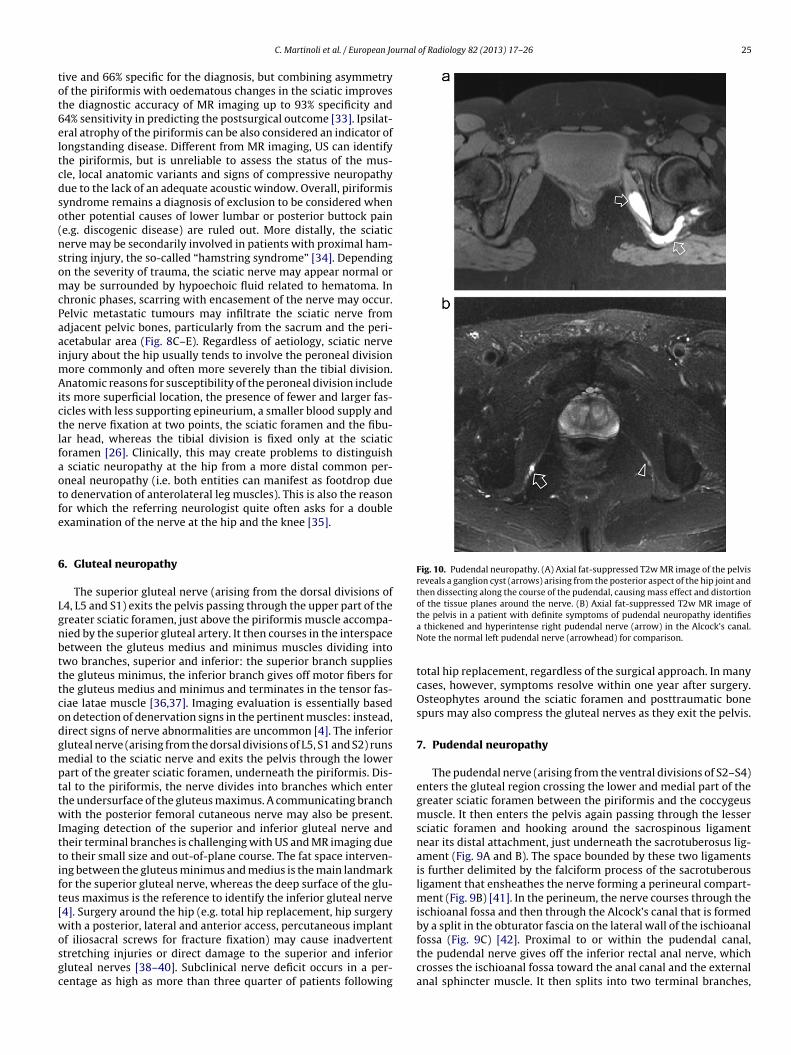
following surgery performed in lithotomy position [19]. Inflamma-tory reaction in the perineural fat can be found in these patients [4].Bone fractures in the pelvic ring and acetabulum, metallic hard-ware (e.g. periacetabular screw and cement leakage), acetabularlabral cysts and obturator hernia may also cause nerve entrapment(Fig. 6) [20–22].
5. Sciatic neuropathy
The sciatic nerve (arising from the ventral divisions of L2through S3), the largest nerve in the body, enters the gluteal regionpassing through the greater sciatic foramen accompanied by theinferior gluteal and the posterior femoral cutaneous nerve. Fromthe beginning, the sciatic nerve is formed by two distinct trunks – alarger tibial (medial) and a smaller peroneal (lateral) – enclosed ina common nerve sheath. In most cases, the nerve exits the greatersciatic foramen inferior to the piriformis muscle; in some individu-als, however, the nerve as a whole or part of it may pass through thepiriformis or cranial to it (Fig. 7A and B) [23,24]. Some anomalies
of the piriformis (e.g. a double-headed muscle) can be also encoun-tered in association with an anomalous nerve course (Fig. 7C). Distalto the piriformis, the sciatic nerve then runs halfway between theischial tuberosity and the greater trochanter, passing posterior to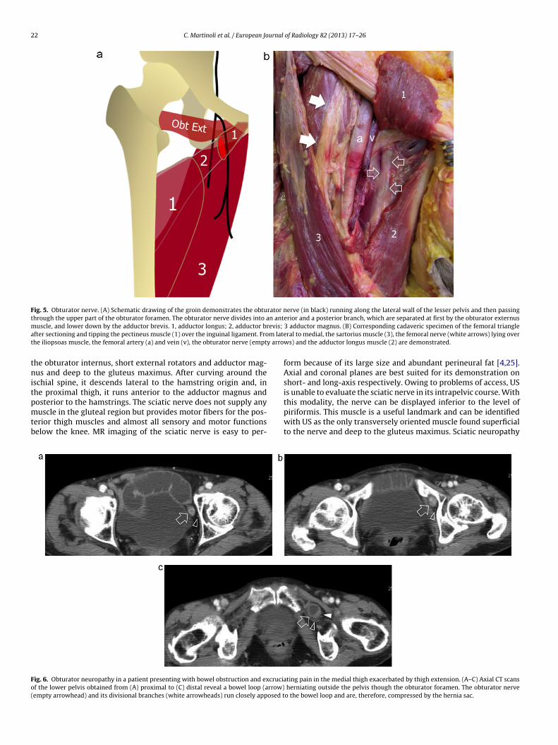
22 C. Martinoli et al. / European Journal of Radiology 82 (2013) 17– 26
Fig. 5. Obturator nerve. (A) Schematic drawing of the groin demonstrates the obturator nerve (in black) running along the lateral wall of the lesser pelvis and then passingthrough the upper part of the obturator foramen. The obturator nerve divides into an anterior and a posterior branch, which are separated at first by the obturator externusm evis;
a m latet y arro
tnitpmtb
Fo(
uscle, and lower down by the adductor brevis. 1, adductor longus; 2, adductor brfter sectioning and tipping the pectineus muscle (1) over the inguinal ligament. Frohe iliopsoas muscle, the femoral artery (a) and vein (v), the obturator nerve (empt
he obturator internus, short external rotators and adductor mag-us and deep to the gluteus maximus. After curving around the
schial spine, it descends lateral to the hamstring origin and, inhe proximal thigh, it runs anterior to the adductor magnus and
osterior to the hamstrings. The sciatic nerve does not supply anyuscle in the gluteal region but provides motor fibers for the pos-erior thigh muscles and almost all sensory and motor functionselow the knee. MR imaging of the sciatic nerve is easy to per-
ig. 6. Obturator neuropathy in a patient presenting with bowel obstruction and excrucif the lower pelvis obtained from (A) proximal to (C) distal reveal a bowel loop (arrow)empty arrowhead) and its divisional branches (white arrowheads) run closely apposed t
3 adductor magnus. (B) Corresponding cadaveric specimen of the femoral triangleral to medial, the sartorius muscle (3), the femoral nerve (white arrows) lying over
ws) and the adductor longus muscle (2) are demonstrated.
form because of its large size and abundant perineural fat [4,25].Axial and coronal planes are best suited for its demonstration onshort- and long-axis respectively. Owing to problems of access, USis unable to evaluate the sciatic nerve in its intrapelvic course. With
this modality, the nerve can be displayed inferior to the level ofpiriformis. This muscle is a useful landmark and can be identifiedwith US as the only transversely oriented muscle found superficialto the nerve and deep to the gluteus maximus. Sciatic neuropathyating pain in the medial thigh exacerbated by thigh extension. (A–C) Axial CT scans herniating outside the pelvis though the obturator foramen. The obturator nerveo the bowel loop and are, therefore, compressed by the hernia sac.

C. Martinoli et al. / European Journal of Radiology 82 (2013) 17– 26 23
Fig. 7. Sciatic nerve. (A) Schematic drawing of the posterior aspect of the hip shows relevant regional structures, including the sacrotuberous (1) and sacrospinous (2)ligaments, the piriformis (3), the superior (4a) and inferior (4b) gemellus, the quadratus femoris (5), the hamstrings (6), the obturator internus (7), and the gluteus medius(8) and minimus (9) that insert into the greater trochanter (asterisk). The sciatic nerve (empty black arrow) exits the pelvis passing through the greater sciatic foramen asindividual tibial (medial) and peroneal (lateral) components embedded in a common nerve sheath. It then continues down passing deep to the piriformis and lateral to theconjoined tendon of the long head of the biceps and semitendinosus. The superior gluteal nerve (empty white arrow) leaves the pelvis through the sciatic foramen above thepiriformis and then divides into a superior and inferior branch. The posterior femoral cutaneous nerve descends alongside the medial aspect of the sciatic nerve accompaniedby the inferior gluteal artery. After sending branches for the lower gluteus maximus and overlying skin, this nerve divides in a perineal (black arrowhead) and descending(white arrowhead) branch. (B) Corresponding cadaveric view of the posterior hip after removal of the gluteus maximus demonstrates the insertions of the piriformis (3),superior (4a) and inferior (4b) gemellus, quadratus femoris (5), gluteus medius (8) and minimus (9) on the greater trochanter (asterisk). Observe the piriformis musclec l neurv he scim erior g
itatslpnvictacp
overing the greater sciatic foramen and intervening between the superior gluteaiew of a double-headed (Pf1 and Pf2) piriformis muscle. There is a high division of tuscle and the medial (S2) component crossing the muscle inferiorly. Note the sup
ncludes a spectrum of traumatic, compressive, ischemic, neoplas-ic and idiopathic causes [26]. Traumatic injuries are most oftenssociated with femoral fracture, hip fracture dislocation and pos-erior thigh compartment syndrome. Concerning iatrogenic causes,ciatic nerve entrapment most often occurs in the retroacetabu-ar region secondary to total hip arthroplasty [26–29]. In theseatients, electrophysiology has revealed a prevalence of subclinicalerve injury in a percentage as high as 70% [30]. The cause may beariably related to the use of a posterior approach, limb lengthen-ng, compression from hematoma, extruded methyl-methacrylate,ementless femoral fixation and laceration from a screw used to fix
he acetabular cup. Other reported lesions related to the postoper-tive period include injury from ischemia and positioning duringardiac surgery, lithotomy position and neurosurgical operativeositions [26,27]. In these cases, MR imaging may reveal a focalovascular bundle and the sciatic nerve. ST, sacrotuberous ligament. (C) Cadavericatic nerve with its lateral (S1) component passing in between the two bellies of theluteal nerve (empty white arrows).
increase in the nerve size and abnormal signal intensity, whereasUS demonstrates a loss of the fascicular echotexture and a focalhypoechoic pattern (Fig. 8A and B) [4]. Obliteration of the fat planesaround the nerve may also be noted reflecting scarring tissue.Anomalies of the piriformis muscle can cause sciatic nerve com-pression, the so called “piriformis syndrome”, a controversial entityin which symptoms are often nonspecific and electrophysiology isdifficult to perform due to the deep location of the nerve [31,32].In this syndrome, a hypertrophied muscle secondary to gait distur-bances, excessive lumbar lordosis and hip flexion deformities cancause crowding of the greater sciatic foramen resulting in nerve
compression. However, muscle asymmetry does not seem a reliablesign of nerve entrapment because side variations in the piriformissize are often found in asymptomatic individuals [4]. Consideringmuscle asymmetry alone, MR imaging has proved to be 46% sensi-
24 C. Martinoli et al. / European Journal of Radiology 82 (2013) 17– 26
Fig. 8. Sciatic neuropathies. (A and B) Selective involvement of the lateral component of the sciatic nerve in a patient who had foot drop and sensory disturbances in theperoneal nerve distribution after prolonged pelvic surgery in a lithotomy position. (A) Axial T1w image of the sciatic nerve at the level of the ischial tuberosity (asterisk)demonstrates a swollen and hypointense lateral (peroneal) component (empty arrow) of the sciatic reflecting intraneural fibrotic changes. The medial (tibial) componentof the sciatic nerve is normal (arrowhead). (B) Corresponding US image reveals hypoechoic changes in the lateral component (arrow) of the sciatic and preserved fascicularechotexture in the medial one (arrowhead). (C–E) Tumour infiltration of the sciatic nerve in a patient with sacral metastases from breast cancer. C Coronal T1w and (D and E)transverse Gd-enhanced fat-suppressed MR images shows extensive infiltration of the piriformis muscle (arrow) and the sciatic nerve (arrowheads) which appears swollenand characterized by marked contrast enhancement.
Fig. 9. Pudendal nerve. (A) Schematic drawing of the pelvis illustrates the pudendal nerve exiting the pelvis through the lower part of the greater gluteal foramen and thenentering the gluteal region again passing through the lesser sciatic foramen in close relationship with the sacrotuberous (1) and the sacrospinous (2) ligaments. After crossingthe sacrotuberous ligament, the pudendal nerve (arrow) redirects its course anteriorly entering the Alcock’s canal, a restricted tunnel underneath the fascia (arrowheads)of the obturator internus muscle (3). (B) Cadaveric dissection shows the sacrotuberous ligament (1) inserting into the ischial tuberosity (asterisk) and the pudendal nerve(empty arrows) and artery (white arrowheads) as they cross underneath it. Note the location of the falciform process (empty arrowheads) of the sacrotuberous ligament,which is possible site for pudendal nerve entrapment. 2, sacrospinous ligament. (C) Axial T1w MR image demonstrates the pudendal nerve (arrow) running close to theobturator internus muscle (OInt) in the Alcock’s canal. Note the position of the sacrotuberous (1) and the sacrospinous (2) ligaments and the ischial tuberosity (asterisk).
urnal of Radiology 82 (2013) 17– 26 25
tot6eltcdso(nsomcPaaimAictlfaotfe
6
LgnbtttcodgmpttwIttift[wosgc
Fig. 10. Pudendal neuropathy. (A) Axial fat-suppressed T2w MR image of the pelvisreveals a ganglion cyst (arrows) arising from the posterior aspect of the hip joint andthen dissecting along the course of the pudendal, causing mass effect and distortionof the tissue planes around the nerve. (B) Axial fat-suppressed T2w MR image of
C. Martinoli et al. / European Jo
ive and 66% specific for the diagnosis, but combining asymmetryf the piriformis with oedematous changes in the sciatic improveshe diagnostic accuracy of MR imaging up to 93% specificity and4% sensitivity in predicting the postsurgical outcome [33]. Ipsilat-ral atrophy of the piriformis can be also considered an indicator ofongstanding disease. Different from MR imaging, US can identifyhe piriformis, but is unreliable to assess the status of the mus-le, local anatomic variants and signs of compressive neuropathyue to the lack of an adequate acoustic window. Overall, piriformisyndrome remains a diagnosis of exclusion to be considered whenther potential causes of lower lumbar or posterior buttock paine.g. discogenic disease) are ruled out. More distally, the sciaticerve may be secondarily involved in patients with proximal ham-tring injury, the so-called “hamstring syndrome” [34]. Dependingn the severity of trauma, the sciatic nerve may appear normal oray be surrounded by hypoechoic fluid related to hematoma. In
hronic phases, scarring with encasement of the nerve may occur.elvic metastatic tumours may infiltrate the sciatic nerve fromdjacent pelvic bones, particularly from the sacrum and the peri-cetabular area (Fig. 8C–E). Regardless of aetiology, sciatic nervenjury about the hip usually tends to involve the peroneal division
ore commonly and often more severely than the tibial division.natomic reasons for susceptibility of the peroneal division include
ts more superficial location, the presence of fewer and larger fas-icles with less supporting epineurium, a smaller blood supply andhe nerve fixation at two points, the sciatic foramen and the fibu-ar head, whereas the tibial division is fixed only at the sciaticoramen [26]. Clinically, this may create problems to distinguish
sciatic neuropathy at the hip from a more distal common per-neal neuropathy (i.e. both entities can manifest as footdrop dueo denervation of anterolateral leg muscles). This is also the reasonor which the referring neurologist quite often asks for a doublexamination of the nerve at the hip and the knee [35].
. Gluteal neuropathy
The superior gluteal nerve (arising from the dorsal divisions of4, L5 and S1) exits the pelvis passing through the upper part of thereater sciatic foramen, just above the piriformis muscle accompa-ied by the superior gluteal artery. It then courses in the interspaceetween the gluteus medius and minimus muscles dividing intowo branches, superior and inferior: the superior branch supplieshe gluteus minimus, the inferior branch gives off motor fibers forhe gluteus medius and minimus and terminates in the tensor fas-iae latae muscle [36,37]. Imaging evaluation is essentially basedn detection of denervation signs in the pertinent muscles: instead,irect signs of nerve abnormalities are uncommon [4]. The inferiorluteal nerve (arising from the dorsal divisions of L5, S1 and S2) runsedial to the sciatic nerve and exits the pelvis through the lower
art of the greater sciatic foramen, underneath the piriformis. Dis-al to the piriformis, the nerve divides into branches which enterhe undersurface of the gluteus maximus. A communicating branchith the posterior femoral cutaneous nerve may also be present.
maging detection of the superior and inferior gluteal nerve andheir terminal branches is challenging with US and MR imaging dueo their small size and out-of-plane course. The fat space interven-ng between the gluteus minimus and medius is the main landmarkor the superior gluteal nerve, whereas the deep surface of the glu-eus maximus is the reference to identify the inferior gluteal nerve4]. Surgery around the hip (e.g. total hip replacement, hip surgeryith a posterior, lateral and anterior access, percutaneous implant
f iliosacral screws for fracture fixation) may cause inadvertenttretching injuries or direct damage to the superior and inferiorluteal nerves [38–40]. Subclinical nerve deficit occurs in a per-entage as high as more than three quarter of patients following
the pelvis in a patient with definite symptoms of pudendal neuropathy identifiesa thickened and hyperintense right pudendal nerve (arrow) in the Alcock’s canal.Note the normal left pudendal nerve (arrowhead) for comparison.
total hip replacement, regardless of the surgical approach. In manycases, however, symptoms resolve within one year after surgery.Osteophytes around the sciatic foramen and posttraumatic bonespurs may also compress the gluteal nerves as they exit the pelvis.
7. Pudendal neuropathy
The pudendal nerve (arising from the ventral divisions of S2–S4)enters the gluteal region crossing the lower and medial part of thegreater sciatic foramen between the piriformis and the coccygeusmuscle. It then enters the pelvis again passing through the lessersciatic foramen and hooking around the sacrospinous ligamentnear its distal attachment, just underneath the sacrotuberosus lig-ament (Fig. 9A and B). The space bounded by these two ligamentsis further delimited by the falciform process of the sacrotuberousligament that ensheathes the nerve forming a perineural compart-ment (Fig. 9B) [41]. In the perineum, the nerve courses through theischioanal fossa and then through the Alcock’s canal that is formedby a split in the obturator fascia on the lateral wall of the ischioanal
fossa (Fig. 9C) [42]. Proximal to or within the pudendal canal,the pudendal nerve gives off the inferior rectal anal nerve, whichcrosses the ischioanal fossa toward the anal canal and the externalanal sphincter muscle. It then splits into two terminal branches,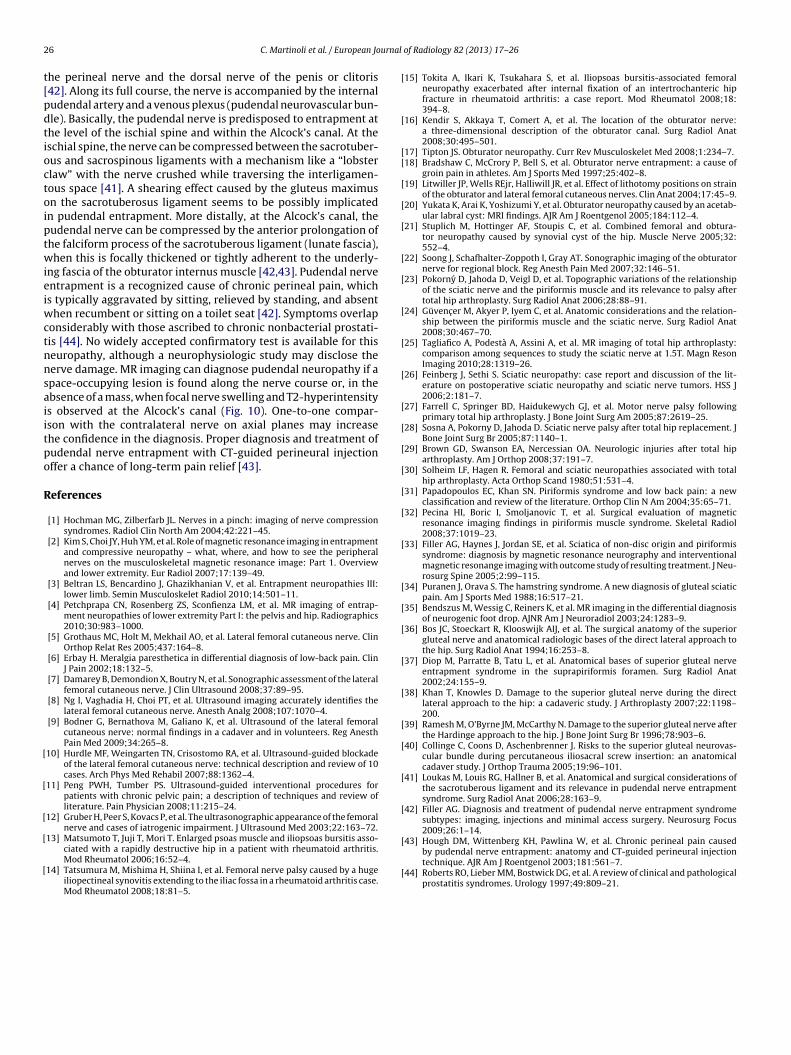
2 urnal
t[pdtioctoiptwieiwctnnsaiitpo
R
[
[
[
[
[
[
[
[[
[
[
[
[
[
[
[
[
[
[
[
[
[
[
[
[
[
[
[
[
[
[
[
[
[43] Hough DM, Wittenberg KH, Pawlina W, et al. Chronic perineal pain caused
6 C. Martinoli et al. / European Jo
he perineal nerve and the dorsal nerve of the penis or clitoris42]. Along its full course, the nerve is accompanied by the internaludendal artery and a venous plexus (pudendal neurovascular bun-le). Basically, the pudendal nerve is predisposed to entrapment athe level of the ischial spine and within the Alcock’s canal. At theschial spine, the nerve can be compressed between the sacrotuber-us and sacrospinous ligaments with a mechanism like a “lobsterlaw” with the nerve crushed while traversing the interligamen-ous space [41]. A shearing effect caused by the gluteus maximusn the sacrotuberosus ligament seems to be possibly implicatedn pudendal entrapment. More distally, at the Alcock’s canal, theudendal nerve can be compressed by the anterior prolongation ofhe falciform process of the sacrotuberous ligament (lunate fascia),hen this is focally thickened or tightly adherent to the underly-
ng fascia of the obturator internus muscle [42,43]. Pudendal nerventrapment is a recognized cause of chronic perineal pain, whichs typically aggravated by sitting, relieved by standing, and absent
hen recumbent or sitting on a toilet seat [42]. Symptoms overlaponsiderably with those ascribed to chronic nonbacterial prostati-is [44]. No widely accepted confirmatory test is available for thiseuropathy, although a neurophysiologic study may disclose theerve damage. MR imaging can diagnose pudendal neuropathy if apace-occupying lesion is found along the nerve course or, in thebsence of a mass, when focal nerve swelling and T2-hyperintensitys observed at the Alcock’s canal (Fig. 10). One-to-one compar-son with the contralateral nerve on axial planes may increasehe confidence in the diagnosis. Proper diagnosis and treatment ofudendal nerve entrapment with CT-guided perineural injectionffer a chance of long-term pain relief [43].
eferences
[1] Hochman MG, Zilberfarb JL. Nerves in a pinch: imaging of nerve compressionsyndromes. Radiol Clin North Am 2004;42:221–45.
[2] Kim S, Choi JY, Huh YM, et al. Role of magnetic resonance imaging in entrapmentand compressive neuropathy – what, where, and how to see the peripheralnerves on the musculoskeletal magnetic resonance image: Part 1. Overviewand lower extremity. Eur Radiol 2007;17:139–49.
[3] Beltran LS, Bencardino J, Ghazikhanian V, et al. Entrapment neuropathies III:lower limb. Semin Musculoskelet Radiol 2010;14:501–11.
[4] Petchprapa CN, Rosenberg ZS, Sconfienza LM, et al. MR imaging of entrap-ment neuropathies of lower extremity Part I: the pelvis and hip. Radiographics2010;30:983–1000.
[5] Grothaus MC, Holt M, Mekhail AO, et al. Lateral femoral cutaneous nerve. ClinOrthop Relat Res 2005;437:164–8.
[6] Erbay H. Meralgia paresthetica in differential diagnosis of low-back pain. ClinJ Pain 2002;18:132–5.
[7] Damarey B, Demondion X, Boutry N, et al. Sonographic assessment of the lateralfemoral cutaneous nerve. J Clin Ultrasound 2008;37:89–95.
[8] Ng I, Vaghadia H, Choi PT, et al. Ultrasound imaging accurately identifies thelateral femoral cutaneous nerve. Anesth Analg 2008;107:1070–4.
[9] Bodner G, Bernathova M, Galiano K, et al. Ultrasound of the lateral femoralcutaneous nerve: normal findings in a cadaver and in volunteers. Reg AnesthPain Med 2009;34:265–8.
10] Hurdle MF, Weingarten TN, Crisostomo RA, et al. Ultrasound-guided blockadeof the lateral femoral cutaneous nerve: technical description and review of 10cases. Arch Phys Med Rehabil 2007;88:1362–4.
11] Peng PWH, Tumber PS. Ultrasound-guided interventional procedures forpatients with chronic pelvic pain; a description of techniques and review ofliterature. Pain Physician 2008;11:215–24.
12] Gruber H, Peer S, Kovacs P, et al. The ultrasonographic appearance of the femoralnerve and cases of iatrogenic impairment. J Ultrasound Med 2003;22:163–72.
13] Matsumoto T, Juji T, Mori T. Enlarged psoas muscle and iliopsoas bursitis asso-
ciated with a rapidly destructive hip in a patient with rheumatoid arthritis.Mod Rheumatol 2006;16:52–4.14] Tatsumura M, Mishima H, Shiina I, et al. Femoral nerve palsy caused by a hugeiliopectineal synovitis extending to the iliac fossa in a rheumatoid arthritis case.Mod Rheumatol 2008;18:81–5.
[
of Radiology 82 (2013) 17– 26
15] Tokita A, Ikari K, Tsukahara S, et al. Iliopsoas bursitis-associated femoralneuropathy exacerbated after internal fixation of an intertrochanteric hipfracture in rheumatoid arthritis: a case report. Mod Rheumatol 2008;18:394–8.
16] Kendir S, Akkaya T, Comert A, et al. The location of the obturator nerve:a three-dimensional description of the obturator canal. Surg Radiol Anat2008;30:495–501.
17] Tipton JS. Obturator neuropathy. Curr Rev Musculoskelet Med 2008;1:234–7.18] Bradshaw C, McCrory P, Bell S, et al. Obturator nerve entrapment: a cause of
groin pain in athletes. Am J Sports Med 1997;25:402–8.19] Litwiller JP, Wells REjr, Halliwill JR, et al. Effect of lithotomy positions on strain
of the obturator and lateral femoral cutaneous nerves. Clin Anat 2004;17:45–9.20] Yukata K, Arai K, Yoshizumi Y, et al. Obturator neuropathy caused by an acetab-
ular labral cyst: MRI findings. AJR Am J Roentgenol 2005;184:112–4.21] Stuplich M, Hottinger AF, Stoupis C, et al. Combined femoral and obtura-
tor neuropathy caused by synovial cyst of the hip. Muscle Nerve 2005;32:552–4.
22] Soong J, Schafhalter-Zoppoth I, Gray AT. Sonographic imaging of the obturatornerve for regional block. Reg Anesth Pain Med 2007;32:146–51.
23] Pokorny D, Jahoda D, Veigl D, et al. Topographic variations of the relationshipof the sciatic nerve and the piriformis muscle and its relevance to palsy aftertotal hip arthroplasty. Surg Radiol Anat 2006;28:88–91.
24] Güvenc er M, Akyer P, Iyem C, et al. Anatomic considerations and the relation-ship between the piriformis muscle and the sciatic nerve. Surg Radiol Anat2008;30:467–70.
25] Tagliafico A, Podestà A, Assini A, et al. MR imaging of total hip arthroplasty:comparison among sequences to study the sciatic nerve at 1.5T. Magn ResonImaging 2010;28:1319–26.
26] Feinberg J, Sethi S. Sciatic neuropathy: case report and discussion of the lit-erature on postoperative sciatic neuropathy and sciatic nerve tumors. HSS J2006;2:181–7.
27] Farrell C, Springer BD, Haidukewych GJ, et al. Motor nerve palsy followingprimary total hip arthroplasty. J Bone Joint Surg Am 2005;87:2619–25.
28] Sosna A, Pokorny D, Jahoda D. Sciatic nerve palsy after total hip replacement. JBone Joint Surg Br 2005;87:1140–1.
29] Brown GD, Swanson EA, Nercessian OA. Neurologic injuries after total hiparthroplasty. Am J Orthop 2008;37:191–7.
30] Solheim LF, Hagen R. Femoral and sciatic neuropathies associated with totalhip arthroplasty. Acta Orthop Scand 1980;51:531–4.
31] Papadopoulos EC, Khan SN. Piriformis syndrome and low back pain: a newclassification and review of the literature. Orthop Clin N Am 2004;35:65–71.
32] Pecina HI, Boric I, Smoljanovic T, et al. Surgical evaluation of magneticresonance imaging findings in piriformis muscle syndrome. Skeletal Radiol2008;37:1019–23.
33] Filler AG, Haynes J, Jordan SE, et al. Sciatica of non-disc origin and piriformissyndrome: diagnosis by magnetic resonance neurography and interventionalmagnetic resonange imaging with outcome study of resulting treatment. J Neu-rosurg Spine 2005;2:99–115.
34] Puranen J, Orava S. The hamstring syndrome. A new diagnosis of gluteal sciaticpain. Am J Sports Med 1988;16:517–21.
35] Bendszus M, Wessig C, Reiners K, et al. MR imaging in the differential diagnosisof neurogenic foot drop. AJNR Am J Neuroradiol 2003;24:1283–9.
36] Bos JC, Stoeckart R, Klooswijk AIJ, et al. The surgical anatomy of the superiorgluteal nerve and anatomical radiologic bases of the direct lateral approach tothe hip. Surg Radiol Anat 1994;16:253–8.
37] Diop M, Parratte B, Tatu L, et al. Anatomical bases of superior gluteal nerveentrapment syndrome in the suprapiriformis foramen. Surg Radiol Anat2002;24:155–9.
38] Khan T, Knowles D. Damage to the superior gluteal nerve during the directlateral approach to the hip: a cadaveric study. J Arthroplasty 2007;22:1198–200.
39] Ramesh M, O’Byrne JM, McCarthy N. Damage to the superior gluteal nerve afterthe Hardinge approach to the hip. J Bone Joint Surg Br 1996;78:903–6.
40] Collinge C, Coons D, Aschenbrenner J. Risks to the superior gluteal neurovas-cular bundle during percutaneous iliosacral screw insertion: an anatomicalcadaver study. J Orthop Trauma 2005;19:96–101.
41] Loukas M, Louis RG, Hallner B, et al. Anatomical and surgical considerations ofthe sacrotuberous ligament and its relevance in pudendal nerve entrapmentsyndrome. Surg Radiol Anat 2006;28:163–9.
42] Filler AG. Diagnosis and treatment of pudendal nerve entrapment syndromesubtypes: imaging, injections and minimal access surgery. Neurosurg Focus2009;26:1–14.
by pudendal nerve entrapment: anatomy and CT-guided perineural injectiontechnique. AJR Am J Roentgenol 2003;181:561–7.
44] Roberts RO, Lieber MM, Bostwick DG, et al. A review of clinical and pathologicalprostatitis syndromes. Urology 1997;49:809–21.