european respiratory society guidelines’for the management ... · the european respiratory...
TRANSCRIPT

European Respiratory Societyguidelines for the managementof adult bronchiectasis
Eva Polverino1, Pieter C. Goeminne2,3, Melissa J. McDonnell4,5,6,Stefano Aliberti 7, Sara E. Marshall8, Michael R. Loebinger9,Marlene Murris10, Rafael Cantón11, Antoni Torres12, Katerina Dimakou13,Anthony De Soyza14,15, Adam T. Hill16, Charles S. Haworth17,Montserrat Vendrell18, Felix C. Ringshausen19, Dragan Subotic20,Robert Wilson9, Jordi Vilaró21, Bjorn Stallberg22, Tobias Welte19,Gernot Rohde23, Francesco Blasi7, Stuart Elborn9,24, Marta Almagro25,Alan Timothy25, Thomas Ruddy25, Thomy Tonia26, David Rigau27 andJames D. Chalmers28
@ERSpublicationsThe publication of the first ERS guidelines for bronchiectasis http://ow.ly/wQSO30dU0nE
Cite this article as: Polverino E, Goeminne PC, McDonnell MJ, et al. European Respiratory Societyguidelines for the management of adult bronchiectasis. Eur Respir J 2017; 50: 1700629 [https://doi.org/10.1183/13993003.00629-2017].
ABSTRACT Bronchiectasis in adults is a chronic disorder associated with poor quality of life andfrequent exacerbations in many patients. There have been no previous international guidelines.
The European Respiratory Society guidelines for the management of adult bronchiectasis describe theappropriate investigation and treatment strategies determined by a systematic review of the literature.
A multidisciplinary group representing respiratory medicine, microbiology, physiotherapy, thoracicsurgery, primary care, methodology and patients considered the most relevant clinical questions (for bothclinicians and patients) related to management of bronchiectasis. Nine key clinical questions weregenerated and a systematic review was conducted to identify published systematic reviews, randomisedclinical trials and observational studies that answered these questions. We used the GRADE approach todefine the quality of the evidence and the level of recommendations. The resulting guideline addresses theinvestigation of underlying causes of bronchiectasis, treatment of exacerbations, pathogen eradication, longterm antibiotic treatment, anti-inflammatories, mucoactive drugs, bronchodilators, surgical treatment andrespiratory physiotherapy.
These recommendations can be used to benchmark quality of care for people with bronchiectasis acrossEurope and to improve outcomes.
This article has supplementary material available from erj.ersjournals.com
Received: March 26 2017 | Accepted after revision: July 10 2017
The guidelines published by the European Respiratory Society (ERS) incorporate data obtained from a comprehensiveand systematic literature review of the most recent studies available at the time. Health professionals are encouraged totake the guidelines into account in their clinical practice. However, the recommendations issued by this guideline maynot be appropriate for use in all situations. It is the individual responsibility of health professionals to consult othersources of relevant information, to make appropriate and accurate decisions in consideration of each patient’s healthcondition and in consultation with that patient and the patient’s caregiver where appropriate and/or necessary, and toverify rules and regulations applicable to drugs and devices at the time of prescription.
This document was endorsed by the ERS Executive Committee and by the European Society of Clinical Microbiologyand Infectious Disease in August 2017.
Copyright ©ERS 2017
https://doi.org/10.1183/13993003.00629-2017 Eur Respir J 2017; 50: 1700629
TASK FORCE REPORTERS GUIDELINES

Affiliations: 1Servei de Pneumologia, Hospital Universitari Vall d’Hebron (HUVH), Institut de Recerca Valld’Hebron (VHIR); Fundación Clínic, Hospital Clínic de Barcelona, Universitat de Barcelona, IDIBAPS, CIBERESBarcelona, Barcelona, Spain. 2Dept of Respiratory Medicine, AZ Nikolaas, Sint-Niklaas, Belgium. 3Dept ofRespiratory Medicine, UZ Leuven, Leuven, Belgium. 4Dept of Respiratory Medicine, Galway UniversityHospitals, Galway, Ireland. 5Lung Biology Group, National University of Ireland, Galway, Ireland. 6Institute ofCell and Molecular Biology, Newcastle University, Newcastle upon Tyne, UK. 7Dept of Pathophysiology andTransplantation, Università degli Studi di Milano, Internal Medicine Department, Respiratory Unit and AdultCystic Fibrosis Center Fondazione IRCCS Cà Granda Ospedale Maggiore Policlinico Milan, Milan, Italy. 8Deptof Clinical Research, Immunology and Physiological Sciences, Wellcome, London, UK. 9Host Defence Unit,Royal Brompton Hospital, Imperial College, London, UK. 10Service de Pneumologie, Hôpital Larrey, CHU deToulouse, Toulouse, France. 11Servicio de Microbiología, Hospital Universitario Ramón y Cajal and InstitutoRamón y Cajal de Investigación Sanitaria (IRYCIS), Madrid, Spain. 12Servei de Pneumologia, Hospital Clínic deBarcelona, Universitat de Barcelona, IDIBAPS, CIBERES Barcelona, Barcelona, Spain. 135th Pulmonary Dept,“Sotiria” Chest Hospital, Athens, Greece. 14Institute of Cellular Medicine, Newcastle University, Newcastleupon Tyne, UK. 15Bronchiectasis Service, Freeman Hospital, Newcastle upon Tyne, UK. 16Dept of RespiratoryMedicine, Royal Infirmary and University of Edinburgh, Edinburgh, UK. 17Cambridge Centre for Lung Infection,Papworth Hospital, Cambridge, UK. 18Bronchiectasis Group, Girona Biomedical Research Institute (IDIBGI), DrTrueta University Hospital, Girona, Spain. 19Dept of Respiratory Medicine, Hannover Medical School, Memberof the German Centre for Lung Research, Hannover, Germany. 20Clinic for Thoracic Surgery - Clinical Centreof Serbia, University of Belgrade, Belgrade, Serbia. 21FCS Blanquerna. Physical Activity and Health Group,Universitat Ramon Llull, Barcelona, Spain. 22Dept of Public Health and Caring Science, Family Medicine andPreventive Medicine, Uppsala University, Uppsala, Sweden. 23Dept of Respiratory Medicine, MaastrichtUniversity Medical Center, Maastricht, The Netherlands. 24Queen’s University Belfast, Belfast, UK. 25EuropeanLung Foundation (ELF)/EMBARC bronchiectasis patient advisory group. 26Institute of Social and PreventiveMedicine, University of Bern, Bern, Switzerland. 27Iberoamerican Cochrane Center, Barcelona, Spain.28College of Medicine, University of Dundee, Ninewells Hospital and Medical School, Dundee, UK.
Correspondence: James D. Chalmers, Scottish Centre for Respiratory Research, Ninewells Hospital andMedical School, Dundee, DD1 9SY, UK. E-mail: [email protected]
Scope and objectivesThis guideline provides evidence-based recommendations for the management of adult patients withbronchiectasis. It only applies to patients with clinically significant bronchiectasis, defined by the presenceof both permanent bronchial dilatation on computed tomography (CT) scanning and the clinicalsyndrome of cough, sputum production and/or recurrent respiratory infections. Radiological bronchiectasismay be evident in healthy asymptomatic individuals, particularly in the elderly [1] or may occur, forexample, due to traction in interstitial lung disease. Such radiological bronchiectasis without clinicalsymptoms are not addressed in this guideline. The following conditions are also excluded: cystic fibrosisbronchiectasis, which has a distinct pathophysiology and treatment pathway, children with bronchiectasis,treatment of primary immunodeficiencies and non-tuberculous mycobacteria (NTM), where diseasespecific therapy is indicated. The majority of these clinical issues are addressed in other guidelines.
This guideline document does not address clinical and radiological diagnosis of bronchiectasis but ratherfocuses on key questions in management. Areas such as smoking cessation, nutrition, influenza andpneumococcal vaccination among other aspects of general management are not specifically addressed inthis document. Readers are referred to relevance guidelines and national policies. A guideline documentcannot address the full complexity of a disease such as bronchiectasis, hence all recommendations shouldbe interpreted taking into account the clinical circumstances and patients’ perceptions, values andpreferences.
Table 1 provides a framework to understand the recommendations made in this document [2, 3].
The target audience for this guideline are all stakeholders involved bronchiectasis care. This includesspecialists in respiratory medicine, infectious diseases, clinical microbiology, general internists, specialistsin thoracic surgery, primary care physicians, pharmacists, respiratory physiotherapists, specialist nurses,regulatory authorities, pharmaceutical companies and policy makers. The guideline is also to informpeople with bronchiectasis to help them to discuss with their care teams and to access appropriate care.
IntroductionBronchiectasis is a chronic respiratory disease characterised by a clinical syndrome of cough, sputumproduction and bronchial infection, and radiologically by abnormal and permanent dilatation of thebronchi. The objectives of treatment in bronchiectasis are to prevent exacerbations, reduce symptoms,
Support statement: The task force was funded by the European Respiratory Society. Funding information for this articlehas been deposited with the Crossref Funder Registry.
Conflict of interest: D. Rigau and T. Tonia act as methodologists for the European Respiratory Society. All otherdisclosures can be found alongside this article at erj.ersjournals.com
https://doi.org/10.1183/13993003.00629-2017 2
ERS GUIDELINES | E. POLVERINO ET AL.

improve quality of life and stop disease progression. Cough and sputum production, along withbreathlessness are the most frequent symptoms but rhinosinusitis, fatigue, haemoptysis and thoracic painare also common [4]. Quality of life impairment in bronchiectasis is equivalent in terms of scores on theSt George’s Respiratory Questionnaire (SGRQ) to severe chronic obstructive pulmonary disease (COPD),idiopathic pulmonary fibrosis and other disabling respiratory diseases [5, 6].
Exacerbations of bronchiectasis are key targets for therapy as they are major determinants of healthcarecosts. They are associated with increased airways and systemic inflammation [7] and progressive lungdamage [8, 9]. In addition, more severe and more frequent exacerbations are associated with worse qualityof life, daily symptoms [10], lung function decline [11], and mortality [9]. Consequently, the majority oftherapeutic interventions are aimed at reducing exacerbations. Despite current treatment approaches,European registry data shows that approximately 50% of European bronchiectasis patients have two ormore exacerbations per year and one third require at least one hospitalisation per year [12].
Our understanding of what causes symptoms and exacerbations is based on the vicious cycle concept, withkey components of the disease being chronic bronchial infection, inflammation, impaired mucociliaryclearance and structural lung damage. Treatment is primarily based on the principles of preventing orsuppressing acute and chronic bronchial infection, improving mucociliary clearance and reducing theimpact of structural lung disease (figure 1).
Chronic airways infection, most frequently with Haemophilus influenzae and Pseudomonas aeruginosa andless frequently with Moraxella catarrhalis, Staphylococcus aureus and Enterobacteriaceae, stimulate andsustain lung inflammation. Persistent isolation of these organisms in sputum or bronchoalveolar lavage isassociated with an increased frequency of exacerbations, worse quality of life and increased mortality[13, 14]. This is particularly the case with P. aeruginosa infection. A systematic review of observationalstudies identified that P. aeruginosa infection is associated with a three-fold increase in mortality risk, analmost seven-fold increase in risk of hospital admission and an average of one additional exacerbation perpatient per year [15].
Inflammation in bronchiectasis is primarily neutrophilic and closely linked to persistent bacterial infection.Excessive neutrophilic inflammation is linked to an increased frequency of exacerbations and rapid lungfunction decline through degradation of airway elastin, among other mechanisms [16–19]. The availabledata also support a role for cell-mediated immunity, specifically T-cells in the pathophysiology ofbronchiectasis, but the role of other inflammatory cells is less clear [17].
Mucociliary clearance is impaired by the impact of structural bronchiectasis, airway dehydration, excessmucus volume and viscosity. More than 70% of bronchiectasis patients expectorate sputum daily withhighly variable sputum volumes. Treatment aims to prevent mucus stasis and the associated mucusplugging, airflow obstruction and progressive lung damage [20].
Structural changes in the lung associated with disease include bronchial dilatation, bronchial wallthickening, and mucus plugging as well as small airways disease and emphysema. More than 50% ofpatients have airflow obstruction, but restrictive, mixed ventilatory pattern and preserved lung function are
TABLE 1 Understanding the recommendations made in this document
Target group Strong recommendations# Conditional (weak) recommendations
Patients All or almost all informed people would choose therecommended choice for or against anintervention.
Most informed people would choose the recommended course ofaction, but a substantial number would not.
Clinicians Most patients should receive the recommendedcourse of action.
Recognise that different choices will be appropriate for differentpatients. Clinicians and other healthcare providers need to devotemore time to the process of shared decision making by which theyensure that the informed choice reflects individual values andpreferences; decision aids and shared decision making areparticularly useful.
Policy makers The recommendation can be adopted as a policy inmost situations.
Policy making will require substantial debate and involvement ofmany stakeholders.
#: strong recommendations based on high quality evidence will apply to most patients for whom these recommendations are made, but theymay not apply to all patients in all conditions; no recommendation can take into account all of the unique features of individual patients andclinical circumstances.
https://doi.org/10.1183/13993003.00629-2017 3
ERS GUIDELINES | E. POLVERINO ET AL.

also frequently observed. Breathlessness is due to the impact of airflow obstruction, impaired gas transfer,exercise deconditioning and the impact of comorbidities [21–24]. Breathlessness is one of the strongestpredictors of mortality [9, 14]. Therapies may aim to treat airflow obstruction (e.g. bronchodilators), toimprove exercise capacity (pulmonary rehabilitation), or to remove poorly functioning or diseased lung(e.g. surgery).
Bronchiectasis has long been a neglected disease. The prevalence of bronchiectasis has been estimated at53 to 566 cases per 100000 inhabitants. Prevalence increases with age and female gender [25–29].
QUINT et al. [28] described that age-adjusted mortality rate for bronchiectasis was 1437.7 per 100000.Several longitudinal studies have described up to a 30% mortality at 1-year follow-up after suffering anexacerbation [30, 31], particularly in the presence of COPD [32].
The economic burden of this disease has been estimated to be similar to COPD; this increases with diseaseseverity, hospitalisations, need for intensive care, and use of inhaled antibiotics [25, 26, 30, 33, 34]. Notherapies are currently specifically licensed by regulatory authorities in Europe or the USA for thetreatment of bronchiectasis. Historically, treatment has been extrapolated from the management of cysticfibrosis bronchiectasis, but randomised clinical trials and clinical experience has demonstrated thattreatment responses are different and that specific guidance for bronchiectasis not due to cystic fibrosis isnecessary [35, 36].
National guidelines are available in Europe: the Spanish guidelines (SEPAR) were published in 2008 [37]and the British Thoracic Society (BTS) guidelines were published in 2010 [38]. Aspects of management ofacute exacerbations in bronchiectasis were addressed in the European Respiratory Society (ERS)/EuropeanSociety for Clinical Microbiology and Infectious Diseases lower respiratory tract infections guidelinespublished in 2011 [39]. However, to date, there are no international guidelines for the management ofadult bronchiectasis published and no national guidelines published in Europe in the past 5 years.
MethodsThis guideline was developed by a European Respiratory Society bronchiectasis task force chaired byE. Polverino (Spain) and J.D. Chalmers (UK). The task force included specialists in respiratory medicinewith recognised expertise in the management of patients with lung infections, as well as a microbiologist,an immunologist, a physiotherapist, a general practitioner, a thoracic surgeon, three patient representativesfrom the European Multicentre Bronchiectasis Audit and Research Collaboration (EMBARC)/EuropeanLung Foundation (ELF) bronchiectasis patient advisory group and two ERS methodologists.
The guideline panel held four face-to-face meetings, beginning in January 2015. The most relevant clinicalquestions on the management of bronchiectasis in adults (for both clinicians and patients) were debated.A total of nine clinical questions were formulated using the PICO format (Patients, Intervention,Comparison, Outcomes) and systematic reviews were conducted to answer these specific questions, untilSeptember 2016 when the final guideline recommendations were discussed and agreed. Regularteleconferences and discussions via e-mail around individual topics were held. The patient representativeswere actively involved in all discussions as full members of the guideline committee, provided input intothe final recommendations and will be involved in developing a lay version of the guideline.
Chronic bronchial infectionLong-term inhaled or oral antibiotic therapy
Eradication of new pathogenic microorganisms
Antibiotic treatment of exacerbations
Impaired mucociliary clearanceLong-term mucoactive treatments
Airway clearance
Structural lung diseaseLong-term bronchodilator therapy
Surgery
Pulmonary rehabilitation
InflammationLong-term anti-inflammatory
therapies
FIGURE 1 Treatments for bronchiectasis considered in this guideline according to the vicious cycle concept ofbronchiectasis.
https://doi.org/10.1183/13993003.00629-2017 4
ERS GUIDELINES | E. POLVERINO ET AL.

Disclosure of potential conflicts of interestCommittee members disclosed all potential conflicts of interest according to ERS policy. Conflictedmembers were asked to abstain from discussions and voting on recommendations in which they wereconsidered to have potential conflicts. Compliance with the conflict of interest policy was monitored bythe chairs. The methodologists were non-voting members of the panel.
Systematic reviewAn experienced external librarian designed and ran a search strategy using MeSH terms and keywords foreach clinical question, in collaboration with the methodologists. More details of the search strategy areshown in the supplementary material. The search retrieved 3038 records; after removal of duplicates andexclusion of citations that did not meet the established inclusion criteria, a total of 48 references wereincluded in the evidence summaries (figure 2; supplementary material).
Assessment of the level of evidence and degree of recommendationsThe panel selected outcomes of interest for each clinical question a priori, based on their relativeimportance to adult patients with bronchiectasis and to clinical decision making (supplementary material).
We followed the GRADE approach to assess the confidence in the evidence (quality) and the degree ofrecommendations [2]. Recommendations are graded as strong or conditional after considering the qualityof the evidence, the balance of desirable and undesirable consequences of compared management options,the assumptions about the relative importance of outcomes, the implications for resource use, and theacceptability and feasibility of implementation [40].
Evidence summary of findings tables and evidence to decisions frameworks were generated for eachclinical question (supplementary material) [41]. Based on these formats, the panel formulated the clinicalrecommendations and decided on their strength by consensus, or, if required, by voting. Following theGRADE approach, strong recommendations are worded as “we recommend”, while conditionalrecommendations are worded as “we suggest”.
Question 1: Is standardised testing for the cause of bronchiectasis beneficial whencompared with no standardised testing?RecommendationsWe suggest the minimum bundle of aetiological tests in adults with a new diagnosis of bronchiectasis(conditional recommendation, very low quality of evidence) is: 1) differential blood count; 2) serumimmunoglobulins (total IgG, IgA and IgM); and 3) testing for allergic bronchopulmonary aspergillosis(ABPA).
It is expected that sputum culture is undertaken for monitoring purposes of bacterial infection.Mycobacterial culture may be helpful in selected cases where NTM are suspected as an aetiological causeof bronchiectasis. Additional tests may be appropriate in response to specific clinical features, or inpatients with severe or rapidly progressive disease.
Summary of the evidenceThe SEPAR and BTS guidelines have previously recommended a routine “bundle” of tests at diagnosis toidentify possible underlying causes of bronchiectasis [37, 38]. Our systematic review identified nopublications which directly addressed whether routine aetiological investigation protocols provide benefitcompared to clinically driven investigations or no testing. Four observational studies were identified whichdescribe the percentage of adult patients (7−37%) whose management changed following investigation ofaetiology while no other relevant outcomes were reported [42–45].
Justification of the recommendationMeasurement of circulating white cell count and differential is suggested in all patients. The presence oflymphopenia or neutropenia may suggest primary or secondary immune deficiency, while lymphocytosismay suggest secondary immune deficiency as a consequence of haematological malignancy.
Serum IgA, IgM, IgG are generally tested together, and we have considered them jointly. Low IgG, with orwithout low IgM or low IgA may indicate a defective antibody production that is an important modifiablecause of bronchiectasis, and 2–8% of patients with bronchiectasis have common variable immunedeficiency [42–44]. Importantly, in these cases immunoglobulin replacement treatment can result insignificant improvement in short and long-term outcomes. The cost of serum immunoglobulin testing islow and testing is readily available.
https://doi.org/10.1183/13993003.00629-2017 5
ERS GUIDELINES | E. POLVERINO ET AL.

The geographic distribution of ABPA is thought to be variable, but establishing diagnosis altersmanagement [45]. Hence the panel advises routine screening of all patients for ABPA at diagnosis. Thegenerally recommended screening tests for ABPA are total serum IgE, specific IgG to Aspergillus, andspecific IgE to Aspergillus or, as an alternative, skin prick tests to Aspergillus [46, 47].
A range of other tests may be appropriate in specific circumstances. In patients with radiological featuresof NTM or clinical features such as weight loss, haemoptysis, rapid deterioration or symptomsnon-responsive to standard therapy, three sequential daily sputum cultures for mycobacterial cultures or asingle bronchoalveolar lavage should be considered [48]. Some authorities recommend measuring antibodyresponses to S. pneumoniae 23 valent polysaccharide vaccine (PPV23) in order to identify individuals withspecific polysaccharide antibody deficiency [37, 38]. Failure to make an antibody response to PPV23(four-fold increase in titre at 4–6 weeks) may suggest a defect in carbohydrate antigen responses. However,due to the large variability in individual antibody response to PPV23 and in testing protocols, this
Records screened after duplicates removedn=138 systematic reviews
n=1997 clinical trials
n=903 observational studies
Records excluded by title/abstractn=97 systematic reviews
n=1894 clinical trials
n=849 observational studies
Full-text articles assessed for eligibilityn=41 systematic reviews
n=103 clinical trials
n=54 observational studies (PICO 1)
Full-text articles excludedn=21 systematic reviews
n=80 clinical trials
n=49 observational studies
PICO 1: 5PICO 4: 7PICO 7: 6
PICO 2: 1PICO 5: 10PICO 8: 1
PICO 3: 0PICO 6: 3PICO 9: 8
PICO 1: 0PICO 4: 3PICO 7: 2
PICO 2: 12PICO 5: 26PICO 8: 7
PICO 3: 8PICO 6: 10PICO 9: 35
Studies included up to July 2015n=20 systematic reviews
n=23 clinical trials
n=5 observational studies (PICO 1)
Identification of relevant references after July 2015
n=2 systematic reviews (PICO 8 and 9)
n=1 clinical trial
PICO 1: 0PICO 4: 6PICO 7: 1
PICO 2: 0PICO 5: 8PICO 8: 0
PICO 3: 0PICO 6: 3PICO 9: 2
PICO 1: 0PICO 4: 2PICO 7: 0
PICO 2: 3PICO 5: 2PICO 8: 0
PICO 3: 0PICO 6: 2
PICO 9: 14
Studies included up to 2015n=22 systematic reviews
n=24 clinical trials
n=5 observational studies (PICO 1)
FIGURE 2 PRISMA flow diagram.
https://doi.org/10.1183/13993003.00629-2017 6
ERS GUIDELINES | E. POLVERINO ET AL.

evaluation should not be performed without specialist support. Testing for cystic fibrosis withmeasurement of sweat chloride, other biomarkers of cystic fibrosis transmembrane conductance regulator(CFTR)-mediated chloride ion transport and CFTR gene mutation analysis should be considered in youngadults or with specific clinical features of cystic fibrosis, such as upper lobe predominance ofbronchiectasis on chest CT, the presence of nasal polyposis and/or chronic rhinosinusitis, recurrentpancreatitis, male primary infertility and/or malabsorption. Testing for primary ciliary dyskinesia withnasal nitric oxide, high-speed video analysis, transmission electron microscopy, immunofluorescence and/or genetic testing should be considered for patients with several of the following features: persistent wetcough since childhood, situs anomalies, congenital cardiac defects, nasal polyposis and/or chronicrhinosinusitis, chronic middle ear disease with or without hearing loss, a history of neonatal respiratorydistress or neonatal intensive care admittance in term infants. Refer to the ERS guidelines for the diagnosisof primary ciliary dyskinesia for more information [49]. The presence of basal emphysema or early onsetairflow obstruction could suggest the need to exclude alpha1-antitrypsin deficiency. There are a wide rangeof other causes of bronchiectasis many of which can be identified by history, physical examination and CTscanning. We do not recommend routine testing of autoantibodies to screen for connective tissue disease,but evidence of connective tissue disease should be sought by history and physical examination.
The suggested bundle is justified by the fact that, despite the lack of strong evidence, selected tests canconsiderably alter the clinical management of bronchiectasis by indicating specific therapeuticinterventions such as immunoglobulin replacement, corticosteroids or antifungal treatment. Theseinterventions imply significant potential benefits for some individuals, and minimal undesirable effectsfrom testing for others. The patient advisory group reported that patients placed a high value onidentifying the underlying cause of bronchiectasis.
Implementation considerationsThe standard tests recommended in this bundle should be available in the majority of healthcare systemsand should not present major implementation issues.
Question 2: Are courses of 14–21 days of systemic antibiotic therapy compared toshorter courses (<14 days) beneficial for treating adult bronchiectasis patients withan acute exacerbation?RecommendationWe suggest acute exacerbations of bronchiectasis should be treated with 14 days of antibiotics (conditionalrecommendation, very low quality of evidence).
Summary of the evidenceBronchiectasis patients are typically given prolonged courses of antibiotics of 14 days’ duration forinfective exacerbations. This recommendation is given in previous guidelines for bronchiectasis [37, 38]. Itis based on expert consensus and studies that documented good clinical outcomes with such treatmentregimens. However, the evidence base for this duration is poor.
The published literature was assessed as to whether shorter (<14 days) courses of antibiotics would be asclinically effective or be associated with any harm compared to 14–21 days of therapy. There was no directevidence of benefit favouring either 14–21 days or shorter courses of antibiotic therapy. The only datacomes from an indirect comparison of response at day 7 versus day 14 in 53 patients, all receivingciprofloxacin (with or without inhaled tobramycin) for 14 days. After pooling both study arms, bacterialload (MD: +0.23 cfu·mL-1 higher at day 14, 95% CI −1.55 to +2.01) and forced expiratory volume in 1 s(FEV1) (MD: +0.01 L at day 14, 95% CI −0.51 to +0.53) were similar at 7 and 14 days with wideconfidence intervals including both benefit and harm. No data was available for clinical outcome such assubsequent quality of life and exacerbations (supplementary material) [50].
Data from other studiesSome authors have shown a favourable impact of 14 days of antibiotics for treatment of a bronchiectasisexacerbation. One study of 32 exacerbations treated with 14 days of intravenous antibiotics demonstratedsignificant improvement in 24-h sputum volume, bacterial clearance, C-reactive protein, incremental walktest and SGRQ, but no improvement in spirometry [51]. A further study of 34 patients treated withintravenous antibiotics for 14 days demonstrated a reduction in sputum bacterial load and markers ofairway inflammation after antibiotic treatment [7].
Justification of the recommendationsIn the absence of any direct data comparing longer and shorter courses of antibiotics, we suggestcontinuing the usual practice of treating acute exacerbations of bronchiectasis with 14 days of antibiotics
https://doi.org/10.1183/13993003.00629-2017 7
ERS GUIDELINES | E. POLVERINO ET AL.

on the basis of the patient’s prior microbiology testing and the severity of the exacerbation. Patients havediverse views on the duration of antibiotics for exacerbations, with some preferring longer courses, andother patients wishing to use shorter courses if possible.
Implementation considerationsIt is possible that shorter courses of antibiotics may be appropriate in some cases. The task force panelsuggests that mild exacerbations, exacerbations in mild patients, those associated with pathogens moresensitive to antibiotics (e.g. S. pneumoniae), or patients with a rapid return to baseline state may benefitfrom shorter courses, but evidence supporting shorter course treatment is lacking. Otherwise, in patientswith lack of recovery by 14 days of antibiotic therapy we suggest re-evaluation of the patient’s clinicalcondition and a new microbiological investigation. Sending a sputum sample at the start of anexacerbation is helpful to guide choice of antibiotics in the event of inadequate response to initial therapy.Due to variations in antibiotic use and healthcare practices across Europe, we do not address choice ofspecific antibiotics, or the role of combination versus monotherapy in this guideline.
Further research studies assessing the optimal duration of antibiotics are recommended.
Question 3: Is eradication treatment beneficial for treating bronchiectasis patientswith a new isolate of a potentially pathogenic microorganism in comparison to noeradication treatment?RecommendationsWe suggest that adults with bronchiectasis with a new isolation of P. aeruginosa should be offerederadication antibiotic treatment (conditional recommendation, very low quality of evidence).
We suggest not offering eradication antibiotic treatment to adults with bronchiectasis following newisolation of pathogens other than P. aeruginosa (conditional recommendation, very low quality of evidence)
Summary of the evidenceEradication treatment refers to any antibiotic treatment given with the express intention of achievingcomplete clearance of the pathogen from the airway. In bronchiectasis, eradication treatment regimensvary, but there is some evidence suggesting that a regimen including a nebulised antibiotic achieves greaterrates of clearance and clinical benefits than intravenous treatment alone in achieving clearance of P.aeruginosa [52].
Chronic airway infection in adult patients with bronchiectasis is frequent and usually associated withworse outcomes such as more exacerbations and poorer quality of life [15, 53]. Definitions of chronicairway infection in bronchiectasis are not established but a systematic review identified that the mostfrequent definition used in bronchiectasis studies is two or more isolates of the same organism at least3 months apart in 1 year [15]. Unfortunately, in patients with persistent infections, there is little evidenceabout the beneficial effects of pathogen eradication beyond P. aeruginosa.
We could not identify any randomised controlled trial directly addressing the question. Therefore weincluded two studies that investigated whether eradication treatment in adult patients with bronchiectasisimproved clinical outcomes compared to the patient’s own baseline [54, 55]. Pooled analysis providessome evidence of the potential benefits of P. aeruginosa eradication in terms of negative sputum samples,frequency of subsequent exacerbations and quality of life, but the evidence is indirect and considered oflow quality.
In particular, the retrospective observational study of WHITE et al. [55] analysed different eradicationtreatment regimens: i.v. antibiotics (12 cases), i.v. antibiotics followed by inhaled antibiotics (13 cases), andoral ciprofloxacin alone. 25 patients across all groups received 3 months of inhaled colistin. Initialclearance rate from sputum was 80%, but 54% of all patients remained P. aeruginosa free at follow-up andthe exacerbation rate fell from 3.93 to 2.09 per year after the eradication treatment. In addition, two thirdsof patients experienced clinical improvement although lung function remained unchanged.
ORRIOLS et al. [54] performed a 15-month single-masked, randomised controlled trial in 35 patients withearly P. aeruginosa infection. These patients received initial therapy with i.v. ceftazidime or tobramycinfollowed by 3 months of 300 mg of nebulised tobramycin b.d. or placebo. At the end of follow-up(12 months), 54% of patients were free of P. aeruginosa in the tobramycin group versus 29% in the placebogroup. Despite some potential methodological limitations, this study showed that the median time torecurrence of P. aeruginosa was longer in the treatment arm compared to placebo and numbers ofexacerbations and hospital admissions were lower in the nebulised tobramycin group. The impact oferadication treatments on the development of antibiotic resistance was not extensively studied. This studyhad no control group and so is limited in terms of informing whether eradication is effective.
https://doi.org/10.1183/13993003.00629-2017 8
ERS GUIDELINES | E. POLVERINO ET AL.

There is no clear evidence to support one regimen over another, and therefore figure 3 illustrates somecommonly used regimes.
Justification of the recommendationThe poor clinical outcomes associated with chronic P. aeruginosa infection, the data from oneobservational study and the clinical experience in cystic fibrosis suggests that P. aeruginosa eradicationmay positively influence important clinical outcomes including exacerbation frequency.
There is no evidence to support eradication of organisms other than P. aeruginosa and in organisms thatare not so clearly associated with poorer outcomes, the risk-benefit ratio is less in favour of eradicationtreatment.
Implementation considerationsIdentification of new isolates of P. aeruginosa requires regular sputum surveillance which has resourceimplications. We suggest as a minimum that patients should have a sputum sample sent when clinicallystable once per year. In circumstances where the date of acquisition of P. aeruginosa is uncertain, a clinicaljudgement must be made on the likely success or otherwise of an eradication attempt. This guideline doesnot address attempted eradication of chronic P. aeruginosa infection, where the infection has been presentfor many years, as this is thought unlikely to be successful. The quality of evidence is low and furtherresearch is also needed on potential side effects of eradication therapies and, particularly, the emergence ofresistance or new infections.
Question 4: Should long-term anti-inflammatory agents be used in adult patientswith bronchiectasis?Recommendation:We suggest not offering treatment with inhaled corticosteroids to adults with bronchiectasis (conditionalrecommendation, low quality of evidence).
We recommend not offering statins for the treatment of bronchiectasis (strong recommendation, lowquality of evidence).
We suggest that the diagnosis of bronchiectasis should not affect the use of inhaled corticosteroids inpatients with comorbid asthma or COPD (best practice advice, indirect evidence).
Oral fluoroquinolone OR
intravenous antibiotics PLUS
inhaled antibiotics, e.g.ciprofloxacin 750 mg b.i.d.
plus inhaled colistin
First/new isolation of P. aeruginosa
Continued
inhaled antibiotics
Total duration
3 months
Initial phase
2 weeks
Intravenous antibiotics, e.g.beta-lactam plus aminoglycoside
Inhaled antibiotics,
e.g. colistin/tobramycin/gentamicin
Total duration
3 months
Initial phase
2 weeks
Oral fluoroquinolone
e.g. ciprofloxacin 750 mg b.i.d.
Consider repeat sample to
confirm persistent P. aeruginosa
Inhaled antibiotics,
e.g. colistin/tobramycin/gentamicin
Intravenous antibiotics, e.g. beta-lactam plus aminoglycoside
Total duration
3 months
Initial phase
2 weeks
FIGURE 3 Three possible and alternative eradication treatment pathways based on what is commonly used in clinical practice. After each step it isrecommended to repeat sputum sampling for Pseudomonas aeruginosa and to progress to the next step if the culture remains positive.
https://doi.org/10.1183/13993003.00629-2017 9
ERS GUIDELINES | E. POLVERINO ET AL.

We considered only studies of anti-inflammatory drugs that were at least 3 months in duration. Althoughmacrolides may have anti-inflammatory activity their role in bronchiectasis is discussed within PICOquestion 5 of these guidelines (regarding antibiotics). We identified six systematic reviews [56–61] andthree studies that met our inclusion criteria [62–64].
HERNANDO et al. [64] reported a double-blind randomised controlled trial over 6 months with 77 patientsallocated to inhaled budesonide 400 μg b.d. or placebo with a primary outcome of lung function. TSANG
et al. [62] reported a trial of inhaled fluticasone versus placebo over 12 months in 86 patients withco-primary end-points of 24 h sputum volume and annual exacerbation frequency. MANDAL et al. [63]studied atorvastatin in 30 patients over 6 months compared to a matched group receiving placebo with aprimary outcome improvement in cough related quality of life measured by the Leicester coughquestionnaire. Overall the three studies included only 193 patients. Two of the studies assessed the effectsof anti-inflammatories on exacerbations with a wide confidence interval (rate ratio (RR) 0.99, 95% CI0.76–1.30) [62, 64]. Hence no clear benefit on reducing exacerbations was noted.
The effect on quality of life, using the SGRQ, was only reported in two studies (123 patients) [63, 64],with an observed improvement of 0.91 points (below the minimal clinically significant difference of 4points, 95% CI −4.51 to +6.33). All three studies reported FEV1 and forced vital capacity (FVC) as lungfunction outcomes [62–64]. No significant benefit was seen with any of the treatments studied for lungfunction.
The study design and small number of patients make these studies not optimal for safety assessment.Across the three studies the pooled estimate of suffering any adverse event was RR 2.75 (95% CI1.21–6.25) as compared to control. The adverse effect profile of both inhaled corticosteroids and statinshas been well described.
The increase in pooled adverse events was largely driven by MANDAL et al. [63] who reported that adverseevents led to withdrawal from the atorvastatin group (one case of headache, one of diarrhoea, and two ofcombined diarrhoea and headache). One was withdrawn due to liver function abnormalities at 3 months.10 (33%) patients receiving atorvastatin had an adverse event versus three (10%) allocated placebo(difference 23%, 95% CI 3–43; p=0.02).
For the inhaled corticosteroid trials in bronchiectasis, adverse event reporting was incomplete. Known andfrequent local adverse events across all diseases include dysphonia and oropharyngeal candidiasis. Moresevere adverse events include: alteration of the hypothalamic-pituitary-adrenal axis function, pneumonia,increased intraocular pressure, formation of cataracts and decreased bone density.
Justification of recommendationsThere are no large trials of anti-inflammatory therapies in bronchiectasis and the existing studies showminimal and, in most cases, no clinically significant benefits. The increased frequency of adverse events,particularly with statins, justifies a recommendation against their use. The guideline panel concludes thatinhaled corticosteroids do not have a role in the routine management of bronchiectasis. Inhaledcorticosteroids have an established role in the treatment of asthma and a proportion of patients withCOPD. In the absence of specific data in adult patients with bronchiectasis and these two conditions, theguideline panel concludes that the presence of bronchiectasis alone should not lead to a decision towithdraw inhaled corticosteroids from patients with established asthma or COPD.
Implementation considerationsWe recommend randomised controlled trials of inhaled corticosteroids in bronchiectasis who are naïve toinhaled corticosteroid therapy. Inhaled corticosteroid use is, however, already widespread in bronchiectasis.In those already treated with inhaled corticosteroids and no clear history of asthma or COPD arandomised controlled trial of inhaled corticosteroid withdrawal may help define true utility of this widelyprescribed therapy.
Question 5: Is long-term antibiotic treatment (⩾3 months) compared to notreatment beneficial for treating adult bronchiectasis patients?RecommendationsWe suggest offering long-term antibiotic treatment for adults with bronchiectasis who have three or moreexacerbations per year (conditional recommendation, moderate quality evidence).
All subsequent recommendations refer to patients with three or more exacerbations per year.
We suggest long-term treatment with an inhaled antibiotic for adults with bronchiectasis and chronicP. aeruginosa infection (conditional recommendation, moderate quality evidence).
https://doi.org/10.1183/13993003.00629-2017 10
ERS GUIDELINES | E. POLVERINO ET AL.

We suggest long-term treatment with macrolides (azithromycin, erythromycin) for adults withbronchiectasis and chronic P. aeruginosa infection in whom an inhaled antibiotic is contraindicated, nottolerated or not feasible (conditional recommendation, low quality evidence).
We suggest long-term treatment with macrolides (azithromycin, erythromycin) in addition to or in placeof an inhaled antibiotic, for adults with bronchiectasis and chronic P. aeruginosa infection who have ahigh exacerbation frequency despite taking an inhaled antibiotic (conditional recommendation, low qualityevidence).
We suggest long-term treatment with macrolides (azithromycin, erythromycin) for adults withbronchiectasis not infected with P. aeruginosa (conditional recommendation, moderate quality evidence).
We suggest long-term treatment with an oral antibiotic (choice based on antibiotic susceptibility andpatient tolerance) for adults with bronchiectasis not infected with P. aeruginosa in whom macrolides arecontraindicated, not tolerated or ineffective (conditional recommendation, low quality evidence).
We suggest long-term treatment with an inhaled antibiotic for adults with bronchiectasis not infected withP. aeruginosa in whom oral antibiotic prophylaxis is contraindicated, not tolerated or ineffective(conditional recommendation, low quality of evidence).
We identified eight systematic reviews [65–72] and 17 relevant studies for this clinical question [36, 52,73–86]. Our evidence summary suggests that long-term antibiotic use, pooling both inhaled and oralantibiotic data, reduces the number of exacerbations, time to first exacerbation, sputum purulence andbreathlessness in adults with bronchiectasis; however, they are also associated with more adverse eventsand bacterial resistance.
Three randomised controlled trials evaluating the effect of nebulised antibiotics in adults withbronchiectasis suggested beneficial effects on exacerbation frequency and/or time to first exacerbation [65,76, 78, 83]. In a study involving 144 adults with bronchiectasis and P. aeruginosa infection, colistin 1MUdelivered twice daily through the I-neb was not associated with a statistically significant improvement intime to first exacerbation compared to placebo [83]. However, in a pre-planned analysis in adherentindividuals (defined as taking ⩾81% of doses recorded by the I-neb), the median (25th quartile) time toexacerbation was 168 (65) days versus 103 (37) days in the colistin and placebo groups, respectively(p=0.038). A similar treatment effect was reported in a study evaluating nebulised liposomal ciprofloxacinin 42 adult patients with bronchiectasis and P. aeruginosa infection [78]. A 12-month single blind study ofnebulised gentamicin in 65 adults with bronchiectasis predominantly infected with H. influenzae (n=26,46%) or P. aeruginosa (n=24, 42%) showed significant benefits including fewer exacerbations compared to0.9% saline-treated patients [76].
Three randomised controlled trials showed beneficial effects of macrolide antibiotics (azithromycin orerythromycin) on exacerbation frequency in adults with bronchiectasis: EMBRACE (141 patients onazithromycin or placebo for 6 months) [77], BAT (83 patients on azithromycin or placebo for 12 months)[80] and BLESS (117 patients on erythromycin or placebo for 12 months) [79]. The EMBRACE studyshowed the rate of event-based exacerbations was 0.59 per patient in the azithromycin group and 1.57 perpatient in the placebo group in the 6-month treatment period (RR 0.38, 95% CI 0.26–0.54; p<0.0001); theBAT study showed the median (interquartile range) number of exacerbations in the azithromycin groupwas 0 (0–1), compared with 2 (1–3) in the placebo group (p<0.001); and the BLESS study showederythromycin significantly reduced protocol defined exacerbations compared to placebo (mean 1.29, 95%CI 0.93–1.65 versus 1.97, 95% CI 1.45–2.48 per patient per year; p<0.003). Doses used in clinical trials orin clinical practice range from 250 mg azithromycin daily, 500 mg or 250 mg three times per week, anderythromycin 400 mg twice daily.
Historical randomised controlled trials evaluating penicillin and tetracycline based antibiotic regimens alsosuggest some benefit in adults with bronchiectasis, with two long-term studies reporting less days off workand reduced sputum purulence with oxytetracycline [84] or amoxicillin treatment [73].
Important adverse events were reported with long-term antibiotic treatment. Diarrhoea was more commonwith oral antibiotics than placebo in the macrolide studies, although treatment discontinuation was rare[66–70]. There was also a 28% increase in the proportion of macrolide-resistant commensal oropharyngealStreptococci after 12 months treatment with erythromycin and a macrolide resistance rate of 88% following12 months of azithromycin [79, 80]. In contrast, there was no antimicrobial resistance reported after 6–12 months of nebulised colistin, dual release liposomal ciprofloxacin or gentamicin [76, 78, 83]. Whilethese specific nebulised preparations were well tolerated, two phase III trials reported more frequenttreatment-related adverse events (1.4 and 1.8 times greater) and discontinuations (2.1 and 6.7 timesgreater) associated with nebulised aztreonam compared to placebo [36]. The most commonly reported
https://doi.org/10.1183/13993003.00629-2017 11
ERS GUIDELINES | E. POLVERINO ET AL.

adverse events were breathlessness, cough and increased sputum production. The incidence of potentialtreatment-related adverse events such as QTc prolongation with macrolides, tinnitus/hearing loss withmacrolides and inhaled aminoglycosides, and renal dysfunction with inhaled aminoglycosides is notknown in people with bronchiectasis, but should be considered when weighing up the potential benefitsand harms of long-term antibiotic treatment.
Justification of recommendationsThe overall balance of desirable effects (particularly fewer exacerbations), undesirable effects (particularlygastrointestinal upset and antimicrobial resistance) and patient values favours long-term antibiotic treatmentin selected patients (figure 4). For individuals with P. aeruginosa, the currently available evidence supportscontinuous use of nebulised colistin [83] or gentamicin [76]). Nebulised aztreonam is not recommended dueto the lack of efficacy with regard to quality of life improvement over two treatment cycles and a highadverse event rate reported in the pivotal phase III trials [36]. Due to the relatively low number ofparticipants with P. aeruginosa in the macrolide studies, the use of macrolide antibiotics is suggested as asecond-line option in patients with this organism [77, 79, 80]. However, for individuals with no evidence ofP. aeruginosa infection, macrolide treatment is suggested as first-line treatment due to the high-qualityevidence for exacerbation reduction and an acceptable side-effect profile [77, 79, 80].
Although the macrolide studies included a minimum exacerbation frequency of one [77], two [79] or three[80] exacerbations in the year preceding enrolment as an entry criterion, the mean exacerbation frequencyin the year prior to enrolment in each of the three studies was ⩾3. Due to potential undesirable effects, thesuggested threshold for starting long-term antibiotic treatment is ⩾3 exacerbations per year. However, thisthreshold may be reduced for individuals with: a history of severe exacerbation, relevant comorbiditiessuch as primary/secondary immunodeficiency, patients in whom exacerbations are having a significantimpact on their quality of life or those with more severe bronchiectasis [9].
Before considering the prescription of long-term antibiotics, general aspects of bronchiectasis managementneed to be optimised, such as airway clearance and treating modifiable underlying causes. Carefulcharacterisation of sputum pathogens (bacteria, mycobacteria and fungi) before and after implementationof long-term antibiotics is essential to direct antibiotic choices, monitor resistance patterns and identifytreatment emergent organisms. Drug toxicity monitoring is also required, most notably with macrolidesand inhaled aminoglycosides.
Implementation considerationsThe use of inhaled antibiotics is associated with a 10–32% risk of bronchospasm [76, 87] and a supervisedtest dose with pre- and post-spirometry is recommended. Prior inhalation of a short-acting bronchodilatormay prevent bronchospasm and, therefore, is advisable.
≥3 exacerbations per year
Optimise airway clearance
Treat underlying causes
Lack of response or intolerance
Lack of response
or intoleranceInadequate response
Non-P. aeruginosa infectionP. aeruginosa infection
Long-term inhaled
antibiotic treatment
Long-term
macrolide treatment
Long-term targeted
oral antibioticCombined oral and
inhaled antibiotic
treatment
FIGURE 4 Summary of recommendations for long-term antibiotic treatment.
https://doi.org/10.1183/13993003.00629-2017 12
ERS GUIDELINES | E. POLVERINO ET AL.

Prior to long-term treatment with macrolides, we recommend excluding active NTM infection becausemacrolide monotherapy can increase the risk of macrolide resistance in NTM.
No cost-effectiveness studies were identified regarding the use of long-term antibiotics in adult patientswith bronchiectasis and further research will be required to determine if cyclical or continuous treatment(possibly involving combinations of preparations) is optimal in terms of exacerbation frequency reduction,treatment burden and risk of antimicrobial resistance.
Figure 4 summarises the approach to long-term antibiotic treatment in adults with bronchiectasissummarising the above guideline recommendations.
Question 6: Is long-term mucoactive treatment (⩾3 months) compared to notreatment beneficial for treating adult bronchiectasis patients?RecommendationWe suggest offering long-term mucoactive treatment (⩾3 months) in adult patients with bronchiectasiswho have difficulty in expectorating sputum and poor quality of life and where standard airway clearancetechniques have failed to control symptoms (weak recommendation, low quality evidence).
We recommend not to offer recombinant human DNase to adult patients with bronchiectasis (strongrecommendation, moderate quality evidence)
Summary of the evidenceAirway clearance adjuncts such as mucolytics and hyperosmolar agents alter mucus viscosity and/orenhance mucociliary clearance. We identified three systematic reviews [88–90] and five relevant studiesmeeting inclusion criteria for this clinical question [35, 91–94]. Of these five randomised controlled trials,two were performed with dry powder mannitol at doses of 320 mg (n=343) [93] and 400 mg twice daily(n=461) [94], one with nebulised recombinant human DNase at a dose of 2.5 mg twice daily (n=349) [35],and two with nebulised hypertonic saline (one with 4 mL 7% once daily [91] or 5 mL 6% twice daily [92],n=28 and n=40 respectively). Only in two studies [35, 93] was the treatment compared to placebo. In onestudy with mannitol [94] and in both studies with hypertonic saline [91, 92], the treatment was comparedto low dose mannitol (50 mg twice daily) [94] and isotonic saline [91], respectively. Three previousmeta-analyses of mucoactive and inhaled hyperosmolar agents in bronchiectasis prior to the most recentmannitol study [93] found insufficient evidence to draw firm conclusions on the effect of inhaledmucoactive and hyperosmolar treatment due to the significant differences in methodology, patient groupsand findings amongst the limited data available [88–90].
Patients with ⩾2 exacerbations in the previous year and a baseline minimum SGRQ score of 30 who receivedmannitol showed a significantly greater improvement in total SGRQ score compared to controls (low dosemannitol), although the difference between arms did not reach the minimal clinically important difference forthe total SGRQ score [93]. An improvement in SGRQ components was shown in patients without chronic P.aeruginosa infection and with no long-term antibiotic treatment who received hypertonic saline 7% [92].
None of the mucoactive agents significantly reduced the number of exacerbations, and the exacerbationrate was higher in the rhDNase group compared with placebo [35]. In patients with ⩾2 exacerbations inthe previous year, mannitol increased the time to first exacerbation [94]. In one study with hypertonicsaline 7% there were reductions in health care utilisation when comparing prospectively collected databetween hypertonic saline and isotonic saline phases [92].
In four studies, a tolerance test was performed at first administration: patients with mannitol-inducedbronchospasm (16%) [93, 94] or a decrease in FEV1 of more than 10% [92] or 15% [91] after inhalationof hypertonic saline (7% and 6%, respectively) were excluded.
In the 3-month study, 1.8% of patients randomised to mannitol experienced bronchospasm and 1.3%reported dyspnoea as opposed to none in the placebo group [93]. In the 13-month study, 20.2% ofpatients in the mannitol arm and 16.7% in the control group experienced adverse events related to studymedication, most of which were judged to be mild or moderate [94]. Hypertonic saline was well toleratedwith the number of patients with adverse events similar to those of the control groups.
The mannitol studies both showed a significantly increased 24 h sputum weight after treatment comparedto the control arms, consistent with improved mucociliary clearance [93, 94]. Mean 24 h sputum weightdecreased progressively during the study in both mannitol and control arms of both studies, but remainedhigher in the mannitol arms throughout.
No change in lung function was observed in the studies with mannitol [93, 94] or hypertonic saline 6%[91]. However, a significant improvement in FEV1 and FVC was shown with hypertonic saline 7% at3 months [92]. In contrast, a decrease in FEV1 was demonstrated in patients treated with RhDNase.
https://doi.org/10.1183/13993003.00629-2017 13
ERS GUIDELINES | E. POLVERINO ET AL.
There is insufficient evidence to permit evaluation of the use of oral mucolytics such as carbocisteine forbronchiectasis [89].
Justification of the recommendationIn summary, despite the wide heterogeneity in studies (agent used, study design and treatment duration),overall the literature showed a small improvement in the time to first exacerbation with a slightly elevatedbut acceptable adverse event profile with inhaled long-term mucoactive agents. The reportedimprovements in quality of life indicate that a proportion of patients will experience a significant benefitwith these agents, but many patients will not.
The current research evidence and the ELF/EMBARC bronchiectasis patient advisory group suggest thatpatients give intermediate value to this treatment and acknowledge difficulties with its administration.Mucoactive therapy is time-consuming and the therapeutic equipment, in the case of nebulisers, may bedifficult to take outside of the patient’s home.
Implementation considerationsThe indication and type of treatment given should be tailored to each individual patient according to theirbaseline symptom profile (frequency and severity of exacerbations, quality of life, bronchial hyperreactivity,and sputum viscosity), baseline lung function and patient preferences. We suggest testing tolerance priorto starting therapy and to consider beta-agonist premedication.
Larger studies should be considered in the future to investigate optimal treatment, dosages, durations andcombinations.
Question 7: Is long-term bronchodilator treatment (⩾3 months) compared to notreatment beneficial for adult bronchiectasis patients?RecommendationsWe suggest not routinely offering long-acting bronchodilators for adult patients with bronchiectasis(conditional recommendation, very low quality of evidence)
We suggest to offer long acting bronchodilators for patients with significant breathlessness on anindividual basis (weak recommendation, very low quality of evidence).
We suggest using bronchodilators before physiotherapy, inhaled mucoactive drugs, as well as beforeinhaled antibiotics, in order to increase tolerability and optimise pulmonary deposition in diseased areas ofthe lungs (good practice point, indirect evidence).
We suggest that the diagnosis of bronchiectasis should not affect the use of long acting bronchodilators inpatients with comorbid asthma or COPD (good practice point, indirect evidence) [95, 96].
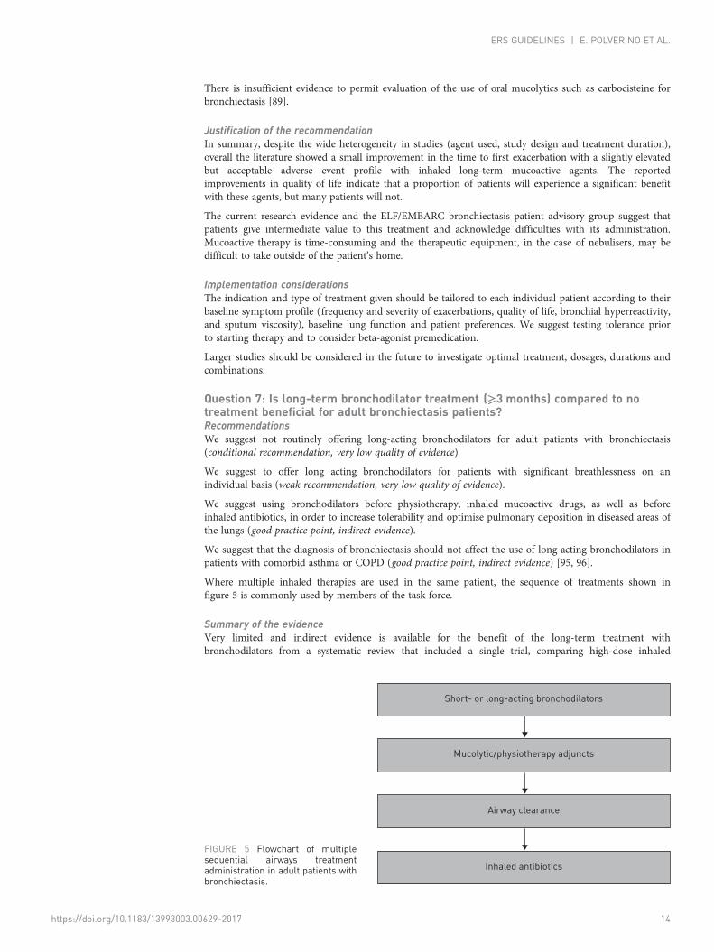
Where multiple inhaled therapies are used in the same patient, the sequence of treatments shown infigure 5 is commonly used by members of the task force.
Summary of the evidenceVery limited and indirect evidence is available for the benefit of the long-term treatment withbronchodilators from a systematic review that included a single trial, comparing high-dose inhaled
FIGURE 5 Flowchart of multiplesequential airways treatmentadministration in adult patients withbronchiectasis.
Short- or long-acting bronchodilators
Mucolytic/physiotherapy adjuncts
Airway clearance
Inhaled antibiotics
https://doi.org/10.1183/13993003.00629-2017 14
ERS GUIDELINES | E. POLVERINO ET AL.

corticosteroids to medium-dose inhaled corticosteroid/long acting beta-agonist combination [97]. Theresults from this study indicate some positive effects on symptom control/symptomatic improvement, inparticular decreased dyspnoea, better cough control, better health-related quality of life (measured bySGRQ symptoms domain), and reduced use of β2-agonist rescue medication. Specific side-effects weregenerally mild (tremor, nervousness and tachycardia). A systematic review identified major methodologicaland reporting concerns relating to this trial [56]. However, extrapolating evidence from populations withother obstructive airway diseases some bronchiectasis subpopulations may benefit from bronchodilators, inparticular subjects with chronic obstructive airflow limitation (FEV1/FVC <0.7; with or without FEV1
reversibility to bronchodilators), or associated asthma in combination with inhaled corticosteroids [95, 96].
Justification of the recommendationsWe suggest the use of bronchodilators in patient with significant breathlessness due to the feasibility ofapplication, the easy availability at a primary care level, the comparatively low treatment costs, and aputatively positive ratio of benefits to adverse events. Appropriate inhalation device selection and inhalertechnique training are recommended. If treatment with bronchodilators does not result in a reduction insymptoms it should be discontinued. There is no evidence to support the use of bronchodilators routinelyas part of the management of bronchiectasis patients without symptomatic breathlessness. According toboth research evidence and patient advisory group feedback, it seems that patients regard this as a low riskand low burden intervention.
Implementation considerationsThe intervention is easy to administer and acceptable to the majority of patients. Furtherinvestigator-driven research on the benefit of bronchodilators in bronchiectasis in various clinicalsituations is needed.
Question 8: Are surgical interventions more beneficial compared to standard(non-surgical) treatment for adult bronchiectasis patients?RecommendationWe suggest not offering surgical treatments for adult patients with bronchiectasis with the exception ofpatients with localised disease and a high exacerbation frequency despite optimisation of all other aspectsof their bronchiectasis management (weak recommendation, very low quality of evidence).
Summary of the evidenceThe rationale for surgical treatment of bronchiectasis is to break the vicious circle of bronchiectasis byremoving the lung segments that are no longer functional, and preventing the contamination of adjacentlung zones. The most frequent indication for the operation is recurrent infections with chronic symptomssuch as productive cough, purulent sputum and haemoptysis [98, 99].
Lobectomy is the most frequently performed operation, but numerous options have been described (e.g.segmentectomy and pneumonectomy) [100–102]. Surgery is the procedure of choice for massivehaemoptysis refractory to bronchial artery embolisation, but emergency surgery in unstable patients isassociated with higher morbidity and mortality reaching 37% [103]. Although bilateral bronchiectasis(reported in 5.8 to 30% of surgical series) are not an absolute contraindication for surgery [104], otheroptions such as prolonged conservative treatment or bronchial artery embolisation are frequently used asan alternative. The video-assisted thoracoscopic surgery (VATS) is often preferred to better preserve lungfunction or reduce scarring. In comparison with open surgery, VATS has been reported to producecomparable symptomatic improvement (94 versus 88%), but with shorter hospital stay, fewercomplications (17.5 versus 23.7%) and less pain after VATS procedures [105]. Contraindications to VATSinclude major parenchymal or pleural fibrosis, and calcified nodes close to the hilar vessels.
No randomised controlled trials of surgical treatment versus standard care were identified. A meta-analysisincluded 38 observational studies with 5541 patients, dealing with efficacy and safety of different surgicalinterventions for adult patients with bronchiectasis focused on three main outcomes: mortality, morbidity(adverse events) and quality of life improvement (symptomatic changes defined as reduction or alleviationof preoperative symptoms) [98].
The pooled mortality from 29 studies that focused on adult patients was 1.4% (95% CI 0.8%–2.5%) [98].Post-operative pooled morbidity for adults was analysed in 26 observational studies and was 16.2% (95%CI,12.5%–19.8%) [98]. It needs to be emphasised that there are no data comparing morbidity to continuedmedical non-surgical management alone. Moreover, according to the aforementioned studies, some of themorbidity is considered relatively minor (air leak, atelectasis, wound infection). Symptomatic changes wereanalysed in 26 observational studies. In the pooled meta-analysis, complete alleviation of symptoms was
https://doi.org/10.1183/13993003.00629-2017 15
ERS GUIDELINES | E. POLVERINO ET AL.

seen in 71.5% (95% CI 68–74.9) and reduction of preoperative symptoms was seen in 20.2% of the adultpopulation (95% CI 17.3–23.1) [98]. Other research has shown that extent of residual bronchiectasis andP. aeruginosa infection were reported as unfavourable prognostic factors [99].
Justification of the recommendationsOverall, surgical interventions seem to be beneficial only in very carefully selected patients requiring thebest risk-benefit profile of improved symptoms against the morbidity associated with surgery. Feedbackfrom the ELF/EMBARC patient advisory group suggests that patients would choose surgery only if therewas no effective medical option for treatment and this feedback informs the recommendation.
Implementation considerationsInvolvement of an experienced surgeon in partnership with an expert respiratory physician is advisable ifsurgical treatment is being considered. Attention should be paid to pre-operative nutritional status andpulmonary rehabilitation. More research is needed on surgical interventions. Although a randomised trialwould be very challenging future studies should include a matched control population with meticulousdescription of other treatments used in both populations.
Question 9: Is regular physiotherapy (airway clearance and/or pulmonaryrehabilitation) more beneficial than control (no physiotherapy treatment) in adultbronchiectasis patients?RecommendationsWe suggest that patients with chronic productive cough or difficulty to expectorate sputum should betaught an airway clearance technique (ACT) by a trained respiratory physiotherapist to perform once ortwice daily (weak recommendation, low quality of evidence).
We recommend that adult patients with bronchiectasis and impaired exercise capacity should participatein a pulmonary rehabilitation programme and take regular exercise. All interventions should be tailored tothe patient’s symptoms, physical capability and disease characteristics (strong recommendation, high qualityof evidence).
Summary of the evidence (figure 6)In bronchiectasis, it is a common belief that physiotherapy can improve mucus clearance and reduce lunginflammation and risk of infection. In addition, it is well accepted by patients. Respiratory physiotherapyincludes ACTs and pulmonary rehabilitation [106, 107]. ACTs consist of breathing techniques, e.g. activecycle of breathing and autogenic drainage, sometimes combined with an instrument, e.g. flutter or
Chest physiotherapy
Mucus problems
Positioning
Postural
drainage
AD
ELTGOL
Slow
expiration
Forced
expiration
PEP Oscillatory
PEP
PEP mask
T-PEP
Flutter
Acapella
HFCWO
ACBT
Cough
Huffing
Instrumental
techniques
Aerobic
training
Strength
training
Inspiratory
muscle
training (IMT)
Expiratory
flow
modification
Exercise
intolerance
Physical
activity
Counselling
Respiratory/muscle
weakness
FIGURE 6 Chest physiotherapy interventions flow chart based on clinical experience from the task force panel. AD: autogenic drainage; ELTGOL:total slow expiration with open glottis and infralateral position; ACBT: active cycle of breathing techniques; PEP: positive expiratory pressure;T-PEP: temporary positive expiratory pressure; HFCWO: high frequency chest wall oscillation.
https://doi.org/10.1183/13993003.00629-2017 16
ERS GUIDELINES | E. POLVERINO ET AL.
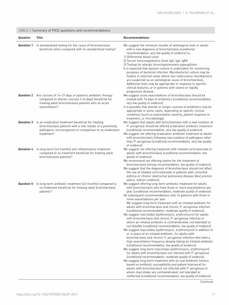
TABLE 2 Summary of PICO questions and recommendations
Question Title Recommendations
Question 1 Is standardised testing for the cause of bronchiectasisbeneficial when compared with no standardised testing?
We suggest the minimum bundle of aetiological tests in adultswith a new diagnosis of bronchiectasis (conditionalrecommendation, very low quality of evidence) is:
1) Differential blood count2) Serum immunoglobulins (total IgG, IgA, IgM)3) Testing for allergic bronchopulmonary aspergillosisIt is expected that sputum culture is undertaken for monitoringpurposes of bacterial infection. Mycobacterial culture may behelpful in selected cases where non-tuberculous mycobacteriaare suspected as an aetiological cause of bronchiectasis.Additional tests may be appropriate in response to specificclinical features, or in patients with severe or rapidlyprogressive disease.
Question 2 Are courses of 14–21 days of systemic antibiotic therapycompared to shorter courses (<14 days) beneficial fortreating adult bronchiectasis patients with an acuteexacerbation?
We suggest acute exacerbations of bronchiectasis should betreated with 14 days of antibiotics (conditional recommendation,very low quality of evidence).
It is possible that shorter or longer courses of antibiotics may beappropriate in some cases, depending on specific clinicalconditions (such as exacerbation severity, patient response totreatment, or microbiology).
Question 3 Is an eradication treatment beneficial for treatingbronchiectasis patients with a new isolate of a potentiallypathogenic microorganism in comparison to no eradicationtreatment?
We suggest that adults with bronchiectasis with a new isolation ofP. aeruginosa should be offered eradication antibiotic treatment(conditional recommendation, very low quality of evidence).
We suggest not offering eradication antibiotic treatment to adultswith bronchiectasis following new isolation of pathogens otherthan P. aeruginosa (conditional recommendation, very low qualityof evidence).
Question 4 Is long-term (⩾3 months) anti-inflammatory treatmentcompared to no treatment beneficial for treating adultbronchiectasis patients?
We suggest not offering treatment with inhaled corticosteroids toadults with bronchiectasis (conditional recommendation, lowquality of evidence).
We recommend not offering statins for the treatment ofbronchiectasis (strong recommendation, low quality of evidence).
We suggest that the diagnosis of bronchiectasis should not affectthe use of inhaled corticosteroids in patients with comorbidasthma or chronic obstructive pulmonary disease (best practiceadvice, indirect evidence).
Question 5 Is long-term antibiotic treatment (⩾3 months) compared tono treatment beneficial for treating adult bronchiectasispatients?
We suggest offering long-term antibiotic treatment for adultswith bronchiectasis who have three or more exacerbations peryear (conditional recommendation, moderate quality of evidence).
All subsequent recommendations refer to patients with three ormore exacerbations per year.We suggest long-term treatment with an inhaled antibiotic foradults with bronchiectasis and chronic P. aeruginosa infection(conditional recommendation, moderate quality of evidence).
We suggest macrolides (azithromycin, erythromycin) for adultswith bronchiectasis and chronic P. aeruginosa infection inwhom an inhaled antibiotic is contraindicated, not tolerated ornot feasible (conditional recommendation, low quality of evidence).
We suggest macrolides (azithromycin, erythromycin) in addition toor in place of an inhaled antibiotic, for adults withbronchiectasis and chronic P. aeruginosa infection who have ahigh exacerbation frequency despite taking an inhaled antibiotic(conditional recommendation, low quality of evidence).
We suggest long-term macrolides (azithromycin, erythromycin)for adults with bronchiectasis not infected with P. aeruginosa(conditional recommendation, moderate quality of evidence).
We suggest long-term treatment with an oral antibiotic (choicebased on antibiotic susceptibility and patient tolerance) foradults with bronchiectasis not infected with P. aeruginosa inwhom macrolides are contraindicated, not tolerated orineffective (conditional recommendation, low quality of evidence).
Continued
https://doi.org/10.1183/13993003.00629-2017 17
ERS GUIDELINES | E. POLVERINO ET AL.

Acapella, that modify expiratory flow and volumes or produce chest wall oscillations in order to increasemucus clearance [108–112]. The principal effect obtained by ACTs is an increase in sputum volume [108,112, 113] and a reduced impact of cough on quality of life [109, 114]. Interesting, but still preliminarydata, shows reduced peripheral airways obstruction, less inflammatory cells in sputum and improvedexercise capacity after ACTs [109, 112, 114]. The aim of a pulmonary rehabilitation programme is toimprove exercise tolerance and quality of life through a tailored standardised exercise protocol [115–117].
We identified three systematic reviews [106, 118, 119] and several additional trials. We included a total of14 clinical trials in our analysis [91, 108, 110–112, 114–117, 120–124].
The pooled analysis shows that pulmonary rehabilitation has a clear impact on exercise capacityimmediately after the programme and a nonsignificant trend to improved quality of life (SGRQ) [116, 117,
TABLE 2 Continued
Question Title Recommendations
We suggest long-term treatment with an inhaled antibiotic foradults with bronchiectasis not infected with P. aeruginosa inwhom oral antibiotic prophylaxis is contraindicated, nottolerated or ineffective (conditional recommendation, low qualityof evidence).Long-term antibiotic therapy should be considered only afteroptimisation of general aspects of bronchiectasis management(airway clearance and treating modifiable underlying causes).
Question 6 Is long-term mucoactive treatment (⩾3 months) compared tono treatment beneficial for treating adult bronchiectasispatients?
We suggest offering long-term mucoactive treatment (⩾3 months)in adult patients with bronchiectasis who have difficulty inexpectorating sputum and poor quality of life and wherestandard airway clearance techniques have failed to controlsymptoms (weak recommendation, low quality of evidence).
We recommend not offering recombinant human DNase to adultpatients with bronchiectasis (strong recommendation, moderatequality of evidence).
Question 7 Is long-term bronchodilator treatment (⩾3 months)compared to no treatment beneficial for adultbronchiectasis patients?
We suggest not routinely offering long-acting bronchodilators foradult patients with bronchiectasis (conditional recommendation,very low quality of evidence).
We suggest offering long acting bronchodilators for patients withsignificant breathlessness on an individual basis (weakrecommendation, very low quality of evidence).
We suggest using bronchodilators before physiotherapy, includinginhaled mucoactive drugs, as well as before inhaled antibiotics,in order to increase tolerability and optimise pulmonarydeposition in diseased areas of the lungs (good practice point,indirect evidence).
We suggest that the diagnosis of bronchiectasis should not affectthe use of long acting bronchodilators in patients withcomorbid asthma or chronic obstructive pulmonary disease(good practice point, indirect evidence) [95, 96].
Question 8 Are surgical interventions more beneficial compared tostandard (non-surgical) treatment for adult bronchiectasispatients?
We suggest not offering surgical treatments for adult patientswith bronchiectasis with the exception of patients with localiseddisease and a high exacerbation frequency despite optimisationof all other aspects of their bronchiectasis management (weakrecommendation, very low quality of evidence).
Question 9 Is regular physiotherapy (airway clearance and/or pulmonaryrehabilitation) more beneficial than control (nophysiotherapy) in adult bronchiectasis patients?
We suggest that patients with chronic productive cough ordifficulty to expectorate sputum should be taught an airwayclearance technique by a trained respiratory physiotherapist toperform once or twice daily (weak recommendation, low qualityof evidence).
We recommend that adult patients with bronchiectasis andimpaired exercise capacity should participate in a pulmonaryrehabilitation programme and take regular exercise. Allinterventions should be tailored to the patient’s symptoms,physical capability and disease characteristics (strongrecommendation, high quality of evidence).
https://doi.org/10.1183/13993003.00629-2017 18
ERS GUIDELINES | E. POLVERINO ET AL.

119, 122, 123]. However, the unpooled study of MANDAL et al. [122] (pulmonary rehabilitation duration(8 weeks) shorter than for pooled studies) described that improvements on exercise capacity and patient’squality of life may be maintained for a longer period of time. Finally, there is one publication showing animpact of pulmonary rehabilitation (8 weeks of supervised exercise training and review of ACTs)decreasing the frequency of exacerbations (median 1 (interquartile range 1–3) versus 2 (1–3); p=0.012)over 12-month follow-up and longer time to first exacerbation (8 versus 6 months; p=0.047) [116]. Thereported impact of both ACTs and pulmonary rehabilitation on pulmonary function is not clinicallyimportant [107–109, 112, 121].
Justification of the recommendationsThe evidence for airways clearance techniques is weak because the studies are small and poorlycomparable due to methodological issues. However, most studies demonstrated a significant increase ofsputum volume. The evidence is stronger for pulmonary rehabilitation, showing improvements in exercisecapacity, cough symptoms and quality of life, and possibly a reduction in exacerbations. The benefits ofpulmonary rehabilitation are achieved in 6 to 8 weeks and maintained for between 3 to 6 months. Finally,there are no relevant adverse effects and the bronchiectasis patients advisory group value the intervention.
Implementation considerationsThe research priorities in physiotherapy are: larger controlled studies with clinical outcomes(exacerbations, cough and quality of life); larger controlled studies including physiotherapy training plusmucoactive agents such as hypertonic saline; the role of pulmonary rehabilitation on exacerbations; andfinally, the compliance with these interventions over a longer period of time (>12 months) [125].
SummaryThe recommendations are summarised in table 2. Management of bronchiectasis aims to reduceexacerbations, reduce symptoms, improve quality of life [126, 127] and reduce the risk of futurecomplications such as lung function decline [128] and severe exacerbations [129]. Treatment decisionsmust balance the potential beneficial effects of the intervention against the burden of treatment and therisk of adverse events. It is important to take into account the patients values and preferences in alltreatment decisions, alongside the history of exacerbations, quality of life [126, 127], severity of disease [9]and underlying aetiology [22], all of which can impact on the patients long-term outcome [130–132].
The purpose of clinical guidelines is to improve the quality of patient care and to promote safe, effectiveand cost-effective treatment. The majority of recommendations in this guideline are conditional and basedon low quality evidence. One outcome of this guideline should be to promote further research into theoptimal treatment of patients with bronchiectasis. Bronchiectasis is a rapidly evolving field and ourrecommendations will require revision as additional data becomes available in the coming years.
AcknowledgementsThe authors acknowledge Valentina Balasso for assistance with the literature search. Guideline development wassupported by the European Bronchiectasis Network (EMBARC) and the EMBARC/European Lung Foundationbronchiectasis patient advisory group.
References1 Winter DH, Manzini M, Salge JM, et al. Aging of the lungs in asymptomatic lifelong nonsmokers: findings on
HRCT. Lung 2015; 193: 283–290.2 Brozek JL, Akl EA, Compalati E, et al. Grading quality of evidence and strength of recommendations in clinical
practice guidelinesPart 3 of 3. The GRADE approach to developingrecommendations. Allergy 2011; 64:1109–1116.
3 Andrews J, Guyatt G, Oxman AD, et al. GRADE guidelines: 14. Going from evidence to recommendations: Thesignificance and presentation of recommendations. J Clin Epidemiol 2013; 66: 719–725.
4 King PT, Holdsworth SR, Freezer NJ, et al. Characterisation of the onset and presenting clinical features of adultbronchiectasis. Respir Med 2006; 100: 2183–2189.
5 Raghu G, King TE, Behr J, et al. Quality of life and dyspnoea in patients treated with bosentan for idiopathicpulmonary fibrosis (BUILD-1). Eur Respir J 2010; 35: 118–123.
6 Tashkin DP, Celli B, Senn S, et al. A 4-year trial of tiotropium in chronic obstructive pulmonary disease. N EnglJ Med 2008; 359: 1543–1554.
7 Chalmers JD, Smith MP, McHugh BJ, et al. Short- and long-term antibiotic treatment reduces airway andsystemic inflammation in non-cystic fibrosis bronchiectasis. Am J Respir Crit Care Med 2012; 186: 657–665.
8 Sheehan RE, Wells AU, Copley SJ, et al. A comparison of serial computed tomography and functional change inbronchiectasis. Eur Respir J 2002; 20: 581–587.
9 Chalmers JD, Goeminne P, Aliberti S, et al. The bronchiectasis severity index. An international derivation andvalidation study. Am J Respir Crit Care Med 2014; 189: 576–585.
10 Aliberti S, Lonni S, Dore S, et al. Clinical phenotypes in adult patients with bronchiectasis. Eur Respir J 2016; 47:1113–1122.
https://doi.org/10.1183/13993003.00629-2017 19
ERS GUIDELINES | E. POLVERINO ET AL.

11 Kapur N, Masters IB, Chang AB. Longitudinal growth and lung function in pediatric non-cystic fibrosisbronchiectasis: What influences lung function stability? Chest 2010; 138: 158–164.
12 Chalmers JD, Aliberti S, Polverino E, et al. The EMBARC European Bronchiectasis Registry: protocol for aninternational observational study. ERJ Open Res 2016; 2: 00081-2015.
13 Loebinger MR, Wells AU, Hansell DM, et al. Mortality in bronchiectasis: a long-term study assessing the factorsinfluencing survival. Eur Respir J 2009; 34: 843–849.
14 McDonnell MJ, Aliberti S, Goeminne PC, et al. Multidimensional severity assessment in bronchiectasis: ananalysis of seven European cohorts. Thorax 2016; 71: 1110–1118.
15 Finch S, McDonnell MJ, Abo-Leyah H, et al. A comprehensive analysis of the impact of Pseudomonas aeruginosacolonization on prognosis in adult bronchiectasis. Ann Am Thorac Soc 2015; 12: 1602–1611.
16 Wilson CB, Jones PW, O’Leary CJ, et al. Systemic markers of inflammation in stable bronchiectasis. Eur Respir J1998; 12: 820–824.
17 Tsang KW, Chan KN, Ho PL, et al. Sputum elastase in steady-state bronchiectasis. Chest 2000; 117: 420–426.18 Chalmers JD, Moffitt KL, Suarez-Cuartin G, et al. Neutrophil elastase activity is associated with exacerbations
and lung function decline in bronchiectasis. Am J Respir Crit Care Med 2017; 195: 1384–1393.19 Chalmers JD, Hill AT. Mechanisms of immune dysfunction and bacterial persistence in non-cystic fibrosis
bronchiectasis. Mol Immunol 2013; 55: 27–34.20 Snijders D, Dominguez BF, Calgaro S, et al. Mucociliary clearance techniques for treating non-cystic fibrosis
bronchiectasis: Is there evidence? Int J Immunopathol Pharmacol 2015; 28: 150–159.21 McDonnell MJ, Aliberti S, Goeminne PC, et al. Comorbidities and the risk of mortality in patients with
bronchiectasis: an international multicentre cohort study. Lancet Respir Med 2016; 4: 969–979.22 Du Q, Jin J, Liu X, et al. Bronchiectasis as a comorbidity of chronic obstructive pulmonary disease: A systematic
review and meta-analysis. PLoS One 2016; 11: e0150532.23 Navaratnam V, Muirhead CR, Hubbard RB, et al. Critical care admission trends and outcomes in individuals
with bronchiectasis in the UK. QJM 2016; 109: 523–526.24 Navaratnam V, Millett ER, Hurst JR, et al. Bronchiectasis and the risk of cardiovascular disease: a
population-based study. Thorax 2017; 72: 161–166.25 Ringshausen FC, de Roux A, Pletz MW, et al. Bronchiectasis-associated hospitalizations in Germany, 2005-2011:
a population-based study of disease burden and trends. PLoS One 2013; 8: e71109.26 Weycker D, Edelsberg J, Oster G, et al. Prevalence and economic burden of bronchiectasis. Clin Pulm Med 2005;
12: 205–209.27 Kwak HJ, Moon J-YY, Choi YW, et al. High prevalence of bronchiectasis in adults: analysis of CT findings in a
health screening program. Tohoku J Exp Med 2010; 222: 237–242.28 Quint JK, Millett ER, Joshi M, et al. Changes in the incidence, prevalence and mortality of bronchiectasis in the
UK from 2004 to 2013: A population-based cohort study. Eur Respir J 2016; 47: 186–193.29 Chang AB, Grimwood K, Mulholland EK, et al. Bronchiectasis in Indigenous children in remote Australian
communities. Med J Aust 2002; 177: 200–204.30 Seitz AE, Olivier KN, Steiner CA, et al. Trends and burden of bronchiectasis-associated hospitalizations in the
United States, 1993-2006. Chest 2010; 138: 944–949.31 Finklea JD, Khan G, Thomas S, et al. Predictors of mortality in hospitalized patients with acute exacerbation of
bronchiectasis. Respir Med 2010; 104: 816–821.32 Goeminne PC, Nawrot TS, Ruttens D, et al. Mortality in non-cystic fibrosis bronchiectasis: A prospective cohort
analysis. Respir Med 2014; 108: 287–296.33 de la Rosa D, Martinez-Garcia MA, Olveira C, et al. Annual direct medical costs of bronchiectasis treatment:
Impact of severity, exacerbations, chronic bronchial colonization and chronic obstructive pulmonary diseasecoexistence. Chron Respir Dis 2016; 13: 361–371.
34 Gursel G. Does coexistence with bronchiectasis influence intensive care unit outcome in patients with chronicobstructive pulmonary disease? Heart Lung 2006; 35: 58–65.
35 O’Donnell AE, Barker AF, Ilowite JS, et al. Treatment of idiopathic bronchiectasis with aerosolized recombinanthuman DNase I. Chest 1998; 113: 1329–1334.
36 Barker AF, O’Donnell AE, Flume P, et al. Aztreonam for inhalation solution in patients with non-cystic fibrosisbronchiectasis (AIR-BX1 and AIR-BX2): Two randomised double-blind, placebo-controlled phase 3 trials. LancetRespir Med 2014; 2: 738–749.
37 Vendrell M, de Gracia J, Olveira C, et al. Normativa SEPAR: diagnóstico y tratamiento de las bronquiectasias.Arch Bronconeumol 2008; 44: 629–640.
38 Pasteur MC, Bilton D, Hill AT, British Thoracic Society Bronchiectasis non-CF Guideline Group. BritishThoracic Society guideline for non-CF bronchiectasis. Thorax 2010; 65: Suppl. 1, i1–i58.
39 Woodhead M, Blasi F, Ewig S, et al. Guidelines for the management of adult lower respiratory tractinfections--full version. Clin Microbiol Infect 2011; 17: Suppl. 6, E1–59.
40 Andrews JC, Schünemann HJ, Oxman AD, et al. GRADE guidelines: 15. Going from evidence torecommendation—determinants of a recommendation’s direction and strength. J Clin Epidemiol 2013; 66:726−735.
41 Alonso-Coello P, Schünemann HJ, Moberg J, et al. GRADE Evidence to Decision (EtD) frameworks: a systematicand transparent approach to making well informed healthcare choices. 1: Introduction. BMJ 2016; 353: i2016.
42 Anwar GA, McDonnell MJ, Worthy SA, et al. Phenotyping adults with non-cystic fibrosis bronchiectasis: Aprospective observational cohort study. Respir Med 2013; 107: 1001–1007.
43 Shoemark A, Ozerovitch L, Wilson R. Aetiology in adult patients with bronchiectasis. Respir Med 2007; 101:1163–1170.
44 Pasteur MC, Helliwell SM, Houghton SJ, et al. An investigation into causative factors in patients withbronchiectasis. Am J Respir Crit Care Med 2000; 162: 1277–1284.
45 Lonni S, Chalmers JD, Goeminne PC, et al. Etiology of non-cystic fibrosis bronchiectasis in adults and itscorrelation to disease severity. Ann Am Thorac Soc 2015; 12: 1764–1770.
46 Greenberger PA. When to suspect and work up allergic bronchopulmonary aspergillosis. Ann Allergy AsthmaImmunol 2013; 111: 1–4.
https://doi.org/10.1183/13993003.00629-2017 20
ERS GUIDELINES | E. POLVERINO ET AL.

47 Knutsen AP, Bush RK, Demain JG, et al. Fungi and allergic lower respiratory tract diseases. J Allergy ClinImmunol 2012; 129: 280–291.
48 Griffith DE, Aksamit T, Brown-Elliott BA, et al. An official ATS/IDSA statement: Diagnosis, treatment, andprevention of nontuberculous mycobacterial diseases. Am J Respir Crit Care Med 2007; 175: 367–416.
49 Lucas JS, Barbato A, Collins SA, et al. European Respiratory Society guidelines for the diagnosis of primaryciliary dyskinesia. Eur Respir J 2017; 49: 1601090.
50 Bilton D, Henig N, Morrissey B, et al. Addition of inhaled tobramycin to ciprofloxacin for acute exacerbations ofPseudomonas aeruginosa infection in adult bronchiectasis. Chest 2006; 130: 1503–1510.
51 Murray MP, Turnbull K, MacQuarrie S, et al. Assessing response to treatment of exacerbations of bronchiectasisin adults. Eur Respir J 2009; 33: 312–317.
52 Orriols R, Roe J. Inhaled antibiotic therapy in non-cystic fibrosis patients with bronchiectasis and chronicbroncial infection by Pseudomonas aeruginosa. Respir Med 1999; 93: 476–480.
53 McDonnell MJ, Jary HR, Perry A, et al. Non cystic fibrosis bronchiectasis: A longitudinal retrospectiveobservational cohort study of Pseudomonas persistence and resistance. Respir Med 2015; 109: 716–726.
54 Orriols R, Hernando R, Ferrer A, et al. Eradication therapy against Pseudomonas aeruginosa in non-cysticfibrosis bronchiectasis. Respiration 2015; 90: 299–305.
55 White L, Mirrani G, Grover M, et al. Outcomes of Pseudomonas eradication therapy in patients with non-cysticfibrosis bronchiectasis. Respir Med 2012; 106: 356–360.
56 Goyal V, Chang AB. Combination inhaled corticosteroids and long-acting beta2-agonists for children and adultswith bronchiectasis. Cochrane Database Syst Rev 2014; CD010327.
57 Steele K, Greenstone M, Lasserson JA. Oral methyl-xanthines for bronchiectasis. Cochrane Database Syst Rev2001; CD002734.
58 Lasserson T, Holt K. Oral corticosteroids for bronchiectasis (stable and acute exacerbations). Cochrane DatabaseSyst Rev 2001; CD002162.
59 Corless JA, Warburton CJ. Leukotriene receptor antagonists for non-cystic fibrosis bronchiectasis. CochraneDatabase Syst Rev 2000; CD002174.
60 Kapur N, Chang AB. Oral non steroid anti-inflammatories for children and adults with bronchiectasis. CochraneDatabase Syst Rev 2007; CD006427.
61 Pizzutto SJ, Upham JW, Yerkovich ST, et al. Inhaled non-steroid anti-inflammatories for children and adultswith bronchiectasis. Cochrane Database Syst Rev 2016; CD007525.
62 Tsang KW, Tan KC, Ho PL, et al. Inhaled fluticasone in bronchiectasis: a 12 month study. Thorax 2005; 60:239–243.
63 Mandal P, Chalmers JD, Graham C, et al. Atorvastatin as a stable treatment in bronchiectasis: A randomisedcontrolled trial. Lancet Respir Med 2014; 2: 455–463.
64 Hernando R, Drobnic ME, Cruz MJ, et al. Budesonide efficacy and safety in patients with bronchiectasis not dueto cystic fibrosis. Int J Clin Pharm 2012; 34: 644–650.
65 Brodt AM, Stovold E, Zhang L. Inhaled antibiotics for stable non-cystic fibrosis bronchiectasis: a systematicreview. Eur Respir J 2014; 44: 382–393.
66 Gao Y, Guan W, Xu G, et al. Macrolide therapy in adults and children with non-cystic fibrosis bronchiectasis: asystematic review and meta-analysis. PLoS One 2014; 9: e90047.
67 Shi ZL, Peng H, Hu XW, et al. Effectiveness and safety of macrolides in bronchiectasis patients: a meta-analysisand systematic review. Pulm Pharmacol Ther 2014; 28: 171–178.
68 Wu Q, Shen W, Cheng H, et al. Long-term macrolides for non-cystic fibrosis bronchiectasis: A systematic reviewand meta-analysis. Respirology 2014; 19: 321–329.
69 Zhuo GY, He Q, Xiang-Lian L, et al. Prolonged treatment with macrolides in adult patients with non-cysticfibrosis bronchiectasis: Meta-analysis of randomized controlled trials. Pulm Pharmacol Ther 2014; 30: 1–9.
70 Fan LC, Lu HW, Wei P, et al. Effects of long-term use of macrolides in patients with non-cystic fibrosisbronchiectasis: a meta-analysis of randomized controlled trials. BMC Infect Dis 2015; 15: 160.
71 Yang JW, Fan LC, Lu HW, et al. Efficacy and safety of long-term inhaled antibiotic for patients with noncysticfibrosis bronchiectasis: a meta-analysis. Clin Respir J 2016; 10: 731–739.
72 Hnin K, Nguyen C, Carson K V, et al. Prolonged antibiotics for non-cystic fibrosis bronchiectasis in children andadults. Cochrane Database Syst Rev 2015; 8: CD001392.
73 Currie DC, Garbett ND, Chan KL, et al. Double-blind randomized study of prolonged higher-dose oralamoxycillin in purulent bronchiectasis. Q J Med 1990; 76: 799–816.
74 Cymbala AA, Edmonds LC, Bauer MA, et al. The disease-modifying effects of twice-weekly oral azithromycin inpatients with bronchiectasis. Treat Respir Med 2005; 4: 117–122.
75 Drobnic ME, Suñé P, Montoro JB, et al. Inhaled tobramycin in non-cystic fibrosis patients with bronchiectasisand chronic bronchial infection with Pseudomonas aeruginosa. Ann Pharmacother 2005; 39: 39–44.
76 Murray MP, Govan JRW, Doherty CJ, et al. A randomized controlled trial of nebulized gentamicin in non-cysticfibrosis bronchiectasis. Am J Respir Crit Care Med 2011; 183: 491–499.
77 Wong C, Jayaram L, Karalus N, et al. Azithromycin for prevention of exacerbations in non-cystic fi brosisbronchiectasis (EMBRACE): A randomised, double-blind, placebo-controlled trial. Lancet 2012; 380: 660–667.
78 Serisier DJ, Bilton D, De Soyza A, et al. Inhaled, dual release liposomal ciprofloxacin in non-cystic fibrosisbronchiectasis (ORBIT-2): a randomised, double-blind, placebo-controlled trial. Thorax 2013; 68: 812–817.
79 Serisier DJ, Martin ML, McGuckin MA, et al. Effect of long-term, low-dose erythromycin on pulmonaryexacerbations among patients with non-cystic fibrosis bronchiectasis: the BLESS randomized controlled trial.JAMA 2013; 309: 1260–1267.
80 Altenburg J. Effect of azithromycin maintenance treatment on infectious exacerbations among patients withnon–cystic fibrosis bronchiectasis. JAMA 2013; 309: 1251–1259.
81 De Diego A, Milara J, Martinez-Moragón E, et al. Effects of long-term azithromycin therapy on airway oxidativestress markers in non-cystic fibrosis bronchiectasis. Respirology 2013; 18: 1056–1062.
82 Tabernero Huguet E, Gil Alaña P, Alkiza Basañez R, et al. Inhaled colistin in elderly patients with non-cysticfibrosis bronchiectasis and chronic Pseudomonas aeruginosa bronchial infection. Rev española Geriatr y Gerontol2015; 50: 111–115.
https://doi.org/10.1183/13993003.00629-2017 21
ERS GUIDELINES | E. POLVERINO ET AL.

83 Haworth CS, Foweraker JE, Wilkinson P, et al. Inhaled colistin in patients with bronchiectasis and chronicPseudomonas aeruginosa infection. Am J Respir Crit Care Med 2014; 189: 975–982
84 PROLONGED antibiotic treatment of severe bronchiectasis; a report by a subcommittee of the AntibioticsClinical Trials (non-tuberculous) Committee of the Medical Research Council. Br Med J 1957; 2: 255–259.
85 Lourdesamy AI, Muthukumaru U. Efficacy of azithromycin in the treatment of bronchiectasis. Respirology 2014;19: 1178–1182.
86 Liu J, Zhong X, He Z, et al. Effect of Low-Dose, long-term roxithromycin on airway inflammation andremodeling of stable noncystic fibrosis bronchiectasis. Mediators Inflamm 2014; 2014: 708608.
87 Barker AF, Couch L, Fiel SB, et al. Tobramycin solution for inhalation reduces sputum Pseudomonas aeruginosadensity in bronchiectasis. Am J Respir Crit Care Med 2000; 162: 481–485.
88 Hart A, Sugumar K, Milan SJ, et al. Inhaled hyperosmolar agents for bronchiectasis. Cochrane Database Syst Rev2014; 5: CD002996.
89 Wilkinson M, Sugumar K, Milan SJ, et al. Mucolytics for bronchiectasis. Cochrane Database Syst Rev 2014; 5:CD001289.
90 Wills P, Greenstone M. Inhaled hyperosmolar agents for bronchiectasis. Cochrane Database Syst Rev 2002; 1:CD002996.
91 Nicolson CHH, Stirling RG, Borg BM, et al. The long term effect of inhaled hypertonic saline 6% in non-cysticfibrosis bronchiectasis. Respir Med 2012; 106: 661–667.
92 Kellett F, Robert NM. Nebulised 7% hypertonic saline improves lung function and quality of life inbronchiectasis. Respir Med 2011; 105: 1831–1835.
93 Bilton D, Daviskas E, Anderson D, et al. Phase 3 randomized study of the efficacy and safety of inhaled drypowder mannitol for the symptomatic treatment of non-cystic fibrosis bronchiectasis. Chest 2013; 144: 215–225.
94 Bilton D, Tino G, Barker AF, et al. Inhaled mannitol for non-cystic fibrosis bronchiectasis: a randomised,controlled trial. Thorax 2014; 69: 1073–1079.
95 Vogelmeier CF, Criner GJ, Martinez FJ, et al. Global Strategy for the Diagnosis, Management, and Prevention ofChronic Obstructive Lung Disease 2017 Report: GOLD Executive Summary. Eur Respir J 2017; 49: 1700214.
96 Reddel HK, Bateman ED, Becker A, et al. A summary of the new GINA strategy: A roadmap to asthma control.Eur Respir J 2015; 46: 622–639.
97 Martínez-García MÁ, Soler-Cataluña JJ, Catalán-Serra P, et al. Clinical efficacy and safety ofbudesonide-formoterol in non-cystic fibrosis bronchiectasis. Chest 2012; 141: 461–468.
98 Fan L-C, Liang S, Lu H-W, et al. Efficiency and safety of surgical intervention to patients with non-cystic fibrosisbronchiectasis: a meta-analysis. Sci Rep 2015; 5: 17382.
99 Hiramatsu M, Shiraishi Y, Nakajima Y, et al. Risk factors that affect the surgical outcome in the management offocal bronchiectasis in a developed country. Ann Thorac Surg 2012; 93: 245–250.
100 Hoffman E. The late results of the conservation of the apical segment of the lower lobe in resections forbronchiectasis. Thorax 1955; 10: 137–141.
101 Collis J. Fate of the lower apical segment in resections for bronchiectasis. Thorax 1953; 8: 323–325.102 Tanaka H, Matsumura A, Okumura M, et al. Pneumonectomy for unilateral destroyed lung with pulmonary
hypertension due to systemic blood flow through broncho-pulmonary shunts. Eur J Cardiothoracic Surg 2005; 28:389–393.
103 Gourin A, Garzon AA. Operative treatment of massive hemoptysis. Ann Thorac Surg 1974; 18: 52–60.104 Schneiter D, Meyer N, Lardinois D, et al. Surgery for non-localized bronchiectasis. Br J Surg 2005; 92: 836–839.105 Zhang P, Zhang F, Jiang S, et al. Video-assisted thoracic surgery for bronchiectasis. Ann Thorac Surg 2011; 91:
239–243.106 Lee AL, Burge AT, Holland AE. Airway clearance techniques for bronchiectasis. Cochrane database Syst Rev
2015; 11: CD008351.107 Bradley J, Moran F, Greenstone M. Physical training for bronchiectasis. Cochrane Database Syst Rev 2002;
CD002166.108 Guimarães FS, Moço VJR, Menezes SLS, et al. Effects of ELTGOL and Flutter VRP1® on the dynamic and static
pulmonary volumes and on the secretion clearance of patients with bronchiectasis. Rev Bras Fisioter 2012; 16:108–113.
109 Nicolini A, Cardini F, Landucci N, et al. Effectiveness of treatment with high-frequency chest wall oscillation inpatients with bronchiectasis. BMC Pulm Med 2013; 13: 21.
110 Eaton T, Young P, Zeng I, et al. A randomized evaluation of the acute efficacy, acceptability and tolerability offlutter and active cycle of breathing with and without postural drainage in non-cystic fibrosis bronchiectasis.Chron Respir Dis 2007; 4: 23–30.
111 Naraparaju S, Vaishali K, Venkatesan P, et al. A comparison of the Acapella and a threshold inspiratory muscletrainer for sputum clearance in bronchiectasis-A pilot study. Physiother Theory Pract 2010; 26: 353–357.
112 Figueiredo PHS, Zin WA, Guimarães FS. Flutter valve improves respiratory mechanics and sputum production inpatients with bronchiectasis. Physiother Res Int 2012; 17: 12–20.
113 Patterson JE, Bradley JM, Hewitt O, et al. Airway clearance in bronchiectasis: A randomized crossover trial ofactive cycle of breathing techniques versus acapella? Respiration 2005; 72: 239–242.
114 Murray MP, Pentland JL, Hill AT. A randomised crossover trial of chest physiotherapy in non-cystic fibrosisbronchiectasis. Eur Respir J 2009; 34: 1086–1092.
115 Faulkner J, Gerhard J, Stoner L, et al. Self-paced walking within a diverse topographical environment elicits anappropriate training stimulus for cardiac rehabilitation patients. Rehabil Res Pract 2012; 2012: 140871.
116 Lee AL, Hill CJ, Cecins N, et al. The short and long term effects of exercise training in non-cystic fibrosisbronchiectasis – a randomised controlled trial. Respir Res 2014; 15: 44.
117 Zanini A, Aiello M, Adamo D, et al. Effects of pulmonary rehabilitation in patients with non-cystic fibrosisbronchiectasis: a retrospective analysis of clinical and functional predictors of efficacy. Respiration 2015; 89:525–533.
118 Bradley J, Moran F, Greenstone M. Physical training for bronchiectasis. Cochrane Database Syst Rev 2002;CD002166.
https://doi.org/10.1183/13993003.00629-2017 22
ERS GUIDELINES | E. POLVERINO ET AL.

119 Lee AL, Hill CJ, McDonald CF, et al. Pulmonary rehabilitation in individuals with non-cystic fibrosisbronchiectasis: a systematic review. Arch Phys Med Rehabil 2016; 774–782.e1.
120 Patterson JE, Bradley JM, Elborn JS. Airway clearance in bronchiectasis: a randomized crossover trial of activecycle of breathing techniques (incorporating postural drainage and vibration) versus test of incrementalrespiratory endurance. Chron Respir Dis 2004; 1: 127–130.
121 Gokdemir Y, Karadag-Saygi E, Erdem E, et al. Comparison of conventional chest physiotherapy andhigh-frequency chest wall oscillation in primary ciliary dyskinesia. Pediatr Pulmonol 2014; 49: 611–616.
122 Mandal P, Sidhu MK, Kope L, et al. A pilot study of pulmonary rehabilitation and chest physiotherapy versuschest physiotherapy alone in bronchiectasis. Respir Med 2012; 106: 1647–1654.
123 Newall C, Stockley RA, Hill SL. Exercise training and inspiratory muscle training in patients with bronchiectasis.Thorax 2005; 60: 943–948.
124 Liaw M-Y, Wang Y-H, Tsai Y-C, et al. Inspiratory muscle training in bronchiectasis patients: a prospectiverandomized controlled study. Clin Rehabil 2011; 25: 524–536.
125 Aliberti S, Masefield S, Polverino E, et al. Research priorities in bronchiectasis: a consensus statement from theEMBARC Clinical Research Collaboration. Eur Respir J 2016; 48: 632–647.
126 Wilson CB, Jones PW, O’Leary CJ, et al. Validation of the St. George’s respiratory questionnaire inbronchiectasis. Am J Respir Crit Care Med 1997; 156: 536–541.
127 Quittner AL, O‘Donnell AE, Salathe MA, et al. Quality of Life Questionnaire-Bronchiectasis: final psychometricanalyses and determination of minimal important difference scores. Thorax 2015; 70: 12–20.
128 Martínez-García MA, Soler-Cataluña J-J, Perpiñá-Tordera M, et al. Factors associated with lung function declinein adult patients with stable non-cystic fibrosis bronchiectasis. Chest 2007; 132: 1565–1572.
129 Chalmers JD, Aliberti S, Blasi F. Management of bronchiectasis in adults. Eur Respir J 2015; 45: 1446–1462.130 Goeminne PC, Scheers H, Decraene A, et al. Risk factors for morbidity and death in non-cystic fibrosis
bronchiectasis: a retrospective cross-sectional analysis of CT diagnosed bronchiectatic patients. Respir Res 2012;13: 21.
131 Hill AT, Haworth CS, Aliberti S, et al. Pulmonary exacerbation in adults with bronchiectasis: a consensusdefinition for clinical research. Eur Respir J 2017; 49: 1700051.
132 Spinou A, Siegert RJ, Guan WJ, et al. The development and validation of the bronchiectasis health questionnaire.Eur Respir J 2017; 49: 1601532.
ERS GUIDELINES | E. POLVERINO ET AL.