evaluation and management of splenic injury in blunt ... 4/version-5... · patients and...
TRANSCRIPT

IOSR Journal of Dental and Medical Sciences (IOSR-JDMS)
e-ISSN: 2279-0853, p-ISSN: 2279-0861.Volume 15, Issue 4 Ver. V (Apr. 2016), PP 01-19
www.iosrjournals.org
DOI: 10.9790/0853-1504050119 www.iosrjournals.org 1 | Page
Evaluation and Management of Splenic Injury In Blunt
Abdominal Trauma
Dr Nikhila Pinjala1, Dr.N.Nageswara Rao
2; Dr.M.MallikarjunaReddy
3
1. Junior Resident, Department of General Surgery,NRI Medical College, Chinakakani
2. Professor, Department of General Surgery NRI Medical College .Chinakakani
3.AssistantProfessor, Department of General Surgery, NRI Medical College, Chinakakani
Abstract: With a view to prevent the immediate and late complications of operative procedures of spleen,
especially the risk of Overwhelming post-splenectomy syndrome (OPSS), non operativemanagement has been
proposed when the haemodynamic condition of the patient permits. This study was done to evaluate the
prevalence, severity and mode of splenic trauma, management techniques (non operative& operative) and
complications amongst the blunt abdominal trauma cases admitted in NRI General Hospital, a tertiary referral
centre between the period October 2013 to September 2015
Patients And Methods:Fortypatients admitted to NRIGH, with splenic injuries from blunt abdominal trauma
between October’2013 to September’2015 were included in the study. For every patient, serial monitoring of
clinical and haematological data was done. For every case FASTand CECT-Abdomen was done to arrive at an
accurate assessment of the severity of splenic and concomitant injuries.
Results:In our study 28patients were managed non-operatively, while 12 underwent various operative
procedures. Grades I, II, and III spleen injury was significantly associated with non-operative treatment, while
Grade-IV and V were associated with splenorhaphy or splenectomy (p < 0.001). Comparing the non-operative
and operative groups, the length of hospital stay was 8 and 11.6 days, while the average blood transfusion
volume given was 2 units and 3.3 units respectively .
Interpretation And Conclusion:The present study confirms the ability to preserve an increasing number of
traumatised spleens by non-operative management. This has become possible as a consequence of increasing
experience and confidence in pursuing a non-operative approach based on accurate diagnostic methods. The
choice between operative and non-operative management of splenic injuries should be based mainly on clinical
evaluation. USG/CECT-scan of abdomen were important tools in the diagnostic pathway and in decision-
making. It is worth noting that a 'safe' grade of spleen injury does not exist, since even minor lesions can lead to
massive haemoperitoneum and shock requiring emergency splenectomy. In view of the well known early and
late complications of splenectomy, spleen preservation should be considered as theprinciple choice in selected
cases.
Keywords: Blunt abdominal trauma ; spleeninjury ; non operativemanagement.
I. Introduction The spleen is one of the most commonly injured intra-abdominal organs. The diagnosis and prompt
management of potentially life-threatening hemorrhage is the primary goal. The preservation of functional
splenic tissue is secondary and in selected patients it may be accomplished by using non-operative management
or operative salvage techniques1.Liver and spleen are the two most common organs that are injured following
blunt abdominal trauma2. Non-operative management of these injuries has evolved over the past two decades
3
Only splenic injuries can be found in about one third of abdominal trauma and in 25–30% of patients who
suffered a traffic accident (Buccoliero and Ruscelli, 2010). When the spleen is injured, blood may be released
into the abdomen and the amount of bleeding depends on the size of the injury. A hematoma of the spleen does
not bleed into the abdomen at first but may rupture and bleed in the first few days after injury, although rupture
sometimes does not occur for weeks or months. An injured or ruptured spleen can make the abdomen painful
and tender. Blood in the abdomen acts as an irritant and causes pain. The pain is in the left side of the abdomen
just below the rib cage. Sometimes the pain is felt in the left shoulder. The abdominal muscles contract
reflexively and feel rigid. If enough blood leaks out, blood pressure falls and people feel light -headed, have
blurred vision and confusion, and lose consciousness.
Doctors usually perform ultrasonography or computed tomography (CT) of the abdomen if they suspect an
injury to the spleen. Rarely, if doctors suspect a severe hemorrhage, surgery is done immediately to make a

Evaluation And Management Of Splenic Injury In Blunt Abdominal Trauma
DOI: 10.9790/0853-1504050119 www.iosrjournals.org 2 | Page
diagnosis and control the bleeding. People with severe bleeding are resuscitated with intravenous fluids and
blood transfusions.
Hemodynamically stable patients with spleen injuries detected by CT are managed non-operatively. Anatomical
CT grading was an ineffective exclusion criteria for NOM or embolisation for splenic trauma.[4]Focused
assessment with sonography for trauma (FAST) examination has replaced diagnostic peritoneal lavage as
diagnostic modality. In hemodynamically stable patients with intra-abdominal fluid detected with FAST,
MDCT scanning with intravenous contrast is the gold standard diagnostic modality.
Splenic injuries occur worldwide both in developing and industrialized countries. The common causes include
road traffic accidents, fall from height, penetrating injuries such as gunshot and stabbing1,2. Following the first
successful total splenectomy in the 16thcentury, total splenectomy came to be regarded as the main mode of
treatment for splenic injuries; however, with the recognition of increased incidence of systemic infection
following splenectomy by encapsulated organisms: and soon after the understanding of the immunological as
well as the anatomy of the organ, the treatment of splenic injury shifted from total splenectomy to splenic
preservation.3,4
This can be achieved by conservative means, angiography and embolization or operative salvage. Operative
salvage can be by splenorrhaphy, partial splenectomy, subtotal splenectomy or deliberate auto transplantation5,6
.The treatment method employed depends on the grade of splenic injury, heamodynamic stability of the patient,
associated injuries, anaesthetic technique, laboratory back-up and the experience of the surgeon.7-9
As surgeons have become more comfortable with nonoperative management of splenic injuries in both children
and adults,1- 4 the traditional indications for nonoperative management have liberalized. This trend is pushed
further by today's managed care environment, as physicians and administrators look for ways to cut costs
without sacrificing quality of care. We wondered if relaxing the criteria for nonoperative management or
changing the monitoring and follow-up was potentially harmful to patients.
Determining the actual frequency of splenic injuries with precision is not possible. Hospital discharge data may
not document the injury if there are numerous, more serious injuries or diseases. A general consensus of trauma
admissions at Level 1 trauma centers across the country suggests splenic injury occurs in as many as 25% of the
average 800-1200 admissions for blunt trauma per year.
II. Aims And Objectives\
To estimate the prevalence, severity and mode of splenic trauma.
To evaluate various available investigations for detection of splenic injuries.
To evaluate various modalities of treatment and common complications.
III. Materials And Methods This is a prospective observational study carried out in theDepartment of General Surgery, NRI
General hospital, a major referral center in coastal Andhra Pradesh, from October 2013 to September 2015.
During this period a total number of 150 cases of blunt abdominal trauma have presented to the casualty, out of
which 40 had various grades of splenic injury.
.
Method Of Collection Of Data: Data was collected from the patients and/or their attendants. Demographic data collected included the
age, sex, occupation and nature and time of accident leading to the injury. Documentation of patients,
whichincluded, identification, history, clinicalfindings, diagnostictests, operativefindings, operativeprocedure,
complications during the stay in the hospital and during subsequent follow-up period, were all recorded on a
proforma specially prepared. The decision for OM or NOM depended on the outcome of the clinical
examination and results of diagnostic tests. The cases were followed and complications noted.
Patients selected for NOM were given bed rest and subjected to serial clinical examination which
included hourly pulse rate, blood pressure, respiratory rate and repeated clinical examination of abdomen and
other systems. Abdominal ultrasonography was used on need basis during hospital stay.

Evaluation And Management Of Splenic Injury In Blunt Abdominal Trauma
DOI: 10.9790/0853-1504050119 www.iosrjournals.org 3 | Page
IV. Observation And Results
TABLE - 1 : AGE DISTRIBUTION Age in Years No.of Patients Percentage
14-19 4 10
20-29 8 20
30-39 14 35
40-49 4 10
50-59 4 10
60-69 5 12.5
70-79 1 2.5
Total 40 100
Maximum incidence of splenic injury is observed in age range of 30-39years.
TABLE - 2 : SEX DISTRIBUTION Gender No.of Patients Percentage
Male 30 75%
Female 10 25%
Total 40 100%
Maximum (75%) incidence of splenic injury is observed in Males.
TABLE - 3 : MODE OF INJURY Mode of Injury No.ofPateints Percentage
RTA 25 62.5%
Assaults 8 20%
Fall from heights 6 15%
Sport Injuries 1 2.5%
Total 40 100%
No Of Patients, 14-19 years, 7
No Of Patients, 20-29 years, 14
No Of Patients, 30-39 years, 23
No Of Patients, 40-49 years, 6
No Of Patients, 50-59 years, 6
No Of Patients, 60-69 years, 9
No Of Patients, 70-79 years, 2
No
.of
Cas
es
Age group in Yrs.
Age distribution
75
25
Sex Distribution
Male
Female

Evaluation And Management Of Splenic Injury In Blunt Abdominal Trauma
DOI: 10.9790/0853-1504050119 www.iosrjournals.org 4 | Page
RTAs were the most common cause in Splenic Injuries (62.5%).
TABLE - 4 : GENERAL CONDITION General Condition No.of Patients Percentage
Stable 14 35%
Unstable 26 65%
Total 40 100%
Out of 26 Unstable cases 15 cases treated conservatively and out of 14 stable patients 13 were managed
conservatively.
TABLE - 5 : SIGNS AND SYMPTOMS Signs and Symptoms No.of Patients
Abdominal Pain 36
Abdominal distension 19
Guarding and rigidity 22
Abdominal tenderness 34
Rebound tenderness 22
Vomiting 13
Haematuria 11
Pulse Rate>100/min 20
Blood Pressure<90mmHg 26
Pallor 14
Free Fluid 13
Absent bowel sounds 10
Tenderness in the lower chest 11
No.ofPateints, RTA, 25
No.ofPateints, Assaults, 8
No.ofPateints, Fall from heights, 6
No.ofPateints, Sport
Injuries, 1
No
.of
Cas
es
Mode of Injury
N o Of PatientsStable
1435%
N o Of Patients
Un stable26
65%
General Condition
Stable
Un stable

Evaluation And Management Of Splenic Injury In Blunt Abdominal Trauma
DOI: 10.9790/0853-1504050119 www.iosrjournals.org 5 | Page
Splenic Injury is more commonly associated with abdominal pain and abdominal tenderness.
Table - 6a : Time Interval Between Trauma And Arrival At Casualty Hours No. of Cases Percentage
0-5 7 17.5
6 - 10 12 30
11 - 15 10 25
16-20 2 5
21-25 6 15
26-30 1 2.5
31-35 1 2.5
36-40 1 2.5
Splenic injury patients presented to the casualty more commonly in the time range of 6-15 hours.
Table - 6 B : Time Interval Between Trauma And Surgery Hours No. of Cases Percentage(%)
0-5 1 7.69%
6 - 10 4 30.77%
11 – 15 4 30.77%
16-20 2 15.38%
21-25 1 7.69%
26-30 1 7.69%
13 100
Table - 6c : Time Interval Between Trauma And Initiation Of Conservative Management Hours No. of Cases Percentage(%)
0-5 5 18.52%
6-10 7 25.93%
11-15 5 18.52%
Abdo minal pain
Abdo minal disten sion
Guarding and rigi dity
Abdo minal tenderness
Rebou nd tendern ess
Time Interval 0-5
6 - 10
11 - 15
16-20
21-25
26-30
31-35
36-40

Evaluation And Management Of Splenic Injury In Blunt Abdominal Trauma
DOI: 10.9790/0853-1504050119 www.iosrjournals.org 6 | Page
16-20 1 3.7%
21-25 6 22.22%
26-30 1 3.7%
31-35 1 3.7%
36-40 1 3.7%
27 100
Table - 7 : Ultrasound Examination Injury Present Injury Absent
Test Positive 33(True Positive) 4(False Positive)
Test Negative 7(False Negative) 106(True Negative)
40 110
Sensitivity = True Positive / True positive + False Negative 33/40 = 82.5%
Specificity = True Negative / True Negative + False Positive
106/110 = 96.36%
Table - 8 : Grading Of Splenic Injury8a: Cect Grading
GRADE No of Patients Percentage
Grade I 14 35%
Grade II 10 25%
Grade III 7 17.5%
Grade IV 4 10%
Grade V 5 12.5%
8b: Grade Of Splenic Injury (Peroperative) Grade No.ofPateints Percentage
Grade I 1 7.69%
Grade II 1 7.69%
Grade III 3 23.08%
Grade IV 3 23.08%
Grade V 5 38.46%
13 100
Series1, 0-5, 1
Series1, 6-10, 4
Series1, 11 –15, 4
Series1, 16-20, 2
Series1, 21-25, 1
Series1, 26-30, 1
Series1, 31-35, 0
Series1, 36-40, 0
Series2, 0-5, 5
Series2, 6-10, 7
Series2, 11 –15, 5
Series2, 16-20, 1
Series2, 21-25, 6
Series2, 26-30, 1
Series2, 31-35, 1
Series2, 36-40, 1
No
.of
Cas
es
Time in Hrs.
Comparison between Trauma, Surgery and Conservative Management
No of Patients, Gr
ade I, 14
No of Patients, Gr
ade II, 10 No of Patients, Gr
ade III, 7 No of Patients, Gr
ade IV, 4
No of Patients, Gr
ade V, 5
No
.of
Pat
ien
ts
CECT Grading

Evaluation And Management Of Splenic Injury In Blunt Abdominal Trauma
DOI: 10.9790/0853-1504050119 www.iosrjournals.org 7 | Page
8c: Grade Of Splenic Injury (Conservative) Grade No.ofPateints Percentage
Grade I 13 48.15%
Grade II 9 33.33%
Grade III 4 14.81%
Grade IV 1 3.70%
Grade V 0 -
27 100.00
Majority of Grade I & Grade II were conservatively managed.
Table - 9 : Management Options Frequency Percentage
Splenectomy 7 17.5%
Splenorrhaphy 4 10%
Laparoscopic haemoevacution 1 2.5%
Non Operative 28 70%
40 100
Majority of splenic injury patients were managed Non-operatively (conservative).
Table 10 : Associated Injuries: Organ Injured No of
No.ofPateints, Grade
I, 1
No.ofPateints, Grade
II, 1
No.ofPateints, Grade
III, 3
No.ofPateints, Grade
IV, 3
No.ofPateints, Grade
V, 5No
.of
Pat
ien
ts
GRADE OF SPLENIC INJURY (PEROPERATIVE)
No.of Pateints, Grade I, 13
No.of Pateints, Grade II, 9
No.of Pateints, Grade III, 4 No.of
Pateints, Grade IV, 1
No.of Pateints, Grade V, 0
Grade of splenic injury (Conservative) Grade I
Grade II
Grade III
Grade IV
Grade V
FrequencySplenecto
my7…
FrequencySplenorrh
aphy4…
FrequencyLaparosco
pic haemoe…
FrequencyNon
Operative28…
FrequencySplenectomy
Splenorrhaphy
Laparoscopic haemoevacution
Non Operative

Evaluation And Management Of Splenic Injury In Blunt Abdominal Trauma
DOI: 10.9790/0853-1504050119 www.iosrjournals.org 8 | Page
Patients
Spleen 40
Liver 03
Bowel 04
Bone 01
Ribs 08
Associated Injuries Along With Spleen
Most common injuries associated with splenic injury were left ribs.
Table - 11 : Haemoperitoneum Amount of Blood No.of Patients
<500 ml 21
500-1000ml 7
1000-1500ml 7
>2000ml 5
Blood loss in majority of splenic injury patients is less than 500 ml.
Table - 12 : Blood Transfusion12A : Total Number of cases received blood transfusion Grade of Injury No of cases received blood
transfusion
I 10
II 9
III 7
IV 4
V 5
No of Patients , Spleen,
40
No of Patients , Liver, 3
No of Patients , Bowel,
4
No of Patients , Bone, 1
No of Patients , Ribs, 8N
o.o
f C
ase
s
Organs
Organs Injured
No of Patients,
<500 ml, 21
No of Patients,
500-1000 m
l, 7
No of Patients,
1 000-1500 ml, 7
No of Patients,
> 2000 ml, 5N
o.o
f C
ase
s
Haemoperitoneum
No of cases received
blood transfusion
, I, 10
No of cases received
blood transfusion
, II, 9
No of cases received
blood transfusion
, III, 7
No of cases received
blood transfusion
, IV, 4
No of cases received
blood transfusion
, V, 5
No
.of
Cas
es
Grades
Blood Transfusion cases with Grading

Evaluation And Management Of Splenic Injury In Blunt Abdominal Trauma
DOI: 10.9790/0853-1504050119 www.iosrjournals.org 9 | Page
12B : Total Number of conservatively managed cases, received blood transfusion Type No of patients
received Blood
I 9
II 8
I II 4
IV 1
Total 22
Total no. of conservatively managed cases :27
% of cases conservatively managed which received blood transfusion
( 22/27) X 100 = 81 %
12C : Total Number of surgically managed cases, received blood transfusion Grade No of patients
received Blood
I 1
II 1
III 3
IV 3
V 5
Total 13
Total no. of surgically managed cases :13% of cases surgically managed which required blood transfusion :
(13/13) X 1 00 = 100%
Comment: All the surgically managed cases (100%) required blood transfusion while only 81% of
conservatively managed cases required blood transfusion.
Average blood transfusion for conservatively managed cases is 2.04 units while surgically managed cases
required blood transfusion of 3.2 units on average.
TABLE - 13 : Morbidity Complications No.of Patients Percentage
Wound Infection 9 69.2%
Wound dehiscence 6 46.1%
Intra Abdominal Collection 6 46.1%
Pancreatic fistula 1 7.6%
Respiratory complication 3 23.07%
No of patients received
blood, I, 9
No of patients received
blood, II, 8
No of patients received blood, I …
No of patients received
blood, IV…
No
.of
Cas
es
Grades
Blood received Patients
No of
patients
received
Blood, I, 2
No of
patients
received
Blood, II, 2
No of
patients
received
Blood, III, 4
No of
patients
received
Blood, IV, 5No
.of
Ca
ses
Patients received Blood

Evaluation And Management Of Splenic Injury In Blunt Abdominal Trauma
DOI: 10.9790/0853-1504050119 www.iosrjournals.org 10 | Page
Mortality:
• A total of two patients have died in our study, one died after splenectomy with associated injuries.
• One patient died after being managed conservatively
• Mortality in the study is 5%
V. Discussion Trauma is the leading cause of death in persons under 45 years of age, with 10% of these fatalities
attributable to abdominal injury. Indian statistics reveal a disproportionate involvement of younger age groups
(15- 25 yrs). The Indian fatality rates for trauma are 20 times that for developed countries. About 30% of such
deaths are thought to be preventable. Swift recognition of injury with prompt and appropriate treatment to
reduce morbidity and mortality is the goal of modern trauma care and hence accurate diagnosis is essential.
Blunt Abdominal Trauma (BAT) has often proved to be the trauma surgeon's nemesis, due to the
multitude of its manifestations68
. The recent trend is heavily in favour of NOM of abdominal solid visceral
injuries given the various sophisticated and highly accurate non-invasive imaging tools at the trauma surgeon's
disposal today. However, the feasibility and safety of such an approach, especially in a limited-resource set-up,
hamstrung by the non-availability of ICU and advanced imaging/interventional techniques like CT and
angiography, has often been a contentious issue68
.
This prospective study was undertaken to evaluate the pattern of splenic injury arising from BAT with
special reference to its management and outcome in the setting of a hospital having better surgical ICU and CT
support.
In 1893, Reigner published the first documented successful splenectomy in the German literature.
Operative mortality rates remained high until the 1950s, when new and rapid advancements in surgical and
anesthesia sciences occurred. Non operative care during this period was predominantly fatal. Prior to the advent
of CT scanning, physical examination and diagnostic procedures such as diagnostic peritoneal lavage (DPL) and
were the only diagnostic methods. Minor splenic injury was probably frequently missed, while major injury
prompting laparotomy for hypotension or physical findings was the normal.
In a recent review, El Matbouly et al55
found that 25% of blunt abdominal trauma accounted for splenic
injury, proper selection of these patients based on the clinical and radiological findings for OM or NOM will
decrease morbidity and mortality. In the present study also 26% of blunt abdominal trauma was associated with
splenic injury.
In a 3 year study conducted by Ting-Min Hsieh et al69
150 patients presented with high-grade BHSI, of
whom 91 and 59 had BHI and BSI, respectively. The majority of the study subjects were men (62%), with a
mean age of 31.9 ± 16.3 years (range, 3–77). The most common causes of high-grade BHI were motorcycle
collision (n = 55, 60.4%), motor vehicle collision (n = 18, 19.8%), falls from greater height (n = 7, 7.7%) or
from own height (n = 4, 4.4%), pedestrian struck (n = 3, 3.3%), assaults (n = 2, 2.2%), and bicycle collision (n =
2, 2.2%).
In another study conducted by John L. Kendall et al70
during a 2-year study period, 7,369 patients were
admitted to the observation unit. Of these, 1,277 (17%) were observed specifically for BAT. The median age of
the study sample was 31 (IQR: 23–42) years, and 715 (66%) were male. The most common mechanisms
resulting in BAT were motor vehicle collision (73%).
In the present study, the majority of subjects were determined to be in the age group between 20-39
years and were male (75%). Road traffic accidents were the commonest mode of trauma (62.5%). These results
are in correlation with the above mentioned studies.This group represents the economically active age and
portrays an economic loss to the family and the nation and the reason for their high incidence of splenic injuries
reflects their high activity levels and participation in high-risk activities.

Evaluation And Management Of Splenic Injury In Blunt Abdominal Trauma
DOI: 10.9790/0853-1504050119 www.iosrjournals.org 11 | Page
STUDY Age Sex Mode of injury
Ting-Min Hsieh et al69 mean age 31.5 Male (62%) RTA
John L. Kendall et al70 mean age 31 Male (66%) RTA
Present study 20 - 39 years Male(75%) RTA
The fact that the economically productive age-group were mostly involved demands an urgent public
policy response. Male predominance in the present study is due to their increased participation in high-risk
activities. Identification of risk taking behavior among trauma patients has potential significance for the
prevention of injuries. Road traffic accidents have been reported to be the commonest cause of blunt splenic
injuries in most studies as supported by the present study High incidence of road traffic accidents in our study
may be attributed to recklessness and negligence of the driver, poor maintenance of vehicles, driving under the
influence of alcohol or drugs and complete disregard of traffic laws. Improvement in road conditions,
prevention of overloading of commuter vehicles, maintenance of vehicles and encouraging enforcement of
traffic laws will decrease the frequency and extent of these injuries.
In our study the most common symptoms and signs that the patients presented with were abdominal
pain (90%) and abdominal tenderness (85%) which are in correlation with the findings in the study conducted
by John L. Kendall et al. Most patients with minor focal injury to the spleen present with complaints of right
upper quadrant abdominal pain. Left shoulder tenderness may also be present as a result of subdiaphragmatic
nerve root irritation with referred pain.
With free intraperitoneal blood, diffuse abdominal pain, peritoneal irritation, and rebound tenderness
are more likely. If the intra-abdominal bleeding exceeds 5-10% of blood volume, clinical signs of early shock
may manifest. Signs include tachycardia, tachypnea, restlessness, and anxiety. Patients may have mild pallor
noted by friends and family. Clinical signs include decreased capillary refill and decreased pulse pressure. With
increasing blood loss into the abdominal cavity, abdominal distension, peritoneal signs, and overt shock may be
observed. Hypotension in a patient with a suspected splenic injury, especially if young and previously healthy,
is a grave sign and should prompt immediate evaluation and intervention.
Despite the fact that injury-arrival time did not significantly affect the outcome of our patients in term
of length of hospital stay and mortality, the author of the present study still believes that prolonged injury-
arrival time contributes significantly to high morbidity and mortality among patients. Early presentation to
hospitals and definitive treatment of these injuries has been reported to reduce mortality and morbidity
associated with the disease72
.
In the present study, none of our patients had received any pre-hospital care at the site of injury and
majority of them were brought in by relatives, friends or police.Similar observations have been noted in other
studies in developing countries71
The lack of advanced pre-hospital care in our environment coupled with
ineffective ambulance system for transportation of patients to hospitals is a major challenge in providing care
for trauma patients and have contributed significantly to poor outcome of these patients due to delay in
definitive management.
In a study conducted by Bhatacharya B et al72
, it was mentioned that rib fractures remain as markers for
increased likelihood of solid organ injuries following blunt trauma regardless of modality by which they are
diagnosed – chest x-ray or CT scan. Such rib fractures detected on CT scanning but missed on chest x-ray still
remain as the markers of increased likelihood of solid organ injury. In addition, such patients are also likely to
have spine and pelvic fractures, and they should surveyed.
The findings of the present study are in correlation with the above study. Rib fractures were commonly
associated injuries. The pattern of associated injuries in this study is in agreement with findings from other
studies done elsewhere72
. The presence of associated injuries is an important determinant of the outcome of
splenic injury patients73
. In the present study, the presence of associated injuries was found to be significantly
associated with both mortality and length of hospital stay (morbidity). Early recognition and treatment of
associated injuries is important in order to reduce mortality and morbidity associated with splenic injuries.
Ali Feyzi et al74
conducted a study on the diagnostic accuracy of ultrasonography in detection of blunt
abdominal trauma and comparison of early and late ultrasonography 24 hours after trauma. Sensitivity,
specificity, negative predictive value, positive predictive value and accuracy of ultrasound were 97%, 98.1%,
99.7%, 83% and 98% respectively. Results obtained from this study indicate that negative ultrasound findings
associated with negative clinical observation virtually exclude abdominal injury, and confirmation by
performing other tests is unnecessary.
In a study conducted by Golett, Orlando MD et al76
accuracy of ultrasonography (US) in detecting
abdominal lesions and free fluid collections in patients with blunt abdominal trauma was evaluated in 250
patients. The overall sensitivity of US in detecting free fluid collection was 98% (51 of 52 cases) with a
specificity of 99% and a positive predictive value of 100%. The overall sensitivity was 93% in spleen injuries,
80% in liver injuries, and 100% in kidney lesions with a positive predictive value of 93%, 100%, and 100%, and
a specificity of 99%, 100%, and 100%, respectively.
Evaluation And Management Of Splenic Injury In Blunt Abdominal Trauma
DOI: 10.9790/0853-1504050119 www.iosrjournals.org 12 | Page
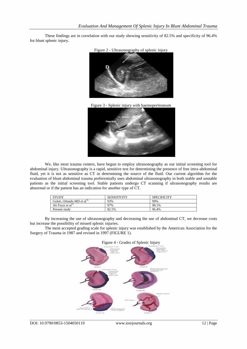
These findings are in correlation with our study showing sensitivity of 82.5% and specificity of 96.4%
for blunt splenic injury.
Figure 2 - Ultrasonography of splenic injury
Figure 3 - Splenic injury with haemoperitoneum
We, like most trauma centers, have begun to employ ultrasonography as our initial screening tool for
abdominal injury. Ultrasonography is a rapid, sensitive test for determining the presence of free intra-abdominal
fluid, yet it is not as sensitive as CT in determining the source of the fluid. Our current algorithm for the
evaluation of blunt abdominal trauma preferentially uses abdominal ultrasonography in both stable and unstable
patients as the initial screening tool. Stable patients undergo CT scanning if ultrasonography results are
abnormal or if the patient has an indication for another type of CT.
STUDY SENSITIVITY SPECIFICITY
Golett, Orlando MD et al76 93% 99%
Ali Feyzi et al75 97% 98.1%
Present study 82.5% 96.4%
By increasing the use of ultrasonography and decreasing the use of abdominal CT, we decrease costs
but increase the possibility of missed splenic injuries.
The most accepted grading scale for splenic injury was established by the American Association for the
Surgery of Trauma in 1987 and revised in 1997 (FIGURE 1).
Figure 4 - Grades of Splenic Injury

Evaluation And Management Of Splenic Injury In Blunt Abdominal Trauma
DOI: 10.9790/0853-1504050119 www.iosrjournals.org 13 | Page
In general, the lower the injury grade the more likely the patient can be managed non-operatively.
However, CT scan is notorious for underestimating injury grade,6 so injury grade alone should not guide the
surgeon for management.
Many studies have been conducted to evaluate the imaging characteristics of splenic trauma with CT
and to address the outcome of conservative treatment.. At most institutions, CT is the modality of choice for
evaluation of blunt abdominal trauma. Overall, sensitivity and specificity are high for detection of splenic
trauma.Haemoperitoneum almost always accompanies splenic injury. Uncommonly, a perisplenic clot is present
without evidence for capsular disruption, which has been reported in approximately 9% of patients and is
termed the sentinel clot (11) .
The CT appearance of intraperitoneal blood depends on the age and physical state of the clot.
Immediately after haemorrhage, intraperitoneal blood has the same attenuation as circulating blood of 20-30
HU. However, attenuation values less than 20 HU are a frequent finding in the acute setting (12).The proposed
reason for this is that blood, being a strong peritoneal irritant, causes a local inflammatory response with
transudation of fluid across the peritoneum. Transudate fluid mixes with and dilutesthe blood before coagulation
begins, decreasing the attenuation. Within hours, a clot forms.
Hemoperitoneum does not indicate whether active hemorrhage is present. Repeat imaging, as clinically
warranted, can aid in detecting ongoing hemorrhage. Increasing hematoma size or changes in character contrary
to the expected sequence are indications of continued hemorrhage. In most instances, hemoperitoneum
significantly resolves within 1 week.
In one study, intra-abdominal hematoma with a stable appearance 3-7 days after injury was suggestive
of continued hemorrhage. Depending on the physical state of the existing hematoma, fresh blood appears either
relatively hypoattenuating or hyperattenuating.On contrast-enhanced CT, extrasplenic extravasation of contrast
material rarely is seen. When extravasation occurs, patients most likely have hemodynamic instability and
proceed to laparotomy.
However, an intraparenchymal vascular blush may appear as single or multiple well-defined areas of
contrast material collection when a bolus injection is performed. Hyperattenuating areas represent localized
areas of contrast material extravasation from pseudoaneurysms or arteriovenous fistulas (14, 15).
Pseudoaneurysm formation is reportedly a delayed finding in 10% of patients with splenic injury (16).The
presence of a pseudoaneurysm is a strong predictor of NOM failure. Davis et al found that of patients in whom
conservative treatment failed, CT scans in 67% demonstrated a contrast blush. Note that 74% of
pseudoaneurysms were not documented on the initial CT scan; this observation provides strong support for
repeat examination in patients who receive conservative treatment (16).
Many authors have attempted to develop grading systems and delineate specific findings to predict the
need for laparotomy and assess the success of conservative treatment. Resciniti et al proposed a CT scoring
system to address the need as follows (17).
Splenic parenchyma Intact - 0
Laceration (thin, linear defect) - 1
Fracture (thick, irregular defect) - 2
Shattered – 3
Splenic capsule Intact - 0
Perisplenic fluid present – 1
Abdominal fluid No fluid - 0
Any fluid except perisplenic – 1
Pelvic fluid No fluid - 0
Any pelvic fluid – 1
In adult patients with a total CT score of less than 2.5, nonsurgical treatment was successful in all
patients. A score of 2.5 or more is correlated with a 46% likelihood of successful nonsurgical treatment. In one
study, all pediatric patients younger than 17 years had successful conservative treatment without delayed
complications irrespective of the score. A subsequent study elucidated potential errors of the scoring system,
particularly in discriminating subcapsular from perisplenic fluid and accounting for interobserver variability(
18). However, 13 of 15 patients treated nonsurgically who had a score of less than 2.5 had favorable outcomes.
The limitations of CT scanning are few but possibly important. The most detrimental limitation to
confident interpretation of a CT scan is motion artifact. The sensitivity for the detection of a splenic injury

Evaluation And Management Of Splenic Injury In Blunt Abdominal Trauma
DOI: 10.9790/0853-1504050119 www.iosrjournals.org 14 | Page
decreases precipitously if the patient cannot remain still on the scanning table. Adequate sedation is essential in
such patients. Overall, the sensitivity and specificity of CT in the detection of splenic injury is close to 100% .
Figure 5 : SubcapsularSplenic Hematoma And Laceration From Capsule To Hilum With
Intraparenchymal Hematoma
Figure 6 - Shattered spleen
Other investigations - Angiography
Splenic trauma can produce a wide variety of angiographic findings, either directly or indirectly.
Indirect signs include displacement of the spleen from the abdominal wall and avascular parenchymal areas
from hematoma. Parenchymal hematoma usually demonstrates hazy borders with splaying of the surrounding
vessels.
The most reliable angiographic sign of splenic trauma is contrast-material extravasation, either
parenchymal or extrasplenic. At times, extravasation may be observed only after the administration of
vasopressin or epinephrine. These medications enhance detection of vascular injury by increasing precapillary
arteriolar resistance. Abrupt cutoff of vessels, vessel wall irregularity, pseudoaneurysms, and early filling of
splenic vein are findings of traumatic injury.
The liberalization of treatment for blunt splenic trauma over the last decade has increased the role of
angiography. Interest in splenic angiography has also been sparked by the inadequacy of CT grading systems to
predict successful nonoperative treatment in certain patients. As a result, angiography has been used to elucidate
risk factors for delayed complications of splenic injury. The literature reports an approximate 5% incidence of
delayed hemorrhage more than 4 days after injury (30)
Figure 7 - Splenic Artery Aneurysm With Intraparenchymal Hematoma

Evaluation And Management Of Splenic Injury In Blunt Abdominal Trauma
DOI: 10.9790/0853-1504050119 www.iosrjournals.org 15 | Page
The criteria for nonoperative management of splenic injuries in adults have traditionally included (1)
Hemodynamic stability after minimal fluid resuscitation; (2) Documentation of splenic injury by imaging techniques; (3) Absence of a serious associated intra-abdominal
injury; (4) No altered level of consciousness that may interfere with serial abdominal examinations; and (5) Age
younger than 55 years.18,67
Recently, there has been a trend toward liberalization of these criteria, as more
surgeons become comfortable with nonoperative management.77-79
At our institution, there are no specific guidelines for management of blunt splenic injuries. The only definite
requirement for nonoperative management is that the patient be hemodynamically stable. Age is not considered
a contraindication, nor is the presence of a head injury. Transfusion remains a variable that changes from patient
to patient.
Risk of transfusion and nonoperative management of splenic injury have remained controversial,80
despite the decreased risk of transfusion-related infections.81
In the past, patients selected for nonoperative management were routinely prescribed several days of
bed rest, given nothing by mouth, and had nasogastric decompression. The patient's hemoglobin level and
abdominal examination results were checked frequently during the first 24 hours and then less frequently as the
patient's condition dictated. Follow-up CT scans were done to document resolution of the injury.82,83
The overall
duration of hospitalization for isolated splenic injury was 5 to 10 days, depending on the patient and the degree
of injury.
This scenario is being challenged in today's managed care environment. We rarely use nasogastric
decompression for the isolated splenic injury. Patients are fed and mobilized much quicker than in the past
because we are being asked to discharge patients from the hospital sooner. Follow-up studies are obtained only
when indicated by the clinical examination results.84
Robert J. Baker36
, MD, Chicago, Ill: This manuscript is concise, to the point, and it adds significantly to the
body of information about nonoperative management of splenic trauma in adults.It is important that the age of
the patient was not a contraindication to nonoperative management.
The literature is replete with contributions, largely before 1990 but also in more recent papers,
proposing that patients older than 55 years should not be managed nonoperatively. There are patients in this
group, and the oldest in the manuscript was 91 years of age, who were managed without operation. The current
trend is to do just that. The second issue relates to CT scanning in splenic trauma. A number of authors have
adopted the Buntain classification of splenic trauma, grading it 1 to 6, proposing that this is a viable way to
differentiate patients who should be operated on from those best treated nonoperatively.There are 2 major
concerns with nonoperative treatment, the first of which is that no other injuries be missed; there were no
missed injuries in this series. The other is that with nonoperative therapy, splenic salvage is often compromised
after a delay and it may then not be possible to repair the spleen when operation becomes necessary.
In the current study, 27 patients out of 40 were managed conservatively. Most being Grade I injuries.
Only one Grade IV splenic injury was treated conservatively who died during treatment. At our institution,
advanced age is not a contraindication for NOM. An important factor in our decision making is whether
comorbid disease exists. Elderly patients appear to have a higher failure rate. If they have comorbid disease,
failure may lead to an adverse outcome. As far as the use of the CT scan results to decide whether early
operations would be performed, I believe that we follow the national trend. Grade 1, 2, and 3 splenic injuries
would be managed nonoperatively unless the patient is hemodynamically unstable or has evidence of a hollow
viscus injury. 84% of the conservatively managed patients required blood transfusions. The patients that were
treated surgically had injuries of Grade III and above, all of whom required blood transfusions post operatively.
In the present study, more than 65% of patients had grade III and above splenic injuries which is
agreement with other studies in developing countries85
Carlin et al74
found that the need for splenectomy was
most significantly correlated with higher grades of splenic injury as supported by the present study.In recent
years the policy of spleen conservation at operation has been established due to its important role in cellular and
humoral immunity and the danger of overwhelming sepsis in asplenic patients49,86-89
.
The recognition that patients without a spleen have an increased risk of death from overwhelming
infection, led surgeons to consider methods of splenic preservation and with the introduction of the CT scan,
non-operative management became popular and then predominant14
Today, 90% of blunt pediatric splenic injuries and about 60-70% of adult ones are managed non-
operatively in the West and other developed countries85,90,91
. In the present study, 30% of patients were treated
operatively and (17.5%) of patients underwent splenectomy. High incidence of splenectomy in our study is
attributed to number of patients with higher grades of splenic injury. Also, unlike in western countries where
patients present within few hours of injury and in relatively stable clinical state92
most of our patients (65%)
presented to the A & E department in poor clinical state within 6 - 15 hours of injury. Sclafani et al35
and
Hagiwara et al38
have described SAE techniques dependent on angiographic findings. The visualization of

Evaluation And Management Of Splenic Injury In Blunt Abdominal Trauma
DOI: 10.9790/0853-1504050119 www.iosrjournals.org 16 | Page
extrasplenic extravasation was treated with selective Gelfoam embolization or superselective gelatin sponge
particle injection, respectively, followed by main SAE by means of coil occlusion. Main SAE alone was
performed if intraparenchymal contrast-material extravasation was the only finding.
The treatment of posttraumatic arteriovenous fistulas and pseudoaneurysms appears to require a
different approach. Arteriovenous fistulas probably remain patent after main SAE, and they have been reported
by Hagiwara et al38
.Many investigators have reported the use of superselective coil embolization without main
SAE to be successful in these patients93
The complication rate of SAE appears to be sufficiently low that it is
not a significant concern compared with that of splenectomy. Data by Mozes et al showed a 2.4% (3 of 126)
mortality within the first 6 months, compared with an 8% (2 of 25) mortality associated with splenectomy. Both
deaths related to splenectomy were associated with postoperative pancreatitis. Statistics reported by Mozes et al
were based on the embolization of no more than 60-70% of splenic tissue (34). Others have confirmed the
unacceptably high morbidity and mortality rates involved with excessive tissue embolization or attempted
nonsurgical splenectomy. Morbidity rates as high as 79% (35) and mortality rates ranging from 12% (36) to
43% (35) have been reported in the literature.
Lack of dedicated trauma centers for caring of trauma patients is a major problem in our community
and the intensive care unit (ICU) at our hospital is unable to cope up with a large number of trauma patients as a
result majority of patients are still admitted and managed in general surgical wards which are not well equipped
in managing trauma patients. In the present study, ICU admission was influenced by injury grade, amount of
haemoperitoneum, transfusion requirements, presence of coagulopathy, associated injuries or presence of
comorbidity.
The presence of complications has an impact on the final outcome of patients presenting with splenic
injuries as supported by the present study. Splenic injuries are commonly associated with other injuries and
these may complicate the management and affect the outcome91
. The pattern of complications in the present
study is similar to what was reported by others88,91
Early recognition and management of complications following splenic injury is of paramount in
reducing the morbidity and mortality resulting from these injuries.
The length of hospital stay has been reported to be an important measure of morbidity among trauma
patients. Prolonged hospitalization is associated with an unacceptable burden on resources for health and
undermines the productive capacity of the population through time lost during hospitalization and disability94
.
The overall length of hospitalization for both survivors and non-survivors in our study were found to be higher
than that reported by other authors91,73
. This can be explained by the presence of severe trauma patients and
large number of patients with associated injuries.The overall mortality rate in this study was 5%88,73
. Factors
responsible for mortality in our study included advanced patient's age, associated injuries, trauma scores, grade
of splenic injuries, admission systolic blood pressure ≤ 90 mmHg, estimated blood loss > 2000 ml,
postoperative complications
Post-splenectomy vaccination against encapsulated organisms is highly recommended for all
splenectomised patients for trauma before their discharge from hospital, with re-vaccination every 5-10 years
and additional antibiotic prophylaxis to compensate for the documented occasional vaccination failure95,96
.
However, in our environment, the majority of patients post splenectomy fail to attend the follow-up clinic,
making further management in those patients problematic. For these reasons, every attempt must be made for
splenic salvage.
This observation calls for training of junior surgical staff in methods of splenic salvage
(splenorrhaphy). In the present study, our patients received post-splenectomy vaccination. Post-vaccination
health education should be given to all splenectomised patients regarding the risk, the importance of prompt
diagnosis and treatment of infection, and the need for strong compliance with anti-malarial prophylaxis.
Self discharge by patient against medical advice is a recognized problem in our setting and this is
rampant, especially amongst trauma patients90
. Similarly, poor follow up visits after discharge from hospitals
remain a cause for concern. These issues are often the results of poverty, long distance from the hospitals and
ignorance. Delayed presentation, lack of Focused Assessment using Sonography in Trauma (FAST) and
irregular availability of CT scan (due to breakdown or inability of patients to afford), unavailability of
interventional radiology, inadequate ICUs, limited vaccination, discharge against medical advice, and the large
number of loss to follow up were the major limitations of this study. Also, since our duration of follow up was
relatively short, we could not estimate the long term outcome of both surgical and non-surgical management of
splenic injuries. However, despite these limitations, the study has provided local data that can be utilized by
health care providers to plan for preventive strategies as well as establishment of management guidelines for
patients with traumatic splenic injuries. The challenges identified in the management of patients with splenic
injuries in our setting need to be addressed, in order to deliver optimal trauma care for the victims of splenic
injuries.

Evaluation And Management Of Splenic Injury In Blunt Abdominal Trauma
DOI: 10.9790/0853-1504050119 www.iosrjournals.org 17 | Page
Nonsurgical management is becoming the preferred treatment method for adult patients who are
hemodynamically stable and have blunt splenic injuries. To attempt nonsurgical management, it is important to
identify and characterize not only the splenic injury but also any concurrent injury to the solid viscera,
mesentery and bowel, or retroperitoneum that may require surgery38,97,98
.
In this study, nonsurgical management ultimately was successful in 26 (70%) of the 40 patients who
presented with blunt splenic injuries.
VI. Conclusion This was a prospective study of 40 cases of splenic injuries amongst blunt abdominal trauma patients in
NRI General Hospital, Chinakakani from a period between October 2013 - September 2015.
Conclusions from the study :
1. Splenic injuries were mostly seen in the age group of 20-39 years (55%), which form the young and
economically productive group. Males were predominantly affected (75%).
2. Road traffic accidents were the most common cause in splenic injuries (62.5%).
3. A thorough and repeated clinical examination and appropriate diagnostic investigations lead to successful
treatment in these patients.
4. Conservative management has increased acceptance and is successful in selected patients, guided by
modern imaging modalities.
5. Ultrasound examination is 82.5% sensitive and 96.4% specific in identifying spleen injuries and free fluid,
and is a useful tool in rapid assessment and evaluation of blunt trauma patients.
6. The most common injuries associated with splenic injury in the present study were left rib fractures(20%).
7. Associated extra abdominal injuries like head, thoracic and orthopedic injuries were found in two cases in
the present study. This significantly influenced the morbidity .
8. Post operative complications like wound infection, dehiscence, respiratory infections and pancreatic fistula
were noted in operatively managed patients and this study showed a mortality of 5%.
9. In this clinical study at our tertiary care center, most of the splenic injuries in BAT were managed non-
operatively (67.5%).
References [1]. Maung AA , KaplanLJ , FrankelHL , CollinsKA; Management of splenic injury in the adult trauma patient . Available from
http://www.uptodate.com/contents/managemen t-of-splenic-injury-in-the-adult-trauma-patient.
[2]. Visrutaratna P, Na-Chiangmai W; Computed tomography of blunt abdominal trauma in children. Singapore Med J ., 2008;49 (4): 352–358.
[3]. Koo TY , RaYM , LeeSE , ChoiIS , YoonDS , Jo YJ et al .; Extension of nonoperative management on spleen injury with
judicious selection and embolization; 10 years of experience. J Korean Surg Soc .,2011; 80: 56 – 60
[4]. Buccoliero ,F. and Ruscelli ,P. Splenic trauma. In the management of trauma.From the territory to the Trauma Center. Edited by:
Cenammo A. Napoli: Italian Society of Surgery, 2010. p. 138 - 50.
[5]. Cohn SM, Arango JI, Myers JG; Computed tomography grading systems poorly predict the need for intervention after spleen and liver injuries.Am Surg., 2009; 75:133-139
[6]. Ameh EA, Chirdan LB, Nmadu PT. Blunt abdominal trauma in children: epidemiology, management, and management problems in
a developing country. Pediatr Surg Int 2000;16(7): 505-509. [7]. Demetriades D, Hadjizacharia P, Constantinou C, Brown C, Inaba K, Rhee P, Salim A. Selective nonoperative management of
penetrating abdominal solid organ injuries. Ann Surg 2006;244(4): 620-628.
[8]. Harbrecht BG. Is anything new in adult blunt splenic trauma? Am J Surg 2005;190(2): 273-278. [9]. Reihner E, Brismar B. Management of splenic trauma--changing concepts. Eur J Emerg Med 1995;2(1): 47-51.
[10]. Reddy CG, Chalasani V, Pathma-Nathan N. Splenic preservation: an additional haemostatic measure during mesh splenorrhaphy.
ANZ J Surg 2004;74(7): 596-597. [11]. Acs G, Furka I, Miko I, Szendroi T, Hajdu Z, Sipka S, Jr., Barath S, Aleksza M, Csipo I, Balo E, Balint A, Fekete K. [Comparative
hematologic and immunologic studies of patients with splenectomy and spleen autotransplantation]. Magy Seb 2005;58(2): 74-79.
[12]. Mooney DP, Rothstein DH, Forbes PW. Variation in the management of pediatric splenic injuries in the United States. J Trauma 2006;61(2): 330-333; discussion 333.
[13]. Lunca S, Romedea N, Morosanu C, Mihalache C, Mihalache S. [Traumatic injuries to the spleen in adults]. Rev Med Chir Soc Med
Nat Iasi 2005;109(2): 281-285. [14]. Upadhyaya P. Conservative management of splenic trauma: history and current trends. Pediatr Surg Int 2003;19(9-10): 617-627.
[15]. Cogbill THMoore EEJurkovich GJMorris JAMucha Jr PShackford SR Nonoperative management of blunt splenic trauma: a
multicenter experience. J Trauma. 1989;291312- 1317 [16]. Bond SJEichelberger MRGotschall CSSivit CJRandolph JG Nonoperative management of blunt hepatic and splenic injury in
children. Ann Surg. 1996;223286- 289
[17]. Elmore JRClark DEIsler RJHorner WR Selective nonoperative management of blunt splenic trauma in adults. Arch Surg. 1989;124581- 586
[18]. Smith Jr JSCooney RNMucha Jr P Nonoperative management of the ruptured spleen: a revalidation of criteria. Surgery.
1996;120745- 750 [19]. Javed NQ, Zahid AQ, Parkash A, Abdul SM. Gun-shot perforation of gut and associated injuries. JSurg Pak 2001; 6: 21-3.
[20]. Bisharat N, Omar H, Lavil, Raza R. Risk of infection and death among postsplenectomy patients. J Infect 2001; 43: 182-6.
[21]. Lucas CE. Splenic Trauma-Choice of Management . Ann. Surg. 1991;213:98–112. [22]. Hoyt DB. Symposium on nonoperative management of liver and spleen trauma:introduction. World J.Surg. 2001;25:1388.
[23]. Uranüs S, Pfeifer J. Nonoperative management of blunt splenic injury. World J.Surg. 2001;25:1405–1407. [PubMed]

Evaluation And Management Of Splenic Injury In Blunt Abdominal Trauma
DOI: 10.9790/0853-1504050119 www.iosrjournals.org 18 | Page
[24]. McClusky III DA, Skandalakis LJ, Colborn GL. Surgical History. Tribute to a Triad: History of Splenic Anatomy,Physiology,and
Surgery-Part 2. . World Journal of Surgery. 1999;23:514–526. [PubMed]
[25]. McCort JJ. 1986 President´s Address. Caring for the major Trauma Victim: the role of radiology. Radiology. 1987;163:1–9. [26]. Mufti TS1, Akbar I, Ahmed S Experience with splenic trauma in Ayub Teaching Hospital, Abbottabad. J Ayub Med Coll
Abbottabad. 2007 Jul-Sep;19(3):3-5.
[27]. Shoaib Ahmed Gangat, Anjum Rehman, Amir Ali Khaskhali, Mohd Ayub, Saher Fatima, Iqbal Ahmed Memon. Splenic trauma - management in relation to mode and grade Pak J Surg Jan - Mar 2008;24(1):2-4.
[28]. Radwan MM, Abu-Zidan FM. Focussed Assessment Sonograph Trauma (FAST) and CT scan in blunt abdominal trauma: surgeon’s perspective. African health sciences. 2006;6(3):187-190.
[29]. Shapiro MJ, Krausz C, Durham RM, Mazuski JE. Overuse of splenic scoring and computed tomographic scans. J Trauma 1999;
47:651-8. [30]. Peitzman AB, Heil B, Rivera L, et al. Blunt splenic injury in adults: Multi-institutional Study of the Eastern Association for the
Surgery of Trauma. J Trauma 2000; 49:177-87; discussion 187-9.
[31]. W. Michael Asher, Echographic Evaluation of Splenic Injury after Blunt TraumaRSNA February 1976 Volume 118, Issue 2 DOI: http://dx.doi.org/10.1148/118.2.411
[32]. Federle MP1, Courcoulas AP, Powell M, Ferris JV, Peitzman AB.Blunt splenic injury in adults: clinical and CT criteria for
management, with emphasis on active extravasation. Radiology. 1998 Jan;206(1):137-42. [33]. Mirvis SE, Whitley NO, Gens DR. Blunt splenic trauma in adults: CT-based classification and correlation with prognosis and
treatment. Radiology. 1989;171:33–9.
[34]. M A Malangoni, J I Cué, M E Fallat, S J Willing, and J D Richardson Evaluation of splenic injury by computed tomography and its impact on treatment. Ann Surg. 1990 May; 211(5): 592–599. PMCID: PMC1358230
[35]. Sclafani SJ1, Weisberg A, Scalea TM, Phillips TF, Duncan AO Blunt splenic injuries: nonsurgical treatment with CT, arteriography,
and transcatheter arterial embolization of the splenic artery. Radiology. 1991 Oct;181(1):189-96. [36]. Becker CD1, Spring P, Glättli A, Schweizer W. Blunt splenic trauma in adults: can CT findings be used to determine the need for
surgery? AJR Am J Roentgenol. 1994 Feb;162(2):343-7.
[37]. Sutyak, John P. MD; Chiu, William C. MD; D'Amelio, Louis F. MD; Amorosa, Judith K. MD; Hammond, Jeffrey S. MD. Computed Tomography Is Inaccurate in Estimating the Severity of Adult Splenic Injury The Journal of Trauma: Injury, Infection,
and Critical Care September 1995 Vol. 39 - Issue 3: pp 514-518
[38]. A Hagiwara, T Yukioka, S Ohta, T Nitatori, H Matsuda, and S ShimazakiNonsurgical management of patients with blunt splenic injury: efficacy of transcatheter arterial embolization.American Journal of Roentgenology1996167:1, 159-166
[39]. Goan, Yih-Gang MD; Huang, Mu-Shun MD; Lin, Jer-Ming MD Nonoperative Management for Extensive Hepatic and Splenic
Injuries with Significant Hemoperitoneum in Adults Journal of Trauma-Injury Infection & Critical Care: August 1998 - Volume 45 - Issue 2 - pp 360-364
[40]. Brasel KJ, DeLisle CM, Olson CJ, et al. Splenic injury: trends in evaluation and management. J Trauma 1998;44:283-6.
[41]. Shanmuganathan K, Mirvis SE, Boyd-Kranis R, Takada T, Scalea TM. Nonsurgical management of blunt splenic injury: use of CT criteria to select patients for splenic arteriography and potential endovascular therapy. Radiology. 2000; 217: 75–82.
[42]. Myers JG, Dent DL, Stewart RM, et al. Blunt splenic injuries: dedicated trauma surgeons can achieve a high rate of nonoperative success in patients of all ages. J Trauma 2000;48:801-5
[43]. Andrew B Peitzman,Failure of Observation of Blunt Splenic Injury in Adults: Variability in Practice and Adverse Consequences. J
Am Coll Surg Vol. 201, No. 2, August 2005 p179-187. [44]. Cocanour, Christine S Age Should Not Be a Consideration for Nonoperative Management of Blunt Splenic Injury Journal of
Trauma-Injury Infection & Critical Care: April 2000 - Volume 48 - Issue 4 - pp 606-612.
[45]. Stassen NA, Bhullar I, Cheng JD, et al. Selective nonoperative management of blunt splenic injury: an Eastern Association for the Surgery of Trauma practice management guideline. J Trauma Acute Care Surg. 2012;73:S294-300.
[46]. W E Longo Nonoperative management of adult blunt splenic trauma. Criteria for successful outcome. Ann Surg. 1989 Nov; 210(5):
626–629. [47]. Koehler, Successful Laparoscopic Splenorrhaphy Using Absorbable Mesh for Grade III Splenic Injury: Report of a Case August
1994 - Volume 4 - Issue 4Surg Laparosc Endosc. 1994 Aug;4(4):311-5.
[48]. Liu, Po Ping Use of Splenic Artery Embolization as an Adjunct to Nonsurgical Management of Blunt Splenic Injury Journal of Trauma-Injury Infection & Critical Care: April 2004 - Volume 56 - Issue 4 - pp 768-773.
[49]. Haan, James Nonoperative Management of Blunt Splenic Injury: A 5-Year Experience Journal of Trauma-Injury Infection &
Critical Care: March 2005 - Volume 58 - Issue 3 - pp 492-498 [50]. Sinha S, Raja S, Lewis M. Recent Changes in the Management of Blunt Splenic Injury: Effect on Splenic Trauma Patients and
Hospital Implications. Annals of The Royal College of Surgeons of England. 2008;90(2):109-112.
[51]. Stassen, Nicole Selective nonoperative management of blunt splenic injury: an Eastern Association for the Surgery of Trauma practice management guideline. J Trauma Acute Care Surg. 2012 Nov;73(5 Suppl 4):S294-300. doi:
10.1097/TA.0b013e3182702afc.
[52]. Beuran M, Gheju I, Venter M, Marian R, Smarandache R. Non-operative management of splenic trauma. Journal of Medicine and Life. 2012;5(1):47-58.
[53]. Liu S, Lei J, Zhi Z, Zhang Y, et al.Management of Traumatic Splenic Rupture in Adults: A Single Center's Experience in Mainland
China.Hepato-gastroenterology[2014, 61(132):966-971]. [54]. Berg RJ1, Inaba K2, Okoye O1, Pasley J1, Teixeira PG1, Esparza M1, Demetriades D1The contemporary management of penetrating
splenic injury. Injury. 2014 Sep;45(9):1394-400. doi: 10.1016/j.injury.2014.04.025. Epub 2014 Apr 18.
[55]. El-Matbouly M, El-Menyar A, Al-Thani H, et al. Traumatic Brain Injury in Qatar: Age Matters—Insights from a 4-Year Observational Study. The Scientific World Journal. 2013;2013:354920. doi:10.1155/2013/354920.
[56]. Arpit Bansal EXPERIENCE WITH SPLENIC TRAUMA IN JEEVAN JYOTI HOSPITAL, ALLAHABAD,U.P. INDIA
International Journal of Development Research, Vol. 05, Issue, 09pp.5510-5513 [57]. Gibney EJ. Non-operative management of blunt splenic injury –works well in about a quarter of patients. B.M.J. 1991;302:1553–
1554.
[58]. Knudson MM, Maull KI. Nonoperative management of solid organ injuries. Surgical clinics of North America. 1999;79:1357–1371 [59]. Britt LD, Cole FJ. ˝Alternative ˝ surgery in trauma management. Arch Surg. 1998;133:1177–1181.
[60]. Velmahos GC, Chan LS, Kamel E. Nonoperative management of splenic injuries; have we gone too far ? . Arch Surg.
2000;135:674–681.

Evaluation And Management Of Splenic Injury In Blunt Abdominal Trauma
DOI: 10.9790/0853-1504050119 www.iosrjournals.org 19 | Page
[61]. Cocanour CS, Moore FA, Ware DN. Delayed complications of nonoperative management of blunt adult splenic trauma. Arch Surg.
1998;133:619–625.
[62]. Pachter HL, Guth AA, Hofstetter SR. Changing patterns in the management of splenic trauma: the impact of nonoperative management. Ann Surg. 1998;227:708–717.
[63]. Garber BG, Mmath P, Fairfull-Smith RJ. Management of adult splenic injuries in Ontario: a population-based study. CJS.
2000;43:283–288 [64]. Dulchavsky SA, Lucas CE, Ledgerwood AM. Wound healing of the injured spleen with and without splenorrhaphy. J Trauma.
1987;27:1155–1160.
[65]. Kluger Y, Rabau M, Rub R. Comparative study of splenic wound healing in young and adult rats. J Trauma. 1999;47:261–264. [66]. Benya EC, Bulas DI, Eichelberger MR. Splenic injury from blunt abdominal trauma in children: follow-up evaluation with CT.
1995;195:685–688.
[67]. Longo WE, Baker CC, Mc.Millen MA. Nonoperative management of adult blunt splenic trauma;criteria for successful outcome. Ann Surg. 1989;210:626–630.
[68]. Mohapatra S, Pattanayak SP, et al. Options in the management of solid visceral injuries from blunt abdominal trauma. IJS 2003; 65:
263-268. [69]. Hsieh T-M, Cheng Tsai T, Liang J-L, Che Lin C. Non-operative management attempted for selective high grade blunt hepatosplenic
trauma is a feasible strategy. World Journal of Emergency Surgery : WJES. 2014;9:51. doi:10.1186/1749-7922-9-51.
[70]. Kendall JL, Kestler AM, Whitaker KT, Adkisson M-M, Haukoos JS. Blunt Abdominal Trauma Patients Are at Very Low Risk for Intra-Abdominal Injury after Emergency Department Observation. Western Journal of Emergency Medicine. 2011;12(4):496-504.
doi:10.5811/westjem.2010.11.2016.
[71]. Chalya PL, Mabula JB, Giiti G, et al. Splenic injuries at Bugando Medical Centre in northwestern Tanzania: a tertiary hospital experience. BMC Research Notes. 2012;5:59. doi:10.1186/1756-0500-5-59.
[72]. Bhattacharya B, Fieber J, Schuster K, Davis K, Maung A. ―Occult‖ rib fractures diagnosed on computed tomography scan only are
still a risk factor for solid organ injury. Journal of Emergencies, Trauma, and Shock. 2015;8(3):140-143. doi:10.4103/0974-2700.160706.
[73]. Iribhogbe PE, Okolo CJ. Management of Splenic Injuries in a University Teaching Hospital in Nigeria. West Afr J Med.
2009;28(5):309–312. [74]. Carlin AM, Tyburski JG, Wilson RF, Steffes C. Factors affecting the outcome of patients with splenic trauma. Am Surg.
2002;68(3):232–239.
[75]. Feyzi A, Rad MP, Ahanchi N, Firoozabadi J. Diagnostic accuracy of ultrasonography in detection of blunt abdominal trauma and comparison of early and late ultrasonography 24 hours after trauma. Pakistan Journal of Medical Sciences. 2015;31(4):980-983.
[76]. Goletti O1, Ghiselli G, Lippolis PV, Chiarugi M, Braccini G, Macaluso C, Cavina E. The role of ultrasonography in blunt
abdominal trauma: results in 250 consecutive cases. J Trauma. 1994 Feb;36(2):178-81. [77]. Archer LPOrgers FBShackford SR Selective nonoperative management of liver and spleen injuries in neurologically impaired
adult patients. Arch Surg. 1996;131309- 315 Coburn MCPfeifer JDeLuca FG Nonoperative management of splenic and hepatic trauma in the multiply injured pediatric and adolescent patient. Arch Surg. 1995;130332- 338
[78]. Keller MSSartorelli KHVane DW Associated head injury should not prevent nonoperative management of spleen or liver injury in children. J Trauma. 1996;41471- 475
[79]. Luna GKDellinger EP Nonoperative observation therapy for splenic injuries: a safe therapeutic option? Am J Surg. 1987;153462-
468 [80]. Schreiber GBBusch MPKleinman SHKorelitz JJ The risk of transfusion-transmitted viral infections: the retrovirus epidemiology
donor study. N Engl J Med. 1996;3341685- 1690.
[81]. Sugg SLGerndt SJHamilton BJFrancis IRTaheri PARodriquez JL Pseudoaneurysms of the intraparenchymal splenic artery after blunt abdominal trauma: a complication of nonoperative therapy and its management. J Trauma. 1995;39593- 595.
[82]. Kluger YPaul DBRaves JJ et al. Delayed rupture of the spleen: myths, facts, and their importance. J Trauma. 1994;36568- 571.
[83]. Lawson DEJacobson JASpizarny DLPranikoff T Splenic trauma: value of follow-up CT. Radiology. 1995;19497- 100 [84]. O I Alatise Splenic injuries in a semi urban hospital in Nigeria East Cent Afr J Surg 2008 Vol 13 (95-100) .
[85]. Tan KK, Chiu MT, Vijayan A. Management of isolated splenic injuries after Blunt Trauma: an institution's experience over 6 years.
Med J Malaysia. 2010;65(4):306–8. [86]. Kamel R, Manar ET, Kamel RR. Surgery of the spleen: splenic conservation. J Surg Pakistan. 2000;5:20–8.
[87]. Agbakwuru EA, Akinkuolie AA, Sowande OA, Adisa OA, Alatise OI, Onakpoya UU, Uhumwango O, Adesukanmi ARK. Splenic
Injuries in a Semi Urban Hospital in Nigeria. East and Cen Afr J of Surg. 2008;13(1):95–100. [88]. Harbrecht BG. Is anything new in adult blunt splenic trauma? Am J Surg. 2005;190(2):273–278. doi:
10.1016/j.amjsurg.2005.05.026.
[89]. Ohanaka EC, Osime U, Okonkwo CE. A five year review of splenic injuries in the University of Benin Teaching Hospital, Benin City, Nigeria. West Afr J Med. 2001;20(1):48–51.
[90]. Osifo OD, Enemudo RE, Ovueni ME. Splenic injuries in children: the challenges of non-operative management in a developing
country. J Indian Assoc Pediatr Surg. 2007;12(4):209–213. doi: 10.4103/0971-9261.40837. [91]. Schwab CW. Selection of nonoperative management candidates. World J Surg. 2001;25:1389–1392
[92]. Godley CD, Warren RL, Sheridan RL, McCabe CJ. Nonoperative management of blunt splenic injury in adults: age over 55 years
as a powerful indicator for failure. J Am Coll Surg. 1996 Aug. 183(2):133-9. [93]. Kang EG, Sharma GK, Lozano R. The global burden of injuries. Am J Public Health. 2000;90:523–526.
[94]. Davidson RN, Wall RA. Prevention and management of infections in patients without a spleen. Clin Microbiol Infect.
2001;7(12):657–660. doi: 10.1046/j.1198-743x.2001.00355.x. [PubMed][Cross Ref] [95]. El-Alfy MS, El-Sayed MH. Overwhelming post-splenectomy infection: is quality of patient knowledge enough for prevention?
Hematol J. 2004;5(1):77–80. doi: 10.1038/sj.thj.6200328
[96]. Gavant ML, Schurr M, Flick PA, Croce MA, Fabian TC, Gold RE. Predicting clin-ical outcome of nonsurgical management of blunt splenic injury: using CT to reveal abnormalities of splenic vasculature. AJRAm J Roentgenol 1997; 168:207–212
[97]. Davis KA, Fabian TC, Croce MA, et al.Improved success in nonoperative man- agement of blunt splenic injuries: embo-lization of
splenic artery pseudoaneu- rysms. J Trauma 1998; 44:1008 –1018.