evaluation of actinomycetes culture media for control of ... wasana... · 732 main export products...
TRANSCRIPT

International Journal of Agricultural Technology 2015 Vol. 11(3):731-746
Available online http://www.ijat-aatsea.com ISSN 2630-0192 (Online)
Evaluation of Actinomycetes Culture Media for Control of
Bakanae Disease of Rice Seeds
Sittivate, W. and Nalumpang, S.*
Department of Entomology and Plant Pathology, Faculty of Agriculture, Chiang Mai
University, Chiang Mai 50200, Thailand.
Sittivate, W. and Nalumpang, S. (2015). Evaluation of actinomycetes culture media for control
of bakanae disease of rice seeds. International Journal of Agricultural Technology 11(3):731-
746.
Abstract Six strains of actinomycetes, OMA60-45, OMA60-46, CSA60-25, SEA120-12,
SEA120-25 and SEA120-31, isolated from natural soil samples from Suthep-Pui National Park,
Chiang Mai, Thailand, were evaluated for their antifungal activities against Fusarium
moniliforme isolate Pk105St_62 causing bakanae disease of rice. There were no significant
differences observed among the actinomycete strains in inhibitory effects on the colony growth
of F. moniliforme (61.3 – 68.0%) when screened using the dual culture method. Then, cultures
of the pathogen were grown on two actinomycete culture media, a non-filtered culture medium
(NF) and filtered culture medium (F), by incubation with shaking in an enzyme production
medium (EPM) for 3, 5 and 7 d. The non-filtered culture medium inhibited colony growth of F.
moniliforme by 30.8 to 59.0%, while the filtered type caused a 22.5 to 51.4% inhibition. The
5- and 7-d-old culture media inhibited conidial germination of F. moniliforme by 22.8 – 99.6%
and 28.3 – 98.6%, respectively. Moreover, abnormal morphological changes occurred in
conidia treated with culture media of OMA60-45 and OMA60-46. Furthermore, at 21 d after
treatment, the percentage of rice seed infected by F. moniliforme was significantly reduced by
treatment with the NF media of both strains (43.50 and 62.50%, respectively) compared to 100%
infection in the control, with no effects on rice germination. This reduction was equivalent to
treatment with a commercial fungicide (captan; Orthocide® 50) (38.75%). In addition, seeds
treated with the NF of OMA60-45 and OMA60-46resulted in 75.0 and 70.0% germination of
rice seeds, respectively, in pathogen infested soil, which was equivalent to treatment with
captan (70.0%), while the germination of non-treated seeds was only 5.0%. Keyword: Fusarium moniliforme, rice, bakanae, actinomycetes, non-filtered and filtered
culture medium
Introduction
Rice (Orysa sativa L.) is one of the most important staple foods for the
world’s rapidly increasing population (Sasaki, 2005). For Thailand, Thai
jasmine rice (cv. Kao Dok Mali 105, KDML 105) is the long grain rice which is
well known for its fragrance and taste all around the world, and is one of the
*Corresponding author: Nalumpang, S.; Email: [email protected]

732
main export products of the central and north eastern region of Thailand.
Therefore, rice production in Thailand, especially Thai jasmine rice, represents
a significant portion of the Thai economy. However, one of major diseases
contributing to loss in the production of rice is bakanae disease caused by the
fungal pathogen, Fusarium moniliforme. The typical symptoms of Bakanae are
slender, chlorotic and elongated primary leaves that seem to be induced by the
production of gibberellin by the pathogen (Ou, 1987; Amoah et al., 1995).
However, not all infected seedlings show these symptoms, as crown rot is also
seen, resulting in stunted rice plants (Ou, 1987; Amoah et al., 1995). Crop
losses caused by Bakanae may reach 40% (Ou, 1987). The pathogen can be
both seed-borne and soilborne. Generally, the seed-borne inoculum provides
initial foci for secondary infection. Under favorable environmental conditions,
infected plants in different foci have the capacity to produce numerous conidia
that subsequently infect proximal healthy plants (Ou, 1985).
Biological control has been widely studied as an alternative method of
controlling plant diseases, since the increasing use of fungicides has caused
development of pathogen resistance, problems with environmental pollution
and human and animal health risks (Compant et al., 2005). Actinomycetes are
responsible for the production of about half of the discovered bioactive
secondary metabolites, notably antibiotics (Berdy, 2005) comprising three
quarters of all known products, and Streptomyces spp. are especially prolific
antibiotic producers (Lechevalier, 1989; Mansour et al. 1996; Saadoun and
Gharaibeh, 2003). They also have an important capacity for the degradation of
polymers such as lignin, chitin, cellulose and starch (Crawford et al., 1988;
Nolan et al., 1988) which improves their biocontrol potential against plant
pathogens (Getha et al., 2005; Minuto et al., 2006). Therefore, the objective of
present study was to investigate the potential of soilborne actinomycetes as
biocontrol agents in vitro and in vivo for controlling bakanae disease of rice.
Material and methods
Pathogen
Fusarium moniliforme isolate Pk105St_62 (sensitive to the fungicide
carbendazim), present in the culture collection of the Laboratory of the
Department of Entomology and Plant Pathology of Chiang Mai University,
Thailand, isolated from naturally infected rice stems, was used for experimental
inoculations. Cultures were grown on potato dextrose agar (PDA) at room
temperature (RT) for 7 d before use.

International Journal of Agricultural Technology 2015 Vol. 11(3): 731-746
733
Antagonists
This study used six actinomycete strains; OMA60-45, OMA60-46,
CSA60-25, SEA120-12, SEA120-25 and SEA120-31, that were previously
isolated from natural soil samples from Suthep-Pui National Park, Chiang Mai,
Thailand. Strains were grown on glucose yeast malt agar (GYM, Shirling and
Gottlieb, 1966) at RT for 10 d.
Antifungal tests on Petri dishes
Antagonism tests of the six actinomycetes strains against F. moniliforme
isolate Pk105St_62 were carried out on Petri dishes containing GYM using the
dual culture method (Fokkema, 1978). The isolates of the antagonistic strains
were streaked (4 cm) at 2 cm apart from one side of a Petri dish for 4 d earlier
than pathogen inoculation, preassuming the slow growth of these actinomycetes
in culture and their secondary metabolite production when incubated at RT. A
5-mm mycelial disc of a 7-d-old pathogen culture was placed on the other side
of the dish. Paired cultures incubated at RT for 14 days. Dishes inoculated only
with test pathogens served as controls. The experiment was done using a
Completely Randomized Design (CRD) with three replications (plates). The
growth of the pathogen in both the test and control experiments was recorded.
Data were collected as percent inhibition of colony growth (PICG) (modified
from Soytong, 1989; Lokesha and Benagi, 2007).
Preparation of culture media
Six actinomycete strains were prepared as non-filtered culture medium
(NF) and filtered culture medium (F) by using a modification of the method of
Saengnak et al. (2014). The cultures were separately grown on an enzyme
production medium (EPM) by incubation with shaking for 3, 5 and 7 days, and
centrifuged for 20 min at 6,000 rpm (4oC) and the supernatants were collected
as the NF. The supernatant was then filtrated through membrane filter pore size
0.2-μm (Minisart®) to get the F (Chareunrat, 1999).
Antifungal activities of the culture media
Efficacy on mycelial growth
Inhibition of pathogen growth by the test compounds was carried out on
PDA according to the agar well diffusion method (modified from Perez et al.,
1990). The PDA consisted of two layers, only the upper layer was inoculated.

734
Twenty µl of each filtrate were pipetted into 5-mm-diameter wells. Agar plugs
from 7-day-old PDA cultures of F. moniliforme strains were transferred to the
center of the plates, and incubated at RT for 10 d. EPM without actinomycetes
and a commercial biofungicide (Bacillus subtilis AP-01; Laminar®) at the
manufacturer’s recommended concentration served as the negative and positive
controls, respectively. Data were collected as percent inhibition of radial
growth (PIRG) modified from Soytong (1989) and Lokesha and Benagi (2007).
Three replications were used arranged in a Two-Factor Factorial Design
arranged in a CRD. Factor A represented different treatments: A1 = NF of
OMA60-45, A2 = F of OMA60-45, A3 = NF of OMA60-46, A4 = F of
OMA60-46, A5 = NF of CSA60-25, A6 = F of CSA60-25, A7 = NF of
SEA120-25, A8 = F of SEA120-25, A9 = NF of SEA120-12, A10 = F of
SEA120-12, A11 = NF of SEA120-31, A12 = F of SEA120-31, A13 = Bs AP-
01 (Laminar®) and A14 = EPM. Factor B represented incubation period of the
culture medium: B1 = 3 d, B2 = 5 d and B3 = 7 d.
Effect on conidial germination
Fusarium moniliforme was prepared as conidial suspensions, and
adjusted to 1×10⁵ conidia/ml using a haemacytometer. An equal volume (100
µl) of treated conidial suspensions from each culture medium was mixed and
spread onto a papery GYM plate, then cut into 1×1 cm sections and placed on a
microscope slide. Conidial suspensions mixed with equal volumes of EPM
served as the negative control. The slide cultures were incubated at RT and
checked for conidial germination at 12 h. In this context germination was
defined as a germ tube that had developed to longer than the cell width. Three
replications were used arranged in a Two-Factor Factorial Design arranged in a
CRD. Factor A represented different treatments: A1 = NF of OMA60-45, A2 =
F of OMA60-45, A3 = NF of OMA60-46, A4 = F of OMA60-46, A5 = NF of
CSA60-25, A6 = F of CSA60-25, A7 = NF of SEA120-25, A8 = F of SEA120-
25, A9 = NF of SEA120-12, A10 = F of SEA120-12, A11 = NF of SEA120-31,
A12 = F of SEA120-31 and A13 = distilled water. Factor B represented
incubation period of the culture medium: B1 = 3 d, B2 = 5 d and B3 = 7 d. To
estimate the percent germination, a total of 300 conidia were examined from
each treatment (100 per replicate).

International Journal of Agricultural Technology 2015 Vol. 11(3): 731-746
735
Effects of culture media on bakanae disease of rice seeds
Preparation of rice seeds
Rice seeds (Oryza sativa L.) cv. Thai Jasmine rice (Khao Dawk Mali 105,
KDML 105) were surface sterilized with 10% Chlorox (1% sodium
hypochlorite, NaOCl) for 5 min followed by thorough rinsing with sterilized
water. Then, sterilized seeds were air dried by blotting on sterile filter paper.
Preparation of pathogen inoculum
F. moniliforme Pk105St_62, 7-d-old culture grown in Petri dishes with
PDA at RT, was flooded with 10 ml of sterile distilled water (Singleton et al.,
1992). Mycelia were dislodged by scraping the surface of the agar culture with
a sterile loop needle. The mycelial suspension was then filtered through sterile
cheese cloth. The concentration of conidia in the suspension was determined
with a haemocytometer and adjusted to 1 × 10⁴ conidia/ml.
Surface-sterilized rice seeds were inoculated by immersion in the
pathogen suspension for 15 min and air dried. Then, inoculated-seeds were
immersed in 5-d-old culture media (seedNF
or seedF) at a rate of 100 seeds per 5
ml for 60 min and then air dried. Sterilized water (seedsw
), and commercial
fungicide (seedcap
), (captan; Orthocide® 50)
at the manufacturer’s recommended
concentration served as the negative and positive controls, respectively.
Germination data was assessed by the blotter method (ISTA, 1999), then
collected as percent of germinated seed and infected seed at 7, 14 and 21 d after
treatment. Four replications were used arranged in a Two-Factor Factorial
Design and in a CRD (100 seeds per replicate).
Effects of culture media as biofungicides under greenhouse conditions
Preparation of pathogen inoculum
Sorghum seeds served as the inoculating media after boiling, sterilization
and packing in polythene film (100 g/bag). F. moniliforme Pk105St_62
colonies on PDA were cut from a peripheral part of a colony with a 5-mm-
diameter cork borer, then transferred to sterilized sorghum seeds in a bag (10
discs/bag) and mixed well. The inoculated bags were incubated at RT for 14 d
in the dark before testing.
Pathogen inoculation
Fusarium-inoculum was mixed with a soil-compost mixture (3:1) from
Mea Hia Agricultural Research, Demonstrative and Training Center, Chiang

736
Mai University, Thailand, in a proportion of 100 g inoculum/kg of soil and
incubated in 6-in-diameter pots for 10 d.
Rice seeds of KDML 105 were immersed in the NF (seedNF
), captan
(seedcap
) or sterilized water (seed
sw) (100 seeds/5 ml) for 60 min and air dried.
Various methods of treatment included: (T1); seedsw
sown in sterilized soil, (T2);
seedsw
sown in Fusarium-infested soil for 5 d, (T3); seedsw
sown in Fusarium-
infested soil for 10 d, (T4); seedcap
sown in Fusarium-infested soil for 5 d, (T5);
seedcap
sown in Fusarium-infested soil for 10 d, (T6); seedNF
of OMA60-45
sown in Fusarium-infested soil for 5 d, (T7); seedNF
of OMA60-45 sown in
Fusarium infested soil for 10 d, (T8); seedNF
of OMA60-46 sown in Fusarium-
infested soil for 5 d and (T9); seedNF
of OMA60-46 sown in Fusarium-infested
soil for 10 d. T1 and T2-T3 served as negative and positive controls,
respectively. T4-T5 served as evaluation of the commercial fungicide. Pots
containing five seeds of each treatment were transferred to a greenhouse. Four
replications were used arranged in a Randomized Completely Block Design
(RCBD) (5 seeds per replicate). Data were collected at 14 d after inoculation as
percent seedling survival.
Statistical analyses
Treatment means were statistically compared using the Least Significant
Difference (LSD) at P≤0.05.
Results and discussion
Antifungal tests in Petri dishes
The six actinomycetes were not significantly different in inhibition of the
colony growth of F. moniliforme Pk105St_62, ranging from 61.3 to 68.0%
(Figure 1), and were categorized as having high antagonistic activity (Soytong,
1989).
Figure 1. Percentage inhibition of the colony growth of Fusarium moniliforme Pk105St_62
caused by the six actinomycete strains on glucose yeast malt agar (GYM) after 14 d. Radial
growth means the colony size of F. moniliforme
Per
cen
t in
hib
itio
n
Actinomycetes

International Journal of Agricultural Technology 2015 Vol. 11(3): 731-746
737
Antifungal activities of the culture media
Efficacy on mycelial growth
The NF culture media of the actinomycetes inhibited colony growth of F.
moniliforme Pk105St_62 from 30.8 to 59.0%, while the F type produced a 22.5
to 51.4% inhibition. However, the commercial biofungicide, B. subtilis AP-01
(Laminar®) and the enzyme production medium inhibited the colony growth by
only 19.3 to 39.3% and 3.0 to 10.6%, respectively. Moreover, our results
showed that the incubation periods of actinomycetes culture media did not
significantly affect the colony growth of F. moniliforme Pk105St_62. Among
the six actinomycete strains, OMA60-45 and OMA60-46 produced the
significantly highest inhibition of colony growth, followed by SEA120-31,
SEA120-12, SEA120-28 and CSA60-25. More importantly, the NF produced
higher average inhibitory effects than the F (Table 1).
Effect on conidial germination
The NF actinomycetes culture media inhibited conidial germination of F.
moniliforme Pk105St_62 ranging from 22.8 to 99.6%, while F type produced a
23.7 to 99.1% inhibition. Inhibition of conidial germination in the negative
control was only 8.30 to 16.2%. Furthermore, the actinomycetes culture media
incubated for 3 d produced the significantly lowest inhibition of conidial
germination of F. moniliforme Pk105St_62, 26.3 to 47.3%, comparable to
incubation periods of 5 and 7 days which had inhibitions ranging from 22.8 to
99.6% and 28.3 to 98.6%, respectively. Among the six actinomycetes strains,
OMA60-45 and OMA60-46 were significantly highest in inhibition of conidial
germination, followed by SEA120-31, SEA120-12, SEA120-28 and CSA60-25
which were not significantly different. In addition, NF media produced average
inhibitory effects equivalent to the F media (Table 2).
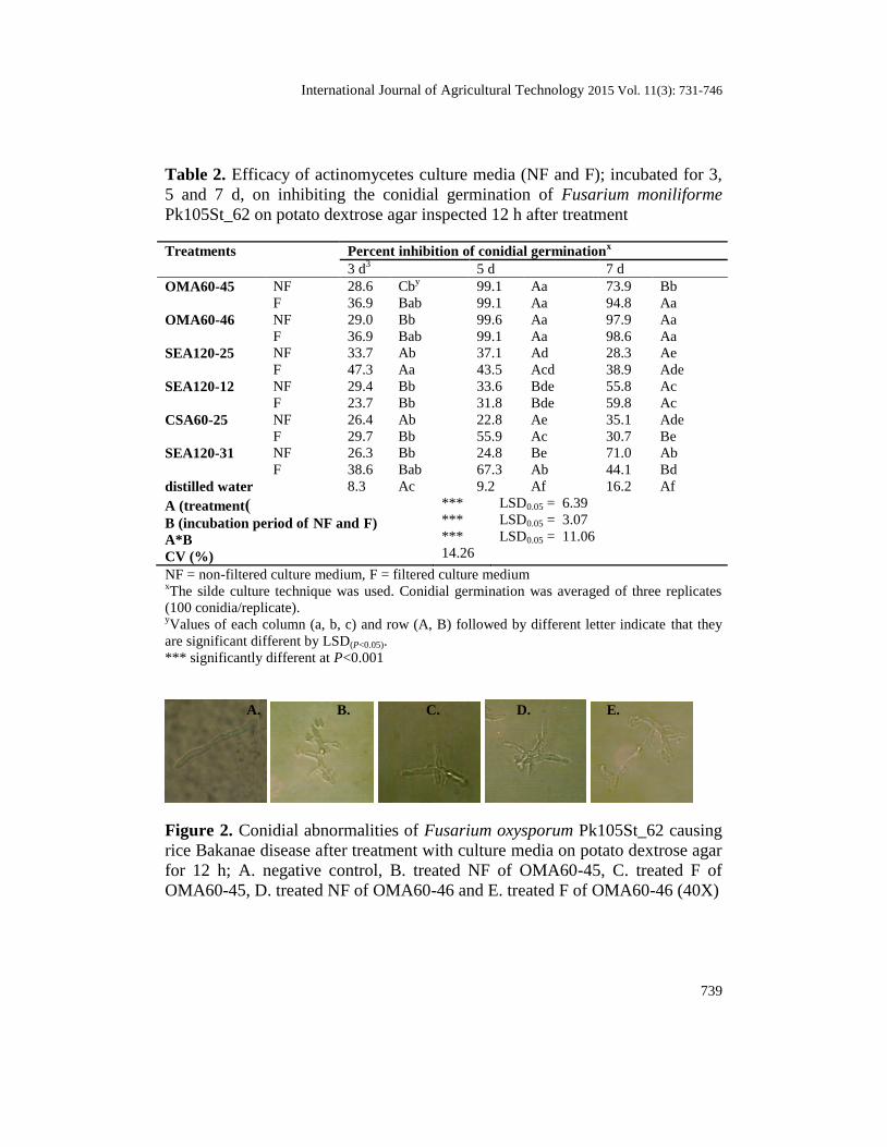
Moreover, conidia treated with culture media of OMA60-45 and
OMA60-46 appeared abnormal. They showed branched hyphae, formed
segments and could not develop into mycelium, while the mycelium of
untreated conidia was elongated (Figure 2). Therefore, these two effective
strains were selected to test in next series of experiments.

738
Table 1. Inhibition of colony growth of Fusarium moniliforme Pk105St_62 by
actinomycete culture media (NF and F) after incubation for 3, 5 and 7 d
Treatments Percent inhibition of colony growthx
3 d 5 d 7 d
OMA60-45 NF 49.0 Aaby
53.4 Aab 47.6 Aab
F 41.4 Aab 51.4 Aab 48.7 Aab
OMA60-46 NF 39.4 Bab 58.4 Aa 51.7 ABa
F 36.5 Ab 48.7 Aab 47.2 ABab
SEA120-25 NF 37.5 Ab 41.5 Ab 34.5 Abc
F 37.5 Ab 20.8 Bc 34.5 Abc
SEA120-12 NF 42.2 Aab 39.8 Ab 33.5 Abc
F 39.5 Aab 40.9 Ab 29.8 Abc
CSA60-25 NF 40.5 Aab 42.6 Ab 30.8 Abc
F 36.5 Ab 23.5 Bc 22.5 BCbc
SEA120-31 NF 50.0 Aa 35.6 Bbc 44.6 ABab
F 44.2 Aab 38.1 Ab 34.2 Abc
Bs AP-01 (Laminar®) 19.3 Bc 36.9 Ab 39.3 Ab
EPM 10.6 Ac 6.6 Ad 3.0 Ad
A (treatment)
B (incubation period of NF and F)
A*B
CV (%)
***
ns
***
20.20
LSD0.05 = 7.06
LSD0.05 = 12.22
NF = non-filtered culture medium, F = filtered culture medium, Bs = Bacillus subtilis xThe agar well method was used. Radial growth was the average of three replicates.
yValues of each column (a, b, c) and row (A, B) followed by different letter indicate that they
are significant different by LSD(P<0.05).
ns: non significant *** significantly different at P<0.001
International Journal of Agricultural Technology 2015 Vol. 11(3): 731-746
739
Table 2. Efficacy of actinomycetes culture media (NF and F); incubated for 3,
5 and 7 d, on inhibiting the conidial germination of Fusarium moniliforme
Pk105St_62 on potato dextrose agar inspected 12 h after treatment
Treatments Percent inhibition of conidial germinationx
3 d3 5 d 7 d
OMA60-45 NF 28.6 Cby 99.1 Aa 73.9 Bb
F 36.9 Bab 99.1 Aa 94.8 Aa
OMA60-46 NF 29.0 Bb 99.6 Aa 97.9 Aa
F 36.9 Bab 99.1 Aa 98.6 Aa
SEA120-25 NF 33.7 Ab 37.1 Ad 28.3 Ae
F 47.3 Aa 43.5 Acd 38.9 Ade
SEA120-12 NF 29.4 Bb 33.6 Bde 55.8 Ac
F 23.7 Bb 31.8 Bde 59.8 Ac
CSA60-25 NF 26.4 Ab 22.8 Ae 35.1 Ade
F 29.7 Bb 55.9 Ac 30.7 Be
SEA120-31 NF 26.3 Bb 24.8 Be 71.0 Ab
F 38.6 Bab 67.3 Ab 44.1 Bd
distilled water 8.3 Ac 9.2 Af 16.2 Af
A (treatment)
B (incubation period of NF and F)
A*B
CV (%)
***
***
***
14.26
LSD0.05 = 6.39
LSD0.05 = 3.07
LSD0.05 = 11.06
NF = non-filtered culture medium, F = filtered culture medium xThe silde culture technique was used. Conidial germination was averaged of three replicates
(100 conidia/replicate). yValues of each column (a, b, c) and row (A, B) followed by different letter indicate that they
are significant different by LSD(P<0.05).
*** significantly different at P<0.001
Figure 2. Conidial abnormalities of Fusarium oxysporum Pk105St_62 causing
rice Bakanae disease after treatment with culture media on potato dextrose agar
for 12 h; A. negative control, B. treated NF of OMA60-45, C. treated F of
OMA60-45, D. treated NF of OMA60-46 and E. treated F of OMA60-46 (40X)
A. B. C. D. E.

740
Effects of culture media on bakanae disease of rice seeds
At 7 d after treatment, rice seeds of all treatments were not significantly
different in seed infection; seeds infected by the pathogen ranged from 0.75 –
2.00%. The treatments clearly differed at 14 d after incubation. SeedNF
of
OMA60-45, OMA60-46 and seedF of OMA60-45 were infected with pathogen
at levels of 25.50, 44.50 and 51.00%, respectively, which were not significantly
different the seedcap
treatment (38.75%). In contrast, the highest infected seed
was found in seedF of OMA60-46 (80.50%), which did not significantly differ
from the pathogen inoculated seed (65.50%). Finally, seedNF
of OMA60-45 had
the significantly lowest infection (43.50%) and was equivalent to seedcap
(38.75%) at 21 d after incubation, followed by seedNF
of OMA60-46 and seedF
of OMA60-45. Conversely, seedF and inoculated seeds were completely
(100%) infected by the pathogen, while F. moniliforme was isolated at a low
rate (3.00% in non-treated seeds (Table 3).
Percent seed germination of rice immersed in various treatments were
successively collected at 7, 14 and 21 d after treatment, which found an over
90% germination. No significant differences in seed germination were observed
among treatments during the test period (Table 4).
Table 3. Effects of actinomycetes culture media (NF and F); incubated for 5 d,
on reduction Fusarium moniliforme Pk105St_62 infected on rice seeds
inspected 7, 14 and 21 d after treatment
Treatments Percent seed infectionx
7 d 14 d 21 d
non treated control 1.00 1.75 Ey 3.00 e
inoculated control 1.00 65.50 ab 100.00 a
captan (Orthocide® 50) 0.75 38.75 cd 38.75 d
OMA60-45 NF 0.75 25.50 d 43.50 cd
F 2.00 51.00 bc 76.75 b
OMA60-46 NF 1.25 44.50 cd 62.50 bc
F 1.25 80.50 a 100.00 a
F-test ns *** ***
LSD0.05 - 20.16 19.28
CV (%) 109.69 31.2 21.62
NF = non-filtered culture medium, F = filtered culture medium
xThe blotter method was used. Percent seed infection was averaged of four replicates (100
seeds/replicate). yValues of each column (a, b, c) and row (A, B) followed by different letter indicate that they
are significantly different by LSD(P<0.05).
ns: non significantly *** significantly different at P<0.001

International Journal of Agricultural Technology 2015 Vol. 11(3): 731-746
741
Table 4. Effects of actinomycete culture media (NF and F); incubated for 5 d,
on rice seed germination after inoculation with Fusarium moniliforme
Pk105St_62, inspected 7, 14 and 21 d after treatment
Treatments Percent seed germinationx
7 d 14 d 21 d
non treated control 97.00 98.00 98.00
inoculated control 91.00 92.50 92.50
captan (Orthocide® 50) 92.00 92.00 94.00
OMA60-45 NF 95.00 95.00 96.00
F 95.00 95.50 95.50
OMA60-46 NF 93.50 95.00 96.00
F 93.50 95.00 96.00
F-test ns ns ns
LSD0.05 - - -
CV (%) 3.46 3.27 3.22
NF = non-filtered culture medium, F = filtered culture medium
xThe blotter method was used. Percent seed germination was averaged of four replicates (100
seeds/replicate).
ns: non significant
Effects of culture media as biofungicides under greenhouse conditions
At 10 d after inoculation with the pathogen in soil, seedNF
of OMA60-45
and OMA60-46 before sowing had equivalent germinations to seedcap
, 75, 70
and 70%, respectively. In contrast, non-treated seed sown in infested and
sterilized soil had germinations of 5 and 95%, respectively (Table 5; Figure 3).
Table 5. Effects of non-filtrated actinomycete culture media (NF); incubated
for 5 d, on rice seeds germination after inoculation with Fusarium moniliforme
Pk105St_62 in soil under greenhouse conditions, inspected 14 d after treatment
xPercent seed germination was the average of four replicates (5 seeds/replicate).
yValues of each column (a, b, c) followed by different letter indicate that they are significantly
different by LSD(P<0.05).
*** significantly different at P<0.001
Treatments Percent seed germinationx
non treated control 95.0 ay
inoculated control 5.0 c
captan (Orthocide® 50) 70.0 b
OMA60-45 75.0 ab
OMA60-46 70.0 b
F-test ***
LSD0.05 21.97
CV (%) 22.63

742
Figure 3. Effects of non-filtered actinomycete culture media (NF); incubated for 5 d, on rice
seeds germination in soil infested with Fusarium moniliforme Pk105St_62 under greenhouse
conditions, inspected 14 d after treatment; A. non treated control, B. inoculated control, C. rice
seeds treated with captan (Orthocide® 50), D. rice seeds treated with the NF of OMA60-45 and
E. rice seeds treated with the NF of OMA60-46
Discussion
Selected actinomycetes, especially in the genus Streptomyces, have been
used in many studies for the direct biocontrol of various plant diseases (Yuan
and Crawford, 1995; Abd-Allah, 2001; Neeno-Eckwall et al., 2001; Getha and
Vikineswary, 2002; Shekhar et al., 2006). The present investigation evaluated
the potential of six actinomycetes; OMA60-45, OMA60-46, CSA60-25,
SEA120-12, SEA120-25 and SEA120-31, to control Fusarium moniliforme
Pk105St_62 causing bakanae disease of rice. These six actinomycetes were
isolated from natural soil samples from Suthep-Pui National Park, Chiang Mai,
Thailand; which was the same source of actinomycete isolates NSP1, NSP2,
NSP3, NSP4, NSP5 and NSP6 (Suwan et al., 2012a; Suwan et al., 2012b;
Saengnak et al., 2013; Saengnak et al., 2014), and based on their morphology
(data not shown), thus they most probably belong to the genus Streptomyces.
Their bioactive component is chitinase (Totree et al., 2011). This enzyme has
ability to inhibit fungal pathogens, and this is one of several properties
associated with actinomycetes that might explain their ability to act as
biocontrol agents.
In the current study these six actinomycetes were tested for their
inhibitory capabilities as non-filtered culture media (NF) and filtered culture
media (F), by incubation with shaking in an enzyme production medium (EPM)
for 3, 5 and 7 d. At 3 d they showed moderately inhibitory effects on the colony
growth of F. moniliforme Pk105St_62, but 5-d-old and 7-d-old culture media
showed higher activities on inhibiting conidial germination and some caused
morphological abnormalities in conidia. Similarly Saengnak (2012) reported
that 5-d-old culture media of the actinomycete NSP (1-6) were significantly
highest in inhibiting conidial germination of Colletotrichum gleosporioides
A. B. C. D. E.

International Journal of Agricultural Technology 2015 Vol. 11(3): 731-746
743
NDM_F012 causing mango anthracnose. Conversely, 3-d-old culture media
had no effects on conidial germination, although they gave the significantly
highest colony growth inhibition of the pathogen. Furthermore, Saengnak et al.
(2013) reported that conidia of F. oxysporum f. sp. capsici, causing chili wilt
disease, appeared abnormal after being treated with the culture media of NSP
(1-6). Jaipin and Nalumpang (2014) also found abnormalities in conidia of C.
gleosporioides, causing strawberry anthracnose, after being treated with the
culture media of NSP (1-6). In addition, Soares et al. (2006) reported ability of
Streptomyces sp. (AC26) on inhibiting the fungal pathogen Curvularia
eragrostides and C. gloeosporioides causing leaf spot of yam, their abilities
were related to concentration of a secondary metabolite associated with
Streptomyces sp.
Furthermore, actinomycetes culture media have ability to reduce the
fungal infection in rice seeds, but no effects on seed germination. Moreover,
rice seeds associated with the non-filtrated actinomycetes culture media could
germinate in pathogen infested soil under greenhouse conditions. These results
indicated that both actinomycetes culture media have potential as biofungicides.
Similarly, Mutitu et al. (2008) evaluated the antibiotic metabolites from two
antagonistic actinomycetes isolates for the control of late blight of tomatoes in
the greenhouse. The metabolites were found to give a significant control in the
management of late blight and delayed the onset of the disease. Punngram1et al.
(2012) investigated the antagonistic activities of the actinomycete RF 16-12,
isolated from rice field soil, against of three rice fungal pathogens: Fusarium
moniliforme, Helminthosporium oryzae and Rhizoctonia solani. They found
that a spore suspension of RF 61-61 was effective in controlling F. moniliforme
on rice seed with no effect on the root and shoot development of rice. The
molecular identfication showed that the RF 16-12 shared 99.86% similarity
with Streptomyces yogyakartensis NBRC 100779T. Wellington (2003) reported
that the strain DAUFPE 11470 showed great effectiveness for controlling a
number of fungal pathogens of seeds, roots and shoots including Aspergillus
spp., Cephalosporium acremonium, Curvularia lunata, Drechslera maydis and
Fusarium subglutinans. Our results indicate that the strains OMA60-45 and
OMA60-46 reduce the incidence of seed pathogenic fungi and have potential as
biological control agents. However, an efficient method of seed treatment with
the biological control agents must be developed before they can become
agriculturally practical. Finally, the use of these strains at the field level will
provide the basis to expand their use to replace chemical fungicides.

744
Conclusion
The current study demonstrated the potential of actinomycetes isolated
from natural soil samples from Suthep-Pui National Park, Chiang Mai,
Thailand, as as biological control agents against bakanae disease of rice caused
by F. monoliforme. The application of non-filtered culture medium (NF) and
filtered culture medium (F) extracts showed good in vitro antifungal properties,
and were also effective under greenhouse conditions. Hence, using these strains
as biofungicides in rice fields may be feasible and practical. The actual
biocontrol mechanisms of these actinomycetes remain to be studied.
Acknowledgements
This study was kindly supported by the Graduate School, Chiang Mai University.
References
Abd-Allah, E. F. (2001). Streptomyces plicatus as a model biocontrol agent. Flora
Microbiology 46:309-314.
Amoah, B. K., Rezanoor, H. N., Nicholson, P. and MacDonald, M. V. (1995). Variation in the
Fusarium section Liseola: pathogenicity and genetic studies of Fusarium moniliforme
Sheldon from different hosts in Ghana. Plant Pathology 44:563-572.
Berdy, J. (2005). Bioactive microbial metabolites. Journal of Antibiotics 58:1-26.
Chareunrat, S. (1999). Inhibitory effect of culture filtrate of chitinase producing mold on
Cladosporium sp., Fusarium sp. and Lasiodiplodia sp. (Master’s thesis). Science in
Agriculture, The Graduate School, Chiang Mai University, Chiang Mai.
Compant, J., Duffy, B., Nowak, J., CIA-Ment, C. and Barka, E. A. (2005). Use of growth
promoting bacteria for biocontrol of plant diseases: Principles, Mechanisms of action,
and future aspects. Applied and Environmental Microbiology 71:4951-4959.
Crawford, D. L., Goodfellow, M., Williams, S. T. and Mordarski, M. (1988). Biodegradation of
agricultural and urban wastes. Actinomycetes In: biotechnology. San Diego: Wiley Inter
science. pp. 433-459.
Fokkema, N. J. (1978). Fungal antagonism in the phylosphere. Annals of Applied Biology
89:115-117.
Getha, K., Vikineswary, S., Wong, W. H., Seki, T., Ward, A. and Goodfellow, M. (2005).
Evaluation of Streptomyces sp strain g10 for suppression of Fusarium wilt and
rhizosphere colonization in pot - grown banana plantlets. Journal of Industrial
Microbiology and Biotechnology 32:24-32.
Getha, K. and Vikineswary, S. (2002). Antagonistic effects of Streptomyces
violaceusniger strain G10 on Fusarium oxysporum f.sp. cubense race 4: Indirect
evidence for the role of antibiosis in the antagonistic process. Journal of Industrial
Microbiology and Biotechnology 25:303-310.
Jaipin, W. and Nalumpang, S. (2014). Effect of soil actinomycetes on growth of carbendazim-
resistant Colletotrichum gloeosporioides causing strawberry anthracnose. Journal of
agricultural 30:1-10.

International Journal of Agricultural Technology 2015 Vol. 11(3): 731-746
745
Lechevalier, H. A. (1989). The Actinomycetes III, A practical guide to generic identification of
Actinomycetes. Bergey's Manual of Systematic Bacteriology. Baltimore: Williams and
Wilkins Company. pp. 2344-2347.
Lokesha, N. M. and Benagi, V. L. (2007). Biocontrol management of pigeonpea dry root
caused by Macrophomina phaseolina. Karnataka Journal of Agricultural Sciences
20:54-56.
Mansour, F. A., El-Shirbiny, S. A. and El-Metwaly, N. A. (1996). Biosynthesis of
dimethyltetracycline by a strain of Streptomyces. Egyptian Journal of Microbiology
31:237-255.
Minuto, A., Spadaro, D., Garibaldi, A. and Gullino, M. L. (2006). Control of soil borne
pathogens of tomato using a commercial formulation of Streptomyces griseoviridis and
solarization. Crop Protection 25:468-475.
Mutitu, E. W., Muiru, W. M. and Mukunya, D. M. (2008). Evaluation of antibiotic metabolites
from actinomycete isolates for the control of late blight of tomatoes under greenhouse
conditions. Asian Journal of Plant Sciences 7:284-290.
Neeno-Eckwall, E. C. and Schottel, J. L. (2001). Competition and antibiosis in the biological
control of potato scab. Canadian Journal of Microbiology 47:332-340.
Nolan, R. D., Cross, T., Goodfellow, M., Williams, S. T. and Mordarski, M. (1988). Isolation
and screening of, actinomycetes. In: Actinomycetes in biotechnology. Orlando:
Academic Press.
Nome, N. F., Barreto, D. and Docampo, D. M. (2010). Seedborne Pathogens. Seeds: Trade,
Production and Technology 114-126.
Ou, S. H. (1985). Rice Diseases Second edition. England: Commonwealth Mycol. Inst., Kew.
Ou, S. H. (1987) Rice Diseases Second edition. Slough, UK: CAB International.
Perez, C., Pauli, M. and Bazerque, P. (1990). An antibiotic assay by well diffusion method.
Acta Biologiae Experimentalis 15:113-115.
Punngraml, N., Thamchaipenet, A. and Duangmal, K. (2011). Actinomycetes from rice field
soil and their activities to inhibit rice fungal pathogens. Proceedings of the 49th
Kasetsart University Annual Conference, Bangkok, Thailand, 1-4 Febauary 2011.
Saadoun, I. and Gharaibeh, R. (2003). The Streptomyces flora of Badia region of Jordan and its
potential as a source of antibiotics active against antibiotic- resistant bacteria. Journal of
Arid Environments 53:365-371.
Saengnak, V. (2012). Phylogeny and efficacy of soil chitinolytic actinomycetes in controlling
mango anthracnose. (Master’s thesis). Science in Agriculture, The Graduate School,
Chiang Mai University, Chiang Mai. 200 pp.
Saengnak, V., Chaisiri, C. and Nalumpang, S. (2013). Antagonistic Streptomyces species can
protect chili plants against wilt disease caused by Fusarium. Journal of Agricultural
Technology 9:1895-1908.
Saengnak, V., Jaipin, W. and Nalumpang, S. (2014). Evaluation of Streptomyces sp. culture
media extracts for control of strawberry anthracnose disease. Journal of Agricultural
Technology 10:105-117.
Sasaki, T. (2005). The map-based sequence of the rice genome. Nature 436:793-800.
Shekhar, N., Bhattacharya, D., Kumar, D. and Gupta, R. K. (2006). Biocontrol of wood-rotting
fungi with Streptomyces violaceusniger XL2. Canadian Journal of Microbiology 52:805-
808.
Shirling, E. B. and Gottlieb, D. (1966). Methods for characterization of Streptomyces species.
Internation Journal of Systematic Bacteriology 16:313-340.

746
Singleton, L. L., Mihail, J. D. and Rush, C. M. (1992). Methods for research on soil-borne
phytopathogenic fungi. St Paul, MN, USA: American Phytopathological Society Press.
264 pp.
Soares, A. C. F., Sousa, C. S., Garrido, M. S., Perea, J. O. and Almeida, N. S. (2006). Soil
Streptomycetes with in vitro activity against the yam pathogens Curvularia eragrostides
and Colletotrichum gloeosporioides. Journal of Brazilian Journal of Microbiology
37:456-461.
Soytong, K. (1989). Biological Control of Plant Pathogens. 326 pp.
Suwan, N., Boonying, W., and Nalumpang, S. (2012a). Antifungal activity of soil
actinomycetes to control chilli anthracnose caused by Colletotrichum gloeosporioides.
Journal of Agricultural Technology 8:725-737.
Suwan, N., To-anun, C., Soytong, K. and Nalumpang, S. (2012b). Evaluation of Streptomyces-
biofungicide to control chili anthracnose in pot experiment. Journal of Agricultural
Technology 8:1663-1676
Thotree, P. (2011). Efficiency of actinomyces culture medium for controlling Colletotrichum sp.
causing chilli anthracnose disease. Journal of Agricultural Science 42:163-166.
Wellington, B. (2003). Biological control of maize seed pathogenic fungi by use of
actinomycetes. Biological Control 48:233-240.
Yuan, W. M. and Crawford, D. L. (1995). Characterization of Streptomyces lydicus WYEC108
as a potential biocontrol agent against fungal root and seed rots. Applied and
Environmental Microbiology 612:3119-3128.
(Received: 29 January 2014, accepted: 28 February 2015)