evaluation of children with global developmental delay/intellectual
TRANSCRIPT

Evaluation of Children With Global Developmental Delay/Intellectual Disability
Dinesh Talwar, MD
Center for Neurosciences

Disclosures
I have one relevant financial relations I need to disclose:
I receive research support from Pfizer. This relationship will in no
way affect the discussion we will be having about Global
Developmental Delay and Intellectual Disability.

Objectives
Define and discuss Global Developmental Delay and Intellectual Disability.
Discuss the screening options and etiologies for both Global Developmental Delays and Intellectual Disability.
Discuss the importance of identifying a specific etiology for children with Global Developmental Delays.

Definitions
Global Developmental Delay (GDD)
Term typically for children < 6 years of age
1-3% of children in this age group
Significant Delay (> 2 SD) in 2 or more of the following domains
Gross/Fine Motor
Speech/Language
Cognition
Social/Personal
Activities of daily living
Many, but not all, may later manifest intellectual disability

Definitions
Intellectual Disability
Usually applied to children > 6 years of age
IQ testing in children > 6 years is more valid
Mild Intellectual Disability
IQ 50-70
Moderate to severe Intellectual Disability
!Q < 50

Identification
Developmental Surveillance
Formal screening, parental reports
Identification of biological and social risk factors
Identification of environmental influences –culture, parental skills, neglect, opportunity
Attempt to differentiate between GDD, speech-language delay and autism
Early intervention
May improve outcome
Diverse etiologies

Investigation of Etiology
History and Physical – may yield an etiology in 30-40% of cases
Hearing and visual assessments
EEG – particularly useful with concomitant history of seizures
CT/MRI scan of the brain, MR spectroscopy
Blood tests – lead level, thyroid function
Cytogenetic studies – karyotype, subteleomeric FISH, Chromosomal microarray, whole exome sequencing
Testing for inborn errors of metabolism

History and Physical
History
Prenatal and Birth History
Developmental History
Family History, consanguinity, history of fetal/infant demise
Past history of acute events, episodes of decompensation, seizures
Physical examination
Dysmorphic features
Cutaneous abnormalities
Organomegaly
Cardiac examination
Neurological examination
Congenital abnormalites

Cutaneous Examination

Cutaneous Examination

Seizures and Developmental Delay Children with developmental delay have a significantly
higher incidence of epilepsy (10-20%)
Epileptic Encephalopathies
Conditions in which the epilepsy and the associated EEG abnormalities contribute, at least in part, to the developmental delay
Often show evidence for developmental regression or slowing/arrest of development
Seen most often with onset of epilepsy in infancy

EEG in Developmental Delay
Has confirmatory value if there is a history indicative or suggestive of seizures, encephalopathy or a particular epileptic syndrome
Important to recognize subtle seizures
Infantile spasms
Drop attacks, head drops, atonic seizures
Myoclonic seizures
Staring spells
Of considerable value if there is a history of regression in speech-language or regression in milestones
Epilepsy aphasia syndromes – Landau Kleffner syndrome
Epileptic encephalopathies – Dravet syndrome etc.
EEG yield in developmental delay without definite seizures is low and may reveal non-specific epileptiform abnormalities

Neuroimaging in Developmental Delay
Screening with CT/MRI is quite useful in determining an etiology of developmental delay
MRI is superior to CT
CT abnormalities – 30%
MRI abnormalities – 48-65%
Presence of physical findings such as focal deficits, microcephaly increase the likelihood of abnormalities on neuroimaging

Neuroimaging in Developmental Delay
MRI abnormalities
CNS malformations – lissencephaly, polymicrogyria, pachygyria, heterotopias, cortical dysplasias, schizencephaly
Cerebral atrophy
White matter disease/abnormalities
Post-ischemic lesions
Phakomatosis – Neurofibromatosis, Tuberous Sclerosis
Specific malformations – Dandy Walker, Joubert syndrome

Lissencephaly
From: Lissencephaly. Kato & Dobyns. Human Molecular Genetics 2003, Vol 12

Pachygyria
4-month old female with developmental delay. Developed infantile spasms at 6 months of age.

Subcortical Band Heterotopias

Hemispheric Malformations of Cortical Development
T2 weighted sagittal image
Predominantly posterior involvement
Posterior dysplastic and pachygyric cortex
Moderately increased volume on the right with midline shift in the occipital area
Ex vacuo effect anteriorly
Hemispheric malformations of cortical development. Gupta et al. Neurology 2004;62(6, Suppl 3):S20-26.

Focal Cortical Dysplasia
Presented at age 8 months with infantile spasms. Currently 7-years old with seizures well controlled.

Focal Cortical Dysplasia with Neoplasm
7-Year old child with intractable seizures, starting at age 2 months.
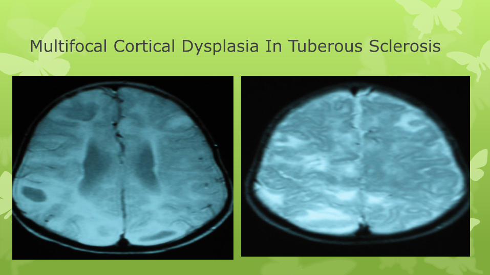
Multifocal Cortical Dysplasia In Tuberous Sclerosis
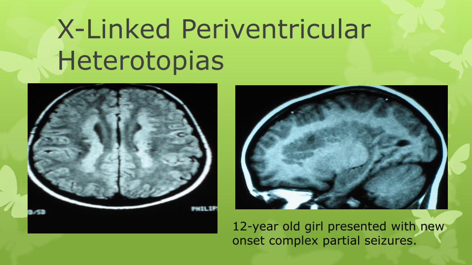
X-Linked Periventricular Heterotopias
12-year old girl presented with new onset complex partial seizures.

Schizencephaly
Textbook of Neuroradiology. Osborn.

Polymicrogyria/Cortical Dysplasia
Presented at age 8 months with partial secondarily GTC seizures; Subsequently developed infantile spasms.

Polymicrogyria/Cortical Dysplasia

Inborn Errors of Metabolism
Screening for inborn errors of metabolism has a yield of 0.2-4.6% depending on clinical features and extent of testing performed
Higher yield if newborn screening was not done
Factors suggestive of inborn error
Positive family history, consanguinity, history of fetal demise or unexplained deaths in childhood
Episodic decompensation, episodic seizures, episodes of encephalopathy
Developmental regression
Organomegaly, multiple organ dysfunction
Dietary selectivity, unusual odors
Hearing loss
Coarsening of facial features
MRI findings of abnormal myelination and/or basal ganglia signal abnormalities

Inborn Errors of Metabolism Screening tests for inborn errors of metabolism
Plasma amino acids, ammonia, acylcarnitine profile, serum uric acid
Urine organic acids
Urine and plasma creatine and guanidinoacetate
Serum transferring electrofocusing (carbohydrate deficient glycoprotein)
Plasma Very Long Chain Fatty Acids (VLCFAs), phytanic acid
Serum 7-dehydrocholesterol (Smith-Lemli-Opitz)
Urine mucopolysaccharides and sialic acid
Blood for lysosomal enzyme deficiencies
CSF glucose, lactate, pyruvate, glycine, organic acids, folate, and neurotransmitter metabolites
CPK, TSH and free T4

Inborn Errors of Metabolism
Testing for congenital disorders of glycosylation
Yield up to 1.4%
Testing for creatine synthesis and transport disorders
Yield up to 2.8%
Potentially treatable

Cytogenetic testing Genomic-wide testing for DNA rearrangements
G-Band Karyotyping
Detects chromosomal structural changes with a resolution of 3-5 million base pairs (3-5 Mb)
Subteleomeric FISH testing
Detect copy number changes (deletions or duplications) for which specific probes are constructed
Resolution 0f 1 Mb
Microarray tests
Oligonucleotide probes
Resolution of 30,000 – 35,000 base pairs (kb)

Cytogenetic Studies
• Chromosomal microarray testing is abnormal on average in 7.8% of subjects with GDD/ID and in 10.6% of those with syndromic features
• Interpretation
• Diagnostic – previously causative
• Possibly diagnostic – absent in unaffected parents
• Uncertain significance – inherited from an unaffected parent
• Results are often complex and require help of a medical geneticist.

Cytogenetic Studies
Chromosomal Microarray has limitations. It can identify only unbalanced copy number changes. Not sensitive for
Inversions
Balanced insertions
Reciprocal translocations
Polyploidy
Low level mosaicism (<20-25%)
Rearrangements in repeat sequences
Point mutations
Duplications/deletions undetectable test’s resolution

Cytogenetic studies
Karyotype studies are abnormal in > 4% of subjects with GDD/ID and in 18.6% of those with syndromic features
Subteleomeric FISH testing is abnormal in
3.5% of subjects with GDD/ID
4.2% of those with syndromic features
0.5% of those with mild impairment
7.4% of those with moderate/severe impairment

Fragile X-syndrome
Most common inherited disorder causing global developmental delay
CGG Trinucleotide repeats
5-40 repeats – Normal
41-55 repeats – Intermediate
56-200 repeats – Premutation
> 200 repeats – Full mutation
Prevalence of full mutation 1:3700 to 1:8900
Prevalence of premutation 1:1000
FMRI testing has a yield of at least 2% in males and females with mild GDD/ID

X-Linked Genetic Testing
X-Linked intellectual disability (XLID) accounts for 10% of cases of ID
> 70 cloned genes cause XLID
Family history often definitely points to or is suggestive of an X-Linked mutation
Common X-Linked genes
FMRI – Fragile X syndrome
ARX
JAR1D1C
SLC6A8
Testing for XLID genes positive in 42% males from definite X-Linked families, 17% males from possible X-Linked families
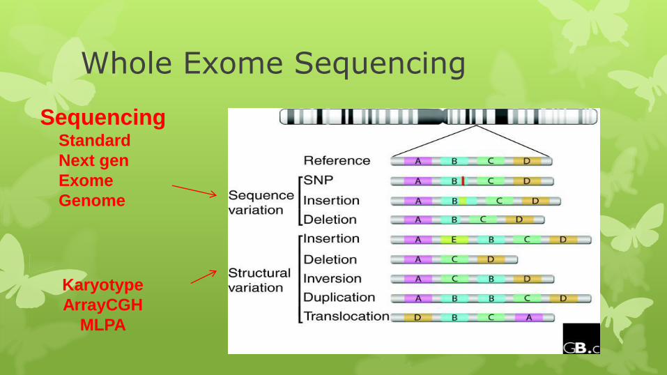
Whole Exome Sequencing
Sequencing Standard
Next gen
Exome
Genome
Karyotype
ArrayCGH
MLPA

ArrayCGH vs Next Gen Sequencing

Benefits of identifying a specific etiology Relieves anxiety/uncertainty
Limits further costly and invasive diagnostic tests
Improves understanding of treatment and prognosis
Helps to anticipate and manage associated medical and behavioral comorbidities
Empowers caregivers to become involved in support and research networks
Counseling for recurrence risk
Prevent recurrence – screening for carriers, prenatal screening

Conclusions Global developmental is seen in 1-3% of children below
the age of 6 years
History and Physical may give important clues to the underlying diagnosis
While blood tests and testing for inborn errors of metabolism may be helpful, MRI scan of the brain and cytogenetic studies provide the most information
There are significant benefits to the identification of a specific etiology including improving understanding of treatment and prognosis