evaluation of combined artificial intelligence and
TRANSCRIPT

Original Investigation | Imaging
Evaluation of Combined Artificial Intelligence and Radiologist Assessmentto Interpret Screening MammogramsThomas Schaffter, PhD; Diana S. M. Buist, PhD, MPH; Christoph I. Lee, MD, MS; Yaroslav Nikulin, MS; Dezső Ribli, MSc; Yuanfang Guan, PhD; William Lotter, PhD;Zequn Jie, PhD; Hao Du, BEng; Sijia Wang, MSc; Jiashi Feng, PhD; Mengling Feng, PhD; Hyo-Eun Kim, PhD; Francisco Albiol, PhD; Alberto Albiol, PhD;Stephen Morrell, B Bus Sc, MiF, M Res; Zbigniew Wojna, MSI; Mehmet Eren Ahsen, PhD; Umar Asif, PhD; Antonio Jimeno Yepes, PhD; Shivanthan Yohanandan, PhD;Simona Rabinovici-Cohen, MSc; Darvin Yi, MSc; Bruce Hoff, PhD; Thomas Yu, BS; Elias Chaibub Neto, PhD; Daniel L. Rubin, MD, MS; Peter Lindholm, MD, PhD;Laurie R. Margolies, MD; Russell Bailey McBride, PhD, MPH; Joseph H. Rothstein, MSc; Weiva Sieh, MD, PhD; Rami Ben-Ari, PhD; Stefan Harrer, PhD;Andrew Trister, MD, PhD; Stephen Friend, MD, PhD; Thea Norman, PhD; Berkman Sahiner, PhD; Fredrik Strand, MD, PhD; Justin Guinney, PhD;Gustavo Stolovitzky, PhD; and the DM DREAM Consortium
Abstract
IMPORTANCE Mammography screening currently relies on subjective human interpretation.Artificial intelligence (AI) advances could be used to increase mammography screening accuracy byreducing missed cancers and false positives.
OBJECTIVE To evaluate whether AI can overcome human mammography interpretation limitationswith a rigorous, unbiased evaluation of machine learning algorithms.
DESIGN, SETTING, AND PARTICIPANTS In this diagnostic accuracy study conducted betweenSeptember 2016 and November 2017, an international, crowdsourced challenge was hosted to fosterAI algorithm development focused on interpreting screening mammography. More than 1100participants comprising 126 teams from 44 countries participated. Analysis began November18, 2016.
MAIN OUTCOMES AND MEASUREMENTS Algorithms used images alone (challenge 1) or combinedimages, previous examinations (if available), and clinical and demographic risk factor data (challenge2) and output a score that translated to cancer yes/no within 12 months. Algorithm accuracy forbreast cancer detection was evaluated using area under the curve and algorithm specificitycompared with radiologists’ specificity with radiologists’ sensitivity set at 85.9% (United States) and83.9% (Sweden). An ensemble method aggregating top-performing AI algorithms and radiologists’recall assessment was developed and evaluated.
RESULTS Overall, 144 231 screening mammograms from 85 580 US women (952 cancer positive�12 months from screening) were used for algorithm training and validation. A second independentvalidation cohort included 166 578 examinations from 68 008 Swedish women (780 cancerpositive). The top-performing algorithm achieved an area under the curve of 0.858 (United States)and 0.903 (Sweden) and 66.2% (United States) and 81.2% (Sweden) specificity at the radiologists’sensitivity, lower than community-practice radiologists’ specificity of 90.5% (United States) and98.5% (Sweden). Combining top-performing algorithms and US radiologist assessments resulted ina higher area under the curve of 0.942 and achieved a significantly improved specificity (92.0%) atthe same sensitivity.
CONCLUSIONS AND RELEVANCE While no single AI algorithm outperformed radiologists, anensemble of AI algorithms combined with radiologist assessment in a single-reader screeningenvironment improved overall accuracy. This study underscores the potential of using machine
(continued)
Key PointsQuestion How do deep learning
algorithms perform compared with
radiologists in screening mammography
interpretation?
Findings In this diagnostic accuracy
study using 144 231 screening
mammograms from 85 580 women
from the United States and 166 578
screening mammograms from 68 008
women from Sweden, no single artificial
intelligence algorithm outperformed US
community radiologist benchmarks;
including clinical data and prior
mammograms did not improve artificial
intelligence performance. However,
combining best-performing artificial
intelligence algorithms with single-
radiologist assessment demonstrated
increased specificity.
Meaning Integrating artificial
intelligence to mammography
interpretation in single-radiologist
settings could yield significant
performance improvements, with the
potential to reduce health care system
expenditures and address resource
scarcity experienced in population-
based screening programs.
+ Invited Commentary
+ Supplemental content
Author affiliations and article information arelisted at the end of this article.
Open Access. This is an open access article distributed under the terms of the CC-BY License.
JAMA Network Open. 2020;3(3):e200265. doi:10.1001/jamanetworkopen.2020.0265 (Reprinted) March 2, 2020 1/15
Downloaded From: https://jamanetwork.com/ on 04/09/2022

Abstract (continued)
learning methods for enhancing mammography screening interpretation.
JAMA Network Open. 2020;3(3):e200265.
Corrected on March 30, 2020. doi:10.1001/jamanetworkopen.2020.0265
Introduction
Mammography screening is one of the most widely deployed tools for early breast cancer detectionand has been shown to decrease mortality in multiple randomized clinical trials.1 However, screeningmammography is imperfect with 1 in 8 cancers missed at time of interpretation in US communitypractice.2 Roughly 9% to 10% of the 40 million US women who undergo routine breast screeningeach year are recalled for additional diagnostic imaging; only 4% to 5% of women recalled areultimately diagnosed as having breast cancer.2 These false positives lead to preventable harms,included patient anxiety, benign biopsies, and unnecessary intervention or treatment.3 High false-positive rates incur significant resources and contribute to the annual $10 billion mammographyscreening costs in the United States.4
Currently, mammograms are interpreted by radiologists and rely on human visual perception toidentify relevant traits,5 leaving its benefit dependent on subjective human interpretation tomaximally extract all diagnostic information from the acquired images.6 In 1998, computer-assisteddetection software was developed for mammography with the hopes of improving radiologistperformance; however, computer-assisted detection has not improved interpretive accuracy.7,8
Recent deep learning advances, and the ever increasing large computational power and digitalmammography (DM) data availability, renewed the interest in evaluating whether more sophisticatedmodels based on quantitative imaging features can match or even outperform human interpretationalone.9-17 Such efforts could aid in improving specificity and overall performance in single-radiologist settings. In double-radiologist interpretive settings such as in Europe, highly accuratealgorithms could alleviate the person power needed for double-radiologist interpretation andconsensus.
Throughout the last decade, crowdsourced competitions or challenges have been popularizedas highly effective mechanisms for engaging the international scientific community to solve complexscientific problems.18,19 The Dialogue on Reverse Engineering Assessment and Methods (DREAM)initiative has run dozens of biomedical challenges, establishing robust and objective computationalbenchmarks in multiple disease areas and across multiple data modalities.19 This report describes theDM DREAM challenge, which was designed to develop and validate breast cancer detectionalgorithms to determine whether machine learning methods applied to mammography data canimprove screening accuracy.20,21
Methods
The study followed the Standards for Reporting of Diagnostic Accuracy (STARD) reporting guideline.22
We conducted an international crowdsourced challenge to assess whether artificial intelligence (AI)algorithms could meet or beat radiologists’ interpretive screening mammography performance.21 Thechallenge asked participants to develop algorithms inputting screening mammography data andoutputting a score representing the likelihood that a woman will be diagnosed with breast cancer withinthe next 12 months (eAppendix 2 in the Supplement). Digital mammogram images included differentviews (eTable 2 in the Supplement) from the most recent screening examination. Subchallenge 2provided access to images for the current and, when available, previous screening examinations, aswell as clinical and demographic risk factor information typically available to interpreting radiologists(eAppendix 3 and eTable 1 in the Supplement).
JAMA Network Open | Imaging Evaluation of Combined Artificial Intelligence and Radiologist Assessment to Interpret Screening Mammograms
JAMA Network Open. 2020;3(3):e200265. doi:10.1001/jamanetworkopen.2020.0265 (Reprinted) March 2, 2020 2/15
Downloaded From: https://jamanetwork.com/ on 04/09/2022
The DM DREAM challenge was hosted between November 2016 and November 2017. Therewere 4 phases (eAppendix 1 and eFigure 1 in the Supplement): open phase (September 2016-November 2016), leaderboard phase (November 2016-March 2017), and validation phase (March2017-May 2017), which together constitute the competitive phase and the community phase (July2017-November 2017). A first data set was provided by Kaiser Permanente Washington (KPW)(eAppendix 5 in the Supplement), which was used during the competitive and community phases. Asecond data set was provided by the Karolinska Institute (KI) in Sweden (eAppendix 5 in theSupplement), which was only used for trained algorithm validation. To protect data privacy, both datasets were securely protected behind a firewall and were not directly accessible to challengeparticipants, who had to submit their algorithms for automated training and testing behind thefirewall (eAppendix 7 in the Supplement).
In the competitive phase, the KPW data set was randomly split into 3 data sets matched on age,body mass index, and race/ethnicity (eAppendix 3 in the Supplement): leaderboard phase training(50%), leaderboard phase evaluation (20%) (eTable 3 in the Supplement), and final evaluation dataset (30%) (eTable 4 in the Supplement). The leaderboard phase allowed competitors to trainalgorithms using the KPW training data or external (public or private) data and submit their trainedalgorithms for evaluation. To minimize overfitting, a maximum of 9 submissions per team werescored in the leaderboard data set with publicly posted results.20 During the validation phase,participants were scored using the KPW final evaluation data set (Figure 1A) using area under thecurve (AUC) and partial AUC as evaluation metrics (eAppendix 6 in the Supplement) assessed at theexamination level, on which the final ranking of team performances was determined.
The 8 top-performing competitive phase teams were invited to collaborate during thecommunity phase to further refine their algorithms (Figure 1B). The output of this phase was anensemble model, consisting of a weighted aggregation of algorithm predictions into a new algorithmcalled the challenge ensemble method (CEM). The CEM model was further integrated with theradiologists’ assessment into another ensemble model called the CEM+R model. The CEM and CEM+Rmodels were trained using the training and leaderboard data of the competitive phase, withperformance assessed using the KPW and KI final evaluation data sets (Figure 1C and eTable 5 in theSupplement).
Data Sources and CharacteristicsKaiser Permamente Washington provided the primary data set for the challenge, with images linkedto curated data as part of the Breast Cancer Surveillance Consortium.21 These data includeprospectively collected patient-reported breast cancer risk factor information linked to breastimaging data, radiologist interpretations and recommendations, and benign and malignant breasttumor biopsy results. An additional independent validation data set was provided by the KI andcomprised women screened in the Stockholm region of Sweden between April 2008 to December2012 with comparable data provided by KPW. Both data sets were deidentified full-field DMs. Thiscollaboration received institutional review board approval at Sage Bionetworks with a waiver ofconsent and Health Insurance Portability and Accountability Act.
Each screening examination was labeled as cancer negative or cancer positive at the breastlevel, defined as a tissue biopsy yielding an invasive cancer or ductal carcinoma in situ positive resultwithin 1 year of the screening examination. Images were weakly labeled, meaning the presence orabsence of cancer was reported per screening examination but not the actual location of cancer oneach image. Breast-level performance was used in the competitive phase, whereas examination-levelperformance was used in the collaborative phase to make results directly comparable to radiologists’performance (eAppendix 4, eAppendix 6, and eFigure 2 in the Supplement).
Training and Evaluating ModelsChallenge data providers required the data to be protected from being downloaded, viewed, orotherwise directly accessed by participants. Therefore, we used a model-to-data approach23 that
JAMA Network Open | Imaging Evaluation of Combined Artificial Intelligence and Radiologist Assessment to Interpret Screening Mammograms
JAMA Network Open. 2020;3(3):e200265. doi:10.1001/jamanetworkopen.2020.0265 (Reprinted) March 2, 2020 3/15
Downloaded From: https://jamanetwork.com/ on 04/09/2022

required participants to submit containerized algorithms (ie, software that was executed on aparticipant’s behalf in a secure cloud environment) using the IBM and Amazon cloud platforms. Bothclouds were donated especially for this challenge (eAppendix 7 and eFigures 3-5 in the Supplement).The model-to-data system was implemented in Synapse (Sage Bionetworks),24 a web-basedplatform for running biomedical challenges. Participants were asked to structure their models in theform of a lightweight portable machine image called a Docker image.25
Ensemble MethodIt has been shown that aggregating the predictions of a set of heterogeneous algorithms can improveperformance over the individual algorithms.26,27 We developed the CEM and CEM+R ensembleclassifiers using a meta-learner stacking method26 (Appendix 8 in the Supplement). Given the datafor a screening examination, each individual algorithm outputs a confidence level (a number between0 and 1) indicative of the likelihood estimated by the algorithm that the woman will develop breastcancer within a year of the screening test. The CEM ensemble algorithm takes as inputs theconfidence levels of each of the community phase 8 top-performing methods. The CEM+R takes thesame inputs as the CEM ensemble plus the radiologist assessment, represented with a 1 if the woman
Figure 1. Training and Evaluation of Algorithms During the Digital Mammography DREAM Challenge
The digital mammographyDREAM challenge
A
Ensembling modelsB
Ensembling modelsand radiologist
C
Submittedmodels
Teams
Organizersand
selected teams
Organizersand
selected teams
Public/privatedata sets
Secured, GPU-accelerated cloud
Final teamranked
performance
KPW trainingdata set
KPW evaluationdata set
Refinedmodels
Breast cancerpredictions
Breast cancerpredictions
Id Conf.
Scoring: AUCpAUC
AUCspecificity atradiologists’
sensitivity
Id Conf.
Id Conf.
Challenge ensemblemethod (CEM)
KPW evaluationdata set
Karolinskaevaluation
data set
Radiologistassessment
Breast cancerpredictions
AUCspecificity atradiologists’
sensitivity
Id Conf.
Id Conf.
CEM + R
KPW evaluationdata set
Karolinskaevaluation
data set
Training and evaluation Kaiser PermamenteWashington (KPW) and Karolinskia Institute data werenot directly available to challenge participants; theywere stored behind a firewall in a secure cloud (graybox). To access the data, participants submittedmodels to be run behind the firewall, in the graphicsprocessing unit (GPU)–accelerated cloud (IBM). A,Training and evaluation of models submitted by teamsduring the Digital Mammography Dialogue on ReverseEngineering Assessment and Methods (DREAM)Challenge. B, A subset of the 8 best models in theevaluation KPW dataset were combined into theChallenge Ensemble Method (CEM), trained using theKPW training set and evaluated in the KPW andKarolinska Institute evaluation datasets. C, Wedeveloped a final ensemble method incorporatingradiologists' interpretation in a method calledCEM+radiologist (CEM+R). AUC indicates area underthe curve; pAUC, partial area under the curve.
JAMA Network Open | Imaging Evaluation of Combined Artificial Intelligence and Radiologist Assessment to Interpret Screening Mammograms
JAMA Network Open. 2020;3(3):e200265. doi:10.1001/jamanetworkopen.2020.0265 (Reprinted) March 2, 2020 4/15
Downloaded From: https://jamanetwork.com/ on 04/09/2022
was recalled or a 0 otherwise. This input information is combined to generate the CEM or the CEM+Rensemble prediction. For the meta-learner classifier, we chose an elastic net regularized logisticregression28 and the R package caret for construction (R Foundation for Statistical Computing).29 Wetrained the meta-learner (ie, tuned the parameters of the logistic regression) on the KPW trainingdata using 10-fold crossvalidation. Final performance assessment was done by applying exactly oncethe CEM and CEM+R methods to the evaluation KPW and KI data sets.
Statistical AnalysisAnalysis began November 18, 2016. We used AUC as our primary metric for evaluating and rankingalgorithm performance during the competitive phase. To assess the algorithms’ sensitivity andspecificity, we computed the radiologists’ sensitivity for the data set under study: 85.9% for KPW and77.1% (single reader) and 83.9% (consensus reading) for KI, which served as the algorithms’specificity prediction threshold. Spearman correlation was used to test for rank distribution similarityof AUCs and specificities between the data sets. We used binomial proportion CIs to compare thesignificance between specificity of radiologists and CEM+R. The DeLong test of significance was usedto compare the area under the receiver operating characteristic curve of 2 correlated receiveroperating characteristic curves. One-tailed P values had a statistical significance threshold of .05. Allanalyses were completed in R statistical software, version 3.5.1 (R Foundation for StatisticalComputing).
Results
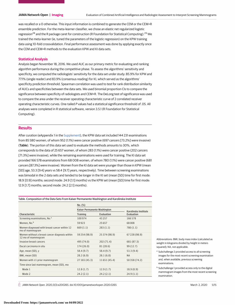
After curation (eAppendix 1 in the Supplement), the KPW data set included 144 231 examinationsfrom 85 580 women, of whom 952 (1.1%) were cancer positive (697 cancers [73.2%] were invasive)(Table). The portion of this data set used to evaluate the methods amounts to 30%, whichcorresponds to the data of 25 657 women, of whom 283 (1.1%) were cancer positive (202 cancers[71.3%] were invasive), while the remaining examinations were used for training. The KI data setprovided 166 578 examinations from 68 008 women, of whom 780 (1.1%) were cancer positive (681cancers [87.3%] were invasive). Women from the KI data set were younger than those in KPW (mean[SD] age, 53.3 [9.4] years vs 58.4 [9.7] years, respectively). Time between screening examinationswas bimodal in the 2 data sets and tended to be longer in the KI set (mean [SD] time for first mode:18.9 [0.9] months; second mode: 24.9 [1.1] months) vs the KPW set (mean [SD] time for first mode:12.9 [1.7] months; second mode: 24.2 [2.1] months).
Table. Composition of the Data Sets From Kaiser Permanente Washington and Karolinska Institute
Characteristic
No. (%)
Kaiser Permanente WashingtonKarolinska InstituteEvaluationTraining Evaluation
Screening examinations, No.a 100 974 43 257 166 578
Women, No.b 59 923 25 657 68 008
Women diagnosed with breast cancer within 12mo of mammogram
669 (1.1) 283 (1.1) 780 (1.1)
Women without a breast cancer diagnosis within12 mo of mammogram
59 254 (98.9) 25 374 (98.9) 67 228 (98.9)
Invasive breast cancers 495 (74.0) 202 (71.4) 681 (87.3)
Ducal carcinoma in situ 174 (26.0) 81 (28.6) 99 (12.7)
Age, mean (SD), y 58.4 (9.7) 58.4 (9.7) 53.3 (9.4)
BMI, mean (SD) 28.2 (6.9) 28.1 (6.8) NA
Women with ≥1 prior mammogram 27 165 (45.3) 11 651 (45.4) 50 358 (74.2)
Time since last mammogram, mean (SD), mo
Mode 1 12.8 (1.7) 12.9 (1.7) 18.9 (0.9)
Mode 2 24.2 (2.1) 24.2 (2.1) 24.9 (1.1)
Abbreviations: BMI, body mass index (calculated asweight in kilograms divided by height in meterssquared); NA, not applicable.a Subchallenge 2 provided access to all screening
images for the most recent screening examinationand, when available, previous screeningexaminations.
b Subchallenge 1 provided access only to the digitalmammogram images from the most recent screeningexamination.
JAMA Network Open | Imaging Evaluation of Combined Artificial Intelligence and Radiologist Assessment to Interpret Screening Mammograms
JAMA Network Open. 2020;3(3):e200265. doi:10.1001/jamanetworkopen.2020.0265 (Reprinted) March 2, 2020 5/15
Downloaded From: https://jamanetwork.com/ on 04/09/2022

Submitted AlgorithmsThe DM DREAM challenge included more than 1100 individuals participating, comprising 126 teamsfrom 44 countries (eAppendix 9 in the Supplement). Thirty-one teams submitted their methods forfinal validation in the competitive phase on the KPW evaluation set (Figure 2A). In subchallenge 1,median AUC performance for all teams was 0.611 (interquartile range, 0.54-0.77), with the best-performing method achieving a 0.855 AUC and a specificity of 68.5% at sensitivity of KPWradiologists of 85.9%. The AUC had little improvement when algorithms were able to use clinical,demographic, and longitudinal information (AUC = 0.858 with specificity = 66.3% at sensitivity85.9%). We observed no improvement across teams in performance measured by AUC (P = .51;t = 0.024) when comparing their results in subchallenge 2 to subchallenge 1 (Figure 2B).
To assess the generalizability of these methods, we evaluated the top 20 methods on the KIdata set. The best-performing method on the KPW data achieved top performance on KI data(AUC = 0.903; specificity = 81.2% at the 83.9% KI radiologists’ sensitivity). Ranking individualmethods on the data sets were found to be significantly correlated by AUC (r = 0.98; 95% CI, 0.95-1.00; P < .001; Figure 2C) and by specificity at Breast Cancer Surveillance Consortium’s averagesensitivity2 of 86.9% (r = 0.96; 95% CI, 0.92-1.00; P < .001; Figure 2D).
Figure 2. Performance of the Algorithms Submitted at the End of the Competitive Phase
1.0
0.8
0.6
0.4
0.2
0
1.0
0.8
0.6
0.4
0
0.2
1.0
0.8
0.6
0.4
0.2
0
Spec
ifici
ty a
t Sen
sitiv
ity o
f 85.
9%
AUC
Performance of algorithms on KPW evaluation set (end of competitive phase)
A
1.00.80.60.4
Karo
linsk
a In
stitu
te S
ubse
t
Kaiser Permanente Washington Set1.00.80.4 0.60 0.2
0.9
0.8
0.7
0.6
0.5
AUC
Availability of clinical and longitudinal dataB
AUCC Specificity at sensitivity of 87%D
YesNo
P = .51
r = 0.98
Karo
linsk
a In
stitu
te S
ubse
t
Kaiser Permanente Washington Set1.00.80.4 0.60 0.2
r = 0.96
KPW radiologists
Submitted algorithms
Individual algorithm performance submitted at theend of the competitive phase on Kaiser PermamenteWashington (KPW) and Karolinska Institute data. A,Area under the curve (AUC) and specificity computedat KPW radiologists' sensitivity of 85.9% of 31 methodssubmitted to the Digital Mammography DigitalMammography Dialogue on Reverse EngineeringAssessment and Methods Challenge and evaluated onKPW evaluation set. B, The performance of methodsis not significantly higher when clinical, demographic,and longitudinal data are provided. C-D, The AUC andspecificity computed at Breast Cancer SurveillanceConsortium’s sensitivity of 86.9% of methodsevaluated on the KPW evaluation set generalize to theKarolinska Institute data.
JAMA Network Open | Imaging Evaluation of Combined Artificial Intelligence and Radiologist Assessment to Interpret Screening Mammograms
JAMA Network Open. 2020;3(3):e200265. doi:10.1001/jamanetworkopen.2020.0265 (Reprinted) March 2, 2020 6/15
Downloaded From: https://jamanetwork.com/ on 04/09/2022

Ensemble Models and Radiologists’ PredictionsWe evaluated whether an ensemble approach could improve overall performance accuracy. Focusingfirst on the KPW evaluation data, the CEM significantly increased performance (AUC = 0.895;P < .001; z = 6.7) when compared with the best-performing team (AUC = 0.858) (Figure 3A). Tocompare the dichotomous screening interpretation (recall/no recall) of the radiologist withcontinuous AI predictions, we examined the specificity using the fixed sensitivity of the radiologist ineach of the cohorts. At the KPW radiologist sensitivity of 85.9%, the specificity of the top model,CEM, and radiologist was 66.3%, 76.1%, and 90.5%, respectively (Figure 4A). Because AI wasconsistently inferior to the radiologists’ performance, we evaluated whether CEM+R could improveperformance. Evaluating the CEM+R on KPW data yielded an AUC of 0.942, with 92% specificity(95% CI, 91.7%-92.3%) (Figure 3A and Figure 4A), higher than the radiologists’ specificity of 90.5%(95% CI, 90.1%-90.9%; P < .001).
Accuracy assessments of the CEM and CEM+R models were repeated in patient subpopulationsby invasive vs ductal carcinoma in situ, age group, and time since examination. We observed that theCEM+R model consistently resulted in a significantly higher specificity compared with theradiologists’ assessments alone except for women with ductal carcinoma in situ in KPW (Figure 4A),women with at least 1 previous mammogram done 9 months or earlier in KPW (resulting in a tiebetween radiologists and CEM+R) (Figure 4A) and women in the oldest age groups in both KPW andKI evaluation data sets (Figure 4A and Figure 4B).
Because the KI data set includes data from a countrywide screening program that completesbiennial screening with each mammogram undergoing double reading by 2 radiologists, we used thefirst KI reader interpretation to directly compare with the US data set. Like the KPW analysis, theCEM method achieved a higher AUC (0.923) compared with the top-performing model AUC (0.903)(Figure 3B) on the KI data set. At the first readers’ sensitivity of 77.1%, the specificity of the top model,CEM, and radiologist was 88%, 92.5%, and 96.7%, respectively (Figure 4B). The CEM+R specificitywas 98.5% (95% CI, 98.4%-98.6%) (KI AUC: 0.942; Figure 3B and Figure 4B), higher than theradiologist alone specificity of 96.7% (95% CI, 96.6%-96.8%; P < .001).
We evaluated whether our ensemble method could be generalized to the double-readingcontext. Using consensus readings (double reading) from the KI data set, we found sensitivity andspecificity of 83.9% and 98.5%, respectively, which outperformed the first readers’ sensitivity andspecificity of 77.1% and 96.7%, respectively. The CEM+R algorithm, using the consensus readers’
Figure 3. Receiver Operating Characteristic Curves of the Best Individual CEM and CEM+R Methods
1.0
0.8
0.6
0.4
0.2
1.0
0.8
0.6
0.4
0.2
0
Sens
itivi
ty (T
PR)
1–Specificity (FPR)
KPW evaluation setA
1.00 0.2 0.4 0.6 0.8
1.0
0.8
0.6
0.4
0.2
0
Sens
itivi
ty (T
PR)
1–Specificity (FPR)
KI evaluation set (single radiologist)B
1.00 0.2 0.4 0.6 0.80
Sens
itivi
ty (T
PR)
1–Specificity (FPR)
KI evaluation set (consensus)C
1.00 0.2 0.4 0.6 0.8
AUC = 0.903AUC = 0.923AUC = 0.955
AUC = 0.858AUC = 0.895AUC = 0.942
AUC = 0.903AUC = 0.923AUC = 0.942
Best-performing model in competitive phase CEM + RCEM
Receiver operating characteristic curves of the best individual method (orange),challenge ensemble method (CEM) (light blue), and challenge ensemble method +radiologist (CEM+R) method (dark blue) in Kaiser Permamente Washington (KPW) (A)and Karolinska Institute (KI) (B-C) data sets for single radiologist and consensus. The
black cross reports the sensitivity and specificity achieved by the radiologist(s) in thecorresponding cohort. AUC indicates area under the curve; FPR, false-positive rate; TPR, true-positive rate.
JAMA Network Open | Imaging Evaluation of Combined Artificial Intelligence and Radiologist Assessment to Interpret Screening Mammograms
JAMA Network Open. 2020;3(3):e200265. doi:10.1001/jamanetworkopen.2020.0265 (Reprinted) March 2, 2020 7/15
Downloaded From: https://jamanetwork.com/ on 04/09/2022

Figure 4. Comparison of the Specificity of Radiologist(s) and CEM+R on Kaiser Permanente Washington (KPW) and Karolinska (KI) Data
85 1009590
BestApproachCharacteristic
AllIndividuals
Cancer-PositiveIndividuals
Cancer-NegativeIndividuals
Sensitivity,%
Specificity, %
KPW evaluation setA
All individuals
Invasive and negatives
DCIS and negatives
25 657
25 576
25 455
283
202
81
25 374
25 374
25 374
85.9
81.7
96.3
CEM + R
CEM + R
Radiologist
Age, y
40-49
50-59
60-69
≥70
5268
8655
8020
3489
29
80
114
58
5239
8575
7906
3431
79.3
86.3
84.2
93.1
CEM + R
CEM + R
CEM + R
Radiologist
Time since most recent examination
No previous examination
9-21 mo
≥21 mo
14 006
5990
5661
164
70
49
13 842
5920
5612
86.0
81.4
91.8
CEM + R
Tie
Tie
85 1009590
BestApproachCharacteristic
AllIndividuals
Cancer-PositiveIndividuals
Cancer-NegativeIndividuals
Sensitivity,%
Specificity, %
KI evaluation set (single radiologist)B
All individuals
Invasive and negatives
DCIS and negatives
67 831
67 739
67 220
703
611
92
67 128
67 128
67 128
77.1
76.1
83.7
CEM + R
CEM + R
CEM + R
Age, y
40-49
50-59
60-69
≥70
26 324
18 351
17 509
5643
173
170
300
60
26 151
18 181
17 209
5583
64.7
77.1
82.0
88.3
CEM + R
CEM + R
CEM + R
Tie
Time since most recent examination
No previous examination
9-21 mo
≥21 mo
17 472
15 946
34 412
286
96
321
17 186
15 850
34 091
79.0
55.2
81.9
CEM + R
CEM + R
CEM + R
85 1009590
BestApproachCharacteristic
AllIndividuals
Cancer-PositiveIndividuals
Cancer-NegativeIndividuals
Sensitivity,%
Specificity, %
KI evaluation set (consensus radiologist)C
All individuals
Invasive and negatives
DCIS and negatives
67 831
67 739
67 220
703
611
92
67 128
67 128
67 128
83.9
83.3
88.0
Radiologist
Radiologist
CEM + R
Age, y
40-49
50-59
60-69
≥70
26 324
18 351
17 509
5643
173
170
300
60
26 151
18 181
17 209
5583
73.4
83.5
88.7
91.7
Radiologist
Tie
Radiologist
Tie
Time since most recent examination
No previous examination
9-21 mo
≥21 mo
17 472
15 946
34 412
286
96
321
17 186
15 850
34 091
87.1
61.5
87.9
Radiologist
Tie
Radiologist
Comparison of the specificity of radiologist(s) and challenge ensemble method +radiologist (CEM+R) for different clinical/demographic conditions on KPW and KI data.For each condition, we report the CI of the specificity of radiologist (blue) and CEM+R
(orange) computed at the sensitivity of radiologists. A best performing approach can beidentified when the 2 CIs do not overlap. DCIS indicates ductal carcinoma in situ.
JAMA Network Open | Imaging Evaluation of Combined Artificial Intelligence and Radiologist Assessment to Interpret Screening Mammograms
JAMA Network Open. 2020;3(3):e200265. doi:10.1001/jamanetworkopen.2020.0265 (Reprinted) March 2, 2020 8/15
Downloaded From: https://jamanetwork.com/ on 04/09/2022

calls, did not significantly improve the consensus interpretations alone (98.1% vs 98.5% specificity,respectively). This observation persisted in subpopulation analyses, where consensus radiologistinterpretations outperformed the CEM+R ensemble across nearly all subpopulations (Figure 4C).
Top-Performing Algorithmic MethodsThe most accurate competitive phase solution was a custom neural network designed for thechallenge, initially trained on strongly labeled external data, and subsequently refined using thechallenge training data set with 3 teams tied for second (Therapixel model in eAppendix 10 andeFigures 6 and 7 in the Supplement). A second strategy was an adaptation of the Faster R-CNN30
object detection framework for mammography,31 which was only trained on external data sets withlocation annotation for lesions (Dezso Ribli’s model in eFigure 8 in the Supplement). Another modelused a combination of a higher resolution method to detect calcifications with lower resolutionmethod for masses. A fourth method used a custom neural network with multiple differentresolution views of the mammograms32 (DeepHealth’s model in eFigure 9 in the Supplement).
Discussion
The results from our study underscore the promise of using deep learning methods for enhancing theoverall accuracy of mammography screening. While no single AI algorithm outperformed UScommunity radiologist benchmarks,2 an ensemble of AI algorithms combined with single-radiologistassessment was associated with an improved overall mammography performance. Surprisingly,there was no additional improvement in performance when models had access to clinical variables orprior examinations. It is possible that participants did not fully exploit this information, especially theuse of prior imaging examinations from the same women. This suggests that future algorithmdevelopment would do well to focus on the use of prior images from the same women to detectbreast cancer. Furthermore, including additional clinical features not provided in this challenge mayresult in improved performance.33 With more than 1100 participants worldwide from 44 countries,more than 1.2 million images representing 310 827 examinations robustly linked to cancer outcomesfrom 2 population-based screening programs, and a third-party approach for evaluation of AIalgorithms on 2 independent data sets, the DM DREAM challenge represents the most objective andrigorous study of deep learning performance for automated mammography interpretation thus far,to our knowledge.
Our trained CEM+R ensemble method used the top AI algorithms resulting from the challengeand the single-radiologist recall assessment available from the KPW training data set. When theCEM+R method was evaluated in 2 independent data sets that included single-radiologistassessments (KPW and KI evaluation sets), the ensemble method had a higher diagnostic accuracycompared with the single radiologist alone. This conclusion is consistent with a recent studydemonstrating the AUC of a hybrid model that averaged the probability of malignancies estimated bya neural network and an expert radiologist outperformed the AUC of either.17 The improvement ofthe CEM+R method over the radiologist assessment was observed across all women except for thefollowing groups: women 70 years and older in both in the KI and KPW cohorts, women with ductalcarcinoma in situ, and women with at least 1 previous screening mammogram done 9 months ormore earlier in the KPW cohort. In contrast, when the same ensemble method was evaluated usingthe consensus interpretation instead of the first radiologist assessment in the Swedish cohort, theensemble performance did not improve in specificity. This somewhat paradoxical result is likelyowing to the fact that the CEM+R ensemble was trained on the single-radiologist interpretation andthereby the importance given by the algorithm to the radiologist’s final interpretation may have beenless than it would have been if the algorithm had been trained with the consensus interpretations.The performance enhancement of the CEM+R ensemble over the single-reader assessmentunderscores the potential value of AI as a second digital reader in a single-radiologist environmentsuch as the United States. In the double-reading and consensus environment seen in Sweden and
JAMA Network Open | Imaging Evaluation of Combined Artificial Intelligence and Radiologist Assessment to Interpret Screening Mammograms
JAMA Network Open. 2020;3(3):e200265. doi:10.1001/jamanetworkopen.2020.0265 (Reprinted) March 2, 2020 9/15
Downloaded From: https://jamanetwork.com/ on 04/09/2022

many other European countries, the addition of AI may not have as great an effect on improvingoverall diagnostic accuracy, even though it is likely that training an ensemble of AI algorithms andradiologists consensus assessments would improve over the consensus assessments alone. Takentogether, our results suggest that adding AI to mammography interpretation in single-radiologistsettings could yield significant performance improvements, with the potential to reduce health caresystem expenditures and address the recurring radiologist person-power issues experienced inpopulation-based screening programs.
This challenge included 2 large population-based mammography data sets from 2 countries thatprospectively collected consecutive screening examinations linked to clinical and longitudinal datawith robust capture of breast cancer outcomes (�12 months’ follow-up) to inform ground truth.These independent data sets differ by screening interval, cancer composition, radiologist interpretivepractices, and some technical parameters (mean compression force), all of which may contribute tothe algorithm performance differences between these 2 cohorts. The top-performing algorithmachieved specificities of 66.2% and 81.2% in the KPW and KI data sets, respectively, at theradiologists’ sensitivity. We believe that the reason for this difference between the 2 data sets is2-fold. First, the 2 specificities correspond to 2 different sensitivity operating points in the 2 datasets: a sensitivity of 85.9% in the KPW data set and a sensitivity of 83.9% in the KI data set.Everything else being equal, a lower sensitivity in the KI data set will result in a higher specificity.Second, we believe the longer screening intervals in the KI data set may make the KI data set easierfor cancer detection. This is supported by the difference in the AUC between the KPW and KI datasets for the top-performing algorithm (0.858 and 0.903, respectively), despite the fact that thetraining data set provided in the challenge consisted of an independent data set collected at KPW.Despite the important differences between these cohorts and screening programs, the performanceconcordance of the algorithms underscores the generalizability of our findings.
To our knowledge, this was the first study in AI and mammography benchmarking requiringteams to submit their algorithms to the challenge organizers, which permitted the evaluation of theiralgorithms in an unbiased and fully reproducible manner. We believe this to be an important newparadigm for data sharing and cloud-based AI algorithm development,23 allowing highly sensitiveand restricted data such as screening mammograms to be used for public research and AI algorithmassessment. Moreover, as a stipulation of the DREAM organization and challenge funder, our fullydocumented algorithms are freely available to the larger research community for use and assessmentin future studies of automated and semiautomated mammography interpretation.34
LimitationsThis study has some limitations. We recognize it is currently theoretical to combine radiologistinterpretation and AI algorithms. We did not study the interaction of a human interpreter with AIalgorithm results and how AI would influence radiologists’ final assessments is an area requiringgreater research efforts.5,35 Challenge participants were unable to download and manipulate thelarger training and validation image data sets, and mammography images were not strongly labeled,meaning cancer regions were not localized. We observed top-performing challenge teams usingexternal data sets containing spatially annotated tumor information for model development hadsignificantly higher performance in the KPW evaluation data than teams without access to stronglylabeled external data (eFigure 10 in the Supplement). During the community phase with additionaltraining data, the faster R-CNN based approach31 surpassed the top-performing teams’ method,which contributed to the improved performance of the ensemble model. This likely reflects thatalthough our data sets are large, they are limited by the relatively small number of positive cases.Consequently, large comparable data sets with spatial annotation will be needed for training originalalgorithms or vastly larger cohorts will be required to train the next generation of AI models.
JAMA Network Open | Imaging Evaluation of Combined Artificial Intelligence and Radiologist Assessment to Interpret Screening Mammograms
JAMA Network Open. 2020;3(3):e200265. doi:10.1001/jamanetworkopen.2020.0265 (Reprinted) March 2, 2020 10/15
Downloaded From: https://jamanetwork.com/ on 04/09/2022

Conclusions
In summary, the DM DREAM challenge represents the largest objective deep learning benchmarkingeffort in screening mammography interpretation to date. An AI algorithm combined with thesingle-radiologist assessment was associated with a higher overall mammography interpretiveaccuracy in independent screening programs compared with a single-radiologist interpretationalone. Our study suggests that a collaboration between radiologists and an ensemble algorithm mayreduce the recall rate from 0.095 to 0.08, an absolute 1.5% reduction. Considering thatapproximately 40 million women are screened for breast cancer in the United States each year, thiswould result in more than half a million women annually who would not have to undergounnecessary diagnostic work-up. Confirmation of these estimates will require additional validationand testing in clinical settings.
ARTICLE INFORMATIONAccepted for Publication: December 26, 2019.
Published: March 2, 2020. doi:10.1001/jamanetworkopen.2020.0265
Correction: This article was corrected on March 30, 2020, to fix an error in the Author Affiliations.
Open Access: This is an open access article distributed under the terms of the CC-BY License. © 2020 Schaffter Tet al. JAMA Network Open.
Corresponding Author: Gustavo Stolovitzky, PhD, IBM Translational Systems Biology and NanobiotechnologyProgram, IBM Thomas J. Watson Research Center, 1101 Kitchawan Rd, Yorktown Heights, NY 10598 ([email protected]).
Author Affiliations: Computational Oncology, Sage Bionetworks, Seattle, Washington (Schaffter, Hoff, Yu,Chaibub Neto, Friend, Guinney); Kaiser Permanente Washington Health Research Institute, Seattle, Washington(Buist); University of Washington School of Medicine, Seattle (Lee); Therapixel, Paris, France (Nikulin); Departmentof Physics of Complex Systems, ELTE Eötvös Loránd University, Budapest, Hungary (Ribli); Department ofComputational Medicine and Bioinformatics, Michigan Medicine, University of Michigan, Ann Arbor (Guan);DeepHealth Inc, Cambridge, Massachusetts (Lotter); Tencent AI Lab, Shenzhen, China (Jie); National University ofSingapore, Singapore (Du); Integrated Health Information Systems Pte Ltd, Singapore (Wang); Department ofElectrical and Computer Engineering, National University of Singapore, Singapore (J. Feng); National UniversityHealth System, Singapore (M. Feng); Lunit Inc, Seoul, Korea (Kim); Instituto de Física Corpuscular (IFIC), CSIC–Universitat de València, Valencia, Spain (F. Albiol); Universitat Politecnica de Valencia, Valencia, Valenciana, Spain(A. Albiol); Centre for Medical Image Computing, University College London, Bloomsbury, London, UnitedKingdom (Morrell); Tensorflight Inc, Mountain View, California (Wojna); University of Illinois at Urbana-Champaign,Urbana (Ahsen); IBM Research Australia, Melbourne, Australia (Asif, Jimeno Yepes, Yohanandan, Harrer); IBMResearch Haifa, Haifa University Campus, Mount Carmel, Haifa, Israel (Rabinovici-Cohen, Ben-Ari); StanfordUniversity, Stanford, California (Yi); Department of Biomedical Data Science, Radiology, and Medicine (BiomedicalInformatics), Stanford University, Stanford, California (Rubin); Department of Physiology and Pharmacology,Karolinska Institutet, Stockholm, Sweden (Lindholm); Department of Diagnostic, Molecular and InterventionalRadiology, Icahn School of Medicine at Mount Sinai, New York, New York (Margolies); Department of Pathology,Molecular and Cell-Based Medicine, Icahn School of Medicine at Mount Sinai, New York, New York (McBride);Department of Genetics and Genomic Sciences, Icahn School of Medicine at Mount Sinai, New York, New York(Rothstein); Department of Population Health Science and Policy, Department of Genetics and Genomic Sciences,Icahn School of Medicine at Mount Sinai, New York, New York (Sieh); Fred Hutchinson Cancer Research Center,Seattle, Washington (Trister); Bill and Melinda Gates Foundation, Seattle, Washington (Norman); Center forDevices and Radiological Health, Food and Drug Administration, Silver Spring, Maryland (Sahiner); Department ofOncology-Pathology, Karolinska Institutet, Stockholm, Sweden (Strand); Breast Radiology, Karolinska UniversityHospital, Stockholm, Sweden (Strand); IBM Research, Translational Systems Biology and Nanobiotechnology,Thomas J. Watson Research Center, Yorktown Heights, New York (Stolovitzky).
The DM DREAM Consortium Authors: Lester Mackey, PhD; Joyce Cahoon, MS; Li Shen, PhD; Jae Ho Sohn, MD,MS; Hari Trivedi, MD; Yiqiu Shen, MS; Ljubomir Buturovic, PhD; Jose Costa Pereira, PhD; Jaime S. Cardoso, PhD;Eduardo Castro, MSc; Karl Trygve Kalleberg, MD, PhD; Obioma Pelka, MSc; Imane Nedjar, MSc; Krzysztof J. Geras,PhD; Felix Nensa, MD; Ethan Goan, BE; Sven Koitka, MSc; Luis Caballero, PhD; David D. Cox, PhD; PavitraKrishnaswamy, PhD; Gaurav Pandey, PhD; Christoph M. Friedrich, PhD; Dimitri Perrin, PhD; Clinton Fookes, PhD;
JAMA Network Open | Imaging Evaluation of Combined Artificial Intelligence and Radiologist Assessment to Interpret Screening Mammograms
JAMA Network Open. 2020;3(3):e200265. doi:10.1001/jamanetworkopen.2020.0265 (Reprinted) March 2, 2020 11/15
Downloaded From: https://jamanetwork.com/ on 04/09/2022

Bibo Shi, PhD; Gerard Cardoso Negrie, MSc; Michael Kawczynski, MS; Kyunghyun Cho, PhD; Can Son Khoo, BSc;Joseph Y. Lo, PhD; A. Gregory Sorensen, MD; Hwejin Jung, PhD.
Affiliations of The DM DREAM Consortium Authors: DeepHealth Inc, Cambridge, Massachusetts (Sorensen);Instituto de Física Corpuscular (IFIC), CSIC–Universitat de València, Valencia, Spain (Caballero); Department ofGenetics and Genomic Sciences, Icahn School of Medicine at Mount Sinai, New York, New York (Pandey); MicrosoftNew England Research and Development Center, Cambridge, Massachusetts (Mackey); North Carolina StateUniversity, Raleigh (Cahoon); Icahn School of Medicine at Mount Sinai, New York, New York (L. Shen); Departmentof Radiology and Biomedical Imaging, University of California, San Francisco, San Francisco (Sohn); EmoryUniversity, Atlanta, Georgia (Trivedi); New York University, New York (Y. Shen, Cho); Clinical Persona, East PaloAlto, California (Buturovic); Institute for Systems and Computer Engineering, Technology and Science, Porto,Portugal (Pereira, Cardoso, Castro); KolibriFX, Oslo, Norway (Kalleberg); Department of Computer Science,University of Applied Sciences and Arts, Dortmund, Germany (Pelka, Koitka, Friedrich); Department of Diagnosticand Interventional Radiology and Neuroradiology, University Hospital Essen, Essen, Germany (Pelka, Nensa);Biomedical Engineering Laboratory Tlemcen University, Tlemcen, Algeria (Nedjar); Department of Radiology, NYUSchool of Medicine, New York, New York (Geras, Koitka); Queensland University of Technology, Brisbane, Australia(Goan, Perrin, Fookes); MIT-IBM Watson AI Lab, IBM Research, Cambridge, Massachusetts (Cox); Institute forInfocomm Research, A*STAR, Singapore (Krishnaswamy); Icahn Institute for Data Science and GenomicTechnology, New York, New York (Pandey); Carl E. Ravin Advanced Imaging Laboratories, Department ofRadiology, Duke University School of Medicine, Durham, North Carolina (Shi); Satalia, London, United Kingdom(Cardoso Negrie); Bakar Computational Health Sciences Institute, University of California, San Francisco, SanFrancisco (Kawczynski); University College London, London, United Kingdom (Khoo); Department of Radiology,Duke University School of Medicine, Durham, North Carolina (Lo); Korea University, Seoul, Korea (Jung).
Author Contributions: Drs Schaffter and Stolovitzky had full access to all of the data in the study and takeresponsibility for the integrity of the data and the accuracy of the data analysis. Drs Schaffter, Buist, and Leecontributed equally as co–first authors. Drs Guinney and Stolovitzky contributed equally as co–senior authors.
Concept and design: Schaffter, Buist, Lee, Nikulin, Ribli, Jie, J. Feng, M. Feng, Kim, A. Albiol, Yepes, Yi, Yu, Margolies,McBride, Sieh, Ben-Ari, Harrer, Trister, Friend, Norman, Sahiner, Guinney, Stolovitzky, Y. Shen, Pereira, Castro,Pelka, Goan, Pandey, Cardoso Negrie.
Acquisition, analysis, or interpretation of data: Schaffter, Buist, Lee, Nikulin, Ribli, Guan, Lotter, Jie, Du, Wang, J.Feng, M. Feng, F. Albiol, Morrell, Wojna, Ahsen, Asif, Yepes, Yohanandan, Rabinovici-Cohen, Hoff, Chaibub Neto,Rubin, Lindholm, Margolies, McBride, Rothstein, Sieh, Ben-Ari, Harrer, Norman, Sahiner, Strand, Guinney,Stolovitzky, Mackey, Cahoon, L. Shen, Sohn, Trivedi, Buturovic, Pereira, Cardoso, Kalleberg, Nedjar, Geras, Nensa,Koitka, Caballero, Cox, Krishnaswamy, Friedrich, Perrin, Fookes, Shi, Kawczynski, Cho, Khoo, Lo, Sorensen, Jung.
Drafting of the manuscript: Schaffter, Buist, Lee, Nikulin, Ribli, Lotter, J. Feng, M. Feng, Kim, Morrell, Ahsen,Yohanandan, Hoff, Yu, Rubin, Sieh, Ben-Ari, Friend, Strand, Guinney, Stolovitzky, Mackey, Trivedi, Nedjar, CardosoNegrie, Kawczynski, Khoo, Jung.
Critical revision of the manuscript for important intellectual content: Schaffter, Buist, Lee, Ribli, Guan, Jie, Du, Wang,M. Feng, F. Albiol, A. Albiol, Morrell, Wojna, Asif, Yepes, Rabinovici-Cohen, Yi, Hoff, Chaibub Neto, Lindholm,Margolies, McBride, Rothstein, Sieh, Ben-Ari, Harrer, Trister, Norman, Sahiner, Strand, Guinney, Stolovitzky,Cahoon, L. Shen, Sohn, Y. Shen, Buturovic, Pereira, Cardoso, Castro, Kalleberg, Pelka, Geras, Nensa, Goan, Koitka,Caballero, Cox, Krishnaswamy, Pandey, Friedrich, Perrin, Fookes, Shi, Cho, Lo, Sorensen.
Statistical analysis: Schaffter, Nikulin, Ribli, Lotter, Du, Wang, M. Feng, A. Albiol, Morrell, Wojna, Ahsen,Yohanandan, Chaibub Neto, McBride, Rothstein, Sieh, Ben-Ari, Harrer, Sahiner, Guinney, Stolovitzky, Mackey,Cahoon, L. Shen, Y. Shen, Pereira, Nedjar, Goan, Caballero, Perrin, Cardoso Negrie, Kawczynski, Cho, Khoo.
Obtained funding: Buist, Lee, M. Feng, Trister, Friend, Norman, Guinney, Stolovitzky, Nensa.
Administrative, technical, or material support: Schaffter, Buist, Ribli, Guan, Jie, J. Feng, Morrell, Wojna, Asif, Yepes,Rabinovici-Cohen, Hoff, Yu, Rubin, Lindholm, Margolies, McBride, Rothstein, Sieh, Ben-Ari, Friend, Norman,Strand, Guinney, Stolovitzky, L. Shen, Nensa, Koitka, Cox, Pandey, Sorensen, Jung.
Supervision: Schaffter, Buist, Lee, M. Feng, F. Albiol, Yepes, Margolies, Sieh, Ben-Ari, Harrer, Norman, Guinney,Stolovitzky, Cardoso, Cox, Friedrich, Fookes.
Conflict of Interest Disclosures: Dr Buist reported grants to Kaiser Permanente Washington from the ArnoldFoundation, National Institutes of Health, Patient Centered Outcomes Research Institute, and the Agency forHealthcare Research and Quality during the conduct of the study. Dr Lee reports a research grant from GEHealthcare to their institution; textbook royalties from McGraw-Hill, Oxford University Press, and Wolters KluwerHealth; resesarch consulting fees from GRAIL Inc for work on a data safety monitoring board; and personal fees forserving on the editorial board of the Journal of the American College of Radiology. Dr Nikulin reports that thesolution they submitted for this challenge (which won first place) became the base of the product currently being
JAMA Network Open | Imaging Evaluation of Combined Artificial Intelligence and Radiologist Assessment to Interpret Screening Mammograms
JAMA Network Open. 2020;3(3):e200265. doi:10.1001/jamanetworkopen.2020.0265 (Reprinted) March 2, 2020 12/15
Downloaded From: https://jamanetwork.com/ on 04/09/2022

developed by Therapixel (where they currently work). Drs Rabinovici-Cohen, Ben-Ari, and Stolovitzky report thatIBM, which has employees who work in the area of screening mammography using artificial intelligence, is theiremployer. Drs Margolies and McBride report grants from Laura and John Arnold Foundation Grant subaward toIcahn School of Medicine at Mount Sinai for the Digital Mammography DREAM challenge during the conduct of thestudy. Drs Rothstein and Sieh report grants from Laura and John Arnold Foundation during the conduct of thestudy. Dr Ben-Ari had patents to P201801121US01 and P20170845US01 pending and patents to US10037601 andUS9918686B2 issued. Dr Sohn reported grants from National Institute of Biomedical Imaging and Bioengineeringduring the conduct of the study. Dr Kalleberg reported personal fees from Age Labs AS outside the submittedwork. Dr Cox reported an equity stake in DeepHealth Inc. Dr Kawczynski reported personal fees from Genentechand Roche outside the submitted work. Dr Cho reported serving on the advisoty board of Lunit outside thesubmitted work. Dr Sorensen reported employment with DeepHealth Inc during the conduct of the study;personal fees from Siemens Healthineers, Konica Minolta, Hitachi, and National Institutes of Health; and grantfunding from National Institutes of Health, National Science Foundation, and the US Air Force. No other disclosureswere reported.
Funding/Support: Funding for the Digital Mammography DREAM challenge was provided by the Laura and JohnArnold Foundation. Drs Buist and Lee are supported by the National Cancer Institute (grants HHSN26120110003and P01CA154292). Dr Lee is also supported by the National Cancer Institute (grant R37CA240403) and theAmerican Cancer Society (grant 126947-MRSG-14-160-01-CPHPS). Dr Guinney is supported by the National CancerInstitute (grant 5U24CA209923).
Role of the Funder/Sponsor: The funders had no role in the design and conduct of the study; collection,management, analysis, and interpretation of the data; preparation, review, or approval of the manuscript; anddecision to submit the manuscript for publication.
The DM DREAM Consortium: Lester Mackey, PhD (Microsoft Research, Cambridge, MA); Hossein Azizpour, PhD(Division of Robotics, Perception, and Learning, KTH Royal Institute of Technology, Stockholm, Sweden); JoyceCahoon, MS (North Carolina State University, Raleigh, NC); Kevin Smith, PhD (School of Electrical Engineering andComputer Science, KTH Royal Institute of Technology, Stockholm, Sweden; Science for Life Laboratory, Solna,Sweden); Bibo Shi, PhD (Carl E. Ravin Advanced Imaging Laboratories, Department of Radiology, Duke UniversitySchool of Medicine, Durham, NC); Li Shen, PhD (Icahn School of Medicine at Mount Sinai, New York, NY); Jae HoSohn, MD, MS (University of California San Francisco, Radiology and Biomedical Imaging, San Francisco, CA); HariTrivedi, MD (Emory University, Atlanta, GA); Yiqiu Shen (New York University, New York, NY); Ljubomir Buturovic,PhD (Clinical Persona Inc, East Palo Alto, CA); Jose Costa Pereira, PhD (INESC TEC, Porto, Portugal); Jaime S.Cardoso, PhD (INESC TEC and University of Porto, Porto, Portugal); Michael Kawczynski, MS (Bakar ComputationalHealth Sciences Institute, University of California, San Francisco, San Francisco, CA); Eduardo Castro, MSc (INESCTEC, Campus da Faculdade de Engenharia da Universidade do Porto, Porto, Portugal); Karl Trygve Kalleberg, MD,PhD (KolibriFX, Oslo, Norway); Obioma Pelka, MSc (Department of Computer Science, University of AppliedSciences and Arts, Dortmund, Germany; Department of Diagnostic and Interventional Radiology andNeuroradiology, University Hospital Essen, Essen, Germany); Imane Nedjar, MSc (Biomedical EngineeringLaboratory Tlemcen University, Tlemcen, Algeria); Kyunghyun Cho, PhD (New York University, New York);Krzysztof J. Geras, PhD (Department of Radiology, NYU School of Medicine, New York, NY); Felix Nensa, MD(Department of Diagnostic and Interventional Radiology and Neuroradiology, University Hospital Essen, Essen,Germany); B.E. Ethan Goan, PhD (Queensland University of Technology, Brisbane, Australia); Sven Koitka, MSc(Department of Computer Science, University of Applied Sciences and Arts Dortmund, Dortmund, Germany;Department of Diagnostic and Interventional Radiology and Neuroradiology, University Hospital Essen, Essen,Germany); Can Son Khoo, BSc (University College London, London, United Kingdom); Luis Caballero, PhD(Instituto de Física Corpuscular [IFIC], CSIC–Universitat de València, Valencia, Spain); Joseph Y. Lo, PhD(Department of Radiology, Duke University School of Medicine, Durham, North Carolina); David D. Cox, PhD(MIT-IBM Watson AI Lab, IBM Research, Cambridge, MA); Pavitra Krishnaswamy, PhD (Institute for InfocommResearch, A*STAR, Singapore); A. Gregory Sorensen, MD (DeepHealth, Inc, Cambridge MA); Hwejin Jung, PhD(Korea University, Seoul, Republic of Korea); Bibo Shi, PhD (Carl E. Ravin Advanced Imaging Laboratories,Department of Radiology, Duke University School of Medicine, Durham, NC); Gerard Cardoso Negrie, MSc (Satalia,London, United Kingdom); Michael Kawczynski, MS (Bakar Computational Health Sciences Institute, University ofCalifornia, San Francisco, San Francisco, CA); Kyunghyun Cho, PhD (New York University, New York, NY); Can SonKhoo, BSc (University College London, London, United Kingdom); Joseph Y. Lo, PhD (Department of Radiology,Duke University School of Medicine, Durham, NC) (eAppendix 11 in the Supplement).
Disclaimer: IBM and Amazon donated computer and storage resources. The mention of commercial products,their sources, or their use in connection with material reported herein is not to be construed as either an actual orimplied endorsement of such products by the US Department of Health and Human Services.
JAMA Network Open | Imaging Evaluation of Combined Artificial Intelligence and Radiologist Assessment to Interpret Screening Mammograms
JAMA Network Open. 2020;3(3):e200265. doi:10.1001/jamanetworkopen.2020.0265 (Reprinted) March 2, 2020 13/15
Downloaded From: https://jamanetwork.com/ on 04/09/2022
REFERENCES1. Nelson HD, Tyne K, Naik A, Bougatsos C, Chan BK, Humphrey L; U.S. Preventive Services Task Force. Screeningfor breast cancer: an update for the U.S. Preventive Services Task Force. Ann Intern Med. 2009;151(10):727-737,W237-42. doi:10.7326/0003-4819-151-10-200911170-00009
2. Lehman CD, Arao RF, Sprague BL, et al. National performance benchmarks for modern screening digitalmammography: update from the Breast Cancer Surveillance Consortium. Radiology. 2017;283(1):49-58. doi:10.1148/radiol.2016161174
3. Nelson HD, Pappas M, Cantor A, Griffin J, Daeges M, Humphrey L. Harms of breast cancer screening: systematicreview to update the 2009 US Preventive Services Task Force recommendation. Ann Intern Med. 2016;164(4):256-267. doi:10.7326/M15-0970
4. O’Donoghue C, Eklund M, Ozanne EM, Esserman LJ. Aggregate cost of mammography screening in the UnitedStates: comparison of current practice and advocated guidelines. Ann Intern Med. 2014;160(3):145. doi:10.7326/M13-1217
5. Houssami N, Lee CI, Buist DSM, Tao D. Artificial intelligence for breast cancer screening: opportunity or hype?Breast. 2017;36:31-33. doi:10.1016/j.breast.2017.09.003
6. Trister AD, Buist DSM, Lee CI. Will machine learning tip the balance in breast cancer screening? JAMA Oncol.2017;3(11):1463-1464. doi:10.1001/jamaoncol.2017.0473
7. Fenton JJ, Taplin SH, Carney PA, et al. Influence of computer-aided detection on performance of screeningmammography. N Engl J Med. 2007;356(14):1399-1409. doi:10.1056/NEJMoa066099
8. Lehman CD, Wellman RD, Buist DSM, Kerlikowske K, Tosteson AN, Miglioretti DL; Breast Cancer SurveillanceConsortium. Diagnostic accuracy of digital screening mammography with and without computer-aided detection.JAMA Intern Med. 2015;175(11):1828-1837. doi:10.1001/jamainternmed.2015.5231
9. Obermeyer Z, Emanuel EJ. Predicting the future: big data, machine learning, and clinical medicine. N Engl JMed. 2016;375(13):1216-1219. doi:10.1056/NEJMp1606181
10. Esteva A, Kuprel B, Novoa RA, et al. Dermatologist-level classification of skin cancer with deep neuralnetworks. Nature. 2017;542(7639):115-118. doi:10.1038/nature21056
11. Poplin R, Varadarajan AV, Blumer K, et al. Prediction of cardiovascular risk factors from retinal fundusphotographs via deep learning. Nat Biomed Eng. 2018;2(3):158-164. doi:10.1038/s41551-018-0195-0
12. Bender E. Challenges: crowdsourced solutions. Nature. 2016;533(7602):S62-S64. doi:10.1038/533S62a
13. Mayo RC, Kent D, Sen LC, Kapoor M, Leung JWT, Watanabe AT. Reduction of false-positive markings onmammograms: a retrospective comparison study using an artificial intelligence-based CAD. J Digit Imaging. 2019;32(4):618-624. doi:10.1007/s10278-018-0168-6
14. Rodríguez-Ruiz A, Krupinski E, Mordang JJ, et al. Detection of breast cancer with mammography: effect of anartificial intelligence support system. Radiology. 2019;290(2):305-314. doi:10.1148/radiol.2018181371
15. Rodriguez-Ruiz A, Lång K, Gubern-Merida A, et al. Stand-alone artificial intelligence for breast cancer detectionin mammography: comparison with 101 radiologists. J Natl Cancer Inst. 2019;111(9):916-922. doi:10.1093/jnci/djy222
16. Conant EF, Toledano AY, Periaswamy S, et al. Improving accuracy and efficiency with concurrent use of artificialintelligence for digital breast tomosynthesis. Radiol Artif Intell. 2019;1(4):e180096. doi:10.1148/ryai.2019180096
17. Wu N, Phang J, Park J, et al. Deep neural networks improve radiologists’ performance in breast cancerscreening [published online October 7, 2019]. IEEE Trans Med Imaging. doi:10.1109/TMI.2019.2945514
18. Krizhevsky A, Sutskever I, Hinton GE. ImageNet classification with deep convolutional neural networks.Commun ACM. 2017;60(6):84-90. doi:10.1145/3065386
19. Saez-Rodriguez J, Costello JC, Friend SH, et al. Crowdsourcing biomedical research: leveraging communities asinnovation engines. Nat Rev Genet. 2016;17(8):470-486. doi:10.1038/nrg.2016.69
20. SAGE Bionetworks. Digital mammography DREAM challenge. https://sagebionetworks.org/research-projects/digital-mammography-dream-challenge/. Accessed January 22, 2020.
21. Breast Cancer Surveillance Consortium. https://www.bcsc-research.org/. Accessed August 30, 2019.
22. Bossuyt PM, Reitsma JB, Bruns DE, et al; STARD Group. STARD 2015: an updated list of essential items forreporting diagnostic accuracy studies. Radiology. 2015;277(3):826-832. doi:10.1148/radiol.2015151516
23. Guinney J, Saez-Rodriguez J. Alternative models for sharing confidential biomedical data. Nat Biotechnol.2018;36(5):391-392. doi:10.1038/nbt.4128
24. SAGE Bionetworks. Synapse. https://www.synapse.org/. Accessed January 22, 2020.
25. Docker. https://www.docker.com/. Accessed January 22, 2020.
JAMA Network Open | Imaging Evaluation of Combined Artificial Intelligence and Radiologist Assessment to Interpret Screening Mammograms
JAMA Network Open. 2020;3(3):e200265. doi:10.1001/jamanetworkopen.2020.0265 (Reprinted) March 2, 2020 14/15
Downloaded From: https://jamanetwork.com/ on 04/09/2022
26. Wolpert DH. Stacked generalization. Neural Netw. 1992;5(2):241-259. doi:10.1016/S0893-6080(05)80023-1
27. Whalen S, Pandey OP, Pandey G. Predicting protein function and other biomedical characteristics withheterogeneous ensembles. Methods. 2016;93:92-102. doi:10.1016/j.ymeth.2015.08.016
28. Friedman J, Hastie T, Tibshirani R. Regularization paths for generalized linear models via coordinate descent.J Stat Softw. 2010;33(1):1-22. doi:10.18637/jss.v033.i01
29. Kuhn M. A Short Introduction to the Caret Package. Published July 16, 2015. http://citeseerx.ist.psu.edu/viewdoc/download?doi=10.1.1.696.4901&rep=rep1&type=pdf. Accessed January 22, 2020.
30. NIPS Proceedings. Faster R-CNN: towards real-time object detection with region proposal networks. https://papers.nips.cc/paper/5638-faster-r-cnn-towards-real-time-object-detection-with-region-proposal-networks. AccessedJanuary 22, 2020.
31. Ribli D, Horváth A, Unger Z, Pollner P, Csabai I. Detecting and classifying lesions in mammograms with deeplearning. Sci Rep. 2018;8(1):4165. doi:10.1038/s41598-018-22437-z
32. Lotter W, Sorensen G, Cox D. A Multi-scale CNN and curriculum learning strategy for mammogramclassification. In: Cardoso M (ed); Deep Learning in Medical Image Analysis and Multimodal Learning for ClinicalDecision Support. New York, NY: Springer International Publishing; 2017:169-177. doi:10.1007/978-3-319-67558-9_20
33. Akselrod-Ballin A, Chorev M, Shoshan Y, et al. Predicting breast cancer by applying deep learning to linkedhealth records and mammograms. Radiology. 2019;292(2):331-342. doi:10.1148/radiol.2019182622
34. GitHub. Code for classifying mammograms using an ensemble of models from the DREAM D.M. Challenge.https://github.com/Sage-Bionetworks/DigitalMammographyEnsemble. Accessed January 22, 2020.
35. Lee CI, Elmore JG. Artificial intelligence for breast cancer imaging: the new frontier? J Natl Cancer Inst. 2019;111(9):875-876. doi:10.1093/jnci/djy223
SUPPLEMENT.eFigure 1. The Timeline of the Competitive Phase of the DM ChallengeeFigure 2. The Screening Process in Stockholm, SwedeneFigure 3. A Training Submission Comprises Two Docker Containers, a Preprocessing Step Followed By a TrainingStepeFigure 4. Participant Submission Workflow During the DM ChallengeeFigure 5. Execution of Inference SubmissionseFigure 6. Architecture of the Deep Neural Network Implemented by the Team Therapixel at the End of theCompetitive Phase of the ChallengeeFigure 7. Comparison Between a Scanned, Film Mammogram Image From DDSM Dataset (Left) and a DigitalMammogram Images From the DM Challenge Dataset Provided by KPW (right)eFigure 8. Outline of the Faster-RCNN Approach for MammographyeFigure 9. For the DREAM Challenge, Predictions Were Made on a Single-Image Basis and Averaged Across Viewsto Generate Breast-Level ScoreseFigure 10. Area Under the Curve (AUC) of the Methods That Have Been Reported as A) Having Been Trained onStrongly Labelled Data (Private or Public Datasets) and B) Using an Ensemble of Models Instead of a Single Modelin the Validation Phase of the ChallengeeAppendix 1. Challenge TimelineeAppendix 2. Challenge QuestionseAppendix 3. Preparation of the Challenge DatasetseAppendix 4. Radiologist Recall AssessmenteAppendix 5. Challenge DatasetseAppendix 6. Challenge Baseline Method and ScoringeAppendix 7. Training and Evaluating Models in the CloudeAppendix 8. Combining Model Predictions Into EnsembleseAppendix 9. Participation in the ChallengeeAppendix 10. Best-Performing Models Submitted at the End of the Competitive PhaseeAppendix 11. DM DREAM ConsortiumeTable 1. Covariates Included in the Exam Metadata File Available for Training and for Evaluation in Sub-Challenge1 (SC1) and Sub-Challenge 2 (SC2)eTable 2. Mammography Views Listed in the KPW DataseteTable 3. The DM Challenge Dataset Used During Leaderboard PhaseeTable 4. The DM Challenge Dataset Used During the Validation PhaseeTable 5. Content of the Karolinska Set in Sub-Challenge 1 and 2 FormatseReferences.
JAMA Network Open | Imaging Evaluation of Combined Artificial Intelligence and Radiologist Assessment to Interpret Screening Mammograms
JAMA Network Open. 2020;3(3):e200265. doi:10.1001/jamanetworkopen.2020.0265 (Reprinted) March 2, 2020 15/15
Downloaded From: https://jamanetwork.com/ on 04/09/2022