evaluation of deuterated 18f- and 11c-labeled choline analogs for
TRANSCRIPT

1
Evaluation of deuterated 18F- and 11C-labeled choline analogs for cancer
detection by positron emission tomography – Supplemental data
Timothy H. Witney, Israt S. Alam, David R. Turton, Graham Smith, Laurence Carroll, Diana
Brickute, Frazer J. Twyman, Quang-Dé Nguyen, Giampaolo Tomasi, Ramla O. Awais and
Eric O. Aboagye.
Supplementary Methods
Synthesis of Radiotracers
General
Materials were used as purchased without further purification. 1,2-2H4-Dimethylethanolamine
(DMEA) was a custom synthesis by Target Molecules Ltd (Southampton, UK). Water for
irrigation was from Baxter (Deerfield, IL, USA) and soda lime was purchased from VWR
(Lutterworth, Leicestershire, UK). 0.9 % sodium chloride for injection was from Hameln
pharmaceuticals Ltd (Gloucester, UK) a 0.045% solution of NaCl was prepared from this
stock and water for irrigation. Lithium aluminum hydride (0.1 M in THF) and hydriodic acid
(57%) were from ABX (Radeburg, Germany). Methylene ditosylate was obtained from the
Huayi Isotope Company (Toronto, Canada). All other chemicals were from Sigma-Aldrich
Co. Ltd (Poole, Dorset, UK). For 11C-methylations on the iPhase 11C-PRO, iPhase
disposable synthesis kits were obtained from iPhase Technologies Pty Ltd (Melbourne,
Australia). For 18F-fluoromethylations on the GE FASTlab (GE Healthcare, Chalfont St.
Giles, UK) the partly assembled GE FASTlab cassette contained a FASTlab water bag, N2
filter, pre-conditioned QMA cartridge and reaction vessel. Waters Sep-Pak Accell CM light,

2
tC18 light and tC18 Plus cartridges were obtained from Waters Corporation (Milford, Ma.,
USA).
Synthesis of 11C-Choline and 11C-1,2-2H4-choline
11C-Methyl iodide was prepared using a standard wet chemistry method. Briefly, 11C-carbon
dioxide was transferred to the iPhase platform via a custom attached cryogenic trap and
reduced to 11C-methane using lithium aluminum hydride (0.1 M in THF) (200 uL) over 1 min
at RT. Concentrated hydroiodic acid (200 µL) was then added to the reactor vessel and the
mixture heated to 140°C for 1 min. 11C-methyl iodide was then distilled through a short
column containing soda lime and phosphorus pentoxide desiccant into a 2 mL stainless steel
loop containing the precursor dimethylethanolamine or 1,2-2H4-dimethylethanolamine (20
µl). The methylation reaction was allowed to proceed at room temperature for 2.5 min. The
crude product was then flushed on to a CM cartridge using ethanol (20 mL) at a flow rate of 5
mL /min. The CM cartridge had previously been pre-conditioned with 0.045 % sodium
chloride (5 mL) then water (5 mL). The CM cartridge was then washed with aqueous
ammonia (0.08 %, 15 mL) then water (10 mL). The choline product was then eluted from the
cartridge using sodium chloride solution (0.045 %, 10 mL).
Synthesis of 18F-fluoromethyl-1,2-2H4-choline
The system was configured with an eluent vial comprising of 1:4 K2CO3 solution in
water:Kryptofix K222 solution in acetonitrile (1.0 mL), 180 mg K2CO3 in water (10.0 mL) and
120 mg Kryptofix K222 in acetonitrile (10.0 mL), methylene ditosylate (4.2-4.4 mg) in

3
acetonitrile (2 % water;1.25 mL), precursor 1,2-2H4-dimethylethanolamine (150 µl) in
anhydrous acetonitrile (1.4 mL).
Fluorine-18 drawn onto system and immobilised on Waters QMA light cartridge then eluted
with 1 mL of a mixture of carbonate and kryptofix into the reaction vessel. After the
K[18F]F/K222/K2CO3 drying cycle was complete, methylene ditosylate in acetonitrile (2 %
water; 1.25 mL) was added and reaction vessel heated to 110°C for 10 minutes. The reaction
was quenched with water (3 mL) and the resulting mixture was passed through both t-C18
light and t-C18 plus cartridges (pre-conditioned with acetonitrile and water; 2 mL each); 15%
acetonitrile in water was then passed through the cartridges. After completion of the clean-up
cycle, methylene ditosylate was trapped on the t-C18 light cartridge and 18F-fluoromethyl
tosylate (together with 18F-tosyl fluoride) was retained on the t-C18 plus, with other reactants
going to waste. The washing cycles ethanol→vacuum→nitrogen were employed to clean the
reaction vessel after this first stage of radiosynthesis. The reaction vessel and the t-C18 plus
cartridge with immobilized 18F-fluoromethyl tosylate were then simultaneously dried under a
stream of nitrogen. 18F-fluoromethyl tosylate was then eluted from the t-C18 plus cartridge
with 150 µl of 1,2-2H4-dimethylethanolamine in 1.4 mL of acetonitrile into the reaction
vessel. The reactor vessel was then heated to 110°C for 15 minutes then cooled and the
reaction vessel contents washed with water on to a CM cartridge (conditioned with 2 mL
water). The cartridge was washed by withdrawing ethanol from the bulk ethanol vial and
passing it through CM cartridge; the washing cycle was repeated once followed by 0.08 %
ammonia solution (4.5 mL). The CM cartridge then was subjected to final washes with
ethanol followed by water. The product, 18F-fluoro-[1,2-2H2]choline, was washed off the CM
cartridge with 0.09% sodium chloride solution (4.5 mL) to afford 18F-fluoro-[1,2-2H2]choline
in sodium chloride buffer as the final formulated product.

4
Assessment of Chemical/Radiochemical Purity
11C-Choline, 11C-1,2-2H4-choline and 18F-fluoro-[1,2-2H2]choline were analyzed for
chemical/radiochemical purity on a Metrohm ion chromatography system (Runcorn, UK)
with a Metrosep C4 cation column (250 × 4.0 mm) attached. The mobile phase was 3 mM
Nitric acid: Acetonitrile (75:25 v/v) running in isocratic mode at 1.5 mL/min. All radiotracers
were >95 % radiochemical purity after formulation.
Kinetic analysis in HCT116 tumors
A 2-tissue irreversible compartmental model was employed to fit the TACs, as has been
previously established for 11C-choline (1, 2). An estimate of the whole blood TAC
(wbTAC(t)) was derived from the PET image itself, as described above. As the wbTAC was
obtained from one voxel only it was relatively noisy. Therefore it was fitted with a sum of 3
exponentials from the peak on and the fitted function was used as input function in the kinetic
modeling (after metabolite correction, see below). The parent fraction values, pf, were
calculated from plasma metabolite analysis: at 2, 15, 30 and 60 minutes they were
[0.96,0.55,0.47,0.26] for 18F-D4-choline, [0.92,0.25,0.20,0.12] for 11C-choline and
[0.91,0.18,0.08,0.03] for 11C-D4-choline, respectively. The pf values were fitted to a sum of
two exponentials with the constraint pf(t=0)=1 to obtain the function pf(t). The parent whole
blood TAC wbTACPAR(t) was then computed by multiplying wbTAC(t) and pf(t) and used as
input function to estimate the parameters K1 (mL/cm3/min), k2 (1/min), k3 (1/min) and Vb
(unitless). The steady state net irreversible uptake rate constant Ki (mL/cm3/min) was
calculated from the estimated microparameters as K1k3 / (k2 + k3). Because the quality of fits
obtained using the wbTACPAR(t) as only input function to the model was poor, and because
18F-D4-choline, 11C-choline and 11C-D4-choline are quickly metabolized in vivo in the

5
mouse, a double input (DI) model accounting for the contribution of metabolites to the tissue
TAC was also considered (3). In the DI model we employed the metabolite whole blood
TAC wbTACMET(t) computed as wbTAC(t)x[1-pf(t)] together with wbTACPAR(t) as input
function; the parent tracer was modeled with a 2-tissue irreversible model whereas a simple
1-tissue reversible model was used to describe the metabolite kinetics, thus computing the
metabolite influx and efflux K1’ and k2’ in addition to the parameters estimated for the parent.
The standard Weighted Non-Linear Least Squares (WNLLS) was used as estimation
procedure. WNLLS minimizes the Weighted Residual Sum of Squares (WRSS) function
2
1
)](),([)( iMODEL
n
iii tCptCwpWRSS −=
= (A)
with )( itC and it indicating respectively the decay-corrected concentration computed from
the PET image and the mid-time of the i-th frame and n denoting number of frames. In Eq.1
weights wi were set to
)exp()( ii
i
ttC λΔ
(B)
with iΔ and λ representing the duration of the i-th frame and the half-life of 18F (for 18F-D4-
choline) or 11C (for 11C-choline and 11C-D4-choline) (4). WNLLS estimation was performed
with the Matlab function lsqnonlin; parameters were constrained to be positive but no upper
bound was applied.

6
In vitro 18F-D4-choline uptake
Cells (5 x 10⁵) were plated into 6-well plates the night prior to analysis. On the day of the
experiment, fresh growth medium, containing 40 µCi 18F-D4-choline, was added to
individual wells. Cell uptake was measured following incubation at 37°C in a humidified
atmosphere of 5% CO2 for 60 min. Plates were subsequently placed on ice, washed 3 times
with ice-cold PBS and lysed in RIPA buffer (Thermo Fisher Scientific Inc., Rockford, IL,
USA; 1 mL, 10 min). Cell lysate was transferred to counting tubes and decay-corrected
radioactivity was determined on a gamma counter (Cobra II Auto-Gamma counter, Packard
Biosciences Co, Pangbourne, UK). Aliquots were snap-frozen and used for protein
determination following radioactive decay using a BCA 96-well plate assay (Thermo Fisher
Scientific Inc., Rockford, IL, USA). Data were expressed as percent of total radioactivity per
mg protein. For hemicholinium-3 treatment (5 mM; Sigma-Aldrich), cells were incubated
with the compound 30 min prior to addition of radioactivity and for the duration of the uptake
time course.
Western blots
Western blotting was performed using standard techniques (22, 23). Cells were harvested
and lysed in RIPA buffer (Thermo Fisher Scientific Inc., Rockford, IL, USA). Membranes
were probed using a rabbit anti-human choline kinase alpha polyclonal antibody (Sigma-
Aldrich Co. Ltd, Poole, Dorset, UK; 1:500). A rabbit anti-actin antibody (Sigma-Aldrich Co.
Ltd, Poole, Dorset, UK; 1:5000) was used as a loading control and a peroxidase-conjugated
donkey anti-rabbit IgG antibody (Santa Cruz Biotechnology Inc., Santa Cruz, CA, USA;
1:2500) as the secondary antibody. Proteins were visualized using the Amersham ECL kit
(GE Healthcare, Chalfont St Giles, Bucks, UK). Blots were scanned (Bio-Rad GS-800

7
Calibrated Densitometer; Bio-Rad, Hercules, CA, USA) and signal quantification was
performed by densitometry using scanning analysis software (Quantity One; Bio-Rad).
For analysis of tumor choline kinase expression, tumors at ~ 100 mm3 were excised, placed
in a Precellys 24 lysing kit 2 mL tube (Bertin Technologies, Montigny-le-Bretonneux,
France), containing 1.4 mm ceramic beads, and snap-frozen in liquid nitrogen. For
homogenization, 1 mL of RIPA buffer was added to the lysing kit tubes, which were
homogenized in a Precellys 24 homogenizer (6500 RPM; 2 x 17 s with 20 s interval). Cell
debris were removed by centrifugation prior to western blotting as described above.
Biodistribution studies
11C-choline, 11C-D4-choline (~18.5 MBq) and 18F-D4-choline (~3.7 MBq) were each injected
via the tail vein of anaesthetized BALB/c nude mice. The mice were maintained under
anesthesia and sacrificed by exsanguination via cardiac puncture at 2, 15, 30 or 60 min post
radiotracer injection to obtain blood, plasma, heart, lung, liver, kidney and muscle. Tissue
radioactivity was determined on a gamma counter (Cobra II Auto-Gamma counter, Packard
Biosciences Co, Pangbourne, UK) and decay corrected. Data were expressed as percent
injected dose per gram of tissue.

8
Supplementary figures
A
B
Supplementary figure 1. Schematic of the models used to describe the parent radiotracer
(18F-D4-choline, 11C-choline or 11C-D4-choline) kinetics. A, Single Input 3k model
(irreversible binding of the parent). pIF (parent Input Function) indicates the concentration of
the parent tracer in arterial plasma. K1 (mL/mL/min) and k2 (1/min) are the rate constants of
transfer from plasma to tissue and from tissue to plasma, respectively. k3 (1/min) represents
the rate at which the parent tracer is phosphorylated. B, Double Input [3+2]k model
(irreversible binding of the parent, reversible binding of the metabolite). pIF, K1, k2 and k3
have the meaning described above. mIF (metabolite Input Function) indicates the
concentration of the labeled metabolite (18F-D4-betaine, 11C-D4-betaine or 11C-betaine) in

9
arterial plasma. K1’ (mL/mL/min) and k2’ (1/min) are the rate constants of transfer from
plasma to tissue and from tissue to plasma of the metabolite.
Supplementary figure 2. Biodistribution time course of 11C-choline (A), 11C-D4-choline (B)
and 18F-D4-choline (C) in BALB/c nude mice. Approximately 18.5 MBq of 11C-labeled
tracer or 3.7 MBq of 18F was administered i.v. into anaesthetized animals prior to sacrifice at

10
indicated time points. Tissues were excised, weighed and counted, with counts normalized to
injected dose/g wet weight tissue. Mean values (n = 3) and SEM are shown.
Supplementary figure 3. Analyte identification on radio-chromatograms. Representative
radio-chromatograms of 18F-D4-choline-treated HCT116 cell lysates. A, 1h uptake of 18F-
D4-choline into HCT116 cells followed by cell lysis and 1h incubation with vehicle at 37oC.
B, 1h uptake of 18F-D4-choline into HCT116 cells followed by cell lysis and 1h incubation
with alkaline phosphatase dissolved in vehicle. The labeled peaks are: 1, 18F-D4-choline; 2,
18F-D4-phosphocholine.

11
Supplementary figure 4. Choline oxidase treatment of 18F-D4-choline. A, Representative
radio-chromatogram of 18F-D4-choline. B, 18F-D4-choline chromatogram following 20 min
treatment with choline oxidase. C, 18F-D4-choline chromatogram following 40 min treatment.
The labelled peaks are: 1, 18F-D4-betainealdehyde; 2, 18F-D4-betaine; 3, 18F-D4-choline.

12
Supplementary figure 5. Correlation between total kidney activity and % radioactivity
retained as phosphocholine. Data were derived from 11C-choline, 11C-D4-choline and 18F-
D4-choline uptake values and metabolism at 2, 15, 30 and 60 min post tracer injection.
Supplementary figure 6. 11C-choline (○), 11C-D4-choline (▲) and 18F-D4-choline (■) PET
imaging analysis in HCT116 tumors. The tumor time versus radioactivity curve (TAC) over
the initial 14 min of the dynamic PET scans to illustrate subtle variations in tracer kinetics.
Mean ± SEM (n = 4 mice per group).
13
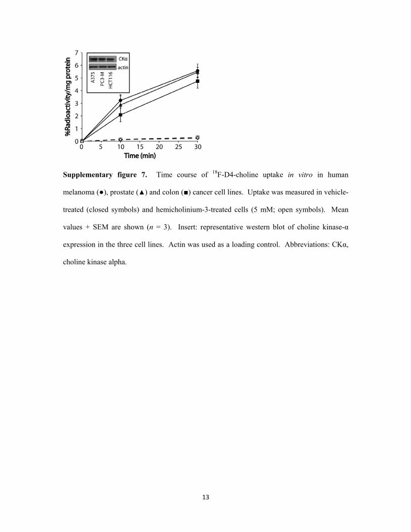
Supplementary figure 7. Time course of 18F-D4-choline uptake in vitro in human
melanoma (●), prostate (▲) and colon (■) cancer cell lines. Uptake was measured in vehicle-
treated (closed symbols) and hemicholinium-3-treated cells (5 mM; open symbols). Mean
values + SEM are shown (n = 3). Insert: representative western blot of choline kinase-α
expression in the three cell lines. Actin was used as a loading control. Abbreviations: CKα,
choline kinase alpha.

14
Supplementary Table
Supplementary table 1. Kinetic parameters from dynamic 18F-D4-choline PET in tumors.
Decay-corrected uptake values at 60 min (NUV60) and the area under the curve (AUC) were
taken from tumor TACs. Flux constant measurements, K1’, Ki and k3 were obtained by fitting
tumor TAC and derived input function, corrected for radioactive plasma metabolites of 18F-
D4-choline, to a 2-tissue irreversible model of tracer delivery and retention. Mean values (n
= 3) ± SEM are shown.
NUV60 AUC K1’ Ki k3
HCT116 1.81 ± 0.11 114.5 ± 7.0 0.142 ± 0.027 0.008 ± 0.001 0.039 ± 0.003
A375 1.71 ± 0.14 107.3 ± 7.7 0.111 ± 0.021 0.006 ± 0.002 0.030 ± 0.008
PC3-M 1.97 ± 0.07 121.3 ± 3.1 0.090 ± 0.007 0.009 ± 0.002 0.040 ± 0.006
References
1. Kenny LM, Contractor KB, Hinz R, Stebbing J, Palmieri C, Jiang J, et al.
Reproducibility of [11C]choline-positron emission tomography and effect of trastuzumab.
Clin Cancer Res. 2010;16:4236-45.
2. Sutinen E, Nurmi M, Roivainen A, Varpula M, Tolvanen T, Lehikoinen P, et al.
Kinetics of [(11)C]choline uptake in prostate cancer: a PET study. Eur J Nucl Med Mol
Imaging. 2004;31:317-24.

15
3. Huang SC, Yu DC, Barrio JR, Grafton S, Melega WP, Hoffman JM, et al. Kinetics
and modeling of L-6-[18F]fluoro-dopa in human positron emission tomographic studies. J
Cereb Blood Flow Metab. 1991;11:898-913.
4. Tomasi G, Bertoldo A, Bishu S, Unterman A, Smith CB, Schmidt KC. Voxel-based
estimation of kinetic model parameters of the L-[1-(11)C]leucine PET method for
determination of regional rates of cerebral protein synthesis: validation and comparison with
region-of-interest-based methods. J Cereb Blood Flow Metab. 2009;29:1317-31.