evaluation of handwriting movement kinematics: from an ... · keywords: kinematics, magnetic...
TRANSCRIPT
fnhum-10-00488 September 27, 2016 Time: 19:44 # 1
METHODSpublished: 29 September 2016
doi: 10.3389/fnhum.2016.00488
Edited by:Mikhail Lebedev,
Duke University, USA
Reviewed by:Raoul Huys,
Centre National de la RechercheScientifique, France
Chris Lange-Küttner,London Metropolitan University, UK
*Correspondence:Marco Bove
Received: 10 June 2016Accepted: 14 September 2016Published: 29 September 2016
Citation:Bisio A, Pedullà L, Bonzano L,
Ruggeri P, Brichetto G and Bove M(2016) Evaluation of HandwritingMovement Kinematics: From an
Ecological to a Magnetic ResonanceEnvironment.
Front. Hum. Neurosci. 10:488.doi: 10.3389/fnhum.2016.00488
Evaluation of Handwriting MovementKinematics: From an Ecological to aMagnetic Resonance EnvironmentAmbra Bisio1, Ludovico Pedullà1,2, Laura Bonzano2, Piero Ruggeri1,Giampaolo Brichetto3 and Marco Bove1*
1 Department of Experimental Medicine, Section of Human Physiology and Centro Polifunzionale di Scienze Motorie,University of Genoa, Genoa, Italy, 2 Department of Neuroscience, Rehabilitation, Ophthalmology, Genetics, Maternal andChild Health, University of Genoa, Genoa, Italy, 3 Scientific Research Area, Italian Multiple Sclerosis Foundation, Genoa, Italy
Writing is a means of communication which requires complex motor, perceptual, andcognitive skills. If one of these abilities gets lost following traumatic events or due toneurological diseases, handwriting could deteriorate. Occupational therapy practitionersprovide rehabilitation services for people with impaired handwriting. However, todetermine the effectiveness of handwriting interventions no studies assessed whetherthe proposed treatments improved the kinematics of writing movement or hadan effect at the level of the central nervous system. There is need to find newquantitative methodologies able to describe the behavioral and the neural outcomesof the rehabilitative interventions for handwriting. In the present study we proposed acombined approach that allowed evaluating the kinematic parameters of handwritingmovements, acquired by means of a magnetic resonance-compatible tablet, and theirneural correlates obtained simultaneously from a functional magnetic resonance imagingexamination. Results showed that the system was reliable in term of reproducibility ofthe kinematic data during a test/re-test procedure. Further, despite the modificationswith respect to an ecological writing movement condition, the kinematic parametersacquired inside the MR-environment were descriptive of individuals’ movement features.At last, the imaging protocol succeeded to show the activation of the cerebralregions associated with the production of writing movement in healthy people. Fromthese findings, this methodology seems to be promising to evaluate the handwritingmovement deficits and the potential alterations in the neural activity in those individualswho have handwriting difficulties. Finally, it would provide a mean to quantitatively assessthe effect of a rehabilitative treatment.
Keywords: kinematics, magnetic resonance-compatible tablet, fMRI, test–retest, ecological validity
INTRODUCTION
Handwriting is historically believed to be one of the most difficult fine motor skills to learn. It takesyears of practice before a person has mastered the mature handwriting skill. Despite the difficultiesthis ability can pose to the learners, handwriting is one of the first motor expertise that childrenacquire at school and is nowadays part of the individual motor-cultural baggage. Although the
Frontiers in Human Neuroscience | www.frontiersin.org 1 September 2016 | Volume 10 | Article 488
fnhum-10-00488 September 27, 2016 Time: 19:44 # 2
Bisio et al. Handwriting Movements in MR Scanner
increasing use of personal computer has reduced the timededicated to handwriting, adults still use handwriting tocommunicate with others, to record ideas and for creativeexpression.
Handwriting is a complex functional activity, which involvesfine motor skills, cognitive and visual-perceptual processing(Mary-Ann, 1992; Feder and Majnemer, 2007). In adults, theability to handwrite can be affected, or even lost, due toneurological diseases. For instance, handwriting deficits arecommon after stroke (Simpson et al., 2015) and in people withmultiple sclerosis (Wellingham-Jones, 1991; Schenk et al., 2000),Parkinson’s disease (Van Gemmert et al., 1999, 2001; Lange et al.,2006), and obsessive-compulsive disorders (Mavrogiorgou et al.,2001).
The restoration of handwriting is matter of the occupationaltherapy treatments. Unfortunately, many of the conventionalmethods used to evaluate handwriting movements during theclinical routine are not standardized and are dependent onthe therapist’s expertise (Hoy et al., 2011; Van Drempt et al.,2011). From two recent reviews on retraining methodologiesfor handwriting ability (Hoy et al., 2011; Yancosek and Howell,2011) emerged that no studies assessed the effect of theproposed treatment on the kinematics of writing movementor on the central nervous system activity. Therefore, it wouldof great importance to quantitatively describe handwritingmotor performance and to investigate brain activity duringhandwriting. The application of this combined methodologycould also allow assessing possible changes in the kinematicsand in the neural activations after a rehabilitation treatment.This is one of the reasons why new digital approaches, whichmake use of technology for data collection and kinematicevaluation, have been developed. To this concern, recent studieson handwriting movements were carried out by means ofdigitizing tablets that allow a quantitative analysis of thekinematic parameters of the writing trace (Mergl et al., 1999; VanGemmert et al., 1999, 2001; Schenk et al., 2000; Mavrogiorgouet al., 2001; Vuillermot et al., 2009; Accardo et al., 2013).Other studies investigated handwriting by using algorithms ableto translate electromiographic signals generated by hand andforearm muscles into handwriting traces (Linderman et al., 2009;Okorokova et al., 2015).
A step further toward a comprehensive description of thelaws that govern handwriting is the acquisition of the neuralcorrelates of writing movements with the aim to investigatethe relationship between the behavioral data and the cerebralactivity. In this context, functional magnetic resonance imaging(fMRI) has become a powerful tool to study the functionalorganization of the brain (Menon, 2001; Savoy, 2001), allowingthe acquisition of brain activation during the motor task. Thus,it is also useful to develop MR-compatible devices to performa kinematic recording of the writing movement during fMRIexamination, even though it poses a problem related to thevalidity of the kinematic data because of the postural constraintsthe subject is forced to maintain in the MR scanner. Untilnow some efforts have been done in this direction, but onlyfew studies tackled this issue (Katanoda et al., 2001; Siebneret al., 2002; Reithler et al., 2006; Reitz et al., 2013; Karimpoor
et al., 2015) and none of them tested whether the kinematicfeatures of the subject’s movements obtained in ecological writingconditions (i.e., when the subject is seated at a table) are preservedwhen the task is performed inside the MR scanner. Indeed,one might hypothesize that when a person moves inside thenarrow space of the MR environment the handwriting movementperformance changes. Furthermore, among these studies, onlyKarimpoor et al., 2015 provided a simultaneous visual feedbackof the subject’s performance showing a reconstruction of theparticipant’s hand during handwriting together with the writtentrace. Although this system was demonstrated to improve thequality of the kinematic measurements with respect to a non-visual feedback condition, this kind of visual feedback could alsobe distracting because the subject’s focus may move from therecognition of the hand to the written trace or also other detailsof the displayed image.
Aim of the present work was to implement a methodologyable to quantitatively characterize the kinematics of handwritingmovements and the related neural substrates. Indeed, in thisstudy we considered the temporal and spatial patterns ofhandwriting, omitting the evaluation of writing per se. Toachieve this goal we: (a) assessed the reproducibility of repeatedmeasurements of handwriting movements acquired by means ofa newly designed and developed MR-compatible tablet outsidethe magnet bore, (b) tested the reliability of the kinematicdata acquired inside the MR scanner, where participants hada visual simultaneous visual feedback of their written trace, bycomparing them with those obtained in ecological condition, and(c) investigated the neural correlates associated to handwriting bymeans of fMRI sequences.
MATERIALS AND METHODS
This study was composed of two experiments: Experiment 1assessed the reproducibility of the kinematic data acquiredwith the MR-compatible tablet by means of a test–retestapproach. Since this was the first study that used this system(i.e., the combination of this MR-compatible tablet with ourcustom-made acquisition software) and this methodology, itwas crucial to test its reliability. Experiment 2 tested whetherthe individual kinematic features associated with handwritingmovements performed in ecological conditions are maintainedwhen participants performed the task inside the MR scanner.Further, in a sub-group of subjects, who took part to Experiment2, we tested whether the proposed kinematic paradigm was ableto evoke the activation of the cerebral areas usually active duringa handwriting task.
ParticipantsA total of 44 subjects were enrolled in this study. Twenty-two of them (12 females and 10 males, mean age ± SD =25.0 ± 5.6 years) participated in Experiment 1, whereas 22 (14females and 8 males, mean age ± SD = 24.2 ± 6.1 years)took part in Experiment 2. Seven out of the 22 subjects whotook part to Experiment 2 were also recruited for an fMRIexamination. Participants who manifested MR contraindications,
Frontiers in Human Neuroscience | www.frontiersin.org 2 September 2016 | Volume 10 | Article 488
fnhum-10-00488 September 27, 2016 Time: 19:44 # 3
Bisio et al. Handwriting Movements in MR Scanner
as claustrophobia, pregnancy, and presence of a pacemakerand other metallic parts not magnetic resonance compatiblewere not considered eligible for Experiment 2. All subjectswere right-handed according to the Edinburgh HandednessInventory (Oldfield, 1971) and naive to the specific purpose ofthis study. People with neurological disturbances were excludedfrom the study. Informed consent was obtained according toa procedure approved by the local ethics committee (ComitatoEtico Regione Liguria, IRCCS Azienda Ospedaliera UniversitariaSan Martino—IST, Genoa, Italy) and to the Declaration ofHelsinki.
Equipment for Handwriting MovementAcquisitionHardwareAn innovative MR-compatible tablet, the SMART TAB (E.M.S.,S.r.l., Bologna), was used to acquire handwriting movementsduring the evaluation sessions outside and inside the MR scanner.The SMART TAB consists of a touch-sensitive tablet, a plastic-made stylus, a USB controller box, and a cable that connectsthe tablet with the controller outside the magnet room. Allthe equipment inside the magnet room is non-ferromagnetic.The surface of the tablet is the AccuTouch Five-Wire ResistiveTouchscreens (Elo Touch Solutions, Inc., Milpitas, CA, USA),where both X and Y measurements are made on a stable rear glasslayer. The touch surface (spatial resolution: 4096 DPI, temporalresolution: 10 ms) is mounted within a frame that delimits thesensitive area while offering some protection from unintentionaltouches. The subject can use the fingers or the plastic stylus towrite on the tablet (Touch Activation Force less than 113 g). The
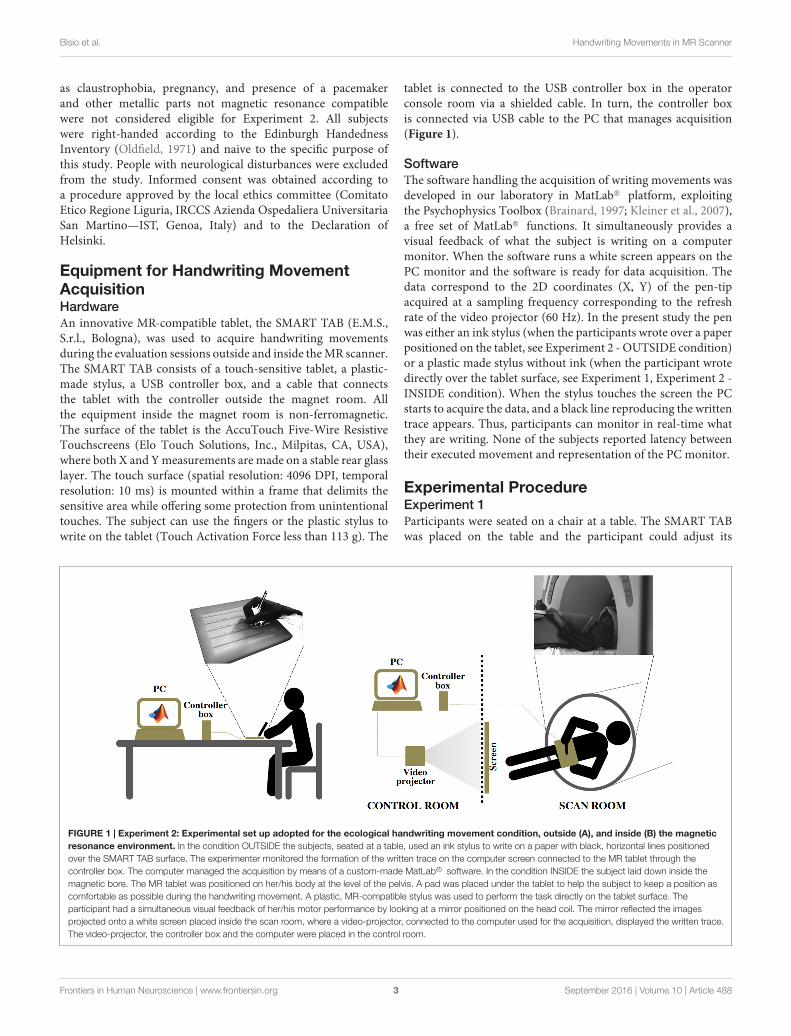
tablet is connected to the USB controller box in the operatorconsole room via a shielded cable. In turn, the controller boxis connected via USB cable to the PC that manages acquisition(Figure 1).
SoftwareThe software handling the acquisition of writing movements wasdeveloped in our laboratory in MatLab R© platform, exploitingthe Psychophysics Toolbox (Brainard, 1997; Kleiner et al., 2007),a free set of MatLab R© functions. It simultaneously provides avisual feedback of what the subject is writing on a computermonitor. When the software runs a white screen appears on thePC monitor and the software is ready for data acquisition. Thedata correspond to the 2D coordinates (X, Y) of the pen-tipacquired at a sampling frequency corresponding to the refreshrate of the video projector (60 Hz). In the present study the penwas either an ink stylus (when the participants wrote over a paperpositioned on the tablet, see Experiment 2 - OUTSIDE condition)or a plastic made stylus without ink (when the participant wrotedirectly over the tablet surface, see Experiment 1, Experiment 2 -INSIDE condition). When the stylus touches the screen the PCstarts to acquire the data, and a black line reproducing the writtentrace appears. Thus, participants can monitor in real-time whatthey are writing. None of the subjects reported latency betweentheir executed movement and representation of the PC monitor.
Experimental ProcedureExperiment 1Participants were seated on a chair at a table. The SMART TABwas placed on the table and the participant could adjust its
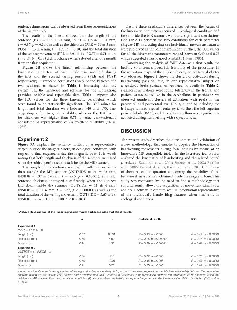
FIGURE 1 | Experiment 2: Experimental set up adopted for the ecological handwriting movement condition, outside (A), and inside (B) the magneticresonance environment. In the condition OUTSIDE the subjects, seated at a table, used an ink stylus to write on a paper with black, horizontal lines positionedover the SMART TAB surface. The experimenter monitored the formation of the written trace on the computer screen connected to the MR tablet through thecontroller box. The computer managed the acquisition by means of a custom-made MatLab R© software. In the condition INSIDE the subject laid down inside themagnetic bore. The MR tablet was positioned on her/his body at the level of the pelvis. A pad was placed under the tablet to help the subject to keep a position ascomfortable as possible during the handwriting movement. A plastic, MR-compatible stylus was used to perform the task directly on the tablet surface. Theparticipant had a simultaneous visual feedback of her/his motor performance by looking at a mirror positioned on the head coil. The mirror reflected the imagesprojected onto a white screen placed inside the scan room, where a video-projector, connected to the computer used for the acquisition, displayed the written trace.The video-projector, the controller box and the computer were placed in the control room.
Frontiers in Human Neuroscience | www.frontiersin.org 3 September 2016 | Volume 10 | Article 488
fnhum-10-00488 September 27, 2016 Time: 19:44 # 4
Bisio et al. Handwriting Movements in MR Scanner
position to feel as comfortable as possible. Once a comfortableposition was achieved the participants were requested to keepthe orientation of the tablet and of their forearm constant.A plastic, MR-compatible stylus was used to perform the taskdirectly on the tablet surface. Participants were required to lookat the computer screen where the written trace was displayedsimultaneously to the motor task. They were asked to write theItalian sentence “Il carro sale al colle” (i.e., “The wagon goes upthe hill”), keeping the text in a single line. This sentence waschosen because it was short and easy for the subject to keep it ina single line, and because it was composed of simple words verypopular in the Italian language. The sentence was repeated fivetimes. Participants were tested twice (PRE and POST sessions,five repetitions of the sentence for each session), one monthapart, to assess possible changes in measurements taken under thesame condition and to define the motor performance parametersshowing good repeatability (i.e., test–retest reliability).
Experiment 2Handwriting movement acquisitionThis experiment was composed of two sessions, which involved22 participants. The first session (OUTSIDE) was aimed atevaluating handwriting movement outside the magnet bore, inecological condition, i.e., participants were seated on a chair ata table. As in Experiment 1, the tablet laying on the table waspositioned by the participant to feel as comfortable as possible.In addition, in order to mimic a conventional handwritingcondition while recording movement kinematics, a paper withblack, horizontal lines was positioned over the SMART TABsurface and the subject was provided with an ink stylus. In thisway, the participant had a visual feedback over the paper of whatshe/he was writing and could appreciate the normal resistance ofthe pen over the paper during the handwriting movement; in themeantime, the tablet acquired the trajectory of the stylus and theexperimenter could simultaneously monitor the writing motoroutput on the computer screen. The subject was asked to writethree times the Italian sentence “Il sole scalda” (i.e., “The sunwarms”) when a “go” signal was provided by the experimenter.As in Experiment 1, this sentence was chosen because it wascomposed of simple words very popular in the Italian language.We adopted a shorter sentence with respect to that of Experiment1 in order to make the task easier for the subject inside the MRscanner.
The second session (INSIDE) took place inside the magnetbore. The subject laid down with the SMART TAB positioned onher/his body at the level of the pelvis. The forearm was positionedover an MR-compatible support made of soft material, adjustablein height to minimize the subject’s forearm movements. A padwas placed under the tablet to help the subject to keep a positionas comfortable as possible during the handwriting movement.A plastic, MR-compatible stylus was used to perform the taskdirectly on the tablet surface. The participant had a simultaneousvisual feedback of her/his motor performance by looking at amirror positioned on the head coil. The mirror reflected theimages projected onto a white screen placed inside the magnetroom, where a video-projector, connected to the computer usedfor the acquisition, displayed the written trace. The horizontal
lines over the writing space were physically created by mountingequidistant, black cotton threads over the white screen inside themagnet room. The task was adapted to cover a temporal intervalof 30 s, which corresponded to the active task blocks of thefMRI acquisition (performed only by seven subjects, see MagneticResonance Imaging Protocol). For this reason, participants wereinstructed to write the sentence on a first line and to repeat it onthe lines below until a “stop” signal was provided. Each subjectcompleted three task blocks, containing at least one completesentence each, interleaved with 30-s periods of rest. The sentencewas the same adopted for the OUTSIDE condition. Duringrest blocks subjects had to handle the stylus without movingand to keep the eyes opened, as in the task blocks, to avoidspurious findings related to visual activations. Each session waspreceded by a familiarization phase which allowed subjects totrain themselves with the SMART TAB in this non-conventionalwriting condition.
Magnetic resonance imaging protocolSeven out of 22 subjects, who executed the two experimentalsessions of the Experiment 2, during the INSIDE session werealso examined by means of an fMRI test. MRI examination wasperformed on a 1.5 T MR system (Signa Excite HDxt, GeneralElectric Healthcare, Waukesha, WI, USA) and included thefollowing series covering the whole brain: axial FLAIR sequence(slice thickness= 5 mm; TR= 9002 ms; TE= 97.5 ms; inversiontime = 2250 ms; flip angle = 90◦; FOV = 240 mm × 240 mm;matrix= 512× 512) to exclude incidental findings in the enrolledsubjects; axial T2-weighted sequence (slice thickness = 5 mm;TR = 6300 ms; TE = 123.7 ms; FOV = 260 mm × 260 mm;matrix = 256 × 256) used as structural reference for the fMRIacquisition; T2∗-weighted single-shot EPI sequences (32 slices;slice thickness = 4 mm; gap = 0.5 mm; TR = 3000 ms;TE = 40 ms; FOV = 260 mm × 260 mm; matrix = 64 × 64)for fMRI. Particularly, each fMRI run included 63 brain volumes;the first 3 volumes were discarded because of non-steadymagnetization. Within each run the subject performed thehandwriting motor task (i.e., active task) alternatively with a restcondition (i.e., control), according to a block designed paradigmconsisting of 30-sec active task periods alternating with 30-scontrol periods (10 brain volumes per block).
Data AnalysisData TreatmentBehavioral dataThe kinematic parameters of handwriting movements werecomputed by means of a custom-made MatLab R© software. Thesoftware automatically detected the beginning and the end ofthe sentence on the basis of the module of the velocity profile(computed over the two dimensions of the tablet, x and y): thefirst and the last instants (in the first line, in case of multiple lines)in which the velocity was greater than zero value correspondedto the beginning and the end of the sentence, respectively. Totalduration (s) (i.e., the time employed by the subject to write anentire sentence), movement length (mm), and thickness (mm) ofthe sentence were considered as outcome parameters. The lengthcorresponded to the size of the segment which connects the first
Frontiers in Human Neuroscience | www.frontiersin.org 4 September 2016 | Volume 10 | Article 488
fnhum-10-00488 September 27, 2016 Time: 19:44 # 5
Bisio et al. Handwriting Movements in MR Scanner
and the last points of the sentence. The thickness was computedas the vertical distance between the top of the highest letter andthe bottom of the lowest one.
Neuroimaging dataSPM12 software (Wellcome Department of ImagingNeuroscience, London, UK) was used for fMRI processing(Friston et al., 1995). For each participant, the first image wasused as a reference to which all the subsequent scans wererealigned, and the 6 parameters describing the rigid bodytransformation between each source image and the referenceimage were used to re-sample each image to apply motioncorrection. Then, slice timing was applied to minimize timing-errors between slices and the functional images were normalizedto the Montreal Neurological Institute (MNI) template brainimage using a 12-parameter affine transformation, re-sampledto 2 mm × 2 mm × 2 mm voxels and smoothed with an 8 mmfull-width at half-maximum isotropic Gaussian kernel to increasethe signal-to-noise ratio.
Statistical AnalysisExperiment 1A paired t-test compared the mean kinematic parameters(average computed across the five trials) acquired for each subjectthe first time the participant used the MR-tablet with thoseacquired one month later. Further, to assess the reliability of theacquisition, a linear regression model (POST = a ∗ PRE + b,where a and b refer to the slope and intercept values of theregression line, respectively) tested the relationship among thesingle-values of the kinematic parameters acquired during thefirst evaluation and after one month on the same subjects.Pearson’s correlation coefficients are provided together with theassociated p-value, the slopes and the intercepts values of the
regression lines. To be more accurate the test–retest reliabilityof the motor performance parameters was assessed also by theIntraclass Correlation Coefficient (ICC) and the correspondingp-value (Shrout and Fleiss, 1979).
Experiment 2To compare participants’ motor performance inside and outsidethe magnetic bore, the mean kinematic parameters (averagecomputed across the thre trials) were compared by meansof paired t-tests. To specifically assess the reliability of thebehavioral acquisitions performed inside the magnetic bore withrespect to that in ecological condition (outside the magnet), alinear regression model (OUTSIDE = a ∗ INSIDE + b) testedthe relationship between the kinematic parameters in the twoexperimental conditions. Pearson’s correlation coefficients areprovided together with the slopes (a) and the intercepts (b)values of the regression lines and the associated p-values. Asin Experiment 1, the ICC and the corresponding p-value werecomputed for each parameter.
Concerning the fMRI data, a general linear model was usedto identify the voxels with task-related signal changes at theindividual level. Task-related t contrast images were created foreach subject, with a height threshold of p < 0.05 FWE-correctedand extent threshold arbitrarily set at k= 50 voxels.
Data in the text are reported as means± SD.
RESULTS
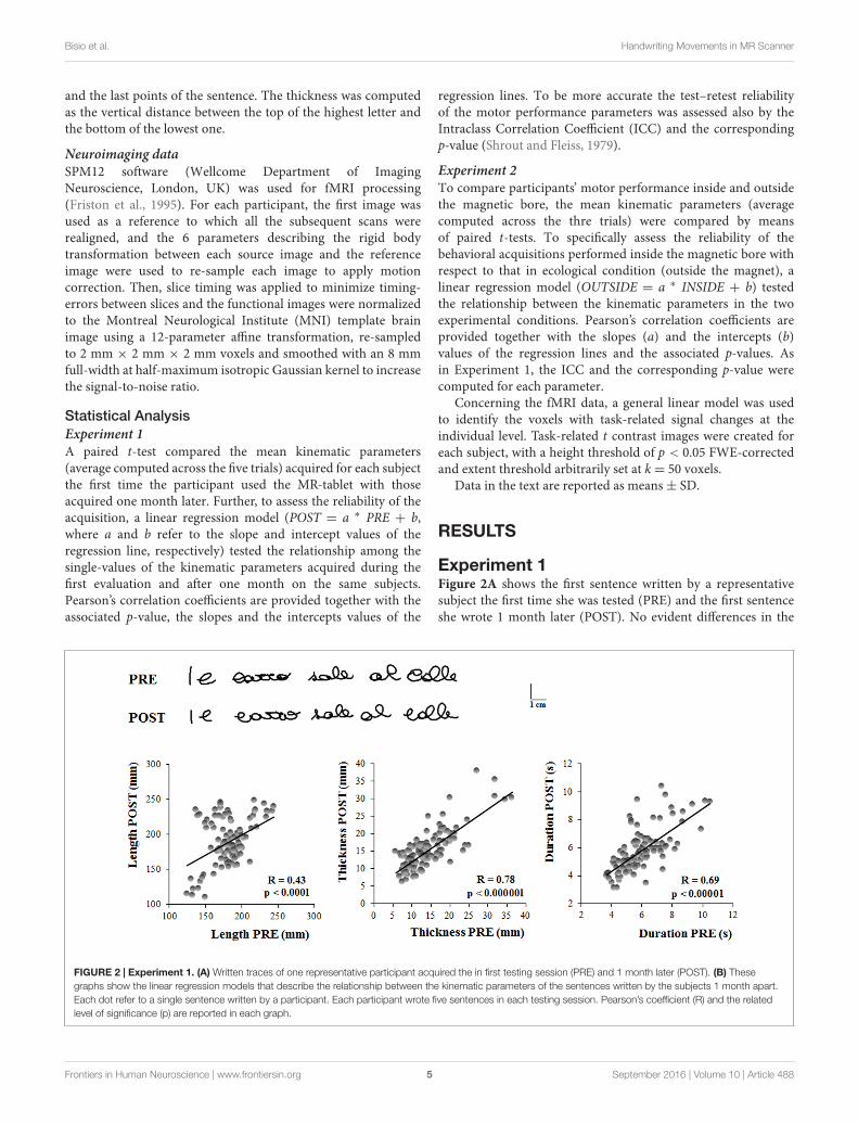
Experiment 1Figure 2A shows the first sentence written by a representativesubject the first time she was tested (PRE) and the first sentenceshe wrote 1 month later (POST). No evident differences in the
FIGURE 2 | Experiment 1. (A) Written traces of one representative participant acquired the in first testing session (PRE) and 1 month later (POST). (B) Thesegraphs show the linear regression models that describe the relationship between the kinematic parameters of the sentences written by the subjects 1 month apart.Each dot refer to a single sentence written by a participant. Each participant wrote five sentences in each testing session. Pearson’s coefficient (R) and the relatedlevel of significance (p) are reported in each graph.
Frontiers in Human Neuroscience | www.frontiersin.org 5 September 2016 | Volume 10 | Article 488
fnhum-10-00488 September 27, 2016 Time: 19:44 # 6
Bisio et al. Handwriting Movements in MR Scanner
sentence dimensions can be observed from these representationsof the written trace.
The results of the t-tests showed that the length of thesentence (PRE = 183 ± 23 mm, POST = 189.47 ± 31 mm;t = 0.97, p = 0.34), as well as the thickness (PRE = 14 ± 5 mm,POST = 15 ± 4 mm; t = 1.71, p = 0.10) and the total durationof the writing movement (PRE = 6.01 ± 1 s, POST = 5.71 ± 1 s;t = 1.37, p= 0.18) did not change when retested after one monthfrom the first acquisition.
Figure 2B shows the linear relationship between thekinematic parameters of each single trial acquired duringthe first and the second testing session (PRE and POST,respectively). Significant correlations were found between thetwo sessions, as shown in Table 1, indicating that thesystem (i.e., the hardware and software for the acquisition)provided reliable and repeatable data. Table 1 reports alsothe ICC values for the three kinematic parameters whichwere found to be statistically significant. The ICC values forlength and total duration were between 0.40 and 0.75, thussuggesting a fair to good reliability, whereas the ICC valuefor thickness was higher than 0.75, a value conventionallyconsidered as representative of an excellent reliability (Fleiss,1986).
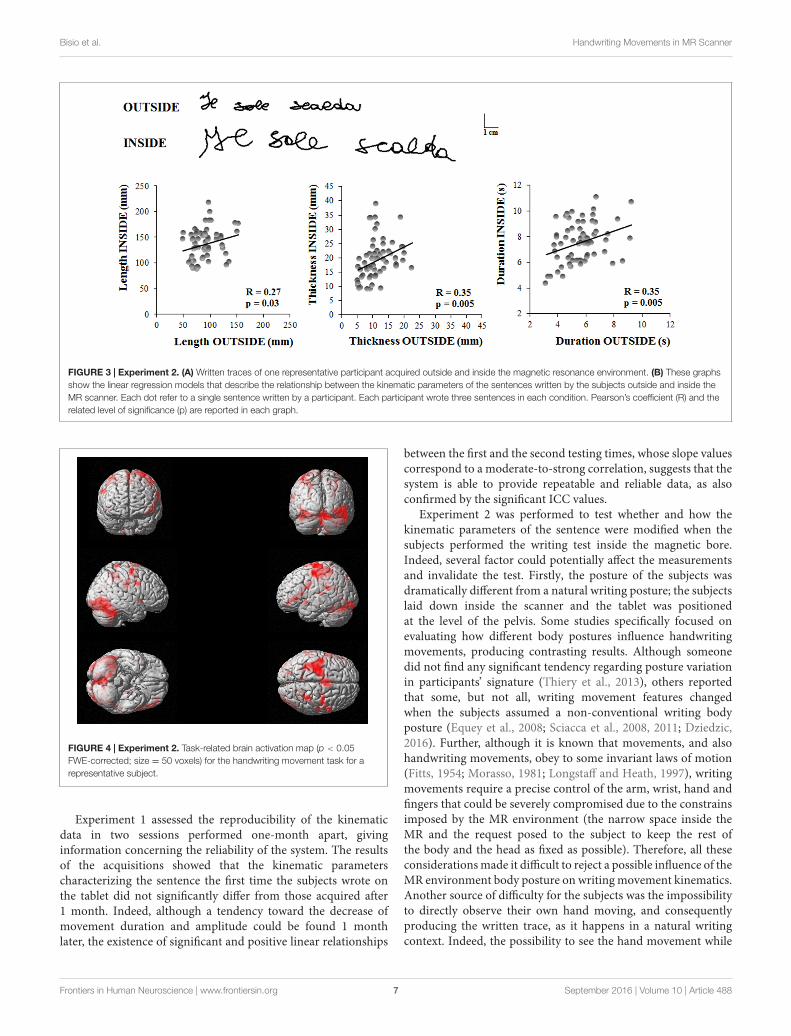
Experiment 2Figure 3A displays the sentence written by a representativesubject outside the magnetic bore, in ecological condition, withrespect to that acquired inside the magnetic bore. It is worthnoting that both length and thickness of the sentence increasedwhen the subject performed the task inside the MR scanner.
The length of the sentence was significantly longer insidethan outside the MR scanner (OUTSIDE = 91 ± 23 mm,INSIDE = 137 ± 29 mm; t = 6.45, p < 0.00001). Similarly,sentence thickness increased significantly when the subjectslaid down inside the scanner (OUTSIDE = 11 ± 4 mm,INSIDE = 19 ± 6 mm; t = 6.22, p < 0.00001), as well as thetotal duration of the writing movement (OUTSIDE= 5.65± 1 s,INSIDE= 7.56± 1 s; t = 5.88, p < 0.00001).
Despite these predictable differences between the values ofthe kinematic parameters acquired in ecological condition andthose inside the MR scanner, we found significant correlations(see Table 1) between the two conditions for each parameter(Figure 3B), indicating that the individuals’ movement featureswere preserved in the MR environment. Further, the ICC valuesfor all the kinematic parameters ranged between 0.40 and 0.75which suggested a fair to good reliability (Fleiss, 1986).
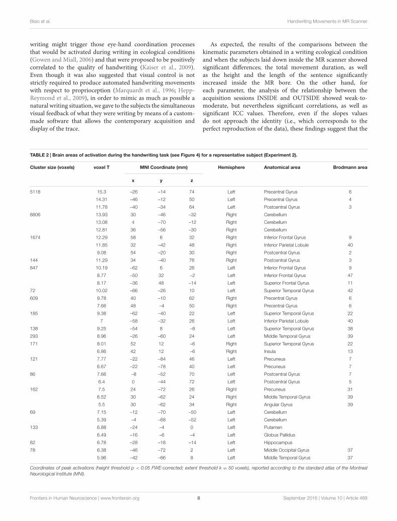
Concerning the analysis of fMRI data, as a first result, thehealthy volunteers showed full feasibility of the procedure. Inthe activation maps of the single subjects, no artifactual clusterwas observed. Figure 4 shows the clusters of activation duringhandwriting (task vs. rest) in one representative subject ona rendered brain surface. As reported in details in Table 2,significant activations were found bilaterally in the frontal andparietal areas, as well as in the cerebellum. In particular, weobserved significant clusters of activation with peaks in theprecentral and postcentral gyri (BA 3, 4, and 6) including theleft superior and medial frontal gyri. Further, the left superiorparietal lobule (BA 7), and the right cerebellum were significantlyactivated during handwriting with respect to rest.
DISCUSSION
The present study describes the development and validation ofa new methodology that enables to acquire the kinematics ofhandwriting movements during fMRI studies by means of aninnovative MR-compatible tablet. In the literature few studiesanalyzed the kinematics of handwriting and the related neuralcorrelates (Katanoda et al., 2001; Siebner et al., 2002; Reithleret al., 2006; Reitz et al., 2013; Karimpoor et al., 2015), and noneof them raised the question concerning the reliability of thebehavioral measurement obtained inside the magnetic bore. Thisstudy was motivated by the need to find a methodology thatsimultaneously allows the acquisition of movement kinematicsand brain activity, in order to acquire information representativeof the individual’s handwriting features when she/he is inecological conditions.
TABLE 1 | Description of the linear regression model and associated statistical results.
a b Statistical results ICC
Experiment 1POST = a ∗ PRE +b
Length (mm) 0.57 84.34 R = 0.43, p < 0.0001 R = 0.42, p < 0.00001
Thickness (mm) 0.75 4.52 R = 0.78, p < 0.000001 R = 0.78, p < 0.00001
Duration (s) 0.74 1.33 R = 0.69, p < 0.00001 R = 0.69, p < 0.00001
Experiment 2OUTSIDE = a ∗ INSIDE + b
Length (mm) 0.34 106 R = 0.27, p = 0.035 R = 0.75, p < 0.00001
Thickness (mm) 0.55 12.91 R = 0.35, p = 0.005 R = 0.57, p < 0.00001
Duration (s) 0.4 5.23 R = 0.35, p = 0.005 R = 0.42, p < 0.00001
a and b are the slope and intercept values of the regression line, respectively. In Experiment 1 the linear regressions modeled the relationship between the parametersacquired during the first testing (PRE) session and 1 month later (POST), whereas in Experiment 2 the relationship between the parameters of the sentence inside andoutside the MR scanner. Pearson’s correlation coefficient (R) and the related probability are reported together with the Intrarclass Correlation Coefficient (ICC) and itsp-value.
Frontiers in Human Neuroscience | www.frontiersin.org 6 September 2016 | Volume 10 | Article 488
fnhum-10-00488 September 27, 2016 Time: 19:44 # 7
Bisio et al. Handwriting Movements in MR Scanner
FIGURE 3 | Experiment 2. (A) Written traces of one representative participant acquired outside and inside the magnetic resonance environment. (B) These graphsshow the linear regression models that describe the relationship between the kinematic parameters of the sentences written by the subjects outside and inside theMR scanner. Each dot refer to a single sentence written by a participant. Each participant wrote three sentences in each condition. Pearson’s coefficient (R) and therelated level of significance (p) are reported in each graph.
FIGURE 4 | Experiment 2. Task-related brain activation map (p < 0.05FWE-corrected; size = 50 voxels) for the handwriting movement task for arepresentative subject.
Experiment 1 assessed the reproducibility of the kinematicdata in two sessions performed one-month apart, givinginformation concerning the reliability of the system. The resultsof the acquisitions showed that the kinematic parameterscharacterizing the sentence the first time the subjects wrote onthe tablet did not significantly differ from those acquired after1 month. Indeed, although a tendency toward the decrease ofmovement duration and amplitude could be found 1 monthlater, the existence of significant and positive linear relationships
between the first and the second testing times, whose slope valuescorrespond to a moderate-to-strong correlation, suggests that thesystem is able to provide repeatable and reliable data, as alsoconfirmed by the significant ICC values.
Experiment 2 was performed to test whether and how thekinematic parameters of the sentence were modified when thesubjects performed the writing test inside the magnetic bore.Indeed, several factor could potentially affect the measurementsand invalidate the test. Firstly, the posture of the subjects wasdramatically different from a natural writing posture; the subjectslaid down inside the scanner and the tablet was positionedat the level of the pelvis. Some studies specifically focused onevaluating how different body postures influence handwritingmovements, producing contrasting results. Although someonedid not find any significant tendency regarding posture variationin participants’ signature (Thiery et al., 2013), others reportedthat some, but not all, writing movement features changedwhen the subjects assumed a non-conventional writing bodyposture (Equey et al., 2008; Sciacca et al., 2008, 2011; Dziedzic,2016). Further, although it is known that movements, and alsohandwriting movements, obey to some invariant laws of motion(Fitts, 1954; Morasso, 1981; Longstaff and Heath, 1997), writingmovements require a precise control of the arm, wrist, hand andfingers that could be severely compromised due to the constrainsimposed by the MR environment (the narrow space inside theMR and the request posed to the subject to keep the rest ofthe body and the head as fixed as possible). Therefore, all theseconsiderations made it difficult to reject a possible influence of theMR environment body posture on writing movement kinematics.Another source of difficulty for the subjects was the impossibilityto directly observe their own hand moving, and consequentlyproducing the written trace, as it happens in a natural writingcontext. Indeed, the possibility to see the hand movement while
Frontiers in Human Neuroscience | www.frontiersin.org 7 September 2016 | Volume 10 | Article 488
fnhum-10-00488 September 27, 2016 Time: 19:44 # 8
Bisio et al. Handwriting Movements in MR Scanner
writing might trigger those eye-hand coordination processesthat would be activated during writing in ecological conditions(Gowen and Miall, 2006) and that were proposed to be positivelycorrelated to the quality of handwriting (Kaiser et al., 2009).Even though it was also suggested that visual control is notstrictly required to produce automated handwriting movementswith respect to proprioception (Marquardt et al., 1996; Hepp-Reymond et al., 2009), in order to mimic as much as possible anatural writing situation, we gave to the subjects the simultaneousvisual feedback of what they were writing by means of a custom-made software that allows the contemporary acquisition anddisplay of the trace.
As expected, the results of the comparisons between thekinematic parameters obtained in a writing ecological conditionand when the subjects laid down inside the MR scanner showedsignificant differences; the total movement duration, as wellas the height and the length of the sentence significantlyincreased inside the MR bore. On the other hand, foreach parameter, the analysis of the relationship between theacquisition sessions INSIDE and OUTSIDE showed weak-to-moderate, but nevertheless significant correlations, as well assignificant ICC values. Therefore, even if the slopes valuesdo not approach the identity (i.e., which corresponds to theperfect reproduction of the data), these findings suggest that the
TABLE 2 | Brain areas of activation during the handwriting task (see Figure 4) for a representative subject (Experiment 2).
Cluster size (voxels) voxel T MNI Coordinate (mm) Hemisphere Anatomical area Brodmann area
x y z
5118 15.3 –26 –14 74 Left Precentral Gyrus 6
14.31 –46 –12 50 Left Precentral Gyrus 4
11.78 –40 –34 64 Left Postcentral Gyrus 3
8806 13.93 30 –46 –32 Right Cerebellum
13.08 4 –70 –12 Right Cerebellum
12.81 36 –56 –30 Right Cerebellum
1674 12.29 58 6 32 Right Inferior Frontal Gyrus 9
11.85 32 –42 48 Right Inferior Parietal Lobule 40
9.08 54 –20 30 Right Postcentral Gyrus 2
144 11.29 34 –40 76 Right Postcentral Gyrus 3
847 10.19 –62 6 26 Left Inferior Frontal Gyrus 9
8.77 –50 32 –2 Left Inferior Frontal Gyrus 47
8.17 –36 48 –14 Left Superior Frontal Gyrus 11
72 10.02 –66 –26 10 Left Superior Temporal Gyrus 42
609 9.78 40 –10 62 Right Precentral Gyrus 6
7.66 48 –4 50 Right Precentral Gyrus 6
185 9.38 –62 –40 22 Left Superior Temporal Gyrus 22
7 –58 –32 26 Left Inferior Parietal Lobule 40
138 9.25 –54 8 –8 Left Superior Temporal Gyrus 38
293 8.96 –26 –60 24 Left Middle Temporal Gyrus 39
171 8.01 52 12 –6 Right Superior Temporal Gyrus 22
6.86 42 12 –6 Right Insula 13
121 7.77 –22 –84 46 Left Precuneus 7
6.67 –22 –78 40 Left Precuneus 7
86 7.66 –8 –52 70 Left Postcentral Gyrus 7
6.4 0 –44 72 Left Postcentral Gyrus 5
162 7.5 24 –72 26 Right Precuneus 31
6.52 30 –62 24 Right Middle Temporal Gyrus 39
5.5 30 –62 34 Right Angular Gyrus 39
69 7.15 –12 –70 –50 Left Cerebellum
5.39 –4 –68 –52 Left Cerebellum
133 6.88 –24 –4 0 Left Putamen
6.49 –16 –6 –4 Left Globus Pallidus
82 6.78 –28 –18 –14 Left Hippocampus
78 6.38 –46 –72 2 Left Middle Occipital Gyrus 37
5.96 –42 –66 8 Left Middle Temporal Gyrus 37
Coordinates of peak activations (height threshold p < 0.05 FWE-corrected; extent threshold k = 50 voxels), reported according to the standard atlas of the MontrealNeurological Institute (MNI).
Frontiers in Human Neuroscience | www.frontiersin.org 8 September 2016 | Volume 10 | Article 488
fnhum-10-00488 September 27, 2016 Time: 19:44 # 9
Bisio et al. Handwriting Movements in MR Scanner
individual features that characterize the subject’s movement withrespect to the other participants were sufficiently preserved in theMR environment despite the previously mentioned difficulties.Namely, a subject who was slower than others in ecologicalwriting condition was slower during fMRI, too. Therefore,although with some obvious differences, the kinematic evaluationof writing movements acquired inside the MR scanner canbe considered sufficiently descriptive of the individual motorbehavior and of the individual differences among subjects.Although this methodology is informative about the temporaland spatial features of handwriting, this study was limited tothe description of these kinematic features of writing movement,without assessing differences in the morphology and topology ofthe letters. This limitation could be considered in future studiesthat, applying the methodology here proposed, will provide amore comprehensive description of the handwriting outcome.
During the fMRI study, the task required an overt movementexecution, which involved hand and fingers’ movements (theforearm was positioned over an MR-compatible supportto minimize the subject’s forearm movements); however,participants were able to maintain the head stable and thisguaranteed to reduce motion artifacts. No image artifacts due tothe presence of the MR-compatible tablet were observed. Further,the fMRI analysis presented here for a representative subjectshowed the significant activations of brain regions which havebeen previously described as candidate cortical sites for writing,i.e., the posterior part of the left middle frontal gyrus and theleft superior parietal lobule (Vernea and Merory, 1975; Bassoet al., 1978), together with the right cerebellum (Katanoda et al.,2001; Purcell et al., 2011; Planton et al., 2013). Further, bilateralactivations in the motor areas were observed in line with aprevious EEG study (Rupasov et al., 2012). Therefore, one couldaccept this protocol as effective in engaging the neural networksinvolved in handwriting tasks.
CONCLUSION
In the present study we proposed a combined approach thatallows evaluating at the same time the kinematic parameters
of handwriting movements and the related brain activity. Thesystem has proven capable of providing reliable kinematicdata. Then, despite the expected modifications with respectto an ecological writing movement condition, the kinematicparameters acquired inside the MR scanner were largelydescriptive of individuals’ movement features. Further, fMRIresults indicate that the cerebral regions activated in thiscondition were those expected to be involved in an handwritingtask in healthy people. Following all these findings, we suggestthat this methodology can be promising to evaluate thebehavioral performance and simultaneously the brain activationsin persons with handwriting difficulties, and provides also a toolto quantitatively evaluate the effects of a rehabilitation treatment.Further studies are also needed to investigate the correlationsbetween the handwriting kinematic parameters and the imagingdata in a large number of healthy participants and people withhandwriting deficits, where abnormal brain activations mightcorrespond to altered motor behavior.
AUTHOR CONTRIBUTIONS
AB conceived and designed the work, performed the acquisition,analyzed and interpreted the data, wrote the manuscript. LPperformed the acquisition, analyzed and interpreted the data,revised the manuscript. LB conceived and designed the work,analyzed the data, revised the manuscript. PR interpreted thedata, drafted and revised the manuscript, gave the final approvalof the version to be published. GB interpreted the data, draftedand revised the manuscript, gave the final approval of theversion to be published. MB conceived and designed the work,interpreted the data, revised the manuscript, gave the finalapproval of the version to be published.
ACKNOWLEDGMENT
This study was supported by FISM - Fondazione Italiana SclerosiMultipla - Cod. 2014/R/5.
REFERENCESAccardo, A. P., Genna, M., and Borean, M. (2013). Reprint of “Development,
maturation and learning influence on handwriting kinematics.” Hum. Mov. Sci.32, 999–1009. doi: 10.1016/j.humov.2013.08.002
Basso, A., Taborelli, A., and Vignolo, L. A. (1978). Dissociated disorders of speakingand writing in aphasia. J. Neurol. Neurosurg. Psychiatry 41, 556–563. doi:10.1136/jnnp.41.6.556
Brainard, D. H. (1997). The psychophysics toolbox. Spat. Vis. 10, 433–436. doi:10.1163/156856897X00357
Dziedzic, T. (2016). The influence of lying body position on handwriting. J. ForensicSci. 61, 178–183. doi: 10.1111/1556-4029.12948
Equey, C., Marquis, R., and Mazzella, W. (2008). Influence of writing posture onthe dimensions of signatures. J. Am. Soc. Quest. Doc. Exam. 10, 53–59.
Feder, K. P., and Majnemer, A. (2007). Handwriting development, competency,and intervention. Dev. Med. Child Neurol. 49, 312–317. doi: 10.1111/j.1469-8749.2007.00312.x
Fitts, P. M. (1954). The information capacity of the human motor system incontrolling the amplitude of movement. J. Exp. Psychol. 47, 381–391. doi:10.1037/h0055392
Fleiss, J. (1986). The Design and Analisys of Clinical Experiments. New York, NY:John Wiley & Sons.
Friston, K. J., Holmes, A. P., Poline, J. B., Grasby, P. J., Williams, S. C., Frackowiak,R. S., et al. (1995). Analysis of fMRI time-series revisited. Neuroimage 2, 45–53.doi: 10.1006/nimg.1995.1007
Gowen, E., and Miall, R. C. (2006). Eye-hand interactions in tracing anddrawing tasks. Hum. Mov. Sci. 25, 568–585. doi: 10.1016/j.humov.2006.06.005
Hepp-Reymond, M. C., Chakarov, V., Schulte-Mönting, J., Huethe, F., andKristeva, R. (2009). Role of proprioception and vision in handwriting. BrainRes. Bull. 79, 365–370. doi: 10.1016/j.brainresbull.2009.05.013
Hoy, M. M. P., Egan, M. Y., and Feder, K. P. (2011). A systematic review ofinterventions to improve handwriting. Can. J. Occup. Ther. 78, 13–25. doi:10.2182/cjot.2011.78.1.3
Frontiers in Human Neuroscience | www.frontiersin.org 9 September 2016 | Volume 10 | Article 488
fnhum-10-00488 September 27, 2016 Time: 19:44 # 10
Bisio et al. Handwriting Movements in MR Scanner
Kaiser, M.-L., Albaret, J.-M., and Doudin, P.-A. (2009). Relationship betweenvisual-motor integration, eye-hand coordination, and quality of handwriting.J. Occup. Ther. Sch. Early Interv. 2, 87–95. doi: 10.1080/19411240903146228
Karimpoor, M., Tam, F., Strother, S. C., Fischer, C. E., Schweizer, T. A., and Graham,S. J. (2015). A computerized tablet with visual feedback of hand positionfor functional magnetic resonance imaging. Front. Hum. Neurosci. 9:150. doi:10.3389/fnhum.2015.00150
Katanoda, K., Yoshikawa, K., and Sugishita, M. (2001). A functional MRI studyon the neural substrates for writing. Hum. Brain Mapp. 13, 34–42. doi:10.1002/hbm.1023
Kleiner, M., Brainard, D. H., and Pelli, D. G. (2007). What’s new in Psychtoolbox-3?Perception 36:S14. doi: 10.1068/v070821
Lange, K. W., Mecklinger, L., Walitza, S., Becker, G., Gerlach, M., Naumann, M.,et al. (2006). Brain dopamine and kinematics of graphomotor functions. Hum.Mov. Sci. 25, 492–509. doi: 10.1016/j.humov.2006.05.006
Linderman, M., Lebedev, M. A., and Erlichman, J. S. (2009). Recognitionof handwriting from electromyography. PLoS ONE 4:e6791. doi:10.1371/journal.pone.0006791
Longstaff, M. G., and Heath, R. A. (1997). Space-time invariance in adulthandwriting. Acta Psychol. 97, 201–214. doi: 10.1016/S0001-6918(97)00015-2
Marquardt, C., Gentz, W., and Mai, N. (1996). “On the role of vision in skilledhandwriting: basic and applied issues,” in Handwriting and Drawing Research,eds M. Simner, C. Leehdam, and A. Thomassen (Amsterdam: IOS Press), 87–97.
Mary-Ann, B. (1992). Understanding and assessing handwriting difficulty:perspectives from the literature. Aust. Occup. Ther. J. 39, 7–15. doi:10.1111/j.1440-1630.1992.tb01751.x
Mavrogiorgou, P., Mergl, R., Tigges, P., El Husseini, J., Schröter, A., Juckel, G.,et al. (2001). Kinematic analysis of handwriting movements in patients withobsessive-compulsive disorder. J. Neurol. Neurosurg. Psychiatry 70, 605–612.doi: 10.1136/jnnp.70.5.605
Menon, R. S. (2001). Imaging function in the working brain with fMRI. Curr. Opin.Neurobiol. 11, 630–636. doi: 10.1016/S0959-4388(00)00260-9
Mergl, R., Tigges, P., Schröter, A., Möller, H. J., and Hegerl, U. (1999). Digitizedanalysis of handwriting and drawing movements in healthy subjects: methods,results and perspectives. J. Neurosci. Methods 90, 157–169. doi: 10.1016/S0165-0270(99)00080-1
Morasso, P. (1981). Spatial control of arm movements. Exp. Brain Res. 42, 223–227.doi: 10.1007/BF00236911
Okorokova, E., Lebedev, M., Linderman, M., and Ossadtchi, A. (2015).A dynamical model improves reconstruction of handwriting frommultichannel electromyographic recordings. Front. Neurosci. 9:389. doi:10.3389/fnins.2015.00389
Oldfield, R. C. (1971). The assessment and analysis of handedness: the Edinburghinventory. Neuropsychologia 9, 97–113. doi: 10.1016/0028-3932(71)90067-4
Planton, S., Jucla, M., Roux, F. E., and Demonet, J. F. (2013). The “handwritingbrain”: a meta-analysis of neuroimaging studies of motor versus orthographicprocesses. Cortex 49, 2772–2787. doi: 10.1016/j.cortex.2013.05.011
Purcell, J. J., Turkeltaub, P. E., Eden, G. F., and Rapp, B. (2011). Examining thecentral and peripheral processes of written word production through meta-analysis. Front. Psychol. 2:239. doi: 10.3389/fpsyg.2011.00239
Reithler, J., Reithler, H., Van Den Boogert, E., Goebel, R., and Van Mier, H.(2006). Resistance-based high resolution recording of predefined 2-dimensionalpen trajectories in an fMRI setting. J. Neurosci. Methods 152, 10–17. doi:10.1016/j.jneumeth.2005.08.012
Reitz, F., Richards, T., Wu, K., Boord, P., Askren, M., Lewis, T., et al. (2013).A low-cost, computer-interfaced drawing pad for FMRI studies of dysgraphiaand dyslexia. Sensors 13, 5099–5108. doi: 10.3390/s130405099
Rupasov, V. I., Lebedev, M. A., Erlichman, J. S., Lee, S. L., Leiter, J. C.,and Linderman, M. (2012). Time-dependent statistical and correlation
properties of neural signals during handwriting. PLoS ONE 7:e43945. doi:10.1371/journal.pone.0043945
Savoy, R. L. (2001). History and future directions of human brain mapping andfunctional neuroimaging. Acta Psychol. (Amst) 107, 9–42. doi: 10.1016/S0001-6918(01)00018-X
Schenk, T., Walther, E. U., and Mai, N. (2000). Closed- and open-loop handwritingperformance in patients with multiple sclerosis. Eur. J. Neurol. 7, 269–279. doi:10.1046/j.1468-1331.2000.00068.x
Sciacca, E., Langlois-Peter, M. B., Gilhodes, J. C., Margot, P., and Velay, J. L.(2008). The range of handwriting variability under different writing conditions.J. Forensic Doc. Exam. 19, 5–13.
Sciacca, E., Langlois-Peter, M. B., Margot, P., and Velay, J. L. (2011). Effects ofdifferent postural conditions on handwriting variability. Forensic Doc. Exam.21, 51–60.
Shrout, P. E., and Fleiss, J. L. (1979). Intraclass correlations: uses in assessing raterreliability. Psychol. Bull. 86, 420–428. doi: 10.1037/0033-2909.86.2.420
Siebner, H. R., Limmer, C., Peinemann, A., Bartenstein, P., Drzezga, A., andConrad, B. (2002). Brain correlates of fast and slow handwriting in humans:a PET-performance correlation analysis. Eur. J. Neurosci. 14, 726–736. doi:10.1046/j.0953-816X.2001.01694.x
Simpson, B., McCluskey, A., Lannin, N. A., and Cordier, R. (2015). Feasibility ofhome-based program to improve handwriting after stroke: a pilot study.Disabil.Rehabil. 38, 1–10. doi: 10.3109/09638288.2015.1059495
Thiery, A., Marquis, R., and Montani, I. (2013). Statistical evaluation of theinfluence of writing postures on on-line signatures. Study of the impact of time.Forensic Sci. Int. 230, 107–116. doi: 10.1016/j.forsciint.2012.10.033
Van Drempt, N., Mccluskey, A., and Lannin, N. A. (2011). A review of factors thatinfluence adult handwriting performance. Aust. Occup. Ther. J. 58, 321–328.doi: 10.1111/j.1440-1630.2011.00960.x
Van Gemmert, A. W., Teulings, H. L., Contreras-Vidal, J. L., and Stelmach,G. E. (1999). Parkinson’s disease and the control of size and speed inhandwriting. Neuropsychologia 37, 685–694. doi: 10.1016/S0028-3932(98)00122-5
Van Gemmert, A. W., Teulings, H. L., and Stelmach, G. E. (2001). Parkinsonianpatients reduce their stroke size with increased processing demands. BrainCogn. 47, 504–512. doi: 10.1006/brcg.2001.1328
Vernea, J., and Merory, J. (1975). Frontal agraphia, (including a case report). Proc.Aust. Assoc. Neurol. 12, 93–99.
Vuillermot, S., Pescatore, A., Holper, L., Kiper, D. C., and Eng, K. (2009).Development, maturation and learning influence on handwriting kinematics.J. Neurosci. Methods 177, 452–460. doi: 10.1016/j.jneumeth.2008.10.018
Wellingham-Jones, P. (1991). Characteristics of handwriting of subjectswith multiple sclerosis. Percept. Mot. Skills 73, 867–879. doi: 10.2466/pms.1991.73.3.867
Yancosek, K. E., and Howell, D. (2011). Systematic review of interventions toimprove or augment handwriting ability in adult clients. OTJR Occup. Particip.Heal. 31, 55–63. doi: 10.3928/15394492-20100722-03
Conflict of Interest Statement: The authors declare that the research wasconducted in the absence of any commercial or financial relationships that couldbe construed as a potential conflict of interest.
Copyright © 2016 Bisio, Pedullà, Bonzano, Ruggeri, Brichetto and Bove. Thisis an open-access article distributed under the terms of the Creative CommonsAttribution License (CC BY). The use, distribution or reproduction in other forumsis permitted, provided the original author(s) or licensor are credited and that theoriginal publication in this journal is cited, in accordance with accepted academicpractice. No use, distribution or reproduction is permitted which does not complywith these terms.
Frontiers in Human Neuroscience | www.frontiersin.org 10 September 2016 | Volume 10 | Article 488