evaluation of hydrogen peroxide fumigation and heat
TRANSCRIPT
fbioe-08-602937 November 10, 2020 Time: 15:52 # 1
ORIGINAL RESEARCHpublished: 16 November 2020
doi: 10.3389/fbioe.2020.602937
Edited by:Maria José Saavedra,
Universidade de Trás os Montes eAlto Douro, Portugal
Reviewed by:Ana Cláudia Coelho,
University of Trás-os-Montes and AltoDouro, Portugal
Dana Mitzel,United States Department
of Agriculture, United States
*Correspondence:Irina Häcker
[email protected] F. Schetelig
Specialty section:This article was submitted to
Biosafety and Biosecurity,a section of the journal
Frontiers in Bioengineering andBiotechnology
Received: 04 September 2020Accepted: 20 October 2020
Published: 16 November 2020
Citation:Häcker I, Koller R, Eichner G,
Martin J, Liapi E, Rühl J, Rehling Tand Schetelig MF (2020) Evaluation
of Hydrogen Peroxide Fumigationand Heat Treatment for Standard
Emergency Arthropod Inactivationin BSL-3 Insectaries.
Front. Bioeng. Biotechnol. 8:602937.doi: 10.3389/fbioe.2020.602937
Evaluation of Hydrogen PeroxideFumigation and Heat Treatment forStandard Emergency ArthropodInactivation in BSL-3 InsectariesIrina Häcker1,2* , Roland Koller3, Gerrit Eichner4, Jakob Martin1, Eleni Liapi5,Johanna Rühl1, Tanja Rehling1 and Marc F. Schetelig1,2*
1 Department of Insect Biotechnology in Plant Protection, Institute for Insect Biotechnology, Justus-Liebig-University Gießen,Gießen, Germany, 2 Department of Insect Pest and Vector Control, Division of Bioresources, Fraunhofer Institutefor Molecular Biology and Applied Ecology (IME), Gießen, Germany, 3 Ortner Reinraumtechnik GmbH (Ortner CleanroomsUnlimited), Villach, Austria, 4 Mathematical Institute, Justus-Liebig-University Gießen, Gießen, Germany, 5 Department ofBiochemistry and Biotechnology, University of Thessaly, Larissa, Greece
Climate change and global movements of people and goods have accelerated thespread of invasive species, including insects that vector infectious diseases, whichthreaten the health of more than half of the world’s population. Increasing researchefforts to control these diseases include the study of vector – pathogen interactions,involving the handling of pathogen-infected vector insects under biosafety level (BSL) 2and 3 conditions. Like microbiology BSL-3 laboratories, BSL-3 insectaries are usuallysubjected to fixed-term or emergency room decontamination using recognized methodssuch as hydrogen peroxide (H2O2) or formaldehyde fumigation. While these procedureshave been standardized and approved for the inactivation of diverse pathogens onsurfaces, to date, there are no current standards for effective room-wide inactivationof insects in BSL-3 facilities in case of an emergency such as the accidental releaseof a large number of infected vectors. As H2O2 is often used for standard roomdecontamination in BSL-3 facilities, we evaluated H2O2 fumigation as a potentialstandard method for the safe, room-wide deactivation of insects in BSL-3 insectariesin comparison to heat treatment. To account for physiological diversity in vector insectspecies, six species from three different orders were tested. For the H2O2 fumigationwe observed a strong but also varying resilience across all species. Lethal exposuretime for the tested dipterans ranged from nine to more than 24 h. Furthermore, thecoleopteran, Tribolium castaneum, did not respond to continuous H2O2 exposure for48 h under standard room decontamination conditions. In contrast, temperatures of50◦C effectively killed all the tested species within 2 to 10 min. The response to lowertemperatures (40–48◦C) again showed a strong variation between species. In summary,results suggest that H2O2 fumigation, especially in cases where a gas generator is partof the laboratory equipment, may be used for the inactivation of selected species but
Frontiers in Bioengineering and Biotechnology | www.frontiersin.org 1 November 2020 | Volume 8 | Article 602937

fbioe-08-602937 November 10, 2020 Time: 15:52 # 2
Häcker et al. Arthropod Inactivation in BSL-3 Insectaries
is not suitable as a general emergency insect inactivation method under normal roomdecontamination conditions. In contrast, heat treatment at 48 to 50◦C has the potentialto be developed as an approved standard procedure for the effective inactivation ofinsects in BSL-3 facilities.
Keywords: BSL-3 insectary, vector insects, infectious disease, gene drive, containment, hydrogen peroxide, heatsusceptibility, inactivation procedures
INTRODUCTION
The increasing worldwide threat by emerging and re-emergingvector-borne infectious diseases has boosted related researchactivities to study vector-pathogen interactions and enablethe development of diagnostic tests, vaccines, and drugs. Forexample, fewer than 40 Zika virus publications per year wereregistered in the PubMed database between 1977 and 2015,but more than 1500 per year from 2016–2019 (based onthe PubMed search query “Zika virus”). Research involvingvector-borne diseases often ultimately involves experiments onpathogen-infected insects, requiring specific containment anddecontamination measures, which depend on the biosafetyclassification of the pathogen. There are four globally recognizedbiosafety levels (BSL) defined by the Centers for Disease Controland Prevention in the United States that have been adopted inEurope under Directive 90/679/EEC (Council of the EuropeanUnion, 1990). The same designations are known as containmentlevels in Canada. The lowest biosafety level, BSL-1, is appropriatewhen there is little danger to personnel or the environment.In contrast, the highest level (BSL-4) is required for easilytransmitted pathogens that cause severe to fatal human diseasesfor which there are no available vaccines or treatments.
Most well-known arthropod-borne diseases are classified asBSL-2 or BSL-3, including dengue, chikungunya fever, Zika fever,West Nile fever, Yellow fever, and malaria. In addition, genedrive systems are being developed worldwide to suppress vectorpopulations or replace them with disease-refractory insects. Suchprojects have recently been classified as BSL-3 in Germany,for example, and therefore also require BSL-3 insectaries.General mandatory BSL-2 and BSL-3 precautions cover aspectssuch as special protective personal clothing and equipment,approved disinfectants, and surface and room decontaminationprocedures. However, these regulations were developed formicrobiology laboratories and animal houses. While there areguidelines for arthropod containment laboratories and safeworking procedures (Scott, 2005; Tabachnick, 2006; AmericanCommittee Of Medical Entomology, 2019), to our knowledge,there are no legal regulations for BSL-2 or BSL-3 insectaries.Accordingly, the microbiology safety precautions have beenadapted individually by BSL-3 insectary users, in cooperationwith local authorities, to account for the specific challengesof working with infected arthropods, i.e., small and highlymobile (flying) pathogen carriers (personal communication).This includes best-practice solutions for the containment ofinsects such as air curtains, airlocks, or both. For the inactivationof escapees, the only available solution so far seems to bethe use of contact insecticides, not only for the inactivation
of individual escapees but also for worst-case scenarios likethe accidental release of many insects. However, given theexpectable increasing number of research projects involvinginsect-borne diseases, an effective, standardized, and widelyaccepted room-wide insect inactivation procedure for BSL-2 andBSL-3 insectaries would be important.
Fumigation procedures have been developed for the routineroom-wide decontamination of high-level biosafety facilities suchas BSL-3 and BSL-4 microbiology containment laboratories,animal houses, and hospital environments. They are acceptedas safe and validated disinfection methods. Formamide was firstused for microbiological decontamination, starting in the early20th century (Dreyfus, 1914). It inactivates viruses, microbialcells, and even resistant microbial spores (Rogers et al., 2007;Gordon et al., 2012). It is also recognized as a disinfectant bythe Robert Koch Institute (Robert Koch Institut, 2017). Thevapor is highly effective because it expands to infiltrate alsosmall crevices. However, this leads to increased room pressureand, therefore, to more stringent requirements for the room’sgas impermeability. Consequently, surrounding rooms have tobe closed during the procedure. Furthermore, formamide formstoxic or irritant residues on surfaces, which subsequently needto be cleaned (Cheney and Collins, 1995; Krishnan et al., 2006;Gordon et al., 2012; Kaspari et al., 2014). Finally, since 2014,formamide is classified as a Class 1B carcinogen and Class 2mutagen in Europe and has also been recognized as a humancarcinogen in the United States (U.S Department of Healthand Human Services, 2016). These drawbacks of formamideuse led to the search for effective alternatives such as hydrogenperoxide (H2O2). H2O2 breaks down naturally into water andoxygen, thus not leaving toxic traces or requiring subsequentcleaning measures. Consequently, it has become more widelyused than formamide for room-wide decontaminations in recentyears. It has proven to be effective against a wide range ofbacteria, spores, and viruses in various facility settings (Heckertet al., 1997; Krishnan et al., 2006; Pottage et al., 2010; Bentleyet al., 2012; Goyal et al., 2014; Kaspari et al., 2014; Lemmenet al., 2015; Petit et al., 2017). Depending on the environmentalconditions, H2O2 can react as a weak acid, a potent oxidizing,or a reducing agent. Moreover, it can easily form hydroxylfree radicals, a quality that is utilized for microbiologicaldisinfection/decontamination. It is also used in some BSL-3 insectaries for the mandatory annual microbiological roomdecontamination (personal communication) in Germany. Thus,an H2O2 gas generator is often standard equipment in a BSL-3insectary. H2O2 fumigation would therefore be a good candidatefor the development as a recognized standard procedure for thesafe, room-wide deactivation of insects in BSL-3 insectaries if its
Frontiers in Bioengineering and Biotechnology | www.frontiersin.org 2 November 2020 | Volume 8 | Article 602937

fbioe-08-602937 November 10, 2020 Time: 15:52 # 3
Häcker et al. Arthropod Inactivation in BSL-3 Insectaries
effectiveness against insects of diverse physiologies (as they occurin different vector insects like mosquitoes, sandflies, triatominebugs, or ticks), could be established.
Alternatively, heat treatments can be used to inactivate insects.Already in the early 1900s, heat was extensively evaluatedand applied to eliminate different stored-product insect pestsand mites, predominantly in mills, like the red flour beetleTribolium castaneum, the confused flour beetle T. confusum,the Mediterranean flour moth Ephestia kuehniella, and manyothers (Fields, 1992; Dean, 1993; Dowdy, 1999). While heattreatment for stored product pest control disappeared forsome time, it has been revived and optimized in recentyears as it offers an environmentally friendly alternative tochemical control (Hansen et al., 2011; Subramanyam et al.,2011; Porto et al., 2015; Valenti et al., 2018). Heat was alsomore effective for eliminating such pests in mills or otherbuilding structures like food processing facilities than fumigation.Provided that the heating time was sufficient to allow also lowheat capacity materials to reach the target temperature, the heatalso reached small crevices were the insects typically survivedthe fumigation procedures because they were less exposed tothe gas (Dean, 1993). Similar circumstances could apply toinsectaries, where insects might equally find small crevices hardlypenetrated by the gas, especially when using H2O2, which incontrast to formamide, has no expansion properties. For theefficient inactivation of insects, we compared the effectiveness offumigation to heating using practicable and standard H2O2 roomdecontamination conditions and simulating room temperaturesof up to 50◦C.
MATERIALS AND METHODS
Insect SpeciesWe tested six insect species representing three orders: (1) Aedesaegypti (Orlando strain, adults, mixed-sex; strain sourced fromthe Insect Transformation Facility, University of Maryland,United States), Anopheles stephensi (SD500 strain, adults, mixed-sex; strain provided by Dr. Andrew M. Blackborough, ImperialCollege, London, United Kingdom), Drosophila melanogaster[Oregon-R strain (Lindsley and Grell, 1968), adults, mixed-sex],and Ceratitis capitata (for H2O2 treatment: Vienna 8 strain,adults, males only; for heat treatment: Egypt-II strain, adults,mixed-sex; both strains sourced from the Insect Pest ControlLaboratory, Seibersdorf, Austria) representing the Diptera; (2)Tribolium castaneum (San Bernardino laboratory strain, adults,mixed-sex; strain sourced from Fraunhofer IME-Bioresources)representing the Coleoptera; and (3) Spilostethus pandurus (wildcollection, larvae and adults, mixed-sex; sampled in Portugal)representing the Hemiptera.
Experimental Setup for the H2O2FumigationThe experiments were conducted at the Ortner Reinraum GmbHtesting site using a specially equipped H2O2 decontaminationairlock (Figure 1A). H2O2 gas was generated using an ISU 2.0system (Ortner Reinraum) from a 35% (v/v) H2O2 solution
(Clamarin). The liquid H2O2 was vaporized in a generator-evaporator system to 125–135◦C and mixed with an airstreampreheated to 35◦C flowing at ∼100 m3 h−1. The mixture waspassed through integrated fans, filters, catalysts, and injectionnozzles to achieve a highly turbulent air injection into thechamber. Decontamination was carried out as a dry processin a closed-loop system to prevent H2O2 condensation. In allexperiments, the H2O2 concentration was slowly increased to thefinal concentration (400 or 1000 ppm) over 30 min to simulatea typical room decontamination cycle. Experimental insect cageswere placed on a table within the airlock (Figure 1B). The controlcohorts were kept on a table in the production hall next to theairlock. Reserve insects not used for the experiments were kept inmetal transport boxes within the production hall. The food andwater supply of the insects were checked daily. The penetration ofH2O2 into the experimental vials and cages was monitored usingSteraffirm Vaporized VH2O2 Class I Process Indicators (STERISLife Sciences, Figure 1C). H2O2 concentration was measuredand recorded by the airlock’s control unit (SupplementaryFigure 1) and monitored using a PortaSens II gas detector (ATI,Figure 1D). Temperature and humidity inside and outside ofthe airlock were monitored using Testo 176H1 data loggers(Figure 1E). The temperature inside the airlock varied between25 and 28◦C during the experiments, and the relative humidity(RH) ranged from 50 to 100% (saturation). Depending on theoutside weather conditions, the temperature next to the controlcohorts in the production hall was between 20 and 25◦C, theRH was 40–70%.
The airlock was available for a testing period of 5 days.Due to these time constraints and the required long incubationperiods, each experiment could be conducted only once.Moreover, all long-term exposure experiments at 400 ppmH2O2 featured an interruption lasting ∼9 h during whichthe 1000 ppm experiments were completed and severalinterruptions lasting ∼1 h to assess the survival of the insects(Supplementary Figure 1).
H2O2 Exposure ConditionsFor all performed H2O2 exposures, the exact number ofreplicates and number of individuals per replicate are providedin Supplementary Table 1 (the 0 h values). Moreover, the tableincludes information about the type of housing of the insectsduring the exposure (V = 175 ml Drosophila vial covered withmesh, or “cage” = 20 cm × 20 cm × 20 cm cage with netting).
MosquitoesAedes aegypti and Anopheles stephensi adults were placed in175 ml Drosophila rearing vials (25–40 insects per vial) closedwith a fine mesh, or housed in groups of 46 to 90 insects in20 cm × 20 cm × 20 cm cages containing netting on oppositesides and in the lid for good aeration, and a fabric sleeve onone side for safe access to the cage (Supplementary Figures 2A–C). The insects were fed on 10% sucrose via soaked cottonpads accessible through the containers’ netting. Mosquitoeswere 1 week old at the beginning of the experiments. Theinsects in vials were exposed to 400 ppm H2O2 for 7, 14, or21 h, or to 1000 ppm H2O2 for 5 h. To determine the lethal
Frontiers in Bioengineering and Biotechnology | www.frontiersin.org 3 November 2020 | Volume 8 | Article 602937

fbioe-08-602937 November 10, 2020 Time: 15:52 # 4
Häcker et al. Arthropod Inactivation in BSL-3 Insectaries
FIGURE 1 | Equipment used for the H2O2 fumigation experiments at Ortner Reinraum GmbH. (A) airlock; (B) table within the airlock showing the setup ofexperimental cages and instruments [H2O2 gas sensor (1), see also (D) and data logger for temperature and humidity (2), see also (E)]; (C) Steris Steraffirm chemicalH2O2 indicator strips; (D) ATI Portasens II portable gas detector used inside the airlock; (E) Testo 176H1 data logger for temperature and humidity monitoring insideand outside (control cohorts) of the airlock.
exposure time LT100 at 400 ppm (all individuals dead), threevials and one cage were initially exposed for 7 h (Aedes) or14 h (Anopheles). Exposure was then extended in 1 h intervalsuntil the last individual was dead. Death was confirmed wheninsects no longer responded to tapping the closed vials orstimulation with a brush.
The effect of H2O2 on Aedes eggs was tested with a 3 monthsold egg collection stored under standard rearing conditions ina zipper bag wrapped in a humid paper towel. For the H2O2exposure (400 ppm H2O2 for 7, 14, or 21 h, and 1000 ppmH2O2 for 5 h) the paper with eggs was placed openly onthe table inside the airlock. After H2O2 exposure, the eggswere visually inspected, re-humidified by spraying with water,and stored in a zipper bag. An unexposed control batch wasre-humidified and stored separately. All eggs were countedand hatched approximately 1 week after H2O2 exposure bysubmerging the eggs in degassed water supplemented witha few crumbs of TabiMin fish food (Tetra) to synchronizehatching. Hatched larvae, eggs, and oviposition papers were
transferred to rearing trays the next day and larvae were rearedon TabiMin fish food until pupation. Pupae were collected andcounted daily to determine the pupation rate of the treatedand control eggs.
Ceratitis capitata (Medfly)Flies were housed in standard 150-ml rearing cages with nettingon opposite sides (35 insects per cage) and were supplied withwater and a standard diet of 3:1 (v/v) sugar and yeast extract(Supplementary Figure 2D). Flies were 1 week old at thebeginning of the experiments. H2O2 exposures were performedin standard rearing cages or in 20 cm × 20 cm × 20 cmcages as used for the mosquitoes, with a paper-lined floor. Theflies were supplied with a cup of food and a wet cotton padinside the cage during and after the experiments (SupplementaryFigure 2E). Flies were exposed to 400 ppm H2O2 for a totalof 26 h and to 1000 ppm for 5 h, with interruptions forlethality assessment after 14 and 21 h (400 ppm) or 3 and4 h (1000 ppm). Survival was also monitored for 5 days
Frontiers in Bioengineering and Biotechnology | www.frontiersin.org 4 November 2020 | Volume 8 | Article 602937

fbioe-08-602937 November 10, 2020 Time: 15:52 # 5
Häcker et al. Arthropod Inactivation in BSL-3 Insectaries
(400 ppm) and 6 days (1000 ppm) after conclusion of theH2O2 treatment.
Drosophila melanogasterFlies were housed in 175 ml Drosophila rearing vials covered withmesh (∼30 insects per vial) containing standard Drosophila diet[0.8% (w/v) agar, 1% (w/v) soy flour, 8% (w/v) corn flour, 1.8%(w/v) brewer’s yeast, 8% (w/v) malt, 2.2% (w/v) molasses, 0.2%(w/v) Nipagin, and 0.625% (v/v) Propionic acid] (SupplementaryFigure 2F). Flies were 3–6 days old at the beginning of theexperiments. Three vials were exposed to 400 ppm H2O2 for47 h with lethality assessments after 7, 21, 35, and 42 h.Another two vials were exposed for 40 h and were checkedafter 14, 28, and 35 h. Two further vials were exposed to1000 ppm H2O2 for 5 h with lethality assessments after 3 and4 h. The development of larvae was monitored for 27 daysafter treatment. All the above experiments were conducted inthe presence of food. In addition, one 400 ppm 9 h exposurewas conducted with two vials of flies in the absence of food(Supplementary Figure 2A).
Spilostethus pandurus (Milkweed Bug)Milkweed bugs were reared on organic sunflower seeds in20 cm × 20 cm × 20 cm plastic cages with netting in the lidfor aeration (Supplementary Figure 2G), each cage containing25–27 Sp. pandurus larvae at different larval stages. A few larvaereached the adult stage during the experiments or post-treatmentmonitoring. Cages were sprayed with water twice a day to ensuresufficient humidity. The larvae were exposed to 400 ppm H2O2for 47 h, or 1000 ppm H2O2 for 5 h, as described above forD. melanogaster. Surviving insects were monitored after theexperiment for 5 days.
Tribolium castaneum (Red Flour Beetle)Adult beetles were maintained on organic wheat flour in 100-ml glass vials covered with paper cloth (∼35 animals pervial). The experimental and control cohorts were transferredto 175 ml Drosophila rearing vials covered with mesh andwere provided with a small amount of flour and some sawdust(Supplementary Figure 2H). Beetles were exposed to 400 ppmH2O2 for 47 h, or 1000 ppm H2O2 for 5 h, as described abovefor D. melanogaster. Surviving beetles were monitored after theexperiment for 5 days.
Insect Heat TreatmentsTo investigate the influence of temperature on insect survival,insects were individually placed into closed 1.5-ml reaction tubesand incubated in a heat block at different temperatures forvarying time intervals. For D. melanogaster and C. capitata,cotton wool was placed in the upper part of the reaction tubesto prevent the insects from evading the heat treatment by movinginto the tube’s lid. Each combination of temperature and time wasassessed in 2–4 biological replicates over 9 months (i.e., insects ofdifferent replicates originated from different generations), witheach replicate consisting of 20 individuals. After each incubationinterval, knock-down of the insects was assessed immediately bytapping the tube and/or stimulation with a brush. All insects were
subjected to a 24 h post-treatment monitoring (PTM) to verifythe death, as in many cases, the observed post-incubation knock-down was not caused by death but by heat-induced temporaryunconsciousness, and the insects recovered from the treatmentwithin the following 24 h. Incubation times were elongatedgradually for each temperature until no recovery was observedwithin 24 h post-treatment.
Insects incubated individually in reaction tubes at 25◦C for theduration of the heat treatment plus the 24 h PTM served as non-treatment controls. If multiple temperatures or time intervalswere tested on the same day with insects from the same cage,a single control cohort was used for all tested conditions. ForC. capitata, D. melanogaster, and T. castaneum, insects of mixedsex were used for all the tests. As female Aedes mosquitoes inthe daily rearing procedures are more resilient to stress such asshortage of water or changes in temperature than males (long-term personal observations) and also have a markedly longerlife span (Häcker et al., 2017), only females were used for thesetests. The age of insects used for the temperature tests wasabout 1 week for medfly and 3–10 days for Drosophila. ForAedes, different age groups of females between 1 and 29 dayswere assessed (see specifications in Supplementary Table 2).T. castaneum specimens were of mixed age (from a few days upto several weeks).
Data Evaluation and StatisticsH2O2 TreatmentFor statistical analyses and graphics of H2O2 treatments, Rversion 3.6.3 (R Core Team, 2020) together with the followingadd-on packages was used: readxl (version 1.3.1) (Wickham andBryan, 2019) to import data from Excel files, survival (version3.1–11) (Therneau, 2020) for the Kaplan-Meier survival curvesand their graphs, and Exact (version 2.0) (Calhoun, 2019) for theexact comparisons of two binomial probabilities.
(a) Samples from different biological replicates of treatments(v) and controls (c) were pooled. Since the observed survivaltimes were partially right-censored, the effect of H2O2 treatmentsover time (treatment period plus post-treatment monitoring)was analyzed using the non-parametric Kaplan-Meier estimatortogether with the log-rank test to compare the survival curves ofdifferent treatments. The significance level was set to 0.05.
(b) Selective comparisons of proportions of survivors betweentreatment and control at specific early time points (i.e., theend of H2O2 treatment in cases of post-treatment monitoring)was performed using Boschloo’s exact test (two-sided) of equalproportions for two independent random samples, because therewere no censored survival times at those early time points.
Heat TreatmentThe highest number of alive insects in each treatment cohortrecorded between treatment termination and the end of the 24 hPTM was used for the analysis of survival numbers. The numberof alive insects in the 25◦C control at the respective timepoint wasused as corresponding no-treatment survival number. Box andwhisker charts of the survival numbers were created using the boxand whisker chart function of MS Excel, selecting the inclusivemedian and displaying outliers and the mean.
Frontiers in Bioengineering and Biotechnology | www.frontiersin.org 5 November 2020 | Volume 8 | Article 602937
fbioe-08-602937 November 10, 2020 Time: 15:52 # 6
Häcker et al. Arthropod Inactivation in BSL-3 Insectaries
RESULTS
A 1 h Exposure at 300 ppm H2O2 DoesNot Affect Insect FitnessHydrogen peroxide room decontamination is typicallyperformed at 300–500 ppm H2O2. To our knowledge, thesensitivity of insects to H2O2 fumigation has not yet beeninvestigated. For a first assessment, an initial 1 h test run at300 ppm with one vial each of T. castaneum and Ae. aegyptiwas performed. Initial H2O2 influx into the airlock caused themosquitoes to start flying and mating. After ∼20 min, femalemosquitoes were observed sitting on the bottom of the vial,cleaning their proboscis with their forelegs. The beetles showedno changed behavior. After 1 h, the airlock was aerated, and thevials were removed. At this point, the mosquitoes started flyingagain and both insect species resumed normal, lively behavior,showing that the 1 h fumigation had no effect.
Seven to 21 h Exposures Affect OnlyMosquito ViabilityGiven the initial test results, the H2O2 dose was increasedto 400 ppm and the exposure time to 7 h. In addition toAe. aegypti and T. castaneum (three vials each), now alsothe other species were included [three standard rearing cagesof C. capitata (Supplementary Figure 2D), two cages of Sp.pandurus (Supplementary Figure 2G), and three vials each ofAn. stephensi (Supplementary Figure 2C) and D. melanogaster(Supplementary Figure 2F)]. Chemical H2O2 indicators wereplaced in one Sp. pandurus and one C. capitata cage, in one vial ofT. castaneum, and in two flour containers from which the beetleshad been removed. One indicator strip was placed directly on thetable (Figure 1B) to compare the speed of color change within thecontainers and in the free air space. Moreover, the control cagesfor each species were set up next to the airlock to monitor theinsects’ survival in the absence of H2O2. These cages were alsoused as controls for all subsequent experiments.
During the 30 min visual monitoring of the indicators beforecondensation on the airlock’s glass door obscured the view, itbecame apparent that the insect containers markedly delayedthe increase in H2O2 concentration compared to the free airspace. After 7 h, the indicators in all containers had turned toyellow, proving the gas’s prolonged presence. Visible effects ofthe treatment could be seen with the mosquitoes and medflies.About half of the Aedes mosquitoes (predominantly males) weredead (Figure 2A and Supplementary Table 1), while the restshowed weak movement. An. stephensi was more resistant tothe treatment, with only about 20% deaths (again predominantlymales). Here, death could only be assessed roughly as manyindividuals were still flying or attempting to fly, preventingthe opening of the vials and individual assessment of death(Figure 2B and Supplementary Table 1). Two of the three vials(V1 and V2) of each mosquito species were then placed next tothe controls for the longer-term evaluation of treatment effects,while the third vial (V3) was returned for extended exposure(see below). In the treated vials placed with the controls, noneof the individuals showed improvement of the health condition,
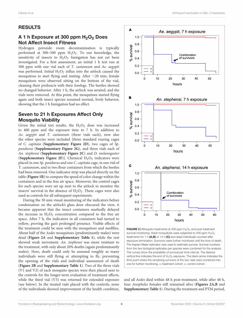
FIGURE 2 | Mosquito treatments at 400 ppm H2O2 and post-treatmentsurvival monitoring. Adult mosquitoes were subjected to 400 ppm H2O2
treatments for 7 h (A,B) or 14 h (C) and dead individuals counted afterexposure termination. Survivors were further monitored until the time of death.The Kaplan-Meier estimator was used to estimate survival. Survival numbersfrom the two biological replicates per species were combined for the analysis.The curves show the probability of survival per time interval. The dashedvertical line indicates the end of H2O2 exposure. The black arrow indicates thetime point where the remaining survivors of the two vials were combined intoone for further monitoring, v, treatment cohort; c, control cohort.
and all Aedes died within 48 h post-treatment, while after 48 h,four Anopheles females still remained alive (Figures 2A,B andSupplementary Table 1). During the treatment and PTM period,
Frontiers in Bioengineering and Biotechnology | www.frontiersin.org 6 November 2020 | Volume 8 | Article 602937

fbioe-08-602937 November 10, 2020 Time: 15:52 # 7
Häcker et al. Arthropod Inactivation in BSL-3 Insectaries
only 4 (Aedes) and 3 (Anopheles) of the more than 90 controlspecimens each died (p < 2−16, Chi-square test).
In medfly containers, all flies were alive and lively after 7 h buthad fallen onto their backs during the exposure and their wingshad stuck to the plastic bottom of the rearing container due tothe humidity. They were able to move their head and legs, andwhen freed, they walked but could not fly. The other three species(D. melanogaster, Sp. pandurus, and T. castaneum) showed novisible effects of the 7 h H2O2 exposure.
One of each mosquito vial, and all D. melanogaster, Sp.pandurus, and T. castaneum containers were returned to theairlock for 14 h. Additionally, fresh containers of each specieswere added to simultaneously assess the effects of 14 and21 h H2O2 exposures (Supplementary Figure 1). At the endof these extended treatments, all Aedes mosquitoes were dead(Supplementary Table 1), whereas 25–30% of the Anopheles(predominantly females) survived exposure for 14 h but wereunable to fly and died within the next 48 h (Figure 2Cand Supplementary Table 1). None of the Anopheles survivedexposure to H2O2 for 21 h. All mosquito specimens from thesetests had a yellowish, bleached appearance.
Fourteen hour exposure of medfly resulted in the sameeffect as observed for 7 h. As the sticking of the flies to theground prevented the unambiguous association of death with theH2O2 treatment, the experimental setup for further experimentswas changed to mosquito cages lined with paper on the floor(Supplementary Figure 2E). D. melanogaster, Sp. pandurus andT. castaneum were still unaffected by the extended treatments.
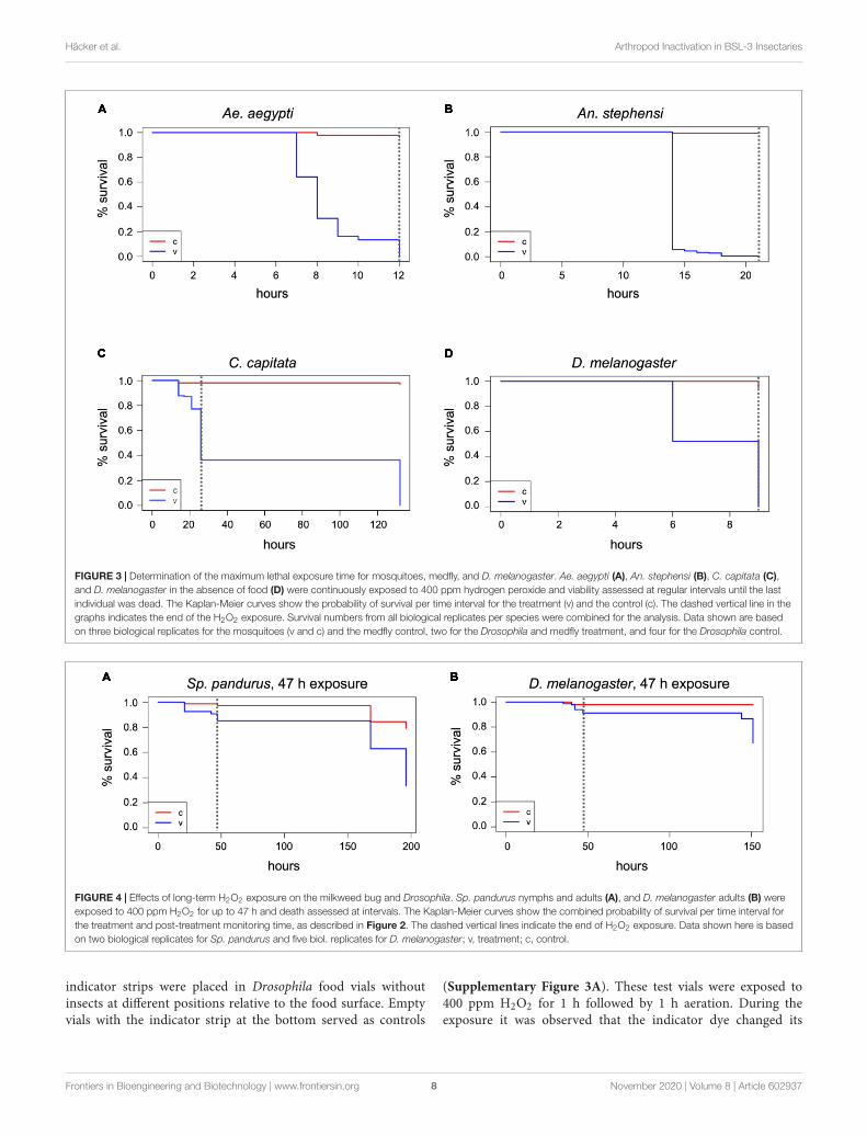
Lethal Exposure Times for Mosquitoes,Medfly, and Drosophila at 400 ppm H2O2Based on the above results, the lethal H2O2 treatment timenecessary to kill all Aedes mosquitoes (LT100) is between 7 and14 h, whereas the LT100 for Anopheles mosquitoes is between 14 hand 21 h. To determine the exact LT100 for both species, one cage(65–90 individuals) and three vials per species (25–40 individualseach) were exposed to 400 ppm H2O2 and checked hourly. TheLT100 for Ae. aegypti, both in cages and vials, was between 10and 12 h (Figure 3A and Supplementary Table 1). The LT100for An. stephensi in the better aerated cage was 15 h, whereas thelast insects died in the vials between 16 and 21 h (Figure 3B andSupplementary Table 1). For both species, the number of deathsduring the treatment was significantly different from the controls(p < 2−16; Chi-square test, based on combined survival numbersof vials and cage).
Using the adjusted cage setup, fresh cohorts of medflies wereexposed to 400 ppm H2O2 for 26 h. After 14 h, several individualswere supine, others were crawling, but none were able to fly,and 17 of 140 flies were dead (Figure 3C and SupplementaryTable 1). After 26 h, 55–60% of the flies had died, compared tonone of the control flies (p < 2.2−16). The survivors of the 26-h treatment were affected, some showing minimal movement,others crawling but unable to fly and often falling into a supineposition. All flies were dead 4.5 days after the end of the treatmentdespite supply of food and water, while in the controls only threeflies died during the whole period (Chi-square test, p < 2−16).
The treated flies showed evidence of H2O2 damage, such as afrosted carapace and dulled eyes (not shown). The medfly LT100could not be determined as the airlock was not available forlonger treatments.
As it could not be excluded that the fly food in the Drosophilarearing containers (Supplementary Figure 2F) changed the effectof the H2O2 on the flies, exposure was repeated with two freshcohorts of flies in empty vials covered with mesh (SupplementaryFigure 2A). In the absence of food all flies were dead within9 h (Figure 3D and Supplementary Table 1). None of the 96control flies outside the airlock (C1–C3, Supplementary Table 1)died during this time. To exclude death in the treatment cohortsdue to the overall more stressful environmental conditions withinthe airlock (temperature, humidity, and lack of food), additionalcontrol incubations were performed in the laboratory underconditions closer to the H2O2 treatment [27◦C, 70% RH (C6, C7),and 28◦C, 37–41% RH (C4, C5), all without food] with similarlyaged flies. Here, only 5% were dead after 9 h (SupplementaryTable 1), showing that the 100% mortality of flies exposed to400 ppm H2O2 for 9 h was statistically significant (Chi-squaretest, p < 2−16) and related to the fumigant rather than theelevated temperature or RH.
Milkweed Bugs and Flour Beetles ShowModerate or No Effects of ExtendedH2O2 ExposuresSpilostethus pandurus and T. castaneum containers weresubjected to an extended 400 ppm H2O2 treatment for a totalof 47 h. In the Sp. pandurus containers, two small larvae haddied after 21 h exposure, both showing a pink to light red colorthat is apparent immediately after ecdysis, when the chitin shellhas not yet hardened and darkened. One larger larva had diedduring ecdysis while still attached to the old chitin shell. All otherindividuals showed no visible effects. At the termination of theexposure eight of the original 54 Sp. pandurus larvae were dead(compared to two of 77 control specimens, p = 0.01275, Chi-square test), but the survivors appeared less lively than the controlinsects. This indicated physical but non-lethal damage inducedby the treatment. Further monitoring for 7 days confirmedthis observation, resulting 36 deaths in the treatment groups,compared to 16 of the 77 control insects (p = 2−7, Figure 4A andSupplementary Table 1).
Remarkably, not a single beetle was dead in the Triboliumvials (experimental or control) after exposure to 400 ppm H2O2for 47 h and 7 days post-treatment. All individuals were livelyand highly viable, showing not the slightest visible effect of thetreatment (Supplementary Table 1).
H2O2 Absorption by Drosophila Fly FoodNeutralizes the Effect on Adult Flies butKills Embryos Deposited in the FoodDuring the H2O2 exposures in the presence of fly food, itwas observed that adult D. melanogaster flies remained mostlyon the food surface and the filter paper placed into the food.To test if the moisture in the food and filter paper may haveprotected the flies by absorbing the reactive molecules, H2O2
Frontiers in Bioengineering and Biotechnology | www.frontiersin.org 7 November 2020 | Volume 8 | Article 602937
fbioe-08-602937 November 10, 2020 Time: 15:52 # 8
Häcker et al. Arthropod Inactivation in BSL-3 Insectaries
FIGURE 3 | Determination of the maximum lethal exposure time for mosquitoes, medfly, and D. melanogaster. Ae. aegypti (A), An. stephensi (B), C. capitata (C),and D. melanogaster in the absence of food (D) were continuously exposed to 400 ppm hydrogen peroxide and viability assessed at regular intervals until the lastindividual was dead. The Kaplan-Meier curves show the probability of survival per time interval for the treatment (v) and the control (c). The dashed vertical line in thegraphs indicates the end of the H2O2 exposure. Survival numbers from all biological replicates per species were combined for the analysis. Data shown are basedon three biological replicates for the mosquitoes (v and c) and the medfly control, two for the Drosophila and medfly treatment, and four for the Drosophila control.
FIGURE 4 | Effects of long-term H2O2 exposure on the milkweed bug and Drosophila. Sp. pandurus nymphs and adults (A), and D. melanogaster adults (B) wereexposed to 400 ppm H2O2 for up to 47 h and death assessed at intervals. The Kaplan-Meier curves show the combined probability of survival per time interval forthe treatment and post-treatment monitoring time, as described in Figure 2. The dashed vertical lines indicate the end of H2O2 exposure. Data shown here is basedon two biological replicates for Sp. pandurus and five biol. replicates for D. melanogaster; v, treatment; c, control.
indicator strips were placed in Drosophila food vials withoutinsects at different positions relative to the food surface. Emptyvials with the indicator strip at the bottom served as controls
(Supplementary Figure 3A). These test vials were exposed to400 ppm H2O2 for 1 h followed by 1 h aeration. During theexposure it was observed that the indicator dye changed its
Frontiers in Bioengineering and Biotechnology | www.frontiersin.org 8 November 2020 | Volume 8 | Article 602937

fbioe-08-602937 November 10, 2020 Time: 15:52 # 9
Häcker et al. Arthropod Inactivation in BSL-3 Insectaries
FIGURE 5 | Lethality induced by short-term exposure to 1000 ppm H2O2. Insects were continuously exposed to 1000 ppm hydrogen peroxide for up to 5 h anddeath assessed in intervals. Survivors were monitored for another 5–8 days after treatment end and death assessed in intervals. Data analysis shown here wasperformed as described in Figure 2 and is based on one large experimental cohort for An. stephensi (A), C. capitata (B), and Sp. pandurus (C), and two biologicalreplicates for D. melanogaster (D). The dashed vertical lines indicate the end of the H2O2 exposure; v, treatment; c, control.
color faster in the empty vials (Supplementary Figures 3B,C),displaying yellow color at the end of the treatment. The degreeof color change in the vials with food depended on the positionof the indicator spot relative to the food surface (SupplementaryFigures 3C–E). Indicator spots positioned partially into a crackin the food changed color only from dark to light purple, whilespots placed directly above the food surface changed towardsorange (Supplementary Figures 3D,E). This indicates that the flyfood indeed neutralized the effect of the reactive H2O2 moleculesand thus protected the flies. Correspondingly, even extendedexposures of 40 h (V4 and V5) or 47 h (V1-V3) in the presence offly food resulted only in low death rates of 5–10% (compared to3% in the controls; p = 2−8; Chi-square test, combined numbersof vials 1–5; Figure 4B and Supplementary Table 1).
H2O2 is a weak acid (pKa = 11.75), a powerful oxidizingagent, and can form free hydroxide radicals. If H2O2 reactivemolecules are absorbed by the food, it might affect the viabilityof the D. melanogaster eggs laid into the food. To verify this, flieswere removed from the treated and control vials 5 days after thelast H2O2 treatment (10 days after the initial transfer of adultsto the vials), and larval development was assessed. Normal larvalfeeding activity was observed in the control vials, but no feedingtraces were visible in any exposed vials. A few early instar larvaewere visible on the food surface in V7 (high ppm treatment),
but they all appeared dead. The food color was lighter in theexposed vials, suggesting H2O2 bleaching. Thirteen days after thelast H2O2 treatment, 1–4 freshly emerged adults and many pupaewere counted in the control vials. The food in V7 showed veryfew and faint traces of larval feeding activity, but no larvae werefound. Four weeks after the last H2O2 treatment, a single pupawas observed in V7, but still no traces of larvae or pupae in anyof the other treated vials. These results indicated that the H2O2reactive molecules indeed killed most embryos embedded in thefood or harmed their larval development.
Increased H2O2 Concentration KillsMosquitoes QuicklyGiven that several species appeared unaffected or only mildlyaffected by prolonged exposure to 400 ppm H2O2, the effect ofa short-term (5 h) exposure to 1000 ppm on all test species wasinvestigated. Again, the mosquitoes were most sensitive to thetreatment. All the Ae. aegypti specimens were killed and bleachedwithin 3 h, the An. stephensi after 5 h, with 90% dead after 3 h(Figure 5A and Supplementary Table 1). In contrast, only ∼10%of the C. capitata specimens were dead after 5 h, although thesurvivors showed the same strongly impaired fitness as observedfor the extended 400 ppm exposure. Six days post-treatment,
Frontiers in Bioengineering and Biotechnology | www.frontiersin.org 9 November 2020 | Volume 8 | Article 602937
fbioe-08-602937 November 10, 2020 Time: 15:52 # 10
Häcker et al. Arthropod Inactivation in BSL-3 Insectaries
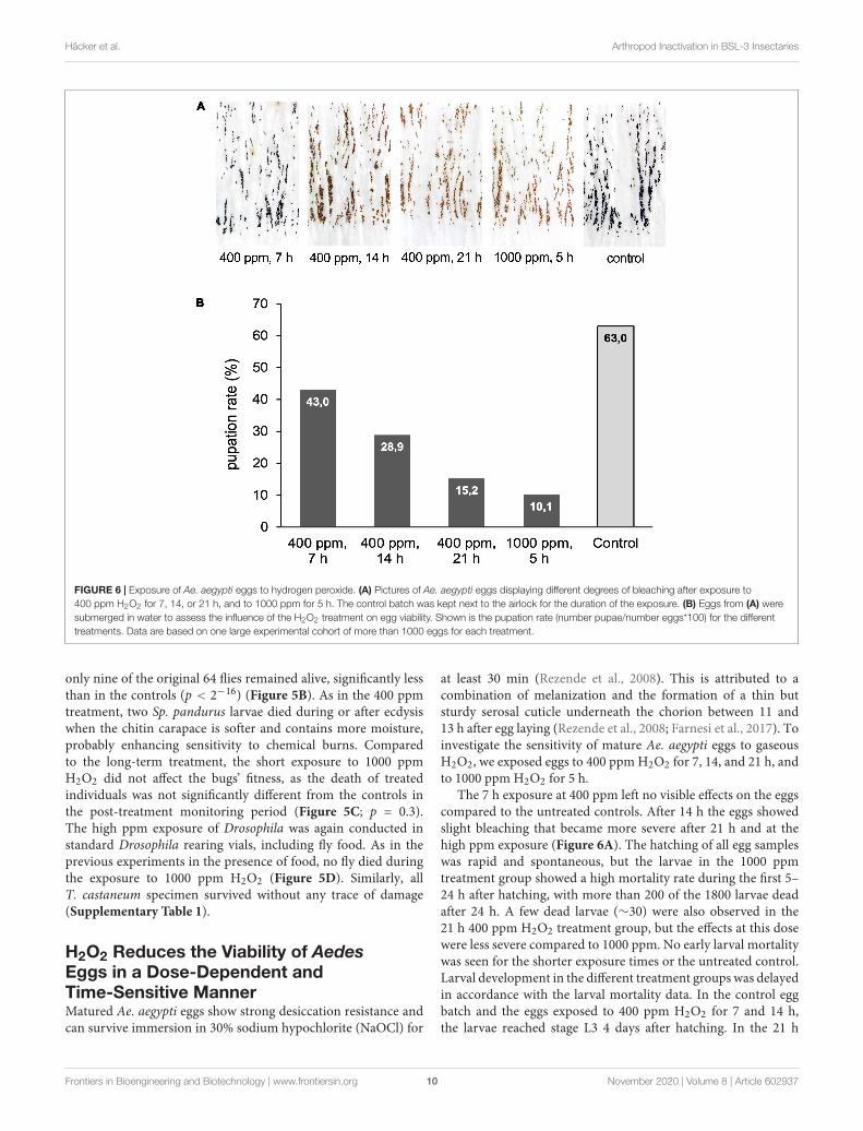
FIGURE 6 | Exposure of Ae. aegypti eggs to hydrogen peroxide. (A) Pictures of Ae. aegypti eggs displaying different degrees of bleaching after exposure to400 ppm H2O2 for 7, 14, or 21 h, and to 1000 ppm for 5 h. The control batch was kept next to the airlock for the duration of the exposure. (B) Eggs from (A) weresubmerged in water to assess the influence of the H2O2 treatment on egg viability. Shown is the pupation rate (number pupae/number eggs*100) for the differenttreatments. Data are based on one large experimental cohort of more than 1000 eggs for each treatment.
only nine of the original 64 flies remained alive, significantly lessthan in the controls (p < 2−16) (Figure 5B). As in the 400 ppmtreatment, two Sp. pandurus larvae died during or after ecdysiswhen the chitin carapace is softer and contains more moisture,probably enhancing sensitivity to chemical burns. Comparedto the long-term treatment, the short exposure to 1000 ppmH2O2 did not affect the bugs’ fitness, as the death of treatedindividuals was not significantly different from the controls inthe post-treatment monitoring period (Figure 5C; p = 0.3).The high ppm exposure of Drosophila was again conducted instandard Drosophila rearing vials, including fly food. As in theprevious experiments in the presence of food, no fly died duringthe exposure to 1000 ppm H2O2 (Figure 5D). Similarly, allT. castaneum specimen survived without any trace of damage(Supplementary Table 1).
H2O2 Reduces the Viability of AedesEggs in a Dose-Dependent andTime-Sensitive MannerMatured Ae. aegypti eggs show strong desiccation resistance andcan survive immersion in 30% sodium hypochlorite (NaOCl) for
at least 30 min (Rezende et al., 2008). This is attributed to acombination of melanization and the formation of a thin butsturdy serosal cuticle underneath the chorion between 11 and13 h after egg laying (Rezende et al., 2008; Farnesi et al., 2017). Toinvestigate the sensitivity of mature Ae. aegypti eggs to gaseousH2O2, we exposed eggs to 400 ppm H2O2 for 7, 14, and 21 h, andto 1000 ppm H2O2 for 5 h.
The 7 h exposure at 400 ppm left no visible effects on the eggscompared to the untreated controls. After 14 h the eggs showedslight bleaching that became more severe after 21 h and at thehigh ppm exposure (Figure 6A). The hatching of all egg sampleswas rapid and spontaneous, but the larvae in the 1000 ppmtreatment group showed a high mortality rate during the first 5–24 h after hatching, with more than 200 of the 1800 larvae deadafter 24 h. A few dead larvae (∼30) were also observed in the21 h 400 ppm H2O2 treatment group, but the effects at this dosewere less severe compared to 1000 ppm. No early larval mortalitywas seen for the shorter exposure times or the untreated control.Larval development in the different treatment groups was delayedin accordance with the larval mortality data. In the control eggbatch and the eggs exposed to 400 ppm H2O2 for 7 and 14 h,the larvae reached stage L3 4 days after hatching. In the 21 h
Frontiers in Bioengineering and Biotechnology | www.frontiersin.org 10 November 2020 | Volume 8 | Article 602937
fbioe-08-602937 November 10, 2020 Time: 15:52 # 11
Häcker et al. Arthropod Inactivation in BSL-3 Insectaries
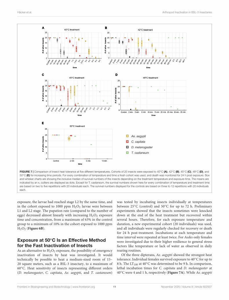
FIGURE 7 | Comparison of insect heat tolerance at five different temperatures. Cohorts of 20 insects were exposed to 40◦C (A), 42◦C (B), 45◦C (C), 48◦C (D), and50◦C (E) for increasing time periods. For every combination of temperature and time a fresh cohort was used, and death was monitored for 24 h post exposure. Boxand whisker charts are showing the inclusive median of survival numbers of the insects depending on the treatment temperature and exposure time. The means areindicated by an x, outliers are displayed as dots. Except for T. castaneum, the survival numbers shown here for every combination of temperature and treatment timeare based on two to five repetitions with 20 individuals each. The survival numbers displayed for the controls are based on three to 12 repetitions with 20 individualseach.
exposure, the larvae had reached stage L2 by the same time, andin the cohort exposed to 1000 ppm H2O2 larvae were betweenL1 and L2 stage. The pupation rate (compared to the number ofeggs) decreased almost linearly with increasing H2O2 exposuretime and concentration, from a maximum of 63% in the controlgroup to a minimum of 10% in the cohort exposed to 1000 ppmH2O2 (Figure 6B).
Exposure at 50◦C Is an Effective Methodfor the Fast Inactivation of InsectsAs an alternative to H2O2 exposure, the possibility of emergencyinactivation of insects by heat was investigated. It wouldtechnically be possible to heat a medium-sized room of 15–20 square meters, such as a BSL-3 insectary, to a maximum of60◦C. Heat sensitivity of insects representing different orders(D. melanogaster, C. capitata, Ae. aegypti, and T. castaneum)
was tested by incubating insects individually at temperaturesbetween 25◦C (control) and 50◦C for up to 72 h. Preliminaryexperiments showed that the insects sometimes were knockeddown at the end of the heat treatment but recovered withinseveral hours. Therefore, for each exposure temperature andduration, a new experimental cohort (20 individuals) was used,and all individuals were regularly checked for recovery or deathfor 24 h post-treatment. Incubations at each temperature andtime interval were repeated at least twice. For Aedes only femaleswere investigated due to their higher resilience to general stressfactors like temperature or lack of water as observed in dailyrearing routines.
Of the three dipterans, Ae. aegypti showed the strongest heattolerance. Individual females survived exposure to 40◦C for up to8 h. The LT100 at 40◦C was determined to be 9 h. In comparison,lethal incubation times for C. capitata and D. melanogaster at40◦C were 4 and 1 h, respectively (Figure 7A). While Ae. aegypti
Frontiers in Bioengineering and Biotechnology | www.frontiersin.org 11 November 2020 | Volume 8 | Article 602937

fbioe-08-602937 November 10, 2020 Time: 15:52 # 12
Häcker et al. Arthropod Inactivation in BSL-3 Insectaries
survival times strongly decreased at higher temperatures, to amaximum of 90 min at 42◦C, 15 min at 45◦C, and 5 min at48◦C, they were in all cases markedly higher than for medfly andDrosophila (Figures 7B–D). At the highest tested temperature,50◦C, Aedes and medfly could withstand a 2-min incubationwithout problems but were dead after 5 min. All Drosophilaspecimen died during the 2-min exposure. As observed for theH2O2 treatments, T. castaneum was again the most resilientspecies, with more than half of the cohort withstanding 50◦Cfor 5 min and displaying longer survival times at all othertemperatures (Figures 7B–E). According to these results, 50◦Cwas defined as lethal temperature for all tested species that killswithin a few minutes. The exact survival numbers for all speciesare provided in Supplementary Table 3.
We moreover selected Ae. aegypti to test the influence ofthe insects’ age on temperature resilience, using mainly fivedifferent age groups of females; 1–7, 5–10, 10–14, 15–17,and 18–29 days post emerging. Based on the survival datafrom the above experiments, 40 and 42◦C were selected asless stringent temperatures for the assay and 48◦C as near-lethal temperature. The results obtained from the differentage groups indicate that around day ten post emerging is acritical age for the heat tolerance of Ae. aegypti females atmoderately elevated temperatures (40 and 42◦C). At short tointermediate incubation times, females younger than 10 daysshowed markedly higher heat resilience than females olderthan 10 days (Figures 8A,B). When approaching the near-lethal incubation time for both temperatures, the survivalnumbers of “young” and “old” females converged, resultingin similar LT100 timepoints. Interestingly, the survival datarecorded for the “young” age groups (1–7 and 5–10) at 40and 42◦C displayed minimal variance between the biologicalreplicates, while the survival times of the older age groups(10–14, 15–17, and 18–29 days) varied strongly betweenreplicates (Figures 8A,B). This variation, however, could notunambiguously be linked to the age difference between the “old”age groups, as in several replicates the oldest females were moreresilient than younger ones (Supplementary Table 2). No age-related difference in heat sensitivity could be observed at 48◦C(Figure 8C).
DISCUSSION
We tested if either H2O2 fumigation or heat treatment hasthe potential to be developed into a standard procedure forthe safe and ethical room-wide decontamination of BSL-3insectaries in emergency situations, such as mass escapes ofpathogen-infected insects. To account for the diversity of vectorinsects potentially held in BSL-3 insectaries, the effectivenessof both methods was tested against insect species representingdifferent orders.
This study shows that heat is a very effective way to kill abroad range of species, with a temperature of 50◦C sufficient tokill all specimens within 10 min under the chosen experimentalconditions. Also, 48◦C effectively killed in less than 1 h. Thesevalues are in accordance with studies in the early 1900s, showing
FIGURE 8 | Comparison of the heat tolerance of Ae. aegypti femalemosquitoes at different age. Influence of age on heat sensitivity wasinvestigated at two moderately increased temperatures, 40 and 42◦C (A,B),and at a near-lethal temperature, 48◦C (C). “Young” females were between 1and 10 days old, “old” females between 10 and 29 days. Lethality for eachexposure temperature and time was assessed in two to four biologicalreplicates (i.e., each replicate was performed with an experimental cohortfrom a different cage). Each cohort consisted of 20 individuals. The box andwhisker charts display the inclusive median of the replicate values for eachexposure time. The means are indicated by an x, outliers are shown as dots.The survival numbers displayed for the controls are based on two to eightrepetitions with 20 individuals each.
that 48–50◦C are effective to kill different stored product insectpests within a few minutes (Dean, 1913). Translating theseresults to a room-wide procedure will require adaptation of
Frontiers in Bioengineering and Biotechnology | www.frontiersin.org 12 November 2020 | Volume 8 | Article 602937

fbioe-08-602937 November 10, 2020 Time: 15:52 # 13
Häcker et al. Arthropod Inactivation in BSL-3 Insectaries
treatment times, as the walls, furnishing, and equipment willdelay heating to the target temperature. Required time andenergy can be estimated using the specific heat capacity andapproximate volume of all the materials (assuming perfect roominsulation without heat loss to the surrounding areas). Likefor room decontaminations by H2O2 fumigation, however, safetreatment times will have to be validated for each specificfacility and species. Temperatures lower than 48◦C are notrecommended, as even under laboratory conditions, extendedtreatment times of several hours were required to reach LT100.Moreover, treatments at sub-lethal temperatures can increasethe risk of heat acclimation, which could decrease treatmentefficacy. Such acclimation has been shown for T. castaneumalready after 5 h at 42◦C (Lu and Liu, 2017). Given a slowoverall room heating rate and material-dependent uneven heat-distribution, such acclimation conditions could be created duringthe heating of a BSL-3 insectary, if for technical or financialreasons the planners would favor long incubation times atlower temperatures over short treatments at high temperatures,and if no fans are used to achieve uniform heating ratesacross the room.
The pronounced heat tolerance observed for T. castaneumin this study has been known for years and was linked toadaptation mechanisms, including the upregulation of heat-shock genes like hsp70, which is strongly upregulated during heatacclimation at 42◦C (Lu and Liu, 2017). Lethal incubation timesat 50◦C observed in this study are similar to published values(Fields, 1992; Mahroof et al., 2003; Yu et al., 2017). Variationsbetween the different studies might be due to the experimentalsetup [size and material of the container and the presence ofother heat-absorbing materials delaying the heating to the targettemperature (e.g., flour)], and the age of the adult beetles.
Our study also revealed a strong difference in tolerance tomoderately elevated temperatures (40–42◦C) between the threedipterans, which could be related to the species’ adaptation totheir natural habitats in tropical and subtropical areas (Aedesand medfly), or in temperate climates (D. melanogaster). Inaddition, tolerance against dehydration might play a role, whichin part is influenced by the insects’ body size, making thesmall Drosophila the most susceptible species. While we didnot control for humidity in our experimental setup, studieswith different insects suggest that controlling for low humidityduring the room-wide heat treatment can improve the efficacy(Mellanby, 1932; Denlinger and Yocum, 1999; Ginsberg et al.,2017). The reasons for the strong variation in temperaturetolerance of Aedes females older than 10 days are unclear. Ithas been shown, however, that nutrient accumulation, density,and temperature during larval development can influence adultquality and life history traits (Couret et al., 2014; Hapairaiet al., 2014; Yahouedo et al., 2014; Oliver and Brooke, 2017;Puggioli et al., 2017), and the tolerance of male Ae. aegypti toheat stress (Sasmita et al., 2019). Similar to T. castaneum, itwas moreover observed for Aedes that pre-exposure of larvaeto sub-lethal temperatures can confer adaptive thermotolerance(Patil et al., 1996). Aedes mosquitoes used in this study weregrown at a constant temperature of 27 ± 0.3◦C, and larvaeand adults were fed with standardized food, excluding an
effect of these factors on variations in temperature tolerance.However, our standard rearing protocols do not control forexact larval densities by counting, but instead, make a visualassessment of the density in the rearing trays and feedthe larvae ad libidum. Therefore, the amount of food perlarva can vary between larval rearing cohorts, which couldlead to variations in nutrient accumulation during larvaldevelopment. This might have influenced the heat toleranceof the “old” females in replicates performed with differentrearing cohorts.
In contrast to the heat treatments, it was impossible todetermine suitable H2O2 fumigation conditions applicable toall tested species in a room-wide scenario. Our results suggestthat the thickness of the chitinous exoskeleton might play asubstantial role in the effects of H2O2 fumigation, as specieswith a thinner exoskeleton (particularly Drosophila, Aedes,and Anopheles) were much more sensitive than species witha thick exoskeleton like the milkweed bug and flour beetle,with the latter appearing to be insensitive to the appliedH2O2 concentrations and treatment times. Higher H2O2 dosesare not applicable for several reasons: first because highconcentrations (≥1000 ppm) would lead to condensation ofthe gas on surfaces, which could damage furnishing andequipment, especially metals could be oxidized, and second,because large quantities of H2O2 would be required for a room-wide application over many hours. The H2O2 exposure resultsobtained for the small dipterans, predominantly the mosquitoes,can probably be transferred to other vector insects of similarphysical constitution like phlebotomine sandflies. Similarly, theresults obtained for the bugs and beetles in our study mightbe extendable to ticks, which also have a thick exoskeletonand can therefore be expected to be essentially insensitive toH2O2 fumigation.
In summary, this study shows that H2O2 fumigationwould only be applicable to a subset of the vector insectstypically held in BSL-2 or BSL-3 insectaries. Its applicabilityas room-wide emergency decontamination would have to beevaluated for each new insect species added to the laboratory,which, in our opinion, prevents H2O2 fumigation from beingestablished as a standard procedure for insect inactivation inBSL-3 insectaries. In contrast, the combined results of theheat treatments suggest 50◦C to be a reliable and universallyapplicable temperature to quickly and irreversibly inactivateinsects of all tested species. The results are probably alsoapplicable to related species and those with similar physicalproperties, unless they have a specific heat tolerance due toadaptation to high temperature conditions in their naturalhabitats. Therefore, heat treatment has the potential to bedeveloped as a standard and commonly recognized room-wideinsect emergency inactivation method.
DATA AVAILABILITY STATEMENT
The original contributions presented in the study are includedin the article/Supplementary Materials, further inquiries can bedirected to the corresponding author/s.
Frontiers in Bioengineering and Biotechnology | www.frontiersin.org 13 November 2020 | Volume 8 | Article 602937
fbioe-08-602937 November 10, 2020 Time: 15:52 # 14
Häcker et al. Arthropod Inactivation in BSL-3 Insectaries
AUTHOR CONTRIBUTIONS
IH designed the experiments. IH, RK, JM, EL, JR, and TRperformed the experiments. IH and GE analyzed the data. IH,GE, and MS wrote the manuscript. All authors contributed to thearticle and approved the submitted version.
FUNDING
This work was supported by projects of the LOEWE CentreDRUID and the LOEWE Centre for Insect Biotechnology andBioresources of the Hessian Ministry of Science and Arts (MS),and the Emmy Noether Program of the German ResearchFoundation SCHE 1833/1-1 (MS).
ACKNOWLEDGMENTS
We thank Dr. Georg Petschenka for providing Spilostethuspandurus, Dr. Eileen Knorr for providing Triboliumcastaneum, Dr. Andrew M. Blackborough for providingAnopheles stephensi, and Joseph Ortner for critical readingof the manuscript.
SUPPLEMENTARY MATERIAL
The Supplementary Material for this article can be foundonline at: https://www.frontiersin.org/articles/10.3389/fbioe.2020.602937/full#supplementary-material
REFERENCESAmerican Committee Of Medical Entomology (2019). Arthropod containment
guidelines, version 3.2. Vec. Born. Zoonot. Dis. 19, 152–173. doi: 10.1089/vbz.2018.2431
Bentley, K., Dove, B. K., Parks, S. R., Walker, J. T., and Bennett, A. M.(2012). Hydrogen peroxide vapour decontamination of surfaces artificiallycontaminated with norovirus surrogate feline calicivirus. J. Hosp. Infect. 80,116–121. doi: 10.1016/j.jhin.2011.10.010
Calhoun, P. (2019). Unconditional Exact Test. 2.0. Available online at: https://CRAN.R-project.org/package=Exact (accessed March 24, 2020).
Cheney, J. E., and Collins, C. H. (1995). Formaldehyde disinfection in laboratories:limitations and hazards. Br. J. Biomed. Sci. 52, 195–201.
Council of the European Union (1990). Council Directive 90/679/EEC of 26November 1990 on the protection of workers from risks related to exposure tobiological agents at work (seventh individual Directive within the meaning ofArticle 16 (1) of Directive 89/391/EEC). Luxemburg: Publications office of theEuropean Union.
Couret, J., Dotson, E., and Benedict, M. Q. (2014). Temperature, larval diet, anddensity effects on development rate and survival of Aedes aegypti (Diptera:Culicidae). PLoS One 9:e87468. doi: 10.1371/journal.pone.0087468
Dean, G. (1913). Milled and stored grain insects. Kansas State Agric. Coll. TechnBull. 189, 139–176.
Dean, G. A. (1993). Further data on heat as a means of controlling mill insects.J. Econ. Entomol. 6, 40–55. doi: 10.1093/jee/6.1.40
Denlinger, D. L., and Yocum, G. D. (1999). “Physiology of heat sensitivity,”in Temperature Sensitivity in Insects and Application in Integrated PestManagement, eds G. Hallman and D. Denlinger (Boulder, CO: Westview Press),7–53. doi: 10.1201/9780429308581-2
Dowdy, A. K. (1999). Mortality of red flour beetle, Tribolium castaneum(Coleoptera: Tenebrionidae) exposed to high temperature and diatomaceousearth combinations. J. Stor. Prod. Res. 35, 175–182. doi: 10.1016/s0022-474x(98)00043-5
Dreyfus, W. M. (1914). Review of formaldehyde fumigation. Am. J. Public Health4, 1046–1049. doi: 10.2105/ajph.4.11.1046
Farnesi, L. C., Vargas, H. C. M., Valle, D., and Rezende, G. L. (2017). Darker eggsof mosquitoes resist more to dry conditions: melanin enhances serosal cuticlecontribution in egg resistance to desiccation in Aedes, Anopheles and Culexvectors. PLoS Negl. Trop. Dis. 11:e0006063. doi: 10.1371/journal.pone.0006063
Fields, P. G. (1992). The control of stored-product insects and mites with extremetemperatures. J. Stor. Products Res. 28, 89–118. doi: 10.1016/0022-474x(92)90018-l
Ginsberg, H. S., Albert, M., Acevedo, L., Dyer, M. C., Arsnoe, I. M., Tsao, J. I.,et al. (2017). Environmental factors affecting survival of immature Ixodesscapularis and implications for geographical distribution of lyme disease: theclimate/behavior hypothesis. PLoS One 12:e0168723. doi: 10.1371/journal.pone.0168723
Gordon, D., Carruthers, B.-A., and Theriault, S. (2012). Gaseous decontaminationmethods in high-containment laboratories. Appl. Biosaf. 17, 31–39. doi: 10.1177/153567601201700107
Goyal, S. M., Chander, Y., Yezli, S., and Otter, J. A. (2014). Evaluating the virucidalefficacy of hydrogen peroxide vapour. J. Hosp. Infect. 86, 255–259. doi: 10.1016/j.jhin.2014.02.003
Häcker, I., Harrell, R. A. I., Eichner, G., Pilitt, K. L., O’Brochta, D. A., Handler,A. M., et al. (2017). Cre/lox-recombinase-mediated cassette exchange forreversible site-specific genomic targeting of the disease vector, Aedes aegypti.Sci. Rep. 7:43883.
Hansen, J. D., Johnson, J. A., and Winter, D. A. (2011). History and use of heatin pest control: a review. Intern. J. Pest Manag. 57, 267–289. doi: 10.1080/09670874.2011.590241
Hapairai, L. K., Marie, J., Sinkins, S. P., and Bossin, H. C. (2014). Effect oftemperature and larval density on Aedes polynesiensis (Diptera: Culicidae)laboratory rearing productivity and male characteristics. Acta Trop.132(Suppl.), S108–S115.
Heckert, R. A., Best, M., Jordan, L. T., Dulac, G. C., Eddington, D. L., and Sterritt,W. G. (1997). Efficacy of vaporized hydrogen peroxide against exotic animalviruses. Appl. Environ. Microbiol. 63, 3916–3918. doi: 10.1128/aem.63.10.3916-3918.1997
Kaspari, O., Lemmer, K., Becker, S., Lochau, P., Howaldt, S., Nattermann, H.,et al. (2014). Decontamination of a BSL3 laboratory by hydrogen peroxidefumigation using three different surrogates for Bacillus anthracis spores. J. Appl.Microbiol. 117, 1095–1103. doi: 10.1111/jam.12601
Krishnan, J., Berry, J., Fey, G., and Wagener, S. (2006). Vaporized hydrogenperoxide-based biodecontamination of a high-containment laboratory undernegative pressure. Appl. Biosaf. 11, 74–80. doi: 10.1177/153567600601100203
Lemmen, S., Scheithauer, S., Hafner, H., Yezli, S., Mohr, M., and Otter, J. A. (2015).Evaluation of hydrogen peroxide vapor for the inactivation of nosocomialpathogens on porous and nonporous surfaces. Am. J. Infect. Control 43, 82–85.doi: 10.1016/j.ajic.2014.10.007
Lindsley, D. L., and Grell, E. H. (1968). Genetic Variations of DrosophilaMelanogaster. Washington, DC: Carnegie Institute of Washington.
Lu, J., and Liu, S. (2017). Influence of acclimation to sublethal temperature onheat tolerance of Tribolium castaneum (Herbst) (Coleoptera: Tenebrionidae)exposed to 50 degrees C. PLoS One 12:e0182269. doi: 10.1371/journal.pone.0182269
Mahroof, R., Subramanyam, B., Throne, J. E., and Menon, A. (2003). Time-mortality relationships for Tribolium castaneum (Coleoptera: Tenebrionidae)life stages exposed to elevated temperatures. J. Econ. Entomol. 96, 1345–1351.doi: 10.1603/0022-0493-96.4.1345
Mellanby, K. (1932). The influence of atmospheric humidity on the thermal deathpoint of a number of insects. J. Exper. Biol. 9, 222–232.
Oliver, S. V., and Brooke, B. D. (2017). The effect of elevated temperatures on thelife history and insecticide resistance phenotype of the major malaria vectorAnopheles arabiensis (Diptera: Culicidae). Malaria J. 16:73.
Frontiers in Bioengineering and Biotechnology | www.frontiersin.org 14 November 2020 | Volume 8 | Article 602937
fbioe-08-602937 November 10, 2020 Time: 15:52 # 15
Häcker et al. Arthropod Inactivation in BSL-3 Insectaries
Patil, N. S., Lole, K. S., and Deobagkar, D. N. (1996). Adaptive larvalthermotolerance and induced cross-tolerance to propoxur insecticide inmosquitoes Anopheles stephensi and Aedes aegypti. Med. Vet. Entomol. 10,277–282. doi: 10.1111/j.1365-2915.1996.tb00743.x
Petit, B. M., Almeida, F. C., Uchiyama, T. R., Lopes, F. O. C., Tino, K. H.,and Chewins, J. (2017). Evaluating the efficacy of hydrogen peroxide vapouragainst foot-and-mouth disease virus within a BSL4 biosafety facility. Lett. Appl.Microbiol. 65, 281–284. doi: 10.1111/lam.12778
Porto, S. M. C., Valenti, F., Cascone, G., and Arcidiacono, C. (2015). Thermalinsulation of a flour mill to improve effectiveness of the heat treatment forinsect pest control. Agric Eng Int: CIGR Journal, Special issue 2015: 18th WorldCongress of CIGR: 94–104.
Pottage, T., Richardson, C., Parks, S., Walker, J. T., and Bennett, A. M.(2010). Evaluation of hydrogen peroxide gaseous disinfection systems todecontaminate viruses. J. Hosp. Infect. 74, 55–61. doi: 10.1016/j.jhin.2009.08.020
Puggioli, A., Carrieri, M., Dindo, M. L., Medici, A., Lees, R. S., Gilles, J. R., et al.(2017). Development of Aedes albopictus (Diptera: Culicidae) larvae underdifferent laboratory conditions. J. Med. Entomol. 54, 142–149. doi: 10.1093/jme/tjw127
R Core Team (2020). R: A Language and Environment for Statistical Computing.Vienna: R Foundation for Statistical Computing.
Rezende, G. L., Martins, A. J., Gentile, C., Farnesi, L. C., Pelajo-Machado, M.,Peixoto, A. A., et al. (2008). Embryonic desiccation resistance in Aedes aegypti:presumptive role of the chitinized serosal cuticle. BMC Dev. Biol. 8:82. doi:10.1186/1471-213X-8-82
Robert Koch Institut (2017). Liste der vom RKI geprüften und anerkanntenDesinfektionsmittel und -verfahren. Bundesgesundheitsblatt. 60, 1274–1297.
Rogers, J. V., Choi, Y. W., Richter, W. R., Rudnicki, D. C., Joseph, D. W., Sabourin,C. L., et al. (2007). Formaldehyde gas inactivation of Bacillus anthracis, Bacillussubtilis, and Geobacillus stearothermophilus spores on indoor surface materials.J. Appl. Microbiol. 103, 1104–1112.
Sasmita, H. I., Tu, W. C., Bong, L. J., and Neoh, K. B. (2019). Effects oflarval diets and temperature regimes on life history traits, energy reservesand temperature tolerance of male Aedes aegypti (Diptera: Culicidae):optimizing rearing techniques for the sterile insect programmes. Parasit. Vect.12:578.
Scott, T. W. (2005). Containment of arthropod disease vectors. ILAR J. 46, 53–61.doi: 10.1093/ilar.46.1.53
Subramanyam, B., Mahroof, R., and Brijwani, M. (2011). Heat treatment of grain-processing facilities for insect management: a histori- cal overview and recentadvances. Stewart Postharvest Rev. 3, 1–11. doi: 10.2212/spr.2011.3.9
Tabachnick, W. J. (2006). Laboratory containment practices for arthropod vectorsof human and animal pathogens. Lab Anim. 35, 28–33. doi: 10.1038/laban0306-28
Therneau, T. M. (2020). Survival: Survival Analysis. 3.1-12. Available online at:https://CRAN.R-project.org/package=survival (accessed March 24, 2020).
U.S Department of Health and Human Services (2016). Formaldehyde, CAS No.50-00-0. Report on Carcinogens, 4th Edn. Washington, DC: U.S. Department ofHealth and Human Services (HHS).
Valenti, F., Porto, S. M. C., Tomasello, N., and Arcidiacono, C. (2018). Enhancingheat treatment efficacy for insect pest control: a case study of a CFD applicationto improve the design and structure of a flour mill. Buildings 8, 48–66.
Wickham, H., and Bryan, J. (2019). readxl: Read Excel Files. 1.3.1. Available: https://CRAN.R-project.org/package=readxl (accessed March 24, 2020).
Yahouedo, G. A., Djogbenou, L., Saizonou, J., Assogba, B. S., Makoutode, M.,Gilles, J. R., et al. (2014). Effect of three larval diets on larval developmentand male sexual performance of Anopheles gambiae s.s. Acta Trop. 132(Suppl.),S96–S101.
Yu, D., Shrestha, B., and Baik, O.-D. (2017). Thermal death kinetics of adultred flour beetle Tribolium castaneum (Herbst) in canola seeds during radiofrequency heating. Intern. J. Food Propert. 20, 3064–3075. doi: 10.1080/10942912.2016.1272609
Conflict of Interest: RK was employed by the company Ortner ReinraumtechnikGmbH.
The remaining authors declare that the research was conducted in the absence ofany commercial or financial relationships that could be construed as a potentialconflict of interest.
Copyright © 2020 Häcker, Koller, Eichner, Martin, Liapi, Rühl, Rehling and Schetelig.This is an open-access article distributed under the terms of the Creative CommonsAttribution License (CC BY). The use, distribution or reproduction in other forumsis permitted, provided the original author(s) and the copyright owner(s) are creditedand that the original publication in this journal is cited, in accordance with acceptedacademic practice. No use, distribution or reproduction is permitted which does notcomply with these terms.
Frontiers in Bioengineering and Biotechnology | www.frontiersin.org 15 November 2020 | Volume 8 | Article 602937