evaluation of neuroprotective effects of natural extracts...
TRANSCRIPT

Rita João Rosado Serranito Ramos
Degree in Biology
Evaluation of Neuroprotective Effects of
Natural Extracts obtained from Portuguese
Agro-food Residues
Dissertation to obtain a Master Degree in Biotechnology
Supervisor: Ana Matias, Ph.D, IBET/ITQB-UNL
Co- Supervisor: Cláudia Santos, Ph.D, IBET/ITQB-UNL
Júri:
Presidente: Prof. Doutor Pedro Miguel Calado Simões
Arguente: Prof. Doutora Margarida Castro Caldas Braga
September 2012

2

Rita João Rosado Serranito Ramos
Degree in Biology
Evaluation of Neuroprotective Effects of
Natural Extracts obtained from Portuguese Agro-food
Residues
Dissertation to obtain a Master Degree in Biotechnology
Supervisor: Ana Matias, Ph.D, IBET/ITQB-UNL Co- Supervisor: Cláudia Santos, Ph.D, IBET/ITQB-UNL
Júri:
Presidente: Prof. Doutor Pedro Miguel Calado Simões
Arguente: Prof. Doutora Margarida Castro Caldas Braga
September 2012

ii

iii
Evaluation of neuroprotective effects of natural extracts obtained from Portuguese agro-food
residues
Copyright Rita Ramos, FCT/UNL, UNL
A Faculdade de Ciências e Tecnologia e a Universidade Nova de Lisboa têm o direito, perpétuo
e sem limites geográficos, de arquivar e publicar esta dissertação através de exemplares
impressos reproduzidos em papel ou de forma digital, ou por qualquer outro meio conhecido ou
que venha a ser inventado, e de a divulgar através de repositórios científicos e de admitir a sua
cópia e distribuição com objetivos educacionais ou de investigação, não comerciais, desde que
seja dado crédito ao autor e editor.

iv
v
Acknowledgments
Gostaria de agradecer a todas as pessoas que direta ou indiretamente contribuíram para a
realização deste trabalho e sem as quais seria impossível conclui-lo. Em primeiro lugar,
gostaria de agradecer à Dra. Ana Matias que foi incansável ao longo de todo este processo,
para o qual esteve sempre disponível para compartilhar as suas ideias e conhecimentos, tendo
sempre uma palavra de incentivo e de apoio quando as coisas corriam menos bem. E por
todas as palavras de motivação e reconhecimento quando tudo corria realmente bem.
Obrigada por toda a confiança e apoio Ana. Gostaria de agradecer à Dra. Cláudia Santos por
todo o apoio, não menos importante. Por todas as palavras de confiança em situações mais
difíceis e por ter sempre uma palavra de apoio e conforto, por todas as ideias e opiniões.
Obrigada por tudo o que me ensinou Cláudia. Obrigada a ambas, foi um prazer enorme
trabalhar convosco. À Dra. Catarina Duarte por ter acreditado em mim e neste trabalho, por ter
sempre uma palavra de motivação e de apoio. Obrigada pelas portas que me abriu. Ao Dr.
Ricardo Boavida Ferreira pela oportunidade pela confiança. Agradeço à Dra. Maria do Rosário
Bronze, à Joana Antunes e a toda equipa do Laboratório de Química Analítica pelo apoio
essencial na identificação de compostos. A todas as pessoas do laboratório de Nutracêuticos e
Libertação Controlada e do laboratório de Biologia da Doença e do Stress que tornaram o
desenvolvimento deste trabalho em muito mais que um simples trabalho. À Dra. Teresa Serra
por estar sempre disponível para ajudar e por tudo o que me ensinou. À Sara Nunes por o
tempo despendido, por todas as opiniões sempre importantes e por toda a ajuda. À Joana
Poejo por toda a paciência, companhia e ajuda. À Ana Nunes pelo conhecimento que partilhou
comigo e por todas as viagens de comboio! Ao Agostinho por toda a ajuda boa disposição. À
Lucélia Tavares por todo o conhecimento partilhado. À Dra. Marta Alves pelos conselhos e
companhia. À Inês Figueira por tudo o que me ensinou e por toda a cumplicidade, obrigada. À
Carolina Jardim e à Diana Macedo pela companhia. À Andreia Gomes, ao Daniel Lopes, ao
João Jorge e ao Pedro Ferreira por todas as conversas e pela companhia espetacular que
foram em mais uma fase deste processo. Sem vocês não teria sido tão gratificante. Gostaria
também de agradecer a todos os meus amigos pelo suporte que são e que sempre foram, pela
companhia, por todos os momentos de descontração e acima de tudo, por estarem sempre
presentes. Às meninas, Cátia Gomes e Nádia Grilo por todas os momentos, opiniões e apoio.
Pela excelente companhia e amizade. Obrigada por terem feito parte desta etapa da minha
vida. Ao Hélder, que foi incansável. Pelo apoio incondicional e por toda a paciência, carinho e
boa disposição. Obrigada, sem ti não era a mesma coisa! À minha família: Aos meus avós, tios
e primas por serem a melhor família do mundo e por todo o orgulho e carinho. Aos meus pais e
à minha avó pelo enorme esforço que fizeram para eu conseguir chegar até aqui e por sempre
acreditarem em mim. Espero que um dia seja possível retribuir tudo aquilo que já fizeram por
mim. À minha irmã que esteve sempre presente e mais uma vez por ter sido imprescindível
para concluir mais uma etapa da minha vida!
Obrigada a Todos, Rita Ramos

vi

vii
Abstract
Countries are currently faced with problems derived from changes in lifespan and an increase in
lifestyle-related diseases. Neurodegenerative disorders such Parkinson’s (PD) and Alzheimer’s
(AD) diseases are an increasing problem in aged societies. Data from World Alzheimer Report
2011 indicate that 36 million people worldwide are living with dementia. Oxidative stress has
been associated with the development of AD and PD. Therefore there is interest to search for
effective compounds or therapies to combat the oxidative damage in these diseases. Current
evidence strongly supports a contribution of phenolic compounds present in fruits and
vegetables to the prevention of neurodegenerative diseases such AD and PD.
The industrial processing of a wide variety of fruits results in the accumulation of by-products
without commercial value. Opuntia ficus-indica (cactus pear) is consumed fresh and processed
like in juice. Prunnus avium (sweet cherry) is consumed fresh but the organoleptics
characteristics of the fruits leads to the smaller and ragged fruits have no commercial value.
Fruit extracts of both species has described to be rich in phenolic compounds and to have high
antioxidant activities due to its composition. The aim of this work was assessing the efficacy of
O. ficus-indica and P. avium by-products extracts obtained with conventional solvent extraction
and pressurized liquid extraction in a neurodegeneration cell model. All extracts have protected
neuroblastoma cells from H2O2-induced death at low, non-toxic levels, which approach to
physiologically-relevant serum concentration. However, cherry extract has a slighter
neuroprotective activity. The protective effect of Opuntia extracts are not conducted by a direct
antioxidant activity since there are not decreases in intracellular ROS levels in cell treated with
extracts and challenged with H2O2, while cherry extract neuroprotection seems to be due to a
direct scavenging activity. Extracts from different biological matrixes seems to protect neuronal
cells trough different cellular mechanisms.
Keywords: Neuroprotection, Agro-food residues, Opuntia ficus-indica, Prunus avium, Natural
extracts.

viii
ix
Resumo
Em países desenvolvidos, o envelhecimento da população está a aumentar assim como a
incidência de doenças relacionadas com a idade e estilo de vida como as doenças
neurodegenerativas. O stress oxidativo tem sido associado ao desenvolvimento de doenças
neurodegenerativas como a doença de Alzheimer (AD) e a doença de Parkinson (PD). Assim
sendo existe um interesse crescente na busca por novos compostos ou terapias eficazes no
combate aos danos provocados pelo stress oxidativo nestas doenças. Evidências apoiam
fortemente um contributo de compostos fenólicos na prevenção de doenças
neurodegenerativas. O processamento industrial de uma grande variedade de frutos resulta na
acumulação de subprodutos sem qualquer valor comercial. Opuntia ficus-indica (figo da índia) é
consumido fresco ou por exemplo processado em sumo. Prunnus avium (cereja) é consumido
preferencialmente fresco, mas o tamanho e as características visuais necessárias leva a que
frutos pequenos e de forma irregular não tenham valor comercial. Extratos de ambos os frutos
têm sido descritos como ricos em compostos fenólicos e com elevada capacidade antioxidante,
devido à sua composição. O objetivo principal desta dissertação é avaliar os efeitos
neuroprotectores de extratos naturais obtidos a partir de resíduos agro-industriais destas duas
espécies com recurso à extração convencional ou com fluidos pressurizados. A avaliação da
capacidade neuroprotectora é realizada recorrendo à linha celular neuronal, proveniente de um
neuroblastoma, SK-N-MC, submetida a um stress oxidativo induzido por uma solução de H2O2.
Todos os extratos demonstraram um efeito neuroprotector em concentrações baixas, não
tóxicas, fisiologicamente relevantes por se encontrarem perto de concentrações séricas. O
efeito protetor dos extratos de Opuntia parece não se dever a uma atividade antioxidante
direta, enquanto a neuroproteção exercida pelo extrato de cereja parece ser devida a uma ação
antioxidante direta. Pode-se concluir que diferentes extratos de matrizes biológicas distintas
parecem proteger as células neuronais por diferentes mecanismos celulares.
Palavras-chave: Neuroprotecção, Resíduos agroindustriais, Opuntia ficus-indica, Prunus
avium, Extratos naturais;

x

xi
Index
1. Introduction ........................................................................................................................... 1
1.1. Neurodegenerative diseases .......................................................................................... 1
1.2. Neurodegeneration and Oxidative Stress ..................................................................... 1
1.2.1. Neurodegeneration Cell Model .......................................................................... 2
1.3. Nutraceuticals and Functional Foods ........................................................................... 3
1.4. Phytochemicals ............................................................................................................... 4
1.5. Extraction Technologies ................................................................................................. 9
1.6. Opuntia ficus-indica ...................................................................................................... 10
1.7. Prunus avium ................................................................................................................. 12
2. Materials and Methods ....................................................................................................... 15
2.1. Materials ......................................................................................................................... 15
2.2. Raw Materials ................................................................................................................. 15
2.3. Preparation of Prunus avium and Opuntia ficus-indica Extracts ............................. 15
2.3.1. Opuntia ficus-indica Extracts ........................................................................... 15
2.3.2. Prunus avium Extract........................................................................................ 16
2.4. Extracts Characterization ............................................................................................. 17
2.4.1. Total Phenolic Content (TPC) ........................................................................... 17
2.4.2. Total Flavonoid Content (TFC) ......................................................................... 18
2.4.3. HPLC – DAD – ED analysis ............................................................................... 18
2.4.4. TLC analysis ...................................................................................................... 19
2.5. Extracts Chemical Antioxidant Activity ...................................................................... 19
2.5.1. Oxygen Radical Absorbance Capacity (ORAC) ............................................. 19
2.5.2. Hydroxyl Radical Adverting Capacity (HORAC) ............................................. 19
2.6. Cell Based Assays......................................................................................................... 20
2.6.1. Cell Culture ........................................................................................................ 20
2.6.2. Cytotoxicity Profile determination ................................................................... 20
2.6.3. Evaluation of Neuroprotective effect ............................................................... 20
2.6.4. Intracellular reactive oxygen species (ROS) production determination ..... 21
2.6.5. Glutathione (GSH) and glutathione disulphide (GSSG) quantification ........ 21
2.7. Statistical analysis ........................................................................................................ 22
3. Results and Discussion ..................................................................................................... 23
3.1. Opuntia ficus-indica ...................................................................................................... 23

xii
3.1.1. Chemical Characterization ............................................................................... 23
3.1.2. Chemical Antioxidant Capacity ........................................................................ 29
3.1.3. Cytotoxicity Profile Determination .................................................................. 30
3.1.4. Evaluation of Neuroprotective Effect .............................................................. 31
3.1.5. Intracelular ROS production determination ................................................... 33
3.1.6. Glutathione (GSH) and Glutathione disulphide (GSSG) Quantification ...... 34
3.2. Prunus avium ................................................................................................................. 35
3.2.1. Chemical Characterization ............................................................................... 35
3.2.1. Chemical antioxidant activity ........................................................................... 38
3.2.2. Cytotoxicity profile determination ................................................................... 38
3.2.3. Neuroprotective effect evaluation ................................................................... 39
3.2.4. Intracellular ROS production determination .................................................. 40
3.2.6. Glutathione (GSH) and Glutathione disulphide (GSSG) Quantification ...... 42
4. Final Conclusions ............................................................................................................... 43
5. Bibliography ........................................................................................................................ 45
6. Annexes ..................................................................................... Error! Bookmark not defined.

xiii
Index of Figures
Figure 1.1 Page 2 The approximate balance of antioxidants and reactive species in
vivo.
Figure 1.2 Page 4 The term "nutraceutical" was coined from "nutrition" and
"pharmaceutical" in 1989 by Stephen DeFelice, MD, founder and
chairman of the Foundation for Innovation in Medicine (FIM),
Cranford, NJ. Adapted from Pandey et al. 2010.
Figure 1.3 Page 6 Classification of dietary phytochemicals (Adapted from Liu, 2004).
Figure 1.4 Page 10 Chemical structure of Amberlite® XAD 16
Figure 1.5 Page 11 Images of Opuntia ficus-indica cladodes and fruits.
Figure 1.6 Page 12 Cherries (Prunus avium) from Saco cultivar collected in “Cova da
Beira” region, Portugal.
Figure 3.1 Page 25 Chromatographic profile of O. ficus-indica from Beja/Serpa fruit by-
products extract (OBS-CSE) obtained using Conventional Solvent
Extraction (CSE) at 280, 360, 420, AND 480 nm.
Figure 3.2 Page 26 Chromatographic profile of O. ficus-indica from Beja/Serpa fruit by-
products extracts obtained using Pressurized Liquid Extraction
(PLE), at 280 nm. PLE – A, PLE – B, and PLE – C.Piscidic acid (1),
Eucomic acid (2), Ferulic acid (3) and Isorhamnetin (4) were
identified.
Figure 3.3 Page 26 Chromatographic profile of O. ficus-indica from Beja/Serpa fruit by-
products extracts obtained using Pressurized Liquid Extraction
(PLE), at 360 nm. PLE – A, PLE – B, and PLE – C.
Figure 3.4 Page 27 Chromatographic profile of O. ficus-indica from Beja/Serpa fruit by-
products extracts obtained using Pressurized Liquid Extraction
(PLE), at 480 nm. PLE – A, PLE – B, and PLE – C.
Figure 3.5 Page 25 Chromatographic profile of O. ficus-indica fruit by-products extracts
obtained with Pressurized Liquid Extraction (PLE) using ED. PLE –
A, PLE – B, and PLE – C. Ferulic acid (1) and Isorhamnetin (2).
Figure 3.6 Page TLC plate of PLE – A, PLE – B, and PLE – C

xiv
Figure 3.7 Page Chemical antioxidant capacity of Opuntia ficus-indica extracts
measured by ORAC and HORAC assays. (a) ORAC (■) expressed
as µmol TE.g-1
dw, and HORAC (■) expressed as µmol CAE.g-1
dw.
(b) ORAC (■) expressed as µmol TE.g-1
GAE, and HORAC (■)
expressed as µmol CAE.g-1
GAE. All values are expressed as mean
of triplicates ± SD.
Figure 3.8 Page Cytotoxicity profile of Opuntia ficus-indica extracts, assessed by
CellTiter-Blue Reagent® Cell Viability Assay (Promega). SK-N-MC
neuroblastoma cells were incubated for 24 h with the extract (0-250
µg GAE mL-1
). Cell viability is expressed as percentage of viable
cells. Values are expressed as percentage relatively to control
(without extract). All values are mean ± SD.
Figure 3.9 Page Neuroprotective effect of Opuntia ficus-indica extracts, assessed by
CellTiter-Blue Reagent Cell Viability Assay (Promega). OBS – CSE
(a),PLE – A (b), PLE – B (c), and PLE – C (d). Cell viability is
expressed as percentage of viable cells. SK-N-MC cells were pre-
incubated with 0.25, 0.5, 1, 2.5, and 5 µg GAE.mL-1
of all extracts for
24 h and then injured by 300 µM H2O2 for 24 h. Statistical differences
between treatments and stressed cells are denoted as *p < 0.05, **p
< 0.01, ***p < 0.001 and statistical differences between treatments
and not-treated cells are denoted as #p < 0.05,
##p < 0.01,
###p <
0.001. All values are means of three independent experiments ± SD.
Figure 3.10 Page Relative ROS production by SK-N-MC neuroblastoma cells pre-
incubated with O. ficus-indica extracts for 2 (■) and 24 h(■) and
submitted to an oxidative stress (300 µM H2O2 for 30 min)ROS were
detected by fluorimetry using H2DCFDA as probe. Statistical
differences in relation with cells not treated are denoted as *p < 0.05,
**p < 0.01, ***p < 0.001. All values are mean ± SD, n=3.
Figure 3.11 Page Quantifications of GSH and ratio GSH/GSSG. After pre-treated for 24
h with O. ficus-indica extracts, SK-N-MC neuroblastoma cells were
subjected to 300 µM H2O2 for 24 h. cells were collected and
analyzed for their content in GSH and GSSG by HPLC. The values
of GSH are expressed in nmol GSH.mg -1
protein. Statistical
differences in relation with cells not treated are denoted as *p < 0.05,
**p < 0.01, ***p < 0.001. All values are mean ± SD, n=3.
Figure 3.12 Page 36 Total phenolic content of crude cherry extract (CCE) and polyphenol-

xv
rich cherry extract (PRCE), expressed as mg GAE.g-1
of extract in a
dry weight basis.
Figure 3.13 Page 36 Chromatographic profile of Polyphenol-Rich Cherry Extract (PRCE)
at 280, 360 e 527nm. Chlorogenic Acid (1), Cyanidin-3-O-glucoside
(2), Cyanidin-3-O-rutinuside (3), Peonidin-3-glucoside (4), Rutin (5),
and Quercetin (6) were indentified.
Figure 3.14 Page Chromatographic profile of Polyphenol-Rich Cherry Extract (PRCE)
using ED. Chlorogenic acid (1), Cyanidin-3-glucoside (2), Cyanidin-3-
rutinoside (3), Rutin (4), Quercetin (5).
Figure 3.15 Page Antioxidant activity of polyphenol-rich cherry extract (PRCE)
measured by ORAC (■) (μmol TE.g-1
dw extract) and HORAC (■)
(μmol CAE.g-1
dw extract) assays. Statistical difference between
ORAC and HORAC values is denoted with different letters. All values
are mean ± SD, n=3.
Figure 3.16 Page Cytotoxicity profile of Prunus avium extract (PRCE), assessed by
CellTiter-Blue Reagent® Cell Viability Assay (Promega). SK-N-MC
neuroblastoma cells were incubated for 24 h with the extract (0-250
µg GAE mL-1
). Cell viability is expressed as percentage of viable
cells. Values are expressed as percentage relatively to control
(without extract). All values are mean ± SD.
Figure 3.17 Page Neuroprotective effect of Prunus avium extract (PRCE), assessed by
CellTiter-Blue Reagent® Cell Viability Assay (Promega). Cell viability
is expressed as percentage of viable cells. SK-N-MC cells were pre-
incubated with 0.25, 0.5, 1, 2.5, and 5 µg GAE.mL-1
of all extracts for
24 h and then injured by 300 µM H2O2 for 24 h. Statistical differences
between treatments and stressed cells are denoted as *p < 0.05, **p
< 0.01, ***p < 0.001 and statistical differences between treatments
and not-treated cells are denoted as #p < 0.05,
##p < 0.01,
###p <
0.001. All values are means of three independent experiments ± SD.
Figure 3.18 Page Relative ROS production by SK-N-MC neuroblastoma cells pre-
incubated with Prunus avium extract (PRCE) for 2h (■) and 24 h(■)
and submitted to an oxidative stress (300 µM H2O2 for 30 min). ROS
were detected by fluorimetry using H2DCFDA as probe. Statistical
differences in relation with cells not treated are denoted as p < 0.05,

xvi
**p < 0.01, ***p < 0.001. All values are means of three independent
experiments ± SD.
Figure 3.19 Page Quantification of GSH and GSH/GSSG ratio. After pre-treated for
24 h with Prunus avium extract PRCE, SK-N-MC neuroblastoma
cells were subjected to 300 µM H2O2 for 24 h. cells were collected
and analyzed for their content in GSH and GSSG by HPLC. The
values of GSH are expressed in nmol GSH.mg -1
protein. All values
are means ± SD.

xvii
Index of Tables
Table 1.1 Page 7 Antioxidant effect of polyphenols in in vivo and in vitro models of
neurotoxicity and neurodegeneration.
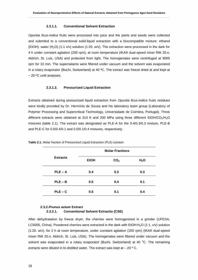
Table 2.1 Page 11 Molar fraction of Pressurized Liquid Extraction (PLE) solution.
Table 3.1 Page Total phenolic content of Opuntia ficus-indica extracts, assessed by
Folin-Ciocalteau method, expressed as mg GAE.g-1
of extract, in a dry
weight basis. Significant statistical differences are denoted with
different letters.
Table 3.2 Page Total flavonoid content of Opuntia ficus-indica extracts, assessed by a
colorimetric method, expressed as mg CE.g-1
of extract, in a dry
weight basis. Significant statistical differences are denoted with
different letters.

xviii

xix
List of abbreviations, acronyms and symbols
Abbreviation Full Form
AAPH 2”,2”-Azobis (2-amidinopropane) dihydrochloride
AD Alzheimer’s Disease
ADT Adsorption Technology
ATP Adenosine tri-phosphate
BBB Blood Brain Barrier
CAE Caffeic Acid Equivalents
CCE Crude Cherry Extract
CE Catechin Equivalents
CNS Central Nervous System
CO2 Carbon Dioxide
CSE Conventional Solvent Extraction
CVD Cardiovascular Diseases
DAD Diode Array Detector
DCFH – DA 2”,7”-Dichlorofluorescin Diacetate
ECACC European Collection of Cell Cultures
ED Electrochemical Detector
EGCG Epigallochatequine -3- gallate
EMEM Eagle Minimum Essential Medium
EtOH Ethanol
ETC Electron Transport Chain
EU European Union
FBS Fetal Bovine Serum
FDA Food and Drug Administration
FL Disodium Fluoresceín
GAE Gallic Acid Equivalents
GPx Glutathione Peroxidase
GR Glutathione Reductase
GSH Glutathione
GSSG Glutathione disulphide
H2O Water
H2O2 Hydrogen Peroxide
HORAC Hydroxyl Radical Adverting Capacity
HPLC High Performance Liquid Chromatography
KA kainate
MeOH Methanol
NMDA N-methyl-D-aspartate

xx
OBS – CSE Opuntia Beja/Serpa – Conventional Solvent Extract
O2- Superoxide anion
OGD Oxygen-Glucose Deprivation
OH. Hydroxyl Radical
ORAC Oxygen Radical Absorbance Capacity
PBS Phosphate Buffer Saline
PD Parkinson’s Disease
PGI Protected Geographical Indication
PLE Pressurized Liquid Extraction
PLE – A Pressurized Liquid Extract – A
PLE – B Pressurized Liquid Extract – B
PLE – C Pressurized Liquid Extract – C
ROS Reactive Oxygen Species
SOD Superoxide Dismutase
SPE Solid Phase Extraction
TE Trolox Equivalents
TFC Total Flavonoid Content
TPC Total Phenolic Content

Evaluation of Neuroprotective Effects of Natural Extracts obtained from Portuguese Agro-food Residues
1
1. Introduction
1.1. Neurodegenerative diseases
Countries are currently faced with problems derived from changes in population demography
and an increase in lifestyle-related diseases. Neurodegenerative disorders such Parkinson’s
(PD) and Alzheimer’s (AD) diseases are an increasing problem in aged societies like the
Europe. There is an increased prevalence of both diseases with age (1). Neurodegenerative
diseases are a heterogeneous debilitating group of degenerative disorders characterized by a
slow and progressive loss of neuronal cells. Such phenomena lead to gradual and progressive
impairments of selective functions of central nervous system (CNS), depending upon the
involved type of neuronal cells, and so far are incurable (2). Data from World Alzheimer Report
2011 indicate that 36 million people worldwide are living with dementia, with numbers doubling
every 20 years to 66 million by 2030, and 115 million by 2050. Alzheimer’s disease has
becoming the most common neurodisorder of today (3, 4). And at financial and social level, AD
represents an impact of €160 billion for the UE27 and €177 billion for whole Europe.
1.2. Neurodegeneration and Oxidative Stress
Neurodegenerative disorders appear to be triggered by multi-factorial events including
neuroinflammation, depletion of endogenous antioxidants and increases in oxidative stress.(1,
5, 6). The high metabolic rate and the low concentration of endogenous antioxidants as well as
the proportion of polyunsaturated fatty acids make the brain tissue particularly susceptible to
oxidative damage (7). The most effective way to produce adenosine tri-phosphate (ATP) at
cellular level is through oxidative phosphorylation within the mitochondria via the electron
transport chain (ETC). The ETC is not completely efficient showing a basal level of electron
leak. The reaction of leaked electrons with molecular oxygen produce a short-lived free radicals
such superoxide anion (O2-), hydroxyl radical (OH
.), and hydrogen peroxide (H2O2) called
reactive oxygen species (ROS)(8, 9). Under normal conditions, mitochondria have an efficient
biochemical defense mechanism to neutralize the effect mediated by these free radicals.
Endogenous defenses are composed of glutathione (GSH), glutathione peroxidise (GPx),
glutathione reductase (GR), superoxide dismutase (SOD), NADPH, vitamins E and C (10, 11).
Generally, intracellular ROS are maintained at low but measurable level and are regulated by
the balance between the rate of production and scavenging by the components of this
biochemical defense mechanism. Oxidative stress is generally caused by an imbalance
between ROS regeneration and antioxidant defenses and can lead to direct cellular organelles
damage such proteins and nucleic acids and this fact can cause mitochondrial damage and
eventual cell death (12).

Evaluation of Neuroprotective Effects of Natural Extracts obtained from Portuguese Agro-food Residues
2
Figure 1.1. The approximate balance of antioxidants and reactive species in vivo.(13)
That is a large body of evidences demonstrating that the accumulation of iron ion species in the
brain, the involvement of mitochondria, the decreased levels of endogenous antioxidants, and
other ROS-mediated pathways are the major pathological factors that contributes to
neurodegenerative diseases like PD and AD (14, 15).
Biological systems have several mechanisms to protect themselves from the ROS
consequences. These antioxidant defenses mechanisms include scavenging of ROS species
and their precursors, binding catalytic metal ions needed for ROS formation, generating and up-
regulating endogenous antioxidant defenses (11). Central nervous system dysfunction has been
observed in all diseases, related to errors in GSH metabolism, suggesting that the brain is
particularly susceptible to alterations in GSH homeostasis. The reason for the sensitivity is
unknown, although may be consider the possibility of the high susceptibility of the brain to
oxidative stress, due to its high oxygen consumption(10).
Glutathione (GSH) plays a central role in the cellular protection mechanism against oxidative
and other forms of stress. Glutathione is present in both reduced (GSH) and oxidized (GSSG)
state and the redox state of the GSH/GSSG couple can serve as an important indicator of cell
redox environment. It has been described that a decrease in neuronal GSH levels leads to
mitochondrial dysfunction and apoptosis.
1.2.1. Neurodegeneration Cell Model
Studies to determine biological effect of specific phytochemicals and plant-based foods involve
experimentation with in vitro and in vivo systems. In vitro systems present advantages as a first
approach, testing their potential effects within cells. They can provide an indication of biological
activity for the phytochemical in question, which can be used to design animal experiments in
the future. The major benefits of the use of in vitro cell models is practical convenience, such as

Evaluation of Neuroprotective Effects of Natural Extracts obtained from Portuguese Agro-food Residues
3
ease of culturing, their relatively low cost, and moderate throughput capabilities (16).
The creation of a cellular model based in the application of oxidative damage in neurons may be
helpful to understand the molecular mechanisms underlying in the development of
neurodegenerative diseases. Therefore, exposure of cultured neurons to relatively low
concentrations of H2O2 induces changes in cell metabolism and lead to a moderate neuronal
death, reproducing what may occur during neurodegeneration process (17).
In the Disease and Stress Biology (DSB) lab, SK-N-MC cell line is used as neuronal cell model
and is a continuous cell line, obtained from human metastic neuroblastoma tissue (17-19).
1.3. Nutraceuticals and Functional Foods
It is well known that consumption of plant-based foods such fruits, vegetables, grains and
cereals have key role in health promotion and disease prevention (20). There has been an
increased concern for consuming health-promoting food products and two new concepts appear
in the market: Nutraceuticals and Functional Foods. The short-term goal of nutraceuticals and
dietary supplements is to improve the quality of life and enhance health status while its long-
term goal is to increase lifespan while maintaining health (20).
The term nutraceutical was coined from nutrition and pharmaceutical (Fig. 1.2) in 1989 by
Stephen DeFelice, founder and chairman of foundation for innovation in medicine, an American
organization which encourages medical health (21). Nowadays the term Nutraceutical is defined
as “a product isolated or purified from the food, generally sold in medicinal form not associated
with food and demonstrated to have a physiological benefit .It also provides benefit against
chronic disease” (21). Such products may range from isolated nutrients, dietary supplements to
genetically engineered designer foods and herbal products and can be found in presentations
similar to drug such pills, extracts or tablets (22).
Figure 1.2. The term "nutraceutical" was coined from "nutrition" and "pharmaceutical" in 1989 by Stephen
DeFelice, MD, founder and chairman of the Foundation for Innovation in Medicine (FIM), Cranford, NJ.
Adapted from Pandey et al. 2010 .
Functional foods represents a type of food that when consumed regularly exert a specific
health-beneficial effect (i. e., a healthier status or a lower risk of disease) beyond their nutritional
properties, and this effect must be scientifically proven (22, 23). From consumers point of view,
Pharmaceutical Nutraceutical Nutrition

Evaluation of Neuroprotective Effects of Natural Extracts obtained from Portuguese Agro-food Residues
4
functional foods and nutraceuticals may offer many benefits such as an increasing of health
value of our diet, help people to live longer, help to avoid particular medicinal conditions, be
perceived to be more "natural” than traditional medicine and less likely to produce unpleasant
side-effects and present food for population with special needs. Some of the most common
bioactive ingredients found in nutraceutical and functional food market are bioactive non-
nutrient plant compounds. These compounds called phytochemicals, have raised interest in
human nutrition because of their potential effects as antioxidants, antiestrogenics, anti-
inflammatory, immunomodulatory and anticarcinogenics (22, 23). Nutraceuticals are able to
exert neuroprotection via a wide range of proposed mechanisms, such as scavenging of free
radicals and ROS, metal chelation, modulation of cell-signalling pathways, and inhibition of
inflammation (24). Most of phytochemical compounds with biological activities present in
nutraceuticals and functional foods are isolated from natural sources using several extraction
techniques.
1.4. Phytochemicals
The composition of foods cannot be reduced to the macronutrients and essential micronutrients.
Foods also contain a large number of other compounds that, although not essential, also have
influence in human health. Several hundreds of phytochemicals such as polyphenols,
carotenoids, glucosinolate, saponins or alkaloids have been identified in foods of plant origin.
Some of these compounds may contribute to explain the beneficial health effects of consuming
fruits and vegetables (25).
Polyphenols constitute a large group of phytochemicals with more than 8000 indentified
compounds (26) and generally, involved in defence against ultraviolet radiation or aggression by
phatogens (27). Polyphenols may be classified in several ways such as a classification based
on structure and function. Simple phenols are substances containing only one aromatic ring and
having at least one phenolic hydroxyl group and have the possibility of have other substituent.
Phenols and polyphenols may occur as unconjugated aglycones or, as conjugates, frequently
with sugar or organic acids. Flavonoids is the most extensively studied polyphenols, all
characterized by a C6-C3-C6 structure, subdivides by the nature of C3 element into anthocyanins,
flavanols, flavanones, flavones, flavonols, isoflavones as shown in figure 1.3 (28).
Epidemiological studies suggest that high dietary intake of polyphenols is associated with
decreased risk of a range of diseases including cardiovascular disease (CVD), specific forms of
cancer and neurodegenerative diseases such AD (29) and PD (30-31). A regular dietary intake
of flavonoid-rich foods and/or beverages has been associated with 50% reduction in the risk of
dementia, a preservation of cognitive performance with ageing, a delay in the onset of
Alzheimer’s disease and a reduction in the risk of developing Parkinson’s disease (1). Many
studies have reported the bioavailability of polyphenolic compounds in the systemic circulation
(32-34). However, less is known about their degree of brain bioavailability, flavanones such as
hesperetin, naringenin and their in vivo metabolites, have been shown to traverse the blood
brain barrier (BBB) in relevant in vitro and in situ models (35). Moreover, several anthocyanins

Evaluation of Neuroprotective Effects of Natural Extracts obtained from Portuguese Agro-food Residues
5
have also been identified in the cortex and cerebellum of rat and pig following feeding with
blueberries.
Together, these results suggest that polyphenols are able to transverse the BBB, in different
degrees and depending on their structure. Thus, such compounds are likely to be candidates for
direct neuroprotective and neuromodulatory actions (1). The contribution of phenolic
compounds to the protection against degenerative diseases as well as their effects on health
has been attributed to their antioxidant activities (36, 37), but also to their cellular modulatory
actions through a direct interaction with receptors or enzymes involved in signal transductions,
such as protein and lipid kinases signaling pathways. Therefore, a wide range of mechanisms
have been described to explain polyphenol’s health benefits such an antioxidant action by
scavenging radicals, a induction of endogenous antioxidants (glutathione peroxidase,
glutathione reductase or superoxide dismutase); iron chelating properties; modulation of genes
related to cell survival/death modulation, gene/protein and cell signaling pathway regulatory
activity as well as regulation of mitochondrial function (38-40).

Evaluation of Neuroprotective Effects of Natural Extracts obtained from Portuguese Agro-food Residues
6
Figure 1.3.Classification of dietary phitochemicals (Adapted from Liu, 2004) . (41)
FlavanonesHydroxy-
benzoic acidsAnthocyanins IsoflavonoidsFlavonesFlavonols
Hydroxy-
cinnamic acidsFlavanols
Phytochemicals
Carotenoids Organosulfur CompoundsNitrogen-containing
CompoundsPhenolicsAlkaloids
Phenolic Acids Flavonoids Stilbennes Coumarins Tannins
Hesperetin
Naringenin
Naringenin
Gallic
Vanillic
Syringic
Gallic acid
Delphinidin
Cyanidin
Peonidin
Pelargonidin
Cyanidin
Genistein
Daidzein
Glicitein
Genistein
Apigenin
Luteolin
Luteolin
Quercetin
Kaempferol
Myricetin
Rutin
Quercetin
p-Coumaric
Caffeic
Chlorogenic
Ferulic
Chlorogenic acid
Catechin
Epicatechin
Catequin

Evaluation of Neuroprotective Effects of Natural Extracts obtained from Portuguese Agro-food Residues
7
Table 1.1. Polyphenols effects in in vivo and in vitro models of neurotoxicity and neurodegeneration (Adapted from Ebrahimi et al. (2012). (14)
Substance Cell line/ Animal model Effect
Aloe-emodin N-methyl-d-aspartate (NMDA)-induced toxicity in retinal ganglion cells (RGCs)
Elevates levels of RNA and protein expression of superoxide dismutase (SOD); Attenuates NMDA-induced apoptosis of RGCs (42)
Curcumin N27 dopaminergic neurons Protects against mitochondrial complex I inhibition (leading to mitochondrial dysfunction) and NS (43)
Curcumin Homocysteine-induced neurotoxicity in rats Reduces Malondialdehyde (MDA)a and Superoxide anion levels; Reduces lipid peroxidation; Improves learning and memory in rats (44)
Curcumin N27 dopaminergic neuronal cell line Increases glutathioneb (GSH) levels (45) Curcumin 3-Nitropropionic acid (3-NP)-induced
neurotoxicity in rats Improves the 3-NP-induced motor and cognitive impairment; Attenuates 3-NP-
induced OS (including lipid peroxidation, reduced GSH and nitrite activity); Restores the decreased succinate dehydrogenasec activity (46)
Epigallocatechingallat (EGCG)
Glucose oxidase-induced neurotoxicity in H 19-7 (a rat neuronal cell line)
Enhances cellular resistance to glucose oxidase-mediated oxidative damage; Elevates heme oxygenase-1d (HO-1) mRNA and protein expression; Activates transcription factor Nrf2e (47)
EGCG Glutamate-induced toxicity in HT22 mouse hippocampus neuronal cells, Kainic acid-induced neurotoxicity in Rats
Reduces glutamate-induced oxidative cytotoxicity; Inactivates the NF-_B signaling pathway; Reduces ROS accumulation and NF-_B transcriptional activity
(48) EGCG Transient global cerebral ischemia
C57BL/6 in mice Reduces the development of delayed neuronal death after transient global
cerebral ischemia in mouse brain (49)
EGCG Age-associated oxidative damage in rat brain Amplifies the activities of enzymic antioxidants like SOD, catalase, glutathione peroxidase, glutathione reductase and glucose-6-phosphate dehydrogenase; Improves the activity of non-enzymic antioxidants like tocopherol, ascorbic acid and glutathione;Ameliorates the MDA and protein carbonyl levels (50)
EGCG Progressive neurotoxic model of long-term serum deprivation in human SH-SY5Y neuroblastoma cells
Decreases protein levels and mRNA expression of the beta subunit of the enzyme prolyl 4-hydroxylase; Decreases protein levels of two molecular chaperones that are associated with HIF regulation, the immunoglobulin-heavy-chain binding protein and the heat shock protein 90 beta (51)
EGCG SOD1-G93A transgenic mice (a model of ALS) Maintains the normal expression of p85a PI3-K, pAkt, and pGSK-3 (molecular signals of survival); Reduces activation of NF-kB and the cleaved form of caspase-3; Reduces microglial activation; Prolongs the life span; Delays the onset of symptoms (52)
Mangiferin Glutamate-induced neurotoxicity in rat cerebral cortex neurons
Prevents neuronal death, oxidative stress and mitochondrial depolarization (53)
Mangiferin 1-Methyl-4-phenyl pyridinium (MPP(+))-induced oxidative stress in the murine neuroblastoma cell
Restores the GSH content (to 60% of control levels), and down-regulates both SOD and catalase mRNA expression (54)

Evaluation of Neuroprotective Effects of Natural Extracts obtained from Portuguese Agro-food Residues
8
line N2A Quenches reactive oxygen intermediates Mangiferin Morin Glutamate-induced neurotoxicity in rat primary
culture of neurons Reduces ROS formation; Activates enzymatic antioxidant system; Restores
mitochondrial membrane potential (55)
Polyphenol-rich Hedeoma multiflorum extract
Biochemical assay on rat brain Homogenates Inhibits lipid peroxidation; Scavenges 2,2_-diphenyl-1-picrylhydrazyl (DPPH) radicals
(56) Polyphenol-rich osmanthus fragrans extract
Glutamate, arachidonic acid, and 6 hydroxydopamine-induced neurotoxicity in rat primary cortical neurons
Scavenges DPPH and hydroxyl anions Inhibits lipid peroxidation
(57) Red wine polyphenol compound
Rat model of ischemic cerebral stroke Prevent the burst of excitatory amino acids in response to ischemia; Reduce brain infarct volumes; Enhance the residual cerebral blood flow during occlusion and reperfusion; Modulate expression of proteins involved in the maintenance of neuronal caliber and axon formation (58)
Resveratrol Lipopolysaccaride (LPS)-induced dopaminergic neurodegeneration in rat
Reduces NADPHg oxidase-mediated generation of ROS; Inhibits microglia activation; Attenuates the activation of MAPK and NF-_B signaling pathways; Implies neuroprotection against LPS-induced dopaminergic neurodegeneration (59)
Resveratrol Glutamate-induced toxicity in mice primary culture of neurons; Optimized ischemic-reperfusion stroke model in mice
Induces heme oxygenase 1d (HO-1) in dose- and time-dependent manner; Protects mouse neurons, subjected to an optimized ischemic-reperfusion stroke model; Protects neurons against ecitotoxicity (60)
Resveratrol 1-methyl-4-phenyl-1,2,3,6 tetrahydropyridine (MPTP)- induced Parkinson in mice
Protects from MPTP-induced motor coordination impairment, hydroxyl radical overloading, and neuronal loss; Scavenges free radicals (61)
Resveratrol MPP(+)-induced neurotoxicity in dopaminergic neurons of midbrain slice culture
Prevents accumulation of ROS, depletion of cellular glutathione, and cellular oxidative damage induced by MPP(+); Activates sirtuin family of NAD-dependent histone deacetylases (62)
Resveratrol A-beta induced toxicity in neurons from a mouse model (Tg2576 line) and mouse neuroblastoma (N2a) cells
Maintains normal expression of peroxiredoxins and mitochondrial structural genes; Maintains normal mitochondrial function
(63)
Tea polyphenols NMDA-induced neurotoxicity in mice Attenuates the increased production of synaptosomal ROS; Reduces the deteriorative ROS-sensitive Na
(+), K(+)-ATPase and Mg
(2+)-ATPase activity (64)

Evaluation of Neuroprotective Effects of Natural Extracts obtained from Portuguese Agro-food Residues
9
1.5. Extraction Technologies
Amongst fruits, vegetables and herbs, agricultural and industrial by-products are attractive
sources for extraction of compounds with nutraceutical interest such pigments or natural
antioxidants. Special attention has been focused on the extraction from inexpensive or residual
sources from agricultural industries. Some studies have already been done on by-products,
which could be potential sources of antioxidants (65). Once the industrial processing of a wide
variety of fruits results in the accumulation of large quantities of residues, the recovery of these
food industry by-products could reduce waste disposal problems and serve as a potential new
source of phytochemicals that can be used in nutraceutical and functional food market creating
value-added applications. In order to obtain such value compounds, extraction techniques have
been investigated. Conventional Solvent Extraction (CSE) is a traditional extraction method
characterized by a solid-liquid principle. Solid-liquid extraction can be defined as a mass
transport phenomenon in which solids contained in a solid matrix migrate into a solvent brought
into contact with the matrix. The single-stage system represents the complete operation of
contacting the solid matrix with the fresh solvent. However, these techniques have, generally,
negative environmental impact and some drawbacks such as long extraction times, large
amounts of initial matrix and organic solvents and lower extraction yields (65).
An attractive and alternative methodology to conventional extraction, to produce extracts for
pharmaceutical or nutraceutical application is Pressurized Liquid Extraction (PLE). The
technique involves the utilization of water and/or organic solvents at considerable elevated
temperatures (313 – 473 K) and pressures (3.3 – 20.3 MPa). Liquid carbon dioxide is found to
be a good extraction solvent in food industry due to its physicochemical properties, low toxicity
and price. Since carbon dioxide is non-polar specie it is not suitable solvent to extract polar
polyphenols. The addition of solvents like ethanol increases the solvating power of carbon
dioxide and the extraction yield of phenolic compounds. Ethanol is a permitted co-solvent in
food industry.The advantages of PLE technique are usually related with the usage of carbon
dioxide (66) that has been described as a “green solvent” and gives to the method improved
characteristics in terms of mass transfer and of solvation properties(66, 67). Extraction time can
be decreased using high temperature and high pressure: there is enhanced diffusivity of the
solvent and, at the same time, there is the possibility of working under an inert atmosphere and
with protection from light (68).
Adsorption technology (ADT) is a commonly applied process to recover bioactive compounds
from plant materials and consists on a solid phase extraction (SPE). SPE consists in a
partitioning of compounds between two phases. The compounds presents in the liquid phase
and these analytes must have a greater affinity for the solid phase than for the sample matrix.
Compounds retained on the solid phase can be removed at a later stage by eluting with a
solvent with a higher affinity for the analytes (elution or desorption step). The different retention
and elution mechanisms are due to intermolecular forces between the analyte, the active sites
on the surface of the solid phase and the liquid phase or matrix (69).

Evaluation of Neuroprotective Effects of Natural Extracts obtained from Portuguese Agro-food Residues
10
The adsorption capacity of an adsorbent for a solute may vary with processing conditions such
as temperature and pH value of solution. The elution step is usually performed with alcohols, in
particular ethanol as it is acceptable for food and pharmaceutical applications.
The adsorption processes can be performed in a batch model (70) or in continuous, where the
adsorbent is packet into a column (71). The use of resins for food production proposes is
regulated by the US Food and Drug Administration (FDA) and the Council of Europe.
Figure 1.4. Chemical structure of Amberlite® XAD 16.
The selection of adsorbents is critical for the development of an adsorption process. Adsorbent
characteristics, such as particle size, surface, area and porosity should be taken in
consideration in each case study. For the purification of polyphenols various adsorbents have
been used (72) One of the most commonly applied is a polymeric resin Amberlite® XAD16
(Figure 1.7) (73).
1.6. Opuntia ficus-indica
Opuntia ficus-indica (cactus pear) is the larger member of Cactaceae family that mainly grows in
arid and semi-arid regions and is largely distributed in Mexico, much Latin America, South Africa
and Mediterranean area(74). Opuntia fruits are oval and elongated berries of 67 to 216 g weigh.
Cactus pear fruits have a thick pericarp (peel) and a juicy pulp with many seeds. The pulp is the
edible part of the fruit and is mainly composed of water (84-90%) and reducing sugars (10-
15%). Fruit seeds represent about 10-15% of the edible pulp and are usually discarded as
waste after pulp extraction (75). The high season for harvesting Opuntia cactus fruits is between
the end of July and November in the Mediterranean regions (76). Fruit and stems of Opuntia
species have been used in folk medicine for burns, wounds, edema, bronchial asthma,
hypertension, indigestion, and type II diabetes (77). They nutritional properties have long been
known and it is also used in traditional medicine. In the industrialized countries of Mediterranean
area, cladodes are not consumed but the fruits are largely used as nutritional sources (76). The
plant is used mainly for fruit production and the fruit are consumed as fresh fruit or juice.
Although in some countries it is used as a vegetable for fodder (78, 79).
C C C C
C C C
H H
H H HH
H
H
H
H
H
n

Evaluation of Neuroprotective Effects of Natural Extracts obtained from Portuguese Agro-food Residues
11
Figure 1.5. Images of Opuntia ficus-indica cladodes and fruits.
Cactus pear was largely ignored by the scientific community until the beginning of the 1980’s
when several studies were published on their biological functions. In recent years, investigations
on the chemical and nutritional value of Opuntia spp. have attracted attention in the food,
nutritional and pharmacological sciences (80, 81) . Based on various studies, fruit pulp is
considered a good source of minerals, especially calcium, potassium and magnesium and
phosphorous(74). The concentration in bioactive compounds is dependent on the cultivar,
cultivation site, maturation stage and environmental conditions.
Fruit is commonly consumed fresh but there is increased interest in the transformation into
different products such as juice, jam and jelly among others. After being consumed or
transformed, fruit peels and seeds are discarded and taking in account they composition in
phytochemicals make them very attractive as sources for extraction. After extraction these by-
products can be used as addictive in food preparation or in the pharmaceutical and
nutraceutical market (82). The nutraceutical benefits of Opuntia spp. fruits are believed to be
related to their content in ascorbic acid and phenolic compounds, including flavonoids, and a
mixture of yellow betaxanthin and red betacyanin pigments. Betaxanthins and betacyanins
belong to betalains group that are water soluble compounds described as excellent radical
scavengers.(79)
In vitro studies have demonstrated that a variety of compounds present in Opuntia sp. fruits are
able to exert neuroprotective activity. The methanolic extract of O. Ficus-indica produced dose-
dependent neuroprotective effects on hydroxyl- and superoxide radical-mediated neuronal
damage to mouse primary cultures (83) and the pre-treatment of cultured neurons and glia cells
with methanolic extract of Opuntia fruits inhibited N-methyl-D-aspartate (NMDA)-, kainate (KA)-

Evaluation of Neuroprotective Effects of Natural Extracts obtained from Portuguese Agro-food Residues
12
and oxygen-glucose deprivation (OGD)-induced neurotoxicity dose-dependent.(84) Kim and
collaborators (2006) also have demonstrated that when gerbils receive different doses of the
Opuntia extract, the neuronal damage in the hippocampus was reduced. Moreover Opuntia
polysaccharides as described to exert a neuroprotective activity against a H2O2-induced
oxidative damage in PC12 cells by maintain cell viability, reduce apoptosis and decrease
intracellular ROS levels.(77)
1.7. Prunus avium
Prunus avium (sweet cherries) are very attractive fruits for consumers due to their taste, colour,
sweetness and wealth of nutrients being easily adapted to a regular diet. Portugal produces
more than 15 000 tons of cherries every year. It is in the northeast of the country, in the region
of Beira Interior, where the fruit is more cultivated and the cherries has protected geographical
indication (PGI) registration according to the European Union (EU) regulations.
Figure 1.6. Cherries (Prunus avium) from Saco cultivar collected in “Cova da Beira” region, Portugal.
Saco cultivar is an old traditional cherry. It is a very promising functional fruit due to its powerful
antioxidant attributed to the phenolic composition (85). Serra et al. evaluated nine “Cova da
Beira” cherry varieties in terms of polyphenolic content and bioactivity (antioxidant propert ies)
and it was shown that Saco cherry extract has a strong antioxidant activity. However there is
little information about cherries biological activities. However, due to its small size and weight
the acceptance of Saco cherry by consumers could be compromised since larger fruits are
preferred.
It has been described that sweet cherries cultivars such as Saco contain various anthocyanins
with the total anthocyanins in a range of 19.4 to 95.7 mg.100 g-1
of fresh cherries. Serra and

Evaluation of Neuroprotective Effects of Natural Extracts obtained from Portuguese Agro-food Residues
13
collaborators described that sweet cherries contain cyaniding -3- glucoside and cyaniding-3-
rutinuside being that 77.3 to 86.6 % of the total anthocyanins content. Among hydroxycynamic
acid derivates, cherries had neochlorogenic acid and p-coumaroylquinic acid as the
predominant compounds and the glycosides of peonidin and pelargonidin as minor compounds
(86). Cyaniding -3- glucoside is described as able to transpose the BBB. (87)
Nevertheless, there is little information available on the possible health benefits of phenolic
compounds present in fresh cherries on animal cells exposed to cell-damaging oxidative stress.
Taking this in account the major aim of this work is to obtain and characterize natural extracts
obtained from Opuntia ficus-indica and Prunus avium, residues using different extraction
technologies, evaluate their neuroprotective effect in a H2O2-induced neurodegeneration cellular
model and understand the cellular mechanisms behind their potential bioactivity.

Evaluation of Neuroprotective Effects of Natural Extracts obtained from Portuguese Agro-food Residues
14

Evaluation of Neuroprotective Effects of Natural Extracts obtained from Portuguese Agro-food Residues
15
2. Materials and Methods
2.1. Materials
For phytochemical characterization: Amberlite® XAD 16 and sodium carbonate (Na2CO3) were
purchased from Sigma- Aldrich (St Quentin Fallavier, France), Folin Ciocalteau reagent was
acquired from Panreac (Barcelona, Spain) and gallic acid from Fluka (Germany). Sodium nitrite
(NaNO2) was purchased from Riedel-de-Haën (Seelze, Germany), aluminium chloride (AlCl3)
and sodium hydroxide (NaOH) was obtained from Sigma-Aldrich, in Germany. (+)-Catechin
hydrate was purchased from Sigma (Japan).
For antioxidant activity assays chemicals used were: 2‟,2‟-Azobis (2- amidinopropane)
dihydrochloride (AAPH), 6-hydroxy-2,5,7,8- tetramethylchroman-2-carboxylic acid (Trolox),
caffeic acid (C9H8O4), cobalt floride tetrahydrate (CoF2), hydrogen peroxide (H2O2) and picolinic
acid (C6H5NO2) were purchased from Sigma-Aldrich (St Quentin Fallavier, France). Disodium
fluorescein (FL) was from TCI Europe (Antwerp, Belgium). Sodium chloride (NaCl), potassium
chloride (KCl) and monopotassium phosphate (KH2PO4) were from Sigma-Aldrich (St Quentin
Fallavier, France) and sodium phosphate dibasic dehydrate (Na2HPO4⋅2H2O) from Riedel-de
Haën (Seelze, Germany) were used for phosphate buffer solution preparation (PBS).
Cell based assays were performed using: 2’,7’-dichlorofluorescein diacetate (DCFH-DA),
hydrogen peroxide (H2O2), sodium dodecyl sulphate (SDS), GSH and GSSG standards,
orthophthalaldehyde (OPA), Eagle Minimum Essential Medium (EMEM), glutamine, non-
essential amino acids, sodium pyruvate, were purchased from Sigma-Aldrich (St. Quentin
Fallavier, France), Disodium fluorescein was obtained from TCI Europe (Antwerp, Belgium),
CellTiter – Blue Reagent® Cell Viability Assay was obtained from Promega (San Luis Obispo,
CA, USA). Fetal bovine serum (FBS), Trypsin-EDTA and penicillin-streptomycin (PenStrep)
were obtained from Invitrogen (Gibco, Invitrogen Corporation, Paisley, UK). Phenolic standards
used were: chlorogenic acid from Sigma-Aldrich (St. Louis, MO, USA), and cyanidin-3-
glucoside, cyanidin-3-rutinoside were all from Extrasynthèse (Genay, France). Ultra-pure water
(18.2 MΩ.cm) was obtained from a Millipore Direct Q3 UV system (Millipore, USA).
2.2. Raw Materials
Cactus pear (Opuntia ficus-indica) fruits were collected at Beja/Serpa, Portugal, in September
2011. After remove the spikes, fruits were processed using a kitchen robot (UFESA, LC5005,
China) and the peels and seeds were collected. The waste (peels and seeds) was kept at
-20oC.
Sweet cherries (Prunus avium), from Saco cultivar, grown in Cova da Beira region, Portugal,
were harvested by hand between May and June of 2011. The whole fruits were processed with
seeds and stalks, milled and frieze dried. Milled cherries were kept at – 20 ºC.
2.3. Preparation of Prunus avium and Opuntia ficus-indica Extracts
2.3.1. Opuntia ficus-indica Extracts
Evaluation of Neuroprotective Effects of Natural Extracts obtained from Portuguese Agro-food Residues
16
2.3.1.1. Conventional Solvent Extraction
Opuntia ficus-indica fruits were processed into juice and the peels and seeds were collected
and submited to a conventional solid-liquid extraction with a biocompatible mixture: ethanol
(EtOH): water (H2O) (1:1 v/v) solution (1:20, w/v). The extraction were processed in the dark for
4 h under constant agitation (200 rpm), at room temperature (IKA® dual-speed mixer RW 20.n,
Aldrich, St. Luis, USA) and protected from light. The homogenates were centrifuged at 9000
rpm for 10 min. The supernatants were filtered under vacuum and the solvent was evaporated
in a rotary evaporator (Buchi, Switzerland) at 40 ºC. The extract was freeze dried at and kept at
– 20 ºC until analyses.
2.3.1.2. Pressurized Liquid Extraction
Extracts obtained during pressurized liquid extraction from Opuntia ficus-indica fruits residues
were kindly provided by Dr. Hermínio de Sousa and his laboratory team group (Laboratory of
Polymer Processing and Supercritical Technology, Universidade de Coimbra, Portugal). Three
different extracts were obtained at 313 K and 200 MPa using three different EtOH/CO2/H2O
mixtures (table 2.1). The extract was designated as PLE-A for the 0.4/0.3/0.3 mixture, PLE-B
and PLE-C for 0.5/0.4/0.1 and 0.5/0.1/0.4 mixtures, respectively.
Table 2.1. Molar fraction of Pressurized Liquid Extraction (PLE) solution.
2.3.2. Prunus avium Extract
2.3.2.1. Conventional Solvent Extractio (CSE)
After dehydratation by freeze dryer, the cherries were homogenized in a grinder (UFESA,
LC5005, China). Powdered cherries were extracted in the dark with EtOH:H2O (1:1, v/v) solution
(1:20, w/v), for 2 h at room temperature, under constant agitation (200 rpm) (IKA® dual-speed
mixer RW 20.n, Aldrich, St. Luis, USA). The homogenates were filtered under vacuum and the
solvent was evaporated in a rotary evaporator (Buchi, Switzerland) at 40 oC. The remaining
extracts were diluted in bi-distilled water. The extract was kept at – 20 º C.
Extracts
Molar Fractions
EtOH CO2 H2O
PLE – A 0.4 0.3 0.3
PLE – B 0.5 0.4 0.1
PLE – C 0.5 0.1 0.4

Evaluation of Neuroprotective Effects of Natural Extracts obtained from Portuguese Agro-food Residues
17
2.3.2.2. Adsorption Technology (AD)
To concentrate in anthocyanins of Prunus avium extract obtained on 2.3.2.1. section, food
grade macroporous resin Amberlite® XAD-16 was used as adsorbent. This resin is allowed for
food applications by the U.S. Food and Drug Administration Code of Federal Regulation Title 21
(88).
The preconditioning of the adsorbent was done by an extensive wash with abundant distilled
water to remove salts and impurities, and was then dried at 70 oC for 24 h. The dried resin was
immersed in EtOH (96%) for 12 h. EtOH was then replaced by bi-distilled water through
washing.
The production of cherry polyphenols rich extract was performed in batch mode. Briefly,
aqueous crude cherry extract (CCE) obtained previously (section 2.3.2.1) were maintained in
contact with the resin in a ratio of 80 mg GAE.g-1
of resin, protected from light and submitted to
200 rpm of agitation, during 4 h. After 4 h of contact, supernatant was removed and resin
washed three times with bi-distilled water. Anthocyanins were then eluted two times with EtOH
(96 %). The ethanolic fraction were concentrated by evaporation, freeze dried and kept at – 20
ºC.
2.4. Extracts Characterization
The extracts were analyzed for their chemical composition and antioxidant capacity against
peroxyl and hydroxyl radicals. Composition in polyphenols was determined using Folin-
Ciocalteau colorimetric method and flavonoids content was performed by the AlCl3
complexation method (89) modified by Tavares et al, 2010. Antioxidant activity was assessed
using two different in vitro chemical assays, ORAC and HORAC adapted by Feliciano et al,
2009 and Serra et al, 2011, respectively. Extracts phenolic profile were obtained by High
Performance Liquid Chromatography with Diode Array Detector and Electrochemical Detector
(HPLC – DAD – ED).
2.4.1. Total Phenolic Content (TPC)
Total phenolic content present in extracts was determined according to the Folin- Ciocalteau
method. To perform the colorimetric method, 20 µL of the appropriate dilutions of extracts were
added to 1580 µL of distilled water. Than 100 µL of the Folin Ciocalteau reagent was added.
The reaction was neutralized with 300 µL of sodium carbonate. The absorbance, at 765 nm of
each sample was measured after 40 min at 40 ºC of incubation, in a Genesys10uv spectrometer
(Thermo Spectronic, New York, USA). The results were expressed as means of independent
triplicates as mg of Gallic Acid Equivalents (GAE) per gram of dry weight extract (mg GAE.g-1
dw). A calibration curve was made using Gallic Acid as standard.

Evaluation of Neuroprotective Effects of Natural Extracts obtained from Portuguese Agro-food Residues
18
2.4.2. Total Flavonoid Content (TFC)
The total flavonoid content was determined using a spectrophotometric method adapted to 96-
well microplate by Tavares et al, 2010. Briefly, 25 µL of each sample, 125 µL of distilled water
and 7.5 µL of NaNO2 5% (v/v) was added to each well of a 96-well plate. The plate was
incubated for 6 min at room temperature. Than 15 µL of a 10% AlCl3 (v/v) solution was added.
After 5 min of incubation, 100 µL of NaOH 1 M was added and the solution of each well mixed.
The absorbance was measured at 510 nm in a microplate reader (BioTekTM
Power Wave XS).
(+)-Catechin hydrate, minimum 98% (w/w) was used as standard, and the results are expressed
in means of triplicates as mg catechin equivalents (CE) per mg of dry weight extract
(mg CE.mg-1
dw).
2.4.3. HPLC – DAD – ED analysis
HPLC analysis of Opuntia ficus-indica and Prunus avium fruit residues extracts were performed
by Analytical Group of IBET (Oeiras, Portugal) coordinated by Dr. Rosário Bronze. Briefly the
analysis were performed on a Waters® Alliance 2695 (Waters) equipped with a quaternary
pump, solvent degasser, auto sampler and column oven, coupled to a Photodiode Array
Detector Waters 996 PDA (Waters). A pre-column (RP-18, 5 μm, Lichrocart) and reverse phase
column (RP-18, 2.5 µm, from Manu-cart) with oven at 35 ºC were used for separation. The
gradient mobile phase consisted of 0.1 % phosphoric acid p.a. in ultra pure water (A): 0.1 %
phosphoric acid p.a. in LC-MS grade acetonitrile (B) and ultra pure water at a flow rate of 0.30
mL.min-1
. The injection volume was 5 μL.Photodiode Array Detector was used to scan
wavelength absorption from 210 to 600 nm.
For HPLC quantification of compounds in cherry extract, the mobile phase used consisted of a
gradient mixture of eluent A water:formic acid (90:10 v/v) and eluent B acetonitrile:water:formic
acid (40:50:10 v/v/v). The following gradient of eluents was used: 0- 15min from 0 until 20% of
eluent B; 10 min with 20% eluent B; 25–70 min, from 20 until 70% eluent B; 70–75 min, with
70% of eluent B; 75–85 min from 70 until 100% eluent B; 85–90 min, with 100% eluent B; 90-95
min from 100 to 0% of eluent B; and 95-100 min 100% of eluent A. The solvent flow rate was
0.7 mL.min-1
. The injection volume was 20 μL. Acquisition range was set between 190 and 700
nm and chromatogram was monitored at 280, 360, and 527 nm. Chlorogenic acid, Cyanidin
glucosides, rutin and quercetin were quantified using standard compounds. Coefficients of
variation on the HPLC quantifications were <5% and final concentrations were expressed as
mg.g-1
dry extract.
Evaluation of Neuroprotective Effects of Natural Extracts obtained from Portuguese Agro-food Residues
19
2.4.4. TLC analysis
Presence of terpenoids compounds was assessed in the three PLE extracts by thin layer
chromatography (TLC) using Lupeol as standard. 10 µL of extracts and standard were carefully
placed in a Plate Silica gel 60 F254. The mobile phase was an acetate:methanol:water (77:15:8)
solution. In order to reveal the terpenoid compounds present Liebermann – Burchard reagent
were prepared adding carefully 5 mL of acetic anhydride and 5 mL concentrated sulphuric acid
to 50 mL absolute ethanol, while cooling on ice. The TLC plate is sprayed with freshly prepared
solution, heated for 10 min at 100 ºC and inspected in UV-365nm.
2.5. Extracts Chemical Antioxidant Activity
2.5.1. Oxygen Radical Absorbance Capacity (ORAC)
The oxygen radical absorbance capacity was assessed by ORAC modified by Ou. et al.
2001(90). The oxidation of disodium flourescein (FL) is catalyzed by peroxyl radicals generated
by 2, 2’-azobis(2-amidopropane)dihydrochloride (AAPH) at 37 oC. ORAC assay measure the
ability of the antioxidant compounds present in the sample to inhibit that oxidation.
The protective effect of an antioxidant specie present in the sample is obtained by the difference
between the area under the fluorescence decay curve (AUC) of the sample and the blank, in
which no antioxidant is present. Briefly, sodium fluorescein (150 μL; 2,0x10⁻⁸
M in PBS 75 mM
pH 7,4) and samples or standards (25 μL) were added to a 96-well microplate and incubated
during 10 min at 37 ºC. AAPH (25 μL,1.28x10-2
prepared in PBS 75mM pH 7,4) were added to
each well. FL800 (Bio-Tek Instruments) microplate fluorescence reader was used (λex: 485 nm,
λem: 530 nm). The blank contained PBS (pH 7.4, 75 mM) instead of sample and 5 to
50 µM of Trolox was used to the calibration curve. The results were expresses as means of
independent triplicades as µmol of Trolox Equivalents (TE) per gram of dry weight extract (µmol
TE.g-1
dw).
2.5.2. Hydroxyl Radical Adverting Capacity (HORAC)
HORAC assay was based on a method reported by Ou et al 2002 (91) modified for the FL800
microplate fluorescence reader FL800 (Bio- Tek Instruments). HORAC assay evaluates the
hydroxyl radical prevention capacity by monitoring the fluorescence decay of fluoresceín (FL),
which is used as a probe. The hydroxyl radical was generated by a Co(II)-mediated Fenton like
reaction and the fluorescence decay curve of FL was used to quantify the HORAC value.
Briefly, 10 L of each sample were added to 180 L of FL (4 x 10⁻³
M) and then was added 10
L of a 0.55 M H2O2 solution. The reaction starts when it is added 10 L of CoF2 to each well of
a 96-well microplate at 37 ºC. The FL800 (Bio-Tek Instruments) microplate fluorescence reader
was used (λex: 485 nm, λem: 530 nm). Caffeic acid was used as a standard and the data were

Evaluation of Neuroprotective Effects of Natural Extracts obtained from Portuguese Agro-food Residues
20
expressed as means of independent triplicates as µmol of Caffeic Acid Equivalents (CAE) per
gram of dry weight extract (µmol CAE.g-1
dw).
2.6. Cell Based Assays
2.6.1. Cell Culture
Human Neuroepithelioma from Supra-Orbital Metastasis SK-N-MC cells, obtained from the
European Collection of Cell Cultures (ECACC), were routinely grown in EMEM (Eagle Minimum
Essential Medium, Sigma) supplemented with 2mM L-Glutamine (Sigma), 10% (v/v) heat-
inactivated fetal bovine serum (FBS) (Sigma), 1% (v/v) non-essential amino acids
(NEM)(Sigma), 1 mM sodium pyruvate and 50 U.mL penicillin and 50 g.mL streptomycin.
According to ECACC instructions, stock cells were maintained in 25 cm2 T-flask, incubated at
37 ºC and 5% CO2. Cells are split to sub-confluence of 70 – 80 % confluence using 0.05 % (v/v)
trypsin-EDTA (Gibco).
2.6.2. Cytotoxicity Profile determination
Extracts toxicity assays were performed using CellTiter-Blue® Reagent. Briefly, SK-N-MC cells
were seeded at a density of 2.5x10⁴ cells/well in 96 well plate. After 24 hours, SK-N-M cells
were pre-incubated with the extracts in a range 0-250 µg GAE.mL-1
medium, for 24 h. A control
was performed with cells incubated only with cell culture medium.
After 24 of incubation with the extracts, 20 µL of CellTiter-Blue® Reagent was added to each
well for 3 h. Resazurin present in CellTiter-Blue® Reagent is a dark blue dye that has little
intrinsic fluorescence, is reduced by metabolically active cells into a fluorescent product,
resorfurin. Therefore, fluorescent signal is proportional to the number of viable cells. The
product was quantified by measurement of the fluorescence (λex: 560 nm, λem: 590 nm) on
microplate fluorescence reader FL800 (Bio-Tek Instruments).
2.6.3. Evaluation of Neuroprotective effect
To evaluate the neuroprotective effect of each extract, a neurodegeneration cell model was
used. The model describes the treatment of SK-N-MC neuroblastoma cells with H2O2 to induce
50% of cell death. Cells were seeded at 2.5 x 10⁴ cells per well. After 24 hours of growth the
medium was removed and the cells were washed with PBS. Cells were pre-incubated with non-
toxic concentrations of each extract and after 24 hours, cells were washed again with PBS and
medium was replaced by medium containing 0,5% (v/v) FBS and 300 µM H2O2 for 24h. Cell
viability was monitorized with CellTiter-Blue® Reagent as previously described (Section 2.6.1).

Evaluation of Neuroprotective Effects of Natural Extracts obtained from Portuguese Agro-food Residues
21
2.6.4. Intracellular reactive oxygen species (ROS) production determination
In order to evaluate the ability of extracts o reduce ROS levels produced by cells, the
conversion of 2’,7’ – dichlorofluorescein diacetate (H2DCFDA) to fluorescent 2’,7’ –
dichlorofluorescein (DCF) was monitored. SK-N-MC cells were seeded at 1.25 x 10⁴ cells per
well in a 96-well plate, grown for 24 h, then washed with PBS and pre-incubated with the
different extracts diluted in medium (0.5 % (v/v) FBS) for 2 or 24h. After this pre-incubation, cells
were washed with PBS and incubated for 10 min at 37 ºC with 25 µM H2DCFDA prepared in
PBS. Cells were washed with PBS and then 300 µM H2O2 was added for 30 min. The FL800
(Bio-Tek Instruments) fluorescence microplate reader was used, with flouorescence filters for an
excitation wavelength of 485 20 nm and an emission wavelength of 530 25 nm, over 1h at
37 ºC. ROS generation was calculated as an increase in fluorescent signal compared with cells
not treated with H2O2.
2.6.5. Glutathione (GSH) and glutathione disulphide (GSSG) quantification
In order to quantify GSH and GSSG, cold 10% (w/v) metaphosphoric acid was carefully added
to samples or standards. After incubation (4 ºC for 10 min) and centrifugation (16,000 g during
20 min at 4 ºC) supernatants were transferred into 1.5 ml tubes (50 µL for GSH determination
and 200 µL for determination of GSSG). GSH and GSSG were quantified by HPLC after
derivatization with orthophthalaldehyde, performed accordingly to Kand’ar (83) and already
described in Tavares et al. (84). Briefly, for GSH quantification, 1 ml of 0.1% (w/v) EDTA in
0.1 M sodium hydrogen phosphate, pH 8.0, was added to 50 µL of supernatant. To a 20 µL
portion of this mixture, 300 µL of 0.1% (w/v) EDTA in 0.1 M sodium hydrogen phosphate, and
20 µL of 0.1% (w/v) orthophthalaldehyde (OPA) in methanol, were added. Tubes were
incubated at 25 ºC for 15 min in the dark. The reaction mixture was then stored at 4 ºC until
analysis. For GSSG analysis, 200 µL of supernatant were incubated at 25 ºC with 200 µL of 40
mM N-ethylmaleimide for 25 min in the dark. To this mixture, 750 µL of 0.1 M NaOH were
added. A 20 µL portion was taken and mixed with 300 µL of 0.1 M NaOH and 20 µL of 0.1%
(w/v) OPA. Tubes were incubated for 15 min in the dark at 25 ºC and stored at 4 ºC until
analysis. Chromatographic analysis was accomplished using isocratic elution on a C18
analytical column (Kinetex Column 15 cm x 4.6 mm, 2.6 µm (Phenomenex)) at 40 ºC on an
AcquityTM
Ultra Performance LC system (Waters). The mobile phase was 15% (v/v) methanol in
25 mM sodium hydrogen phosphate, pH 6.0. The flow rate was kept constant at
0.3 mL.min-1
. The excitation and emission wavelengths were set at 350 and 420 nm,
respectively. The amounts of GSH and GSSG were quantified from the corresponding peak
areas, using Empower® Pro 2.0 software. The concentrations of GSH and GSSG in the
samples were determined from standard curves with ranges 0–100 µM for GSH and 0–10 µM
for GSSG.

Evaluation of Neuroprotective Effects of Natural Extracts obtained from Portuguese Agro-food Residues
22
2.7. Statistical analysis
The results reported in this work are the averages of at least three independent experiments
and are represented as the means ± SD. Differences amongst treatments were detected by
analysis of variance with the Tukey HSD (honest significant difference) multiple comparison
test (α = 0.05) using SigmaStat 3.10 (Systat).

Evaluation of Neuroprotective Effects of Natural Extracts obtained from Portuguese Agro-food Residues
23
3. Results and Discussion
3.1. Opuntia ficus-indica
Opuntia ficus-indica fruits were harvested in Beja/Serpa region, Portugal, and were processed
with peel and seeds and separated into juice and residue.
In this work two different extraction methods of Opuntia ficus-indica fruit residues were
compared, Conventional Solvent Extraction (CSE) and Pressurized Liquid Extraction (PLE).
CSE were performed with a EtOH:H2O (1:1 v/v) solution and PLE were performed using
different CO2, H2O and EtOH molar fractions. The three extracts obtained with PLE (PLE – A,
PLE – B and PLE – C) and the extract obtained with CSE (OBS – CSE) was analyzed for their
content and composition of their phytochemical content, namely polyphenolic composition and
presence of terpenes. For the compositional analysis High Performance Liquid Chromatography
with Diode Array Detection and Electrochemical Detection (HPLC–DAD–ED) was used and
Folin-Ciocalteau method was apllied to quantify the total content. The total content in flavonoids
was assessed by a colorimetric assay. All samples were characterized for their chemical
antioxidant activity using two different in vitro assays: ORAC, HORAC. The extracts obtained
from both extraction technologies were analyzed for their neuroprotective effect in SK-N-MC
neuroblastoma cell line.
3.1.1. Chemical Characterization
The product obtained from CSE is a light yellow colored extract and the products of the PLE are
a brown-yellow and very viscous extracts. The total phenolic content was assessed by Folin-
Ciocalteau colorimetric assay (table 3.1).and total flavonoids content was assessed by AlCl3
complexation colorimetric assay (table 3.2).
Table 3.1. Total phenolic content of Opuntia extracts, assessed by Folin-Ciocalteau method, expressed as
mg GAE.g-1
of extract, in a dry weight basis. Significant statistical differences are denoted with different letters. All values are means of three independent experiments ± SD.
Opuntia ficus-indica
Extracts
Total Phenolic Content
TPC± SD
(mg GAE.g-1
dw)
OBS 23.75 a ± 0.47
PLE - A 22.43b ± 0.46
PLE – B 9.12 c ± 0.21
PLE – C 13.29 d ± 0.34

Evaluation of Neuroprotective Effects of Natural Extracts obtained from Portuguese Agro-food Residues
24
Table 3.2. Total flavonoid content of Opuntia extracts, assessed by a colorimetric method, expressed as
mg CE.g-1
of extract, in a dry weight basis. Significant statistical differences are denoted with different letters. All values are means of three independent experiments ± SD.
Opuntia ficus-indica
Extracts
Total Flavonoid Content
TFC± SD
(mg CE.g-1
dw)
OBS – CSE 1.88 ± 0.20
PLE - A 1.51 ± 0.38
PLE – B 1.21 ± 0.44
PLE – C 1.46 ± 0.23
As shown in table 3.1., the three Opuntia extracts obtained with PLE technology present
different phenolic contents. TPC ranged from 9.12 to 22.43 mg GAE.g-1
dw, demonstrating that
the phenolic composition are dependent of the extraction conditions. OBS – CSE is the extract
with higher water content in the solution and the one with higher phenolic content. PLE extract
that shows a higher phenolic content is PLE – A while PLE – B shows the lowest value. The
latter was extracted with a solution with less water resulting in an extract depleted in phenolic
acids.
Total flavonoid content (TFC) ranged between 1.21 and 1.88 mg CE.g-1
dw. Among all Opuntia
extracts, the one obtained with CSE shows the highest content in phenolic compounds and in
flavonoids with 23.75 mg GAE.g-1
dw and 1.88 mg CE.g-1
dw, respectively.
However it’s interesting to note that flavonoids concentrations varied within extraction conditions
of the same matrix. The extract that shows a higher amount of flavonoids per total polyphenols
is PLE – B, that shows 0.133 mg CE.mg-1
GAE (approximately 13 %), being the extract with
lower TPC but with a higher percentage of flavonoids. These percentages of flavonoids content
in Opuntia by-products are in accordance with the values described by Cardador – Martinez.
The study reveal that flavonoid content of Opuntia ficus-indica residues extracts range from 14
to 23.5 % of the fruit peel and is approximately 12 % of phenolic compounds for seeds (82).
In order to analyze and compare the phenolic composition of samples HPLC-DAD-ED was
performed at different UV absorption, namely 280 nm, 360 nm and 480 nm which are the
characteristic wavelength of phenolic compounds, flavonoids and betaxanthins (yellow-orange
pigments), respectively.
When comparing HPLC profiles of O. ficus indica extracts at 280 nm (Figure 3.1. and 3.2.)
some differences were observed between CSE and PLE derived products. OBS – CSE have
higher amount of compounds with short retention time, reflecting compounds with higher

Evaluation of Neuroprotective Effects of Natural Extracts obtained from Portuguese Agro-food Residues
25
polarity and lower molecular weight. This is related with the water content of the extraction
solution, since conventional extract is the one with higher water content (50%).
However, as shown, the phenolic profiles of the three PLE extracts are similar between the
three samples (PLE – A - PLE – C) at 280 (Fig 3.2.) and 360 nm (Fig 3.3.). However PLE – B
seems to be less concentrate in phenolics compounds with lower molecular weight. The most
significant differences between PLE extracts are in the betaxanthins profile at 480 nm (Fig 3.4.).
Furthermore, betaxanthin compounds were identified in both PLE – A and PLE – C, whereas no
betaxanthins pigments were identified in PLE – B. PLE-A and PLE-C extracts presents two and
three different betaxanthins, respectively. The absence of betaxanthins in PLE-B is consistent
with the expected since that betaxanthins are water-soluble pigments and PLE-B extract is the
one that have lower content of water in the extraction solution. The absence of betaxanthins in
OBS – CSE (Fig. 3.1.) could be explained with the viscosity of the matrix. Sugars and fibers
present in high amounts in original matrix could difficult the extraction of bexanthins pigments.
Figure 3.1. Chromatographic profile of O. ficus indica from Beja/Serpa fruit by-products extract (OBS-
CSE) obtained using Conventional Solvent Extraction (CSE) at 280, 360, 420, AND 480 nm.
Minutes
0.0 2.5 5.0 7.5 10.0 12.5 15.0 17.5 20.0 22.5 25.0 27.5 30.0 32.5 35.0 37.5 40.0 42.5 45.0 47.5 50.0 52.5 55.0 57.5 60.0
mA
U
0
50
100
150
200
250
300
350
400
450
mA
U
0
50
100
150
200
250
300
350
400
450Detector 1-280nm
Opuntia 3 H2O/extract
Nov2911038A.dat
Detector 1-360nm
Opuntia 3 H2O/extract
Nov2911038A.dat
Detector 1-Scan-420 nm
Opuntia3H2OExt_420
Detector 1-Scan-480 nm
Opuntia3H2OExt_480

Evaluation of Neuroprotective Effects of Natural Extracts obtained from Portuguese Agro-food Residues
26
Figure 3.2. Chromatographic profile of O. ficus indica from Beja/Serpa fruit by-products extracts obtained using Pressurized Liquid Extraction (PLE), at 280 nm. PLE – A, PLE – B, and PLE – C.Piscidic acid (1),
Eucomic acid (2), Ferulic acid (3) and Isorhamnetin (4) were identified.
Figure 3.3. Chromatographic profile of O. ficus indica from Beja/Serpa fruit by-products extracts obtained
using Pressurized Liquid Extraction (PLE), at 360 nm. PLE – A, PLE – B, and PLE – C.
Minutes
0 10 20 30 40 50 60 70 80 90 100
mA
U
-250
0
250
500
750
1000
1250
1500
1750
2000
2250
2500
mA
U
-250
0
250
500
750
1000
1250
1500
1750
2000
2250
2500
Detector 1-280nm
Opuntia 5
Jul2611027.dat
Detector 1-280nm
Opuntia 8
jul2611030.dat
Detector 1-280nm
Opuntia 9
ago0111036.dat
1
2 34
Minutes
0 10 20 30 40 50 60 70 80 90 100
mA
U
-250
0
250
500
750
1000
1250
1500
1750
2000
2250
2500
mA
U
-250
0
250
500
750
1000
1250
1500
1750
2000
2250
2500
Detector 1-360nm
Opuntia 8
jul2611030.dat
Detector 1-360nm
Opuntia 9
ago0111036.dat
Detector 1-360nm
Opuntia 5
Jul2611027.dat

Evaluation of Neuroprotective Effects of Natural Extracts obtained from Portuguese Agro-food Residues
27
Figure 3.4. Chromatographic profile of O. ficus indica from Beja/Serpa fruit by-products extracts obtained
using Pressurized Liquid Extraction (PLE), at 480 nm. PLE – A, PLE – B, and PLE – C.
In PLE extracts, piscidic acid (1), eucomic acid (2), ferulic acid (3) and Isorhamnetin (4) were
identified by HPLC. Closer to piscidic acid retention time was identified another compound with
similar UV-Spectra. This compound may be a piscidic acid derivative or another compound from
hydroxycinnamic acids class. All the compounds present between 50’ and 70’ are quercetin
derivatives such as isorhamnetins, once they show an absorbance maximum at 360 nm.
Quercetin, kaempferol and isorhamnetin and their derivatives has been reported as the main
compounds found in O. ficus-indica. Cai and collaborators shows that the main flavonoids found
in Opuntia spp. were isorhamnetin 3-O-(2,6-dirhamnosyl)glucoside and isorhamnetin 3-O-D-
rutinuside (94).
Minutes
0 10 20 30 40 50 60 70 80 90 100
mA
U
0
100
200
300
400
500
mA
U
0
100
200
300
400
500
Detector 1-Scan-481 nm
OP5_480nm
Detector 1-Scan-481 nm
OP8_480nm
Detector 1-Scan-481 nm
OP9_480nm
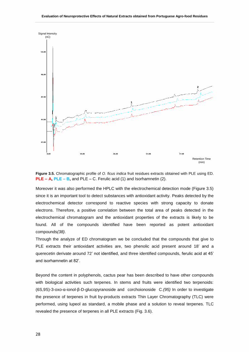
Evaluation of Neuroprotective Effects of Natural Extracts obtained from Portuguese Agro-food Residues
28
Figure 3.5. Chromatographic profile of O. ficus indica fruit residues extracts obtained with PLE using ED.
PLE – A, PLE – B, and PLE – C. Ferulic acid (1) and Isorhamnetin (2).
Moreover it was also performed the HPLC with the electrochemical detection mode (Figure 3.5)
since it is an important tool to detect substances with antioxidant activity. Peaks detected by the
electrochemical detector correspond to reactive species with strong capacity to donate
electrons. Therefore, a positive correlation between the total area of peaks detected in the
electrochemical chromatogram and the antioxidant properties of the extracts is likely to be
found. All of the compounds identified have been reported as potent antioxidant
compounds(38).
Through the analyze of ED chromatogram we be concluded that the compounds that give to
PLE extracts their antioxidant activities are, two phenolic acid present around 18’ and a
querecetin derivate around 72’ not identified, and three identified compounds, ferulic acid at 45’
and isorhamnetin at 82’.
Beyond the content in polyphenols, cactus pear has been described to have other compounds
with biological activities such terpenes. In stems and fruits were identified two terpenoids:
(6S,9S)-3-oxo-α-ionol-β-D-glucopyranoside and corchoionoside C.(95) In order to investigate
the presence of terpenes in fruit by-products extracts Thin Layer Chromatography (TLC) were
performed, using lupeol as standard, a mobile phase and a solution to reveal terpenes. TLC
revealed the presence of terpenes in all PLE extracts (Fig. 3.6).
Signal Intensity
(nC)
Retention Time
(min)
1
2

Evaluation of Neuroprotective Effects of Natural Extracts obtained from Portuguese Agro-food Residues
29
Figure 3.6. TLC plate for assess the presence of terpenoids in O. ficus-indica fruit residues extract
obtained with PLE (PLE – A, PLE – B, and PLE – C in a concentration of 50 mg.mL-1
dw extract).
The presence of a fluorescent band in the same molecular weight of the standard is common to
all PLE extracts. This may reveal that the PLE extracts have lupeol in their composition or a
similar compound with the same molecular weight. TLC reveals also the presence of other
terpenoids compounds with higher molecular weights which identification is under evaluation.
3.1.2. Chemical Antioxidant Capacity
In order to evaluate the chemical antioxidant activities of Opuntia ficus-indica extracts, two
different assays were performed: ORAC and HORAC.
ORAC assay measures the potential to scavenge harmful oxygen species such peroxyl and
HORAC assay analyze the efficacy of the extract to prevent hydroxyl radical formation.
Sample Lupeol PLE – A PLE – B PLE – C
Concentration
(mg.mL-1)
10 50 50 50
Volume (µL) 10 10 10 10

Evaluation of Neuroprotective Effects of Natural Extracts obtained from Portuguese Agro-food Residues
30
Figure 3.7. Chemical antioxidant capacity of Opuntia ficus-indica extracts measured by ORAC and
HORAC assays. (a) ORAC (■) expressed as µmol TE.g-1
dw, and HORAC (■) expressed as µmol CAE.g-1
dw. (b) Antioxidant capacity per unit of phenol ORAC (■) expressed as µmol TE.g-1
GAE, and HORAC (■)
expressed as µmol CAE.g-1
GAE. All values are means of three independent experiments ± SD.
ORAC and HORAC values obtained for O. ficus-indica extracts range from 145 to 481 µmol
TE.g-1
dw, and 167 to 427 µmol CAE.g-1
dw, respectively (Fig. 3.7). As verified for TPC and for
TFC, OBS – CSE had the highest ORAC value. Between Opuntia extracts PLE – B have higher
ORAC and HORAC/TPC ratio, being ORAC/TPC ratio similar to OBS – CSE.
These results suggest that PLE and CSE technologies have the ability to extract different
phenolic compounds from Opuntia fruit residues. While conventional solvent extraction have the
mostly radical chain breaking antioxidants since it presents a higher ORAC/TPC value,
pressurized liquid extracts are mainly constitute for preventive antioxidants showing a higher
HORAC/TPC values. That could be traduced in different in vivo potencies or bioactivities.
3.1.3. Cytotoxicity Profile Determination
Prior to the evaluation of neuroprotective potential of different Opuntia extracts, cytotoxicity
assays were performed using CellTiter-Blue® Reagent (Promega). This reagent is a dark blue
dye that has little intrinsic fluorescence. When present in the cytoplasm of metabolically active
cells is reduced into a fluorescent product, resorfurin. Therefore, fluorescent signal of resorfurin
is proportional to the number of viable cells.
a
bc
bcdd
e
d
0.0
100.0
200.0
300.0
400.0
500.0
600.0
OBS - CSE PLE - A PLE - B PLE - C
a
b
a
ab
cc
d
e
0.00
10.00
20.00
30.00
40.00
50.00
60.00
OBS - CSE PLE - A PLE - B PLE - C
(a) (b)

Evaluation of Neuroprotective Effects of Natural Extracts obtained from Portuguese Agro-food Residues
31
Figure 3.8. Cytotoxicity profile of Opuntia ficus-indica extracts, assessed by CellTiter-Blue Reagent Cell
Viability Assay (Promega). OBS – CSE (a),PLE – A (b), PLE – B (c), and PLE – C (d). SK-N-MC
neuroblastoma cells were incubated for 24 h with the extract (0-250 µg GAE mL-1
). Cell viability is
expressed as percentage of viable cells. Values are expressed as percentage relatively to control (without
extract). All values are means of three independent experiments ± SD.
PLE – B (fig 3.8. (c)), seems to be more toxic extract, while it shows a more pronounced
reduction of cell viability for lower concentrations than the other Opuntia extracts. The extract
that shows a lower toxicity is OBS-CSE (fig 3.8. (a)). All extracts are completely toxic to
concentrations higher than 125 µg GAE.mL-1
. Cytotoxicity profiles are under the expected since
most polyphenols show a bell-shape dose-response curve, presenting cellular toxicity at high
concentrations (96).
Non-toxic concentrations of the extracts selected for neuroprotection assays were 0.25, 0.5, 1,
2.5 and 5 µg GAE.mL-1
. Curiously, the non-toxic range of Opuntia extracts (maximum 5 µg
GAE.mL-1
for all Opuntia extracts) is closer to the values reported for dietary polyphenolic-
derived metabolites in plasma at 4 µM (97). The maximum plasma level of flavonoids in humans
is usually reached between 0.06 and 7.6 µM (33). For quercetin the concentration in human
plasma ranges from 0.1 to 10 µM (34).
3.1.4. Evaluation of Neuroprotective Effect
Today, cell models can provide an indication of biological activity for the phytochemicals in
question. The main advantage of the use of in vitro cell models is the practical convenience,
such as easy culturing, relatively low cost and moderate throughput capabilities (16). Neuronal
0
20
40
60
80
100
120
0 50 100 150 200 250 300
Ce
ll V
iab
ilit
y (%
)
0
20
40
60
80
100
120
0 50 100 150 200 250 300
0
20
40
60
80
100
120
0 50 100 150 200 250 300
Ce
ll V
iab
ilit
y (%
)
Total Phenolic Content (µg GAE mL-1)
0
20
40
60
80
100
120
0 50 100 150 200 250 300
Total Phenolic Content (µg GAE mL-1)
A B
DC
(a) (b)
(c) (d)

Evaluation of Neuroprotective Effects of Natural Extracts obtained from Portuguese Agro-food Residues
32
cell death caused by oxidative stress has been implicated in a variety of brain pathologies
including neurodegenerative diseases such as AD and PD (7).
SK-N-MC cells were pre-incubated for 24 h in the presence or absence of Opuntia extracts and
then challenged with 300 µM H2O2 for 24 h. Neuroprotection was then evaluated by monitoring
cell viability using Cell Titer Blue – Reagent® (Promega). Figure 3.9. shows the different
responses of SK-N-MC cells to stress after pre-incubation with the Opuntia extracts.
Figure 3.9. Neuroprotective effect of Opuntia ficus-indica extracts, assessed by CellTiter-Blue Reagent
Cell Viability Assay (Promega). OBS – CSE (a),PLE – A (b), PLE – B (c), and PLE – C (d). Cell viability is
expressed as percentage of viable cells. SK-N-MC cells were pre-incubated with 0.25, 0.5, 1, 2.5, and 5 µg
GAE.mL-1
of all extracts for 24 h and then injured by 300 µM H2O2 for 24 h. Statistical differences between
treatments and stressed cells are denoted as *p < 0.05, **p < 0.01, ***p < 0.001 and statistical differences
between treatments and not-treated cells are denoted as #p < 0.05,
##p < 0.01,
###p < 0.001. All values are
means of three independent experiments ± SD.
Only OBS – CSE (Figure 3.9. (a)) is able to increase significantly cell viability for 0.25 µg
GAE.mL-1
, while no extract from PLE technology can exert any protective effect. However, all
the extracts in the range of 0.5 to 2.5 µg GAE.mL-1
have the same behavior, exerting a
significant maintenance of cell viability, with no statistical differences between the treatments
with different extracts.
The similarities observed in the intermediary concentrations are not observed in the higher
concentration 5 µg GAE.mL-1
, when PLE – C are not able to protect neuronal cells at the same
level of the remain Opuntia extracts (Figure 3.9. (d)). Comparing the results of neuroprotective
activity with the phenolic profile of Opuntia extracts, it is suggested to exist a negative
correlation of neuroprotection and the presence of betaxhantin pigments, since PLE – C is the
one with possess three compounds from this family.
- -0.25 0.5 1 2.5 5
- + + + + + +
Total Phenolic Content(µg GAE.mL-1)
- - 0.25 0.5 1 2.5 5
H2O2(300 µM)
- + + + + + +
***
###
###***
#***
*** ##***
###***
0
20
40
60
80
100
120
140
***
######
##**
*** ***
***
0
20
40
60
80
100
120
140
Ce
ll V
iab
ility (%
)
***
######
#***
*** ***
***
0
20
40
60
80
100
120
140
***
###
###
###**
***
***
###*
0
20
40
60
80
100
120
140
Cell
Via
bility
(%)
(a) (b)
(c) (d)

Evaluation of Neuroprotective Effects of Natural Extracts obtained from Portuguese Agro-food Residues
33
Kim and co-authors (2006) have already report the neuroprotective effect of a methanolic
extract of O.ficus-indica fruits against NMDA-, KA-, and OGD- induced neuronal injury. The
treatment of neuronal cells with 30, 300 and 1000 µg.mL-1
of Opuntia extract significantly
reduced the neurotoxicity in a dose-dependent way. While interesting results it is important to
note that the concentrations tested by the authors are not dietary relevant and the content in
phenolic compounds of the extracts was not assessed.
3.1.5. Intracelular ROS production determination
The concentration that shows a higher neuroprotective effect was also tested for intracellular
antioxidant capacity. Two different pre-incubation times were tested, at 2 and 24 h, to cover
different timescale events. It was expect that the 2 h pre-incubation would cover the time period
when direct scavenging events caused by the phenolic compounds present in the extracts while
24 h of pre-incubation with extracts could reflect ROS levels influence by indirect effects on
endogenous antioxidant systems.
Figure 3.10. Relative ROS production by SK-N-MC neuroblastoma cells pre-incubated with O. ficus-indica
extracts for 2h (■) and 24 h(■) and submitted to an oxidative stress (300 µM H2O2 for 30 min). ROS were
detected by fluorimetry using H2DCFDA as probe. Statistical differences in relation with cells not treated
are denoted as *p < 0.05, **p < 0.01, ***p < 0.001. All values are means of three independent experiments
± SD.
At both time points, 2 and 24 hours, and in the absence of an imposed oxidative stress all
Opuntia extracts maintain the basal ROS production. H2O2 was used to promote an oxidative
0,0
0,5
1,0
1,5
2,0
2,5
0
0,5
1
1,5
2
2,5
µg GAE.mL-1 - - + + + + + + + +
H2O2
(300 µM)
- + - + - + - + - +
OBS – CSE PLE – A PLE – B PLE – C
RO
S rela
tive
pro
ductio
n

Evaluation of Neuroprotective Effects of Natural Extracts obtained from Portuguese Agro-food Residues
34
stress in cells and in fact, ROS production duplicates after 30 min compared with the control,
however without promoting cell death. With a 2 h pre-incubation, none of the extract reduced
intracellular ROS caused by H2O2-induced stress. With a 24 h pre-incubation, followed by H2O2
stress also none extract reduced intracellular ROS production either. Comparing these results
with those obtained for neuroprotection assay, it is suggested that the neuroprotection carried
out by Opuntia extracts was not mediated by a direct ROS scavenging activity or even by
activating endogenous antioxidant systems. Antioxidant compounds may be characterized into
two types, the direct antioxidants, which are a short-lived redox-active molecules, and indirect
antioxidants, which may or may not show redox activity and are effective by interfering in other
molecular mechanisms (98).
3.1.6. Glutathione (GSH) and Glutathione disulphide (GSSG) Quantification
GSH is the major antioxidant within cells and is involved in maintaining a tight control of redox
status (10) to verify the modifications induced by metabolites pre-treatment and post application
of H2O2 stress, the redox pair GSH/GSSG was quantified. Preliminary results that need to be
confirmed are presented for GSH and GSSG quantifications after a 24h pre-incubation with
Opuntia extracts and an oxidative stress applied with H2O2 (Fig 3.11).
Figure 3.11. Quantifications of GSH and ratio GSH/GSSG after pre-treated for 24 h with O. ficus-indica
extracts, SK-N-MC neuroblastoma cells were subjected to 300 uM H2O2 for 24 h. cells were collected and
analyzed for their content in GSH and GSSG by HPLC. The values of GSH are expressed in nmol
GSH.mg -1
protein. All values are means ± SD.
The cells challenged with H2O2 presented slightly a decrease in the GSH/GSSG ratio as
expected, although not significant in the assay.
Total Phenolic Content
(µg GAE.mL-1)- - 1 1 1 1 1 1 1 1
H2O2
(300 µM)- + - + - + - + - +
0
5
10
15
20
25
30
GS
H/G
SS
G
PLE – A PLE – B PLE – COBS – CSE

Evaluation of Neuroprotective Effects of Natural Extracts obtained from Portuguese Agro-food Residues
35
Cells subjected to a pre-incubation with PLE – A and PLE – C extracts, when challenged with
an oxidative stress, did not show any modifications on GSH/GSSG levels. However, OBS –
CSE seems to promote a decrease in the ratio levels. This decrease after with pre-incubation
with a polyphenolic extract and followed by an oxidative stress was also observed by Tavares
and collaborators (93) and could be due to the mobilization of GSH molecules conducted by
other identities such as proteins. On the contrary, PLE – B extracts promote a slightly increase
in GSH/GSSG ratio. The increment may be related with an effect of the extract in promoting
activities related with restore of GSH. In fact PLE-B extract is the only that after 24 h incubation
reduces the GSH/GSSG ratio in cells, and if followed by stress the cells seems able to recover
to levels higher than the control.
Taking these results in account seems that the neuroprotective effect showed by PLE – B
extract in neuroprotection assay can be due to their capacity of increase GSH/GSSG ratio
levels.
Moreover interesting is the fact that results suggest that the same levels of neuroprotection
capacity at 1 μg GAE.mL-1 observed in all extracts may have different causes. Overall these
preliminary results are very interesting for further confirmations.
It has been shown that curcumin and flavonoids enhance the in vivo synthesis of GSH (99). The
increase in GSH levels in various cell types subjected to polyphenol treatment results from an
increased transcription of the γ-GSS (glutathione synthase) gene (100). GSH may has
multifunctional roles, including being a reducing agent and a cofactor in enzyme function, as
well as playing a role in the chemical antioxidant defense and the detoxification of xenobiotics.
3.2. Prunus avium
Fruits were selected from the ones with no commercial value and were consider waste. Fruits
were processed with peel, seeds and stalks to obtain the extract from the residue. Cherry
extract were obtain trough a conventional solvent extraction (in batch mode) with a H2O:EtOH
(1:1) solution as solvent. To recover and concentrate polyphenols from Saco cherry crude
extract macroporous adsorption resin Amberlite® XAD16 was used as described before
(material and methods, 2.3). Resin Amberlite® XAD16 was chosen due to its low operation
cost, simple handling and high absorption capacity for a large number of compounds, namely
compounds with aromatic rings such as phenolics (72, 88).
3.2.1. Chemical Characterization
The extract obtained from cherry residue is a red/pink powder. TPC obtained by Folin
Ciocalteau method increases after resin concentration (Fig 3.12), it was 10 mg GAE.g-1
dw for
crude extract (CCE) and 375.5 mg GAE.g-1
dw for polyphenol rich cherry extract (PRCE).

Evaluation of Neuroprotective Effects of Natural Extracts obtained from Portuguese Agro-food Residues
36
Figure 3.1Total phenolic content of crude cherry extract (CCE) and polyphenol-rich cherry extract (PRCE),
expressed as mg GAE.g-1
of extract in a dry weight basis. All values are means of three independent
experiments ± SD.
A very important fact is that was a high enrichement in TPC after the submission to AD
technology, proving that Amberlite® XAD16 resin is a good approach to enhance the
concentration in phenolic compounds present in a biological sample. Figure 3.13 shows the
phenolic profile of PRCE at 280, 360 and 527 nm.
Figure 3.13. Chromatographic profile of Polyphenol-Rich Cherry Extract (PRCE) at 280, 360 e 527nm.
Chlorogenic Acid (1), Cyanidin-3-O-glucoside (2), Cyanidin-3-O-rutinuside (3), Peonidin-3-glucoside (4),
Rutin (5), and Quercetin (6) were indentified.
0
50
100
150
200
250
300
350
400
450
CCE PRCE
To
tal P
heno
lic C
onte
nt
(TP
C)
(mg
GA
E.g
-1d
w)
Minutes
0 10 20 30 40 50 60 70 80 90 100 110
mA
U
0
100
200
300
400
500
600
700
800
900
mA
U
0
100
200
300
400
500
600
700
800
900Detector 1-280nmExtracto Cerej a Dil.+v . sobrecargaj ul0912004.dat
Detector 1-360nmExtracto Cerej a Dil.+v . sobrecargaJul0912004.dat
Detector 1-527nmExtracto Cerej a Dil.+v . sobrecargaJul0912004.dat
1
2
3
4
5
6

Evaluation of Neuroprotective Effects of Natural Extracts obtained from Portuguese Agro-food Residues
37
The compounds identified in PRCE by HPLC were a phenolic acid, chlorogenic acid (1), three
anthocyanins namely cyanidin-3-O-glucoside (2), cyanidin-3-O-rutinuside (3), peonidin-3-
glucoside (4), and two flavonoids, rutin (5) and quercetin (6). Anthocyanins are flavonoids
recognized for their promoting health benefits mainly because their strong antioxidant activity
(101, 102). Due to their singular structure, anthocyanins can scavenge ROS and also chelate
metals preventing these way the formation of free radicals by Fenton or Fenton-like
reactions(103).
Through the analyses of ED chromatogram was identified also a flavonol namely Quercetin (Fig
3.14 (5)).
Figure 3.14. Chromatographic profile of PRCE using ED. Chlorogenic acid (1), Cyanidin-3-glucoside (2),
Cyanidin-3-rutinoside (3), Rutin (4) , Quercetin (5).
As showed before, the identification of compounds detected by HPLC with the electrochemical
detection mode is an important tool to detect substances with antioxidant activity. All of the
compounds identified (Figure 3.14.) have been reported as potent antioxidant compounds(38).
Moreover phenolic compounds identified by HPLC – DAD – ED have already been described by
other authors to be the active antioxidant species in cherries. For example, Piccolella and
collaborators (2008) found that flavonoids like epicatechin and anthocyanins, and hydroxyl-
1
2
4
3
5
Signal Intensity
(nC)
Retention Time
(min)

Evaluation of Neuroprotective Effects of Natural Extracts obtained from Portuguese Agro-food Residues
38
cinnamic acids such chlorogenic and neochlorogenic acid are the most active compounds in
scavenging free radicals (104). Previous study with apples and wines have reported good
correlations between the total area of electrochemical chromatogram and chemical antioxidant
activity (105).
3.2.1. Chemical antioxidant activity
Results of antioxidant activity of cherry extract measured by ORAC (μmol TE.g-1
dw extract) and
HORAC (μmol CAE.g-1
dw extract) assays are presented in figure 3.15.
Figure3.15. Antioxidant activity of polyphenol-rich cherry extract (PRCE) measured by ORAC (■) (μmol
TE.g-1
dw extract) and HORAC (■) (μmol CAE.g-1
dw extract) assays. Statistical differences are denoted
with different letters. All values are mean ± SD, n=3.
For ORAC assay, Blando and co-authors (2004)(106) showed that the antioxidant values of
cherries are related with their total anthocyanin content whereas for HORAC assay, no study
has been published for cherry extracts to date. A previous study with apple cultivars good
correlations were obtained between antioxidant values of ORAC and HORAC with catechin,
epicatechin and chlorogenic acid, suggesting that these compounds are the major contributors
of antioxidant capacity in several fruit matrices.
3.2.2. Cytotoxicity profile determination
SK-N-MC cells were incubated with PRCE with concentrations from 0 to 250 µg GAE.mL-1
and
cell viability assayed using CellTiter-Blue® Reagent (Promega) (Fig 3.16). .

Evaluation of Neuroprotective Effects of Natural Extracts obtained from Portuguese Agro-food Residues
39
Figure 3.16. Cytotoxicity profile of Prunus avium extract (PRCE), assessed by CellTiter-Blue Reagent®
Cell Viability Assay (Promega). SK-N-MC neuroblastoma cells were incubated for 24 h with the extract (0-
250 µg GAE mL-1
). Cell viability is expressed as percentage of viable cells. Values are expressed as
percentage relatively to control (without extract). All values are mean ± SD.
Non-toxic concentrations of PRCE selected for neuroprotection assay were 0.25, 0.5, 1, 2.5 and
5 µg GAE.mL-1
. As verified for Opuntia extracts, the higher non-toxic concentration of PRCE (5
µg GAE.mL-1
) is closer to the values reported for anthocyanins present in human plasma, that is
less than 0.15 µM (59).
3.2.3. Neuroprotective effect evaluation
PRCE neuroprotective ability was assayed in the human cell model previously described. SK-N-
MC cells were pre-incubated for 24 h in the presence or absence of PRCE and then challenged
with 300 µM H2O2 for 24 h. Neuroprotection was then evaluated by monitoring cell viability using
Cell Titer Blue – Reagent® (Promega). Figure 3.17. shows the different responses of SK-N-MC
cells to stress.
0
20
40
60
80
100
120
0 50 100 150 200 250 300
Ce
ll V
iab
ilit
y (%
)
Total Phenolic Content (µg GAE.mL-1)

Evaluation of Neuroprotective Effects of Natural Extracts obtained from Portuguese Agro-food Residues
40
Figure 3.17. Neuroprotective effect of Prunus avium extract (PRCE), assessed by CellTiter-Blue Reagent
® Cell Viability Assay (Promega). Cell viability is expressed as percentage of viable cells. SK-N-MC cells were pre-incubated with 0.25, 0.5, 1, 2.5, and 5 µg GAE.mL
-1 of all extracts for 24 h and then injured by
300 µM H2O2 for 24 h. Statistical differences between treatments and stressed cells are denoted as *p < 0.05, **p < 0.01, ***p < 0.001 and statistical differences between treatments and not-treated cells are denoted as
#p < 0.05,
##p < 0.01,
###p < 0.001. All values are means of three independent experiments ±
SD.
As verified for all O. ficus-indica extracts obtained with PLE technology, PRCE can not exert
neuroprotection at the lower tested concentration (0.25 µg GAE.mL-1
). At 0.5 and
1 µg GAE.mL-1
PRCE are able to increase cell viability higher than cells submitted to an
oxidative stress, however the extract can not promote a cell survival at the same scale that the
control cells. In two higher concentrations (2.5 and 5 µg GAE.mL-1
), PRCE are unable to
promote any increase in cell viability.
3.2.4. Intracellular ROS production determination
After measure the chemical antioxidant capacity of PRCE, the cellular antioxidant capacity was
assessed in SK-N-MC cells. H2O2 can generate hydroxyl radicals by Fenton or Fenton-like
reactions(107) and are a stress inductor capable to generate oxidative stress in SK-N-MC cells
with an increase in ROS production (17, 93). After a pre-incubation of cells with the extract
during 2 and 24 h, cell were challenged with H2O2 and cellular levels of ROS were measured
against cells not submitted to H2O2 – induced stress.
***
*** ###
###**
#***
######
0
20
40
60
80
100
120
140
Cell
Via
bil
ity (
%)
Total Phenolic Content
(µg GAE.mL-1
) - - 0.25 0.5 1 2.5 5
H2O2
(300 µM) - + + + + + +

Evaluation of Neuroprotective Effects of Natural Extracts obtained from Portuguese Agro-food Residues
41
Figure 3.18. Relative ROS production by SK-N-MC neuroblastoma cells pre-incubated with Prunus avium
extract (PRCE) for 2h (■) and 24 h(■) and submitted to an oxidative stress (300 µM H2O2 for 30 min). ROS
were detected by fluorimetry using H2DCFDA as probe. Statistical differences in relation with cells not
treated are denoted as p < 0.05, **p < 0.01, ***p < 0.001. All values are means of three independent
experiments ± SD.
In the absence of H2O2, treatment of SK-N-MC cells with PRCE at 1 µg GAE.mL-1
for 2 and 24 h
did not affect endogenous ROS levels (Figure 3.18.). The pre-treatment of neuronal cells with
PRCE during 2 h followed by a H2O2 – induced stress shows a significant decrease of ROS
levels (Figure 3.18. (a)). This effect is not detected at 24 h of PRCE pre-treatment (Figure 3.18.
(b)). As shown by Serra and co-authors (2011) cyaniding 3-glucoside and cyaniding 3-
rutinoside, that are the main phenolics present in PRCE, are compounds with high chemical
antioxidant capacities. Therefore the neuroprotective effect exerted by this extract could be
related with their capacity of exert a direct scavenge of free radicals in neuronal cells, which are
associated with the decrease detected with 2 h incubation.
Total Phenolic Content
(µg GAE.mL-1)- - 1 1
H2O2
(300 µM)- + - +
PRCE
RO
S re
lative
pro
du
ctio
n
***
0
0.5
1
1.5
2
2.5
******
0.00
0.50
1.00
1.50
2.00
2.50

Evaluation of Neuroprotective Effects of Natural Extracts obtained from Portuguese Agro-food Residues
42
3.2.6. Glutathione (GSH) and Glutathione disulphide (GSSG) Quantification
Figure 3.19. Quantification of GSH and GSH/GSSG ratio. After pre-treated for 24 h with Prunus avium
extract (PRCE), SK-N-MC neuroblastoma cells were subjected to 300 µM H2O2 for 24 h. cells were
collected and analyzed for their content in GSH and GSSG by HPLC. The values of GSH are expressed in
nmol GSH.mg -1
protein. All values are means ± SD.
Regarding the ability of PRCE extract to modulate the GSH/GSSG ratio, also a preliminary
assay was performed. Cells pre-incubated with PRCE do not seem to react by altering the
GSH/GSSG balance.suggesting that the neuroprotective effect showed by PRCE is not
mediated by a regulation of GSH/GSSG redox pair.
0
5
10
15
20
25
30
GS
H/G
SS
GPRCE
Total Phenolic Content
(µg GAE.mL-1)- - 1 1
H2O2
(300 µM)- + - +

Evaluation of Neuroprotective Effects of Natural Extracts obtained from Portuguese Agro-food Residues
43
4. Final Conclusions
The aim of this work was to assess the efficacy of O. ficus-indica and P. avium fruit by-products
extracts in a neurodegeneration cell model with SK-N-MC neuroblastoma cell line submitted to
an oxidative injury. All the O. ficus-indica and Prunus avium extracts have protected
neuroblastoma cells from H2O2-induced death at low, non-toxic levels, revealing both species as
promising sources for neuroprotective compounds. This reinforces the importance of the
approach since the concentrations used are found able to protect neuronal cells at
physiologically-relevant serum concentration.
Based on cellular events evaluated the extracts from different biological matrixes seems to
protect neuronal cells trough different cellular mechanisms. While cherry extract are able to
reduce ROS levels, cactus pear extracts are able to modulate the GSH/GSSG homeostasis.
On the other hand the different responses revealed by neuronal cells to extracts obtained with
different extraction technology from the same matrix highlights the influence of chemical
composition. The results suggest that different extraction methods are able to isolate
compounds with differential biological activities and even using the same technology and the
same biological matrix, the conditions of the extraction could influence the phytochemical
content and consequently, the extracts biological activities.
It is now thought more likely that some phytochemicals, including polyphenols, are processed
by the body metabolism as xenobiotics. They stimulate stress- related cell signalling pathways
that result in increased expression of genes that encode cytoprotective genes. This provides
grounds for the hormesis theory, i.e. when mild stress triggers defense mechanisms. In the case
of polyphenols could explain how they could have an indirect antioxidant activity. However,
other phytomolecules could act as antioxidant defences against ROS. Ascorbic acid (Vitamin
C), Vitamin E, glutathione (GSH) and carotenoids are examples of nonenzymatic antioxidant
defenses.
Many in vitro studies are focused on polyphenol-derived health benefits, but using doses
significantly higher than those to which humans are exposed through the diet or that could be
found in the blood (Collins, 2005). This work encompasses a more physiological approach
which takes into account treatment of cell models with relevant in vivo concentrations of
phytochemical metabolites. However and since is food based extract that may be consider to
constitute a supplement/nutraceutical for oral consumption other concerns should be consider
for the more promising extracts. In particular consider the physiochemical changes occurring in
the gastrointestinal tract, absorption and metabolism, these are important factors with a
consequential impact on bioavailability and bioefficacy.

Evaluation of Neuroprotective Effects of Natural Extracts obtained from Portuguese Agro-food Residues
44

Evaluation of Neuroprotective Effects of Natural Extracts obtained from Portuguese Agro-food Residues
45
5. Bibliography
1. Vauzour, D., et al., Polyphenols and human health: prevention of disease and mechanisms of action. Nutrients. 2010. 2(11): p. 1106-31.
2. Carlesi, C., et al., Strategies for clinical approach to neurodegeneration in
Amyotrophic lateral sclerosis. Arch Ital Biol. 2011. 149(1): p. 151-67. 3. Iriti, M., et al., Neuroprotective herbs and foods from different traditional
medicines and diets. Molecules. 2010. 15(5): p. 3517-55. 4. Pandey, K.B. and S.I. Rizvi, Plant polyphenols as dietary antioxidants in human
health and disease. Oxid Med Cell Longev, 2009. 2(5): p. 270-8. 5. Campos-Esparza Mdel, R. and M.A. Torres-Ramos, Neuroprotection by natural
polyphenols: molecular mechanisms. Cent Nerv Syst Agents Med Chem. 2009. 10(4): p. 269-77.
6. Sun, A.Y., et al., Botanical phenolics and brain health. Neuromolecular Med, 2008.
10(4): p. 259-74. 7. Zhao, B., Natural antioxidants for neurodegenerative diseases. Mol Neurobiol,
2005. 31(1-3): p. 283-93. 8. Cui, H., Y. Kong, and H. Zhang, Oxidative stress, mitochondrial dysfunction, and
aging. J Signal Transduct. 2012: p. 646354. 9. Kelsey, N.A., H.M. Wilkins, and D.A. Linseman, Nutraceutical antioxidants as
novel neuroprotective agents. Molecules. 2010. 15(11): p. 7792-814. 10. Ballatori, N., et al., Glutathione dysregulation and the etiology and progression of
human diseases. Biol Chem, 2009. 390(3): p. 191-214. 11. Ramalingam, M. and S.-J. Kim, Reactive oxygen/nitrogen species and their
functional correlations in neurodegenerative diseases. 2012. Journal of Neural Transmission. 119(8): p. 891-910.
12. Facecchia, K., et al., Oxidative toxicity in neurodegenerative diseases: role of
mitochondrial dysfunction and therapeutic strategies. J Toxicol. 2011: p. 683728. 13. Halliwell, B., Free radicals and antioxidants – quo vadis? Trends in
Pharmacological Sciences. 2011. 32(3): p. 125-130. 14. Ebrahimi, A. and H. Schluesener, Natural polyphenols against neurodegenerative
disorders: potentials and pitfalls. Ageing Res Rev. 2012. 11(2): p. 329-45. 15. Dumont, M. and M.F. Beal, Neuroprotective strategies involving ROS in Alzheimer
disease. Free Radic Biol Med. 2011. 51(5): p. 1014-26. 16. Traka, M.H. and R.F. Mithen, Plant science and human nutrition: challenges in
assessing health-promoting properties of phytochemicals. 2011. Plant Cell. 23(7): p. 2483-97.
17. Tavares, L., et al., Neuroprotective effects of digested polyphenols from wild
blackberry species. Eur J Nutr.2012. 18. Tavares, L., et al., Antioxidant capacity of Macaronesian traditional medicinal
plants. 2010. Molecules. 15(4): p. 2576-92.

Evaluation of Neuroprotective Effects of Natural Extracts obtained from Portuguese Agro-food Residues
46
19. Tavares, L., et al., The neuroprotective potential of phenolic-enriched fractions from four Juniperus species found in Portugal. Food Chem. 2012. 135(2): p. 562-70.
20. Shahidi, F., Nutraceuticals and functional foods: Whole versus processed foods.
Trends in Food Science & Technology, 2009. 20(9): p. 376-387. 21. Pandey, M., R.K. Verma, and S.A. Saraf, Nutraceuticals: new era of medicine and
health. Asian Journal of Pharmaceutical and Clinical Research, 2010. vol3(1). 22. Espin, J.C., M.T. Garcia-Conesa, and F.A. Tomas-Barberan, Nutraceuticals: facts
and fiction. Phytochemistry, 2007. 68(22-24): p. 2986-3008. 23. Cencic, A. and W. Chingwaru, The role of functional foods, nutraceuticals, and
food supplements in intestinal health. Nutrients. 2012. 2(6): p. 611-25. 24. Chao, J., et al., Nutraceuticals and their preventive or potential therapeutic value
in Parkinson's disease. Nutr Rev. 2012. 70(7): p. 373-386. 25. Scalbert, A., et al., Databases on Food Phytochemicals and Their Health-
Promoting Effects. Journal of Agricultural and Food Chemistry, 2011. 59(9): p. 4331-4348.
26. Tsao, R., Chemistry and biochemistry of dietary polyphenols. Nutrients. 2010.
2(12): p. 1231-46. 27. Pandey, K.B. and S.I. Rizvi, Plant polyphenols as dietary antioxidants in human
health and disease. Oxidative Medicine and Cellular Longevity, 2009. 2(5): p. 270-278.
28. Moreira, P.I., et al., Oxidative stress mechanisms and potential therapeutics in
Alzheimer disease. J Neural Transm, 2005. 112(7): p. 921-32. 29. Beal, M.F., Mitochondria, oxidative damage, and inflammation in Parkinson's
disease. Ann N Y Acad Sci, 2003. 991: p. 120-31. 30. Conforti, F., et al., In vivo anti-inflammatory and in vitro antioxidant activities of
Mediterranean dietary plants. Journal of Ethnopharmacology, 2008. 116(1): p. 144-151.
31. Paredes-Lopez, O., et al., Berries: improving human health and healthy aging, and
promoting quality life--a review. Plant Foods Hum Nutr. 2010. 65(3): p. 299-308. 32. Young, J., K.W. Wahle, and S.P. Boyle, Cytoprotective effects of phenolic
antioxidants and essential fatty acids in human blood monocyte and neuroblastoma cell lines: surrogates for neurological damage in vivo. Prostaglandins Leukot Essent Fatty Acids, 2008. 78(1): p. 45-59.
33. Quideau, S., et al., Plant Polyphenols: Chemical Properties, Biological Activities,
and Synthesis. Angewandte Chemie-International Edition, 2011. 50(3): p. 586-621. 34. Rahman, I., S.K. Biswas, and P.A. Kirkham, Regulation of inflammation and redox
signaling by dietary polyphenols. Biochem Pharmacol, 2006. 72(11): p. 1439-52. 35. Fraga, C.G., et al., Basic biochemical mechanisms behind the health benefits of
polyphenols. Mol Aspects Med. 2010. 31(6): p. 435-45. 36. Lin, H.J., et al., Aloe-emodin metabolites protected N-methyl-d-aspartate-treated
retinal ganglion cells by Cu-Zn superoxide dismutase. J Ocul Pharmacol Ther, 2007. 23(2): p. 152-71.

Evaluation of Neuroprotective Effects of Natural Extracts obtained from Portuguese Agro-food Residues
47
37. Mythri, R.B., et al., Glutamoyl diester of the dietary polyphenol curcumin offers improved protection against peroxynitrite-mediated nitrosative stress and damage of brain mitochondria in vitro: implications for Parkinson's disease. Mol Cell Biochem. 2011. 347(1-2): p. 135-43.
38. Ataie, A., et al., Neuroprotective effects of the polyphenolic antioxidant agent,
Curcumin, against homocysteine-induced cognitive impairment and oxidative stress in the rat. Pharmacol Biochem Behav. 2010. 96(4): p. 378-85.
39. Harish, G., et al., Bioconjugates of curcumin display improved protection against
glutathione depletion mediated oxidative stress in a dopaminergic neuronal cell line: Implications for Parkinson's disease. Bioorg Med Chem. 2010. 18(7): p. 2631-8.
40. Kumar, P., et al., Possible neuroprotective mechanisms of curcumin in
attenuating 3-nitropropionic acid-induced neurotoxicity. Methods Find Exp Clin Pharmacol, 2007. 29(1): p. 19-25.
41. Romeo, L., et al., The major green tea polyphenol, (-)-epigallocatechin-3-gallate,
induces heme oxygenase in rat neurons and acts as an effective neuroprotective agent against oxidative stress. J Am Coll Nutr, 2009. 28 Suppl: p. 492S-499S.
42. Kang, K.S., et al., Dual beneficial effects of (-)-epigallocatechin-3-gallate on
levodopa methylation and hippocampal neurodegeneration: in vitro and in vivo studies. PLoS One. 2010. 5(8): p. e11951.
43. Park, J.W., et al., Green tea polyphenol (-)-epigallocatechin gallate reduces
neuronal cell damage and up-regulation of MMP-9 activity in hippocampal CA1 and CA2 areas following transient global cerebral ischemia. J Neurosci Res, 2009. 87(2): p. 567-75.
44. Srividhya, R., et al., Attenuation of senescence-induced oxidative exacerbations
in aged rat brain by (-)-epigallocatechin-3-gallate. Int J Dev Neurosci, 2008. 26(2): p. 217-23.
45. Weinreb, O., T. Amit, and M.B. Youdim, A novel approach of proteomics and
transcriptomics to study the mechanism of action of the antioxidant-iron chelator green tea polyphenol (-)-epigallocatechin-3-gallate. Free Radic Biol Med, 2007. 43(4): p. 546-56.
46. Koh, S.H., et al., The effect of epigallocatechin gallate on suppressing disease
progression of ALS model mice. Neurosci Lett, 2006. 395(2): p. 103-7. 47. Lemus-Molina, Y., et al., Mangifera indica L. extract attenuates glutamate-induced
neurotoxicity on rat cortical neurons. Neurotoxicology, 2009. 30(6): p. 1053-8. 48. Amazzal, L., et al., Mangiferin protects against 1-methyl-4-phenylpyridinium
toxicity mediated by oxidative stress in N2A cells. Neurosci Lett, 2007. 418(2): p. 159-64.
49. Campos-Esparza, M.R., M.V. Sanchez-Gomez, and C. Matute, Molecular
mechanisms of neuroprotection by two natural antioxidant polyphenols. Cell Calcium, 2009. 45(4): p. 358-68.
50. Dade, M.M., et al., Antioxidant and cytotoxic properties of an aqueous extract
from the Argentinean plant Hedeoma multiflorum. Pharm Biol. 2011. 49(6): p. 633-9.
51. Lee, H.H., C.T. Lin, and L.L. Yang, Neuroprotection and free radical scavenging
effects of Osmanthus fragrans. J Biomed Sci, 2007. 14(6): p. 819-27.

Evaluation of Neuroprotective Effects of Natural Extracts obtained from Portuguese Agro-food Residues
48
52. Ritz, M.F., et al., Chronic treatment with red wine polyphenol compounds mediates neuroprotection in a rat model of ischemic cerebral stroke. J Nutr, 2008. 138(3): p. 519-25.
53. Zhang, F., et al., Resveratrol protects dopamine neurons against
lipopolysaccharide-induced neurotoxicity through its anti-inflammatory actions. Mol Pharmacol. 2010. 78(3): p. 466-77.
54. Sakata, Y., et al., Resveratrol protects against experimental stroke: putative
neuroprotective role of heme oxygenase 1. Exp Neurol. 2010. 224(1): p. 325-9. 55. Lu, K.T., et al., Neuroprotective effects of resveratrol on MPTP-induced neuron
loss mediated by free radical scavenging. J Agric Food Chem, 2008. 56(16): p. 6910-3.
56. Okawara, M., et al., Resveratrol protects dopaminergic neurons in midbrain slice
culture from multiple insults. Biochem Pharmacol, 2007. 73(4): p. 550-60. 57. Manczak, M., et al., Mitochondria-targeted antioxidants protect against amyloid-
beta toxicity in Alzheimer's disease neurons. J Alzheimers Dis. 2010. 20 Suppl 2: p. S609-31.
58. Chen, C.M., et al., Novel regimen through combination of memantine and tea
polyphenol for neuroprotection against brain excitotoxicity. J Neurosci Res, 2008. 86(12): p. 2696-704.
59. Ignat, I., I. Volf, and V.I. Popa, A critical review of methods for characterisation of
polyphenolic compounds in fruits and vegetables. Food Chemistry. 2011. 126(4): p. 1821-1835.
60. Seabra, I.J., et al., High pressure solvent extraction of maritime pine bark: Study
of fractionation, solvent flow rate and solvent composition. Journal of Supercritical Fluids, 2012. 62: p. 135-148.
61. Mustafa, A. and C. Turner, Pressurized liquid extraction as a green approach in
food and herbal plants extraction: A review. Analytica Chimica Acta, 2011. 703(1): p. 8-18.
62. Grotewold, E., The science of Flavonoids. 2006: Springer Science and Business
Media, Inc. 63. Berrueta, L., B. Gallo, and F. Vicente, A review of solid phase extraction: Basic
principles and new developments. Chromatographia, 1995. 40(7): p. 474-483. 64. Zessner, H., et al., Fractionation of polyphenol-enriched apple juice extracts to
identify constituents with cancer chemopreventive potential. Mol Nutr Food Res, 2008. 52 Suppl 1: p. S28-44.
65. Salim, N., et al., Chemical composition of Opuntia ficus-indica (L.) fruit. African
Journal of Biotechnology, 2009. 8(8): p. 1623-1624. 66. Piga, A., Cactus pear: A fruit of nutraceutical and functional importance. Journal
of the Professional Association for Cactus Development, 2004. 6: p. 9-22. 67. Butera, D., et al., Antioxidant activities of sicilian prickly pear (Opuntia ficus
indica) fruit extracts and reducing properties of its betalains: betanin and indicaxanthin. J Agric Food Chem, 2002. 50(23): p. 6895-901.
68. Huang, X., et al., Neuroprotective and antioxidative effect of cactus
polysaccharides in vivo and in vitro. Cell Mol Neurobiol, 2009. 29(8): p. 1211-21.
Evaluation of Neuroprotective Effects of Natural Extracts obtained from Portuguese Agro-food Residues
49
69. Feugang, J.M., et al., Nutritional and medicinal use of Cactus pear (Opuntia spp.) cladodes and fruits. Front Biosci, 2006. 11: p. 2574-89.
70. Stintzing, F.C., et al., Color, betalain pattern, and antioxidant properties of cactus
pear (Opuntia spp.) clones. J Agric Food Chem, 2005. 53(2): p. 442-51. 71. Fernandez-Lopez, J.A., et al., Determination of antioxidant constituents in cactus
pear fruits. Plant Foods Hum Nutr. 2010. 65(3): p. 253-9. 72. Moussa-Ayoub, T.E., et al., Identification and quantification of flavonol aglycons
in cactus pear (Opuntia ficus indica) fruit using a commercial pectinase and cellulase preparation. Food Chemistry, 2011. 124(3): p. 1177-1184.
73. Cardador-Martinez, A., C. Jimenez-Martinez, and G. Sandoval, Revalorization of
cactus pear (Opuntia spp.) wastes as a source of antioxidants. Ciencia E Tecnologia De Alimentos, 2011. 31(3): p. 782-788.
74. Ha, H.J., et al., Quercetin attenuates oxygen-glucose deprivation- and excitotoxin-
induced neurotoxicity in primary cortical cell cultures. Biol Pharm Bull, 2003. 26(4): p. 544-6.
75. Kim, J.H., et al., Opuntia ficus-indica attenuates neuronal injury in in vitro and in
vivo models of cerebral ischemia. J Ethnopharmacol, 2006. 104(1-2): p. 257-62. 76. Goncalves, B., et al., Effect of ripeness and postharvest storage on the phenolic
profiles of Cherries (Prunus avium L.). J Agric Food Chem, 2004. 52(3): p. 523-30. 77. Serra, A.T., et al., Identification of bioactive response in traditional cherries from
Portugal. Food Chemistry, 2011. 125(2): p. 318-325. 78. Andres-Lacueva, C., et al., Anthocyanins in aged blueberry-fed rats are found
centrally and may enhance memory. Nutr Neurosci, 2005. 8(2): p. 111-20. 79. Scordino, M., et al., Adsorption of flavonoids on resins: Hesperidin. Journal of
Agricultural and Food Chemistry, 2003. 51(24): p. 6998-7004. 80. Michalska, A., et al., Antioxidant contents and antioxidative properties of
traditional rye breads. J Agric Food Chem, 2007. 55(3): p. 734-40. 81. Ou, B., M. Hampsch-Woodill, and R.L. Prior, Development and validation of an
improved oxygen radical absorbance capacity assay using fluorescein as the fluorescent probe. J Agric Food Chem, 2001. 49(10): p. 4619-26.
82. Ou, B., et al., Novel fluorometric assay for hydroxyl radical prevention capacity
using fluorescein as the probe. J Agric Food Chem, 2002. 50(10): p. 2772-7. 83. Kand’ár, R., et al., Determination of reduced and oxidized glutathione in
biological samples using liquid chromatography with fluorimetric detection. Journal of Pharmaceutical and Biomedical Analysis, 2007. 43(4): p. 1382-1387.
84. Tavares, L.l., et al., Neuroprotective effect of blackberry (Rubus sp.) polyphenols
is potentiated after simulated gastrointestinal digestion. Food Chemistry. 2012. 131(4): p. 1443-1452.
85. Cai, W.R., X.H. Gu, and J. Tang, Extraction, Purification, and Characterisation of
the Flavonoids from Opuntia milpa alta Skin. Czech Journal of Food Sciences, 2010. 28(2): p. 108-116.
86. Lee, E.H., et al., Constituents of the stems and fruits of Opuntia ficus-indica var.
saboten. Arch Pharm Res, 2003. 26(12): p. 1018-23.

Evaluation of Neuroprotective Effects of Natural Extracts obtained from Portuguese Agro-food Residues
50
87. Scapagnini, G., et al., Modulation of Nrf2/ARE Pathway by Food Polyphenols: A Nutritional Neuroprotective Strategy for Cognitive and Neurodegenerative Disorders. Molecular Neurobiology, 2011. 44(2): p. 192-201.
88. Manach, C., et al., Bioavailability and bioefficacy of polyphenols in humans. I.
Review of 97 bioavailability studies. American Journal of Clinical Nutrition, 2005. 81(1): p. 230s-242s.
89. Ghosh, D. and A. Scheepens, Vascular action of polyphenols. Mol Nutr Food Res,
2009. 53(3): p. 322-31. 90. Schmitt-Schillig, S., et al., Flavonoids and the aging brain. J Physiol Pharmacol,
2005. 56 Suppl 1: p. 23-36. 91. Biswas, S., J. Bhattacharyya, and A.G. Dutta, Oxidant induced injury of
erythrocyte-role of green tea leaf and ascorbic acid. Mol Cell Biochem, 2005. 276(1-2): p. 205-10.
92. Blando, F., C. Gerardi, and I. Nicoletti, Sour cherry (Prunus cerasus L)
anthocyanins as ingredients for functional foods. Journal of Biomedicine and Biotechnology, 2004(5): p. 253-258.
93. McCune, L.M., et al., Cherries and Health: A Review. Critical Reviews in Food
Science and Nutrition, 2011. 51(1): p. 1-12. 94. Piccolella, S., et al., Antioxidant properties of sour cherries (Prunus cerasus L.):
role of colorless phytochemicals from the methanolic extract of ripe fruits. J Agric Food Chem, 2008. 56(6): p. 1928-35.
95. Feliciano, R., et al., Phenolic Content and Antioxidant Activity of Moscatel
Dessert Wines from the Setúbal Region in Portugal. Food Analytical Methods, 2009. 2(2): p. 149-161.
96. Blando, F., C. Gerardi, and I. Nicoletti, Sour Cherry (Prunus cerasus L)
Anthocyanins as Ingredients for Functional Foods. J Biomed Biotechnol, 2004. 2004(5): p. 253-258.
97. Wijeratne, S.S.K., S.L. Cuppett, and V. Schlegel, Hydrogen peroxide induced
oxidative stress damage and antioxidant enzyme response in Caco-2 human colon cells. Journal of Agricultural and Food Chemistry, 2005. 53(22): p. 8768-8774.
98. Jacob, J.K., et al., Biochemical basis for functional ingredient design from fruits.
Annu Rev Food Sci Technol. 3: p. 79-104. 99. Biswas, S., J. Bhattacharyya, and A.G. Dutta, Oxidant induced injury of
erythrocyte-role of green tea leaf and ascorbic acid. Mol Cell Biochem, 2005. 276(1-2): p. 205-10.
100. Moskaug, J.O., et al., Polyphenols and glutathione synthesis regulation. Am J
Clin Nutr, 2005. 81(1 Suppl): p. 277S-283S. 101. Blando, F., C. Gerardi, and I. Nicoletti, Sour cherry (Prunus cerasus L)
anthocyanins as ingredients for functional foods. Journal of Biomedicine and Biotechnology, 2004(5): p. 253-258.
102. McCune, L.M., et al., Cherries and Health: A Review. Critical Reviews in Food
Science and Nutrition, 2011. 51(1): p. 1-12.

Evaluation of Neuroprotective Effects of Natural Extracts obtained from Portuguese Agro-food Residues
51
103. Miguel, M.G., Anthocyanins: Antioxidant and/or anti-inflammatory activities. Journal of Applied Pharmaceutical Science, 2011: p. 07-15.
104. Piccolella, S., et al., Antioxidant properties of sour cherries (Prunus cerasus L.):
role of colorless phytochemicals from the methanolic extract of ripe fruits. J Agric Food Chem, 2008. 56(6): p. 1928-35. Evaluation of Neuroprotective Effects of Natural Extracts obtained from Portuguese Agro-food Residues
105. Feliciano, R., et al., Phenolic Content and Antioxidant Activity of Moscatel Dessert
Wines from the Setúbal Region in Portugal. Food Analytical Methods, 2009. 2(2): p. 149-161.
106. Blando, F., C. Gerardi, and I. Nicoletti, Sour Cherry (Prunus cerasus L)
Anthocyanins as Ingredients for Functional Foods. J Biomed Biotechnol, 2004. 2004(5): p. 253-258.
107. Wijeratne, S.S.K., S.L. Cuppett, and V. Schlegel, Hydrogen peroxide induced
oxidative stress damage and antioxidant enzyme response in Caco-2 human colon cells. Journal of Agricultural and Food Chemistry, 2005. 53(22): p. 8768-8774.

Evaluation of Neuroprotective Effects of Natural Extracts obtained from Portuguese Agro-food Residues
52