evaluation of right iliac fossa mass-a retrospective … graham-div_manj.pdf · evaluation of right...
TRANSCRIPT
Jemds.com Original Article
J Evolution Med Dent Sci/ eISSN- 2278-4802, pISSN- 2278-4748/ Vol. 5/ Issue 03/ Jan. 11, 2016 Page 200
EVALUATION OF RIGHT ILIAC FOSSA MASS-A RETROSPECTIVE STUDY
G. Raja Billy Graham1, V. Vijayabhasker2 1Professor, Department of General Surgery, Government Chengalpattu Medical College. 2Assistant Professor, Department of General Surgery, Melmaruvathur Adhiparasakthi Institute of Medical Sciences and Research,
Melmaruvathur.
ABSTRACT
Mass in right iliac fossa is one of the common clinical surgical entity with varied etiologies, involving intra peritoneal organs like
vermiform appendix, caucus, ileum, retroperitoneal structures like kidneys, blood vessels etc., and abdominal wall masses like
desmoids tumor. To know the etiology and the various pattern of presentation of right iliac fossa mass in our institution a
retrospective study was designed including all adult patients diagnosed to have a mass in right iliac fossa. Pediatric patients and
female patients having a mass due to gynaecological diseases were excluded from this study. All clinical parameters, radiological
investigations, biochemical investigations, endoscopy, cytology, biopsy were collected and analysed systematically.
KEYWORDS
Carcinoma Caecum, Appendicular Mass, Rif Mass, Psoas Abscess, Ileocaecal Tuberculosis, Crohn’s Disease.
HOW TO CITE THIS ARTICLE: Graham GRB, Vijayabhasker V. Evaluation of right iliac fossa mass-a retrospective study. J Evolution
Med Dent Sci 2016;5(3):200-214, DOI: 10.14260/jemds/2016/45
INTRODUCTION
Right iliac fossa mass may arise from parietal wall or
intraperitoneum or retroperitoneum. Mass may arise from
right iliac regional structures or extended from adjacent
structures.
Various Causes for Right Iliac Fossa Mass Parietal Wall
Masses
Lipoma, desmoid tumor, pyogenic abscess and haematoma,
iliac or appendicular abscess burrowing into abdominal wall.
Intraperitoneal Masses
Appendicular abscess or mass, ileocaecal tuberculosis,
carcinoma caecum, mesenteric adenitis, iliac nodes, typhlitis,
Crohn’s disease, actinomycosis, distended gallblader, ovarian
cysts, fibroid uterus, tubo-ovarian mass, occasionally
intussusception, amoeboma, diverticular disease.
Retroperitoneal Masses
Sarcoma, aneurysm, psoas abscess, undescended testis,
unascended kidney and tumour from ilium and cartilage.
Appendicular pathology is commonest cause for right
iliac fossa mass (Either appendicular mass or abscess). Other
common causes are ileo-caecal tuberculosis and carcinoma
caecum.
Appendicular mass is formed by inflamed appendix
adherent with dilated ileum, greater omentum and caecum.1
Appendicular abscess due to suppuration in an acute
appendicitis or suppuration in an already formed
appendicular mass.1 Abdominal tuberculosis is common in
developing countries like India. It is sixth most common type
Financial or Other, Competing Interest: None. Submission 10-11-2015, Peer Review 11-11-2015, Acceptance 19-11-2015, Published 09-01-2016. Corresponding Author: Dr. G. Raja Billy Graham, No. 53, Vedhachalam Nagar, Chengalpattu, Kanchipuram-603001, Tamilnadu. E-mail: [email protected] DOI:10.14260/jemds/2015/45
of extrapulmonary tuberculosis. Its incidence is high in HIV
infected patients.(2) Ileo-caecal tuberculosis is commonest type
of abdominal tuberculosis due to presence of Peyer’s patches
and stasis of luminal contents favoured by ileo-caecal valve.
Commonest type of carcinoma in caecum is
adenocarcinoma. Third common site in large bowel carcinoma
(12%). Diet with lack of fibres and high fat increases risk.
Dietary vitamins A, C, E (Antioxidants) and Zinc and high fibre
diet reduces the risk. Diagnosis of right iliac fossa mass mainly
depends on complete clinical examination, radiological,
biochemical, microbiological and pathological investigations.
Commonest cause of right iliac fossa mass in our country is
appendicular mass or abscess, ileo-caecal tuberculosis and
carcinoma caecum.
OBJECTIVES OF THE STUDY
To study the aetiology, pattern of presentation, management
and complications in patients with right iliac fossa mass in our
institution.
MATERIALS AND METHODS
This retrospective study was carried out in our institution over
a period of two years from October 2010 to October 2012. All
patients who are provisionally diagnosed to have mass in the
right iliac fossa by clinical evaluation are included in the study.
All datas including age, gender, relevant history, investigations
(Complete blood count, Blood grouping and Rh typing, HIV I
and II, Chest radiograph, Ultrasound abdomen and pelvis, CT
Scan abdomen and pelvis, IVP and barium follow through and
enema, FNAC, Biopsy) were done to conclude the final
diagnosis and appropriate treatment and postoperative
complications and final histopathological reports were
recorded in the standard forms. Cases are selected by following
Inclusion and exclusion criteria;
Inclusion Criteria
All the cases admitted with Right iliac fossa mass in our
institution.
Exclusion Criteria
Female patients with gynaecological diseases. Paediatric age
group (<12 years) patients with right iliac fossa mass.
Jemds.com Original Article
J Evolution Med Dent Sci/ eISSN- 2278-4802, pISSN- 2278-4748/ Vol. 5/ Issue 03/ Jan. 11, 2016 Page 201
OBSERVATION AND RESULTS
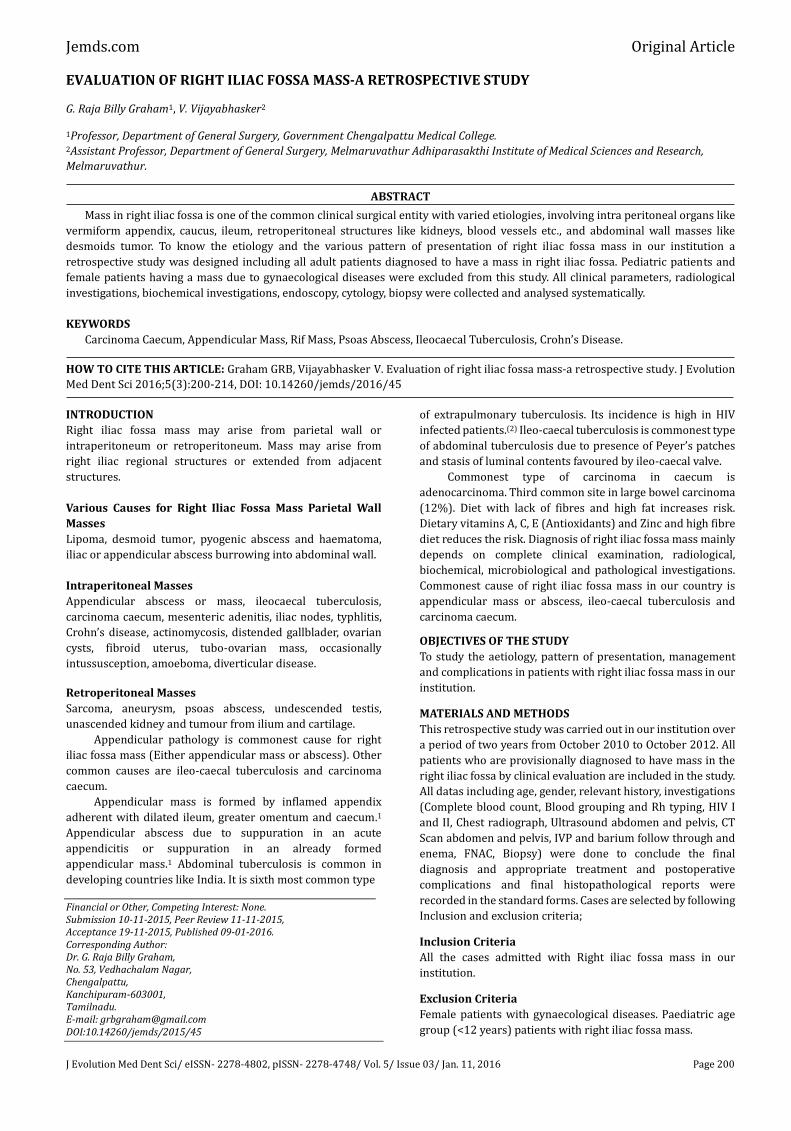
Fig. 1: Incidence of various conditions
In our study of 80 cases, 60% percent of cases are related to appendicular pathology; 20% of cases are related to ileo-caecal
tuberculosis, 12.5% cases are related to carcinoma caecum, 7.5% of cases are related to various type of parietal wall and
retroperitoneal causes like Psoas abscess, Retroperitoneal Schwannoma, Desmoid tumour, Rectus sheath haematoma. One case of
carcinoma caecum was associated with ileo-caecal tuberculosis.
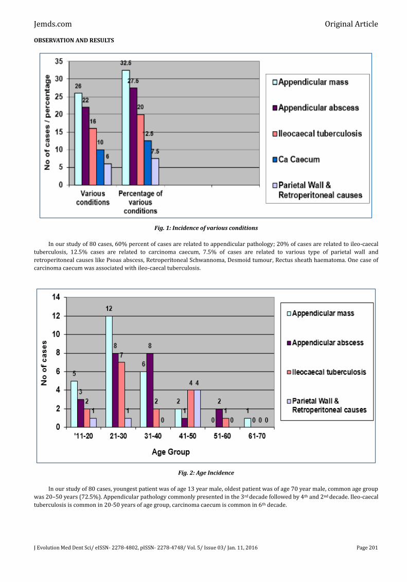
Fig. 2: Age Incidence
In our study of 80 cases, youngest patient was of age 13 year male, oldest patient was of age 70 year male, common age group
was 20–50 years (72.5%). Appendicular pathology commonly presented in the 3rd decade followed by 4th and 2nd decade. Ileo-caecal
tuberculosis is common in 20-50 years of age group, carcinoma caecum is common in 6th decade.
Jemds.com Original Article
J Evolution Med Dent Sci/ eISSN- 2278-4802, pISSN- 2278-4748/ Vol. 5/ Issue 03/ Jan. 11, 2016 Page 202
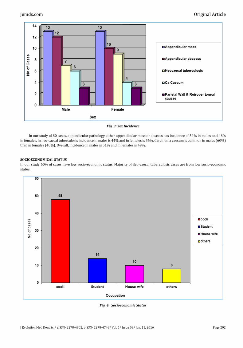
Fig. 3: Sex Incidence
In our study of 80 cases, appendicular pathology either appendicular mass or abscess has incidence of 52% in males and 48%
in females. In ileo-caecal tuberculosis incidence in males is 44% and in females is 56%. Carcinoma caecum is common in males (60%)
than in females (40%). Overall, incidence in males is 51% and in females is 49%.
SOCIOECONOMICAL STATUS
In our study 60% of cases have low socio-economic status. Majority of ileo-caecal tuberculosis cases are from low socio-economic
status.
Fig. 4: Socioeconomic Status
Jemds.com Original Article
J Evolution Med Dent Sci/ eISSN- 2278-4802, pISSN- 2278-4748/ Vol. 5/ Issue 03/ Jan. 11, 2016 Page 203
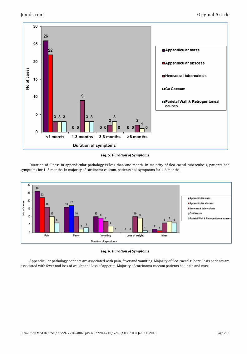
Fig. 5: Duration of Symptoms
Duration of illness in appendicular pathology is less than one month. In majority of ileo-caecal tuberculosis, patients had
symptoms for 1–3 months. In majority of carcinoma caecum, patients had symptoms for 1-6 months.
Fig. 6: Duration of Symptoms
Appendicular pathology patients are associated with pain, fever and vomiting. Majority of ileo-caecal tuberculosis patients are
associated with fever and loss of weight and loss of appetite. Majority of carcinoma caecum patients had pain and mass.
Jemds.com Original Article
J Evolution Med Dent Sci/ eISSN- 2278-4802, pISSN- 2278-4748/ Vol. 5/ Issue 03/ Jan. 11, 2016 Page 204
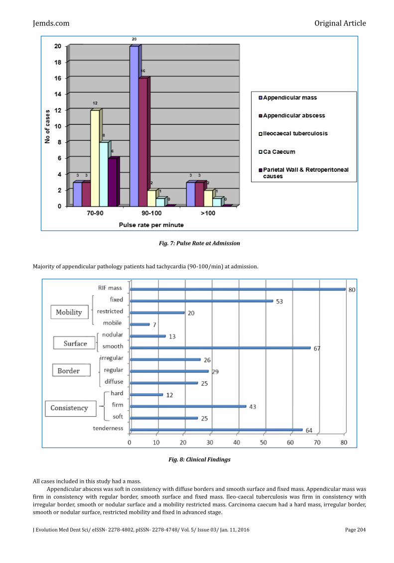
Fig. 7: Pulse Rate at Admission
Majority of appendicular pathology patients had tachycardia (90-100/min) at admission.
Fig. 8: Clinical Findings
All cases included in this study had a mass.
Appendicular abscess was soft in consistency with diffuse borders and smooth surface and fixed mass. Appendicular mass was
firm in consistency with regular border, smooth surface and fixed mass. Ileo-caecal tuberculosis was firm in consistency with
irregular border, smooth or nodular surface and a mobility restricted mass. Carcinoma caecum had a hard mass, irregular border,
smooth or nodular surface, restricted mobility and fixed in advanced stage.
Jemds.com Original Article
J Evolution Med Dent Sci/ eISSN- 2278-4802, pISSN- 2278-4748/ Vol. 5/ Issue 03/ Jan. 11, 2016 Page 205
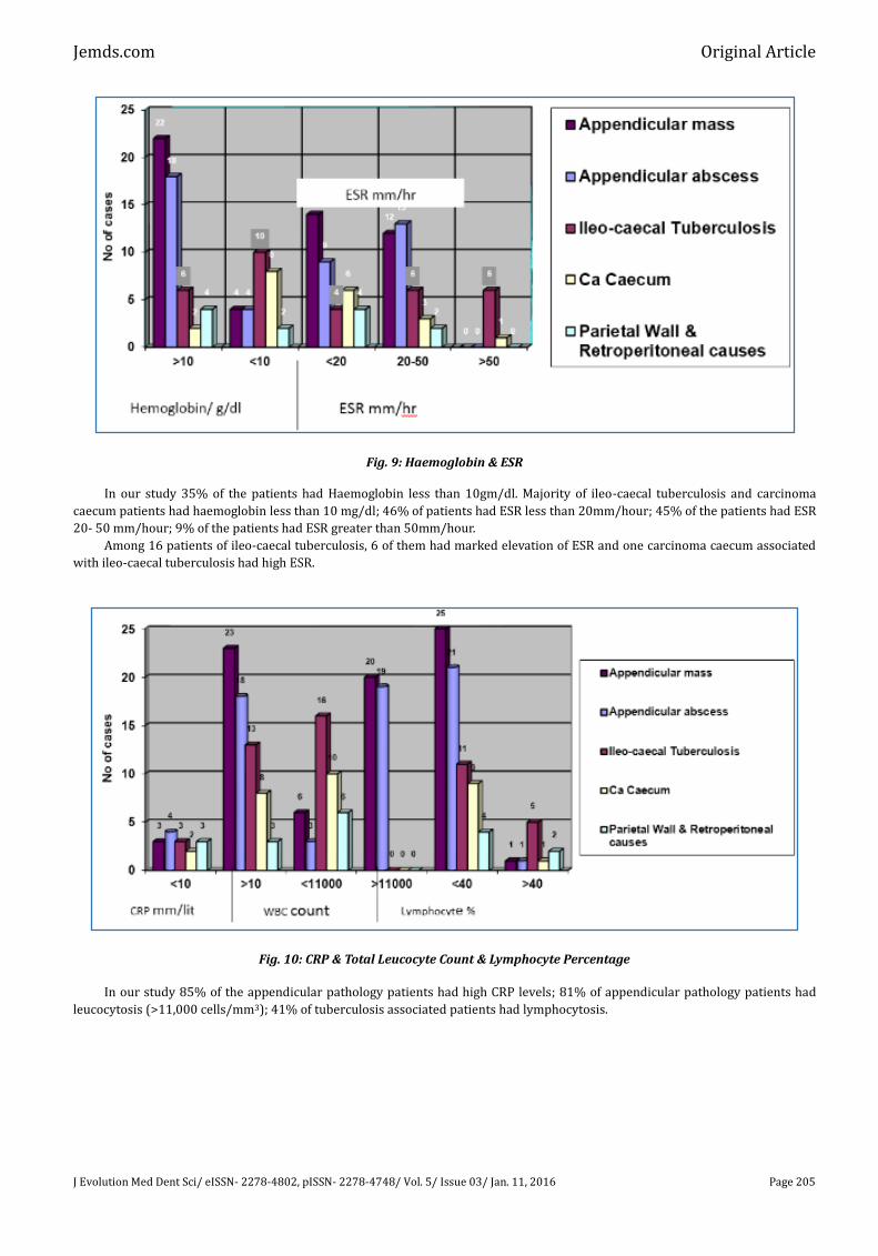
Fig. 9: Haemoglobin & ESR
In our study 35% of the patients had Haemoglobin less than 10gm/dl. Majority of ileo-caecal tuberculosis and carcinoma
caecum patients had haemoglobin less than 10 mg/dl; 46% of patients had ESR less than 20mm/hour; 45% of the patients had ESR
20- 50 mm/hour; 9% of the patients had ESR greater than 50mm/hour.
Among 16 patients of ileo-caecal tuberculosis, 6 of them had marked elevation of ESR and one carcinoma caecum associated
with ileo-caecal tuberculosis had high ESR.
Fig. 10: CRP & Total Leucocyte Count & Lymphocyte Percentage
In our study 85% of the appendicular pathology patients had high CRP levels; 81% of appendicular pathology patients had
leucocytosis (>11,000 cells/mm3); 41% of tuberculosis associated patients had lymphocytosis.
Jemds.com Original Article
J Evolution Med Dent Sci/ eISSN- 2278-4802, pISSN- 2278-4748/ Vol. 5/ Issue 03/ Jan. 11, 2016 Page 206
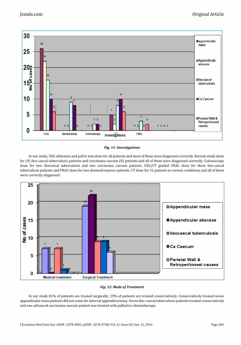
Fig. 11: Investigations
In our study, USG abdomen and pelvis was done for all patients and most of them were diagnosed correctly. Barium study done
for (9) ileo-caecal tuberculosis patients and carcinoma caecum (8) patients and all of them were diagnosed correctly. Colonoscopy
done for two ileocaecal tuberculosis and two carcinoma caecum patients. USG/CT guided FNAC done for three ileo-caecal
tuberculosis patients and FNAC done for two desmoid tumour patients. CT done for 31 patients in various conditions and all of them
were correctly diagnosed.
Fig. 12: Mode of Treatment
In our study 81% of patients are treated surgically; 19% of patients are treated conservatively. Conservatively treated seven
appendicular mass patients did not come for interval appendicectomy. Seven ileo-caecal tuberculosis patients treated conservatively
and one advanced carcinoma caecum patient was treated with palliative chemotherapy.
Jemds.com Original Article
J Evolution Med Dent Sci/ eISSN- 2278-4802, pISSN- 2278-4748/ Vol. 5/ Issue 03/ Jan. 11, 2016 Page 207
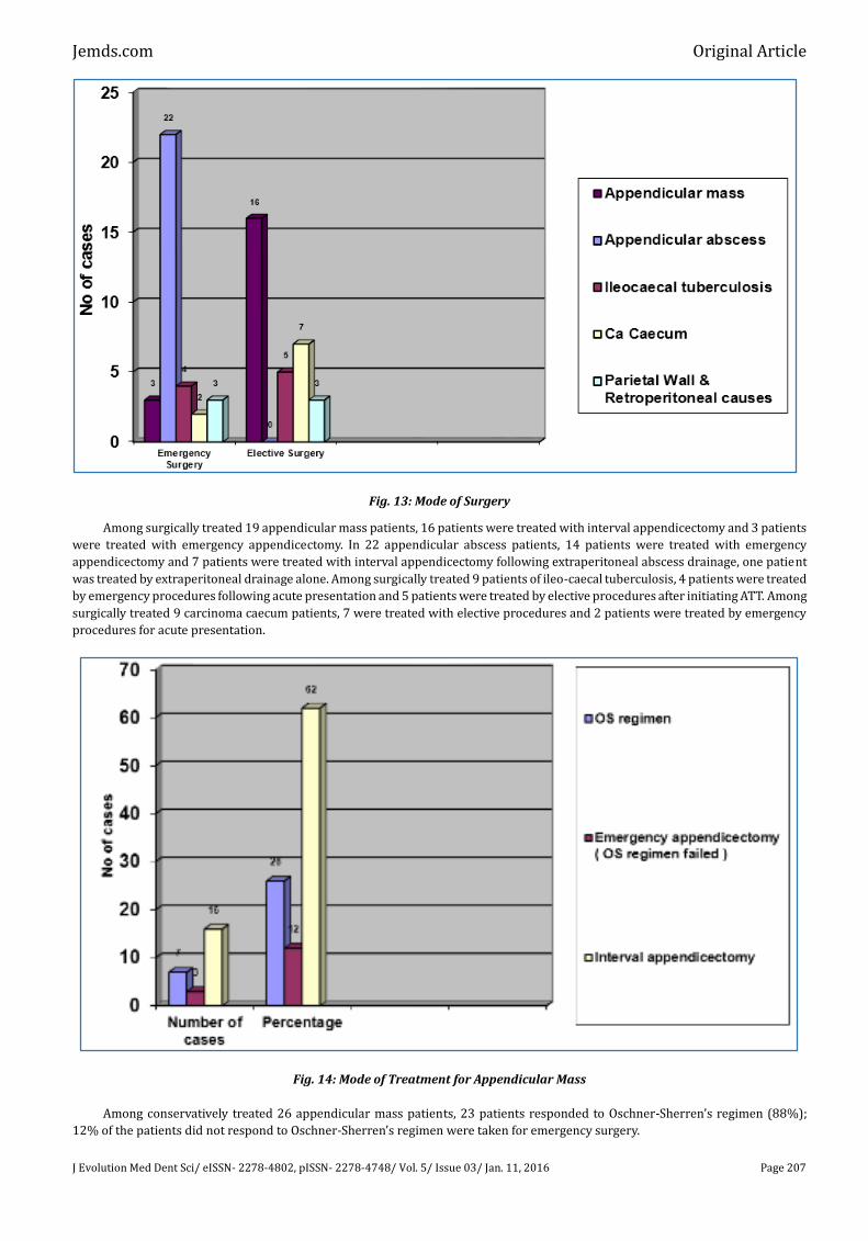
Fig. 13: Mode of Surgery
Among surgically treated 19 appendicular mass patients, 16 patients were treated with interval appendicectomy and 3 patients
were treated with emergency appendicectomy. In 22 appendicular abscess patients, 14 patients were treated with emergency
appendicectomy and 7 patients were treated with interval appendicectomy following extraperitoneal abscess drainage, one patient
was treated by extraperitoneal drainage alone. Among surgically treated 9 patients of ileo-caecal tuberculosis, 4 patients were treated
by emergency procedures following acute presentation and 5 patients were treated by elective procedures after initiating ATT. Among
surgically treated 9 carcinoma caecum patients, 7 were treated with elective procedures and 2 patients were treated by emergency
procedures for acute presentation.
Fig. 14: Mode of Treatment for Appendicular Mass
Among conservatively treated 26 appendicular mass patients, 23 patients responded to Oschner-Sherren’s regimen (88%);
12% of the patients did not respond to Oschner-Sherren’s regimen were taken for emergency surgery.
Jemds.com Original Article
J Evolution Med Dent Sci/ eISSN- 2278-4802, pISSN- 2278-4748/ Vol. 5/ Issue 03/ Jan. 11, 2016 Page 208
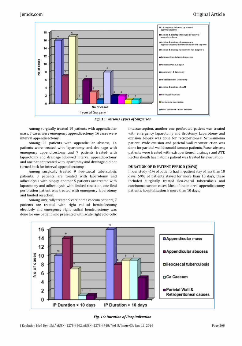
Fig. 15: Various Types of Surgeries
Among surgically treated 19 patients with appendicular
mass, 3 cases were emergency appendicectomy, 16 cases were
interval appendicectomy.
Among 22 patients with appendicular abscess, 14
patients were treated with laparotomy and drainage with
emergency appendicectomy and 7 patients treated with
laparotomy and drainage followed interval appendicectomy
and one patient treated with laparotomy and drainage did not
turned back for interval appendicectomy.
Among surgically treated 9 ileo-caecal tuberculosis
patients, 3 patients are treated with laparotomy and
adhesiolysis with biopsy, another 5 patients are treated with
laparotomy and adhesiolysis with limited resection, one ileal
perforation patient was treated with emergency laparotomy
and limited resection.
Among surgically treated 9 carcinoma caecum patients, 7
patients are treated with right radical hemicolectomy
electively and emergency right radical hemicolectomy was
done for one patient who presented with acute right colo-colic
intussusception, another one perforated patient was treated
with emergency laparotomy and ileostomy. Laparotomy and
excision biopsy was done for retroperitoneal Schwannoma
patient. Wide excision and parietal wall reconstruction was
done for parietal wall desmoid tumour patients. Psoas abscess
patients were treated with extraperitoneal drainage and ATT.
Rectus sheath haematoma patient was treated by evacuation.
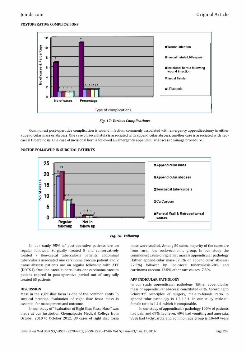
DURATION OF INPATIENT PERIOD (DAYS)
In our study 41% of patients had in-patient stay of less than 10
days; 59% of patients stayed for more than 10 days, these
included surgically treated ileo-caecal tuberculosis and
carcinoma caecum cases. Most of the interval appendicectomy
patient’s hospitalisation is more than 10 days.
Fig. 16: Duration of Hospitalisation
Jemds.com Original Article
J Evolution Med Dent Sci/ eISSN- 2278-4802, pISSN- 2278-4748/ Vol. 5/ Issue 03/ Jan. 11, 2016 Page 209
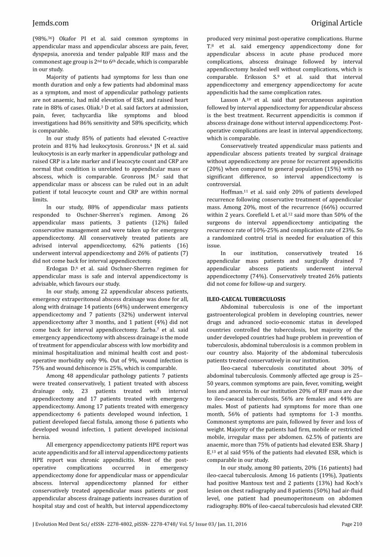
POSTOPERATIVE COMPLICATIONS
Fig. 17: Various Complications
Commonest post-operative complication is wound infection, commonly associated with emergency appendicectomy in either
appendicular mass or abscess. One case of faecal fistula is associated with appendicular abscess; another case is associated with ileo-
caecal tuberculosis. One case of incisional hernia followed an emergency appendicular abscess drainage procedure.
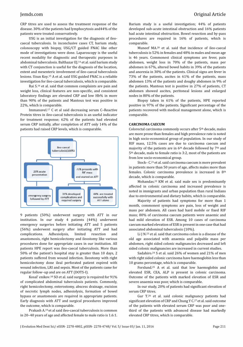
POSTOP FOLLOWUP IN SURGICAL PATIENTS
Fig. 18: Followup
In our study 95% of post-operative patients are on
regular followup. Surgically treated 8 and conservatively
treated 7 ileo-caecal tuberculosis patients, abdominal
tuberculosis associated one carcinoma caecum patient and 2
psoas abscess patients are on regular follow-up with ATT
(DOTS-I). One ileo-caecal tuberculosis, one carcinoma caecum
patient expired in post-operative period out of surgically
treated 65 patients.
DISCUSSION
Mass in the right iliac fossa is one of the common entity in
surgical practice. Evaluation of right iliac fossa mass is
essential for management and outcome.
In our study of “Evaluation of Right Iliac Fossa Mass” was
made at our institution Chengalpattu Medical College from
October 2010 to October 2012; 80 cases of right iliac fossa
mass were studied. Among 80 cases, majority of the cases are
from rural, low socio-economic group. In our study the
commonest cause of right iliac mass is appendicular pathology
(Either appendicular mass-32.5% or appendicular abscess-
27.5%) followed by ileo-caecal tuberculosis-20% and
carcinoma caecum-12.5% other rare causes -7.5%.
APPENDICULAR PATHOLOGY
In our study, appendicular pathology (Either appendicular
mass or appendicular abscess) constituted 60%. According to
Schwartz’ principles of surgery, male-to-female ratio in
appendicular pathology is 1.2-1.3:1, in our study male-to-
female ratio is 1.1:1, which is comparable.
In our study of appendicular pathology 100% of patients
had pain and 69% had fever, 40% had vomiting and anorexia,
88% had tachycardia and common age group is 10–60 years
Jemds.com Original Article
J Evolution Med Dent Sci/ eISSN- 2278-4802, pISSN- 2278-4748/ Vol. 5/ Issue 03/ Jan. 11, 2016 Page 210
(98%.36) Okafor PI et al. said common symptoms in
appendicular mass and appendicular abscess are pain, fever,
dyspepsia, anorexia and tender palpable RIF mass and the
commonest age group is 2nd to 6th decade, which is comparable
in our study.
Majority of patients had symptoms for less than one
month duration and only a few patients had abdominal mass
as a symptom, and most of appendicular pathology patients
are not anaemic, had mild elevation of ESR, and raised heart
rate in 88% of cases. Oliak.3 D et al. said factors at admission,
pain, fever, tachycardia like symptoms and blood
investigations had 86% sensitivity and 58% specificity, which
is comparable.
In our study 85% of patients had elevated C-reactive
protein and 81% had leukocytosis. Gronross.4 JN et al. said
leukocytosis is an early marker in appendicular pathology and
raised CRP is a late marker and if leucocyte count and CRP are
normal that condition is unrelated to appendicular mass or
abscess, which is comparable. Gronross JM.5 said that
appendicular mass or abscess can be ruled out in an adult
patient if total leucocyte count and CRP are within normal
limits.
In our study, 88% of appendicular mass patients
responded to Oschner-Sherren’s regimen. Among 26
appendicular mass patients, 3 patients (12%) failed
conservative management and were taken up for emergency
appendicectomy. All conservatively treated patients are
advised interval appendicectomy, 62% patients (16)
underwent interval appendicectomy and 26% of patients (7)
did not come back for interval appendicectomy.
Erdogan D.6 et al. said Oschner-Sherren regimen for
appendicular mass is safe and interval appendicectomy is
advisable, which favours our study.
In our study, among 22 appendicular abscess patients,
emergency extraperitoneal abscess drainage was done for all,
along with drainage 14 patients (64%) underwent emergency
appendicectomy and 7 patients (32%) underwent interval
appendicectomy after 3 months, and 1 patient (4%) did not
come back for interval appendicectomy. Zarba.7 et al. said
emergency appendicectomy with abscess drainage is the mode
of treatment for appendicular abscess with low morbidity and
minimal hospitalization and minimal health cost and post-
operative morbidity only 9%. Out of 9%, wound infection is
75% and wound dehiscence is 25%, which is comparable.
Among 48 appendicular pathology patients 7 patients
were treated conservatively, 1 patient treated with abscess
drainage only, 23 patients treated with interval
appendicectomy and 17 patients treated with emergency
appendicectomy. Among 17 patients treated with emergency
appendicectomy 6 patients developed wound infection, 1
patient developed faecal fistula, among those 6 patients who
developed wound infection, 1 patient developed incisional
hernia.
All emergency appendicectomy patients HPE report was
acute appendicitis and for all interval appendicectomy patients
HPE report was chronic appendicitis. Most of the post-
operative complications occurred in emergency
appendicectomy done for appendicular mass or appendicular
abscess. Interval appendicectomy planned for either
conservatively treated appendicular mass patients or post
appendicular abscess drainage patients increases duration of
hospital stay and cost of health, but interval appendicectomy
produced very minimal post-operative complications. Hurme
T.8 et al. said emergency appendicectomy done for
appendicular abscess in acute phase produced more
complications, abscess drainage followed by interval
appendicectomy healed well without complications, which is
comparable. Eriksson S.9 et al. said that interval
appendicectomy and emergency appendicectomy for acute
appendicitis had the same complication rates.
Lasson A.10 et al. said that percutaneous aspiration
followed by interval appendicectomy for appendicular abscess
is the best treatment. Recurrent appendicitis is common if
abscess drainage done without interval appendicectomy. Post-
operative complications are least in interval appendicectomy,
which is comparable.
Conservatively treated appendicular mass patients and
appendicular abscess patients treated by surgical drainage
without appendicectomy are prone for recurrent appendicitis
(20%) when compared to general population (15%) with no
significant difference, so interval appendicectomy is
controversial.
Hoffman.11 et al. said only 20% of patients developed
recurrence following conservative treatment of appendicular
mass. Among 20%, most of the recurrence (66%) occurred
within 2 years. Corefield L et al.12 said more than 50% of the
surgeons do interval appendicectomy anticipating the
recurrence rate of 10%-25% and complication rate of 23%. So
a randomized control trial is needed for evaluation of this
issue.
In our institution, conservatively treated 16
appendicular mass patients and surgically drained 7
appendicular abscess patients underwent interval
appendicectomy (74%). Conservatively treated 26% patients
did not come for follow-up and surgery.
ILEO-CAECAL TUBERCULOSIS
Abdominal tuberculosis is one of the important
gastroenterological problem in developing countries, newer
drugs and advanced socio-economic status in developed
countries controlled the tuberculosis, but majority of the
under developed countries had huge problem in prevention of
tuberculosis, abdominal tuberculosis is a common problem in
our country also. Majority of the abdominal tuberculosis
patients treated conservatively in our institution.
Ileo-caecal tuberculosis constituted about 30% of
abdominal tuberculosis. Commonly affected age group is 25–
50 years, common symptoms are pain, fever, vomiting, weight
loss and anorexia. In our institution 20% of RIF mass are due
to ileo-caeacal tuberculosis, 56% are females and 44% are
males. Most of patients had symptoms for more than one
month, 56% of patients had symptoms for 1-3 months.
Commonest symptoms are pain, followed by fever and loss of
weight. Majority of the patients had firm, mobile or restricted
mobile, irregular mass per abdomen. 62.5% of patients are
anaemic, more than 75% of patients had elevated ESR. Sharp J
E.13 et al said 95% of the patients had elevated ESR, which is
comparable in our study.
In our study, among 80 patients, 20% (16 patients) had
ileo-caecal tuberculosis. Among 16 patients (19%), 3patients
had positive Mantoux test and 2 patients (13%) had Koch’s
lesion on chest radiography and 8 patients (50%) had air-fluid
level, one patient had pneumoperitoneum on abdomen
radiography. 80% of ileo-caecal tuberculosis had elevated CRP.
Jemds.com Original Article
J Evolution Med Dent Sci/ eISSN- 2278-4802, pISSN- 2278-4748/ Vol. 5/ Issue 03/ Jan. 11, 2016 Page 211
CRP titres are used to assess the treatment response of the
disease. 30% of the patients had lymphocytosis and44% of the
patients were treated conservatively.
USG is an initial investigation for the diagnosis of ileo-
caecal tuberculosis. In inconclusive cases CT, barium study,
colonoscopy with biopsy, USG/CT guided FNAC like other
mode of investigations were done. Laparoscopy is the useful
recent modality for diagnostic and therapeutic purposes in
abdominal tuberculosis. Balthazar EJ.14 et al. said barium study
with CT conjunction is useful for the diagnosis of the location,
extent and mesenteric involvement of ileo-caecal tuberculosis
lesions. Uzun Koy.15 A et al. said USG guided FNAC is a reliable
investigation for ileo-caecal tuberculosis, which is comparable.
Rai S.16 et al. said that common complaints are pain and
weight loss, clinical features are non-specific, and consistent
laboratory findings are elevated CRP and low Hb% in more
than 90% of the patients and Mantoux test was positive in
22%, which is comparable.
Immanuvel.17 C et al said decreasing serum C–Reactive
Protein titres in ileo-caecal tuberculosis is an useful indicator
for treatment response; 62% of the patients had elevated
serum CRP initially, after completion of ATT only 14% of the
patients had raised CRP levels, which is comparable.
9 patients (50%) underwent surgery with ATT in our
institution. In our study 4 patients (44%) underwent
emergency surgeries before initiating ATT and 5 patients
(56%) underwent surgery after initiating ATT and had
complications. Adhesiolysis, limited resection and
anastomosis, right hemicolectomy and ileostomy like various
procedures done for appropriate cases in our institution. All
patients HPE report was ileo-caecal tuberculosis. More than
50% of the patient’s hospital stay is greater than 10 days, 2
patients suffered from wound infection. Ileostomy with right
hemicolectomy done ileal perforated patient expired with
wound infection, LRI and sepsis. Most of the patients came for
regular follow–up and are on ATT (DOTS-I).
Kosal’ enikov.18 SO et al. said surgery is required for 91%
of complicated abdominal tuberculosis patients. Commonly,
right hemicolectomy, enterostomy, abscess drainage, excision
of necrotic lymph nodes, adhesiolysis, formation of bowel
bypass or anastomosis are required in appropriate patients.
Early diagnosis with ATT and surgical procedures improved
the outcome, which is comparable.
Prakash A.19 et al said ileo-caecal tuberculosis is common
in 20–40 years of age and affected female to male ratio is 1.6:1.
Barium study is a useful investigation; 44% of patients
developed sub-acute intestinal obstruction and 16% patients
had acute intestinal obstruction. Bowel resection and by-pass
procedures are required in 16% of patients, which is
comparable.
Muneef MA.20 et al. said that incidence of ileo-caecal
tuberculosis is 52% in females and 48% in males and mean age
is 46 years. Commonest clinical symptoms are fever, pain
abdomen, weight loss in 70% of the patients, mass per
abdomen in 67%, altered bowel habits in 39% of the patients
and anorexia in 30% of the patients. Clinical signs are fever in
73% of the patients, ascites in 61% of the patients, mass
abdomen 13% of the patients and doughy abdomen in 9% of
the patients. Mantoux test is positive in 27% of patients, CT
abdomen showed ascites, peritoneal lesions and enlarged
nodes in 80% of the patients.
Biopsy taken in 61% of the patients, HPE reported
positive in 97% of the patients. Significant percentage of the
patients recovered with medical management alone, which is
comparable.
CARCINOMA CAECUM
Colorectal carcinoma commonly occurs after 5th decade, males
are more prone than females and high prevalence rate is noted
in high socio-economical group of population. In our study of
RIF mass, 12.5% cases are due to carcinoma caecum and
majority of the patients are in 6th decade followed by 7th and
5th decade, male to female ratio is 3:2, most of the patients are
from low socio-economical group.
Stock– C.21 et al. said carcinoma caecum is more prevalent
in patients more than 50 years of age, affects males more than
females. Colonic carcinoma prevalence is increased in 8th
decade, which is comparable.
Mohandas.22 KM et al. said male sex is predominantly
affected in colonic carcinoma and increased prevalence is
noted in immigrants and urban population than rural Indians
due to environmental and dietary habits, which is comparable.
Majority of patients had symptoms for more than 1
month, commonest symptoms are pain, loss of weight and
mass per abdomen. All cases had hard mobile or fixed RIF
mass; 80% of carcinoma caecum patients were anaemic and
had mild elevation of ESR. Among 10 cases of carcinoma
caecum marked elevation of ESR is present in one case that had
associated abdominal tuberculosis (10%).
Li J N.23 et al. said that carcinoma colon is a disease of the
old age associated with anaemia and palpable mass per
abdomen, right sided colonic malignancies decreased and left
sided colonic malignancies are increased in current studies.
Sadahiro.24 S et al. said 26% of women and 21% of men
with right sided colonic carcinoma have haemoglobin less than
10 grams percentage, which is comparable.
Forslund.25 A et al. said that low haemoglobin and
elevated ESR, CEA, ALP is present in colonic carcinoma.
Outcome of the patients with marked elevation of ESR and
severe anaemia was poor, which is comparable.
In our study, 20% of patients had significant elevation of
serum CRP titres.
Gur T.26 et al. said colonic malignancy patients had
significant elevation of CRP and Chung Y C.27 et al. said outcome
of the patients with elevated serum CRP was poor and one-
third of the patients with advanced disease had markedly
elevated CRP titres, which is comparable.
Jemds.com Original Article
J Evolution Med Dent Sci/ eISSN- 2278-4802, pISSN- 2278-4748/ Vol. 5/ Issue 03/ Jan. 11, 2016 Page 212
USG abdomen and pelvis was done for all patients
(100%), barium study was done for 8 patients, CT scan
abdomen and pelvis was done for all patients. CT scan revealed
RIF mass in 8 patients, right sided colo-colic intussusception in
one patient and pneumoperitoneum in another one patient.
Colonoscopy was done for 20% of the patients.
Storm E.28 et al. said that barium study has 91%
sensitivity for diagnosis of colonic malignancies, which is
comparable.
In our study, 90% of the patients are treated surgically in
our institution. Among 10 patients, majority of the patients (7)
were treated by right radical hemicolectomy (70%) electively
and one patient (10%) with right colo-colic intussusception
underwent emergency right radical hemicolectomy with
primary anastomosis. Another one patient with ileal
perforation, underwent emergency ileostomy and expired
post-operatively due to LRI with septicemia. All other patients
came for regular follow-up and were on anti-cancer
chemotherapy. One patient (10%) with advanced carcinoma
caecum was treated with palliative chemotherapy. For all post-
operative patients HPE reports revealed various types of
adenocarcinoma caecum. Among 10 carcinoma caecum
patients, one patient (10%) had associated abdominal
tuberculosis and was started on ATT prior to chemotherapy.
Herfath.29 C et al. said that treatment of carcinoma
caecum and ascending colon presenting with obstruction, the
treatment of choice was right hemicolectomy with ileostomy
and transverse colostomy, which is comparable in our study.
PARIETAL WALL AND RETROPERITONEAL CAUSES
In our study, both the parietal wall desmoid tumour patients
are females in reproductive age group. USG, CT confirmed the
parietal wall lesion, FNAC and excision biopsy followed by
surgery confirmed the diagnosis.
Aissa.30 A et al. said that desmoid tumour is common in
anterior abdominal wall, more prevalent in pregnancy and
post-partum period and associated with hormonal influence.
USG, CT, MRI and post-operative excisional biopsy reports are
confirmatory investigations, which is comparable.
Etiology of both the psoas abscess patients was
tuberculosis, both underwent extra-peritoneal drainage with
ATT.
Villar F.31 C et al. said 50% of the psoas abscess is
associated with skeletal tuberculosis, which is comparable in
our study.
In our study, one rectus sheath haematoma patient
presented as RIF mass, surgical evacuation of the haematoma
was done as the conservative management failed.
Rajagopal.32 et al. said that abdominal pain in parietal
wall haematoma is a rare entity and is treated conservatively
and rarely require surgical intervention, which is comparable.
In our study, one retroperitoneal Schwannoma patient
presented as RIF mass which was diagnosed by USG/CT/USG
guided core needle biopsy. Surgical excision was done.
Rai BR.33 et al. said that pelvic Schwannoma presented as
RIF mass and right sciatica, which is comparable.
In our study USG abdomen and pelvis is done for all cases
and RIF mass was diagnosed in more than 95% of the patients.
In inconclusive cases, various investigations like CT, barium
study, colonoscopy, USG guided FNAC were done.
According to the Sabiston text book of surgery USG
abdomen had 85% sensitivity and 90% specificity for
diagnosis of appendicular pathology, which is comparable.
Millard FC.34 et al. said USG abdomen correctly diagnosed
97% of RIF masses, so the investigation of choice in RIF mass
is USG abdomen, which is comparable.
Jain R.35 et al. said that sonographic features of
abdominal tuberculosis thickened mesentery and
lymphadenopathy, dilated small bowel loops and ascites were
diagnosed in 98% of the patients, which is comparable.
Martinez.36 Ares D et al. said that USG abdomen is 79%
sensitive and 92% specific for the diagnosis of colonic
carcinoma, which is comparable.
In our study, overall post-operative morbidity is 17% and
mortality is 3%. Common post-operative complication is
wound infection. Most of the post-operative complications are
treated conservatively.
CONCLUSION
Right iliac fossa mass was common in 20 to 50 years of age
group.
Overall incidence was more common in males as compared
to females (1.1:1).Appendicular pathology and Carcinoma
caecum was more common in males as compared to
females. Ileo-caecal tuberculosis was more common in
females.
The diseases were more in people from low socio –
economic status and the commonest symptom was pain in
abdomen.
Appendicular pathology (60%) either in the form of
appendicular mass (32.5%) or appendicular abscess
(27.5%) were the commonest cause of mass in the right
iliac fossa. Ileo-caecal tuberculosis (20%), carcinoma
caecum (12.5%) was the other common causes of mass in
the right iliac fossa.
Ultrasound abdomen was the essential investigation and it
had a sensitivity of greater than 95%.
Normal levels of serum CRP titer essentially ruled out
appendicular pathology. Serial titers were helpful in
assessing the treatment response of ileo-caecal
tuberculosis and in prognosis of carcinoma caecum.
In patients with appendicular mass, initial conservative
management followed by interval appendicectomy had
better results with minimal complications.
In patients with appendicular abscess, abscess drainage
combined with appendicectomy in the same procedure,
had high morbidity compared to patients who underwent
interval appendicectomy following abscess drainage.
A 44% of the patients with ileo-caecal tuberculosis were
managed conservatively with anti-tubercular therapy
(DOTS-I). Surgery was required in another 56% of the
patients, of which 25% of the patients presented with
acute complications needing immediate surgery; 31% of
the patients presented with sub-acute intestinal
obstruction due to adhesions and strictures following the
initiation of anti-tubercular therapy, which later required
surgical intervention.
An 80% of the patients with carcinoma caecum underwent
successful surgical resection; 20% of the patients
presented with acute surgical problem requiring
immediate surgery; 10% of the patients presented with
advanced disease.
Jemds.com Original Article
J Evolution Med Dent Sci/ eISSN- 2278-4802, pISSN- 2278-4748/ Vol. 5/ Issue 03/ Jan. 11, 2016 Page 213
Most of the parietal wall and retroperitoneal conditions
were treated surgically.
Early evaluation and intervention is needed to improve
the patient’s outcome and to reduce the morbidity and
mortality.
LIST OF ABBREVIATIONS
ADA - Adenosine deaminase
ALP - Alkaline phosphatase
ATT - Antitubercular treatment
Ca - Carcinoma
CEA - Carcinoembryonic antigen
CT - Computerised Tomography.
CRP - C-Reactive protein
DVT - Deep vein thrombosis
EGFR - Epidermal growth factor receptor
FNAC - Fine needle aspiration cytology
5-FU - 5-Fluro uracil
HIV - Human immunodeficiency virus
HPE - Histopathological examination
LIF - Left iliac fossa
LRI - Lower respiratory tract infection
MRI - Magnetic resonance imaging
NSAID – Non-steroidal anti-inflammatory drugs.
O-S - Ochsner-Sherren regimen
PCR - Polymerase chain reaction
PET - Positron emission tomography
PPD - Purified protein derivatives
RIF - Right iliac fossa
Tc99 - Technetium 99
TC - Total leucocyte count
TNF - Tumor necrosis factor
USG - Ultrasonogram
VEGF - Vascular endothelial growth factor
WBC - White blood cell
Yrs – Years
BIBLIOGRAPHY
1. Das S. Examination of acute abdomen, chapter 33, a Das
S, examination of acute abdomen, chapter 33, a manual
on clinical surgery, 8th edition, Elsevier, page 435 to 456.
2. Sharma MP, Bhatia V. Abdominal tuberculosis,
Department of Gastroenterology. All India Institute of
Medical Sciences, D II/23, Ansari Nagar, New Delhi 110-
029, India. [email protected], Indian J Med Res.
2004 Oct;120(4):305-15.
3. Oliak D, Yamini D, Udani VM, Lewis RJ, Vargas H, Arnell
T, et al. Can perforated appendicitis be diagnosed
preoperatively based on admission factors? Department
of Surgery, Harbor-UCLA Medical Center, Torrance, CA
90509, USA. J Gastrointest Surg. 2000 Sep-Oct;4(5):470-
4.
4. Grönroos JM, Grönroos P. Leukocyte count and C-
reactive protein in the diagnosis of acute appendicitis.
Department of Surgery, University of Turku, Turku,
Finland. Br J Surg 1999 Apr;86(4):501.
5. Grönroos JM. Clinical suspicion of acute appendicitis - is
the time ripe for more conservative treatment?
Departments of Surgery and Emergency, University of
Turku, Turku, Finland. [email protected], Minim
Invasive Ther Allied Technol. 2011 Jan;20(1):42-5. doi:
10.3109/13645706.2010.496958. Epub 2010 Jul 21.
6. Erdoğan D, Karaman I, Narci A, Karaman A, Cavuşoğlu
YH, Aslan MK, et al. Comparison of two methods for the
management of appendicular mass in children,
Department of Pediatric Surgery, Dr. Sami Ulus
Children’s Hospital, 06080 Ankara, Turkey.
[email protected], Pediatr Surg Int. 2005
Feb;21(2):81-3. Epub 2004 Dec 22.
7. Zarba Meli E, Mazzocchi P, Lepiane P, Dalsasso
G, Giacovazzo F, Salvio A, et al. [The role of surgery in the
treatment of appendicular abscesses]. II Clinical
Chirurgica, Università degli Studi di Roma La Sapienza.
8. Hurme T, Nylamo E. Conservative versus operative
treatment of appendicular abscess. Experience of 147
consecutive patients. Department of Paediatric Surgery,
Turku University Central Hospital, Finland, Ann Chir
Gynaecol. 1995;84(1):33-6. PMID:7645908.
9. Eriksson S, Styrud J. Interval appendicectomy: a
retrospective study, Department of Surgery, Karolinska
Institute at Danderyd Hospital, Sweden. Eur J Surg. 1998
Oct;164(10):771-4; discussion 775.
10. Lasson A, Lundagårds J, Lorén I, et al. Appendiceal
abscesses: primary percutaneous drainage and selective
interval appendicectomy, Department of Diagnostic
Radiology, Malmö University Hospital, University of
Lund, Sweden. Eur J Surg. 2002;168(5):264-9.
11. Hoffmann J, Lindhard A, Jensen HE. Appendix mass:
conservative management without interval
appendectomy. Am J Surg. 1984 Sep;148(3):379-82.
12. Corfield L. Interval appendicectomy after appendiceal
mass or abscess in adults: what is “best practice?”
Department of Surgery, University Hospital Lewisham,
Lewisham High Street, London, SE13 6LH, UK, Surg
Today. 2007;37(1):1-4. Epub 2007 Jan 1.
13. Sharp JF, Goldman M. Abdominal tuberculosis in East
Birmingham--a 16 year study, Department of Surgery,
East Birmingham Hospital, Bordesley Green East, UK,
Postgrad Med J. 1987 Jul;63(741):539-14.
14. Balthazar EJ, Gordon R, Hulnick D. Ileocecal tuberculosis:
CT and radiologic evaluation, Department of Radiology,
New York University Medical Center, NY 10016. AJR Am
J Roentgenol. 1990 Mar;154(3):499-503.
15. Uzunkoy A, Harma M, Harma M. Diagnosis of abdominal
tuberculosis: experience from 11 cases and review of the
literature, Department of Surgery, University of Harran,
Faculty of Medicine, Sanliurfa, Turkey. World J
Gastroenterol. 2004 Dec 15;10(24):3647-9.
16. Rai S, Thomas WM. Diagnosis of abdominal tuberculosis:
the importance of laparoscopy. Department of General
Surgery, Leicester General Hospital, Gwendolen Road,
Leicester LE5 4PW, UK. [email protected], J R Soc
Med. 2003 Dec;96(12):586-8.
17. Immanuel C, Acharyulu GS, Kannapiran M, et al. Acute
phase proteins in tuberculous patients. Tuberculosis
Research Centre (Indian Council of Medical Research),
Madras, Indian. J Chest Dis Allied Sci. 1990 Jan-
Mar;32(1):15-23.
18. Kosul'nikov SO, Kravchenko KV, Tarnopol’skiĭ SA, et al.
[The symptoms and surgical tactics for complicated
forms of the abdominal cavity tuberculosis]. Klin
Khir. 2012 Jan;(1):33-8.
19. Prakash A, Sharma LK, Koshal A, et al. Ileocaecal
tuberculosis, Aust N Z J Surg. 1975 Nov;45(4):371-5.
Jemds.com Original Article
J Evolution Med Dent Sci/ eISSN- 2278-4802, pISSN- 2278-4748/ Vol. 5/ Issue 03/ Jan. 11, 2016 Page 214
20. Muneef MA, Memish Z, Mahmoud SA, Sadoon
SA, Bannatyne R, Khan Y. Tuberculosis in the belly: a
review of forty-six cases involving the gastrointestinal
tract and peritoneum, Dept. of Pediatrics, King Fahad
National Guard Hospital, Riyadh, Saudi Arabia. Scand J
Gastroenterol. 2001 May;36(5):528-32.
21. Stock C, Haug U, Brenner H. Population-based prevalence
estimates of history of colonoscopy or sigmoidoscopy:
review and analysis of recent trends. Division of Clinical
Epidemiology and Aging Research, German Cancer
Research Center, Heidelberg, Germany. Gastrointest
Endosc. 2010 Feb;71(2):366-381.e2. doi:
10.1016/j.gie.2009.06.018. Epub 2009 Oct 20.
22. Mohandas KM, Desai DC. Epidemiology of digestive tract
cancers in India. V. Large and small bowel, Division of
Digestive Diseases and Nutrition, Tata Memorial
Hospital, Mumbai. Indian J Gastroenterol. 1999 Jul-
Sep;18(3):118-21.
23. Li JN, Zhao L, Zheng WY, Miao Z, Tang XY, Qian JM. [The
trends in clinical characteristics of colon cancer in last
two decades], Peking Union Medical College Hospital,
Peking Union Medical College, Chinese Academy of
Medical Sciences, Beijing 100730, China. Zhonghua Nei
Ke Za Zhi. 2010 Mar;49(3):226-9.
24. Sadahiro S, Suzuki T, Tokunaga N, Mukai M, Tajima
T, Makuuchi H, et al. Anemia in patients with colorectal
cancer. Department of Surgery, Tokai University School
of Medicine, Bohseidai, Isehara, Japan. J
Gastroenterol. 1998 Aug;33(4):488-94.
25. Forslund A, Engarås B, Lönnroth C, et al. Prediction of
postoperative survival by preoperative serum
concentrations of anti-p53 compared to CEA, CA 50, CA
242 and conventional blood tests in patients with
colorectal carcinoma. Department of Surgery,
Sahlgrenska University Hospital, Göteborg University,
Sweden. Int J Oncol. 2002 May;20(5):1013-8.
26. Gür T, Demir H, Kotan MÇ. Tumor markers and
biochemical parameters in colon cancer patients before
and after chemotherapy. Division of Biochemistry,
Health Sciences Faculty, Yuzuncu Yil University, Van,
Turkey. [email protected], Asian Pac J Cancer
Prev. 2011;12(11):3147-50.
27. Chung YC, Chang YF. Serum C-reactive protein correlates
with survival in colorectal cancer patients, but is not an
independent prognostic indicator. Department of
Surgery, Hsin-Chu Hospital, No. 25, Lane 442, Sec. 1,
Xhing-Kuo Road, Hsin-Chu City, Taiwan, Republic of
China. [email protected], Eur J Gastroenterol
Hepatol. 2003 Apr;15(4):369-73.
28. Strøm E, Larsen JL. Colon cancer at barium enema
examination and colonoscopy: a study from the county of
Hordaland, Norway. Department of Radiology,
University Hospital of Bergen, Haukeland Hospital,
Norway, Radiology.
1999 Apr;211(1):211-4.
29. Herfarth C, Runkel N. [Surgical standards in primary
colon cancer], Chirurgische Klinik, Universität
Heidelberg.
30. Aïssa A, Alouini-Mekki R, Ben Abdallah A, Enaifar
R, Kobbi I, Stita W, et al. [Update on the management of
desmoids tumors], Service d'imagerie médicale, hôpital
Ibn EL Jazzar, 3100 Kairouan, Tunisie.
[email protected], Gynecol Obstet Fertil. 2012
Feb;40(2):104-8. doi: 10.1016/j.gyobfe.2011.07.055.
Epub 2012 Jan 16.
31. Vilar FC, Neves FF, Colares JK, et al. [Spinal tuberculosis
(Pott’s disease) associated to psoas abscess: report of
two cases and a literature review], Divisão de Moléstias
Infecciosas, Departamento de Clínica Médica, Faculdade
de Medicina de Ribeirão Preto, Universidade de São
Paulo, Av. Bandeirantes 3900, 14048-900 Ribeirão Preto,
SP. Rev Soc Bras Med Trop. 2006 May-Jun;39(3):278-82.
32. Rajagopal AS, Shinkfield M, Voight S, et al. Massive rectus
sheath hematoma. Department of Surgery, St Mary’s
Hospital, Newport, Isle of Wight, P030 5TG, United
Kingdom. [email protected], Am J Surg. 2006
Jan;191(1):126-7.
33. Rai BR, Chaudhary D, Thapa P, Joshi MR, Dangol
UM, Singh DR. Ancient cystic pelvic schwannoma
presenting as a right iliac fossa mass, Department of
Surgery, Kathmandu Medical College Teaching Hospital,
Sinamangal, Nepal. [email protected]. Kathmandu
Univ Med J (KUMJ). 2005 Jul-Sep;3(3):285-8.
34. Millard FC, Collins MC, Peck RJ. Ultrasound in the
investigation of the right iliac fossa mass. Department of
Radiology, Royal Hallamshire Hospital, Sheffield, Br J
Radiol.
1991 Jan;64(757):17-9.
35. Jain R, Sawhney S, Bhargava DK, et al. Diagnosis of
abdominal tuberculosis: sonographic findings in patients
with early disease. Department of Radiodiagnosis. All
India Institute of Medical Sciences, New Delhi, India. AJR
Am J Roentgenol. 1995 Dec;165(6):1391-5.
36. Martínez-Ares D, Martín-Granizo Barrenechea I, Souto-
Ruzo J, Yáñez López J, Pallarés Peral A, Vázquez-Iglesias
JL. The value of abdominal ultrasound in
the diagnosis of colon cancer, Service of Digestive
Diseases. Complejo Hospitalario Universitario Xeral-
Cíes, Vigo, Pontevedra, Spain.
[email protected],Rev Esp Enferm Dig. 2005
Dec;97(12):877-86.