evaluation of surgical results using multivariate analysis
TRANSCRIPT

8/9/2019 Evaluation of Surgical Results Using Multivariate Analysis
http://slidepdf.com/reader/full/evaluation-of-surgical-results-using-multivariate-analysis 1/5
Original article
The tarsal tunnel syndrome:evaluation of surgical results using multivariate analysis
H. Baba1, M. Wada1, S. Annen1, M. Azuchi1, S. Imura1, K. Tomita2
1 Department of Orthopaedic Surgery, Fukui Medical School, Fukui, Japan2 Department of Orthopaedic Surgery, School of Medicine, Kanazawa University, Kanazawa, Japan
Accepted: 29 June 1996
Summary. Thirty-four patients with the tarsal
tunnel syndrome were treated by decompression of the posterior tibial nerve. The condition was bi-lateral in 3 cases. There were 9 men and 25 womenwith an average age at operation of 41 years. Theaverage follow up was for 3.8 years. Multivariateanalysis showed that the outcome is influenced, inorder of importance, by fibrosis around the nerve,the preoperative severity of the condition, a historyof sprained ankle, worker’s compensation, a longhistory, and heavy work. The results were favour-able when there was a short history, the presenceof a ganglion, no sprains, and light work. Mea-surement of the terminal latency of the medial
plantar nerve was valuable in assessing recovery.The precise cause of the syndrome and its effect ontreatment should be considered before operation.
Resume. Nous rapportons ici les resultats desdecompression chirurgicales du tunnel tarsien.Trente-sept nerfs tibiaux avaient ete decomprimessur trente-quatre patients (9 hommes et 25 fem-mes), dont la moyenne d’age etait de 41 ans. L’intervalle moyen des examens de controle etait de 3,8 ans. D’apres les analyses des multiviaria-bles il apparaıt que les resultats sont affectes dansl’ordre: par des fibroses autour du nerf, la gravite de la maladie avant l’operation, des entorses de lacheville, des indemnites sociales, des antecedents pathologiques de longue date, et des travaux pe-nibles. Inversement les resultats sont favorablesdans le cas d’un kyste du ganglion, d’absence de facteurs anterieurs et de travail physique peu pe-
nible. La mesure de la latence finale du nerf
plantaire median a fait ressortir des valeurs si-gnificatives pour l’evolution du syndrome du tun-nel tarsien.
Introduction
The tarsal tunnel syndrome is an entrapment neu-ropathy caused by pressure on the posterior tibialnerve as it passes posterior and inferior to themedial malleolus beneath the fibrous origin of theabductor hallucis muscle [2, 8]. Various factorscontribute to the clinical presentation and symp-
toms. Although the syndrome is well known, theresults of operation vary [3, 9, 14, 15].We evaluated by multivariate analysis the fac-
tors affecting the outcome in a group of patientswith the syndrome.
Patients and methods
The group comprised 34 patients (9 men and 25 women) withan average age of 41 years (range 14 to 73 years) at the time of operation. In 3 women the condition was bilateral. The averageduration of symptoms was 0.9 years (range 1 month to 8 years).Seven patients were hypertensive (systolic pressure 150 mmHg), 3 had diabetes mellitus, and one had chronicobliterative arteriosclerosis. None had clinical evidence of
lumbosacral radiculopathy. Fourteen had a history of asprained ankle, but without major injury to the bones or liga-ments.
The diagnosis was established when one or more of thefollowing conditions were present: local pain, a positiveTinel’s sign; sensory disturbances, and weakness of abductorhallucis. The clinical condition was graded: Grade I – no symptoms; Grade II – pain only; Grade III – pain and numbness; Grade IV – muscle weakness associated with grade III
symptoms.
Reprint requests to: H. Baba, Department of OrthopaedicSurgery, Fukui Medical School, Shimoaizuki 23, Matsuoka,Fukui 910-11, Japan
International Orthopaedics (SICOT) (1997) 21: 67–71
OrthopaedicsInternational
© Springer-Verlag 1997

8/9/2019 Evaluation of Surgical Results Using Multivariate Analysis
http://slidepdf.com/reader/full/evaluation-of-surgical-results-using-multivariate-analysis 2/5
The terminal latency of the posterior tibial nerve was measuredin 27 patients (29 feet). This involved electrical stimulation ata point 1 cm distal and posterior to the medial malleolus andmonitoring evoked responses from the abductor hallucismuscle 1 cm posterior and inferior to the navicular tuberosity.A Neuromatic 200 and a N1500 digital electromyography(EMG) apparatus (Dantec DISA, Scovlunde, Denmark) wasused. A denervation pattern in the abductor hallucis EMG,with positive-sharp wave, was further evidence for the syn-drome. CT and MRI were used in 16 cases to study abnorm-alities of the bones and ligaments.
Operative technique
Epidural or spinal anaesthesia was used in 22 and local an-aesthesia in 12 patients. A curved incision, 3.5 cm long, wasmade 1 cm distal to the medial malleolus and the posteriortibial nerve was exposed proximal to the flexor retinaculum.This was divided with all constricting structures and any lumppresent was excised. The calcaneal branch of the posterior ti-bial nerve was preserved. Two plantar branches were freedbeneath the fibrous origin of abductor hallucis in patients withmuscle weakness or EMG evidence of denervation. The patientwore a short leg-ankle-foot support for 3 weeks and was thenencouraged to resume normal activity.
Follow-up was carried out at an average of 3.8 years (range1 to 7 years) after operation to determine improvement of numbness and sensory disturbances, relief of pain and recoveryof motor strength in the big toe.
Statistical analysis
Multivariate analysis, based on the theory of QuantificationType II programme (Nakayama Books, Tokyo, Japan), wasapplied to the following variables namely gender, age at op-eration, duration of symptoms, causes of nerve compression,preoperative grading, occupation, and precipitating factors.With the definition of the postoperative clinical condition as adependent variable, these categories were entered into multi-variate analysis (Quant 2, Kyoritsu, Tokyo) in a NEC-PC9801ns-T computer (Nihon Electric, Tokyo). The postoperativeclinical state was also examined with regard to the terminallatency using Student’s t -test (StatView™ programme, AbacusConcepts, Berkeley, CA) with P = 0.05 considered significant.
Results
Before operation, 31 patients (37 feet) complainedof pain on the medial side of the sole which wasworse at night in 7. A positive Tinel sign waspresent in every case. Sensory disturbances wererecorded in 28 feet with hyperaesthesia of the soleand calcaneal area in 3 feet, in the medial and
calcaneal area in 17, and in the medial and lateralside of the sole in 8 feet. Weakness of the abductorhallucis was found in 15. The grading was II in 19,III in 13 and IV in 5.
After operation, there was improvement in pain,sensory disturbances and Tinel’s sign (Fig. 1). Atfollow-up, 26 feet were graded as I, 6 as II, 3 as III,and 2 as IV (Fig. 2). Twenty-one feet improved byone grade and 12 by two grades. Three remained inthe same grade, one in grade II and 2 in grade IV.
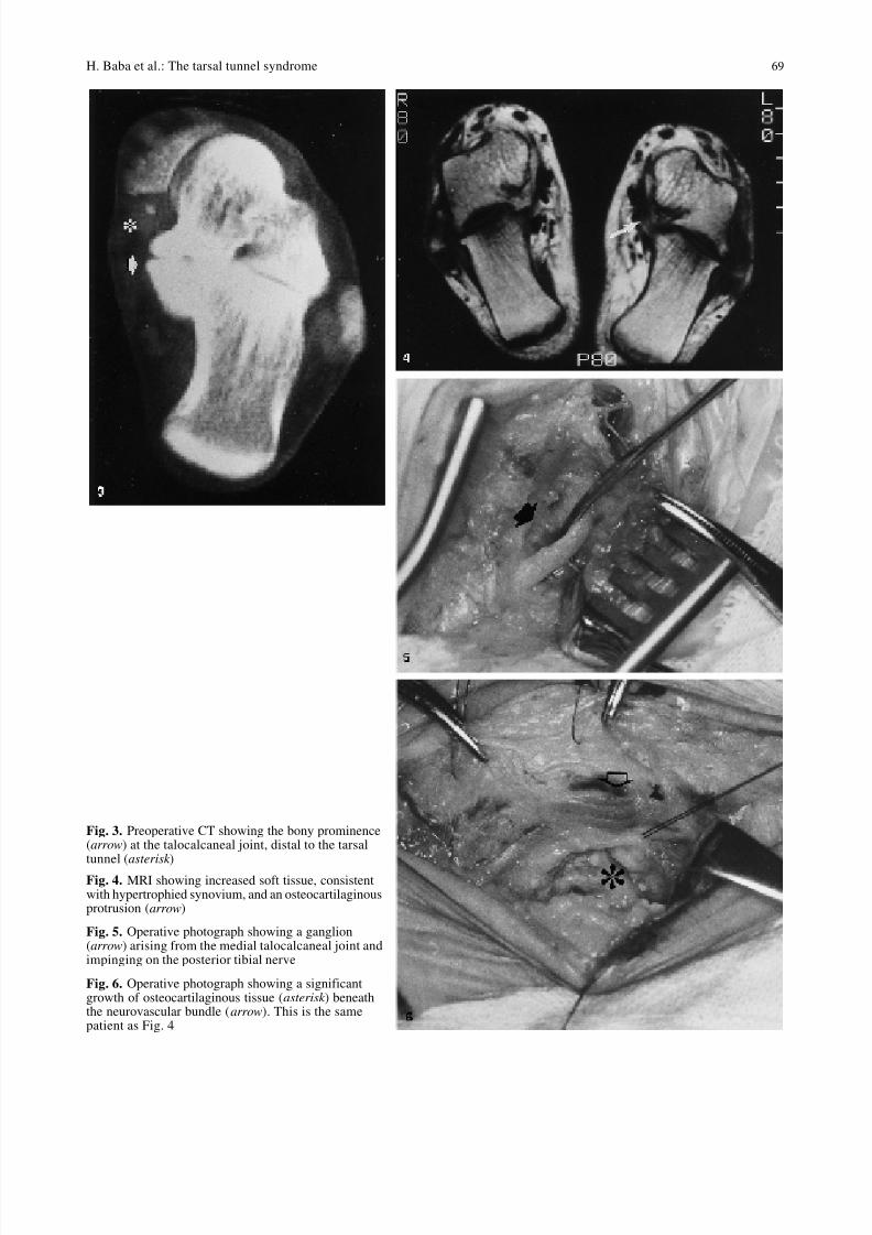
Radiographs showed a bony prominence at themedial talocalcaneal joint in 3 patients (Fig. 3),one showing abnormal growth of soft tissue as well
as an osteocartilaginous protrusion around thenerve (Fig. 4). Talocalcaneal coalition was notfound.
At operation a ganglion, arising from the medialpart of the talocalcaneal joint, was present in13 feet (Fig. 5). The mass impinged on the nerve asit penetrated the tibiocalcaneal ligament in 9, or atthe point between the tibiocalcaneal and posteriortibio-fibular ligaments in 4. Significant fibrosisaround the nerve was present just distal to theflexor retinaculum in 7, 6 of whom had a history of recurrent minor sprains of the ankle. The other
finding included abnormal growth of synoviumand an osteocartilaginous mass around the nervetogether with habitual dislocation of the posteriortibial tendon anteriorly (Fig. 6).
Measurement of terminal latency
Latency in the feet showing one grade of improvement averaged 5.9
0.5 ms (n = 14;
mean standard error, range 4.9 to 7.8 ms). Inthose feet with an improvement of 2 grades the
68 H. Baba et al.: The tarsal tunnel syndrome
Fig. 1. The clinical findings before operation and at the lastfollow-up
Fig. 2. The preoperative and postoperative clinical grades
8/9/2019 Evaluation of Surgical Results Using Multivariate Analysis
http://slidepdf.com/reader/full/evaluation-of-surgical-results-using-multivariate-analysis 3/5
69H. Baba et al.: The tarsal tunnel syndrome
Fig. 3. Preoperative CT showing the bony prominence(arrow) at the talocalcaneal joint, distal to the tarsal
tunnel (asterisk
)Fig. 4. MRI showing increased soft tissue, consistentwith hypertrophied synovium, and an osteocartilaginousprotrusion (arrow)
Fig. 5. Operative photograph showing a ganglion(arrow) arising from the medial talocalcaneal joint andimpinging on the posterior tibial nerve
Fig. 6. Operative photograph showing a significantgrowth of osteocartilaginous tissue (asterisk ) beneaththe neurovascular bundle (arrow). This is the samepatient as Fig. 4

8/9/2019 Evaluation of Surgical Results Using Multivariate Analysis
http://slidepdf.com/reader/full/evaluation-of-surgical-results-using-multivariate-analysis 4/5
latency averaged 5.1 0.5 ms (n = 12, range 4.0 to6.6 ms). The difference between the 2 groups wasstatistically significant (P
0.05). Three patientswith unchanged grades after operation showed alatency 7.0 ms.
Multivariate analysis (Table 1)
The partial correlation coefficient of the analysisaveraged 0.893, showing a high statistical validity.With the exception of gender and age, duration of symptoms, cause of nerve compression, occupa-tion, preceding factors and preoperative gradesshowed a significantly high score of range (partialcorrelation coefficient 0.700). The surgical re-sult was significantly influenced by the presence of symptoms for 12 months (category score–0.371), fibrosis around the nerve (–0.692),worker’s compensation (–0.457), sprain (–0.611)or grade IV before operation (–0.633).
Discussion
The tarsal tunnel syndrome is a clinical entitywhich may present with pain in the foot andweakness of the toes [1, 5, 10]. Entrapment of theposterior tibial nerve, or its branches, is due tointrinsic or extrinsic factors within the tunnelformed by the flexor retinaculum, behind anddistal to the medial malleolus. A thorough searchshould be made to identify the underlying lesion,
but a number of cases arise spontaneously with noobvious cause. The diagnosis is based on clinicalfindings and EMG studies [4], and radiographymay detect the underlying abnormality. Treatmentmay vary depending on the lesion present.
Examination should include palpation of theposterior tibial nerve in the retromalleolar area and
along each of its branches, including the inter-digital nerves. Percussion over the nerve may showa positive Tinel’s sign. The distal tunnel syndromeis indicated when pain on percussion is limited tothe undersurface of the abductor hallucis muscle.Subtle diminution of sensation may be the firstsign, especially after heavy exercise [18] or ex-cessive movement of the foot and ankle. Injectionof local anaesthetic into the tunnel may reducepain temporarily.
Measurement of terminal latency is the standardtest for disturbed motor conduction between thesite of stimulation, usually proximal to the tunnel,
and the abductor hallucis. Kaplan et al demon-strated that latencies measured in both the abductorhallucis and the digiti minimi muscles were sig-nificantly greater than normal in patients with thesyndrome [4]. They also reported a significantdecrease in amplitude and prolongation of motorevoked potentials from the same muscles in thesyndrome. Abnormalities detected from the mus-cular response reflect involvement of funicularmotor integrity with denervation potentials in the
70 H. Baba et al.: The tarsal tunnel syndrome
Table 1. Demographic data showing results of multivariate analysis
Item Category Category score Range (P.C.C.)a
Gender G1: maleG2: female
0.039–0.078
0.193 (0.392)
Age at operation A1: 40 yearsA2: 40’sA3: 50’s
A4: 60 years
–0.1090.029
–0.099
–0.121
0.238 (0.512)
Duration of disease history T1: 6 monthsT2: 6–12 monthsT3: 12 months
0.887–0.183–0.371
0.830 (0.924)
Cause of the disease C1: fibrosisC2: ganglion cystC3: undetectable
–0.6920.733
–0.119
0.914 (0.955)
Occupation O1: light work O2: heavy work O3: worker’s compensation
0.167–0.277–0.457
0.772 (0.851)
Preceding factors P1: nothing remarkableP2: sprain
0.331–0.611
0.972 (0.938)
Preoperative grades G1: grade IIG2: grade IIIG3: grade IV
0.492–0.092–0.633
1.137 (1.005)
a P.C.C.: partial correlation coefficient (average, 0.893)

8/9/2019 Evaluation of Surgical Results Using Multivariate Analysis
http://slidepdf.com/reader/full/evaluation-of-surgical-results-using-multivariate-analysis 5/5
EMG, and further loss of muscle strength. Earlyinvolvement of the neural pathway involved insensory conduction with small funiculi of the tibialnerve would show decreased amplitude and in-creased latency of the sensory action potentialsmeasured across the tunnel.
Oh and Oh et al demonstrated that the terminal
latency test is sensitive in the tarsal tunnel syn-drome with abnormal latency in 52.4% of patients[11, 12]. They considered that examination of sensory conduction in the medial and lateralplantar nerves was a more rewarding test for di-agnosis, and abnormal conduction (either absentspike or slow conduction velocity) was present in90.5% of their cases. Nevertheless terminal latencymay be of value for evaluation of the outcome, asis shown in our cases.
Radiography may detect talocalcaneal coalition,space occupying lesions, or fibrotic soft tissuechanges around the nerve. Bony prominence at thetalocalcaneal joint is reported to cause symptomsin 25% of cases, and a ganglion in 35% [17]. Highresolution CT scanning and MRI may detect fi-brosis or scarring around the nerve and allowsbetter choice of the surgical procedure.
Management depends on the clinical symptomsand the level of disability. Symptoms may be re-lieved by the temporary use of an immobiliser,injections of steroid, anti-inflammatory drugs,physiotherapy or a combination of these. Decom-pression is indicated for persistent pain and para-esthesia. Before operation, specific points of
maximum tenderness should be noted and theseareas explored. All compressive lesions must beexcised and neurolysis is optional if there is scartissue; we carry it out routinely when there issignificant muscle wasting and denervation po-tentials are seen on the EMG.
The differential diagnosis includes entrapmentof the medial plantar nerve below the talus andnavicular bones, known as jogger’s foot, or distaltarsal tunnel entrapment.
The reported results of surgery vary [3, 9, 14,15], possibly because of different methods of as-sessment. We used multivariate analysis which
showed that the results are influenced, in order of importance, by fibrosis around the nerve, the pre-operative severity of the condition (grade IV), ahistory of sprained ankle, worker’s compensation,a long history (
12 months), and heavy work.Conversely, the outcome was favourable when
there was a short history, a ganglion, a pre-operative grade of II, no sprains, and light work.These results are significant, and this type of evaluation provides better clinical assessment andreliably indicates the different factors which in-fluence the outcome.
References
1. Cimino WR (1990) Tarsal tunnel syndrome: review of theliterature. Foot Ankle 11: 47–52
2. DiStefano V, Sack JT, Whittaker R, Nixon JE (1972)Tarsal-tunnel syndrome. Review of the literature and twocase reports. Clin Orthop 88: 76–79
3. Edwards WG, Lincoln CR, Bassett FH III, Goldner JL(1969) The tarsal tunnel syndrome: diagnosis and treat-ment. JAMA 207: 716–720
4. Fu R, DeLisa JA, Kraft GH (1980) Motor nerve latenciesthrough the tarsal tunnel in normal adult subjects: standarddeterminations correlated for temperature and distance.Arch Phys Med Rehab 61: 243–248
5. Goodgold J, Kopell HP, Speilholz NI (1965) The tarsal
tunnel syndrome. Objective diagnostic criteria. N EnglJ Med 273: 742–745
6. Jackson DL, Haglund B (1991) Tarsal tunnel syndrome inathletes. Case reports and review of the literature. AmJ Sports Med 19: 61–65
7. Kaplan PE, Kernohan WT Jr (1981) Tarsal tunnel syn-drome. An electrodiagnostic and surgical correlation.J Bone Joint Surg [Am] 63: 96–99
8. Keck C (1962) The tarsal-tunnel syndrome. J Bone JointSurg [Am] 44: 180–182
9. Kline DG, Hudoson AR (1995) Diagnosis and treatment:tarsal tunnel syndrome: Saunders, Philadelphia,pp 315–316
10. Lam SJS (1967) Tarsal tunnel syndrome. J Bone JointSurg [Br] 49: 87–92
11. Oh SJ (1984) Tarsal tunnel syndrome. Clinical electro-myography and nerve conduction studies. University Park Press, Baltimore, pp 408–410
12. Oh SJ, Arnold PK, Park KH, Kim DE (1991) Electro-physiological improvement following decompression sur-gery in tarsal tunnel syndrome. Muscle Nerve 14:407–410
13. Pfeiffer W, Cracchiolo A III (1994) Clinical results aftertarsal tunnel decompression. J Bone Joint Surg [Am] 76:1222–1230
14. Radin EL (1983) Tarsal tunnel syndrome. Clin Orthop181: 167– 170
15. Ricciardi-Pollini PT, Moneta MR, Falez F (1985) Thetarsal tunnel syndrome: a report of eight cases. Foot Ankle6: 146–149
16. Schon LC (1994) Nerve entrapment neuropathy, and nerve
dysfunction in athletes. Orthop Clin North Am 25: 47–5917. Takakura Y, Kitada C, Sugimoto K, Tanaka Y, Tamai S
(1991) Tarsal tunnel syndrome. Causes and results of op-erative treatment. J Bone Joint Surg [Br] 73: 125–128
18. Zeiss J, Fenton P, Ebraheim N, Cooms RJ (1991) Magneticresonance imaging for ineffectual tarsal tunnel surgicaltreatment. Clin Orthop 264: 264–266
71H. Baba et al.: The tarsal tunnel syndrome