evaluation of the bleeding patient history (inherited or acquired bleeding tendency?) prior invasive...
TRANSCRIPT

EVALUATION OF THE BLEEDING PATIENTEVALUATION OF THE BLEEDING PATIENT
HISTORY (inherited or acquired bleeding tendency?)• Prior invasive procedures, dental extractions• Family history• Medications, alcohol
PHYSICAL EXAM• Mucosal/skin vs soft tissue bleeding?• Bleeding from one or multiple sites?• Mucosal hemangiomas, skin/joint laxity, etc

Platelet defects and vessel disorders: immediate bleeding from skin and mucosal surfaces, petechiae
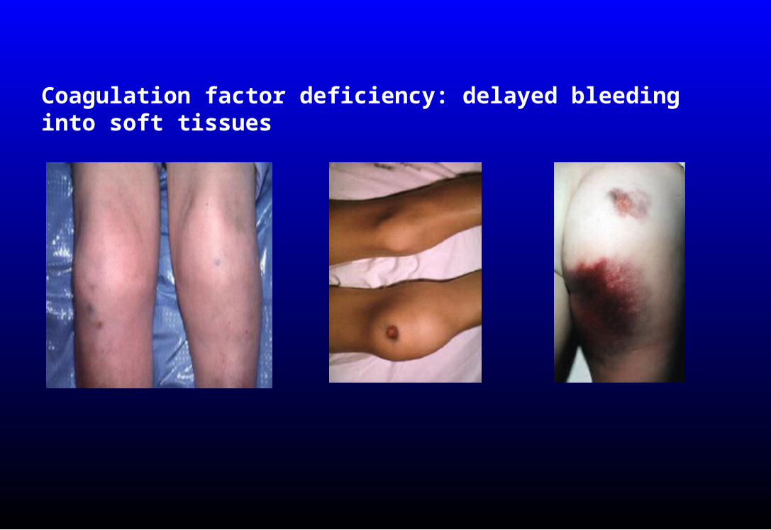
Coagulation factor deficiency: delayed bleeding into soft tissues

Bleeding confined to an operative site is usually due to a severed vessel

Initial laboratory evaluationInitial laboratory evaluation
• Platelet count
• PT/INR
• aPTT
• Fibrinogen
• Platelet function screen

Prothrombin time/INRProthrombin time/INR
Long PT/INR– Liver disease– Vitamin K deficiency– Warfarin or warfarin analogs (rat poison)– DIC– Inherited conditions rare– Won’t detect hemophilia, factor VIII inhibitor,
heparin at therapeutic concentrations
Magnitude of test abnormality usually proportional to clinical severity

aPTTaPTT
Long aPTT– Heparin (therapeutic or contaminant)– Hemophilia A or B– von Willebrand disease (low VIII – but PTT may be normal)– Factor XI deficiency– Factor VIII inhibitor– Contact factor deficiency (do not cause bleeding)– Lupus anticoagulant (does not cause bleeding)– Less sensitive than PT/INR to liver disease, DIC, warfarin
Magnitude of test abnormality not necessarily proportional to clinical severity

Platelet function screenPlatelet function screen
• Replaces the bleeding time• Advantages
– Ex vivo test (no skin incision)– Better standardized– Better sensitivity and specificity
PFA-100

Platelet function screenPlatelet function screen
Sensitive to: Defective platelet adhesion in von Willebrand
disease Platelet dysfunction due to many drugs Inherited platelet disorders
Not useful in patients with moderate or severe Thrombocytopenia
Rarely the only abnormal test in an acute bleeding disorder

Conditions that may cause bleeding with Conditions that may cause bleeding with normal or near-normal screening testsnormal or near-normal screening tests
• Mild hemophilia (factor level 20-30% of normal)
• Von Willebrand disease
• Factor XIII deficiency (very rare)
• Fibrinolytic disorders
• Vascular disorders (Ehlers-Danlos, amyloid, etc)

IMMUNE IMMUNE THROMBOCYTOPENIATHROMBOCYTOPENIA

IMMUNE THROMBOCYTOPENIAIMMUNE THROMBOCYTOPENIA
• ITP• Drug-induced purpura• Heparin-induced thrombocytopenia • Post-transfusion purpura

ITPITP• Autoimmune platelet destruction
• Mild to severe thrombocytopenia
• A diagnosis of exclusion
• Bleeding risk low to moderate in most cases– Hospitalization not always needed
• Rarely begins in hospital
• Other coag tests normal
• Few symptoms other than bruising & petechiae

ITP: initial treatmentITP: initial treatment
• Corticosteroids: prednisone 1 mg/kg or pulse high dose dexamethasone
• IVIG (high dose)
• Splenectomy
• Platelet transfusion sometimes effective

Drug-induced thrombocytopeniaDrug-induced thrombocytopenia
• Severe thrombocytopenia (<5K) not uncommon
• Often begins in hospital, precipitous drop in platelet count
• Bleeding risk moderate to high
• Typically occurs within days to weeks of starting a drug
• Testing for drug-dependent platelet Ab can confirm diagnosis but slow turnaround time

DRUGS MOST LIKELY TO CAUSE DRUGS MOST LIKELY TO CAUSE THROMBOCYTOPENIATHROMBOCYTOPENIA
Hematology 2009;153
**
*
*
*
*
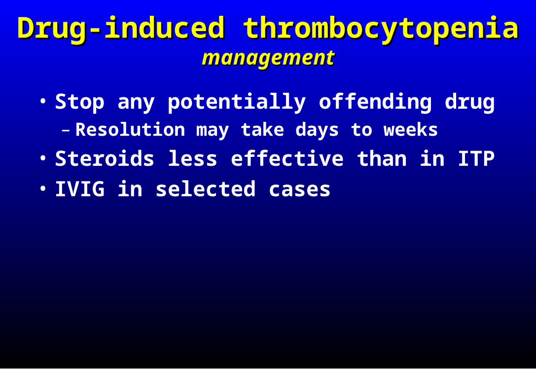
Drug-induced thrombocytopeniaDrug-induced thrombocytopeniamanagementmanagement
• Stop any potentially offending drug– Resolution may take days to weeks
• Steroids less effective than in ITP
• IVIG in selected cases

Heparin-induced thrombocytopeniaHeparin-induced thrombocytopenia• Mild to moderate thrombocytopenia with onset
during hospitalization in most cases
• Onset typically 4-7 days after initial heparin exposure (UFH > LMWH)
• Recent surgery, infection, inflammation increase risk
• Bleeding risk low, thrombosis risk high
• Heparin Ab test very sensitive – HIT can be ruled out if negative
• Rx: Stop heparin (any form), consider alternative anticoagulant (not warfarin)

Post-transfusion purpuraPost-transfusion purpura• Severe, precipitous drop in platelet count, usually a
few days after blood product exposure (FFP, RBC most common)
• Most patients are multigravid women
– Prior exposure to platelet antigen during pregnancy, recall Rxn after transfusion
• Most patients lack a common platelet alloantigen called HPA-1 (~ 2% of population)
• Antibodies directed against HPA-1 on transfused “passenger” platelets, patient’s own platelets are destroyed as “innocent bystanders”

Post-transfusion purpuraPost-transfusion purpura• If suspected notify blood bank to arrange
for appropriate testing and transfusion therapy
• Test for HPA-1 antibodies in patient serum
• Do NOT give platelet transfusion
• If RBC transfusion needed give washed RBC
• High dose IVIG
• Steroids generally not helpful
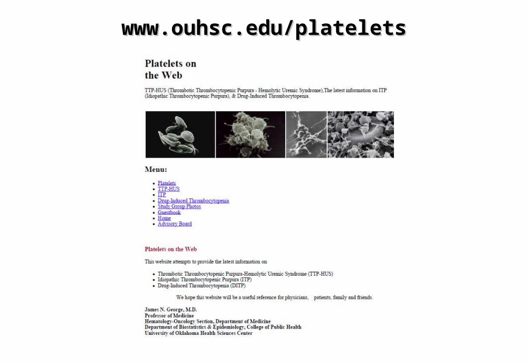
www.ouhsc.edu/plateletswww.ouhsc.edu/platelets

DISSEMINATED DISSEMINATED INTRAVASCULAR INTRAVASCULAR
COAGULATIONCOAGULATION

DISSEMINATED INTRAVASCULAR COAGULATIONDISSEMINATED INTRAVASCULAR COAGULATION
• Rapid formation & lysis of intravascular fibrin• Consumption of clotting factors, platelets,
inhibitors• Life-threatening underlying disease • Bleeding due to uncontrolled fibrinolysis,
thrombocytopenia, clotting factor consumption and tissue injury from underlying disease
• Tissue injury/necrosis due to microvascular occlusion, hypotension, cytokine-mediated endothelial damage
• Most deaths due to underlying disease
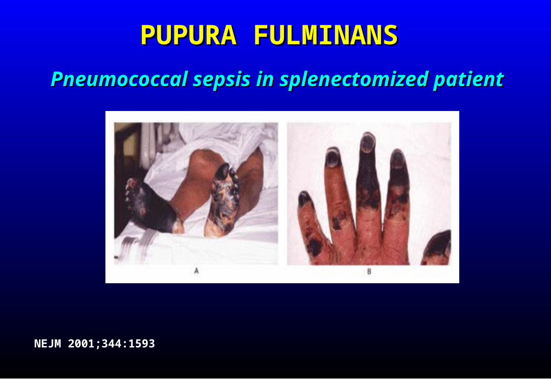
PUPURA FULMINANSPUPURA FULMINANS
Pneumococcal sepsis in splenectomized patientPneumococcal sepsis in splenectomized patient
NEJM 2001;344:1593

DIAGNOSIS OF DICDIAGNOSIS OF DIC
1. Underlying disease capable of causing DIC?
2. Evidence of accelerated clotting factor and platelet consumption, and increased fibrinolysis?
If both present DIC is likely

SCREENING FOR DICSCREENING FOR DIC
• D-dimer (most sensitive)
• PT/INR (PTT less helpful)
• Fibrinogen
• CBC/platelet count
• Fibrin monomer (most specific)

TREATMENT OF DICTREATMENT OF DIC
• TREAT UNDERLYING DISEASE!
• Clotting factor & inhibitor replacement for patients with significant bleeding: Fresh frozen plasma (goal INR ≤ 1.6) Cryoprecipitate (goal fibrinogen ≥ 100) Platelets (goal platelet count 50-75K)
• Pharmacologic inhibitors (selected pts with refractory bleeding) Heparin (low dose) Antifibrinolytics (Amicar, tranexamic acid)

THROMBOTIC THROMBOTIC THROMBOCYTOPENIC THROMBOCYTOPENIC
PURPURA (TTP)PURPURA (TTP)

TTPTTP
• Microangiopathic hemolytic anemia
• Thrombocytopenia
• Organ dysfunction (CNS, renal, other) due to small vessel occlusion
• Untreated mortality rate >90%
• With treatment mortality < 20%
• 1-2 cases/yr @ UWHC

TTPTTP• Caused by autoimmune destruction of
ADAMTS-13 protease that modulates von Willebrand factor multimer size
• Very large multimers clump platelets
• Microthrombi damage RBC and block vessels

TTPTTP
• Thrombocytopenia (may be severe)• Intravascular hemolysis (high LDH, low
haptoglobin, schistocytes in blood smear)• INR, PTT, fibrinogen usually normal• Organ dysfunction
– Neurologic symptoms– Renal dysfunction (hematuria, proteinuria)– Cardiac (arrhythmia)
• ADAMTS-13 activity low (usually <5%)

TTP - DDXTTP - DDX• Cancer (may be occult)• Pregnancy complications (HELLP, etc)• Hemolytic-uremic syndrome (kidney most
prominent target organ)– Shiga toxin
– “Atypical” HUS: genetic component, complement-mediated
• Vasculitis (SLE, scleroderma)• HIV infection• DIC• Drugs (calcineurin inhibitors, mitomycin C,
interferon, clopidogrel, ticlopidine)
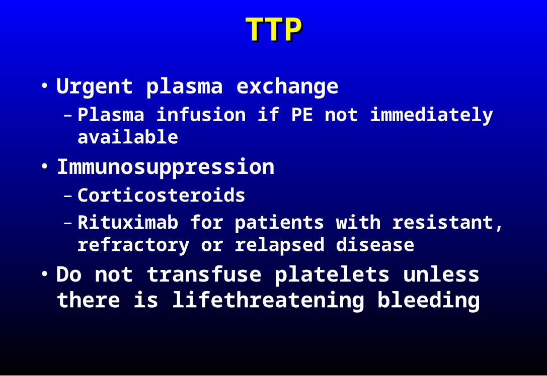
TTPTTP
• Urgent plasma exchange– Plasma infusion if PE not immediately available
• Immunosuppression– Corticosteroids– Rituximab for patients with resistant, refractory
or relapsed disease
• Do not transfuse platelets unless there is lifethreatening bleeding

HEMOPHILIAHEMOPHILIA

HEMOPHILIAHEMOPHILIA
• Inherited deficiency of factor VIII (hemophilia A) or factor IX (hemophilia B)
• Sex-linked inheritance; almost all patients male• Most bleeding into joints, muscles; mucosal and
CNS bleeding uncommon• Severity inversely proportional to factor level:
< 1%: severe, bleeding after minimal injury
1-5%: moderate, bleeding after mild injury
> 5%: mild, bleeding after significant trauma or surgery (may not be diagnosed until adulthood)
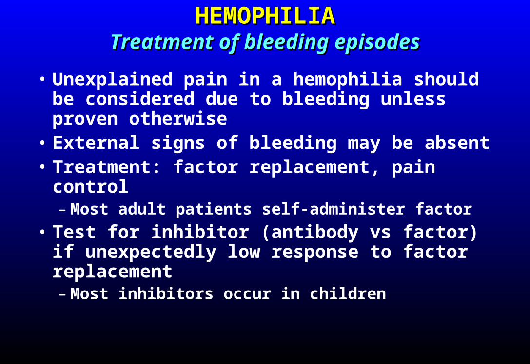
HEMOPHILIAHEMOPHILIATreatment of bleeding episodesTreatment of bleeding episodes
• Unexplained pain in a hemophilia should be considered due to bleeding unless proven otherwise
• External signs of bleeding may be absent• Treatment: factor replacement, pain control
– Most adult patients self-administer factor
• Test for inhibitor (antibody vs factor) if unexpectedly low response to factor replacement– Most inhibitors occur in children

TREATMENT OF BLEEDS IN HEMOPHILIATREATMENT OF BLEEDS IN HEMOPHILIA
• Administer appropriate clotting factor concentrate– 20-40 U/kg for minor bleeds, repeat daily for 2-3 days– 40-50 U/kg for major bleeds, repeat daily for 4-7 days– 50 U/kg q 12 hours for life-threatening bleeds
• 1 U/kg should increase plasma level of factor by about 2%– Initial dose higher with factor IX concentrate (greater
volume of distribution, but longer half-life)

TREATMENT OF HEMOPHILIACS WITH TREATMENT OF HEMOPHILIACS WITH INHIBITORSINHIBITORS
• Call a hematologist• Recombinant factor VIIa• High dose factor VIII (if low titer inhibitor)• Induce tolerance with daily factor infusion ±
immunosuppression

ACQUIRED FACTOR VIII DEFICIENCYACQUIRED FACTOR VIII DEFICIENCY
• Autoantibody to factor VIII (most common autoimmune factor deficiency)
• Most patients elderly• Often presents with severe soft tissue or mucosal
bleeding (different bleeding pattern than inherited hemophilia)
• Laboratory: prolonged aPTT not corrected by mixing with normal plasma, factor VIII activity typically < 10%– Bleeding risk not proportional to factor level– Normal INR and platelet count
• Treatment: rVIIa, immunosuppression
BLOOD PRODUCTS AND BLOOD PRODUCTS AND DRUGS IN THE TREATMENT OF DRUGS IN THE TREATMENT OF
BLEEDING DISORDERSBLEEDING DISORDERS

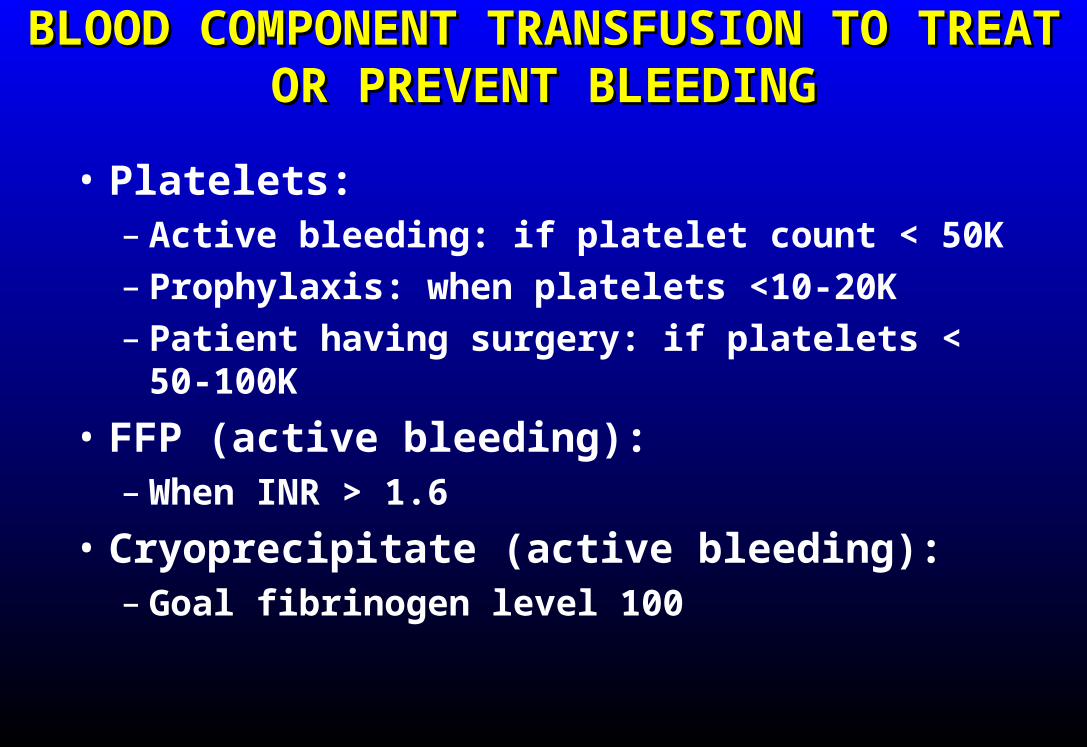
BLOOD COMPONENT TRANSFUSION TO BLOOD COMPONENT TRANSFUSION TO TREAT OR PREVENT BLEEDINGTREAT OR PREVENT BLEEDING
• Platelets:– Active bleeding: if platelet count < 50K– Prophylaxis: when platelets <10-20K– Patient having surgery: if platelets < 50-100K
• FFP (active bleeding):– When INR > 1.6
• Cryoprecipitate (active bleeding):– Goal fibrinogen level 100
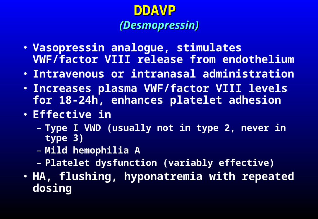
DDAVP DDAVP (Desmopressin)(Desmopressin)
• Vasopressin analogue, stimulates VWF/factor VIII release from endothelium
• Intravenous or intranasal administration• Increases plasma VWF/factor VIII levels for 18-24h,
enhances platelet adhesion• Effective in
– Type I VWD (usually not in type 2, never in type 3)– Mild hemophilia A– Platelet dysfunction (variably effective)
• HA, flushing, hyponatremia with repeated dosing
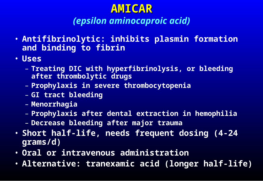
AMICARAMICAR(epsilon aminocaproic acid)
• Antifibrinolytic: inhibits plasmin formation and binding to fibrin
• Uses– Treating DIC with hyperfibrinolysis, or bleeding after
thrombolytic drugs– Prophylaxis in severe thrombocytopenia– GI tract bleeding– Menorrhagia– Prophylaxis after dental extraction in hemophilia– Decrease bleeding after major trauma
• Short half-life, needs frequent dosing (4-24 grams/d)• Oral or intravenous administration• Alternative: tranexamic acid (longer half-life)

Recombinant Factor VIIa (rFVIIa)Recombinant Factor VIIa (rFVIIa)
• Augments tissue factor-induced coagulation• FDA approved for treatment of Factor VIII
inhibitor w/bleeding• Off-label use is common, efficacy unclear:
– Other coagulation inhibitors (including drugs) w/bleeding
– Emergency reversal of coagulopathy (eg, warfarin) in patients with lifethreatening bleeding
• Potent procoagulant → risk of thrombosis• Very expensive!

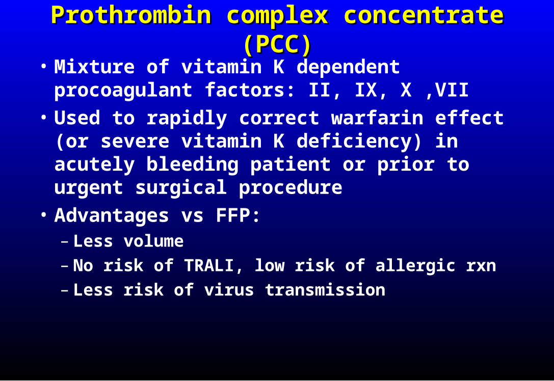
Prothrombin complex concentrate (PCC)Prothrombin complex concentrate (PCC)
• Mixture of vitamin K dependent procoagulant factors: II, IX, X ,VII
• Used to rapidly correct warfarin effect (or severe vitamin K deficiency) in acutely bleeding patient or prior to urgent surgical procedure
• Advantages vs FFP:– Less volume– No risk of TRALI, low risk of allergic rxn– Less risk of virus transmission

VITAMIN KVITAMIN K
Indications:1. Correction of vitamin K deficiency
2. Treatment of warfarin or superwarfarin overdose
3. Prophylaxis in patients at risk for vitamin K deficiency
Oral or IV administration (unreliableabsorption with subcutaneous injection)

ANTICOAGULANT-RELATED ANTICOAGULANT-RELATED BLEEDINGBLEEDING
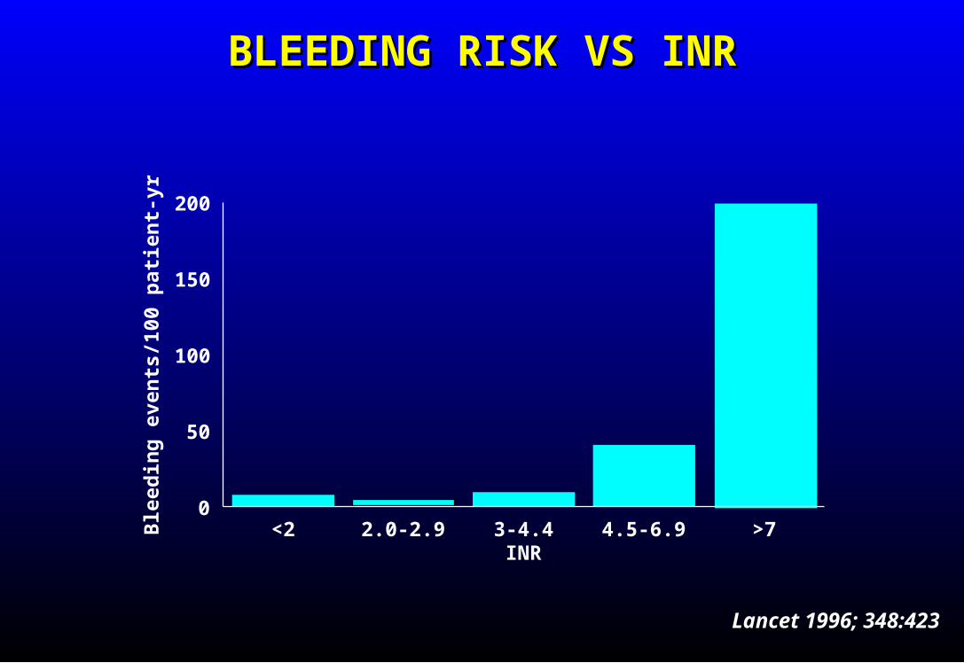
BLEEDING RISK VS INRBLEEDING RISK VS INR
<2 2.0-2.9 3-4.4 4.5-6.9 >7INR
0
50
100
150
200
Ble
edin
g e
ven
ts/1
00 p
atie
nt-
yr
Lancet 1996; 348:423
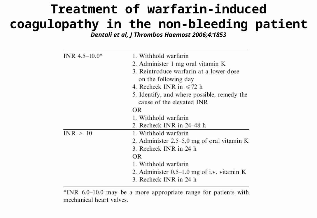
Treatment of warfarin-induced coagulopathy in the non-bleeding patient
Dentali et al, J Thrombos Haemost 2006;4:1853

Treatment of warfarin-induced coagulopathy in the bleeding patient
Dentali et al, J Thrombos Haemost 2006;4:1853

Reversal of heparin and LMWHReversal of heparin and LMWH
• Heparin:– FFP won’t help– Protamine sulfate: 1 mg/100U heparin– Neutralizes 80% of heparin
• LMWH– FFP won’t help– Protamine neutralizes 40% of LMWH

New anticoagulant drugs:New anticoagulant drugs:Dabigatran, rivaroxaban, apixaban, edoxabanDabigatran, rivaroxaban, apixaban, edoxaban
• If PT and aPTT normal overdose is unlikely
• No specific antidote (some in development)
• Activated charcoal if drug taken recently
• PCC ~ 50 U/kg (rivaroxaban, apixaban, edoxaban)
• Consider dialysis (dabigatran only)
• FFP likely to be ineffective, risk of volume overload, TRALI

TRANSFUSION REACTIONSTRANSFUSION REACTIONS

• Intravascular lysis of transfused rbcs by complement, IgM• Causes:
Transfusion of ABO-incompatible blood Transfusion of ABO-incompatible plasma Non-ABO antibodies
• Clinical manifestations: Fever (but most febrile reactions not hemolytic) Back pain Dark or red urine (hemoglobinuria) Bronchospasm Shock DIC Organ failure (esp kidneys) Death
IMMEDIATE HEMOLYTIC TRANSFUSION REACTIONIMMEDIATE HEMOLYTIC TRANSFUSION REACTION
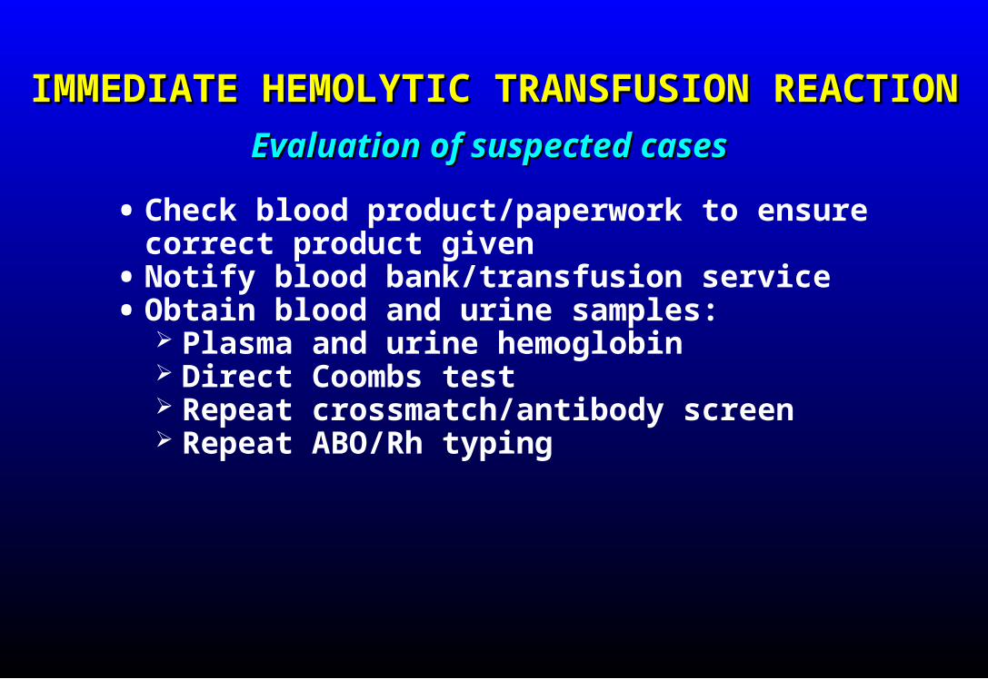
IMMEDIATE HEMOLYTIC TRANSFUSION REACTIONIMMEDIATE HEMOLYTIC TRANSFUSION REACTION
• Check blood product/paperwork to ensure correct product given
• Notify blood bank/transfusion service• Obtain blood and urine samples:
Plasma and urine hemoglobin Direct Coombs test Repeat crossmatch/antibody screen Repeat ABO/Rh typing
Evaluation of suspected casesEvaluation of suspected cases

IMMEDIATE HEMOLYTIC TRANSFUSION REACTIONIMMEDIATE HEMOLYTIC TRANSFUSION REACTION
• Stop transfusion immediately• IV crystalloid or colloid• Maintain BP, heart rate• Maintain airway• Diuresis
fluid, loop diuretic (mannitol may cause volume overload)
• Monitor renal and coagulation status
ManagementManagement

DELAYED HEMOLYTIC TRANSFUSION REACTIONDELAYED HEMOLYTIC TRANSFUSION REACTION
• IgG-mediated lysis of transfused red cells (usually extravascular, non-ABO)
• Usually begins 5-10 days after transfusion
• Jaundice, falling Hct, positive direct Coombs test, fever
• Not generally life-threatening

FEBRILE, NONHEMOLYTIC TRANSFUSION FEBRILE, NONHEMOLYTIC TRANSFUSION REACTIONREACTION
• Cause: cytokines released by leukocytes during storage; antibodies to HLA antigens on transfused or donor PMNS
• Incidence: ≤0.5% of units transfused• More common in multiply transfused recipients• Fever, chills, respiratory distress in severe reactions• Reduced incidence/severity with leukocyte-poor product

TRANSFUSION-RELATED ACUTE LUNG INJURY TRANSFUSION-RELATED ACUTE LUNG INJURY (TRALI)(TRALI)
• Hypoxemia with bilateral pulmonary infiltrates• No increase in central venous or pulmonary artery
pressures• Usually begins acutely within 6 hours of
transfusion• Clinical: acute respiratory distress, fever, chills• Pathophysiology:
1. Underlying lung injury (eg, sepsis, pneumonia) causes PMNs to adhere to pulmonary capillaries
2. Mediators in transfused blood product (neutrophil antibodies, cytokines) activate PMNs with resultant capillary injury

TRANSFUSION-RELATED ACUTE LUNG INJURYTRANSFUSION-RELATED ACUTE LUNG INJURY

TRANSFUSION-RELATED ACUTE LUNG INJURY TRANSFUSION-RELATED ACUTE LUNG INJURY (TRALI)(TRALI)
• Risk: FFP > platelets > RBC• Treatment: stop transfusion (if still in progress);
oxygen; ventilatory support if necessary; pulse corticosteroids

OTHER ACUTE NON-INFECTIOUS OTHER ACUTE NON-INFECTIOUS COMPLICATIONS OF TRANSFUSIONCOMPLICATIONS OF TRANSFUSION
• Allergic reactions• Anaphylaxis (IgA-deficient recipient)• Lung damage from microaggregates (massive
transfusion)• Transfusion-associated circulatory overload
(“TACO”)• Bacterial infection (mainly with platelet transfusion)• Hypothermia (rapid infusion of refrigerated blood)• Citrate toxicity/hypocalcemia (massive transfusion
or apheresis)• Graft-vs-host disease• Air embolism
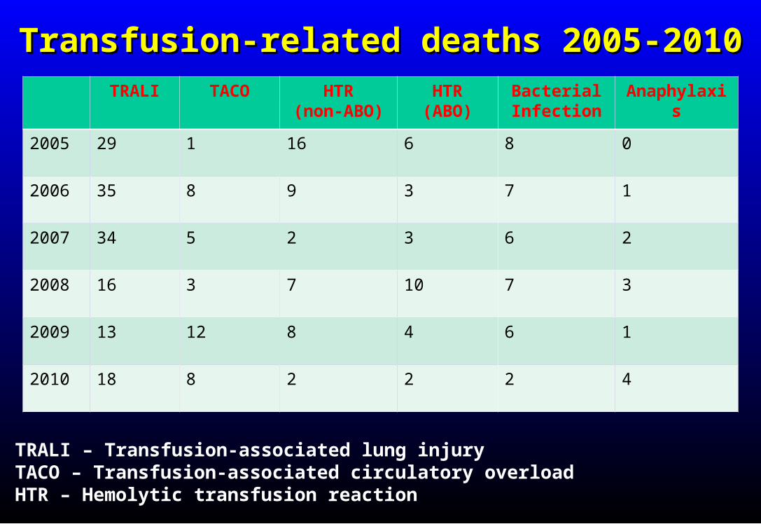
Transfusion-related deaths 2005-2010Transfusion-related deaths 2005-2010TRALI TACO HTR
(non-ABO)HTR
(ABO)BacterialInfection
Anaphylaxis
2005 29 1 16 6 8 0
2006 35 8 9 3 7 1
2007 34 5 2 3 6 2
2008 16 3 7 10 7 3
2009 13 12 8 4 6 1
2010 18 8 2 2 2 4
• TRALI – Transfusion-associated lung injury• TACO – Transfusion-associated circulatory overload• HTR – Hemolytic transfusion reaction